microreview mammalian escherichia · microreview mammalian and escherichia coli signal recognition...
TRANSCRIPT

Molecular Microbiology (1994) 11(1), 9 - 1 3
MicroReview Mammalian and Escherichia coli signal recognition particles
J o e n Luirink,1* and Bernhard Dobberstein2
1Department of Microbiology, Institute of Molecular Biological Sciences, Biocentrum Amsterdam, De Boelelaan 1087, 1081 HV Amsterdam, The Netherlands. 2Zentrum fur Molekulare Bioiogie Heidelberg, lm Neuenheimer Feid 282, 69120 Heidelberg, Germany.
Summary
Recent ev idence from both biochemical and genetic studies indicates that protein targeting to the pro-karyotic cytoplasmic membrane and the eukaryotic endoplasmic reticulum membrane may have more in c o m m o n than previously thought. A ribonucleo-protein particle w a s identified in Escherichia coli that cons is t s of at least one protein (P48 or Ffh) and one RNA molecule (4.5S RNA), both of which exhibit strong s e q u e n c e similarity with constituents of the mammalian signal recognition particle (SRP). Like the mammalian S R P , the E. coli S R P binds specifically to the signal s e q u e n c e of presecretory proteins. Depletion of either P48 or 4.5S RNA affects translation and results in the accumulation of precursors of several secreted proteins. This review d i scusses t h e s e recent studies and speculates on the position of the S R P in the complex network of protein interactions involved in translation and membrane targeting in £ . coli.
Introduction
In both prokaryotic and eukaryotic cells, m a n y proteins are targeted to, inserted into and translocated across biological m e m b r a n e s . T h e cytoplasmic m e m b r a n e (CM) of Escherichia coli and the endop lasmic reticulum ( E R ) m e m brane of the can ine pancreas h a v e been especia l ly popu lar for studies of protein targeting and translocation. In both s y s t e m s the secretory proteins face the s a m e problems. T h e y h a v e to maintain a translocat ion-competent conformation in the cytosol , contact the m e m b r a n e , traverse the m e m b r a n e and then b e released at the trans s ide of
Received 21 July, 1993; revised 13 September, 1993; accepted 14 September, 1993. *For correspondence. Tel. (20) 5484780; Fax (20) 6429202.
the membrane . Most proteins dest ined to insert into or traverse the C M and ER carry part of their targeting information in an /v-terminal signal s e q u e n c e of 1 5 - 3 0 amino acid residues, which contains an essent ia l hydrophobic core region of approximately 10 res idues (von Heijne, 1988). T h e s e signal s e q u e n c e s are structurally similar and often functionally interchangeable between pro-karyotes and eukaryotes , implying c o n s e r v e d underlying m e c h a n i s m s of signal sequence -med ia ted targeting and translocation (von Heijne, 1988). However , until recently, little homology has been observed between c o m p o n e n t s of both export sys tems .
E. coli: the general secretory pathway
In E. coli several soluble and m e m b r a n e proteins have been identified (inititially in genetic studies) that are required at different s t a g e s of the general secretory pathw a y (for an excel lent recent review, s e e Pugs ley , 1993). Pre-proteins interact with molecular c h a p e r o n e s like S e c B , D n a K / D n a J and G r o E L / G r o E S to maintain their translocat ion-competent conformation in the cytosol (Kumamoto , 1991). Little is known about the molecular bas is of the pre -pro te in - chaperone interaction. Recent ev idence indicates that S e c B binds cotranslationally to only a limited subse t of presecretory proteins (Kumamoto and Francetic, 1993). Determination of SecB-b ind ing sites in precursor molecu les h a s met with conflicting results, but most of the available data indicate that S e c B binds to multiple sites in the mature portion of the pre-protein (for discus s i ons on this i s sue , s e e Pugs ley , 1993; Kumamoto , 1991). G r o E L w a s shown to interact with comple ted pre-p-l ac tamase by photocross- l inking (Bochkareva era/. , 1988). Different pre-proteins s e e m to prefer different chaperones but they can b e quite promiscuous w h e n c ircumstances change . For instance, increased levels of G r o E L and D n a J / D n a K can c o m p e n s a t e for the loss of S e c B (Altman et al., 1991; Wild era/ . , 1992). A m o n g these chaperones , S e c B s e e m s the most specif ic for exported proteins. This conclus ion is supported by the fact that S e c B a l so fulfils a 'pilot' function by binding to the membrane -assoc ia ted S e c A protein (Haiti ef al., 1990). S e c A has binding affinity not only for S e c B but a l so for the signal s e q u e n c e and mature domain of the pre-protein (Akita ef al., 1990; Jo ly
10 J. Luirink and B. Dobberstein
and Wickner, 1993). It a lso binds A T P (Lill etai, 1989) and has been implicated in the generation of energy for the translocation process (Schiebel etai, 1991). S e c A interacts with the membrane -embedded S e c Y / S e c E complex (Hartl et ai, 1990) which is a constituent of the so-cal led translo-con, the putative protein pore in the CM. T h e eukaryotic homologue of S e c Y is the S e c 6 1 protein identified first by a genetic screen in yeas t (Desha ies and Schekman, 1987) and most recently by a biochemical approach in mammal ian cells (Gorlich etai, 1992).
Mammalian cells: SRP-medlated transport
In eukaryotic cells, the signal recognition particle (SRP ) recognizes the signal peptide w h e n it protrudes from the ribosome in the cytosol (at 6 0 - 9 0 total amino acid chain length). S R P may be prebound to the ribosome allowing pass ing s e q u e n c e s to be screened (Siegel and Walter, 1988) . T h e S R P is a large ribonucleoprotein complex con sisting of a 7 S R N A and six different polypeptides of 9 , 1 4 , 19, 54, 68 a n d 72 kDa. T h e associat ion between the S R P and the signal peptide lowers the rate of protein synthesis and thereby increases the time span in which the complex can contact the membrane with the nascent chain in a translocation competent conformation (Siegel and Waiter, 1988) . The r ibosome/nascent cha in /SRP complex then binds to the ER membrane via an interaction between S R P and S R P receptor (also called the 'docking protein'). Upon binding to the S R P receptor, the signal peptide is displaced from the S R P in a GTP -dependent process and b e c o m e s available for interactions with components of the putative translocon (Rapoport, 1992). Translation resumes and the nascent chain inserts co-translationally into the E R membrane . Finally, the S R P is released from the membrane -bound complex in a step that requires G T P hydrolysis (Connolly and Gilmore, 1989) . Thus , the S R P is a versatile adaptor which functions a s a 'pilot' and a s a molecular chaperone to guide the nascent secretory protein to the membrane in a trans-location-competent form. T h e different functions have been ascribed to the individual protein components in the S R P in which the 7 S RNA probably p lays a scaffolding role. S R P 9 and S R P 1 4 form a heterodimer which is neces sary for the translation arrest function. S R P 6 8 and S R P 7 2 a lso form a heterodimer which has been implicated in the mechanism of 'docking' to the E R membrane (Siegel and Walter, 1988). S R P 1 9 assists in the binding of S R P 5 4 to the 7 S R N A whereas S R P 5 4 is responsible for binding to the signal sequence (Romisch etai., 1990; Zopf etai, 1990).
S R P 5 4 has a modular structure consisting of an /^terminal G-domain , which contains a conserved GTP-binding motif, and a C-terminal M-domain, which is rich in methionine residues. Several independent studies have shown that the M-domain is responsible for both binding
to the 7S RNA and to the signal s e q u e n c e of the nascent presecretory protein (Zopf et ai, 1990; High and Dobberstein, 1991). Reconstituted S R P containing only the M-domain of S R P 5 4 w a s shown to be able to recognize the signal sequence albeit with lower efficiency than intact S R P , but it was unable to target the ribosome/ nascent chain complex to the E R membrane (Zopf et ai, 1993). The M-domain contains four predicted amphipathic a-hel ices (Bernstein etai, 1989). T h e methionine residues are found at evolutionary conserved positions and thought to line one side of each a-helix. A n attractive model has been put forward in which the helices are juxtaposed with the flexible methionine s ide chains forming a groove which accommodates the large variety of hydrophobic signal s e q u e n c e s (Bernstein et ai, 1989). T h e G-domain increases the efficiency of signal sequence binding and is probably also involved in the binding of the S R P to the a-subunit of the S R P receptor (Zopf et ai, 1993).
A peptide-binding motif similar to the o n e suggested for the interaction between signal s e q u e n c e s and the S R P 5 4 protein has been identified for the major histocompatibility complex (MHC) c lass I and II molecules (Bjorkman et ai, 1987; Brown et ai, 1993). In this case , peptides of nine amino acid residues were found to bind in a groove formed by two a-helices. The oc-helices are arranged s ide by s ide on a platform built by p-pleated sheets . In the c a s e of the signal sequence-binding domain of S R P 5 4 the platform would be formed by the 7S RNA.
Evidence for an E. coli SRP
T h e search for an SRP- l ike particle in E. coli has long been discouraged by the inability to identify SRP- l ike components in genetic screenings for export mutants. These screens identified very successful ly several of the sec genes described above. Recently, this search gained n e w impetus when sequence compar isons revealed the existence of E. coli homologues of S R P 5 4 and S R P 7 S RNA, P48 (also called Ffh for fifty-four homologue) and 4 .5S RNA, respectively (Romisch etai, 1989; Bernstein etai, 1989; Poritz etai, 1988).
T h e P48 gene was initially identified a s an open reading frame upstream of the trmD operon at 56 min of the E. coli chromosome (Bystrom et ai, 1983). P48 is very similar over its entire length to S R P 5 4 and s e e m s to have the s a m e modular structure (Romisch etai, 1989; Bernstein etai, 1989). T h e M-domain of P48 lacks one of the predicted C-terminal amphipathic hel ices of S R P 5 4 . This could explain s o m e of the differences between the pro-karyotic and eukaryotic membrane targeting system.
E coli 4 .5S RNA is o n e of the smallest members of the family of SRP7S- l ike R N A s found in mammal ian cells, plants, yeast , archaebacteria and eubacteria (Larsen and Zwieb, 1991). It forms an extended stem-loop structure,
IIIIIIIIIIIIIIIIIIIIIIIIIIIIIIIIIIIIIIIIIIIIIIIIIIIIHNM
which is homologous to the most highly conserved domain of S R P 7 S RNA.
T w o independent studies gave the first hints that P48 and 4 .5S R N A are part of an SRP- l ike complex in E. coli (Ribes et al., 1990; Poritz et al., 1990). 4 . 5 S R N A was found to be in a complex with P48 in a wild-type E. coli extract (Ribes et al., 1990; Poritz et al., 1990) and to bind to S R P 5 4 in vitro (Poritz era / . , 1990). T o investigate the function of 4 .5S RNA, a strain was constructed which allowed conditional expression of 4 .5S RNA. Depletion of 4 .5S RNA (or overexpression of a dominant lethal 4 .5S RNA allele) showed pleiotropic effects, including an early induction of the heat -shock response, a relatively late inhibition of cell growth and protein synthesis , and finally cell death (Ribes et al., 1990; Poritz era/ . , 1990). Expression of S R P 7 S R N A could partially complement for the loss of 4 .5S RNA in this strain (Ribes et al., 1990). Effects on secretion were limited to a decreased processing of pre-p- lactamase at late time points after depletion (Ribes et al., 1990; Poritz et al., 1990). At the time it w a s not clear whether this w a s a c o n s e q u e n c e of the heat-shock response or of a genuine secretion defect.
Support for the latter possibility c a m e from three recent studies. In an elegant genetic approach similar to the one described above , Phillips and Silhavy (1992) investigated the effects of cellular depletion of the protein component of the RNP , P48. Rather surprisingly, precursor forms of all tested secretory proteins accumulated, which is indicative of a general effect on protein secretion. It must be noted that P48-depleted cells exhibited an elongated cell shape suggesting impaired cell division. i
In a second biochemical study we demonstrated that P48 binds specifically to the signal peptide of nascent pre-secretory proteins (Luirink et al., 1992). Truncated pre-prolactin m R N A (coding for 86 amino acid residues) was translated in a cell-free sytem in the presence of Lys-tRNA carrying a photoactivatable crosslinking group in its side chain. After purification, the r ibosome/nascent chain complexes were incubated with E. coli cell extracts and crosslinking w a s induced. P48 w a s found to be cross -linked to the pre-prolactin signal sequence , but not to a mutated, non-functional signal sequence . Ev idence was obtained that the interaction of P48 with the signal peptide is similar to that of S R P 5 4 : (i) S R P 5 4 competes with P48 for binding to the signal peptide (ii) P48 binds a s part of an R N P containing 4 .5S R N A (iii) P48 binds only to nascent polypeptides, not to polypeptides which have been released from the ribosome. A striking difference, however, is the dependence of P48 binding on the pres e n c e of 4 . 5 S R N A in the cell extract used for crosslinking, whereas S R P 5 4 binds very efficiently in the a b s e n c e of S R P 7 S RNA. T h e binding region of P48 on the 4 .5S R N A w a s recently mapped by site-directed mutagenesis (Wood et al., 1992). P48 binds to two loop structures in
Escherichia coli SRP 11
the central portion of the 4 . 5 S RNA. Interestingly, a correlation w a s found between the inability of mutant 4 .5S RNA molecules to bind P48 and to restore growth in vivo in 4 .5S RNA depleted cells. This indicated that the E. coli S R P indeed functions a s a ribonucleoprotein particle.
In a third study, Bernstein and coworkers (1993) reconstituted mammalian S R P in which S R P 5 4 had been replaced by P48. This chimeric S R P w a s still capable of signal peptide binding and imposing translational arrest, but could not promote translocation of pre-prolactin into the ER. Possibly, P48 does not interact with the eukaryotic S R P receptor.
4.5S RNA and translational regulation
From physiological and genetic studies of the functions of 4 .5S RNA in E. coli a role for this molecule related to translation has been suggested (an excellent discussion of this aspect can be found in a review article by Brown, 1991). Synthesis of 4 . 5 S RNA w a s found to b e co-ordinately regulated with that of ribosomal R N A and transfer RNA (Ikemura and Dahlberg, 1973). Selection of suppressors that reduce the 4 .5S RNA requirement were found in genes coding for components of the translational apparatus, the elongation factor G (EF-G) (Brown, 1987) and in the binding site for 4 .5S RNA on 2 3 S ribosomal RNA or resulted in the increased concentration of s o m e uncharged tRNAs (Brown, 1989). Both the 4 .5S RNA and the P48 have been found to interact with r ibosomes, individually and as an R N P particle. Both were shown to be released from r ibosomes by treatment with puromycin (Brown, 1987; 1989; Luirink et al., 1992). T h e finding that P48 affects the 4 . 5 S R N A requirement a n d the fact that 4 .5S RNA can be found free and complexed to P48 make it likely that free 4 .5S RNA and the R N P complex function in different pathways (Brown, 1991). T h e free form h a s been implicated in modulating the rate of translation to allow proteins to fold properly (Brown, 1991). Such a function would explain the finding that a reduction in the amount of 4.5S RNA leads to misfolding of proteins. W e envisage that the P48/4 .5S R N A complex h a s a more selective function: a s P48 contacts signal s e q u e n c e s in nascent polypeptides this function would be limited to secretory and membrane proteins. Given the function of 4 . 5 S RNA in general translation regulation we would a s s u m e a similar, although more selective function for the P48 /4 .5S RNA complex. It relays information from the nascent chain via the R N A to the translating r ibosome and via its G T P -binding domain to a component yet to be characterized.
Model for chaperone-mediated protein targeting in E. coli
Both cytosolic and proteins destined for secretion have appeared to interact in a hierarchical manner with a
MiiiiiiiiiiiiiiiiiiiiiiiiiiiiiiiiflHWIi
12 J. Luirink and B. Dobberstein
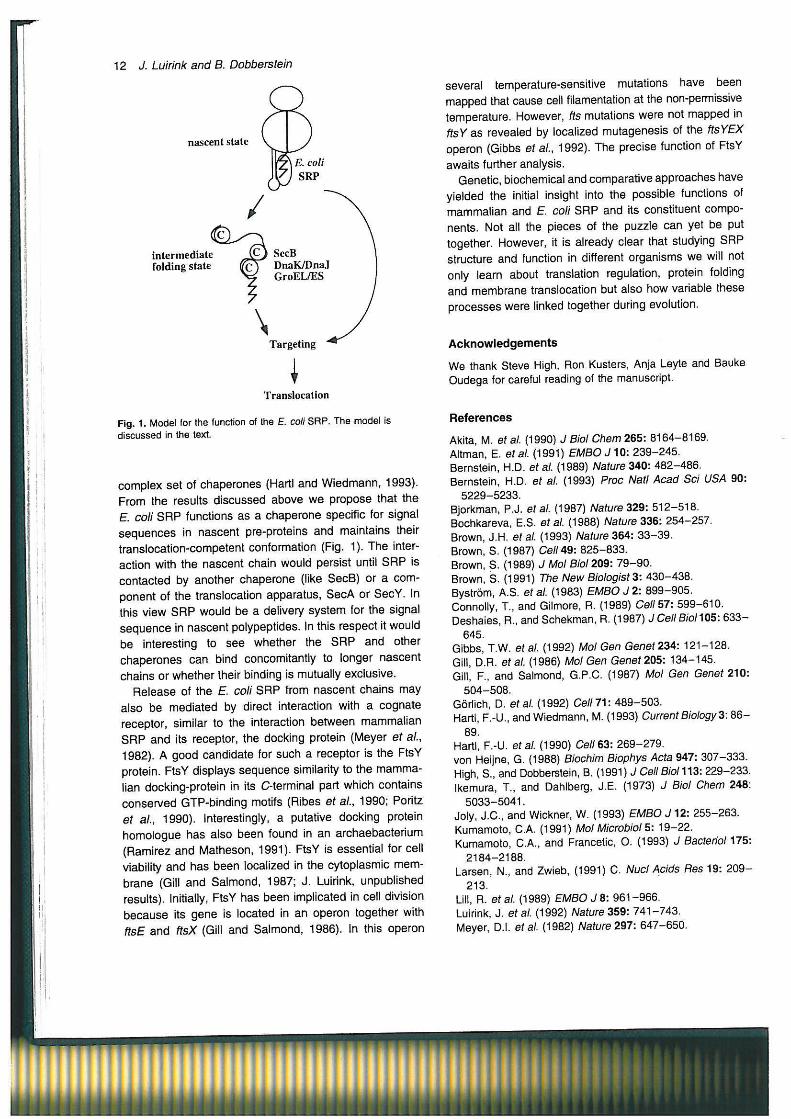
nascent state
intermediate folding state
( C ) SecB DnaK/DnaJ GroEL/ES
Targeting
Translocation
Fig. 1. Model for the function of the E. coli SRP. T h e model is discussed in the text.
complex set of chaperones (Hartl and Wiedmann , 1993). From the results d iscussed above w e propose that the E. coli S R P functions a s a chaperone specific for signal s e q u e n c e s in nascent pre-proteins and maintains their translocation-competent conformation (Fig. 1). T h e interaction with the nascent chain would persist until S R P is contacted by another chaperone (like S e c B ) or a component of the translocation apparatus, S e c A or S e c Y . In this view S R P would be a delivery sys tem for the signal s e q u e n c e in nascent polypeptides. In this respect it would be interesting to s e e whether the S R P and other chaperones can bind concomitantly to longer nascent cha ins or whether their binding is mutually exclusive.
R e l e a s e of the E. coli S R P from nascent chains may also be mediated by direct interaction with a cognate receptor, similar to the interaction between mammal ian S R P and its receptor, the docking protein (Meyer et al., 1982). A good candidate for such a receptor is the FtsY protein. FtsY displays s e q u e n c e similarity to the m a m m a lian docking-protein in its C-terminal part which contains conserved GTP-b inding motifs (Ribes et al., 1990; Poritz et al., 1990). Interestingly, a putative docking protein homologue has a lso been found in an archaebacterium (Ramirez and Matheson, 1991). FtsY is essential for cell viability and has been localized in the cytoplasmic m e m brane (Gill and Sa lmond, 1987; J . Luirink, unpublished results). Initially, FtsY has been implicated in cell division b e c a u s e its g e n e is located in an operon together with ftsE and ftsX (Gill and Sa lmond, 1986). In this operon
several temperature-sensitive mutations have been mapped that cause cell filamentation at the non-permissive temperature. However, fts mutations were not mapped in ftsY a s revealed by localized mutagenesis of the ftsYEX operon (Gibbs et al., 1992). T h e precise function of FtsY awaits further analysis.
Genetic, biochemical and comparative approaches have yielded the initial insight into the possible functions of mammal ian and E. coli S R P and its constituent compo nents. Not all the pieces of the puzzle can yet be put together. However, it is already clear that studying S R P structure and function in different organisms w e will not only learn about translation regulation, protein folding and membrane translocation but also how variable these processes were linked together during evolution.
Acknowledgements
W e thank Steve High, Ron Kusters, Anja Leyte and Bauke Oudega for careful reading of the manuscript.
References
Akita, M. et al. (1990) J Biol Chem 265: 8164-8169. Altman, E. etal. (1991) EMBO J10 : 239 -245 . Bernstein, H.D. etal. (1989) Nature 340: 482-486. Bernstein, H.D. er al. (1993) Proc Natl Acad Sci USA 90:
5229-5233. Bjorkman, P. J . etal. (1987) Nature 329: 512-518. Bochkareva, E.S. etal. (1988) Nature 336: 254-257. Brown, J .H. etal. (1993) Nature 364: 3 3 - 3 9 . Brown, S. (1987) Ce//49: 825 -833 . Brown, S. (1989) J Mol Biol 209: 7 9 - 9 0 . Brown, S. (1991) The New Biologist 3: 430 -438 . Bystrom, A.S. etal. (1983) EMBO J 2: 899-905. Connolly, T., and Gilmore, R. (1989) Ce//57: 599-610. Deshaies, R., and Schekman, R. (1987) J Cell Biol 105: 6 3 3 -
645. Gibbs, T .W. etal. (1992) Mol Gen Genet234: 121-128. Gill, D.R. etal. (1986) Mol Gen Genet205: 134-145. Gill, F., and Salmond, G.P.C. (1987) Mol Gen Genet 210:
504-508 . Gorlich, D. etal. (1992) Ce//71: 489 -503 . Hartl, F.-U., and Wiedmann, M. (1993) Current Biology 3: 8 6 -
89. Hartl, F.-U. ef al. (1990) Cell 63: 269 -279 . von Heijne, G. (1988) Biochim Biophys Acta 947: 307-333. High, S., and Dobberstein, B. (1991) J Cell Biol 113: 229-233. Ikemura, T., and Dahlberg, J .E. (1973) J Biol Chem 248:
5033-5041. Joly, J .C. , and Wickner, W. (1993) EMBO J12: 255-263. Kumamoto, C.A. (1991) Mol Microbiol 5: 19 -22 . Kurnamoto, C.A., and Francetic, O. (1993) J Bacterid 175:
2184-2188. Larsen, N., and Zwieb, (1991) C. Nucl Acids Res 19: 2 0 9 -
213. Lill, R. etal. (1989) EMBO J 8: 961-966. Luirink, J . etal. (1992) Nature 359: 741 -743 . Meyer, D.I. etal. (1982) Nature297: 647 -650 .
Illllllllllllllllillllllllilllllllllllllllllllll

Phillips, G.J., and Silhavy, T.J . (1992) Nature 359: 744-746. Poritz, M.A. etal. (1988) Cell 55: 4 - 6 . Poritz, M.A. etal. (1990) Science 250: 1111-1117. Pugsley, A.P- (1993) Microbiol Rev 57: 50 -108 . Ramirez, C , and Matheson, A T . (1991) Mol Microbiol 5:
1687-1693. Rapoport, T. (1992) Science 258: 931 -936 . Ribes, V. ef a/. (1990) Ce/ /63: 591 -600 .
Escherichia coli SRP
Romisch, K. etal. (1989) Nature 340: 478 -482 . Romisch, K. etal. (1990) J Cell Biol 1793-1802. Schiebel, E. etal. (1991) Ce/ /64: 927 -939 . Siegel, V., and Walter, P. (1988) TIBS 13: 314 -315 . Wild, J . etal. (1992) Genes DevB: 1165-1172. Wood, H. et al. Nucl Acids Res 20: 5919-5925. Zopf, D. etal. (1990) EM BO J 9: 4511 -4517 . Zopf, D. etal. (1993) J Cell Biol 120: 1113-1121.