microorganisms vs. synthetic polymers. … · synthetic polymers used in paint and coating...
TRANSCRIPT
Philosophy Doctorate School In Scienze Molecolari e Biotecnologie Agrarie, Alimentari ed
Ambientali
Philosophy Doctorate Course In Chimica, Biochimica ed Ecologia degli Antiparassitari, XXIV cycle
Philosophy Doctorate Thesis
MICROORGANISMS vs. SYNTHETIC POLYMERS.
ECOLOGY AND BIODEGRADATION
GIACOMUCCI LUCIA No. Matr. R08194
SUPERVISOR: Dr. Francesca Cappitelli COORDINATOR: Prof. Daniele Daffonchio
Academic Year 2010/2011

COVER
Ausonia & Hungaria hotel façade. Detail of a cherub. Source – http://www.hotelhungaria.com/.
____________________________________________________________________Contents
ABSTRACT p. 1
CHAPTER I Introduction p.5
CHAPTER II Aim of the work p.11
CHAPTER III Microbial deterioration of artistic tiles from the façade of the Grande Albergo Ausonia & Hungaria (Venice, Italy)
p.13
CHAPTER IV Degradation of nitrocellulose–based paint by Desulfovibrio desulfuricans ATCC 13541
p.31
CHAPTER V A new non-degenerated primer pair for the specific detection of the nitrite reductase gene in Desulfovibrio genus
p.45
CHAPTER VI Conclusions p.53
APPENDIX I p.55
APPENDIX II p.61

____________________________________________________________________Abstract
1
Abstract Despite synthetic polymers have been considered little or no deteriorable for many years,
now we know they can undergo to chemical, physical and biological damage. Effects of biological degradation on polymers include masking of surface properties due to the presence of microorganisms inhabiting surfaces, embrittlement and loss of stability due to changes in chemical structure of polymers, presence of cracks and swellings due to penetration of microorganisms into the polymer matrix and changes in polymer colour due to excretion of microbial pigments. Microbial deterioration depends on the constitution and the properties of polymer materials as well as environmental conditions. Millions of tons of synthetic polymers used as adhesives, binders, coatings, inks, etc., are produced worldwide every year. In 1997 the worldwide paint and coatings industry represented a mature 50+ billion dollar market and synthetic polymers used in paint and coating industries account for approximately 45-55% of the worldwide decorative market. Deterioration of varnishes and binding media results in chemical changes that lead to an increase in the insolubility and polarity of the material, a reduction of the strength, and a change in colour, among others. As biodeterioration of synthetic polymers seriously compromises the adhesion and durability of the paint as well as its decorative/protective function, identification of the cause of synthetic polymer biodeterioration is of great importance.
Although biodeterioration of synthetic polymers in objects during their lifetime should be avoided to preserve the object function, synthetic polymer materials should be susceptible to degradation once they are disposed of and treated as waste. The major ingredients of paints are the pigment, a material which provides colour, and the binding medium, a film-forming material in which the pigment particles are dispersed and forms the matrix that hardens and binds the pigments on the painted surface. All the synthetic polymers used as paint and coatings binders are belonging to different chemical classes, the most important being acrylics, polyvinylacetate and nitrocellulose-alkyds. Microorganisms can assimilate and/or degrade these synthetic polymers because their chemical bonds are the same as those found in the natural polymeric matter, which is generally easily degraded. Bioremediation is widely used for the clean-up of environmental pollutants using microorganisms and could be successfully applied for the removal of synthetic polymers, including those present in paint and coating formulations.
The aims of this PhD project were to study both aspects of synthetic polymer biodegradation, in particular:
� Characterise the microbial community associated to biodeterioration of an acrylic polymer used as protective and consolidant, in order to identify microorganisms potentially active against synthetic polymers.
� Study bacterial degradation of nitrocellulose in order to develop a bioremediation process to remove nitrocellulose-based paints.
Chapter 3 reports a case-study about how a microbial community changes in the presence
or in the absence of an acrylic polymer used as consolidants. Synthetic polymers have been widely applied as consolidants and protective in cultural heritage field for the treatment of objects and buildings to prevent further deterioration. The long-term efficiency of the consolidative/protective treatments was believed influenced mainly by chemical and physical agents (e.g. UV light and temperature) and therefore lots of synthetic polymers have been used in cultural heritage conservation without testing them against biological deterioration. As a result, treated objects are sometimes in worse conditions than untreated objects and, moreover, biodeterioration of the added materials is an additional cause of damage. The study
Abstract____________________________________________________________________
2
of microbial community changes due to synthetic polymer treatments and the identification of biodeteriogen microorganisms is a crucial step in developing a treatment strategy in cultural heritage conservation.
In this work, we present the first molecular characterisation of a microbial community present on artistic ceramic treated with acrylics. The study was conducted on ceramic tiles of the Grande Albergo Ausonia & Hungaria façade. In 2007 the façade underwent conservation treatment to consolidate severely damaged tiles and to remove dark spots present on its surface. Tiles on the first horizontal register were then treated with the commercial synthetic resin Paraloid B72® (copolymer methylacrylate−ethylmethacrilate), as consolidant and protective product. Soon after the intervention both treated and untreated tiles showed coloured alterations caused by microorganisms between the pottery layer and the glaze.
Samples from treated and untreated areas were initially observed under stereo, epifluorescence and the scanning electron microscope and analysed by Energy-dispersive X-ray spectroscopy (EDX) and micro Fourier transform infrared spectroscopy (FTIR). The results showed that the polymer, identified as Paraloid B72®, was present only in two of the nine treated samples and confirmed the presence of biological alterations in all samples. Paraloid B72® was not found on the surface of the most of the treated samples, probably because of photooxidative depolymerization and the wash out of resin from the tile surfaces. Deteriogen biofilm was present at the interface glaze-pottery in samples without any presence of Paraloid B72®, while in the two samples where we found Paraloid B72®, the biofilm was mainly in the pottery layer.
Microscope techniques together with denaturing gradient gel electrophoresis (DGGE) and sequencing from total DNA extracted from Hungaria samples were used to identify sessile taxa causing coloured alteration. Our results showed that the colour of the deposit present in the tile samples and the greenish alteration on the balcony were most likely mainly due to the presence of cryptoendolithic cyanobacteria and eukaryotic algae respectively. Biodegrading microorganisms related to the presence of synthetic polymers and Paraloid B72® were also found. In particular, Phoma, an uncultured Bacteroidetes and Methylibium sp. were found in all the samples in which we detected Paraloid B72®. Melanised fungi, such as Phoma, are well known as the most damaging fungi able to attack and penetrate stone monument surfaces. Bacteroidetes show hydrolytic activity toward polymeric substances and Methylibium can grow on organic pollutants. In addition, when Paraloid B72® was present, the biofilm was located in a deeper position than in samples without any evidence of the resin. Using DGGE technique it was also proved that the microflora present on the tiles was generally greatly influenced by the environment of the Hungaria hotel. Several microorganisms related to the alkaline environment, the range of the tile pH, and related to the aquatic environment and the pollutants of the Venice lagoon were found.
The rapid reappearing of microbial deterioration soon after the 2007 conservation treatment, which included the use of biocides, was likely favoured by the water content and organic substances, some of them added during the conservation treatment. Therefore, Paraloid B72® was not the best consolidant polymer to be used fort the long-time conservation of the ceramic tile of Hungaria hotel.
Chapter 4 is focused on the capability of Desulfovibrio desulfuricans ATCC 13541 to
attack nitrocellulose as binder in paint. Synthetic polymers used for the manufacture of paint and coatings are polymers belonging to different chemical classes and little information regarding the chemical characteristics and their variation in a population of paints are present in literature. On the base of few works, acrylics, polyvinylacetates and nitrocellulose-alkyd polymers seem to be most used as binders in spray paint formulation. The high presence of nitrocellulose, a uniformly substituted cellulose compound with varying degree of nitration, as component of paint binders should lead to a more environmental attention because the
____________________________________________________________________Abstract
3
presence of nitro compounds materials in the wastewater effluent causes severe environmental problems, high toxicity and provokes serious health problems. Microorganisms are able to degrade nitrocellulose by two pathways: i) cleavage of β–1,4–glucoside bonds that produces nitrooligosaccharides of various length, normally carried out by fungi, and ii) nitrocellulose denitration that reduces the degree of nitro substitution, generally performed by bacteria. Since nitrooligosaccharides have mutagenic properties, the second pathway is preferred over the first for exploitation as a biodegradation pathway. Nitrocellulose undergoes degradation by sulphate–reducing bacteria under anaerobic conditions. In particular, sulphate–reducing bacteria of the genus Desulfovibrio decrease the amount of nitrocellulose powder in media containing this compound. Desulfovibrio spp. firstly reduces the nitration content of nitrocellulose in powder due to a nitroesterase activity of the bacteria and then reduces nitrate to ammonia through the dissimilatory nitrate reduction to ammonia (DNRA). DNRA is a two-step process involving nitrate reduction to nitrite and the subsequent nitrite reduction to ammonium by nitrate– and nitrite reductases. There are several sulphate–reducing bacteria able to reduce nitrate to nitrite, but it appears that this is not a shared feature across the genus Desulfovibrio. In contrast, the dissimilatory reduction of nitrite to ammonium seems to be widespread in Desulfovibrio.
For this study, D. desulfuricans ATCC 13541 was selected because it was used in metal biosorption, and therefore the strain is resistant to the high concentrations of metals that can be encountered in paints. Nitrocellulose was selected in the form of the red spray paint by Motip–Dupli® Autocolor (colour 5–0200) that was confirmed by FTIR spectroscopy as composed by nitrocellulose and a modified polyester resin. At the end of degradation experiments, the capability of D. desulfuricans ATCC 13541 to adhere onto the surface of the paint was assessed by epifluorescence microscopy observations and confirmed by FTIR–ATR spectroscopy that showed the presence of proteinaceous material on the painted surface. Nitrocellulose degradation was followed indirectly by measuring nitrate, nitrite and ammonia concentration in the cultural medium and directly by stereoscope microscopy observation, FTIR spectroscopy and colourimetric measurements of the paint layer. The results proved that, even if slight abiotic degradation of the paint as a consequence of the long immersion time in the culture medium was noticeable, D. desulfuricans was active against Autocolor paint. In particular the bacteria acted with high specificity on the N–O bond of the nitro–substituted cellulose. Moreover, after incubation with D. desulfuricans, paint detachment and fading of Autocolor paint slides were clearly perceptible at first glance. The colour fading of Autocolor paint, proved by changes in CIELAB colour parameters, could be caused by the degradation of the paint and the removal of nitro groups from the nitrocellulose molecule.
In this work, changes in nitrate– and nitrite reductase activity in D. desulfuricans incubated in the presence or in the absence of nitrocellulose as binder of Autocolor paint were also evaluated. It was assessed that the activity of nitrate reductase was equivalent while nitrite reductase activity was higher in D. desulfuricans grown in the presence or in the absence of Autocolor paint.
In Chapter 5 the developement of a new primer pair specific for nrfA gene in
Desulfovibrio genus is reported. DNRA or nitrate ammonification is an anaerobic process in which nitrate is reduced to ammonia with nitrite as intermediate. The ability to carry out DNRA is phylogenetically widespread. Many sulphate-reducing bacteria are able to perform respiratory ammonification in the presence of nitrate when sulphate is absent and/or in low concentration. The first step of DNRA, the nitrate reduction to nitrite, is usually performed by the periplasmic nitrate reductase NapAB, while the second step, the nitrite reduction to ammonium is catalysed by the pentaheme cytochrome c nitrite reductase NrfA. In Desulfovibrio spp., nitrite reductase NrfA plays an important role for those strains able to use nitrate and nitrite rather than sulphate as electron acceptor, and for those bacteria capable of
Abstract____________________________________________________________________
4
reducing nitrite but unable to reduce nitrate. In fact, nitrite is a very toxic compound and the additional function of nitrite reductase allows Desulfovibrio spp. to survive in environments containing nitrite up to millimolar concentrations. As the presence of the nitrite reductase in the Desulfovibrio genus is widespread, the gene nrfA, that encods for the key enzyme of the second step of the DNRA pathway, could be used as a marker for this dissimilatory process.
Multiple alignment of nrfA sequences of Desulfovibrio species, available from two different databases, showed several consensus sequences. nrfA primers were designed into these conserved sequences using Primer3 software and in silico tested by BlastN tool from NCBI and ThermoPhyl software. The results showed that the best primer pair was nrfA-F2 – nrfA-R5. The selected primer pair was then tested firstly on Desulfovibrio and Desulfomicrobium strains from culture collection and secondly on two environmental samples. The results proved that a 850bp, identified by sequencing as a nrfA gene fragment, was successfully amplified using the new primer pair.
nrfA gene sequences obtained from NCBI and KEGG databases, the two environmental samples and Desulfovibrio and Desulfomicrobium culture collection strains were then clustered in OTUs. The results showed that there was a high diversity in nrfA sequences clustered in OTUs and there were OTU groups not represented by any sequence present in databases, confirming that more work should be done to study nrfA gene and DNRA pathway in Desulfovibrio genus.
In conclusion, this project showed that: • Biotechnology provides valid tools to study changes in microbial community structure
due to the presence of synthetic polymers and to identify synthetic polymer biodeteriogen microorganisms.
• Desulfovibrio desulfuricans ATCC 13541 is able to degrade nitrocellulose as binder in paint and likely performs this degradation by the DNRA pathway.
• The new nrfA primers could help for isolating and obtaining more sequences of the nrfA gene from both culture collections and environmental samples and for studying dissimilatory nitrate reduction to ammonia (DNRA) pathway.
Further studies on synthetic polymer biodeterioration should be made for the selection of
the best one to be applied for each specific use in order to prevent the loss of polymer function once it has been applied to a surface.
On the other hand, D. desulfuricans is a promising bacterium in nitrocellulose-based paint bioremediation and nrfA gene could be used to study DNRA metabolism or nitrite detoxifying in bacteria of Desulfovibrio genus, in order to improve nitrocellulose bioremediation process.
___________________________________________________________________Chapter I
5
Introduction Polymers are macromolecules composed of repeating structural units, called monomers,
with a molecular weight generally higher than 1000 g/mol. Monomer units are combined in different ways to deliver different polymers (Eubeler et al. 2009).
Synthetic polymers are polymers obtained by chemically industrial polymerisation of low–molecular weight hydrocarbons (Eubeler et al. 2009; Peris-Vicente et al. 2007). These materials have been firstly synthesised during the Industrial Revolution. In the 20th century their use has become widespread and our daily life could not be imagined without them (Eubeler et al. 2009; Peris-Vicente et al. 2007). Nowadays, synthetic polymers with an extensive range of properties are produced, and widely employed in thousands of totally different products (objects, coatings, artworks) (Gu et al. 2005; Peris-Vicente et al. 2007).
At first, the use of synthetic polymers was considered to overcome problems that occurred to natural polymers, in particular synthetic ones were supposed more resistant to chemical, physical and biological damage than natural materials (Cappitelli et al. 2006; Cappitelli et al. 2005; Favaro et al. 2006). However, it has been found that also synthetic materials can undergo rapid deterioration (Cappitelli et al. 2006; Cappitelli et al. 2008; Hofland 2011; Lucas et al. 2008; Obidi et al. 2009). Deterioration phenomena of synthetic polymers lead to changes in their physical, chemical and optical properties due to chemical (e.g., oxidation), physical (e.g., UV light), and biological deterioration (Cappitelli et al. 2008; Cappitelli et al. 2005; Lucas et al. 2008; Weiss 1997). Among deterioration types, biological deterioration could damage the structure and function of synthetic polymers because microorganisms can assimilate and/or degrade synthetic polymers because their chemical bonds are the same as those found in the natural polymeric matter, which is generally easily degraded (Cappitelli et al. 2005). Microbial deterioration depends on the constitution and the properties of polymer materials as well as environmental conditions (e.g. humidity, weather and atmospheric pollutants) (Gu et al. 2005; Hofland 2011; Lucas et al. 2008). Several different microorganisms, belonging to bacteria, algae and fungi, are involved in biodeterioration of synthetic polymers. Microbiological action occurs when microorganisms form biofilms on surfaces of synthetic polymeric materials under favourable conditions of humidity and temperature (Cappitelli et al. 2007b; Cappitelli et al. 2004; Lucas et al. 2008). The development of different microbial species, in a specific order, increases the biodeterioration, facilitating in this way the production of simple molecules. Recent studies show that also organic dyes and atmospheric pollutants are potential sources of nutrients for some microorganisms. These adsorbed pollutants may also favour the material colonisation by other microbial species (Cappitelli et al. 2007b; Eubeler et al. 2009; Gu et al. 2005; Lucas et al. 2008). The main types of biological damage include: biological coating masking surface properties, increased leaching of additives and monomers that are used as nutrients, production of metabolites (e.g., acids), enzymatic attack, physical penetration and disruption, water accumulation, and excretion of pigments (Cappitelli et al. 2008).
Nowadays, millions of tons of synthetic polymers used as adhesives, binders, coatings, inks, etc., are produced worldwide every year (Cappitelli et al. 2004). In 1997 the worldwide paint and coatings industry represented a mature 50+ billion dollar market and among Europe, Italy, Germany, the United Kingdom, and France dominated the 13+ billion dollar European market (Weiss 1997). The paint and coatings industry is normally divided into three broad market categories: (1) architectural or decorative coatings; (2) original equipment manufacturer (OEM)/product or industrial coatings; and (3) specialty or maintenance
Chapter I___________________________________________________________________
6
coatings. By definition, the decorative market category includes all paint, varnishes, and lacquers sold for direct application to either interior or exterior surfaces buildings, including those used for artistic purpose. The industrial market category includes all paint and coatings that are formulated to specific customer specifications and applied to original, durable equipment within the confines of an established manufacturing process. Specialty or maintenance coatings are paint and coatings that can withstand unusual exposure to corrosion, extreme temperature conditions, as well as prolonged exposure to either hazardous chemicals or water (Weiss 1997). Deterioration of varnishes and binding media results in chemical changes such as cross-linking between chains of polymers, chain scissioning, oxidation of the main chains or side groups and the breakdown of molecules, often accompanied by the formation of highly oxidised products. These structural changes lead to an increase in the insolubility and polarity of the material, a reduction of the strength, and a change in colour, among others (Domenech-Carbo 2008). Considering that among paint and coating market the decorative category accounts for approximately 45-55% of the market share in the worldwide sale of paint and coatings (Weiss 1997) and over the past decades a range of synthetic adhesives, consolidants, and protectives have been applied to enhance long-term preservation of objects and materials (Cappitelli et al. 2007a; Gu et al. 2005; Lucas et al. 2008) biodeterioration of synthetic polymers is a real problem and the use of these materials requires a more critical approach. In particular, as biodeterioration of synthetic polymers seriously compromises the adhesion and durability of the paint as well as its decorative/protective function, identification of the cause of synthetic polymer biodeterioration is of great importance (Cappitelli 2010; Domenech-Carbo 2008; Obidi et al. 2009). To this aim, molecular biology studies are very useful as they provide information about the identity and quantity of microorganism present on a substratum and help in identifying biodeteriogen ones (Cappitelli 2010; Cappitelli et al. 2006).
Although biodeterioration of synthetic polymers should be avoided to preserve their function, synthetic polymer materials should be susceptible to degradation once they are disposed of and treated as waste. As synthetic polymers are chemically synthesised organic compounds hardly degradable, are considered xenobiotic substances (Eubeler et al. 2009; Stenuit et al. 2008; Tarasova et al. 2004). Xenobiotics tend to accumulate in the environment, because they contain structural elements or substituents that do not (or rarely) occur in nature and could not be easily degraded (Stenuit et al. 2008; Tarasova et al. 2004). The chemical properties and quantities of xenobiotics determine their toxicity and persistence in the environment. Environmental problems due to the persistence of chemical pollutants have brought the possibility of long-term environmental disasters into the public conscience. Therefore, various strategies are being developed and further research is currently underway to develop means of protecting the environment (Singh et al. 2006). In polymer research, biodegradation is a useful property to obtain polymers for certain applications where biodegradation enhances the value of an application (e.g., mulching films, food-packaging materials or polymers used in oil-field and gas-field chemicals). The value of the application increases because the products when biodegradable may have lesser or no environmental risk (Eubeler et al. 2009). According to Lucas et al. (2008), the biodegradation of polymeric materials includes several steps and the process can stop at each stage: (i) the combined action of microbial communities, other decomposer organisms or/and abiotic factors that fragment the biodegradable materials into tiny fractions. (ii) Microorganisms secrete catalytic agents (i.e. enzymes and free radicals) able to cleave polymeric molecules reducing progressively their molecular weight. This process, called depolymerisation, generates oligomers, dimers and monomers. (iii) Some molecules are then recognised by receptors of microbial cells and can go across the cytoplasmic membrane. The other molecules stay in the extracellular surroundings and can be the object of different modifications. (iv) Assimilation occurs when transported molecules integrate the microbial metabolism to produce energy, new biomass,
___________________________________________________________________Chapter I
7
storage vesicles and numerous primary and secondary metabolites. (v) Concomitantly, some simple and complex metabolites may be excreted and reach the extracellular surroundings (e.g. organic acids, aldehydes, terpens, antibiotics, etc.). Simple molecules as CO2, N2, CH4, H2O and different salts from intracellular metabolites that are completely oxidised are released in the environment. This final stage is called mineralisation. The main focus in biodegradation research has been on polyesters and on soil or compost biodegradation because this is a major route of entry for packaging material especially developed for this purpose. Biodegradation in aqueous media has mostly been neglected. There are a few guidelines and methods available but almost no data has been published so far (Eubeler et al. 2009). This area can be important because water-soluble polymers (e.g. polyethylene glycol, paints and dyes polymers) are used in especially large amounts today (Eubeler et al. 2009). In recent years attention has been paid to the complete environmental performance of the paints using the life-cycle assessment as a technique that considers the total ecological impact of the product (Hofland 2011).
Paints and coatings are uniformly dispersed mixtures having a viscosity ranging from a thin liquid to a semi-solid paste. The major ingredients of paints are the pigment, a material which is ground to a powder and provides colour, and the binding medium, a film-forming transparent material in which the pigment particles are dispersed and forms the matrix that hardens and binds the pigments on the painted surface (Cappitelli et al. 2005; Obidi et al. 2009). All the synthetic polymers used for the manufacture of paint and coatings are polymers belonging to different chemical classes. Acrylics, polyvinylacetates and alkyds are the synthetic resins mainly used as paint binders (Cappitelli et al. 2006; Cappitelli et al. 2005; Cappitelli et al. 2004; Domenech-Carbo et al. 2006; Osete-Cortina et al. 2006; Weiss 1997). Acrylic resins find use in a variety of paint and coatings that support among others, the automotive, and coil industries. The key attribute of acrylic coatings is their resistance to hydrolysis during extended exterior exposure (weathering). In this respect, acrylic paints exhibit superior performance to others coatings, such as improved corrosion and durability at smaller film thickness (Weiss 1997).
Synthetic polymers have been widely used by artists, e. g. as binders, since 19th century and over the past decades a range of range of synthetic adhesives, consolidants, and protectives have been used for conservation treatment in order to enhance their long-term preservation (Cappitelli et al. 2007a; Cappitelli et al. 2008; Cappitelli et al. 2004; Favaro et al. 2006). Even if synthetic polymer biodeterioration occur also in polymers used in artistic field as materials for conservation and in contemporary collections, the problem is still underestimated (Cappitelli et al. 2008) and the conditions of artistic object treated with synthetic polymers are sometimes worse than the conditions of untreated objects and, often, biodeterioration of the added materials is a cause of the damage (Cappitelli et al. 2006).
The physical and chemical structures of the polymers are the basic properties that affect degradation and biodegradation (Massardier-Nageotte et al. 2006). Unfortunately, little information regarding the chemical characteristics and their variation in a population of paints are present in literature. On the base of these few works, acrylic, ortho-phtalic alkyd and nitrocellulose alkyd polymers seem to be most used as binders in spray paint formulation (Govaert et al. 2004; Zieba-Palus 1999; Zieba-Palus 2005).
The high presence of nitrocellulose, a uniformly substituted cellulose compound with varying degree of nitration, as component of paint binders should lead to a more environmental attention because the presence of nitro compounds materials in the wastewater effluent causes severe environmental problems, high toxicity and provoking serious health problems (Auer et al. 2005; El-Diwani et al. 2009). Nitrocellulose is believed to be very resistant to microbial attack (Auer et al. 2005) because the conformational rigidity of nitrocellulose molecule determined by strong interactions of electronegative ONO2
- groups, restricts its utilization in microbial metabolism (Tarasova et al. 2005). However, also
Chapter I___________________________________________________________________
8
nitrocellulose could be a potential source of carbon and nitrogen for microorganisms. The products of nitrocellulose degradation by natural microflora, nitrooligosacclarides, have high mutagenic activity, therefore nitrocellulose conversion into the natural compound cellulose is of great importance (Petrova et al. 2002). Concerning this, there are some papers dealing with nitrocellulose transformation in anaerobic conditions by the enrichment cultures of sulfidogenic, metanogenic and denitrifying bacteria (Petrova et al. 2002; Tarasova et al. 2005). The sulphate-reducing bacteria are a diverse group of anaerobic bacteria that have the ability to use sulphate as a terminal electron acceptor in the consumption of organic matter, with the concomitant production of H2S. They are ubiquitous in the environment and have crucial roles in the biogeochemical cycling of carbon and sulphur (Daly et al. 2000; Purdy et al. 2002). Sulphate reducers can reduce other sulphur compounds (thiosulphate, sulphite and sulphur) to sulphide or can reduce nitrate and nitrite to ammonium. These microorganisms are not only versatile in their metabolism, but also in the environmental conditions in which they grow (Muyzer et al. 2008). Apart from their importance in nature, sulphate-reducing bacteria can be successfully used in bioremediation of industrial wastes and toxic metals (Barton et al. 2009; Chang et al. 2004; He et al. 2010; Muyzer et al. 2008). Among sulphate-reducing bacteria, there are evidences that bacteria of Desulfovibrio genus can act as the initial agents in utilization of the polymer in a microbial consortium. Nitrates that appear in the medium as a result of hydrolysis of the nitroester bonds in nitrocellulose, are either metabolized by bacteria possessing nitrate and nitrite reductase activities, or can be a source of nitrogen or the electron acceptors for other members of the microbial consortium. Cellulose thus formed can be attacked by microorganisms possessing cellulolytic activity (Tarasova et al. 2004).
Bioremediation is an emerging in situ technology for the clean-up of environmental pollutants using microorganisms. Microbial metabolism is now accepted as a safer and efficient tool for the removal of many xenobiotics (Paul et al. 2005) and biodegradation can be a key feature of synthetic polymers within the frame of sustainable development (Eubeler et al. 2009). In this sense, the study of bioremediation of nitrocellulose used in paint formulation is a fascinating task.
___________________________________________________________________Chapter I
9
References Auer N., Hedger J. N., Evans C. S. (2005). Degradation of nitrocellulose by fungi. Biodegradation 16(3): 229-
236. Barton L. L., Fauque G. D. (2009). Chapter 2 Biochemistry, Physiology and Biotechnology of Sulfate-Reducing
Bacteria. Advances in Applied Microbiology. Allen I. Laskin S. S., Geoffrey M. G., Academic Press. Volume 68: 41-98.
Cappitelli F., Zanardini E., Sorlini C. (2004). The biodeterioration of synthetic resins used in conservation. Macromolecular bioscience 4(4): 399-406.
Cappitelli F., Vicini S., Piaggio P., Abbruscato P., Princi E., Casadevall A., Nosanchuk J. D., Zanardini E. (2005). Investigation of fungal deterioration of synthetic paint binders using vibrational spectroscopic techniques. Macromolecular bioscience 5(1): 49-57.
Cappitelli F., Principi P., Sorlini C. (2006). Biodeterioration of modern materials in contemporary collections: can biotechnology help? Trends in biotechnology 24(8): 350-354.
Cappitelli F., Nosanchuk J. D., Casadevall A., Toniolo L., Brusetti L., Florio S., Principi P., Borin S., Sorlini C. (2007a). Synthetic consolidants attacked by melanin-producing fungi: case study of the biodeterioration of Milan (Italy) cathedral marble treated with acrylics. Applied and environmental microbiology 73(1): 271-277.
Cappitelli F., Principi P., Pedrazzani R., Toniolo L., Sorlini C. (2007b). Bacterial and fungal deterioration of the Milan Cathedral marble treated with protective synthetic resins. The Science of the total environment 385(1-3): 172-181.
Cappitelli F., Sorlini C. (2008). Microorganisms attack synthetic polymers in items representing our cultural heritage. Applied and environmental microbiology 74(3): 564-569.
Cappitelli F. (2010). Sythetic polymers. Cultural Heritage Microbiology: Fundamental Studies in Conservation Science. Mitchell R., McNamara C. J., ASM Press: 153-166.
Chang I. S., Groh J. L., Ramsey M. M., Ballard J. D., Krumholz L. R. (2004). Differential expression of Desulfovibrio vulgaris genes in response to Cu(II) and Hg(II) toxicity. Applied and environmental microbiology 70(3): 1847-1851.
Daly K., Sharp R. J., McCarthy A. J. (2000). Development of oligonucleotide probes and PCR primers for detecting phylogenetic subgroups of sulfate-reducing bacteria. Microbiology 146 ( Pt 7): 1693-1705.
Domenech-Carbo M. T., Osete-Cortina L., Canizares J. D., Bolivar-Galiano F., Romero-Noguera J., Fernandez-Vivas M. A., Martin-Sanchez I. (2006). Study of the microbiodegradation of terpenoid resin-based varnishes from easel painting using pyrolysis-gas chromatography-mass spectrometry and gas chromatography-mass spectrometry. Analytical and Bioanalytical Chemistry 385(7): 1265-1280.
Domenech-Carbo M. T. (2008). Novel analytical methods for characterising binding media and protective coatings in artworks. Analytica Chimica Acta 621(2): 109-139.
El-Diwani G., El-Ibiari N. N., Hawash S. I. (2009). Treatment of hazardous wastewater contaminated by nitrocellulose. Journal of Hazardous Materials 167(1-3): 830-834.
Eubeler J. P., Bernhard M., Zok S., Knepper T. P. (2009). Environmental biodegradation of synthetic polymers I. Test methodologies and procedures. Trac-Trends in Analytical Chemistry 28(9): 1057-1072.
Favaro M., Mendichi R., Ossola F., Russo U., Simon S., Tomasin P., Vigato P. A. (2006). Evaluation of polymers for conservation treatments of outdoor exposed stone monuments. Part I: Photo-oxidative weathering. Polymer Degradation and Stability 91(12): 3083-3096.
Govaert F., Bernard M. (2004). Discriminating red spray paints by optical microscopy, Fourier transform infrared spectroscopy and X-ray fluorescence. Forensic Sci Int 140(1): 61-70.
Gu J. G., Gu J. D. (2005). Methods currently used in testing microbiological degradation and deterioration of a wide range of polymeric materials with various degree of degradability: A review. Journal of Polymers and the Environment 13(1): 65-74.
He Q., He Z., Joyner D. C., Joachimiak M., Price M. N., Yang Z. K., Yen H. C., Hemme C. L., Chen W., Fields M. M., Stahl D. A., Keasling J. D., Keller M., Arkin A. P., Hazen T. C., Wall J. D., Zhou J. (2010). Impact of elevated nitrate on sulfate-reducing bacteria: a comparative study of Desulfovibrio vulgaris. Isme Journal 4(11): 1386-1397.
Hofland A. (2011). Alkyd resins: From down and out to alive and kicking. Progress in Organic Coatings In Press, Corrected Proof.
Lucas N., Bienaime C., Belloy C., Queneudec M., Silvestre F., Nava-Saucedo J. E. (2008). Polymer biodegradation: Mechanisms and estimation techniques. Chemosphere 73(4): 429-442.
Massardier-Nageotte V., Pestre C., Cruard-Pradet T., Bayard R. (2006). Aerobic and anaerobic biodegradability of polymer films and physico-chemical characterization. Polymer Degradation and Stability 91(3): 620-627.
Chapter I___________________________________________________________________
10
Muyzer G., Stams A. J. M. (2008). The ecology and biotechnology of sulphate-reducing bacteria. Nature Reviews Microbiology 6(6): 441-454.
Obidi O. F., Aboaba O. O., Makanjuola M. S., Nwachukwu S. C. (2009). Microbial evaluation and deterioration of paints and paint-products. J Environ Biol 30(5 Suppl): 835-840.
Osete-Cortina L., Doménech-Carbó M. T. (2006). Characterization of acrylic resins used for restoration of artworks by pyrolysis-silylation-gas chromatography/mass spectrometry with hexamethyldisilazane. Journal of Chromatography A 1127(1-2): 228-236.
Paul D., Pandey G., Pandey J., Jain R. K. (2005). Accessing microbial diversity for bioremediation and environmental restoration. Trends in biotechnology 23(3): 135-142.
Peris-Vicente J., Lerma-Garcia M. J., Simo-Alfonso E., Domenech-Carbo M. T., Gimeno-Adelantado J. V. (2007). Use of linear discriminant analysis applied to vibrational spectroscopy data to characterize commercial varnishes employed for art purposes. Analytica Chimica Acta 589(2): 208-215.
Petrova O. E., Tarasova N. B., Davydova M. N. (2002). Biotechnological potential of sulfate-reducing bacteria for transformation of nitrocellulose. Anaerobe 8(6): 315-317.
Purdy K. J., Embley T. M., Nedwell D. B. (2002). The distribution and activity of sulphate reducing bacteria in estuarine and coastal marine sediments. Antonie Van Leeuwenhoek 81(1-4): 181-187.
Singh R., Paul D., Jain R. K. (2006). Biofilms: implications in bioremediation. Trends in microbiology 14(9): 389-397.
Stenuit B., Eyers L., Schuler L., Agathos S. N., George I. (2008). Emerging high-throughput approaches to analyze bioremediation of sites contaminated with hazardous and/or recalcitrant wastes. Biotechnology Advances 26(6): 561-575.
Tarasova N. B., Petrova O. E., Davydova M. N., Khairutdinov B. I., Klochkov V. V. (2004). Changes in the nitrocellulose molecule induced by sulfate-reducing bacteria Desulfovibrio desulfuricans 1,388. The enzymes participating in this process. Biochemistry (Mosc) 69(7): 809-812.
Tarasova N. B., Petrova O. E., Faizullin D. A., Davydova M. N. (2005). FTIR-spectroscopic studies of the fine structure of nitrocellulose treated by Desulfovibrio desulfuricans. Anaerobe 11(6): 312-314.
Weiss K. D. (1997). Paint and coatings: A mature industry in transition. Progress in Polymer Science 22(2): 203-245.
Zieba-Palus J. (1999). Application of micro-Fourier transform infrared spectroscopy to the examination of paint samples. Journal of Molecular Structure 511-512: 327-335.
Zieba-Palus J. (2005). Examination of spray paints by the use of reflection technique of microinfrared spectroscopy. Journal of Molecular Structure 744-747: 229-234.
__________________________________________________________________Chapter II
11
Aim of the work The degradation of synthetic polymers can be due to chemical, physical and biological
factors. Biodeterioration of polymeric materials is an interfacial process caused by adhering microorganisms that colonised material surfaces. Effects of degradation on polymers include masking of surface properties due to the presence of microorganisms inhabiting surfaces, embrittlement and loss of stability due to changes in chemical structure of polymers (e.g. reduction in molecular weight due to chain scission or increase due to crosslinking), presence of cracks and swellings due to penetration of microorganisms into the polymer matrix and changes in polymers colour due to excretion of microbial pigments.
Synthetic polymer biodegradation presents undesirable and beneficial aspects. The first is the loss of polymer function once the polymer has been applied to a surface and the second is the positive effect of environmental risk reduction associated to synthetic polymers bioremediation processes.
The current research will investigate both aspects of synthetic polymers biodegradation used in paint and coating formulations, in particular acrylics and nitrocellulose, as paints and coatings hold an important part of synthetic polymer markets. In particular, this PhD thesis will have a twofold goal:
� Characterise the microbial community associated to biodeterioration of an
acrylic polymer used as protective and consolidant, in order to study the community structure and the microbial mechanisms involved in its deterioration. A more knowledge of microbial communities inhabiting synthetic polymer surfaces provide additional information about the relation between microorganisms and synthetic polymer and might be useful in the comprehension of synthetic polymer biodeterioration phenomena. Thus, it is the first step to identify microorganisms potentially active against synthetic polymers.
� Study bacterial degradation of nitrocellulose in order to develop a bioremediation process to remove nitrocellulose-based paints. Based on a better knowledge of biodegradation pathways, more strains could be used in bioremediation applications. In this regards, our aims will be to test the capability of Desulfovibrio desulfuricans, a promising bacteria for nitrocellulose degradation, to degrade nitrocellulose-based paints.

__________________________________________________________________Chapter III
13
Microbial deterioration of artistic tiles from the façade of the Grande Albergo Ausonia & Hungaria (Venice, Italy)1
Abstract The Grande Albergo Ausonia & Hungaria (Venice Lido, Italy) has an Art Nouveau
polychrome ceramic coating on its façade, which was restored in 2007. In the conservation treatment the acrylic resin Paraloid B72® have been applied as consolidant. Soon after the conservation treatment, many tiles of the façade decoration showed coloured alterations putatively attributed to the presence of microbial communities. To confirm the presence of the biological deposit and the stratigraphy of the Hungaria tiles, stereomicroscope, optical and environmental scanning electron microscope observations were made. The characterisation of the microbial community was performed using a PCR–DGGE approach. This study reported the first use of a culture–independent approach to identify the total community present in biodeteriorated artistic tiles. The case–study examined here reveals that the coloured alterations on the tiles were mainly due to the presence of cryptoendolithic cyanobacteria and the treatment with the synthetic resin led to a colonisation by biodeteriogen fungi. In addition, we proved that the microflora present on the tiles was generally greatly influenced by the environment of the Hungaria hotel. We found several microorganisms related to the alkaline environment, which is in the range of the tile pH, and related to the aquatic environment and the pollutants of the Venice lagoon.
Introduction Monuments can be degraded by physical, chemical, and biological factors.
Microorganisms are among the principal biological agents causing biodeterioration. Microbial deterioration is related to both environmental conditions and the physico–chemical properties of construction materials (Giannantonio et al. 2009; Polo et al. 2010). Microorganisms cause damage to stone surfaces through a variety of mechanisms, including chemical reactions with the materials (e. g. microbial excretion of aggressive organic or inorganic acids), physical disruption (e. g. microbial production of extracellular polymeric substances (EPS) that can cause mechanical stresses to the mineral structure), and aesthetic alterations (e. g. the production of pigments) (Cappitelli et al. 2007a; Cunha et al. 2003; Polo et al. 2010; Yoon et al. 2006). Dust, pollutants and, finally, synthetic polymers are potential additional substrates for microorganisms (Cappitelli et al. 2007a; Cappitelli et al. 2007b).
Synthetic polymers have been widely employed both as consolidants and water repellents for the treatment of stone materials in objects and buildings to prevent further deterioration (Cappitelli et al. 2007a; Cappitelli et al. 2007b; Favaro et al. 2006). For long time synthetic polymers used in conservation field have been considered resistant to biodeterioration. However, it has been found that also synthetic materials can undergo rapid deterioration. As a
1 Published as: Giacomucci L., Bertoncello R., Salvadori O., Martini I., Favaro M., Villa F., Sorlini C., Cappitelli F. (2011). Microbial deterioration of artistic tiles from the facade of the Grande Albergo Ausonia & Hungaria (Venice, Italy). Microbial Ecology 62(2): 287-298.
Chapter III__________________________________________________________________
14
result, treated objects are sometimes in worse conditions than untreated objects and, moreover, biodeterioration of the added materials may be a cause of the damage (Cappitelli et al. 2007b). The study of microbial community structure changes due to synthetic polymer treatments is therefore a crucial step in cultural heritage conservation.
The case-study under investigation was the façade of the Grande Albergo Ausonia & Hungaria, one of the most prestigious hotels on the Lido of Venice (Italy). The Hungaria hotel façade is completely covered with Art Nouveau polychrome ceramic tiles. In 2007 the façade underwent conservation treatment to consolidate severely damaged tiles and to remove dark spots present on its surface. Tiles on the first horizontal register were then treated with the commercial synthetic resin Paraloid B72® as consolidant and protective product. After the 2007 conservation treatment some tiles showed coloured alterations between the pottery and the glaze layers, putatively attributed by conservators to microbial growth.
Ceramic has been used as an ornamental material from antiquity. To create ceramics, clay is mixed with water and subsequently air–dried and subjected to fire (Takeuchi 2006). Tiles are generally ceramic plaques glazed on one side.
Microorganisms cause physical deterioration on the external part of ceramics, leading to surface detachment and increased porosity. Biochemical deterioration is due to microbial metabolism that produces acids that can solubilise the original pottery materials (Pereira et al. 2011). The produced salts are often highly water soluble and increase the water content of the porous material. Microbiological deterioration is an underestimated problem as microbial physico–chemical attack cannot be easily distinguished from other sources of damage (Pereira et al. 2011).
The aim of this work was to confirm the microbial deterioration, to characterise the microbial community present on the ceramic tiles of the Grande Albergo Ausonia & Hungaria façade and to study changes in the microbial community due to the presence of Paraloid B72®. To date, only one study concerning microflora present in artistic ceramic tiles using optical microscope observations (Oliveira et al. 2001) and another dealing with the effect of a lichen on sekishu glazed roof–tiles (Felsenstein 1993) are available in the scientific literature.
Methods
Description of Hungaria Hotel In 1914 the Neo−Renaissance façade of the Grande Albergo Ausonia & Hungaria was
coated with Art Nouveau polychrome ceramic tiles covering approximately 800 m2. The façade is ornamented with pilasters made of white and coloured tiles, and high or low relief decorations. Hungaria tiles are painted and glazed soft stoneware; in particular, the glaze process was made through complete immersion in a bath of crushed glass dispersed in water, meaning that they were completely glazed by a vitreous layer (Marata 2008). In 2007 the façade underwent conservation treatment to consolidate severely damaged tiles, e.g. showing cracking, and to remove dark spots present on its surface. Stained tiles were cleaned using water, hydrogen peroxide and sodium hypochlorite. Finally, tiles on the first horizontal register were treated with the commercial synthetic resin Paraloid B72® (copolymer methylacrylate−ethylmethacrilate), as the consolidant and protective product. Soon after the intervention both treated and untreated tiles showed coloured alterations between the pottery layer and the glaze.
__________________________________________________________________Chapter III
15
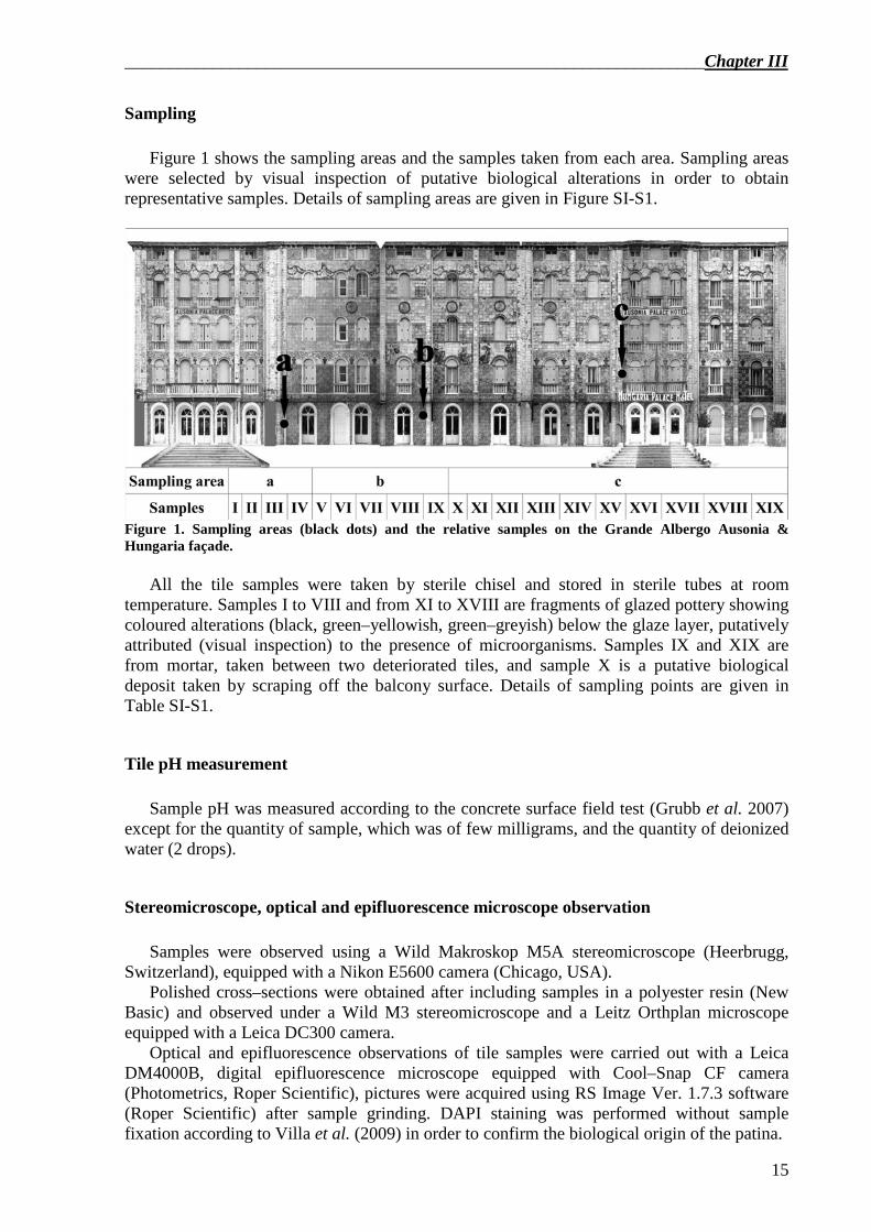
Sampling Figure 1 shows the sampling areas and the samples taken from each area. Sampling areas
were selected by visual inspection of putative biological alterations in order to obtain representative samples. Details of sampling areas are given in Figure SI-S1.
Figure 1. Sampling areas (black dots) and the relative samples on the Grande Albergo Ausonia & Hungaria façade.
All the tile samples were taken by sterile chisel and stored in sterile tubes at room
temperature. Samples I to VIII and from XI to XVIII are fragments of glazed pottery showing coloured alterations (black, green–yellowish, green–greyish) below the glaze layer, putatively attributed (visual inspection) to the presence of microorganisms. Samples IX and XIX are from mortar, taken between two deteriorated tiles, and sample X is a putative biological deposit taken by scraping off the balcony surface. Details of sampling points are given in Table SI-S1.
Tile pH measurement Sample pH was measured according to the concrete surface field test (Grubb et al. 2007)
except for the quantity of sample, which was of few milligrams, and the quantity of deionized water (2 drops).
Stereomicroscope, optical and epifluorescence microscope observation Samples were observed using a Wild Makroskop M5A stereomicroscope (Heerbrugg,
Switzerland), equipped with a Nikon E5600 camera (Chicago, USA). Polished cross–sections were obtained after including samples in a polyester resin (New
Basic) and observed under a Wild M3 stereomicroscope and a Leitz Orthplan microscope equipped with a Leica DC300 camera.
Optical and epifluorescence observations of tile samples were carried out with a Leica DM4000B, digital epifluorescence microscope equipped with Cool–Snap CF camera (Photometrics, Roper Scientific), pictures were acquired using RS Image Ver. 1.7.3 software (Roper Scientific) after sample grinding. DAPI staining was performed without sample fixation according to Villa et al. (2009) in order to confirm the biological origin of the patina.
Chapter III__________________________________________________________________
16
ESEM–EDX observation Observations of the glass surface and polished cross–sections were performed by a Fei
Quanta 200 FEG–ESEM instrument to evaluate the polymer morphology and distribution and the cell size and cell location in tile samples. The semi–quantitative elemental compositions were obtained by an Energy Dispersive X–ray Spectrometer EDAX Genesys, using an accelerating voltage of 25 keV. The samples were observed directly, without any preliminary conductive coating.
MicroFTIR analysis The samples collected were placed on a gold plate and treated with a few drops of CHCl3
to extract any potentially present soluble organic fraction. After gentle solvent evaporation, the soluble residue, distributed as a halo around the sample, was analysed using microFTIR. A Nicolet microscope connected to a Nicolet 560 FTIR system, equipped with a Mercury Cadmium Telluride (MCT) detector and OMNIC32 software, was used for spectra collection. The size of the sample area investigated was about 50×50 µm. The IR spectra were recorded in reflectance mode in the range of 4000–650 cm–1, with a resolution of 4 cm–1.
DNA extraction Before DNA extraction all the samples were ground using a sterile mortar and pestle.
Total DNA was extracted directly from the samples, as described by Polo et al. (2010).
Analysis of the Bacterial community The 16S rRNA gene fragment extracted from the samples (tiles, mortar and putative
biological deposit) were amplified with primers GC−357 F and 907 R with chemical conditions and a thermal cycling program as reported by Polo et al. (2010), except for the dNTP mix concentration of 0.2 µM.
Analysis of fungal community The internal transcribed spacer (ITS) region fragments were amplified by a semi–nested
PCR performed as follows: a first amplification step using the combination of primers NS5 and ITS4 (White et al. 1990) with 1 X of PCR Buffer, 1.8 mM of MgCl2, 0.2 mM of dNTP mix, 0.5 µM of each primer and 0.625 U of Taq DNA polymerase (GoTaq, Promega) in 25 µl PCR reaction; the cycling program consisted in an initial denaturation at 95 °C for 3 min followed by 30 cycles of denaturation at 95 °C for 45 s, annealing at 52 °C for 45 s and extension at 72 °C for 2 min, and a final extension at 72 °C for 10 min. The first PCR product was used as template for a second amplification step performed with the primers ITS4 and GC clamped ITS1 (Gardes et al. 1993) (GC clamp: 5′−CCGGCGCCGCGGCGGGCGGGGCGGGGGCACGGG−3′). The reaction mixture was identical to first–step PCR except for 0.12 mM of dNTP mix and 0.3 µM of each primer. The cycling program consisted in an initial denaturation at 94 °C for 5 min followed by 35 cycles
__________________________________________________________________Chapter III
17
of denaturation at 94 °C for 45 s, annealing at 58 °C for 45 s and extension at 72 °C for 2 min, and a final extension at 72 °C for 10 min.
Analysis of the phototrophic community The 5th dominium of 23S gene fragments were amplified by DGGE−PCR performed with
primers p23SrV−F GC clamped (GC clamp: 5′−CGCCCGCCGCGCGCGGCGGGCGGGGCGGGGGCACGGGGGG−3′) and p23SrV−R (Sherwood et al. 2007) with the following chemical conditions: 1 X of PCR Buffer, 1.8 mM of MgCl2, 0.2 mM of dNTP mix, 0.5 µM of each primer and 2 U of Taq DNA polymerase (Invitrogen) in 50 µl PCR reaction. The thermal cycling program included an initial denaturation at 95 °C for 2 min, followed by 35 cycles consisting of denaturation at 94 °C for 30 s, annealing at 57 °C for 30 s and extension at 72 °C for 30 s, and a final extension step at 72 °C for 10 min.
Denaturing Gradient Gel Electrophoresis (DGGE) and sequencing The DGGE analysis was performed with 6 % polyacrylamide (6 % of a 37:1 acrylamide–
bisacrylamide mixture (Sigma) in a Tris acetate EDTA (TAE) 1 X buffer (Sigma), 0.75 mm thick, 16x10 cm) gels prepared according to Polo et al. (2010). Denaturant gradients were 40 %–70 % for bacteria and the phototrophic community, and 30 %–60 % for the fungal community. The DNA fragments were separated by electrophoresis run for 17 h at 90 V, performed by the D−Code Universal Mutation Detection system (Bio−Rad). The gels were stained by SYBR−Green (Armerscham Pharmacia Biotech) and the results observed by a GelDoc (Bio−Rad) apparatus. Individual lanes of the gel images were straightened and aligned using Adobe Photoshop (Adobe System Incorporated). The excised bands were eluted in 50 µl milli–Q water by incubation at 37 °C for 5 h, re−amplified and identified by sequencing (Primm, Milan). The sequences were analysed using the BLASTN software (www.ncbi.nlm.nih.gov/BLAST) and the Classifier tool by Ribosomal Data Project (http://rdp.cme.msu.edu/classifier/classifier.jsp).
Results
Stereomicroscope, optical, epifluorescence and electronic microscope observation of the coloured alterations
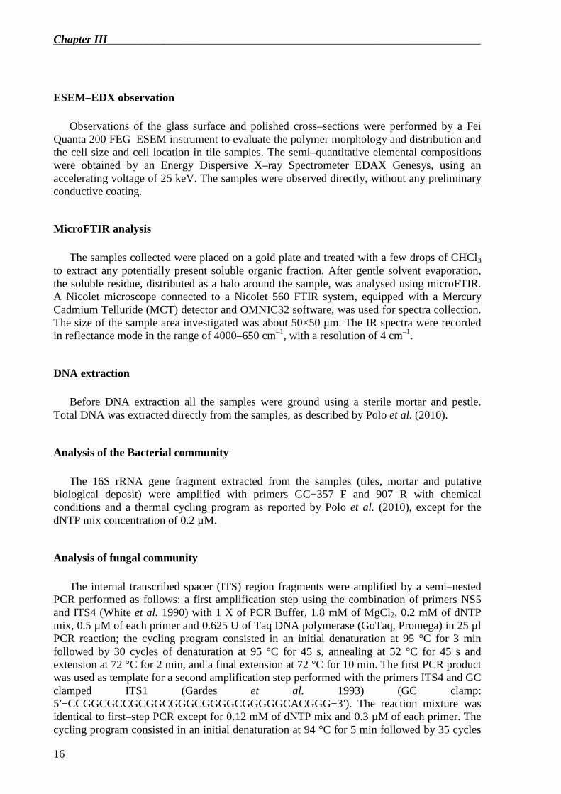
Using a stereomicroscope we observed in samples I to VII and XIII to XVIII the coloured
alteration at the interface glaze-pottery (see Figure 2a–b and Figure 4a) with an average thickness of about 250 µm with a maximum of 800 µm in sample XVI (Figure 2b). In samples XI and XII the coloured alteration was mainly in the pottery layer (see Figure 2f–g and Figure 4b).
The optical, DAPI and electronic microscope (ESEM) observations confirmed the presence of microbiological cells forming a biofilm, which often included tile material (Figs. 2 c, h, e, l). We mainly observed smaller (diameter 1–2 µm) and bigger (diameter 3–4 µm) coccoid cells (Figures 2c and 2h). Due to the small size, and the autofluorescence, the presence of cyanobacteria was hypothesized (Figures. 2d and 2i).
Chapter III__________________________________________________________________
18
Figure 2– Top view of the samples XVI (a) and XII (f) under the stereomicroscope; thin sections of samples XVI (b) and XII (g); yellow arrows indicate coloured alterations. Optical microscope images of biological deposits obtained after grinding samples XVI (c) and XII (h); autofluorescence of samples XVI (d) and XII (i) under Texas Red filter cube; ESEM observation of sample XVI (e); DAPI staining of sample XII (l) under DAPI filter cube.
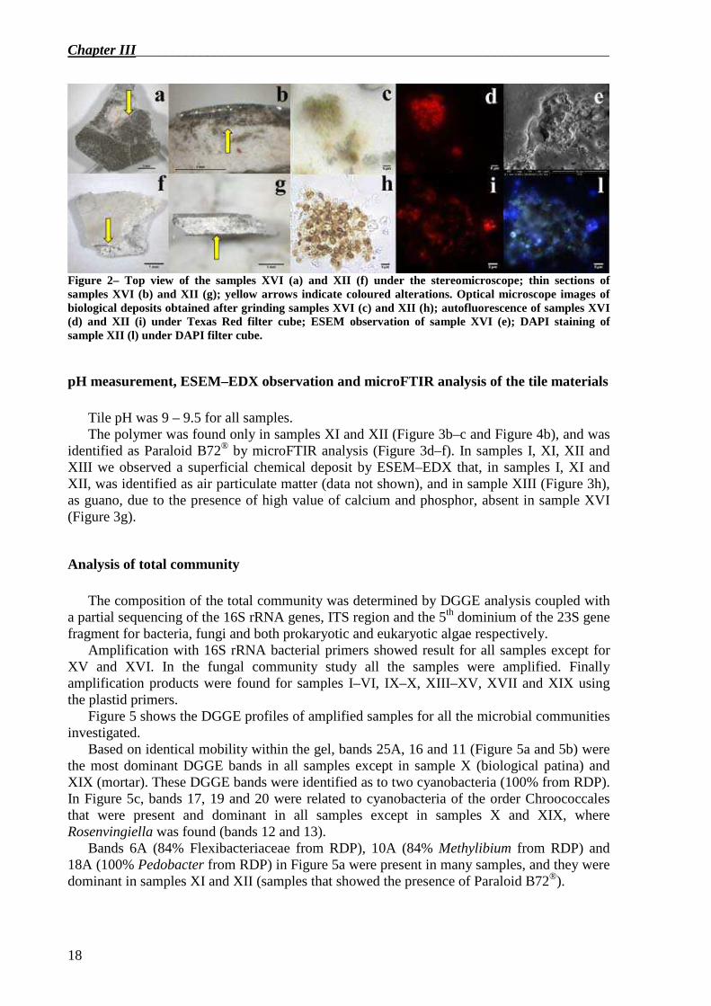
pH measurement, ESEM–EDX observation and microFTIR analysis of the tile materials Tile pH was 9 – 9.5 for all samples. The polymer was found only in samples XI and XII (Figure 3b–c and Figure 4b), and was
identified as Paraloid B72® by microFTIR analysis (Figure 3d–f). In samples I, XI, XII and XIII we observed a superficial chemical deposit by ESEM–EDX that, in samples I, XI and XII, was identified as air particulate matter (data not shown), and in sample XIII (Figure 3h), as guano, due to the presence of high value of calcium and phosphor, absent in sample XVI (Figure 3g).
Analysis of total community The composition of the total community was determined by DGGE analysis coupled with
a partial sequencing of the 16S rRNA genes, ITS region and the 5th dominium of the 23S gene fragment for bacteria, fungi and both prokaryotic and eukaryotic algae respectively.
Amplification with 16S rRNA bacterial primers showed result for all samples except for XV and XVI. In the fungal community study all the samples were amplified. Finally amplification products were found for samples I–VI, IX–X, XIII–XV, XVII and XIX using the plastid primers.
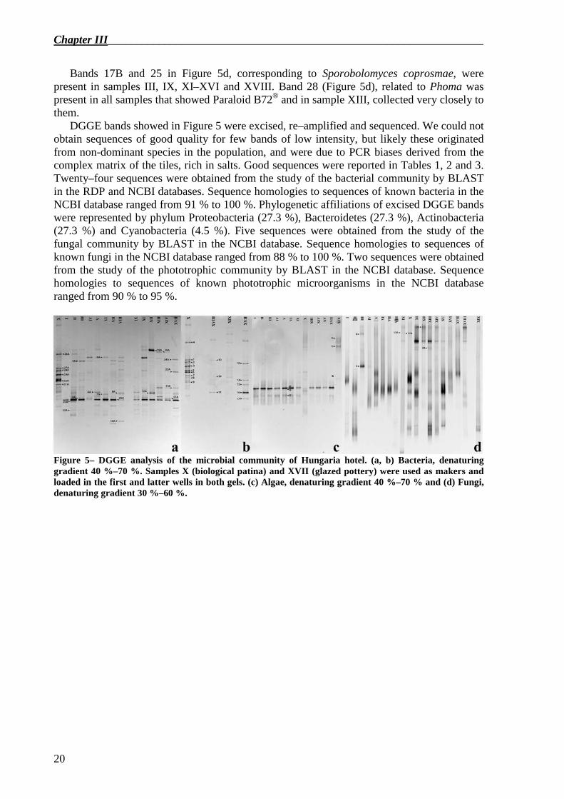
Figure 5 shows the DGGE profiles of amplified samples for all the microbial communities investigated.
Based on identical mobility within the gel, bands 25A, 16 and 11 (Figure 5a and 5b) were the most dominant DGGE bands in all samples except in sample X (biological patina) and XIX (mortar). These DGGE bands were identified as to two cyanobacteria (100% from RDP). In Figure 5c, bands 17, 19 and 20 were related to cyanobacteria of the order Chroococcales that were present and dominant in all samples except in samples X and XIX, where Rosenvingiella was found (bands 12 and 13).
Bands 6A (84% Flexibacteriaceae from RDP), 10A (84% Methylibium from RDP) and 18A (100% Pedobacter from RDP) in Figure 5a were present in many samples, and they were dominant in samples XI and XII (samples that showed the presence of Paraloid B72®).
__________________________________________________________________Chapter III
19
Figure 3– Back scattered electron images collected from the surfaces of samples I (a), XI (b) and XII (c) at magnification 50x, 80x and 100x respectively. Pottery is the grey layer; the glaze layer is in white, while the white arrows clearly indicate the dark amorphous coating corresponding to the applied polymer. IR spectra of the amorphous material collected from sample XI (d), sample XII (e) and reference spectra of commercial product Paraloid B72 (f). The good fitting of absorbance patterns collected from the samples XI and XII with the reference spectra confirms the acrylic polymer nature of the coating on the tiles surfaces. EDX spectra collected from the outmost surface of samples XVI (g) and XIII (h). (g) EDX spectrum without any presence of deposits on the surface. The presence of Ca3(PO4)2 is suggested by the elements Ca and P, highlighted by the black arrows on spectra (h).
Figure 4– Schematic representation of samples I–VIII and XIII–XVIII, which are characterised by the absence of the acrylic resin (a) and samples XI–XII, which are characterised by the presence of the synthetic polymer (b). PB72 stands for the acrylic polymer-based Paraloid B72. The star indicates a richer microbial community, with the presence of black fungi, such as Phoma, and bacteria as Methylibium sp.
Chapter III__________________________________________________________________
20
Bands 17B and 25 in Figure 5d, corresponding to Sporobolomyces coprosmae, were present in samples III, IX, XI–XVI and XVIII. Band 28 (Figure 5d), related to Phoma was present in all samples that showed Paraloid B72® and in sample XIII, collected very closely to them.
DGGE bands showed in Figure 5 were excised, re–amplified and sequenced. We could not obtain sequences of good quality for few bands of low intensity, but likely these originated from non-dominant species in the population, and were due to PCR biases derived from the complex matrix of the tiles, rich in salts. Good sequences were reported in Tables 1, 2 and 3. Twenty–four sequences were obtained from the study of the bacterial community by BLAST in the RDP and NCBI databases. Sequence homologies to sequences of known bacteria in the NCBI database ranged from 91 % to 100 %. Phylogenetic affiliations of excised DGGE bands were represented by phylum Proteobacteria (27.3 %), Bacteroidetes (27.3 %), Actinobacteria (27.3 %) and Cyanobacteria (4.5 %). Five sequences were obtained from the study of the fungal community by BLAST in the NCBI database. Sequence homologies to sequences of known fungi in the NCBI database ranged from 88 % to 100 %. Two sequences were obtained from the study of the phototrophic community by BLAST in the NCBI database. Sequence homologies to sequences of known phototrophic microorganisms in the NCBI database ranged from 90 % to 95 %.
Figure 5– DGGE analysis of the microbial community of Hungaria hotel. (a, b) Bacteria, denaturing gradient 40 %–70 %. Samples X (biological patina) and XVII (glazed pottery) were used as makers and loaded in the first and latter wells in both gels. (c) Algae, denaturing gradient 40 %–70 % and (d) Fungi, denaturing gradient 30 %–60 %.
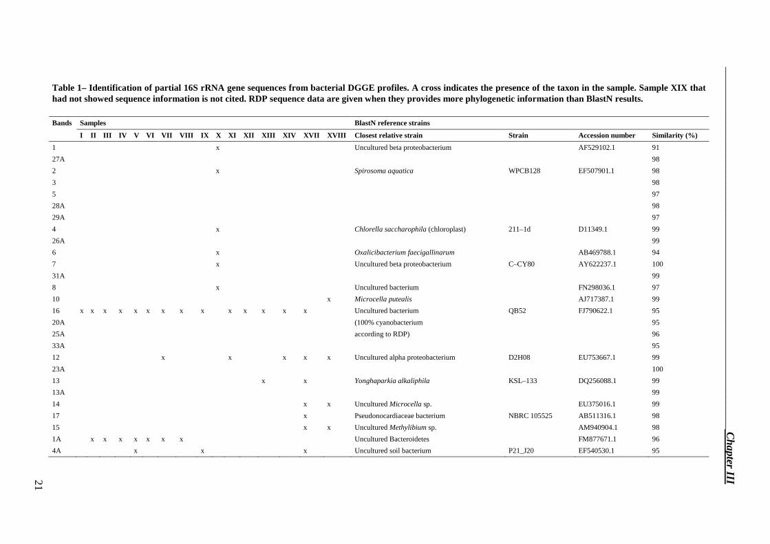
Table 1– Identification of partial 16S rRNA gene sequences from bacterial DGGE profiles. A cross indicates the presence of the taxon in the sample. Sample XIX that had not showed sequence information is not cited. RDP sequence data are given when they provides more phylogenetic information than BlastN results.
Bands Samples BlastN reference strains
I II III IV V VI VII VIII IX X XI XII XIII XIV XV II XVIII Closest relative strain Strain Accession number Similarity (%)
1 x Uncultured beta proteobacterium AF529102.1 91
27A 98
2 x Spirosoma aquatica WPCB128 EF507901.1 98
3 98
5 97
28A 98
29A 97
4 x Chlorella saccharophila (chloroplast) 211–1d D11349.1 99
26A 99
6 x Oxalicibacterium faecigallinarum AB469788.1 94
7 x Uncultured beta proteobacterium C–CY80 AY622237.1 100
31A 99
8 x Uncultured bacterium FN298036.1 97
10 x Microcella putealis AJ717387.1 99
16 x x x x x x x x x x x x x x Uncultured bacterium QB52 FJ790622.1 95
20A (100% cyanobacterium 95
25A according to RDP) 96
33A 95
12 x x x x x Uncultured alpha proteobacterium D2H08 EU753667.1 99
23A 100
13 x x Yonghaparkia alkaliphila KSL–133 DQ256088.1 99
13A 99
14 x x Uncultured Microcella sp. EU375016.1 99
17 x Pseudonocardiaceae bacterium NBRC 105525 AB511316.1 98
15 x x Uncultured Methylibium sp. AM940904.1 98
1A x x x x x x x Uncultured Bacteroidetes FM877671.1 96
4A x x x Uncultured soil bacterium P21_J20 EF540530.1 95
__________________________________________________________________ C
hapter III 21
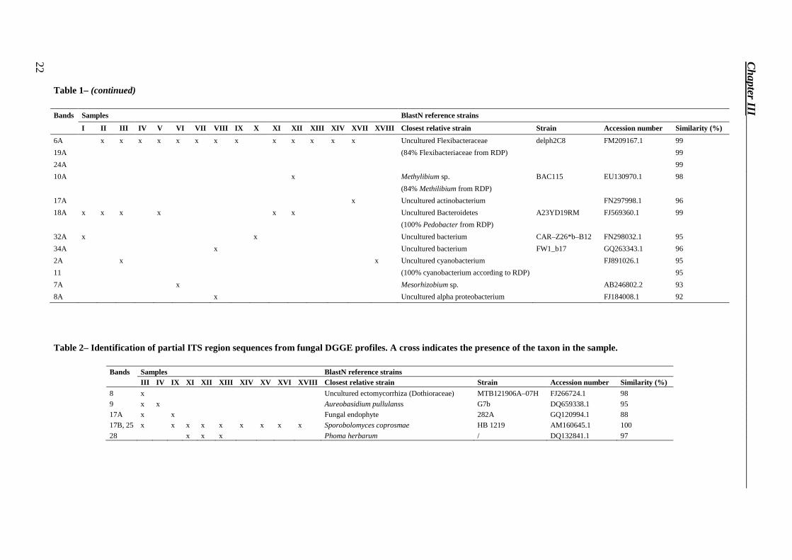
Table 1– (continued)
Bands Samples BlastN reference strains
I II III IV V VI VII VIII IX X XI XII XIII XIV XVII XVIII Closest relative strain Strain Accession number Similarity (%)
6A x x x x x x x x x x x x x Uncultured Flexibacteraceae delph2C8 FM209167.1 99
19A (84% Flexibacteriaceae from RDP) 99
24A 99
10A x Methylibium sp. BAC115 EU130970.1 98
(84% Methilibium from RDP)
17A x Uncultured actinobacterium FN297998.1 96
18A x x x x x x Uncultured Bacteroidetes A23YD19RM FJ569360.1 99
(100% Pedobacter from RDP)
32A x x Uncultured bacterium CAR–Z26*b–B12 FN298032.1 95
34A x Uncultured bacterium FW1_b17 GQ263343.1 96
2A x x Uncultured cyanobacterium FJ891026.1 95
11 (100% cyanobacterium according to RDP) 95
7A x Mesorhizobium sp. AB246802.2 93
8A x Uncultured alpha proteobacterium FJ184008.1 92
Table 2– Identification of partial ITS region sequences from fungal DGGE profiles. A cross indicates the presence of the taxon in the sample.
Bands Samples BlastN reference strains III IV IX XI XII XIII XIV XV XVI XVIII Closest relative strain Strain Accession number Similarity (%)
8 x Uncultured ectomycorrhiza (Dothioraceae) MTB121906A–07H FJ266724.1 98
9 x x Aureobasidium pullulanss G7b DQ659338.1 95 17A x x Fungal endophyte 282A GQ120994.1 88
17B, 25 x x x x x x x x x Sporobolomyces coprosmae HB 1219 AM160645.1 100
28 x x x Phoma herbarum / DQ132841.1 97
Chapter III___________________________________________________
_______________
22
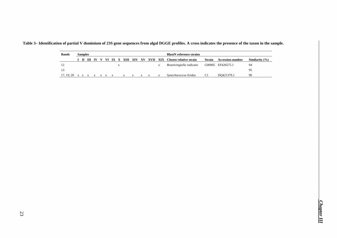
Table 3– Identification of partial V dominium of 23S gene sequences from algal DGGE profiles. A cross indicates the presence of the taxon in the sample.
Bands Samples BlastN reference strains
I II III IV V VI IX X XIII XIV XV XVII XIX Closest relative strain Strain Accession number Similarity (%)
12 x x Rosenvingiella radicans G00005 EF426575.1 94
13 95
17, 19, 20 x x x x x x x x x x x x Synechococcus lividus C1 DQ421379.1 90
__________________________________________________________________ C
hapter III
23
Chapter III__________________________________________________________________
24
Discussion To date, no molecular studies concerning microflora on artistic ceramics are available. A
pioneer study on this topic was carried out by Oliveira et al. (2001), who made optical microscopic observations of glazed ceramic tiles on the façade of Salvador and Belèm buildings (Brazil). However, the above–mentioned author has reported no further investigations apart from the documenting of the presence of Cyanophyta and Bacilliarophyta. Therefore, we present here the first molecular characterisation of a microbial community present on artistic ceramic.
Denaturing gradient gel electrophoresis (DGGE) and sequencing from total DNA extracted from Hungaria samples were used to identify taxa of microorganisms causing coloured alteration and verify that Paraloid B72® was the best consolidants to be used.
The molecular analysis of the total community showed that bacteria, eukaryotic algae and fungi were present. Since the principal deterioration appearing on the Hungaria hotel is a coloured deposit, most likely due to the growth of both prokaryotic and eukaryotic algae, we employed, for the characterisation of the phototrophic community, a recent set of primers (p23SrV) designed by Sherwood and Presting (2007). Here we present the first use of p23SrV primers for DGGE analysis.
Combining the results obtained from sequencing of bacterial 16S and phototrophic 23S fragments we found cyanobacteria in all tile samples -independently on the position of biofilms either below the glaze or in the pottery-, but not in the biological deposit taken from the balcony.
Cyanobacteria are very common on monuments, for example they have been detected on Ca’ d’Oro (Venice) (Praderio et al. 1993), on glazed ceramic tiles from Portugal and Brazil (Oliveira et al. 2001), and, in general, on sculptures and buildings (Cunha et al. 2003; Macedo et al. 2009; Polo et al. 2010) exposed to high humidity, running–off water, high and low temperatures, as well as wetting/drying cycles and high UV exposure (Portillo et al. 2009). Using primers specific for the phototrophic community, bands 17, 19 and 20 were identified as a cyanobacterium of the order Chroococcales, which is the most widespread order present on stone monuments (Macedo et al. 2009; Rindi 2007). In addition to the production of extracellular polymeric substances and pigments, some species of this order are able to precipitate magnesium and calcium (Rindi 2007).
The cyanobacteria (100 % from RDP) detected on the Hungaria tiles using 16S rRNA primers showed high similarity to uncultured bacteria detected in a sample from the Tibetan tundra in which the community was mainly composed by coccoid cyanobacterial cells (Wong et al. 2010).
The presence of coloured deposit in depth into the pottery layer detected by microscope observations of Hungaria tiles with cracks and fractures is indicative of a cryptoendolithic niche. The fact that the microbiological deposit was located between the glaze and the pottery layer made the collect the entire biomass and estimate the quantity of cyanobacteria against the whole community not possible. Endolithic microorganisms, the most widespread of them are cyanobacteria, occur in various habitats, such as hot and cold deserts and were reported to exist also in monuments (de los Rios et al. 2007; Horath et al. 2006; McNamara et al. 2006; Norris et al. 2006; Sigler et al. 2003). In extreme environments, endolithic growth provides protection from low temperature, UV radiation, and desiccation and provides mineral nutrients (Horath et al. 2006; Macedo et al. 2009; McNamara et al. 2006; Norris et al. 2006; Sigler et al. 2003). In Hungaria samples we found small coccoid cells (from 1–2 to 3–4 µm). de los Rios et al. (2007) found spherical to oval shaped cells of 1.1–1.5 µm in size in granite rocks collected in Antarctica, while in dolomite rock in central Switzerland, Horath et al. (2006) detected coccoid cyanobacteria not only as single 3–6 µm cells, but also as
__________________________________________________________________Chapter III
25
multicellular aggregates. On external stone and building surfaces, high light levels may favour endolithic growth by cyanobacteria. Del Monte and Sabbioni (1983) reported a perforating activity of endolithic cyanobacteria inhabiting cracks and fissures in the marble of monuments in Torcello Island, near Venice. Especially when endolithic microorganisms are present, water absorption by the biofilm matrix causes mechanical stress that opens cracks and fissures in the material (de los Rios et al. 2007; McNamara et al. 2006).
The eukaryotic algae of the Hungaria hotel were found only in the mortar samples and on the first floor stone balcony, never on the ceramic tiles, and they were epilithic. In this study, chloroplasts of the phylum Chlorophyta were detected, which was represented by the genus Chlorella (99 % of similarity). Green algae have already been found in Venetian buildings made of stone and marble (Praderio et al. 1993) and, in particular, Chlorella sp. was found on Ca’ d’Oro (Salvadori et al. 1994). The other eukaryotic alga found in this work bands 12 and 13, is a green alga of the order Prasiolales that is widespread in temperate regions, in a wide range of terrestrial and littoral habitats (Rindi 2007). Microorganisms of the order of Prasiolales have been detected in very high amounts on cement and bricks in Galway (Ireland), and at the base of walls, in corners and on protrusions of buildings in Oviedo and León (Spain) (Rindi 2007). Moreover, the abundance of these algae in places affected by bird guano is well–documented (Rindi 2007). We found guano in sample XIII that was collected very close to the balcony where Rosenvingiella was detected (band 13).
Another microorganism present mainly in the sampling area d was the red–coloured yeast Sporobolomyces coprosmae (Weber et al. 2005).
In conclusion, the colour of the deposit present in the tile samples and the greenish alteration on the balcony are most likely mainly due to the presence of cyanobacteria and eukaryotic algae respectively.
Paraloid B72® was detected in samples XI and XII. This synthetic resin, frequently used in the conservation of ceramic tiles (Vaz et al. 2008), was applied during the 2007 restoration of the hotel façade. The fact that no polymers were found on the surface of other samples (XIII–XIX), which had most likely been treated with the resin, could be due to photooxidative depolymerization and the wash out of resin from the tile surfaces (Favaro et al. 2006; Favaro et al. 2005). Phoma was found in all the samples in which we detected Paraloid B72®, and also in one without any evidence of polymer but collected in sampling area d, which was treated in the 2007 restoration. Melanised fungi, such as Phoma, are among the most damaging fungi, attacking and penetrating the surfaces of stone monuments (Cappitelli et al. 2007b). In the study by Cappitelli et al. (2007b) on melanin–producing fungi that attack synthetic polymers used in cultural heritage, the Phoma genus was found. The presence of an uncultured Bacteroidetes (band 18A) and Methylibium sp. (bands 15 and 10A) can be due to the fact that Bacteroidetes show hydrolytic activity toward polymeric substances and Methylibium can grow on organic pollutants (Borin et al. 2009). In addition, when Paraloid B72 was present, the biofilm was located in a deeper position than in samples without any evidence of the resin, as also suggested by Ariño and Saiz-Jimenez (1996).
During this study several microorganisms related to an alkaline environment, which is the range of the tile pH, were found. Among the Bacteroidetes we found two uncultured bacteria (bands 8 and 32A) that were previously found in the Roman Necropolis of the Carmona tomb, carved in a calcarenite bed, an alkaline material. Both microorganisms are taxonomically related to Hymenobacter (100 % from RDP), and were detected in the sample taken from the first floor balcony. Many Hymenobacter species have been isolated from air and alkaline soil, indicating a possible relationship based on desiccation and alkaline tolerance (Schloss et al. 2009). In another study, Pedobacter, the most probable taxon (100 % from RDP) for the band 18A, was detected in alkaline painted and unpainted concrete structures by (Giannantonio et al. 2009). Finally, Bacteroidetes were also detected in the highly alkaline saline soil of the former lake Texcoco in Mexico (Valenzuela-Encinas et al. 2009). Among the
Chapter III__________________________________________________________________
26
Betaproteobacteria found, one is Yonghaparkia alkaliphila (bands 13 and 13A), bacteria that live in natural alkaline environments (Yoon et al. 2006). From the study by Hyvärinen et al. (2002), Actinobacteria were found especially on ceramic products, which may be due to their capability to tolerate alkaline conditions. Related to this, Actinobacteria were also detected in frescoes from two different churches in Siena (Italy), both of them showing an alkaline environment (Milanesi et al. 2009). Among this phylum we found one uncultured Actinobacteria (band 17A) that was previously found in the Roman Necropolis of Carmona tomb, carved in a calcarenite bed and the most related bacteria to the band 10, Microcella putealis, bacteria that live in natural alkaline environments (Tiago et al. 2004).
Taking into account the natural environment in which the hotel is built, the Venice Lagoon, it is not surprising to find microorganisms correlated with aquatic ecosystems and halophilic microorganisms. As an example, Aureobasidium, which was detected in two samples near the ground, is from a study on the diversity of marine yeasts, and was previously found in building and ceramic materials and in natural salterns (Hyvärinen et al. 2002; Jurado et al. 2008). Also black fungi were detected on hypersaline salt pans (Chertov et al. 2004). Bacteroidetes have frequently been found to be dominant in marine ecosystems (Valenzuela-Encinas et al. 2009); moreover, Borin et al. (2009) reported that this phylum was widespread in the Venice Lagoon. Another Bacteroidetes that was detected in the biological deposit present on the balcony is Spirosoma aquatica (bands 2, 3, 5, 28A and 29A), a bacterium isolated from freshwater. Alpha and Beta Proteobacteria were detected during a study about biofilm formation and succession, occurring on the surface of unglazed ceramic tiles in an artificial reef deployed in the northern Gulf of Eilat (Siboni et al. 2007). Finally, also the order of Chroococcales (one of the cyanobacteria detected in tile samples) is abundant in marine and freshwater areas (Rindi 2007). Therefore the high salt concentration of the environment near the Hungaria Hotel is an important factor in the selection of the microflora present on its façade.
The Venice Lagoon is contaminated by heavy metals, hydrocarbons, polycyclic aromatic hydrocarbons, polychlorinated biphenyls from industrial, urban and agricultural sources (Borin et al. 2009). Some of the bacteria detected in this study (without substratum preferences) are correlated with pollutants. The uncultured Microcella sp. (band 14) was found in shoreline environments (Spain) affected by the Prestige oil spill (Tamura et al. 2007), two uncultured Beta Proteobacteria and an uncultured Bacteroidetes (bands 1, 27A, 7, 31A, 1A) were found in studies about the microbial community in sites contaminated by trichloroethene and uranium, and Methylibium sp. (band 10A) was found in filters used in water treatment for the removal of natural organic matter and organic micropollutants (Magic-Knezev et al. 2009).
The massive growth of microorganisms soon after the restoration might be explained by the water content of the tiles. Presence and increase of water inside the ceramic tiles completely glazed by a vitreous layer partially cracked, as for instance in the case of Hungaria hotel, can be physically associated to the phenomena of capillarity interesting masonry buildings. If the walls are covered by a partially permeable or impermeable layer as for instance the vitreous mosaics covering the walls of San Marco Basilica in Venice, the wet zone reaches up to several meters above the ground (Bakolas et al. 1995).
In conclusion, it was proved that the deterioration of the tiles was mainly due to the presence of microorganisms. The rapid reappearing of coloured alterations soon after the 2007 restoration, which included the use of biocides, was likely favoured by the water content and organic substances, some of which added during the conservation treatment. Paraloid B72® was not the best consolidant polymer to be used fort the long-time conservation of the ceramic tile of Hungaria hotel. For a long term maintenance plan, an immediate series of corrective measures, finalised to a more efficient conservation of the Hungaria façade is strongly suggested.

__________________________________________________________________Chapter III
27
SUPPORTING INFORMATION AVAILABLE Additional Figures and Tables are provided in Appendix I.
Chapter III__________________________________________________________________
28
References
Arino X. and SaizJimenez C. (1996). Lichen deterioration of consolidants used in the conservation of stone monuments. Lichenologist 28: 391-394.
Bakolas A., Biscontin G., Contardi V., Franceschi E., Moropoulou A., Palazzi D. and Zendri E. (1995). Thermoanalytical research on traditional mortars in venice. Thermochimica Acta 269-270(0): 817-828.
Borin S., Brusetti L., Daffonchio D., Delaney E. and Baldi F. (2009). Biodiversity of prokaryotic communities in sediments of different sub-basins of the Venice lagoon. Res Microbiol 160(5): 307-314.
Cappitelli F., Nosanchuk J. D., Casadevall A., Toniolo L., Brusetti L., Florio S., Principi P., Borin S. and Sorlini C. (2007a). Synthetic consolidants attacked by melanin-producing fungi: case study of the biodeterioration of Milan (Italy) cathedral marble treated with acrylics. Applied and environmental microbiology 73(1): 271-277.
Cappitelli F., Principi P., Pedrazzani R., Toniolo L. and Sorlini C. (2007b). Bacterial and fungal deterioration of the Milan Cathedral marble treated with protective synthetic resins. The Science of the total environment 385(1-3): 172-181.
Chertov O., Gorbushina A. and Deventer B. (2004). A model for microcolonial fungi growth on rock surfaces. Ecological Modelling 177(3-4): 415-426.
Cunha C. A., Macieira S., Dias J. M., Almeida G., Gonçalves L. L., Costa C., Lampreia J., Huber R., Moura J. J. G., Moura I. and Romão M. J. (2003). Cytochrome c Nitrite Reductase from Desulfovibrio desulfuricans ATCC 27774. Journal of Biological Chemistry 278(19): 17455-17465.
de los Rios A., Grube M., Sancho L. G. and Ascaso C. (2007). Ultrastructural and genetic characteristics of endolithic cyanobacterial biofilms colonizing Antarctic granite rocks. FEMS Microbiol Ecol 59(2): 386-395.
Del Monte M. and Sabbioni C. (1983). Weddellite on limestone in the Venice [Italy] environment. Environmental Science & Technology 17(9): 518-522.
Favaro M., Simon S., Menichelli C., Fassina V. and Vigato P. A. (2005). The Four Virtues of the Porta della Carta, Ducal Palace, Venice: Assessment of the State of Preservation and Re-Evaluation of the 1979 Restoration. Studies in Conservation 50(2): 109-127.
Favaro M., Mendichi R., Ossola F., Russo U., Simon S., Tomasin P. and Vigato P. A. (2006). Evaluation of polymers for conservation treatments of outdoor exposed stone monuments. Part I: Photo-oxidative weathering. Polymer Degradation and Stability 91(12): 3083-3096.
Felsenstein J. (1993). PHYLIP (Phylogeny Inference Package) version 3.5c. Department of Genetics, University of Washington, Seattle, Distributed by the author.
Gardes M. and Bruns T. D. (1993). ITS primers with enhanced specificity for basidiomycetes--application to the identification of mycorrhizae and rusts. Mol Ecol 2(2): 113-118.
Giannantonio D. J., Kurth J. C., Kurtis K. E. and Sobecky P. A. (2009). Molecular characterizations of microbial communities fouling painted and unpainted concrete structures. International Biodeterioration & Biodegradation 63(1): 30-40.
Grubb J. A., Hemant S. L. and Kakade A. M. (2007). Testing pH of concrete. Concrete International 29(4): 78-83.
Horath T., Neu T. and Bachofen R. (2006). An Endolithic Microbial Community in Dolomite Rock in Central Switzerland: Characterization by Reflection Spectroscopy, Pigment Analyses, Scanning Electron Microscopy, and Laser Scanning Microscopy. Microbial Ecology 51(3): 353-364.
Hyvärinen A., Meklin T., Vepsäläinen A. and Nevalainen A. (2002). Fungi and actinobacteria in moisture-damaged building materials — concentrations and diversity. International Biodeterioration & Biodegradation 49(1): 27-37.
Jurado V., Sanchez-Moral S. and Saiz-Jimenez C. (2008). Entomogenous fungi and the conservation of the cultural heritage: A review. International Biodeterioration & Biodegradation 62(4): 325-330.
Macedo M. F., Miller A. Z., Dionisio A. and Saiz-Jimenez C. (2009). Biodiversity of cyanobacteria and green algae on monuments in the Mediterranean Basin: an overview. Microbiology 155(Pt 11): 3476-3490.
Magic-Knezev A., Wullings B. and Van der Kooij D. (2009). Polaromonas and Hydrogenophaga species are the predominant bacteria cultured from granular activated carbon filters in water treatment. Journal of applied microbiology 107(5): 1457-1467.
Marata A., Ed. (2008). Se i Muri potessero parlare. Arbizzano di Negrar (VR) Italy, I Libri di Damoli. McNamara C. J., Perry T. D. t., Bearce K. A., Hernandez-Duque G. and Mitchell R. (2006). Epilithic and
endolithic bacterial communities in limestone from a Maya archaeological site. Microb Ecol 51(1): 51-64. Milanesi C., Baldi F., Borin S., Brusetti L., Ciampolini F., Iacopini F. and Cresti M. (2009). Deterioration of
medieval painting in the chapel of the Holy Nail, Siena (Italy) partially treated with Paraloid B72. International Biodeterioration & Biodegradation 63(7): 844-850.
Norris T. B. and Castenholz R. W. (2006). Endolithic photosynthetic communities within ancient and recent travertine deposits in Yellowstone National Park. FEMS Microbiol Ecol 57(3): 470-483.
__________________________________________________________________Chapter III
29
Oliveira M. M., Sanjad T. B. C. and Bastos C. J. P. (2001). Biological degradation of glazed ceramic tiles. Historical Constructions. Lourenço P. B., Roca P. Guimarães (Portugal), University of Minho: 337-342.
Pereira I. A. C., Ramos A. R., Grein F., Marques M. C., Da Silva S. M. and Venceslau S. S. (2011). A comparative genomic analysis of energy metabolism in sulfate reducing bacteria and archaea. Frontiers in Microbiology 2.
Polo A., Cappitelli F., Brusetti L., Principi P., Villa F., Giacomucci L., Ranalli G. and Sorlini C. (2010). Feasibility of removing surface deposits on stone using biological and chemical remediation methods. Microbial Ecology 60(1): 1-14.
Portillo M. C., Alloza R. and Gonzalez J. M. (2009). Three different phototrophic microbial communities colonizing a single natural shelter containing prehistoric paintings. Science of The Total Environment 407(17): 4876-4881.
Praderio G., Schiraldi A., Sorlini C., Stassi A. and Zanardini E. (1993). Microbiological and calorimetric investigations on degraded marbles from the Cà d'Oro facade (Venice). Thermochimica Acta 227(0): 205-213.
Rindi F. (2007). Diversity, Distribution and Ecology of Green Algae and Cyanobacteria in Urban Habitats. Algae and Cyanobacteria in Extreme Environments. Seckbach J., Springer Netherlands. 11: 619-638.
Salvadori O., Sorlini C. and Zanardini E. (1994). Microbiological and biochemical investigations on stone of the Ca’ Oro façade (Venice). III International Symposium on the Conservation of Monuments in the Mediterranean Basin, Venice.
Schloss P. D., Westcott S. L., Ryabin T., Hall J. R., Hartmann M., Hollister E. B., Lesniewski R. A., Oakley B. B., Parks D. H., Robinson C. J., Sahl J. W., Stres B., Thallinger G. G., Van Horn D. J. and Weber C. F. (2009). Introducing mothur: open-source, platform-independent, community-supported software for describing and comparing microbial communities. Applied and environmental microbiology 75(23): 7537-7541.
Sherwood A. R. and Presting G. G. (2007). Universal primers amplify a 23S rDNA plastid marker in eukaryotic algae and cyanobacteria. Journal of Phycology 43(3): 605-608.
Siboni N., Lidor M., Kramarsky-Winter E. and Kushmaro A. (2007). Conditioning film and initial biofilm formation on ceramics tiles in the marine environment. FEMS Microbiol Lett 274(1): 24-29.
Sigler W. V., Bachofen R. and Zeyer J. (2003). Molecular characterization of endolithic cyanobacteria inhabiting exposed dolomite in central Switzerland. Environmental Microbiology 5(7): 618-627.
Takeuchi J. (2006). Habitat segregation of a functional gene encoding nitrate ammonification in estuarine sediments. Geomicrobiology Journal 23(2): 75-87.
Tamura K., Dudley J., Nei M. and Kumar S. (2007). MEGA4: Molecular Evolutionary Genetics Analysis (MEGA) software version 4.0. Mol Biol Evol 24(8): 1596-1599.
Tiago I., Chung A. P. and Verissimo A. (2004). Bacterial diversity in a nonsaline alkaline environment: heterotrophic aerobic populations. Applied and environmental microbiology 70(12): 7378-7387.
Valenzuela-Encinas C., Neria-Gonzalez I., Alcantara-Hernandez R. J., Estrada-Alvarado I., Zavala-Diaz de la Serna F. J., Dendooven L. and Marsch R. (2009). Changes in the bacterial populations of the highly alkaline saline soil of the former lake Texcoco (Mexico) following flooding. Extremophiles 13(4): 609-621.
Vaz M. F., Pires J. and Carvalho A. P. (2008). Effect of the impregnation treatment with Paraloid B-72 on the properties of old Portuguese ceramic tiles. Journal of Cultural Heritage 9(3): 269-276.
Villa F., Giacomucci L., Polo A., Principi P., Toniolo L., Levi M., Turri S. and Cappitelli F. (2009). N-vanillylnonanamide tested as a non-toxic antifoulant, applied to surfaces in a polyurethane coating. Biotechnology letters 31(9): 1407-1413.
Weber R. W. S., Madhour A., Anke H., Mucci A. and Davoli P. (2005). 2-Hydroxytorularhodin, a New Xanthophyll from the Red Yeast Sporobolomyces coprosmae. Helvetica Chimica Acta 88(11): 2960-2966.
White T. J., Bruns T., Lee S. and Taylor J. (1990). Amplification and direct sequencing of fungal ribosomal RNA genes for phylogenetics. PCR protocols: a guide to methods and applications. Innis M. A., Gelfand D. H., Sninsky J. J., White T. J. San Diego, Academic Press: 315−322.
Wong F., Lacap D., Lau M., Aitchison J., Cowan D. and Pointing S. (2010). Hypolithic Microbial Community of Quartz Pavement in the High-Altitude Tundra of Central Tibet. Microbial Ecology 60(4): 730-739.
Yoon J. H., Kang S. J., Schumann P. and Oh T. K. (2006). Yonghaparkia alkaliphila gen. nov., sp. nov., a novel member of the family Microbacteriaceae isolated from an alkaline soil. Int J Syst Evol Microbiol 56(Pt 10): 2415-2420.

__________________________________________________________________Chapter IV
31
Degradation of nitrocellulose–based paint by Desulfovibrio desulfuricans ATCC 135411
Abstract Nitrocellulose is one of the most commonly used compounds in ammunition and paint
industries and its recalcitrance to degradation has a negative impact on human health and the environment. In this study the capability of Desulfovibrio desulfuricans ATCC 13541 to degrade nitrocellulose as binder in paint was assayed for the first time. Nitrocellulose–based paint degradation was followed by monitoring the variation in nitrate, nitrite and ammonium content in the culture medium using ultraviolet–visible spectroscopy. At the same time cell counts and ATP assay were performed to estimate bacterial density and activity in all samples. Infrared spectroscopy and colourimetric measurements of paint samples were performed to assess chemical and colour changes due to the microbial action. Microscope observations of nitrocellulose–based paint samples demonstrated the capability of the bacterium to adhere to the paint surface and change the paint adhesive characteristics. Finally, preliminary studies of nitrocellulose degradation pathway were conducted by assaying nitrate– and nitrite reductases activity in D. desulfuricans grown in presence or in absence of paint. We found that D. desulfuricans ATCC 13541 is able to transform nitrocellulose as paint binder and we hypothesised ammonification as degradation pathway. The results suggest that D. desulfuricans ATCC 13541 is a good candidate as a nitrocellulose–degrading bacterium.
Introduction Nitrocellulose, one of the most important and oldest cellulose derivative, is principally
used as binder in paint and a bulk ingredient in gunpowder formulations (Auer et al. 2005). In addition, many materials commonly used in daily life, such as printing inks, decorative films, and pharmaceuticals, contain nitrocellulose (El-Diwani et al. 2009; Souza et al. 2005). The explosive and toxic properties of nitrocellulose and its recalcitrance to degradation make the compound harmful for human health and the environment (El-Diwani et al. 2009; Freedman et al. 1996; Souza et al. 2005). In this respect, although nitrocellulose wastes from industries (e.g. ammunition material and paint industries) and household hazardous waste, which consists of waste from a number of household products, contain increasing nitrocellulose content, only limited research effort into nitrocellulose waste treatment have been reported (Auer et al. 2005; El-Diwani et al. 2009; Freedman et al. 2002; Petrova et al. 2002; Slack et al. 2005; Souza et al. 2005). Currently, the disposal of nitrocellulose is carried out by different processes, including membrane separation processes to enhance recovery of the fines, detonation, incineration and controlled low temperature thermal denitration, but these treatments produce dangerous airborne particulates and pan residues containing toxic materials (Auer et al. 2005; Freedman et al. 2002). For this reason, it would be appropriate to develop methods to degrade nitrocellulose in an environmentally safer and more economical
1 Submitted for publication to Biodegradation: L. Giacomucci, F. Toja, P. Sanmartín, L. Toniolo, B. Prieto, F. Villa, F. Cappitelli. Degradation of nitrocellulose–based paint by Desulfovibrio desulfuricans ATCC 13541.
Chapter IV__________________________________________________________________
32
way. Biological methods could be promising because biotransformation could produce environmentally safe products at potentially lower costs (Freedman et al. 1996).
Microorganisms are able to degrade nitrocellulose by two pathways: i) cleavage of β–1,4–glucoside bonds that produces nitrooligosaccharides of various length, normally carried out by fungi (Auer et al. 2005; Freedman et al. 2002; Petrova et al. 2002; Tarasova et al. 2004), and ii) nitrocellulose denitration that reduces the degree of nitro substitution, generally performed by bacteria (El-Diwani et al. 2009; Tarasova et al. 2004; Tarasova et al. 2005). Since nitrooligosaccharides have mutagenic properties (Petrova et al. 2002), the second pathway is preferred over the first for exploitation as a biodegradation pathway. It has been reported that nitrocellulose undergoes degradation by methanogenic or sulphate–reducing bacteria under anaerobic conditions (Freedman et al. 2002; Tarasova et al. 2005). Sulphate–reducing bacteria of the genus Desulfovibrio were reported to decrease the amount of nitrocellulose powder in media containing this compound (Tarasova et al. 2009). The study by (Petrova et al. 2006) reported the presence of nitrate in the culture media containing nitrocellulose, due to the nitroesterase activity of D. desulfuricans 1388. The subsequent nitrate disappearance and ammonium appearance indicated the dissimilatory reduction of nitrate to ammonium (Bursakov et al. 1997; Petrova et al. 2006; Petrova et al. 2002; Tarasova et al. 2004), a process involving nitrate reduction to nitrite and the subsequent nitrite reduction to ammonium by nitrate– and nitrite reductases (Bursakov et al. 1997; Kraft et al. 2011; Moura et al. 2007). There are several sulphate–reducing bacteria able to reduce nitrate to nitrite (Marietou et al. 2009; Moura et al. 1997), but it appears that this is not a shared feature across the genus Desulfovibrio (Marietou et al. 2009; Tarasova et al. 2009). In contrast, the dissimilatory reduction of nitrite to ammonium seems to be widespread in Desulfovibrio, which can synthesise a very active, constitutive nitrite reductase (Marietou et al. 2009; Moura et al. 2007).
To date, studies on nitrocellulose biodegradation have been performed on nitrocellulose powder, with a percentage of nitro groups of being more than 12%, the percentage generally used in the military sector (Auer et al. 2005; Freedman et al. 2002; Petrova et al. 2002; Tarasova et al. 2009; Tarasova et al. 2004). To the best of our knowledge there are no reported studies on the biodegradation of nitrocellulose as component of daily life materials, like nitrocellulose–based paints. The biodegradation of paints is also interesting because substances other than nitrocellulose are present that could interfere with the degradation process. Although the bulk of a paint is pigment and binder, which is the film–forming material in which the pigment particles are dispersed and forms the matrix that binds the pigments on the painted surface, other components like metals, solvent and additives, are present in paint formulations (Cappitelli et al. 2005).
The main aim of this work was to study the capability of Desulfovibrio desulfuricans ATCC 13541 to attack nitrocellulose as binder in paint. Ultraviolet–visible and infrared spectroscopy, microscope observations and reflectance colour techniques were used to correlate nitrocellulose degradation to specific chemical changes in the composition of the paint material. In addition, a preliminary study on the nitrocellulose degradation pathway in Desulfovibrio desulfuricans ATCC 13541 was conducted.
__________________________________________________________________Chapter IV
33
Material and methods
Nitrocellulose–based paint and paint sample preparation The 20th century paint binders are generally made of synthetic and semi–synthetic
polymers including nitrocellulose (Buzzini et al. 2004; Govaert et al. 2004; Zieba-Palus 2005). The red spray paint by Motip–Dupli® Autocolor (colour 5–0200) was selected in this study since most of red spray paints are composed of alkyd–nitrocellulose–based binders, including those produced by Motip® (Govaert et al. 2004; Segalini et al. 2000). Autocolor paint samples were prepared spraying the paint on plain glass slides (Prestige, 7.6 x 2.6 cm). The can was firstly shaken for 3 min per manufacturer's instruction and then the paint sprayed by a moving rate of about 20 cm/s, maintaining nozzle at a distance of 30 cm above the glass slide and the manual pressure on spray valve fixed to allow a uniform spread (Segalini et al. 2000). The painted slides were dried for four months prior to their use in degradation experiments. For each experiment a constant ratio of 4.55 mm2 of Autocolor paint per 1 ml of cultural medium was used.
Degradation experiment The sulphate–reducing bacterium Desulfovibrio desulfuricans ATCC 13541 was selected
for all the degradation experiments. D. desulfuricans was maintained anaerobically in DSMZ 63 medium (Cappitelli et al. 2006), while experiments were performed in modified medium containing half the standard sulphate content. Sulphates were not completely omitted as they may help nitrocellulose degradation (Marietou et al. 2009; Tarasova et al. 2009). Degradation experiments were performed in triplicate with the three following samples: D. desulfuricans cells in the culture medium and an Autocolor painted slide (sample A); the culture medium without microorganisms and an Autocolor painted slide (sample B), analysed to detect any contamination occurrence, and D. desulfuricans cells in the culture medium (sample C). The initial cell concentration was 107 cells/ml and the Autocolor painted slide was held static at 90°.
All samples were incubated under anaerobic conditions for 49 days and analysed at the beginning and at the end of the incubation period. A cell count using the Thoma counting chamber and an ATP assay, performed using the Biomass Detection Kit (Promicol), were performed to monitor bacterial density and activity in all samples.
Nitrate, nitrite and ammonia concentration tests and pH measurements
Nitrate, nitrite and ammonia contents in the culture medium were evaluated by ultraviolet–visible spectroscopy using a UV/Vis 6705 Spectrophotometer (Jenway, UK). Samples were firstly filtered using a 0.22 µm membrane (Millipore, Italy) to eliminate bacterial cells and precipitated FeS particles produced by them that could interfere with the readings. Nitrate and nitrite content was assayed according to (APHA et al. 1998) except for nitrite assay, where the sample volume used has been 1 ml. Ammonia concentration was determined using the Nessler reagent (Merck, Germany). The colourimetric reaction was performed by adding 2 µl sodium–potassium tartrate and 20 µl Nessler reagent to 1 ml of sample. After 15 min, absorbance was evaluated at 420 nm. The nitrate, nitrite and ammonia nitrogen (NO3
−–N, NO2
−–N and NH3–N, respectively) concentrations were calculated using calibration curves previously obtained.
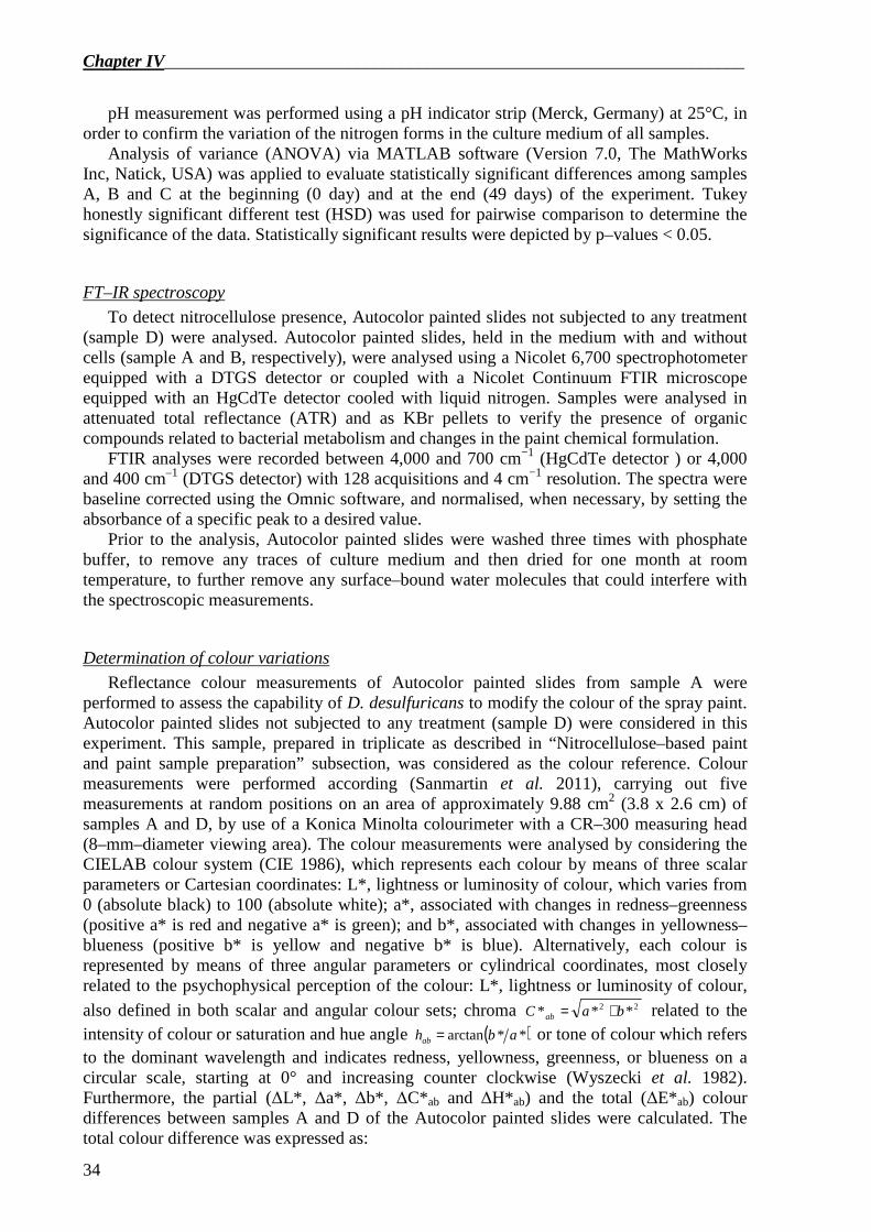
Chapter IV__________________________________________________________________
34
pH measurement was performed using a pH indicator strip (Merck, Germany) at 25°C, in order to confirm the variation of the nitrogen forms in the culture medium of all samples.
Analysis of variance (ANOVA) via MATLAB software (Version 7.0, The MathWorks Inc, Natick, USA) was applied to evaluate statistically significant differences among samples A, B and C at the beginning (0 day) and at the end (49 days) of the experiment. Tukey honestly significant different test (HSD) was used for pairwise comparison to determine the significance of the data. Statistically significant results were depicted by p–values < 0.05.
FT–IR spectroscopy
To detect nitrocellulose presence, Autocolor painted slides not subjected to any treatment (sample D) were analysed. Autocolor painted slides, held in the medium with and without cells (sample A and B, respectively), were analysed using a Nicolet 6,700 spectrophotometer equipped with a DTGS detector or coupled with a Nicolet Continuum FTIR microscope equipped with an HgCdTe detector cooled with liquid nitrogen. Samples were analysed in attenuated total reflectance (ATR) and as KBr pellets to verify the presence of organic compounds related to bacterial metabolism and changes in the paint chemical formulation.
FTIR analyses were recorded between 4,000 and 700 cm−1 (HgCdTe detector ) or 4,000 and 400 cm–1 (DTGS detector) with 128 acquisitions and 4 cm−1 resolution. The spectra were baseline corrected using the Omnic software, and normalised, when necessary, by setting the absorbance of a specific peak to a desired value.
Prior to the analysis, Autocolor painted slides were washed three times with phosphate buffer, to remove any traces of culture medium and then dried for one month at room temperature, to further remove any surface–bound water molecules that could interfere with the spectroscopic measurements.
Determination of colour variations
Reflectance colour measurements of Autocolor painted slides from sample A were performed to assess the capability of D. desulfuricans to modify the colour of the spray paint. Autocolor painted slides not subjected to any treatment (sample D) were considered in this experiment. This sample, prepared in triplicate as described in “Nitrocellulose–based paint and paint sample preparation” subsection, was considered as the colour reference. Colour measurements were performed according (Sanmartin et al. 2011), carrying out five measurements at random positions on an area of approximately 9.88 cm2 (3.8 x 2.6 cm) of samples A and D, by use of a Konica Minolta colourimeter with a CR–300 measuring head (8–mm–diameter viewing area). The colour measurements were analysed by considering the CIELAB colour system (CIE 1986), which represents each colour by means of three scalar parameters or Cartesian coordinates: L*, lightness or luminosity of colour, which varies from 0 (absolute black) to 100 (absolute white); a*, associated with changes in redness–greenness (positive a* is red and negative a* is green); and b*, associated with changes in yellowness–blueness (positive b* is yellow and negative b* is blue). Alternatively, each colour is represented by means of three angular parameters or cylindrical coordinates, most closely related to the psychophysical perception of the colour: L*, lightness or luminosity of colour,
also defined in both scalar and angular colour sets; chroma 22 *** baC ab += related to the intensity of colour or saturation and hue angle ( )**arctan abhab = or tone of colour which refers to the dominant wavelength and indicates redness, yellowness, greenness, or blueness on a circular scale, starting at 0° and increasing counter clockwise (Wyszecki et al. 1982). Furthermore, the partial (∆L*, ∆a*, ∆b*, ∆C*ab and ∆H*ab) and the total (∆E*ab) colour differences between samples A and D of the Autocolor painted slides were calculated. The total colour difference was expressed as:
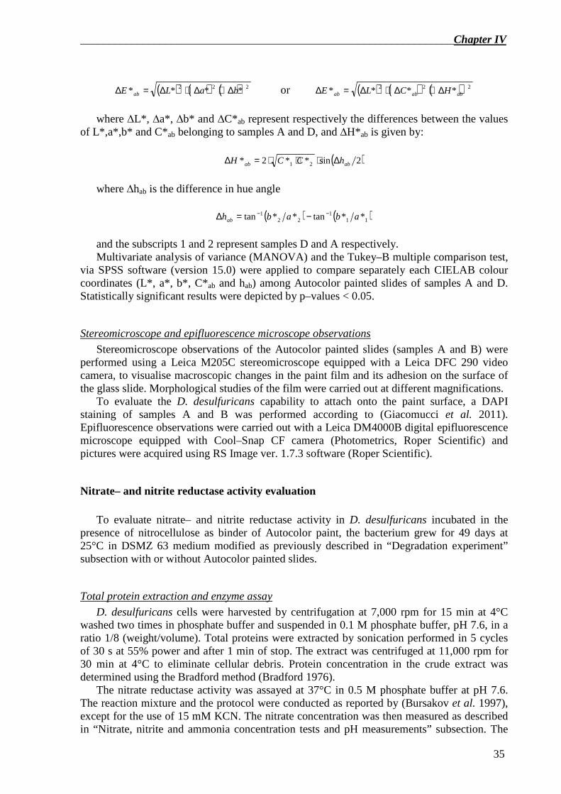
__________________________________________________________________Chapter IV
35
( ) ( ) ( )222 **** baLE ab ∆+∆+∆=∆ or ( ) ( ) ( )222 **** ababab HCLE ∆+∆+∆=∆
where ∆L*, ∆a*, ∆b* and ∆C*ab represent respectively the differences between the values
of L*,a*,b* and C*ab belonging to samples A and D, and ∆H*ab is given by:
( )2sin**2* 21 abab hCCH ∆⋅⋅⋅=∆
where ∆hab is the difference in hue angle
( ) ( )111
221 **tan**tan ababhab
−− −=∆ and the subscripts 1 and 2 represent samples D and A respectively. Multivariate analysis of variance (MANOVA) and the Tukey–B multiple comparison test,
via SPSS software (version 15.0) were applied to compare separately each CIELAB colour coordinates (L*, a*, b*, C*ab and hab) among Autocolor painted slides of samples A and D. Statistically significant results were depicted by p–values < 0.05.
Stereomicroscope and epifluorescence microscope observations
Stereomicroscope observations of the Autocolor painted slides (samples A and B) were performed using a Leica M205C stereomicroscope equipped with a Leica DFC 290 video camera, to visualise macroscopic changes in the paint film and its adhesion on the surface of the glass slide. Morphological studies of the film were carried out at different magnifications.
To evaluate the D. desulfuricans capability to attach onto the paint surface, a DAPI staining of samples A and B was performed according to (Giacomucci et al. 2011). Epifluorescence observations were carried out with a Leica DM4000B digital epifluorescence microscope equipped with Cool–Snap CF camera (Photometrics, Roper Scientific) and pictures were acquired using RS Image ver. 1.7.3 software (Roper Scientific).
Nitrate– and nitrite reductase activity evaluation To evaluate nitrate– and nitrite reductase activity in D. desulfuricans incubated in the
presence of nitrocellulose as binder of Autocolor paint, the bacterium grew for 49 days at 25°C in DSMZ 63 medium modified as previously described in “Degradation experiment” subsection with or without Autocolor painted slides.
Total protein extraction and enzyme assay
D. desulfuricans cells were harvested by centrifugation at 7,000 rpm for 15 min at 4°C washed two times in phosphate buffer and suspended in 0.1 M phosphate buffer, pH 7.6, in a ratio 1/8 (weight/volume). Total proteins were extracted by sonication performed in 5 cycles of 30 s at 55% power and after 1 min of stop. The extract was centrifuged at 11,000 rpm for 30 min at 4°C to eliminate cellular debris. Protein concentration in the crude extract was determined using the Bradford method (Bradford 1976).
The nitrate reductase activity was assayed at 37°C in 0.5 M phosphate buffer at pH 7.6. The reaction mixture and the protocol were conducted as reported by (Bursakov et al. 1997), except for the use of 15 mM KCN. The nitrate concentration was then measured as described in “Nitrate, nitrite and ammonia concentration tests and pH measurements” subsection. The
Chapter IV__________________________________________________________________
36
nitrite reductase activity was assayed as by (Liu et al. 1994), except for the termination of the reaction that was achieved by vigorously shaking of the reaction tube. The nitrite concentration was then measured as described in “Nitrate, nitrite and ammonia concentration tests and pH measurements” subsection. The enzymatic unit is defined by µmol of substrate reduced per minute. To evaluate statistically significant differences among enzymatic activities the t–test, via Microsoft Excel tool for Windows, was applied. Statistically significant results were depicted by p–values < 0.05.
Results
Characterisation of Autocolor® paint Autocolor film (Sample D) was analysed by FTIR spectroscopy for the identification of
the main component of the polymeric blend. The main component of the resin is nitrocellulose and an alkyd fraction (a modified polyester resin) was also present. The characteristic and diagnostic FTIR peaks of nitrocellulose are related to the N–O vibration at 1,655 cm–1 and 1,280 cm–1, while the polyester resin has the characteristic vibration peaks at 1,735 cm–1 (ν C=O) and 1,073 cm–1 (ν C–O), both related to the ester group of the polymer.
The complete characterisation of other components, like pigment, stabiliser or other additives, was beyond the aim of this work.
Degradation experiment Cell counts and ATP content of samples A and C carried out at both the beginning and at
the end of the experiments were not significantly different. Investigations performed on sample B showed that there was no occurrence of contamination during the incubation period (data not shown). UV–Vis spectrophotometric measurements performed in the culture medium showed a decrease in nitrate (Figure 1a) and nitrite (Figure 1b) concentration in samples A and C in comparison to sample B, where they were significantly higher and an increase in ammonia concentration in all three samples (Figure 1c). The final ammonia concentration was not significantly different across all 3 samples (Figure 1c). These results were in agreement with a pH increment in the same sample (data not shown).
Figure 1. Variation of nitrogen forms concentration in the culture medium of samples A, B and C at the beginning (0 day) and at the end (49 days) of the experiments. (a) nitrate nitrogen (p–value = 1.30×10⁻¹³), (b) nitrite nitrogen (p–value = 0), and (c) ammonia nitrogen (p–value = 2.45×10⁻¹¹) concentrations. The histograms represent average values from three independent replicates. According to post–hoc analysis, means sharing the same letter are not significantly different from each other.
__________________________________________________________________Chapter IV
37
FTIR and FTIR–ATR spectroscopy
Bacterial growth and the degradation phenomenon were analysed by FTIR spectroscopy in KBr transmission and ATR. Since transmittance FTIR on KBr pellets shows the absorbance of the bulk material, this was the most adequate technique to investigate the bacterial degradation of the Autocolor paint. As FTIR-ATR analyses give information about surface phenomena they showed that bacteria have colonised the polymeric surface.
Acquired spectra were baseline corrected and normalised on the C–H stretching area using the 2,960 cm–1 peak, related the CH3 asymmetric stretching vibration of the polyester component, which is expected to be the more stable component.
The paint was partially affected by the prolonged immersion in the aqueous culture medium, visible by the slow reduction in the intensity of signal related to the nitric substituent (peaks at 1,655 cm–1 and 1,280 cm–1), suggesting a partial abiotic denitrification process. Furthermore, the polyester component was also slightly affected by the prolonged contact with the buffer as was evident by the reduced intensity of the C=O stretching peak at 1,735 cm–1 and the C–O stretching vibration at 1,073 cm–1, both related to the ester group of the main chain.
Figure 2. (a) FTIR ATR spectra of the Autocolor film (sample D) and the degraded sample A in the range 1,900–700 cm–1. The arrows indicate the variation in intensity related to bacterial compounds on the film surface. (b) FTIR spectra of the Autocolor film (sample D), of the paint incubated with the culture medium (sample B) and incubated in the presence of bacteria (sample A). The arrows indicate the diagnostic peak for the degradation of the nitro–substituent in the nitrocellulose molecules.
The comparison between sample D and sample A FTIR-ATR spectra showed an increase
in signal related to amide groups at 1,540 cm–1 (see black arrow in Figure 2a). The presence of proteinaceous material was also confirmed by the increase of signal in the area between 900 and 1,100 cm–1, related to C–N and N–N stretching vibrations in nitrogen compounds. Furthermore, the relative intensity of the peak at 1,280cm–1 (amide I signal) decreased due to degradation of the N–O substituent. This decrease was not observed for the peak at 1,655 cm–
1 (amide II signal) for the concurrent increase of the amine products which signal falls in the same range (see green arrows in Figure 2a). A noticeable increase in signal in the area from 3,000 to 3,700 cm–1 must be related to NH and OH stretching vibrations due to organic material.
The slight abiotic degradation recorded in the FTIR analysis of the KBr pellets was enhanced by the presence of bacteria which decreased the rate of nitrification of the cellulose-based paint leading to a considerable decrease of the signals at 1,655 cm–1 and 1,280 cm–1 (see black arrow in Figure 2b). The high specificity of the bacterial activity was also confirmed by the invariance of the C–N stretching peak visible at 840 cm–1 which remained stable during all degradation experiments. The degradation of the modified polyester
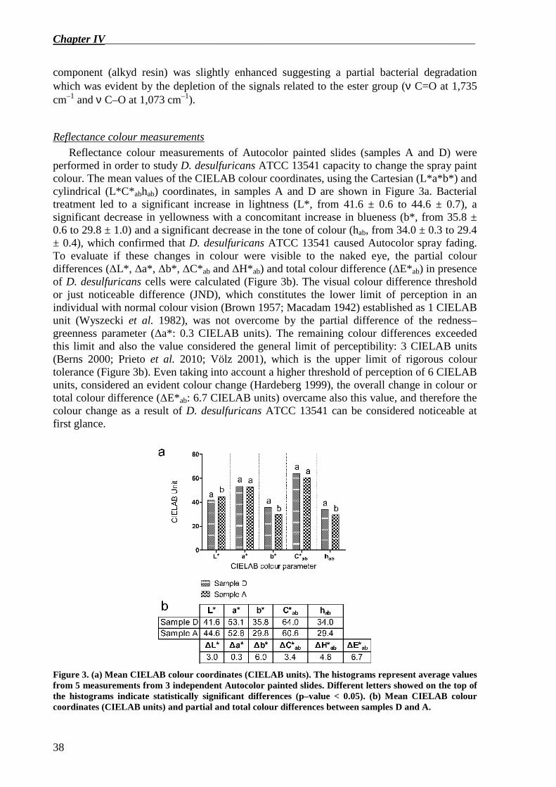
Chapter IV__________________________________________________________________
38
component (alkyd resin) was slightly enhanced suggesting a partial bacterial degradation which was evident by the depletion of the signals related to the ester group (ν C=O at 1,735 cm–1 and ν C–O at 1,073 cm–1).
Reflectance colour measurements
Reflectance colour measurements of Autocolor painted slides (samples A and D) were performed in order to study D. desulfuricans ATCC 13541 capacity to change the spray paint colour. The mean values of the CIELAB colour coordinates, using the Cartesian (L*a*b*) and cylindrical (L*C*abhab) coordinates, in samples A and D are shown in Figure 3a. Bacterial treatment led to a significant increase in lightness (L*, from 41.6 ± 0.6 to 44.6 ± 0.7), a significant decrease in yellowness with a concomitant increase in blueness (b*, from 35.8 ± 0.6 to 29.8 ± 1.0) and a significant decrease in the tone of colour (hab, from 34.0 ± 0.3 to 29.4 ± 0.4), which confirmed that D. desulfuricans ATCC 13541 caused Autocolor spray fading. To evaluate if these changes in colour were visible to the naked eye, the partial colour differences (∆L*, ∆a*, ∆b*, ∆C*ab and ∆H*ab) and total colour difference (∆E*ab) in presence of D. desulfuricans cells were calculated (Figure 3b). The visual colour difference threshold or just noticeable difference (JND), which constitutes the lower limit of perception in an individual with normal colour vision (Brown 1957; Macadam 1942) established as 1 CIELAB unit (Wyszecki et al. 1982), was not overcome by the partial difference of the redness–greenness parameter (∆a*: 0.3 CIELAB units). The remaining colour differences exceeded this limit and also the value considered the general limit of perceptibility: 3 CIELAB units (Berns 2000; Prieto et al. 2010; Völz 2001), which is the upper limit of rigorous colour tolerance (Figure 3b). Even taking into account a higher threshold of perception of 6 CIELAB units, considered an evident colour change (Hardeberg 1999), the overall change in colour or total colour difference (∆E*ab: 6.7 CIELAB units) overcame also this value, and therefore the colour change as a result of D. desulfuricans ATCC 13541 can be considered noticeable at first glance.
Figure 3. (a) Mean CIELAB colour coordinates (CIELAB units). The histograms represent average values from 5 measurements from 3 independent Autocolor painted slides. Different letters showed on the top of the histograms indicate statistically significant differences (p–value < 0.05). (b) Mean CIELAB colour coordinates (CIELAB units) and partial and total colour differences between samples D and A.
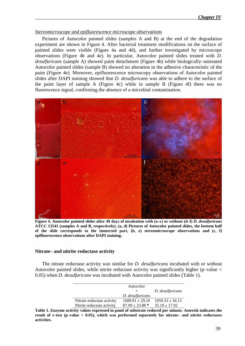
__________________________________________________________________Chapter IV
39
Stereomicroscope and epifluorescence microscope observations
Pictures of Autocolor painted slides (samples A and B) at the end of the degradation experiment are shown in Figure 4. After bacterial treatment modifications on the surface of painted slides were visible (Figure 4a and 4d), and further investigated by microscope observations (Figure 4b and 4e). In particular, Autocolor painted slides treated with D. desulfuricans (sample A) showed paint detachment (Figure 4b) while biologically–untreated Autocolor painted slides (sample B) showed no alteration in the adhesive characteristic of the paint (Figure 4e). Moreover, epifluorescence microscopy observations of Autocolor painted slides after DAPI staining showed that D. desulfuricans was able to adhere to the surface of the paint layer of sample A (Figure 4c) while in sample B (Figure 4f) there was no fluorescence signal, confirming the absence of a microbial contamination.
Figure 4. Autocolor painted slides after 49 days of incubation with (a–c) or without (d–f) D. desulfuricans ATCC 13541 (samples A and B, respectively). (a, d) Pictures of Autocolor painted slides, the bottom half of the slide corresponds to the immersed part, (b, e) stereomicroscope observations and (c, f) epifluorescence observations after DAPI staining.
Nitrate– and nitrite reductase activity The nitrate reductase activity was similar for D. desulfuricans incubated with or without
Autocolor painted slides, while nitrite reductase activity was significantly higher (p–value < 0.05) when D. desulfuricans was incubated with Autocolor painted slides (Table 1).
Autocolor
+ D. desulfuricans
D. desulfuricans
Nitrate reductase activity 1089.91 ± 29.10 1059.33 ± 34.13 Nitrite reductase activity 87.09 ± 23.88 * 35.18 ± 17.92
Table 1. Enzyme activity values expressed in µmol of substrate reduced per minute. Asterisk indicates the result of t–test (p–value < 0.05), which was performed separately for nitrate– and nitrite reductases activities.
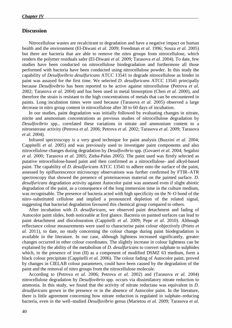
Chapter IV__________________________________________________________________
40
Discussion Nitrocellulose wastes are recalcitrant to degradation and have a negative impact on human
health and the environment (El-Diwani et al. 2009; Freedman et al. 1996; Souza et al. 2005) but there are bacteria that are able to remove the nitro groups from nitrocellulose, which renders the polymer residuals safer (El-Diwani et al. 2009; Tarasova et al. 2004). To date, few studies have been conducted on nitrocellulose biodegradation and furthermore all those performed with bacteria have been conducted using nitrocellulose powder. In this study the capability of Desulfovibrio desulfuricans ATCC 13541 to degrade nitrocellulose as binder in paint was assayed for the first time. We selected D. desulfuricans ATCC 13541 principally because Desulfovibrio has been reported to be active against nitrocellulose (Petrova et al. 2002; Tarasova et al. 2004) and has been used in metal biosorption (Chen et al. 2000), and therefore the strain is resistant to the high concentrations of metals that can be encountered in paints. Long incubation times were used because (Tarasova et al. 2005) observed a large decrease in nitro group content in nitrocellulose after 30 to 60 days of incubation.
In our studies, paint degradation was initially followed by evaluating changes in nitrate, nitrite and ammonium concentrations as previous studies of nitrocellulose degradation by Desulfovibrio spp., correlated these variations in nitrate and ammonium content to a nitroesterase activity (Petrova et al. 2006; Petrova et al. 2002; Tarasova et al. 2009; Tarasova et al. 2004).
Infrared spectroscopy is a very good technique for paint analysis (Buzzini et al. 2004; Cappitelli et al. 2005) and was previously used to investigate paint components and also nitrocellulose changes during degradation by Desulfovibrio spp. (Govaert et al. 2004; Segalini et al. 2000; Tarasova et al. 2005; Zieba-Palus 2005). The paint used was firstly selected as putative nitrocellulose-based paint and then confirmed as a nitrocellulose- and alkyd-based paint. The capability of D. desulfuricans ATCC 13541 to adhere onto the surface of the paint, assessed by epifluorescence microscopy observations was further confirmed by FTIR–ATR spectroscopy that showed the presence of proteinaceous material on the painted surface. D. desulfuricans degradation activity against Autocolor paint was assessed even if slight abiotic degradation of the paint, as a consequence of the long immersion time in the culture medium, was recognisable. The presence of bacteria acted with high specificity on the N–O bond of the nitro–substituted cellulose and implied a pronounced depletion of the related signal, suggesting that bacterial degradation favoured this chemical group compared to others.
After incubation with D. desulfuricans, we observed paint detachment and fading of Autocolor paint slides, both noticeable at first glance. Bacteria on painted surfaces can lead to paint detachment and discolouration (Cappitelli et al. 2009; Pepe et al. 2010). Although reflectance colour measurements were used to characterise paint colour objectively (Prieto et al. 2011), to date, no study concerning the colour change during paint biodegradation is available in the literature. In our case, although lightness increased significantly, greater changes occurred in other colour coordinates. The slightly increase in colour lightness can be explained by the ability of the metabolism of D. desulfuricans to convert sulphate to sulphides which, in the presence of iron (II) as a component of modified DSMZ 63 medium, form a black colour precipitate (Cappitelli et al. 2006). The colour fading of Autocolor paint, proved by changes in CIELAB colour parameters, could have been caused by the degradation of the paint and the removal of nitro groups from the nitrocellulose molecule.
According to (Petrova et al. 2006; Petrova et al. 2002) and (Tarasova et al. 2004) nitrocellulose degradation by Desulfovibrio spp. occurs via dissimilatory nitrate reduction to ammonia. In this study, we found that the activity of nitrate reductase was equivalent in D. desulfuricans grown in the presence or in the absence of Autocolor paint. In the literature, there is little agreement concerning how nitrate reduction is regulated in sulphate–reducing bacteria, even in the well–studied Desulfovibrio genus (Marietou et al. 2009; Tarasova et al.
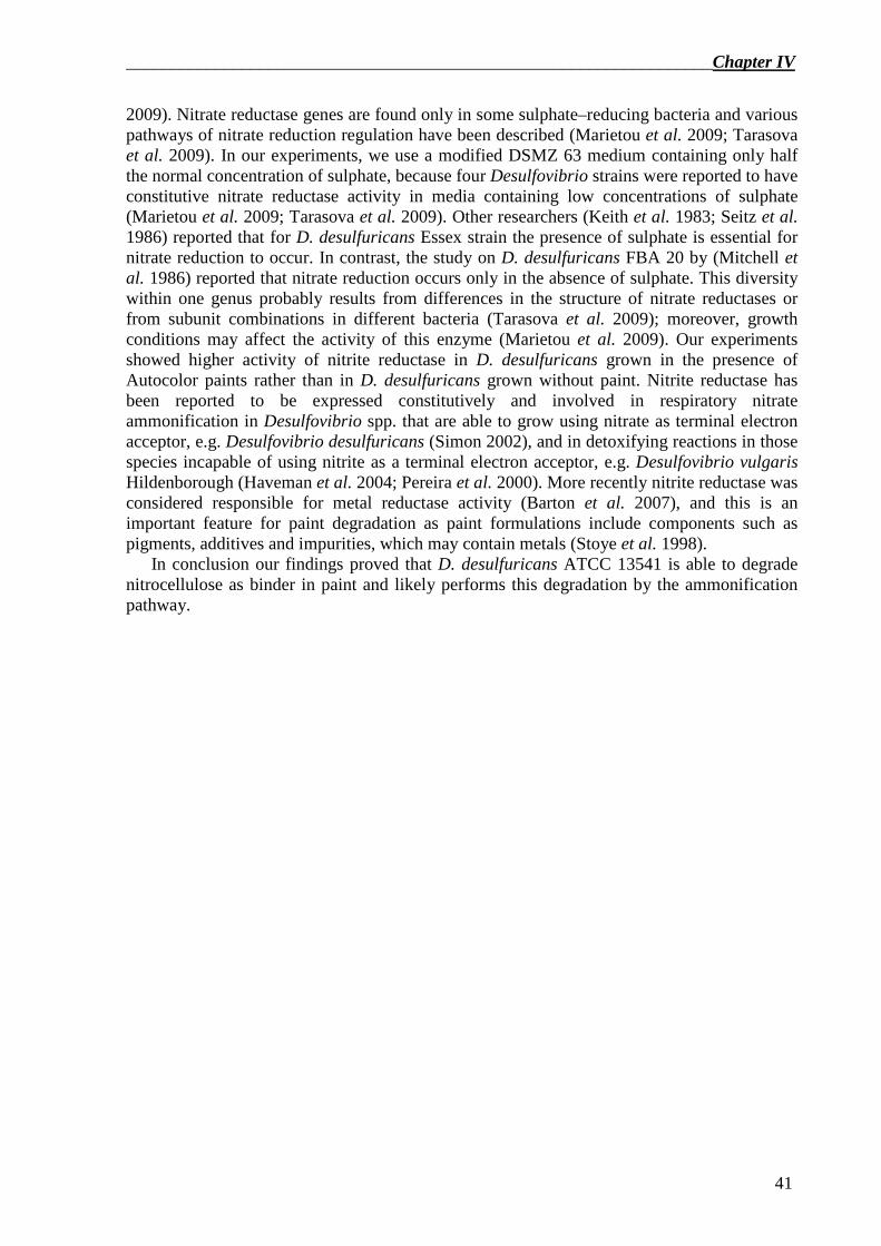
__________________________________________________________________Chapter IV
41
2009). Nitrate reductase genes are found only in some sulphate–reducing bacteria and various pathways of nitrate reduction regulation have been described (Marietou et al. 2009; Tarasova et al. 2009). In our experiments, we use a modified DSMZ 63 medium containing only half the normal concentration of sulphate, because four Desulfovibrio strains were reported to have constitutive nitrate reductase activity in media containing low concentrations of sulphate (Marietou et al. 2009; Tarasova et al. 2009). Other researchers (Keith et al. 1983; Seitz et al. 1986) reported that for D. desulfuricans Essex strain the presence of sulphate is essential for nitrate reduction to occur. In contrast, the study on D. desulfuricans FBA 20 by (Mitchell et al. 1986) reported that nitrate reduction occurs only in the absence of sulphate. This diversity within one genus probably results from differences in the structure of nitrate reductases or from subunit combinations in different bacteria (Tarasova et al. 2009); moreover, growth conditions may affect the activity of this enzyme (Marietou et al. 2009). Our experiments showed higher activity of nitrite reductase in D. desulfuricans grown in the presence of Autocolor paints rather than in D. desulfuricans grown without paint. Nitrite reductase has been reported to be expressed constitutively and involved in respiratory nitrate ammonification in Desulfovibrio spp. that are able to grow using nitrate as terminal electron acceptor, e.g. Desulfovibrio desulfuricans (Simon 2002), and in detoxifying reactions in those species incapable of using nitrite as a terminal electron acceptor, e.g. Desulfovibrio vulgaris Hildenborough (Haveman et al. 2004; Pereira et al. 2000). More recently nitrite reductase was considered responsible for metal reductase activity (Barton et al. 2007), and this is an important feature for paint degradation as paint formulations include components such as pigments, additives and impurities, which may contain metals (Stoye et al. 1998).
In conclusion our findings proved that D. desulfuricans ATCC 13541 is able to degrade nitrocellulose as binder in paint and likely performs this degradation by the ammonification pathway.
Chapter IV__________________________________________________________________
42
References
APHA, AWWA and WEF, Eds. (1998). Standard Methods for the Examination of Water and Wastewater. Washinghton, APHA.
Auer N., Hedger J. N. and Evans C. S. (2005). Degradation of nitrocellulose by fungi. Biodegradation 16(3): 229-236.
Barton L. L., Goulhen F., Bruschi M., Woodards N. A., Plunkett R. M. and Rietmeijer F. J. M. (2007). The bacterial metallome: composition and stability with specific reference to the anaerobic bacterium Desulfovibrio desulfuricans. Biometals 20(3-4): 291-302.
Berns R. S. (2000). Billmeyer and Saltzman’s Principles of Color Technology. New York, Wiley. Bradford M. M. (1976). A rapid and sensitive method for the quantitation of microgram quantities of protein
utilizing the principle of protein-dye binding. Anal Biochem 72(1-2): 248-254. Brown W. R. J. (1957). Color Discrimination of Twelve Observers. J. Opt. Soc. Am. 47(2): 137-143. Bursakov S. A., Carneiro C., Almendra M. J., Duarte R. O., Caldeira J., Moura I. and Moura J. J. (1997).
Enzymatic properties and effect of ionic strength on periplasmic nitrate reductase (NAP) from Desulfovibrio desulfuricans ATCC 27774. Biochem Biophys Res Commun 239(3): 816-822.
Buzzini P. and Massonnet G. (2004). A market study of green spray paints by Fourier transform infrared (FTIR) and Raman spectroscopy. Sci Justice 44(3): 123-131.
Cappitelli F., Vicini S., Piaggio P., Abbruscato P., Princi E., Casadevall A., Nosanchuk J. D. and Zanardini E. (2005). Investigation of fungal deterioration of synthetic paint binders using vibrational spectroscopic techniques. Macromolecular bioscience 5(1): 49-57.
Cappitelli F., Zanardini E., Ranalli G., Mello E., Daffonchio D. and Sorlini C. (2006). Improved methodology for bioremoval of black crusts on historical stone artworks by use of sulfate-reducing bacteria. Applied and environmental microbiology 72(5): 3733-3737.
Cappitelli F., Abbruscato P., Foladori P., Zanardini E., Ranalli G., Principi P., Villa F., Polo A. and Sorlini C. (2009). Detection and elimination of cyanobacteria from frescoes: the case of the St. Brizio Chapel (Orvieto Cathedral, Italy). Microbial Ecology 57(4): 633-639.
Chen B. Y., Utgikar V. P., Harmon S. M., Tabak H. H., Bishop D. F. and Govind R. (2000). Studies on biosorption of zinc(II) and copper(II) on Desulfovibrio desulfuricans. International Biodeterioration & Biodegradation 46(1): 11-18.
CIE (1986). Publication 15–2: colorimetry. Vienna, CIE Central Bureau. El-Diwani G., El-Ibiari N. N. and Hawash S. I. (2009). Treatment of hazardous wastewater contaminated by
nitrocellulose. Journal of Hazardous Materials 167(1-3): 830-834. Freedman D. L., Caenepeel B. M. and Kim B. J. (1996). Biotransformation of nitrocellulose under methanogenic
conditions. Water Science and Technology 34(5-6): 327-334. Freedman D. L., Cashwell J. M. and Kim B. J. (2002). Biotransformation of explosive-grade nitrocellulose under
denitrifying and sulfidogenic conditions. Waste Management 22(3): 283-292. Giacomucci L., Bertoncello R., Salvadori O., Martini I., Favaro M., Villa F., Sorlini C. and Cappitelli F. (2011).
Microbial deterioration of artistic tiles from the facade of the Grande Albergo Ausonia & Hungaria (Venice, Italy). Microb Ecol 62(2): 287-298.
Govaert F. and Bernard M. (2004). Discriminating red spray paints by optical microscopy, Fourier transform infrared spectroscopy and X-ray fluorescence. Forensic Sci Int 140(1): 61-70.
Hardeberg A. Y. (1999). Acquisition and reproduction of color images: colorimetric and multispectral approaches. PhD Thesis, Ecole Nationale Superieure des Telecommunications.
Haveman S. A., Greene E. A., Stilwell C. P., Voordouw J. K. and Voordouw G. (2004). Physiological and gene expression analysis of inhibition of Desulfovibrio vulgaris Hildenborough by nitrite. J Bacteriol 186(23): 7944-7950.
Keith S. M. and Herbert R. A. (1983). Dissimilatory nitrate reduction by a strain of Desulfovibrio desulfuricans. FEMS Microbiol Lett 18(1-2): 55-59.
Kraft B., Strous M. and Tegetmeyer H. E. (2011). Microbial nitrate respiration - Genes, enzymes and environmental distribution. Journal of Biotechnology 155(1): 104-117.
Liu M.-C., Costa C. and Moura I. (1994). Hexaheme nitrite reductase from Desulfovibrio desulfuricans (ATCC 27774). Methods Enzymol. Harry D. Peck J. J. L., Academic Press. Volume 243: 303-319.
Macadam D. L. (1942). Visual Sensitivities to Color Differences in Daylight. J. Opt. Soc. Am. 32(5): 247-273. Marietou A., Griffiths L. and Cole J. (2009). Preferential Reduction of the Thermodynamically Less Favorable
Electron Acceptor, Sulfate, by a Nitrate-Reducing Strain of the Sulfate-Reducing Bacterium Desulfovibrio desulfuricans 27774. J. Bacteriol. 191(3): 882-889.
Mitchell G. J., Jones J. G. and Cole J. A. (1986). Distribution and Regulation of Nitrate and Nitrite Reduction by Desulfovibrio and Desulfotomaculum Species. Archives of Microbiology 144(1): 35-40.
__________________________________________________________________Chapter IV
43
Moura I., Bursakov S., Costa C. and Moura J. J. (1997). Nitrate and nitrite utilization in sulfate-reducing bacteria. Anaerobe 3(5): 279-290.
Moura J. J. G., Gonzalez P., Moura I., Fauque G., Barton L. L. and Hamilton W. A. (2007). Dissimilatory nitrate and nitrite ammonification by sulphate-reducing eubacteria. Sulphate-Reducing Bacteria. Environmental and Engineered Systems. Barton L. L., Hamilton W. A. Cambridge, Cambridge University Press: 241-264.
Pepe O., Sannino L., Palomba S., Anastasio M., Blaiotta G., Villani F. and Moschetti G. (2010). Heterotrophic microorganisms in deteriorated medieval wall paintings in southern Italian churches. Microbiological Research 165(1): 21-32.
Pereira I. A. C., LeGall J., Xavier A. V. and Teixeira M. (2000). Characterization of a heme c nitrite reductase from a non-ammonifying microorganism, Desulfovibrio vulgaris Hildenborough. Biochimica Et Biophysica Acta-Protein Structure and Molecular Enzymology 1481(1): 119-130.
Petrova O. E., Tarasova N. B. and Davydova M. N. (2002). Biotechnological potential of sulfate-reducing bacteria for transformation of nitrocellulose. Anaerobe 8(6): 315-317.
Petrova O. E., Tarasova N. B., Alyabyev A. J., Davydova M. N. and Loseva N. L. (2006). Calorimetric studies of the growth of Desulfovibrio desulfuricans in the presence of nitrocellulose. Thermochimica Acta 445(1): 67-69.
Prieto B., Sanmartín P., Silva B. and Martínez-Verdú F. (2010). Measuring the color of granite rocks: A proposed procedure. Color Research & Application 35(5): 368-375.
Prieto B., Sanmartín P., Pereira-Pardo L. and Silva B. (2011). Recovery of the traditional colours of painted woodwork in the Historical Centre of Lugo (NW Spain). Journal of Cultural Heritage 12(3): 279-286.
Sanmartin P., Villa F., Silva B., Cappitelli F. and Prieto B. (2011). Color measurements as a reliable method for estimating chlorophyll degradation to phaeopigments. Biodegradation 22(4): 763-771.
Segalini S., Chirico A. D., Depinto G. and Pegoraro M. (2000). Antigraffiti removers and antigraffiti coatings. XXV Fatipec Congress, Turin.
Seitz H. J. and Cypionka H. (1986). Chemolithotrophic Growth of Desulfovibrio-Desulfuricans with Hydrogen Coupled to Ammonification of Nitrate or Nitrite. Archives of Microbiology 146(1): 63-67.
Simon J. (2002). Enzymology and bioenergetics of respiratory nitrite ammonification. Fems Microbiology Reviews 26(3): 285-309.
Slack R. J., Gronow J. R. and Voulvoulis N. (2005). Household hazardous waste in municipal landfills: contaminants in leachate. The Science of the total environment 337(1-3): 119-137.
Souza J. V. B., da Silva E. S., da Silva F. T. and Paiva T. C. B. (2005). Fungal treatment of a delignification effluent from a nitrocellulose industry. Bioresource Technology 96(17): 1936-1942.
Stoye D. and Freitag W. (1998). Introduction. Paints, coatings, and solvents. Stoye D., Freitag W. Weinheim, Wiley WCH: 1-10.
Tarasova N. B., Petrova O. E., Davydova M. N., Khairutdinov B. I. and Klochkov V. V. (2004). Changes in the nitrocellulose molecule induced by sulfate-reducing bacteria Desulfovibrio desulfuricans 1,388. The enzymes participating in this process. Biochemistry (Mosc) 69(7): 809-812.
Tarasova N. B., Petrova O. E., Faizullin D. A. and Davydova M. N. (2005). FTIR-spectroscopic studies of the fine structure of nitrocellulose treated by Desulfovibrio desulfuricans. Anaerobe 11(6): 312-314.
Tarasova N. B., Gorshkov O. V. and Petrova O. E. (2009). Activity of Nitrate Reductase in Desulfovibrio vulgaris VKM 1388. Microbiology 78(2): 160–164.
Völz H. G. (2001). Industrial Color Testing. Weinheim, Wiley–VCH. Wyszecki G. and Stiles W. S. (1982). Color science, concepts and methods, quantitative data and formulae. New
York, Wiley. Zieba-Palus J. (2005). Examination of spray paints by the use of reflection technique of microinfrared
spectroscopy. Journal of Molecular Structure 744-747: 229-234.

___________________________________________________________________Chapter V
45
A new non-degenerated primer pair for the specific detection of the nitrite reductase gene in Desulfovibrio genus1
Abstract Dissimilatory nitrate reduction to ammonia is the process in which nitrate is reduced to
ammonia with nitrite as intermediate. The ability to carry out this process is phylogenetically widespread and the gene nrfA, encoding for the key enzyme of the second step of the pathway, could be used as a marker for this dissimilatory process. In this study we developed a new primer pair specific for nrfA gene in Desulfovibrio genus. The specificity of the primer pair was tested both on Desulfovibrio DNA of thirteen species and two wastewater samples. PCR amplifications yielded products of the expected size (850 bp) and sequences obtained from Desulfovibrio DNAs and environmental samples clone libraries matched the Desulfovibrio nrfA gene. Nevertheless we found different nrfA gene sequences in the environment that are not present in the databases. The new primer set could be used for obtaining more sequences of the nrfA gene and improve our knowledge of the dissimilatory nitrate reduction to ammonia pathway.
Introduction Dissimilatory nitrate reduction to ammonia (DNRA) or nitrate ammonification is an
anaerobic process in which nitrate is reduced to ammonia with nitrite as intermediate (Cabello et al. 2009; Einsle et al. 1999; Kraft et al. 2011). The ability to carry out DNRA is phylogenetically widespread (Kraft et al. 2011). Many sulphate-reducing bacteria (SRB) are able to perform respiratory ammonification in the presence of nitrate when sulphate is absent and/or in low concentration, although there has been recent evidence that sulphate reduction is preferred over DNRA if both electron acceptors are present (Kraft et al. 2011; Marietou et al. 2009). In DNRA, nitrate reduction to nitrite is usually performed by the periplasmic nitrate reductase NapAB, while nitrite reduction to ammonium is catalysed by the pentaheme cytochrome c nitrite reductase NrfA, without the release of any intermediate (Cabello et al. 2009; Einsle et al. 1999; Kraft et al. 2011). In SRB and, in particular, in Desulfovibrio, nitrite reductase NrfA plays an important role for those strains able to use nitrate and nitrite instead of sulphate as electron acceptor, and for those bacteria capable of reducing nitrite but unable to reduce nitrate (Cabello et al. 2009; Greene et al. 2003; Haveman et al. 2004; Simon 2002). In fact, nitrite has been reported as a very toxic compound and the additional function of nitrite reductase, the key enzyme involved in the second step of the DNRA pathway, allows Desulfovibrio spp. to survive in environments containing nitrite up to millimolar concentrations (Cabello et al. 2009). The presence of the nitrite reductase in the Desulfovibrio genus is widespread and various species synthesise a very active, constitutive nitrite reductase (Marietou et al. 2009; Moura et al. 2007; Simon 2002).
1 Manuscript in preparation for submission: L. Giacomucci et al. A new non-degenerated primer pair for the specific detection of the nitrite reductase gene in Desulfovibrio genus.
Chaper V___________________________________________________________________
46
Nitrate reducing pathways, such as denitrification, are very well known (Kraft et al. 2011; Mohan et al. 2004). In contrast, few studies have focused on DNRA. Moreover, even if many studies are available on cytochrome c nitrite reductase protein (NrfA) (Cabello et al. 2009; Einsle et al. 1999; Kraft et al. 2011), only few studies are available on the genes encoding this enzyme (Kraft et al. 2011; Mohan et al. 2004). To date, there are only few nrfA sequences available in nucleotide databases, mainly from pathogenic strains and therefore probably not relevant in environmental studies. As a consequence, the isolation of environmentally important species performing DNRA is an important step in order to obtain additional sequences of the nrfA gene (Kraft et al. 2011) and generate new knowledge on the DNRA pathway. As the functional gene nrfA occurs in diverse bacterial taxa, such as Gamma-, Delta- and Epsilon-Proteobacteria and in members of the Bacteroides, available primers are highly degenerated to target the widest range of microorganisms (Kraft et al. 2011; Mohan et al. 2004). Unfortunately, the employment of degenerate primers has different drawbacks, such as the use of touchdown protocols to minimise annealing mismatches and the use of high fidelity Taq polymerases to improve PCR results (Mohan et al. 2004). Nevertheless, errors in DNA sequences or failed amplification could still occur.
The aim of this research was the development of a new primer pair to amplify nrfA gene fragment specifically in Desulfovibrio genus and provide a new tool for studying DNRA pathway.
Methods
Primer design. In order to find conserved domains, amino acid and nucleotide nitrite reductase (NrfA)
sequences of five Desulfovibrio and one Desulfomicrobium strains available in both GenBank (Benson et al. 2011) and KEGG (Kanehisa et al. 2002) gene databases were aligned using ClustalX2 (Larkin et al. 2007). PCR primers were then designed using Primer3 (Rozen et al. 1999), asking the software to design primer sets inside the consensus sequences found by multiple alignment. Few degenerations based on this alignment were manually added. The in silico coverage and specificity of the new primer sets were tested using both the NCBI BLAST tool (http://blast.ncbi.nlm.nih.gov/Blast.cgi) and ThermoPhyl software (Oakley et al. 2011). Finally, hetero/homodimers formation and primer stability of the novel primer sets were evaluated using OligoAnalyzer 3.1 software (http://www.idtdna.com/analyzer/Applications/OligoAnalyzer/); parameters setting for primer characteristic determination were 0.2 µM primer, 1.5 mM Mg++ salt and 0.2 mM dNTPs, and the ∆G temperature was 60°C.
Desulfovibrio strains. DNA from thirteen Desulfovibrio and five Desulfomicrobium strains obtained from both
the American Type Culture Collection (ATCC; Manassas, VA) and the German Collection of Microorganisms and Cell Cultures (DSMZ, Braunschweig, Germany) have been used for testing the specificity of the NrfA PCR assays.
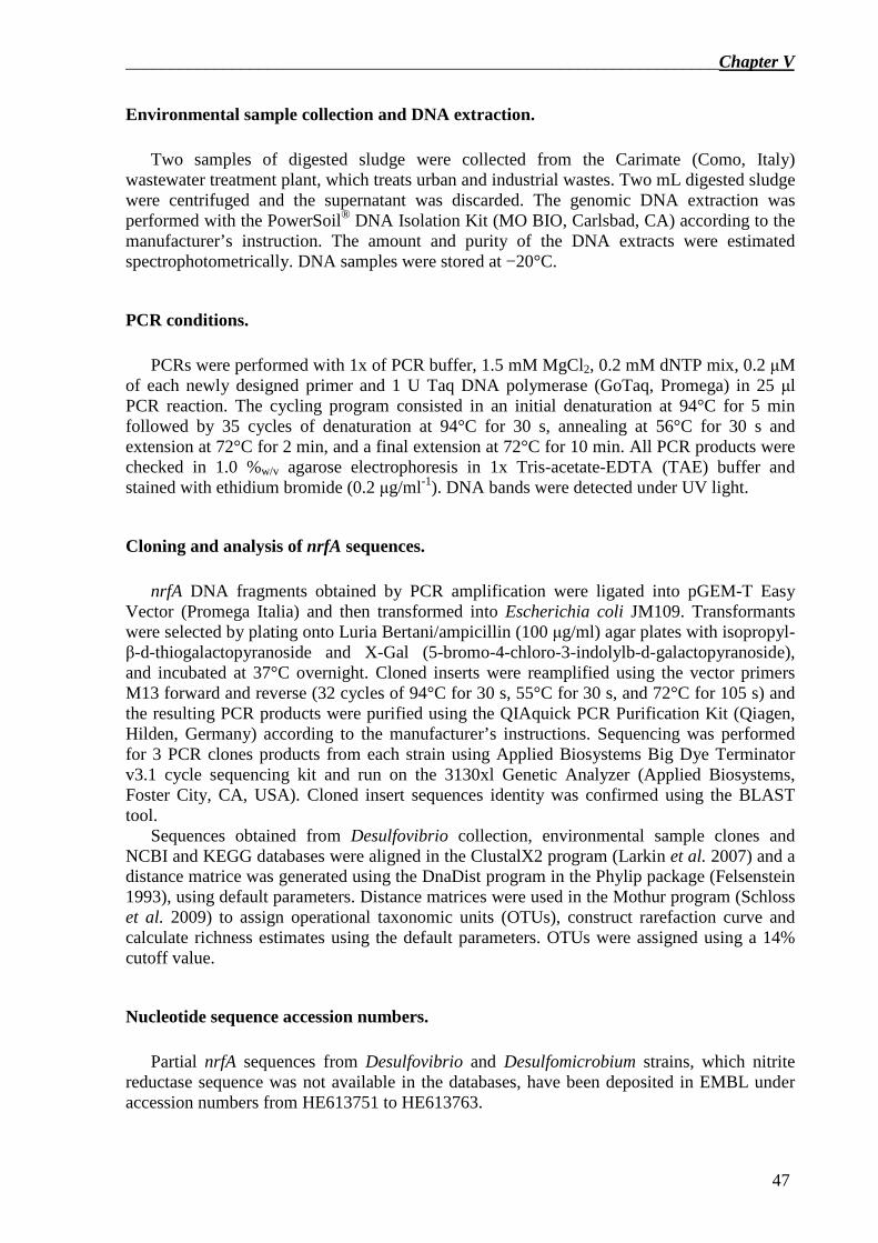
___________________________________________________________________Chapter V
47
Environmental sample collection and DNA extraction. Two samples of digested sludge were collected from the Carimate (Como, Italy)
wastewater treatment plant, which treats urban and industrial wastes. Two mL digested sludge were centrifuged and the supernatant was discarded. The genomic DNA extraction was performed with the PowerSoil® DNA Isolation Kit (MO BIO, Carlsbad, CA) according to the manufacturer’s instruction. The amount and purity of the DNA extracts were estimated spectrophotometrically. DNA samples were stored at −20°C.
PCR conditions. PCRs were performed with 1x of PCR buffer, 1.5 mM MgCl2, 0.2 mM dNTP mix, 0.2 µM
of each newly designed primer and 1 U Taq DNA polymerase (GoTaq, Promega) in 25 µl PCR reaction. The cycling program consisted in an initial denaturation at 94°C for 5 min followed by 35 cycles of denaturation at 94°C for 30 s, annealing at 56°C for 30 s and extension at 72°C for 2 min, and a final extension at 72°C for 10 min. All PCR products were checked in 1.0 %w/v agarose electrophoresis in 1x Tris-acetate-EDTA (TAE) buffer and stained with ethidium bromide (0.2 µg/ml-1). DNA bands were detected under UV light.
Cloning and analysis of nrfA sequences. nrfA DNA fragments obtained by PCR amplification were ligated into pGEM-T Easy
Vector (Promega Italia) and then transformed into Escherichia coli JM109. Transformants were selected by plating onto Luria Bertani/ampicillin (100 µg/ml) agar plates with isopropyl-β-d-thiogalactopyranoside and X-Gal (5-bromo-4-chloro-3-indolylb-d-galactopyranoside), and incubated at 37°C overnight. Cloned inserts were reamplified using the vector primers M13 forward and reverse (32 cycles of 94°C for 30 s, 55°C for 30 s, and 72°C for 105 s) and the resulting PCR products were purified using the QIAquick PCR Purification Kit (Qiagen, Hilden, Germany) according to the manufacturer’s instructions. Sequencing was performed for 3 PCR clones products from each strain using Applied Biosystems Big Dye Terminator v3.1 cycle sequencing kit and run on the 3130xl Genetic Analyzer (Applied Biosystems, Foster City, CA, USA). Cloned insert sequences identity was confirmed using the BLAST tool.
Sequences obtained from Desulfovibrio collection, environmental sample clones and NCBI and KEGG databases were aligned in the ClustalX2 program (Larkin et al. 2007) and a distance matrice was generated using the DnaDist program in the Phylip package (Felsenstein 1993), using default parameters. Distance matrices were used in the Mothur program (Schloss et al. 2009) to assign operational taxonomic units (OTUs), construct rarefaction curve and calculate richness estimates using the default parameters. OTUs were assigned using a 14% cutoff value.
Nucleotide sequence accession numbers. Partial nrfA sequences from Desulfovibrio and Desulfomicrobium strains, which nitrite
reductase sequence was not available in the databases, have been deposited in EMBL under accession numbers from HE613751 to HE613763.

Chaper V___________________________________________________________________
48
Results Multiple alignment of nrfA sequences of Desulfovibrio species, available from GenBank
and KEGG databases, showed several consensus sequences (Figure 1).
Figure 1. Multiple alignment of NrfA sequences used to design nrfA primers. The sequences were retrieved from GenBank and aligned by ClustalX2. Black lines represent the positions of the five consensus amino acid sequences used for designing PCR primers. The full names of the bacteria from which the sequences were derived are Desulfovibrio vulgaris str. Hildenborough, Desulfovibrio vulgaris DP4, Desulfovibrio aespoeensis Aspo-2, Desulfomicrobium baculatum, Desulfovibrio vulgaris str. 'Miyazaki F' and Desulfovibrio desulfuricans ATCC 27774.
Four primers pairs were designed from nucleotide sequences corresponding to the five
amino acid consensus sequences in Figure 1. After the analysis of the characteristic of the degenerated oligonucleotide designed, forward primer from region 2 and reverse primer from region 5 were selected (Table 1).
Primer Sequence (5’ � 3’)
nrfA-F2 ACCTGCTGGAACTGCAARA nrfA-R5 GTGGAAGCCCACGCTGTT
Table 1 Primer sequences for PCR amplification of nrfA. Ambiguity code: R = G or A. Theoretical specificities of the new primer set, tested with both Blast tool from NCBI
(http://blast.ncbi.nlm.nih.gov/Blast.cgi) and ThermoPhyl software (Oakley et al. 2011), showed that primers have generally a higher number of exact matches to the nrfA gene sequences from members of the target group of bacteria for which they were designed (data not shown).
For most of the strains tested, PCR amplification of the partial nrfA gene, using nrfA-F2 and nrfA-R5 primers, resulted in products of the predicted size (Table 2).
No amplification was observed for three of the bacteria tested, in particular Desulfovibrio fructosivorans DSM 3604, Desulfovibrio longus DSM 6739 and Desulfovibrio burkinensis DSM 6830, while DNAs from all Desulfomicrobium spp. tested were successfully amplified and sequenced (Table 2).
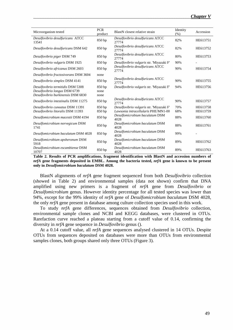
___________________________________________________________________Chapter V
49
Microorganism tested PCR product
BlastN closest relative strain Identity (%)
Accession
Desulfovibrio desulfuricans ATCC 13541
850 bp Desulfovibrio desulfuricans ATCC 27774
82% HE613751
Desulfovibrio desulfuricans DSM 642 850 bp Desulfovibrio desulfuricans ATCC 27774
82% HE613752
Desulfovibrio piger DSM 749 850 bp Desulfovibrio desulfuricans ATCC 27774
80% HE613753
Desulfovibrio vulgaris DSM 1925 850 bp Desulfovibrio vulgaris str. 'Miyazaki F' 90% -
Desulfovibrio africanus DSM 2603 850 bp Desulfovibrio desulfuricans ATCC 27774
90% HE613754
Desulfovibrio fructosivorans DSM 3604 none
Desulfovibrio simplex DSM 4141 850 bp Desulfovibrio desulfuricans ATCC 27774
90% HE613755
Desulfovibrio termitidis DSM 5308 850 bp Desulfovibrio vulgaris str. 'Miyazaki F' 94% HE613756 Desulfovibrio longus DSM 6739 none Desulfovibrio burkinensis DSM 6830 none
Desulfovibrio intestinalis DSM 11275 850 bp Desulfovibrio desulfuricans ATCC 27774
90% HE613757
Desulfovibrio cuneatus DSM 11391 850 bp Desulfovibrio vulgaris str. 'Miyazaki F' 70% HE613758 Desulfovibrio litoralis DSM 11393 850 bp Lawsonia intracellularis PHE/MN1-00 68% HE613759
Desulfomicrobium macestii DSM 4194 850 bp Desulfomicrobium baculatum DSM 4028
88% HE613760
Desulfomicrobium norvegicum DSM 1741
850 bp Desulfomicrobium baculatum DSM 4028
88% HE613761
Desulfomicrobium baculatum DSM 4028 850 bp Desulfomicrobium baculatum DSM 4028
99% -
Desulfomicrobium apsheronum DSM 5918
850 bp Desulfomicrobium baculatum DSM 4028
89% HE613762
Desulfomicrobium escambiense DSM 10707
850 bp Desulfomicrobium baculatum DSM 4028
89% HE613763
Table 2. Results of PCR amplifications, fragment identification with BlastN and accession numbers of nrfA gene fragments deposited in EMBL. Among the bacteria tested, nrfA gene is known to be present only in Desulfomicrobium baculatum DSM 4028.
BlastN alignments of nrfA gene fragment sequenced from both Desulfovibrio collection
(showed in Table 2) and environmental samples (data not shown) confirm that DNA amplified using new primers is a fragment of nrfA gene from Desulfovibrio or Desulfomicrobium genus. However identity percentage for all tested species was lower than 94%, except for the 99% identity of nrfA gene of Desulfomicrobium baculatum DSM 4028, the only nrfA gene present in database among culture collection species used in this work.
To study nrfA gene differences, sequences obtained from Desulfovibrio collection, environmental sample clones and NCBI and KEGG databases, were clustered in OTUs. Rarefaction curve reached a plateau starting from a cutoff value of 0.14, confirming the diversity in nrfA gene sequence in Desulfovibrio genus ().
At a 0.14 cutoff value, all nrfA gene sequences analysed clustered in 14 OTUs. Despite OTUs from sequences deposited on databases were more than OTUs from environmental samples clones, both groups shared only three OTUs (Figure 3).
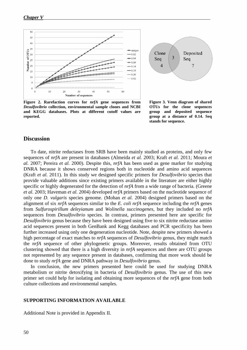
Chaper V___________________________________________________________________
50
Figure 2. Rarefaction curves for nrfA gene sequences from Desulfovibrio collection, environmental sample clones and NCBI and KEGG databases. Plots at different cutoff values are reported.
Figure 3. Venn diagram of shared OTUs for the clone sequences group and deposited sequence group at a distance of 0.14. Seq stands for sequence.
Discussion To date, nitrite reductases from SRB have been mainly studied as proteins, and only few
sequences of nrfA are present in databases (Almeida et al. 2003; Kraft et al. 2011; Moura et al. 2007; Pereira et al. 2000). Despite this, nrfA has been used as gene marker for studying DNRA because it shows conserved regions both in nucleotide and amino acid sequences (Kraft et al. 2011). In this study we designed specific primers for Desulfovibrio species that provide valuable additions since existing primers available in the literature are either highly specific or highly degenerated for the detection of nrfA from a wide range of bacteria. (Greene et al. 2003; Haveman et al. 2004) developed nrfA primers based on the nucleotide sequence of only one D. vulgaris species genome. (Mohan et al. 2004) designed primers based on the alignment of six nrfA sequences similar to the E. coli nrfA sequence including the nrfA genes from Sulfurospirillum deleyianum and Wolinella succinogenes, but they included no nrfA sequences from Desulfovibrio species. In contrast, primers presented here are specific for Desulfovibrio genus because they have been designed using five to six nitrite reductase amino acid sequences present in both GenBank and Kegg databases and PCR specificity has been further increased using only one degeneration nucleotide. Note, despite new primers showed a high percentage of exact matches to nrfA sequences of Desulfovibrio genus, they might match the nrfA sequence of other phylogenetic groups. Moreover, results obtained from OTU clustering showed that there is a high diversity in nrfA sequences and there are OTU groups not represented by any sequence present in databases, confirming that more work should be done to study nrfA gene and DNRA pathway in Desulfovibrio genus.
In conclusion, the new primers presented here could be used for studying DNRA metabolism or nitrite detoxifying in bacteria of Desulfovibrio genus. The use of this new primer set could help for isolating and obtaining more sequences of the nrfA gene from both culture collections and environmental samples.
SUPPORTING INFORMATION AVAILABLE
Additional Note is provided in Appendix II.
___________________________________________________________________Chapter V
51
References
Almeida M. G., Macieira S., Goncalves L. L., Huber R., Cunha C. A., Romao M. J., Costa C., Lampreia J., Moura J. J. and Moura I. (2003). The isolation and characterization of cytochrome c nitrite reductase subunits (NrfA and NrfH) from Desulfovibrio desulfuricans ATCC 27774. Re-evaluation of the spectroscopic data and redox properties. European Journal of Biochemistry 270(19): 3904-3915.
Benson D. A., Karsch-Mizrachi I., Lipman D. J., Ostell J. and Sayers E. W. (2011). GenBank. Nucleic Acids Research 39(suppl 1): D32-D37.
Cabello P., Roldán M. D., Castillo F. and Moreno-Vivián C. (2009). Nitrogen Cycle. Encyclopedia of Microbiology. Moselio S. Oxford, Academic Press: 299-321.
Einsle O., Messerschmidt A., Stach P., Bourenkov G. P., Bartunik H. D., Huber R. and Kroneck P. M. (1999). Structure of cytochrome c nitrite reductase. Nature 400(6743): 476-480.
Felsenstein J. (1993). PHYLIP (Phylogeny Inference Package) version 3.5c. Department of Genetics, University of Washington, Seattle, Distributed by the author.
Greene E. A., Hubert C., Nemati M., Jenneman G. E. and Voordouw G. (2003). Nitrite reductase activity of sulphate-reducing bacteria prevents their inhibition by nitrate-reducing, sulphide-oxidizing bacteria. Environmental Microbiology 5(7): 607-617.
Haveman S. A., Greene E. A., Stilwell C. P., Voordouw J. K. and Voordouw G. (2004). Physiological and gene expression analysis of inhibition of Desulfovibrio vulgaris Hildenborough by nitrite. J Bacteriol 186(23): 7944-7950.
Kanehisa M., Goto S., Kawashima S. and Nakaya A. (2002). The KEGG databases at GenomeNet. Nucleic Acids Research 30(1): 42-46.
Kraft B., Strous M. and Tegetmeyer H. E. (2011). Microbial nitrate respiration - Genes, enzymes and environmental distribution. Journal of Biotechnology 155(1): 104-117.
Larkin M. A., Blackshields G., Brown N. P., Chenna R., McGettigan P. A., McWilliam H., Valentin F., Wallace I. M., Wilm A., Lopez R., Thompson J. D., Gibson T. J. and Higgins D. G. (2007). Clustal W and Clustal X version 2.0. Bioinformatics 23(21): 2947-2948.
Marietou A., Griffiths L. and Cole J. (2009). Preferential Reduction of the Thermodynamically Less Favorable Electron Acceptor, Sulfate, by a Nitrate-Reducing Strain of the Sulfate-Reducing Bacterium Desulfovibrio desulfuricans 27774. J. Bacteriol. 191(3): 882-889.
Mohan S. B., Schmid M., Jetten M. and Cole J. (2004). Detection and widespread distribution of the nrfA gene encoding nitrite reduction to ammonia, a short circuit in the biological nitrogen cycle that competes with denitrification. FEMS Microbiology Ecology 49(3): 433-443.
Moura J. J. G., Gonzalez P., Moura I., Fauque G., Barton L. L. and Hamilton W. A. (2007). Dissimilatory nitrate and nitrite ammonification by sulphate-reducing eubacteria. Sulphate-Reducing Bacteria. Environmental and Engineered Systems. Barton L. L., Hamilton W. A. Cambridge, Cambridge University Press: 241-264.
Oakley B. B., Dowd S. E. and Purdy K. J. (2011). ThermoPhyl: a software tool for selecting phylogenetically optimized conventional and quantitative-PCR taxon-targeted assays for use with complex samples. FEMS Microbiology Ecology 77(1): 17-27.
Pereira I. A. C., LeGall J., Xavier A. V. and Teixeira M. (2000). Characterization of a heme c nitrite reductase from a non-ammonifying microorganism, Desulfovibrio vulgaris Hildenborough. Biochimica Et Biophysica Acta-Protein Structure and Molecular Enzymology 1481(1): 119-130.
Rozen S. and Skaletsky H. (1999). Primer3 on the WWW for General Users and for Biologist Programmers. 132: 365-386.
Schloss P. D., Westcott S. L., Ryabin T., Hall J. R., Hartmann M., Hollister E. B., Lesniewski R. A., Oakley B. B., Parks D. H., Robinson C. J., Sahl J. W., Stres B., Thallinger G. G., Van Horn D. J. and Weber C. F. (2009). Introducing mothur: open-source, platform-independent, community-supported software for describing and comparing microbial communities. Applied and environmental microbiology 75(23): 7537-7541.
Simon J. (2002). Enzymology and bioenergetics of respiratory nitrite ammonification. Fems Microbiology Reviews 26(3): 285-309.

__________________________________________________________________Chapter VI
53
Conclusions Deterioration of synthetic polymers in varnishes and binding media seriously
compromises the adhesion and durability of the paint as well as its decorative/protective function, therefore identification of the cause of synthetic polymer biodeterioration is of great importance. On the base of this work, it is possible to conclude that:
� Biotechnology provides valid tools to study changes in microbial community structure
due to the presence of synthetic polymers and to identify synthetic polymer degrading microorganisms.
� In the Hungaria hotel case-study, coloured alterations on the tiles were mainly due to the presence of cryptoendolithic cyanobacteria and the treatment with the synthetic resin led to a colonisation by biodeteriogen fungi.
� Paraloid B72® is not the best consolidant polymer to be used for the long-time conservation of the ceramic tile of Hungaria hotel.
Other results about the community structure of the Hungaria hotel showed that the
microflora present on the tiles was generally greatly influenced by the Venice lagoon, the environment of the hotel:
� The rapid reappearing of coloured alterations soon after the 2007 restoration, which included the use of biocides, is likely favoured by the water content and organic substances, some of which added during the conservation treatment.
� Several microorganisms related to the alkaline environment, which is in the range of the tile pH, and related to the aquatic environment and the pollutants of the Venice lagoon, are present on the façade of the Hungaria hotel.
� To ensure longer lasting conservation of the Hungaria façade, a more efficient maintenance plan that includes the selection of a better consolidant instead of Paraloid is strongly suggested.
When synthetic polymers objects and paints are disposed of they should be susceptible to
biodegradation. Bioremediation is a safer and efficient tool for the removal of many xenobiotics and can be successfully applied for the removal of synthetic polymers, including those present in paint and coating formulations. The study of the bacterial degradation of nitrocellulose as binder in paint formulations leads to conclude that:
� Desulfovibrio desulfuricans ATCC 13541 is able to adhere onto the surface of the
nitrocellulose-based paint. � The incubation with D. desulfuricans causes paint detachment and fading of
nitrocellulose-based paint. The colour fading could be caused by the degradation of the paint and the removal of nitro groups from the nitrocellulose molecule.
� D. desulfuricans is active against the nitrocellulose-based paint with high specificity on the N–O bond of the nitro–substituted cellulose.
� The activity of nitrate reductase was equivalent while nitrite reductase activity was higher in D. desulfuricans grown in the presence of the nitrocellulose-based paint.
� D. desulfuricans ATCC 13541 is able to degrade nitrocellulose as binder in paint and likely performs this degradation by the dissimilatory nitrate reduction to ammonia pathway.
Chapter VI__________________________________________________________________
54
The dissimilatory nitrate reduction to ammonia (DNRA) is the process in which nitrate is reduced to ammonia with nitrite as intermediate. The ability to carry out DNRA is phylogenetically widespread and the gene nrfA encodes for the key enzyme of the second step of the pathway, the reduction of nitrite to ammonia. For that reason the study of the nrfA gene is an important step to study the DNRA pathway. According to the results obtained in this work, it is possible to conclude that:
� The new primer set allows the amplification of a 850 bp nrfA gene product. � The new primers are specific for the amplification of nrfA gene of Desulfovibrio genus
bacteria, but they may match the nrfA sequence of other phylogenetic groups (e.g. Desulfomicrobium genus).
� Using these primers thirteen new nrfA gene sequences, nine from Desulfovibrio spp. and four from Desulfomicrobium spp., were deposited in the EMBL database.
� Results obtained from OTU clustering showed that there are OTU groups not represented by any sequence present in databases.
� The new primer set can be used for obtaining more sequences of the nrfA gene and improving our knowledge of the dissimilatory nitrate reduction to ammonia pathway.
In conclusion, further studies on synthetic polymer biodeterioration should be made for
the selection of the best one to be applied for each specific use in order to prevent the loss of polymer function once it has been applied to a surface.
On the other hand, D. desulfuricans is a promising bacterium in nitrocellulose-based paint bioremediation and nrfA gene could be used to study DNRA or nitrite detoxifying metabolism in bacteria of Desulfovibrio genus, in order to improve nitrocellulose bioremediation process.
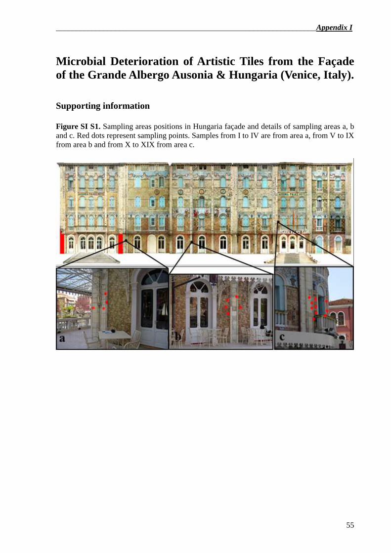
__________________________________________________________________Appendix I
55
Microbial Deterioration of Artistic Tiles from the Façade of the Grande Albergo Ausonia & Hungaria (Venice, Italy).
Supporting information Figure SI S1. Sampling areas positions in Hungaria façade and details of sampling areas a, b and c. Red dots represent sampling points. Samples from I to IV are from area a, from V to IX from area b and from X to XIX from area c.
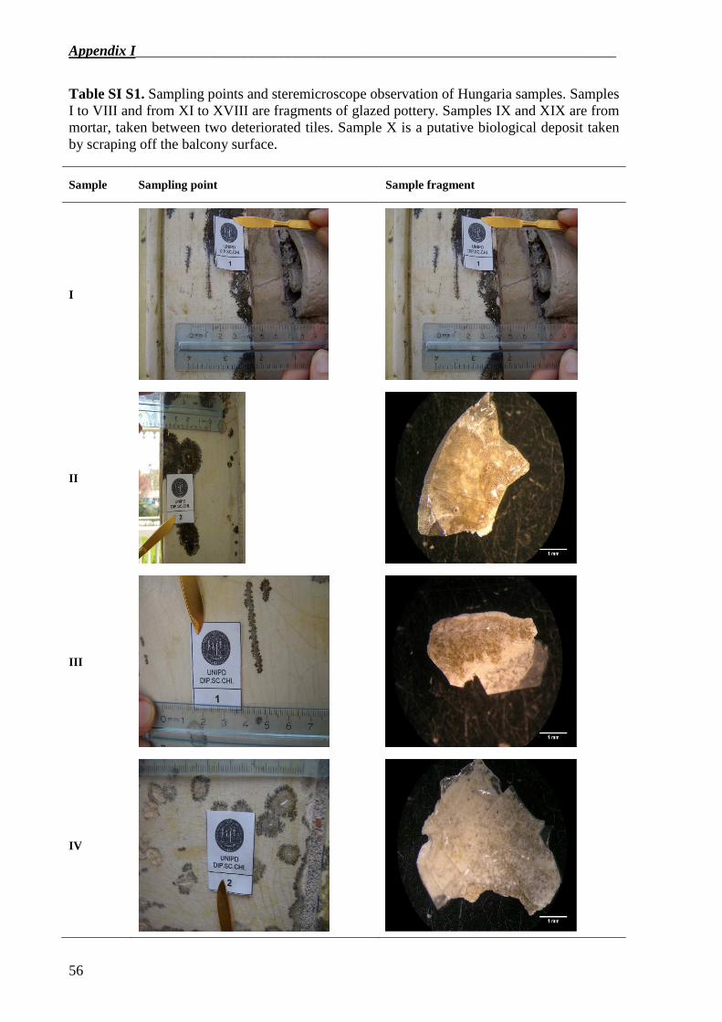
Appendix I__________________________________________________________________
56
Table SI S1. Sampling points and steremicroscope observation of Hungaria samples. Samples I to VIII and from XI to XVIII are fragments of glazed pottery. Samples IX and XIX are from mortar, taken between two deteriorated tiles. Sample X is a putative biological deposit taken by scraping off the balcony surface.
Sample Sampling point Sample fragment
I
II
III
IV
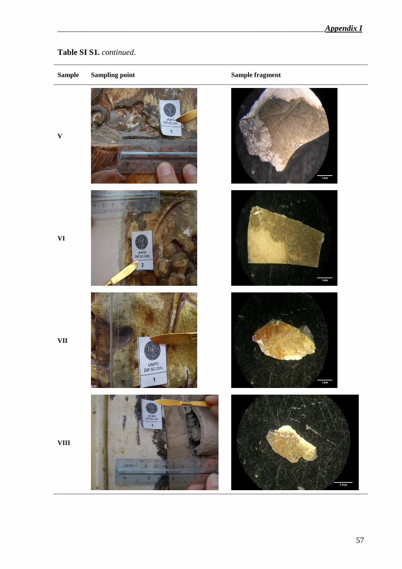
__________________________________________________________________Appendix I
57
Table SI S1. continued.
Sample Sampling point Sample fragment
V
VI
VII
VIII
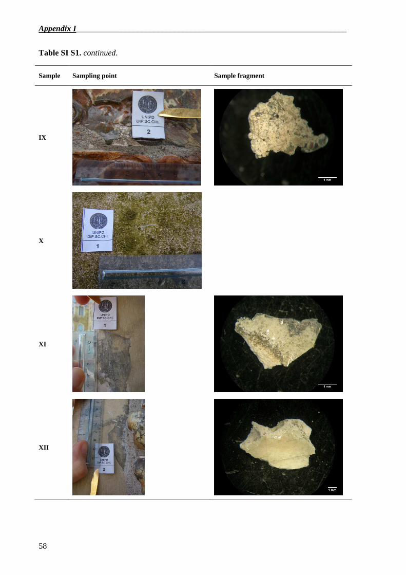
Appendix I__________________________________________________________________
58
Table SI S1. continued.
Sample Sampling point Sample fragment
IX
X
XI
XII
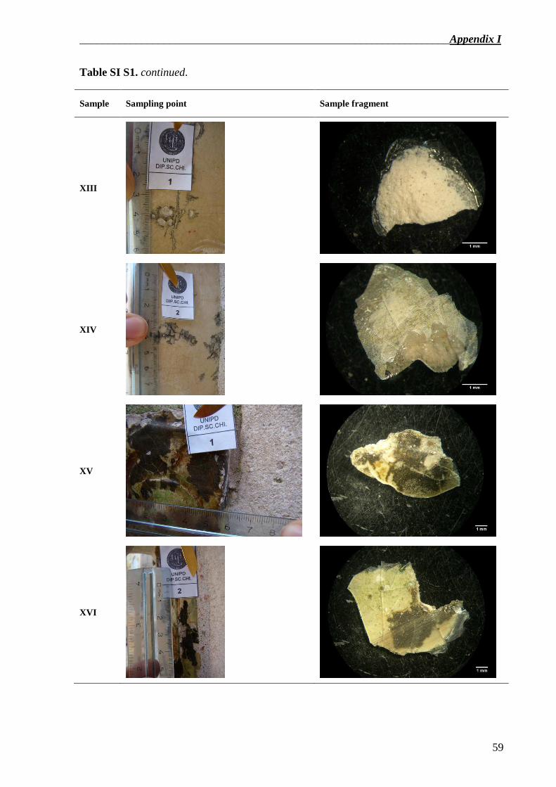
__________________________________________________________________Appendix I
59
Table SI S1. continued.
Sample Sampling point Sample fragment
XIII
XIV
XV
XVI
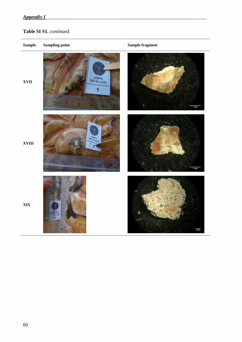
Appendix I__________________________________________________________________
60
Table SI S1. continued.
Sample Sampling point Sample fragment
XVII
XVIII
XIX
_________________________________________________________________Appendix II
61
Nitrate and nitrite reductase genes expression in Desulfovibrio desulfuricans ATCC 13541.
Dissimilatory nitrate reduction to ammonia (DNRA) or nitrate ammonification is a two-
step process in which nitrate is reduced to nitrite and nitrite is subsequently reduced to ammonia (Cabello et al. 2009; Einsle et al. 1999; Kraft et al. 2011). In DNRA, the reduction of nitrate to nitrite is assumed to being mostly catalysed by the periplasmic nitrate reductase complex NapAB (Kraft et al. 2011; Simon 2002), while the second step of DNRA, the reduction of nitrite to ammonia, is catalysed by NrfA, without the release of any intermediate (Cabello et al. 2009; Einsle et al. 1999; Kraft et al. 2011).
DNRA is an anaerobic process performed by a phylogenetically wide range of bacteria and occurs in reductant-rich environments like anaerobic marine sediments, sulphide-thermal vents, and gastrointestinal tracts of animals (Cabello et al. 2009; Kraft et al. 2011). The nrfA gene, coding for NrfA, the pentaheme cytochrome c nitrite reductase, has been found in Gamma-, Delta- and Epsilonproteobacteria and in members of the Bacteroides (Kraft et al. 2011; Mohan et al. 2004; Smith et al. 2007).
Many sulphate-reducing Deltaproteobacteria are able to perform DNRA in the presence of nitrate when sulphate is absent and/or in low concentration (Kraft et al. 2011; Marietou et al. 2009). Sulphate-reducing bacteria (SRB) are microorganisms with a great ecological importance. They have been used in reducing soluble metal oxyanions to insoluble forms, a process of great potential in the bioremediation of toxic heavy metals and radionuclides from groundwater and waste water (Chen et al. 2000; Dong et al. 2009; He et al. 2006; Muyzer et al. 2008). Another biotechnological application of SRB is the removal and reuse of sulphur compounds from waste water and off gases, as for example the removing of hydrogen sulphide from natural gas or biogas (Muyzer et al. 2008) or the reduction oxidized sulphur compounds to sulphide, such as the removal of black crusts from stone surfaces of historical monuments (Cappitelli et al. 2007; Cappitelli et al. 2006). SRB of Desulfovibrio genus have been also studied for the biodegradation of nitrocellulose (Petrova et al. 2002a; Tarasova et al. 2009), a semi-synthetic polymer widely used in objects and paint formulations. Furthermore, according to Petrova et al. (2002b); Tarasova et al. (2004) the nitrocellulose denitration by Desulfovibrio spp action is followed by DNRA process to reduce the nitrate present in the cultural medium.
Functional genes napA and nrfA have been previously used to detect microorganisms able to perform DNRA in the environment and to follow their relative abundance along nitrate or nitrite concentration gradient (Dong et al. 2009). Gene expression studies can help in understanding DNRA pathway of SRB used in bioremediation (Haveman et al. 2005). Thus, it is important to study how SRB respond to nitrate to predict the performance of these bacteria for bioremediation (He et al. 2006).
The aim of this study was to evaluate DNRA pathway in different growth conditions. In particular, Real-Time PCR was used to assess napA and nrfA genes expression changes in the presence of different concentration of nitrate.
Pure culture of sulphate-reducing bacterium Desulfovibrio desulfuricans ATCC 13541
was maintained anaerobically in Widdel’s medium (Widdel et al. 1992). Experiments were performed with an initial cell concentration of 107 cells/ml in modified medium without sulphates, but containing 4.3 or 14.7 mM of nitrate (treatment 1 and 2, respectively) and a control sample was set up in normal Widdel’s medium, that contains 4g/l of sulphate. All samples were incubated at 30 °C under anaerobic conditions for 0, 1, 2 and 3 days. At each
Appendix II_________________________________________________________________
62
time point, three replicates of each sample were sacrificed. A cell count using the Neobauer counting chamber was performed to monitor bacterial density and then all samples were centrifuged at 4000 rpm for 30 min. The resultant pellets were resuspended in 0.5 ml of each culture media, 1 ml of RNAprotect Bacteria Reagent (QIAgen, CA) was added, mixed immediately by vortexing for 5 s to preserve RNA and stored at -80 °C.
RNA from all samples was extracted using RNeasy Mini Kit (QIAgen, CA), according to manufacture instructions, quantified by NanoDrop (Thermo Scientific, USA) and analysed using Agilent 2100 Bioanalyzer (Agilent Technologies, USA) to assess RNA quality. Reverse transcription PCR was set up from 50 µl of total RNA using random primers and GoScript reverse transcriptase (Promega, USA) following suppliers instructions and cDNA obtained was purified using PCR Purification Kit (QIAgen, CA), following manufacture instructions.
331F and 797R primer sequences, for amplifying the 16S rRNA reference gene, were obtained from Smith et al. (2009) (Table 1). Primer sequences for both napA and nfrA were designed using Primer Express v 2.0 software (Applied Biosystems), starting from D. desulfuricans ATCC 13541 target genes sequences previously obtained in our laboratory (Table 1).
For each primer combination, Real-Time PCR assays were carried out within a single assay plate. Each assay contained analytical triplicate cDNA templates for each experimental replicate sample and no-template controls (NTC) in triplicate. Real-Time PCR amplification mixtures contained 2 µl of 1:2 dilution of cDNA template, 12.5 µl of Power SYBR Green PCR master mix (Applied BioSystems), 1 µl of 20 ng/µl of each primer, made up to a total volume of 25 µl with sterile RNase free water. Real-Time PCR amplification and detection for all primer combinations were performed using an ABI 7500 Fast Real-Time PCR System (Applied BioSystems), with the following parameters: 50 °C for 2 min, 95 °C for 10 min, and 40 cycles at 95 °C for 15 s and 60 °C for 1 min. Automatic analysis settings were selected to determine the threshold cycle (Ct) values and baseline settings. Analysis of the relative gene expression data was performed using the 7500 Fast System software v 1.4 (Applied Biosystem), according to the 2-∆∆Ct method (Livak et al. 2001).
Target Primer name Sequence (5’–3’)
Amplicon Length (bp)
Temp. (°C)
Bacterial 16S rRNA gene 331F TTCTACGGGAGGCAGCAG 466 60 797R GGACTACCAGGGTATCTAATCCTGTT D. desulfuricans napA gene
NapA_214F GGCTACACGGCCAACAAGTT 58 59
NapA_271R TGGGATCGAGATGGTTGGA D. desulfuricans nrfA gene
NrfA_368F GCAAGCCCGGTTCTTTTGT 57 60
NrfA_454R TGATCATGCCGACCTTGGA Table 1. Quantitative PCR primer sets targeting small subunit ribosomal RNA genes of bacteria, napA and nfrA gene of D. desulfuricans ATCC 13541. Temp. stands for annealing temperature.
Cell counts of all samples at each time point of the experiment were not significantly
different (Figure 1), therefore 4.3 or 14.7 mM of nitrate instead of 4 g/l of sulphate did not change D. desulfuricans ATCC 13541 growth.
RNA sample quality, checked using the Bioanalyzer, was good for most of the samples. Samples with a low RNA quality were discarded before Real-Time PCR analysis.
Real-Time PCR analysis was conducted on cDNAs to quantify the expression levels of both napA and nrfA genes in order to determine changes in DNRA genes expression in D. desulfuricans grown in the three different nitrate concentrations.
Both genes involved in the DNRA pathway showed different expression levels in the three different growth conditions (Figure 2). In particular, napA gene was expressed at higher level
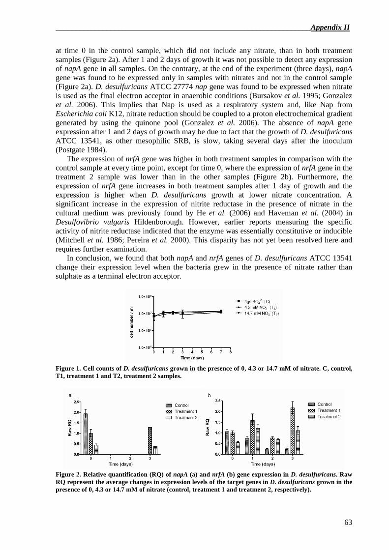
_________________________________________________________________Appendix II
63
at time 0 in the control sample, which did not include any nitrate, than in both treatment samples (Figure 2a). After 1 and 2 days of growth it was not possible to detect any expression of napA gene in all samples. On the contrary, at the end of the experiment (three days), napA gene was found to be expressed only in samples with nitrates and not in the control sample (Figure 2a). D. desulfuricans ATCC 27774 nap gene was found to be expressed when nitrate is used as the final electron acceptor in anaerobic conditions (Bursakov et al. 1995; Gonzalez et al. 2006). This implies that Nap is used as a respiratory system and, like Nap from Escherichia coli K12, nitrate reduction should be coupled to a proton electrochemical gradient generated by using the quinone pool (Gonzalez et al. 2006). The absence of napA gene expression after 1 and 2 days of growth may be due to fact that the growth of D. desulfuricans ATCC 13541, as other mesophilic SRB, is slow, taking several days after the inoculum (Postgate 1984).
The expression of nrfA gene was higher in both treatment samples in comparison with the control sample at every time point, except for time 0, where the expression of nrfA gene in the treatment 2 sample was lower than in the other samples (Figure 2b). Furthermore, the expression of nrfA gene increases in both treatment samples after 1 day of growth and the expression is higher when D. desulfuricans growth at lower nitrate concentration. A significant increase in the expression of nitrite reductase in the presence of nitrate in the cultural medium was previously found by He et al. (2006) and Haveman et al. (2004) in Desulfovibrio vulgaris Hildenborough. However, earlier reports measuring the specific activity of nitrite reductase indicated that the enzyme was essentially constitutive or inducible (Mitchell et al. 1986; Pereira et al. 2000). This disparity has not yet been resolved here and requires further examination.
In conclusion, we found that both napA and nrfA genes of D. desulfuricans ATCC 13541 change their expression level when the bacteria grew in the presence of nitrate rather than sulphate as a terminal electron acceptor.
Figure 1. Cell counts of D. desulfuricans grown in the presence of 0, 4.3 or 14.7 mM of nitrate. C, control, T1, treatment 1 and T2, treatment 2 samples.
Figure 2. Relative quantification (RQ) of napA (a) and nrfA (b) gene expression in D. desulfuricans. Raw RQ represent the average changes in expression levels of the target genes in D. desulfuricans grown in the presence of 0, 4.3 or 14.7 mM of nitrate (control, treatment 1 and treatment 2, respectively).
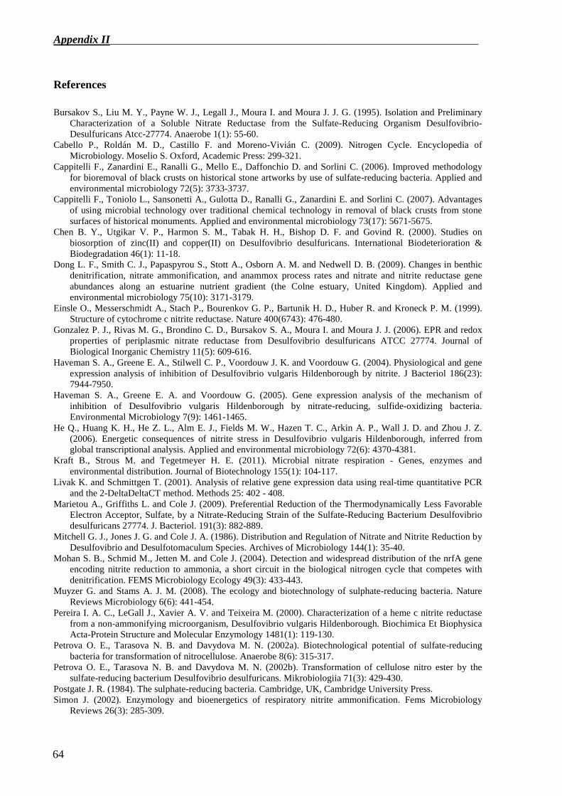
Appendix II_________________________________________________________________
64
References
Bursakov S., Liu M. Y., Payne W. J., Legall J., Moura I. and Moura J. J. G. (1995). Isolation and Preliminary Characterization of a Soluble Nitrate Reductase from the Sulfate-Reducing Organism Desulfovibrio-Desulfuricans Atcc-27774. Anaerobe 1(1): 55-60.
Cabello P., Roldán M. D., Castillo F. and Moreno-Vivián C. (2009). Nitrogen Cycle. Encyclopedia of Microbiology. Moselio S. Oxford, Academic Press: 299-321.
Cappitelli F., Zanardini E., Ranalli G., Mello E., Daffonchio D. and Sorlini C. (2006). Improved methodology for bioremoval of black crusts on historical stone artworks by use of sulfate-reducing bacteria. Applied and environmental microbiology 72(5): 3733-3737.
Cappitelli F., Toniolo L., Sansonetti A., Gulotta D., Ranalli G., Zanardini E. and Sorlini C. (2007). Advantages of using microbial technology over traditional chemical technology in removal of black crusts from stone surfaces of historical monuments. Applied and environmental microbiology 73(17): 5671-5675.
Chen B. Y., Utgikar V. P., Harmon S. M., Tabak H. H., Bishop D. F. and Govind R. (2000). Studies on biosorption of zinc(II) and copper(II) on Desulfovibrio desulfuricans. International Biodeterioration & Biodegradation 46(1): 11-18.
Dong L. F., Smith C. J., Papaspyrou S., Stott A., Osborn A. M. and Nedwell D. B. (2009). Changes in benthic denitrification, nitrate ammonification, and anammox process rates and nitrate and nitrite reductase gene abundances along an estuarine nutrient gradient (the Colne estuary, United Kingdom). Applied and environmental microbiology 75(10): 3171-3179.
Einsle O., Messerschmidt A., Stach P., Bourenkov G. P., Bartunik H. D., Huber R. and Kroneck P. M. (1999). Structure of cytochrome c nitrite reductase. Nature 400(6743): 476-480.
Gonzalez P. J., Rivas M. G., Brondino C. D., Bursakov S. A., Moura I. and Moura J. J. (2006). EPR and redox properties of periplasmic nitrate reductase from Desulfovibrio desulfuricans ATCC 27774. Journal of Biological Inorganic Chemistry 11(5): 609-616.
Haveman S. A., Greene E. A., Stilwell C. P., Voordouw J. K. and Voordouw G. (2004). Physiological and gene expression analysis of inhibition of Desulfovibrio vulgaris Hildenborough by nitrite. J Bacteriol 186(23): 7944-7950.
Haveman S. A., Greene E. A. and Voordouw G. (2005). Gene expression analysis of the mechanism of inhibition of Desulfovibrio vulgaris Hildenborough by nitrate-reducing, sulfide-oxidizing bacteria. Environmental Microbiology 7(9): 1461-1465.
He Q., Huang K. H., He Z. L., Alm E. J., Fields M. W., Hazen T. C., Arkin A. P., Wall J. D. and Zhou J. Z. (2006). Energetic consequences of nitrite stress in Desulfovibrio vulgaris Hildenborough, inferred from global transcriptional analysis. Applied and environmental microbiology 72(6): 4370-4381.
Kraft B., Strous M. and Tegetmeyer H. E. (2011). Microbial nitrate respiration - Genes, enzymes and environmental distribution. Journal of Biotechnology 155(1): 104-117.
Livak K. and Schmittgen T. (2001). Analysis of relative gene expression data using real-time quantitative PCR and the 2-DeltaDeltaCT method. Methods 25: 402 - 408.
Marietou A., Griffiths L. and Cole J. (2009). Preferential Reduction of the Thermodynamically Less Favorable Electron Acceptor, Sulfate, by a Nitrate-Reducing Strain of the Sulfate-Reducing Bacterium Desulfovibrio desulfuricans 27774. J. Bacteriol. 191(3): 882-889.
Mitchell G. J., Jones J. G. and Cole J. A. (1986). Distribution and Regulation of Nitrate and Nitrite Reduction by Desulfovibrio and Desulfotomaculum Species. Archives of Microbiology 144(1): 35-40.
Mohan S. B., Schmid M., Jetten M. and Cole J. (2004). Detection and widespread distribution of the nrfA gene encoding nitrite reduction to ammonia, a short circuit in the biological nitrogen cycle that competes with denitrification. FEMS Microbiology Ecology 49(3): 433-443.
Muyzer G. and Stams A. J. M. (2008). The ecology and biotechnology of sulphate-reducing bacteria. Nature Reviews Microbiology 6(6): 441-454.
Pereira I. A. C., LeGall J., Xavier A. V. and Teixeira M. (2000). Characterization of a heme c nitrite reductase from a non-ammonifying microorganism, Desulfovibrio vulgaris Hildenborough. Biochimica Et Biophysica Acta-Protein Structure and Molecular Enzymology 1481(1): 119-130.
Petrova O. E., Tarasova N. B. and Davydova M. N. (2002a). Biotechnological potential of sulfate-reducing bacteria for transformation of nitrocellulose. Anaerobe 8(6): 315-317.
Petrova O. E., Tarasova N. B. and Davydova M. N. (2002b). Transformation of cellulose nitro ester by the sulfate-reducing bacterium Desulfovibrio desulfuricans. Mikrobiologiia 71(3): 429-430.
Postgate J. R. (1984). The sulphate-reducing bacteria. Cambridge, UK, Cambridge University Press. Simon J. (2002). Enzymology and bioenergetics of respiratory nitrite ammonification. Fems Microbiology
Reviews 26(3): 285-309.

_________________________________________________________________Appendix II
65
Smith C. J., Nedwell D. B., Dong L. F. and Osborn A. M. (2007). Diversity and abundance of nitrate reductase genes (narG and napA), nitrite reductase genes (nirS and nrfA), and their transcripts in estuarine sediments. Applied and environmental microbiology 73(11): 3612-3622.
Smith C. J. and Osborn A. M. (2009). Advantages and limitations of quantitative PCR (Q-PCR)-based approaches in microbial ecology. FEMS Microbiology Ecology 67(1): 6-20.
Tarasova N. B., Petrova O. E., Davydova M. N., Khairutdinov B. I. and Klochkov V. V. (2004). Changes in the nitrocellulose molecule induced by sulfate-reducing bacteria Desulfovibrio desulfuricans 1,388. The enzymes participating in this process. Biochemistry (Mosc) 69(7): 809-812.
Tarasova N. B., Gorshkov O. V. and Petrova O. E. (2009). Activity of Nitrate Reductase in Desulfovibrio vulgaris VKM 1388. Microbiology 78(2): 160–164.
Widdel F. and Bak F. (1992). Gram-negative mesophilic sulfate-reducing bacteria. The Prokaryotes. Balows A., Trüper H. G., Dworkin M., Harder W., Schleifer K. H., Springer-Verlag. IV: 3352-3378.

_______________________________________________________________Collaborations
Prof. Renzo Bertoncello and Ilaria Martini Dipartimento di Scienze Chimiche, Università degli Studi di Padova, via Marzolo 1, 35131 Padova, Italy. Dr. Ornella Salvadori Soprintendenza speciale per il patrimonio storico, artistico ed etnoantropologico e per il polo museale della città di Venezia e dei comuni della Gronda lagunare, Laboratorio Scientifico, Cannaregio 3553, 30131 Venezia, Italy Dr. Monica Favaro Istituto di Chimica Inorganica e delle Superfici, Consiglio Nazionale delle Ricerche, Corso Stati Uniti 4, 35127 Padova, Italy Prof. Lucia Toniolo and Francesca Toja Dipartimento di Chimica, Materiali, e Ingegneria Chimica (CMIC) “G. Natta”, Politecnico di Milano, Via Mancinelli 7, 20131 Milano, Italy. Dr. Beatriz Prieto and Patricia Sanmartin Departamento de Edafoloxía y Química Agrícola, Facultad Farmacia, Universidade de Santiago de Compostela, 15782 Santiago de Compostela, Spain. Dr. Kevin J. Purdy, Prof. Elisabetta Zanardini and Dr. Francis P. Sweeney School of Life Sciences, University of Warwick, CV4 7AL, Coventry, UK.