microglia activation in a pediatric rabbit model of...
TRANSCRIPT
RESEARCH ARTICLE
Microglia activation in a pediatric rabbit model of tuberculousmeningitisElizabeth W. Tucker1,2,3,4,5,*, Supriya Pokkali2,3,6,*, Zhi Zhang1,4, Vincent P. DeMarco2,3,6, Mariah Klunk2,3,6,Elizabeth S. Smith1,4, Alvaro A. Ordonez2,3,6, Marie-France Penet7,8, Zaver Bhujwalla7,8, Sanjay K. Jain2,3,6,‡,§ andSujatha Kannan1,4,‡,§
ABSTRACTCentral nervous system (CNS) tuberculosis (TB) is the most severeform of extra-pulmonary TB and disproportionately affects youngchildren where the developing brain has a unique host response. NewZealand white rabbits were infected withMycobacterium tuberculosisvia subarachnoid inoculation at postnatal day 4-8 and evaluated until4-6 weeks post-infection. Control and infected rabbit kits wereassessed for the development of neurological deficits, bacterialburden, and postmortem microbiologic and pathologic changes. Thepresence of meningitis and tuberculomas was demonstratedhistologically and by in vivo magnetic resonance imaging (MRI).The extent of microglial activation was quantified by in vitroimmunohistochemistry as well as non-invasive in vivo imaging ofactivated microglia/macrophages with positron emission tomography(PET). Subarachnoid infection induced characteristic leptomeningealand perivascular inflammation and TB lesions with central necrosis, acellular rim and numerous bacilli on pathologic examination.Meningeal and rim enhancement was visible on MRI. An intensemicroglial activation was noted in M. tuberculosis-infected animalsin the white matter and around the TB lesions, as evidenced bya significant increase in uptake of the tracer 124I-DPA-713, whichis specific for activated microglia/macrophages, and confirmed byquantification of Iba-1 immunohistochemistry. Neurobehavioralanalyses demonstrated signs similar to those noted in children withdelayedmaturation and development of neurological deficits resultingin significantly worse composite behavior scores in M. tuberculosis-infected animals. We have established a rabbit model that mimics
features of TB meningitis in young children. This model could providea platform for evaluating novel therapies, including host-directedtherapies, against TB meningitis relevant to a young child’sdeveloping brain.
KEY WORDS: Tuberculosis, Pediatric, Microglia, PET, Meningitis,TSPO
INTRODUCTIONGlobally, central nervous system (CNS) tuberculosis (TB)continues to be a devastating disease that disproportionatelyaffects toddler-age children (Be et al., 2009; Jain et al., 2013;Yaramis et al., 1998). CNS TB most commonly presents asmeningitis, but can also present as intracranial tuberculomas or, lessfrequently, tuberculous brain abscesses (Be et al., 2009; Jain et al.,2005; Kumar et al., 2002; Rock et al., 2008). TB meningitis is themost severe form of TB and is associated with significant morbidityand mortality (13-57%) even with the completion of twelve monthsof arduous treatment (Girgis et al., 1998; Rohlwink et al., 2016a;van Well et al., 2009; Yaramis et al., 1998). Children often presentwith signs of increased intracranial pressure, brainstem dysfunction,cranial nerve palsies and stroke resulting in permanent hemiplegiaand quadriplegia (Jain et al., 2013; van Well et al., 2009; Yaramiset al., 1998). Neuroinflammation seems to be a key component ofthe pathological process, leading to exudative meningitis,endovasculitis, infarction and obstructive hydrocephalus seen asmeningeal or post-contrast enhancement on computed tomography(CT) or magnetic resonance imaging (MRI), associated withneurologic disability and poor outcomes (Be et al., 2009; Donaldand Schoeman, 2004; Katti, 2004; Schoeman and Donald, 2013;van Well et al., 2009). Host-directed therapies have been used forthe treatment of TB meningitis with strong evidence for the benefitof adjunctive corticosteroids in decreasing mortality in adults(Prasad and Singh, 2008; Thwaites et al., 2004). Additionally, inchildren, adjunctive corticosteroids have been shown to not only bebeneficial in reducing mortality, but also in reducing neurologicsequelae (Girgis et al., 1991; Schoeman et al., 1997). However,corticosteroid use leads to a nonspecific modulation of the immuneresponse with significant side effects that can limit its use (Ordonezet al., 2014). Therefore, there is an urgent need for the developmentand validation of novel host-directed therapies for CNS TB.
Although animal models of TB meningitis have been previouslydeveloped, they typically mimic adult disease (Tsenova et al., 1999,2005, 2002, 1998; vanWell et al., 2007) and do not take into accountthe effects of injury and immune dysregulation in the developingbrain, which would have significant relevance in childhood disease.Microglia, the resident immune cells in the brain, are the primary hostcells of Mycobacterium tuberculosis and release pro-inflammatorycytokines when activated as a result of TB infection (Curto et al.,Received 29 July 2016; Accepted 8 November 2016
1Department of Anesthesiology and Critical Care Medicine, Division of PediatricAnesthesiology and Critical Care Medicine, Johns Hopkins University School ofMedicine, Baltimore, MD 21287, USA. 2Center for Infection and InflammationImaging Research, Johns Hopkins University School of Medicine, Baltimore, MD21287, USA. 3Center for Tuberculosis Research, Johns Hopkins University Schoolof Medicine, Baltimore, MD 21287, USA. 4Center for Nanomedicine, Johns HopkinsUniversity School of Medicine, Baltimore, MD 21287, USA. 5Department ofAnesthesiology and Critical Care Medicine, Division of Critical Care Medicine,Johns Hopkins All Children's Hospital, St. Petersburg, FL 33701, USA. 6Departmentof Pediatrics, Division of Infectious Diseases, Johns Hopkins University School ofMedicine, Baltimore, MD 21287, USA. 7JHU ICMIC Program, Division of CancerImaging Research, The Russell H. Morgan Department of Radiology andRadiological Science, Johns Hopkins University School of Medicine, Baltimore,MD 21287, USA. 8Sidney Kimmel Comprehensive Cancer Center, Johns HopkinsUniversity School of Medicine, Baltimore, MD 21287, USA.*These two authors contributed equally to this work‡These two authors contributed equally to this work
§Authors for correspondence ([email protected]; [email protected])
S.K.J., 0000-0001-9620-7070
This is an Open Access article distributed under the terms of the Creative Commons AttributionLicense (http://creativecommons.org/licenses/by/3.0), which permits unrestricted use,distribution and reproduction in any medium provided that the original work is properly attributed.
1497
© 2016. Published by The Company of Biologists Ltd | Disease Models & Mechanisms (2016) 9, 1497-1506 doi:10.1242/dmm.027326
Disea
seModels&Mechan
isms
2004; Hernandez Pando et al., 2010; Rock et al., 2005; Yang et al.,2007, 2009; Zucchi et al., 2012).Microglia are not only involvedwithhost-defense (Rock et al., 2004), but also have a crucial role inneurodevelopment. During development, microglia are active inaxonal guidance, neurodevelopmental apoptosis and synaptogenesis(Schafer et al., 2012; Tremblay et al., 2010; Verney et al., 2010). CNSinfection leads to the disruption of normal glial function, therebymaking the immature brain uniquely vulnerable to injury.In this study, we developed a rabbit model that mimics features of
TB meningitis in young children. Rabbits were chosen as theprogression of myelination, microglial presence in the white mattertracts, and brain development follows a pattern that is similar to that inhumans where it starts perinatally and continues in the postnatalperiod, albeit in a more compressed timeframe (Drobyshevsky et al.,2005; Saadani-Makki et al., 2008). Subarachnoid infection withM. tuberculosis led to the development of inflammatory meningitis,characteristic TB lesions and microglial activation as early as twoweeks after infection in this model, corresponding to a postnatal agewhere several neurological functions are still maturing in the rabbit.Neurobehavioral analyses, and non-invasive live animal imagingwithMRI and positron emission tomography (PET), targeting activatedmicroglia/macrophages (Foss et al., 2013; Ordonez et al., 2015), werecorrelated with CNS disease as seen on postmortem examination.
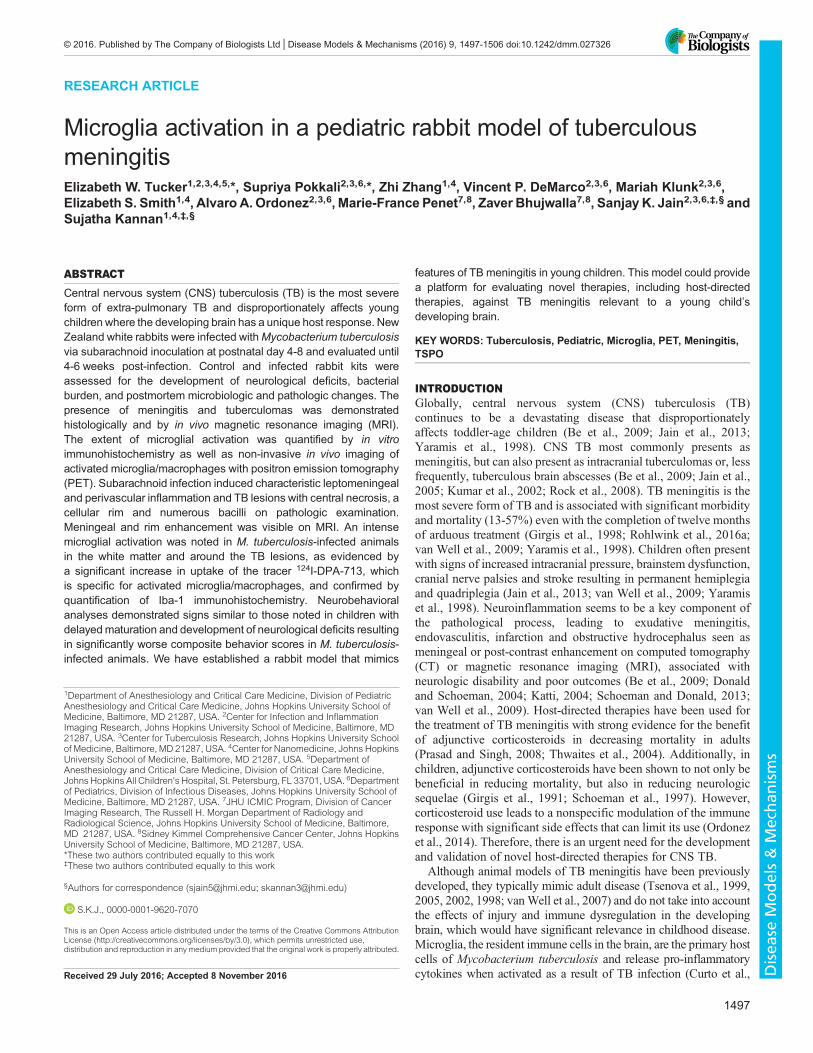
RESULTSEstablishment of CNS infectionWe performed direct inoculation ofM. tuberculosis in post-natal day(PND) 4-8 rabbits via subarachnoid route to produce CNS infection.Mean bacillary implantation (one day after subarachnoid infection)was 4.14±1.18 log10 colony forming units (CFU) (mean±s.d.) andreached 6.31±1.7 log10 14 days post-infection (Fig. 1A). Thebacterial burden remained relatively stable thereafter. Further, weobserved bacterial spread from brain to lungs, which might be aresult of disruption of the blood-brain barrier (BBB) (Fig. 1B),suggesting that like young children, young rabbits have limitedcapacity to prevent dissemination of infection.
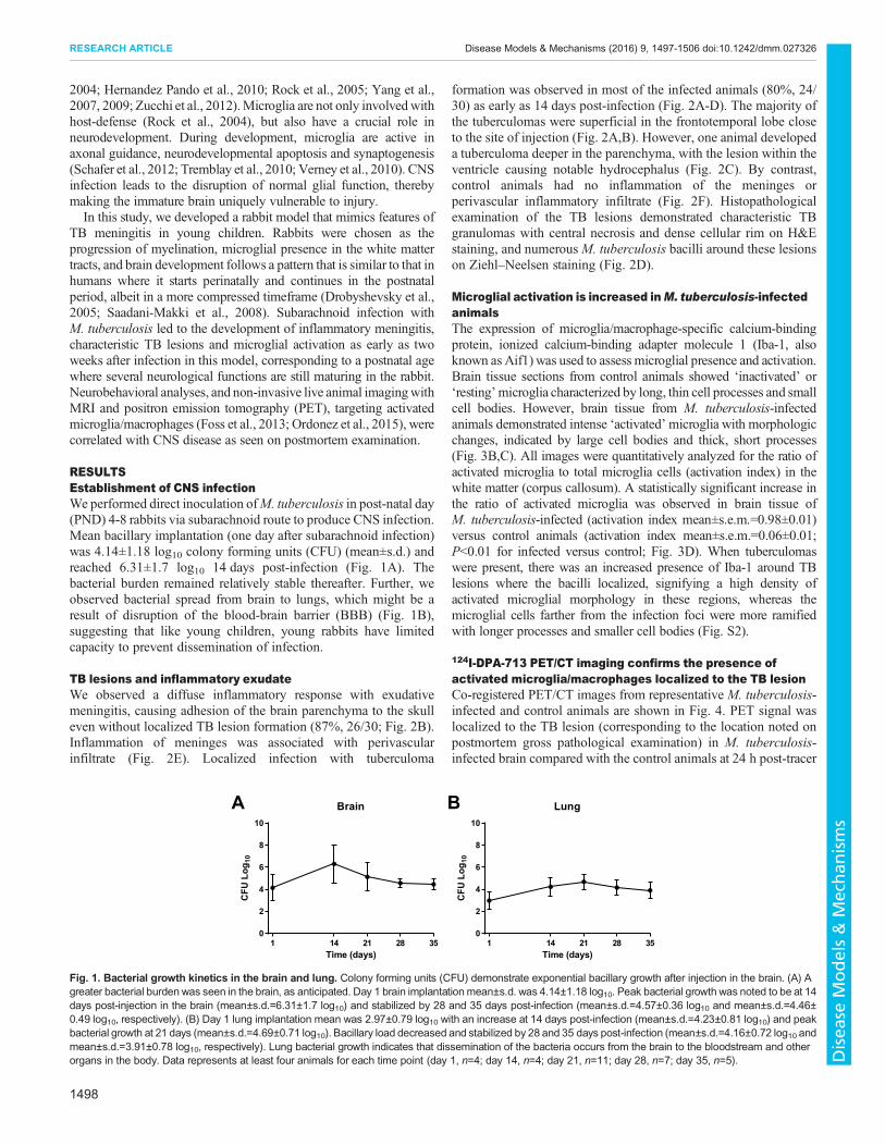
TB lesions and inflammatory exudateWe observed a diffuse inflammatory response with exudativemeningitis, causing adhesion of the brain parenchyma to the skulleven without localized TB lesion formation (87%, 26/30; Fig. 2B).Inflammation of meninges was associated with perivascularinfiltrate (Fig. 2E). Localized infection with tuberculoma
formation was observed in most of the infected animals (80%, 24/30) as early as 14 days post-infection (Fig. 2A-D). The majority ofthe tuberculomas were superficial in the frontotemporal lobe closeto the site of injection (Fig. 2A,B). However, one animal developeda tuberculoma deeper in the parenchyma, with the lesion within theventricle causing notable hydrocephalus (Fig. 2C). By contrast,control animals had no inflammation of the meninges orperivascular inflammatory infiltrate (Fig. 2F). Histopathologicalexamination of the TB lesions demonstrated characteristic TBgranulomas with central necrosis and dense cellular rim on H&Estaining, and numerousM. tuberculosis bacilli around these lesionson Ziehl–Neelsen staining (Fig. 2D).
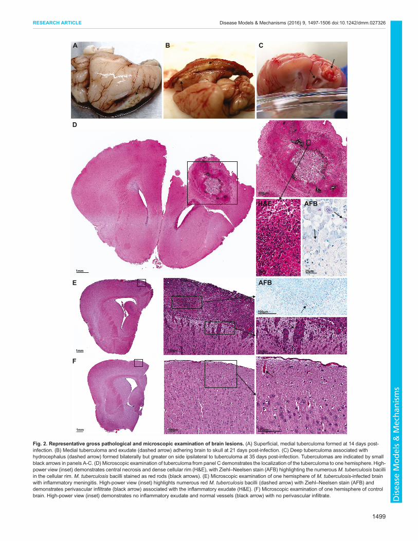
Microglial activation is increased inM. tuberculosis-infectedanimalsThe expression of microglia/macrophage-specific calcium-bindingprotein, ionized calcium-binding adapter molecule 1 (Iba-1, alsoknown as Aif1) was used to assess microglial presence and activation.Brain tissue sections from control animals showed ‘inactivated’ or‘resting’microglia characterized by long, thin cell processes and smallcell bodies. However, brain tissue from M. tuberculosis-infectedanimals demonstrated intense ‘activated’microglia with morphologicchanges, indicated by large cell bodies and thick, short processes(Fig. 3B,C). All images were quantitatively analyzed for the ratio ofactivated microglia to total microglia cells (activation index) in thewhite matter (corpus callosum). A statistically significant increase inthe ratio of activated microglia was observed in brain tissue ofM. tuberculosis-infected (activation index mean±s.e.m.=0.98±0.01)versus control animals (activation index mean±s.e.m.=0.06±0.01;P<0.01 for infected versus control; Fig. 3D). When tuberculomaswere present, there was an increased presence of Iba-1 around TBlesions where the bacilli localized, signifying a high density ofactivated microglial morphology in these regions, whereas themicroglial cells farther from the infection foci were more ramifiedwith longer processes and smaller cell bodies (Fig. S2).
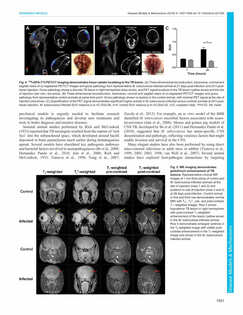
124I-DPA-713 PET/CT imaging confirms the presence ofactivated microglia/macrophages localized to the TB lesionCo-registered PET/CT images from representative M. tuberculosis-infected and control animals are shown in Fig. 4. PET signal waslocalized to the TB lesion (corresponding to the location noted onpostmortem gross pathological examination) in M. tuberculosis-infected brain compared with the control animals at 24 h post-tracer
0
2
4
6
8
10
Brain
Time (days)
CFU
Log 1
0
1 14 21 28 350
2
4
6
8
10
Lung
Time (days)
CFU
Log 1
0
1 14 21 28 35
A B
Fig. 1. Bacterial growth kinetics in the brain and lung. Colony forming units (CFU) demonstrate exponential bacillary growth after injection in the brain. (A) Agreater bacterial burden was seen in the brain, as anticipated. Day 1 brain implantationmean±s.d. was 4.14±1.18 log10. Peak bacterial growth was noted to be at 14days post-injection in the brain (mean±s.d.=6.31±1.7 log10) and stabilized by 28 and 35 days post-infection (mean±s.d.=4.57±0.36 log10 and mean±s.d.=4.46±0.49 log10, respectively). (B) Day 1 lung implantation mean was 2.97±0.79 log10 with an increase at 14 days post-infection (mean±s.d.=4.23±0.81 log10) and peakbacterial growth at 21 days (mean±s.d.=4.69±0.71 log10). Bacillary load decreased and stabilized by 28 and 35 days post-infection (mean±s.d.=4.16±0.72 log10 andmean±s.d.=3.91±0.78 log10, respectively). Lung bacterial growth indicates that dissemination of the bacteria occurs from the brain to the bloodstream and otherorgans in the body. Data represents at least four animals for each time point (day 1, n=4; day 14, n=4; day 21, n=11; day 28, n=7; day 35, n=5).
1498
RESEARCH ARTICLE Disease Models & Mechanisms (2016) 9, 1497-1506 doi:10.1242/dmm.027326
Disea
seModels&Mechan
isms
600μm
1mm 25μm 25μm
A B C
D
H&E AFB
1mm
1mm
100μm
100μm
100μm
100μm
100μm
AFBE
F
Fig. 2. Representative gross pathological and microscopic examination of brain lesions. (A) Superficial, medial tuberculoma formed at 14 days post-infection. (B) Medial tuberculoma and exudate (dashed arrow) adhering brain to skull at 21 days post-infection. (C) Deep tuberculoma associated withhydrocephalus (dashed arrow) formed bilaterally but greater on side ipsilateral to tuberculoma at 35 days post-infection. Tuberculomas are indicated by smallblack arrows in panels A-C. (D) Microscopic examination of tuberculoma from panel C demonstrates the localization of the tuberculoma to one hemisphere. High-power view (inset) demonstrates central necrosis and dense cellular rim (H&E), with Ziehl–Neelsen stain (AFB) highlighting the numerousM. tuberculosis bacilliin the cellular rim. M. tuberculosis bacilli stained as red rods (black arrows). (E) Microscopic examination of one hemisphere of M. tuberculosis-infected brainwith inflammatory meningitis. High-power view (inset) highlights numerous red M. tuberculosis bacilli (dashed arrow) with Ziehl–Neelsen stain (AFB) anddemonstrates perivascular infiltrate (black arrow) associated with the inflammatory exudate (H&E). (F) Microscopic examination of one hemisphere of controlbrain. High-power view (inset) demonstrates no inflammatory exudate and normal vessels (black arrow) with no perivascular infiltrate.
1499
RESEARCH ARTICLE Disease Models & Mechanisms (2016) 9, 1497-1506 doi:10.1242/dmm.027326
Disea
seModels&Mechan
isms
injection [M. tuberculosis-infected standardized uptake value(SUV) mean±s.e.m.=0.70±0.09, control SUV mean±s.e.m.=0.25±0.02; P=0.03; Fig. 4C]. 3D reconstruction videos are available(Movies 1 and 2).
MR imaging demonstrates gadolinium enhancement of TBlesionsRepresentative MR imaging of M. tuberculosis-infected andcontrol animals 28 days post-infection are shown in Fig. 5.
Imaging of M. tuberculosis-infected animals showed post-gadolinium enhancement of the tuberculomas on T1-weightedimages and T2-weighted images demonstrated enlarged ventricleswith enhancement ipsilateral to the tuberculoma. The post-gadolinium enhancement of TB lesions seen in this model issimilar to enhancement of tuberculomas in individuals with CNSTB (Katti, 2004; Rohlwink et al., 2016b).
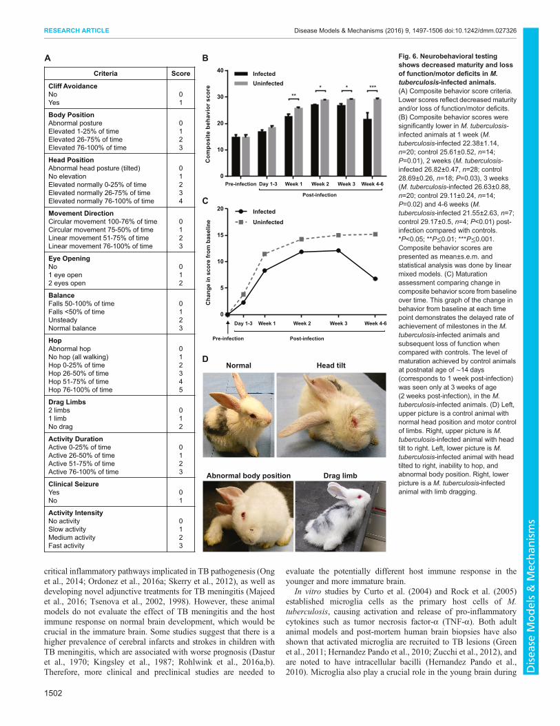
Neurobehavioral testing shows decreased maturity and lossof function and/or motor deficits in M. tuberculosis-infectedanimalsNeurobehavioral scores were compared between the control andM. tuberculosis-infected groups over time. At time 0 (pre-infection)there were no differences between the groups. However, neuro-monitoring over time demonstrated that theM. tuberculosis-infectedanimals developed clinically apparent neurological deficits as earlyas 2 weeks post-infection, with abnormal gait, seizures, head tilt andmotor deficits of the hind limbs (Fig. 6D). Representative videos areshown in Movie 3. The composite behavior score reflects bothdelays in normal development and progressive loss of function.Newborn animals are normally not able to hop and have their eyesclosed and their motor function improves as they mature. However,in the presence of infection there is delayed achievement of normalmilestones such as eye opening, and loss of motor functions. As theinfection progressed, animals began to drag their hind limbs,hopped less frequently or lost normal head and body elevation,leading to a decrease in the scores. A significant decrease in scoreswas seen for the M. tuberculosis-infected animals when comparedwith the control animals by 7 days post-infection, and thisdifference worsened over time (Fig. 6B). A delay in maturationwas seen in the juvenile rabbits with TB meningitis when comparedwith the controls (Fig. 6C). We observed a variation in phenotypicseverity of the M. tuberculosis-infected animals, with severe signsdeveloping in 23% (6/26) animals that developedmeningitis and TBlesions on gross pathologic examination.
DISCUSSIONThe burden of TB in children remains high, and the World HealthOrganization (WHO) estimates 1 million new cases of TB inchildren annually (World Health Organization, 2016). This is asubstantial increase from the prior WHO (2014) estimation, andrepresents the challenges of diagnosing TB in children, which israrely bacteriologically confirmed (Salazar-Austin et al., 2015). TBmeningitis continues to be associated with high morbidity andmortality in young children. Therefore, the development of relevant
Corpus callosum Coronaradiata
Corpus callosum
Coronaradiata
Lateralventricle
Late
ral
vent
ricle
A
Tuberculoma
250μm 250μm
25μm 25μm
Infected Uninfected
0.0
0.5
1.0
1.5
Act
ivat
ed m
icro
glia
/Tot
al m
icro
glia
Lateralventricle
Corpus callosum
Corona radiata
Tuberculomas
C
B
D
**
Fig. 3. Microglia are activated in TB meningitis. (A) Schematicrepresentation of rabbit brain section highlighting lateral ventricles, corpuscallosum, corona radiata and tuberculoma (red dashed line) seen at highermagnification in brain tissue slices in B. (B-C) Representative Iba-1-stainedbrain tissue sections from TB-infected (B,C, left panels) and control (B,C, rightpanels) animals at 21 days post-infection (PND 3-4 weeks) shown under low(B) and high (C) magnification. The low-magnification images (B) show thelateral ventricles (yellow dashed line), corpus callosum, corona radiata andtuberculoma (red dashed line). The high-magnification images (C) are from theregion of the corpus callosum. M. tuberculosis-infected animals (B,C, leftpanels) demonstrates dense, activated microglia with large cell bodies andshortened and/or thickened processes (red arrow) whereas microglia in age-matched controls demonstrate a ‘normal’, ‘resting’ morphology with long, thinprocesses and small cell bodies (red arrow). (D) Quantitative analyses in thecorpus callosum demonstrates a significant increase in percentage ofactivated microglia in M. tuberculosis-infected rabbit kits compared withcontrols. M. tuberculosis-infected mean±s.e.m.=0.98±0.01, n=7; controlmean±s.e.m.=0.06±0.01, n=5; unpaired t-test, **P≤0.01.
1500
RESEARCH ARTICLE Disease Models & Mechanisms (2016) 9, 1497-1506 doi:10.1242/dmm.027326
Disea
seModels&Mechan
isms
preclinical models is urgently needed to facilitate researchinvestigating its pathogenesis and develop new treatments andtools to better diagnose and monitor diseases.Seminal animal studies performed by Rich and McCordock
(1933) reported that TBmeningitis resulted from the rupture of ‘richfoci’ into the subarachnoid space, which developed around bacillideposited in brain parenchyma much earlier during hematogenousspread. Several models have elucidated key pathogenic pathwaysand bacterial factors involved in neuropathogenesis (Be et al., 2008;Hernandez Pando et al., 2010; Jain et al., 2006; Rich andMcCordock, 1933; Tsenova et al., 1998; Yang et al., 2007;
Zucchi et al., 2012). For example, an in vitro model of the BBBidentified M. tuberculosis microbial factors associated with neuro-invasiveness (Jain et al., 2006). Mouse and guinea pig models ofCNS TB, developed by Be et al. (2011) and Hernandez Pando et al.(2010), suggested that M. tuberculosis has strain-specific CNSdissemination and pathology, reflecting virulence factors that mightenable invasion and survival in the CNS.
Many elegant studies have also been performed by using directintra-cisternal infections in adult mice or rabbits (Tsenova et al.,1999, 2005, 2002, 1998; van Well et al., 2007). Several animalstudies have explored host-pathogen interactions by targeting
C
1 240.0
0.5
1.0
1.5
Time (hours)
SUV
*
Fig. 4. 124I-DPA-713 PET/CT imaging demonstrates tracer uptake localizing to the TB lesion. (A) Three-dimensional reconstruction, transverse, coronal andsagittal views of co-registered PET/CT images and gross pathology from representativeM. tuberculosis-infected animal at 21 days post-infection and 24 h post-tracer injection. Gross pathology shows a discrete TB lesion in right hemisphere (blue arrow), and PET signal localizes to the TB lesion (yellow arrow) and the siteof injection (ear vein, red arrow). (B) Three-dimensional reconstruction, transverse, coronal and sagittal views of co-registered PET/CT images and grosspathology from representative control animals at same time point. Gross pathology shows no lesions in the control animal, with minimal PET signal at the site ofinjection (red arrow). (C) Quantification of the PET signal demonstrates significant higher activity inM. tuberculosis-infected versus controls animals at 24 h post-tracer injection. M. tuberculosis-infected SUV mean±s.e.m.=0.70±0.09, n=4; control SUV mean±s.e.m.=0.25±0.02, n=2; unpaired t-test, *P=0.03. He, heart.
Fig. 5. MR imaging demonstratesgadolinium enhancement of TBlesions. Representative coronal MRimages of 1 mm thick slices of control andM. tuberculosis-infected animals at thesite of injection (rows 1 and 2) andposterior to site of injection (rows 3 and 4)at 28 days post-infection. Control animalin first and third row demonstrates normalMRI with T2-, T2*-, pre- and post-contrastT1- weighted images. Row 2 showshypodense TB lesion in right hemispherewith post-contrast T1-weightedenhancement of the lesion (yellow arrow)in the M. tuberculosis-infected animal.Row 4 demonstrates enlarged ventricle inthe T2-weighted image with visible post-contrast enhancement in the T1-weightedimage (red arrow) in the M. tuberculosis-infected animal.
1501
RESEARCH ARTICLE Disease Models & Mechanisms (2016) 9, 1497-1506 doi:10.1242/dmm.027326
Disea
seModels&Mechan
isms
critical inflammatory pathways implicated in TB pathogenesis (Onget al., 2014; Ordonez et al., 2016a; Skerry et al., 2012), as well asdeveloping novel adjunctive treatments for TB meningitis (Majeedet al., 2016; Tsenova et al., 2002, 1998). However, these animalmodels do not evaluate the effect of TB meningitis and the hostimmune response on normal brain development, which would becrucial in the immature brain. Some studies suggest that there is ahigher prevalence of cerebral infarcts and strokes in children withTB meningitis, which are associated with worse prognosis (Dasturet al., 1970; Kingsley et al., 1987; Rohlwink et al., 2016a,b).Therefore, more clinical and preclinical studies are needed to
evaluate the potentially different host immune response in theyounger and more immature brain.
In vitro studies by Curto et al. (2004) and Rock et al. (2005)established microglia cells as the primary host cells of M.tuberculosis, causing activation and release of pro-inflammatorycytokines such as tumor necrosis factor-α (TNF-α). Both adultanimal models and post-mortem human brain biopsies have alsoshown that activated microglia are recruited to TB lesions (Greenet al., 2011; Hernandez Pando et al., 2010; Zucchi et al., 2012), andare noted to have intracellular bacilli (Hernandez Pando et al.,2010). Microglia also play a crucial role in the young brain during
A B
C
DNormal Head tilt
Abnormal body position Drag limb
Criteria Score
Cliff AvoidanceNoYes
01
Body PositionAbnormal postureElevated 1-25% of timeElevated 26-75% of time Elevated 76-100% of time
0123
Head PositionAbnormal head posture (tilted)No elevationElevated normally 0-25% of timeElevated normally 26-75% of timeElevated normally 76-100% of time
01234
Movement DirectionCircular movement 100-76% of timeCircular movement 75-50% of timeLinear movement 51-75% of timeLinear movement 76-100% of time
0123
Eye OpeningNo1 eye open2 eyes open
012
BalanceFalls 50-100% of timeFalls <50% of timeUnsteadyNormal balance
0123
HopAbnormal hopNo hop (all walking)Hop 0-25% of timeHop 26-50% of timeHop 51-75% of timeHop 76-100% of time
012345
Drag Limbs2 limbs1 limbNo drag
012
Activity DurationActive 0-25% of timeActive 26-50% of timeActive 51-75% of timeActive 76-100% of time
0123
Clinical SeizureYesNo
01
Activity IntensityNo activitySlow activityMedium activityFast activity
0123
Pre-infection
Post-infection
Day 1-3 Week 1 Week 2 Week 3 Week 4-6
Pre-infection Post-infection
Day 1-3 Week 1 Week 2 Week 3 Week 4-60
5
10
15
20 Infected
Uninfected
Cha
nge
in s
core
from
bas
elin
e
0
10
20
30
40 InfectedUninfected
Com
posi
te b
ehav
ior
scor
e
****** *
Fig. 6. Neurobehavioral testingshows decreased maturity and lossof function/motor deficits in M.tuberculosis-infected animals.(A) Composite behavior score criteria.Lower scores reflect decreased maturityand/or loss of function/motor deficits.(B) Composite behavior scores weresignificantly lower in M. tuberculosis-infected animals at 1 week (M.tuberculosis-infected 22.38±1.14,n=20; control 25.61±0.52, n=14;P=0.01), 2 weeks (M. tuberculosis-infected 26.82±0.47, n=28; control28.69±0.26, n=18; P=0.03), 3 weeks(M. tuberculosis-infected 26.63±0.88,n=20; control 29.11±0.24, n=14;P=0.02) and 4-6 weeks (M.tuberculosis-infected 21.55±2.63, n=7;control 29.17±0.5, n=4; P<0.01) post-infection compared with controls.*P<0.05; **P≤0.01; ***P≤0.001.Composite behavior scores arepresented as mean±s.e.m. andstatistical analysis was done by linearmixed models. (C) Maturationassessment comparing change incomposite behavior score from baselineover time. This graph of the change inbehavior from baseline at each timepoint demonstrates the delayed rate ofachievement of milestones in the M.tuberculosis-infected animals andsubsequent loss of function whencompared with controls. The level ofmaturation achieved by control animalsat postnatal age of ∼14 days(corresponds to 1 week post-infection)was seen only at 3 weeks of age(2 weeks post-infection), in the M.tuberculosis-infected animals. (D) Left,upper picture is a control animal withnormal head position and motor controlof limbs. Right, upper picture is M.tuberculosis-infected animal with headtilt to right. Left, lower picture is M.tuberculosis-infected animal with headtilted to right, inability to hop, andabnormal body position. Right, lowerpicture is a M. tuberculosis-infectedanimal with limb dragging.
1502
RESEARCH ARTICLE Disease Models & Mechanisms (2016) 9, 1497-1506 doi:10.1242/dmm.027326
Disea
seModels&Mechan
isms

normal development. Microglia are essential for axonal andsynaptic plasticity through axonal guidance, neurodevelopmentalapoptosis, neurogenesis and synaptogenesis, making them vital fornormal development (Cunningham et al., 2013; Schafer et al., 2012;Tremblay et al., 2010; Verney et al., 2010). Therefore, we focusedon developing a model that mimics TB meningitis in toddler-agechildren. The clinical relevance of this model lies in the ability tostudy the microglial response during active white matterdevelopment and myelination mimicking that in children. To date,corticosteroids are the only host-directed therapy proven to decreasemortality and morbidity in children with TB meningitis (Girgiset al., 1991; Prasad and Singh, 2008; Schoeman et al., 1997).Therefore, a better understanding of the role of immune cells inthe developing brain involved in TB meningitis is imperative fordeveloping novel therapeutic strategies that would improveoutcomes while facilitating normal brain development.Subarachnoid infection in our model led to inflammatory
meningitis and formation of characteristic brain tuberculomaswith central necrosis and dense cellular rim in the majority ofM. tuberculosis-infected animals. Diffuse exudative meningitis andhydrocephalus seen on gross pathological examination, as well asMRI with gadolinium enhancement, akin to human disease, werealso noted. Histologically, microglia were robustly activated byM. tuberculosis infection, confirming the integral involvement ofmicroglia described previously by Curto et al.’s (2004) and Rocket al.’s (2005) in vitro studies and in other animal models byHernandez Pando et al. (2010) and Zucchi et al. (2012). In order tonon-invasively monitor microglial inflammation in our model, weused 124I-DPA-713, a newer generation of TSPO ligand, withexcellent signal-to-noise ratios for the detection of activatedmicroglia/macrophages associated with M. tuberculosis infection(Foss et al., 2013; Ordonez et al., 2015). In our model, 124I-DPA-713 PET/CT imaging demonstrated signal localized to the TBlesion, with low background signal in control animals. Detectingmicroglial activation using non-invasive PET in vivo imaging couldpotentially be used as a biomarker for early diagnosis and treatmentmonitoring. The extent of microglial activation using PET imaginghas been shown to correlate with the severity of neurologic injury ina neonatal rabbit model of cerebral palsy (Kannan et al., 2011, 2007)and might also be helpful as a prognostic indicator. First-in-humanstudies using 124I-DPA-713 are currently ongoing. Pathogen-specific imaging modalities to specifically detect bacteria directlycould also substantially enhance the diagnostic and monitoringcapabilities (Ordonez et al., 2016b; Weinstein et al., 2014) andcould be tested in this model in future studies. MRI could also beutilized to monitor response to treatment or predict outcomes.A recent prospective pediatric study by Rohlwink et al. (2016b)demonstrated that MRI evidence of severe infarcts from eithervascular or non-vascular pathology, such as intracranialhypertension or hydrocephalus, was predictive of poor outcomes.Although we did not identify infarcts in our model at the time ofimaging, enlarged ventricles were detected on MRI. It is possiblethat infarcts could be visualized at later time points with diseaseprogression.There was a wide variability in the neurobehavioral scores
correlative of disease severity in M. tuberculosis-infected rabbits,with worsening signs as the infection progressed. The differences incomposite behavior scores noted at the early time points primarilycorrelates with delays in normal developmental milestones that areindicative of the early signs of infection in these animals.Retrospective studies in children with TB meningitis found thatchildren were more likely to present with advanced disease (stage II
with lethargy, nuchal rigidity, seizures and focal neurological signsoften with cranial nerve abnormalities or stage III with hemiplegiaor paraplegia, coma and eventual death) and only 3-10% presentwith mild disease (stage I with fever, headache and loss ofdevelopmental milestones) (Long et al., 2012; vanWell et al., 2009;Yaramis et al., 1998). Our animal model could enable furthercharacterization of pediatric TB meningitis over time to betterdelineate the pathogenesis when the majority of children presentwith advanced disease.
The current study has some limitations. The developmental timeframe is much more compressed in rabbits when compared withhumans. Therefore, in order to study the pathogenesis and themicroglial responses within this compressed developmental timeframe, a larger inoculum than would typically be seen in pediatricCNS TB was used to study the progression of the disease. Webelieve that the timing of the initial insult at a crucial developmentalperiod is important. The younger age of the rabbits and the highinoculum could also explain why M. tuberculosis strain H37Rv,which is typically considered less virulent than the Beijing strains,produced robust disease phenotype in the current study. Futureexperiments with other M. tuberculosis strains, such as from theBeijing family, could elucidate the role of bacterial factors in thepathogenesis of TB meningitis in a developing brain. Finally, directinoculation with M. tuberculosis into the subarachnoid space, asutilized in the current study, does not mimic the natural route ofaerosol infection with secondary hematogenous infection.However, this allows for greater reproducibility and inducesdisease with characteristics similar to humans, enabling furtherinvestigation of the host immune response.
ConclusionsIn summary, we have established a reproducible and clinicallyrelevant rabbit model that mimics key features of TB meningitis inyoung children, who are also disproportionately affected by thisform of TB. Future studies will focus on elucidating the role of glialactivation in TB and its effects on white matter development andneuronal injury, mechanisms of paradoxical reactions to treatments,novel antimicrobial treatment regimens including host-directedtherapies (especially those targeting microglia), and non-invasiveimaging to follow progression of disease and response to treatment.
MATERIALS AND METHODSAll protocols were approved by the Johns Hopkins University (JHU)Biosafety, Radiation Safety, and Animal Care and Use Committeesaccording to the National Institutes of Health guide for the care and useof laboratory animals.
Bacterial strainsAll bacterial stocks were obtained from the laboratory of S.K.J.Logarithmically growing or frozen, titrated stocks of M. tuberculosisH37Rv were used as described previously (Harper et al., 2012; Ordonezet al., 2015, 2016a). Prior to infection, the bacterial suspension was washedand re-suspended in phosphate buffered saline (PBS).
Animal infectionsFreshly prepared M. tuberculosis suspension was inoculated into thesubarachnoid space of male and female New Zealand White rabbits(Robinson Services Inc., Mocksville, NC) at postnatal day (PND) 4-8. Priorto injection, topical anesthesia (lidocaine; Ferndale IP Inc., Ferndale, MI)was applied and dexmedetomidine hydrochloride (0.2 µg g−1; Zoetis,Florham Park, NJ) was provided for sedation. The rabbit was restrained on aboard enabling stabilization of the head, and the bregma was palpated andmarked. A 28-gauge insulin syringe was used to inject 20 µl of bacterialsuspension (over 20 min) into the subarachnoid space via the bregma by
1503
RESEARCH ARTICLE Disease Models & Mechanisms (2016) 9, 1497-1506 doi:10.1242/dmm.027326
Disea
seModels&Mechan
isms
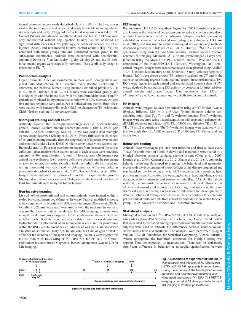
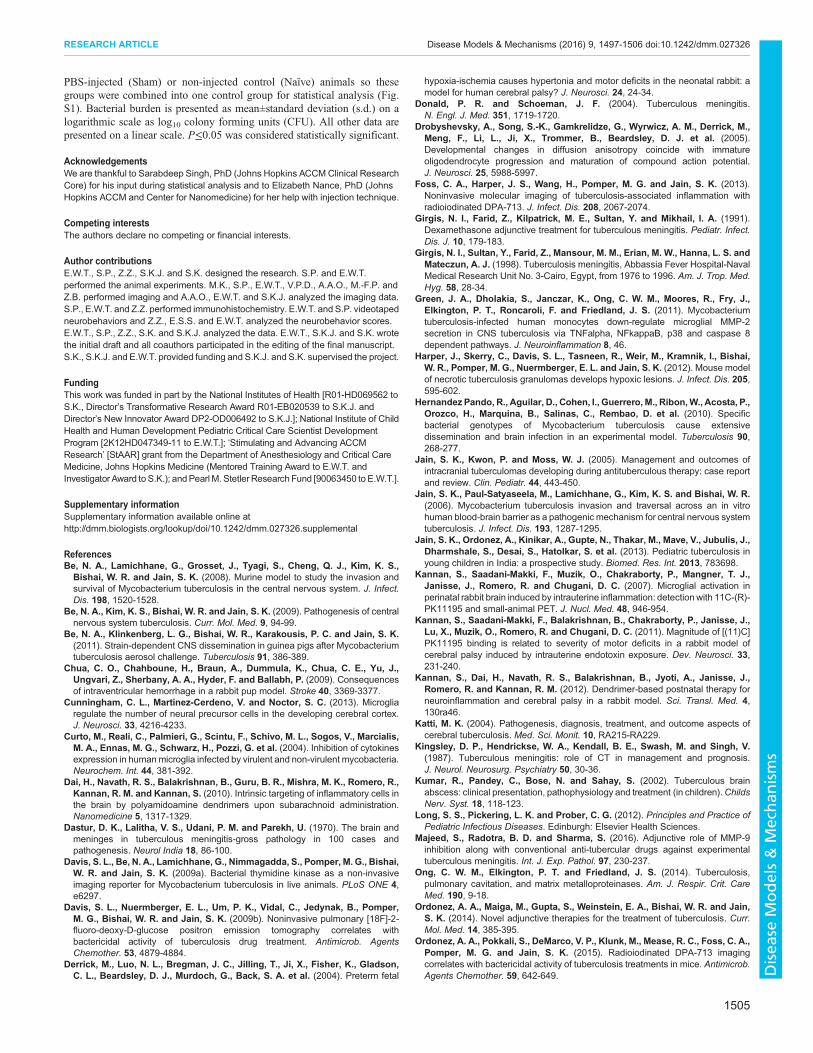
trained personnel as previously described (Dai et al., 2010). The bregmawasused as the injection site as it is open and easily accessible in young rabbits.Average optical density (OD600) of the bacterial suspension was 1.42±0.21.Control (Sham) animals were anesthetized and injected with PBS or wereonly anesthetized without any injection (Naïve). As we observed nosignificant differences in CNS pathology or behavior between the PBS-injected (Sham) and non-injected (Naïve) control animals (Fig. S1), wecombined both these groups into one uninfected control group in thesubsequent experiments. Animals were euthanized with pentobarbitalsodium (120 mg kg−1) at day 1, day 14, day 21, day 28 and day 35 post-infection and organs were aseptically harvested. The overall study design ispresented in Fig. 7.
Postmortem analysisOrgans from M. tuberculosis-infected animals were homogenized andplated onto Middlebrook 7H11 selective plates (Becton Dickinson) toenumerate the bacterial burden using methods described previously (Beet al., 2008; Ordonez et al., 2015). Brains were examined grossly andhistologically with specimens fixed with 4% paraformaldehyde and paraffinembedded or placed in cryoprotective solution with 30% sucrose. At leastfive animals per group were euthanized at indicated time-points. Brain sliceswere stained with hematoxylin-eosin (H&E) to characterize TB lesions andZiehl–Neelsen staining for mycobacterium.
Microglial staining and cell countAntibody against the microglia/macrophage-specific calcium-bindingprotein, ionized calcium-binding adapter molecule 1 (Iba-1, 1:500 goatanti-Iba-1, Abcam, Cambridge, MA, ab107159) was used to stain microgliaas previously described (Zhang et al., 2015). Every fifth section (thicknessof 15 µm) extending caudally from the bregma (site of injection) was stainedand examined under a Leica DM2500microscope (LeicaMicrosystems Inc.,Bannockburn, IL). Four non-overlapping images from the area of the corpuscallosum (representative white matter region) in each section were randomlycaptured at 40× magnification. A total of five sections (20 images) peranimal were evaluated. Iba-1-positive cells were counted and the percentageof activated microglia (bushy, round) to total microglial cells [activated andresting (ramified)] was calculated to determine an activation index aspreviously described (Kannan et al., 2007; Saadani-Makki et al., 2009).Images were analyzed by personnel blinded to experimental groups.Microglial activation was examined 21 days post-infection and data from atleast five animals were analyzed for each group.
Non-invasive imagingLive M. tuberculosis-infected and control animals were imaged within asealed bio-containment bed (Minerve, Esternay, France) modified in-houseto be compliant with biosafety-3 (BSL-3) containment (Davis et al., 2009a,b). Filters (0.22 µm, Whatman) were used at both the inlet and the outlet tocontain the bacteria within the device. For MR imaging, animals wereimaged inside in-house-designed BSL-3 containment devices with nometallic parts. Rabbits were initially sedated with dexmedetomidinehydrochloride for placement of an intravenous access, and for positioningwithin the BSL-3 containment device. Anesthesia was then maintained witha mixture of isoflurane (Henry Schein, Melville, NY) and oxygen titrated toeffect for the duration of transport and imaging. Animals were injected viathe ear vein with 16.24 MBq of 124I-DPA-713 for PET/CT or 5 mmolgadolinium-based contrast (Magnevist, Berlex Laboratories,Wayne, NJ) forMR imaging.
PET imagingRadioiodinated DPA-713, a synthetic ligand for TSPO (translocator proteinalso known as the peripheral benzodiazepine receptor), which is upregulatedon mitochondria in activated microglia/macrophages, has been previouslyvalidated as a marker of activated macrophages in pulmonary TB (Fosset al., 2013) and was used to monitor microglial activation using methodsdescribed previously (Ordonez et al., 2015). Briefly, 124I-DPA-713 wassynthesized using current Good Manufacturing Practices under a researchcontract (3D Imaging, Maumelle, AR). Animals were imaged 21 days post-infection using the Mosaic HP PET (Philips, Bothell, WA) and the CTcomponent of the NanoSPECT/CT (Bioscan, Washington, DC) smallanimal imagers. Images were reconstructed and co-registered using AMIDE1.0.4 (http://amide.sourceforge.net). Spherical (8 mm3 volume) regions ofinterest (ROI) were drawn around TB lesions visualized on CT and in thesame corresponding region (frontal-parietal region) in control animals. TwoROIs were drawn for each animal and standardized uptake values (SUV)were calculated by normalizing ROI activity by correcting for injected dose,animal weight and tracer decay. Data represents four ROIs ofM. tuberculosis-infected animals and two ROIs from a control animal.
MR imagingAnimals were imaged 28 days post-infection using a 9.4T Bruker Avance(Bruker, Billerica, MA) with a Bruker 70 mm diameter volume coil,acquiring multi-slice T2-, T2*- and T1-weighted images. The T2-weightedimages were acquired using a rapid acquisition with relaxation enhancement(RARE) sequence (rare factor of 8, TR of 5000 ms, effective TE of 32 ms,TE of 8 ms, 2 acquisitions). The T2*-weighted images were acquired with afast low angle shot (FLASH) sequence (TR of 500 ms, TE of 6 ms, and oneacquisition).
Behavioral testingAnimals were videotaped pre- and post-infection and then at least every7 days for a minimum of 5 min. Behavior and maturation were scored by ablinded observer using a validated scoring system (Chua et al., 2009;Derrick et al., 2004; Kannan et al., 2012; Zhang et al., 2015). A compositebehavior score was developed to combine the behavioral and maturationscores with the development of motor deficits as the disease progressed andwas based on the following criteria: cliff avoidance, body position, headposition, movement direction, eye opening, balance, hop, limb drag, activityduration, activity intensity and seizure activity (Fig. 6A). As the rabbitsmatured, the composite behavior score increased to its peak. However, asM. tuberculosis-infected animals developed signs of infection, the scoredecreased again, reflecting a regression of milestones and development ofdeficits. Behavioral testing ended when animals met criteria for euthanasiaper our animal protocol. Data from at least 18 animals are presented for eachgroup (30 M. tuberculosis-infected and 18 control animals).
Statistical analysisMicroglial activation and 124I-DPA-713 PET/CT SUV data were analyzedusing t-tests (GraphPad Software Inc., La Jolla, CA). Linear mixed modelsthat accounted for variation among repeated measurements over time withinsubjects were used to estimate the differences between neurobehavioralscores across time and treatment. The analyses were performed using Rversion 3.2.2 (R Foundation for Statistical Computing, Vienna, Austria).Where appropriate, the Bonferroni correction for multiple testing wasapplied. Data are expressed as means±s.e.m. There was no statisticallysignificant difference in behavior or microglial quantification between
Birth
10 14 21 28 35
Postnatalday 4-8
Days
Gross pathology and Immunohistochemistry
Bacillary burden and Neurobehavioral testing
124I-DPA-713 PET/CT imagingIn vivo subarachnoid injection
of M. tuberculosis MRIFig. 7. Schematic of experimental timeline. Invivo subarachnoid injection of M. tuberculosisH37Rv at PND 4-8 represents time-point zero.During the experiment, the bacillary burden wasquantified and neurobehavioral testing wasvideotaped and scored. 124I-DPA-713 PET/CTimaging occurred at 21 days post-infection andMR imaging at 28 days post-infection.
1504
RESEARCH ARTICLE Disease Models & Mechanisms (2016) 9, 1497-1506 doi:10.1242/dmm.027326
Disea
seModels&Mechan
isms
PBS-injected (Sham) or non-injected control (Naïve) animals so thesegroups were combined into one control group for statistical analysis (Fig.S1). Bacterial burden is presented as mean±standard deviation (s.d.) on alogarithmic scale as log10 colony forming units (CFU). All other data arepresented on a linear scale. P≤0.05 was considered statistically significant.
AcknowledgementsWe are thankful to Sarabdeep Singh, PhD (Johns Hopkins ACCMClinical ResearchCore) for his input during statistical analysis and to Elizabeth Nance, PhD (JohnsHopkins ACCM and Center for Nanomedicine) for her help with injection technique.
Competing interestsThe authors declare no competing or financial interests.
Author contributionsE.W.T., S.P., Z.Z., S.K.J. and S.K. designed the research. S.P. and E.W.T.performed the animal experiments. M.K., S.P., E.W.T., V.P.D., A.A.O., M.-F.P. andZ.B. performed imaging and A.A.O., E.W.T. and S.K.J. analyzed the imaging data.S.P., E.W.T. and Z.Z. performed immunohistochemistry. E.W.T. and S.P. videotapedneurobehaviors and Z.Z., E.S.S. and E.W.T. analyzed the neurobehavior scores.E.W.T., S.P., Z.Z., S.K. and S.K.J. analyzed the data. E.W.T., S.K.J. and S.K. wrotethe initial draft and all coauthors participated in the editing of the final manuscript.S.K., S.K.J. and E.W.T. provided funding and S.K.J. and S.K. supervised the project.
FundingThis work was funded in part by the National Institutes of Health [R01-HD069562 toS.K., Director’s Transformative Research Award R01-EB020539 to S.K.J. andDirector’s New Innovator Award DP2-OD006492 to S.K.J.]; National Institute of ChildHealth and Human Development Pediatric Critical Care Scientist DevelopmentProgram [2K12HD047349-11 to E.W.T.]; ‘Stimulating and Advancing ACCMResearch’ [StAAR] grant from the Department of Anesthesiology and Critical CareMedicine, Johns Hopkins Medicine (Mentored Training Award to E.W.T. andInvestigator Award to S.K.); andPearlM. Stetler Research Fund [90063450 to E.W.T.].
Supplementary informationSupplementary information available online athttp://dmm.biologists.org/lookup/doi/10.1242/dmm.027326.supplemental
ReferencesBe, N. A., Lamichhane, G., Grosset, J., Tyagi, S., Cheng, Q. J., Kim, K. S.,Bishai, W. R. and Jain, S. K. (2008). Murine model to study the invasion andsurvival of Mycobacterium tuberculosis in the central nervous system. J. Infect.Dis. 198, 1520-1528.
Be, N. A., Kim, K. S., Bishai, W. R. and Jain, S. K. (2009). Pathogenesis of centralnervous system tuberculosis. Curr. Mol. Med. 9, 94-99.
Be, N. A., Klinkenberg, L. G., Bishai, W. R., Karakousis, P. C. and Jain, S. K.(2011). Strain-dependent CNS dissemination in guinea pigs after Mycobacteriumtuberculosis aerosol challenge. Tuberculosis 91, 386-389.
Chua, C. O., Chahboune, H., Braun, A., Dummula, K., Chua, C. E., Yu, J.,Ungvari, Z., Sherbany, A. A., Hyder, F. and Ballabh, P. (2009). Consequencesof intraventricular hemorrhage in a rabbit pup model. Stroke 40, 3369-3377.
Cunningham, C. L., Martinez-Cerdeno, V. and Noctor, S. C. (2013). Microgliaregulate the number of neural precursor cells in the developing cerebral cortex.J. Neurosci. 33, 4216-4233.
Curto, M., Reali, C., Palmieri, G., Scintu, F., Schivo, M. L., Sogos, V., Marcialis,M. A., Ennas, M. G., Schwarz, H., Pozzi, G. et al. (2004). Inhibition of cytokinesexpression in humanmicroglia infected by virulent and non-virulent mycobacteria.Neurochem. Int. 44, 381-392.
Dai, H., Navath, R. S., Balakrishnan, B., Guru, B. R., Mishra, M. K., Romero, R.,Kannan, R. M. and Kannan, S. (2010). Intrinsic targeting of inflammatory cells inthe brain by polyamidoamine dendrimers upon subarachnoid administration.Nanomedicine 5, 1317-1329.
Dastur, D. K., Lalitha, V. S., Udani, P. M. and Parekh, U. (1970). The brain andmeninges in tuberculous meningitis-gross pathology in 100 cases andpathogenesis. Neurol India 18, 86-100.
Davis, S. L., Be, N. A., Lamichhane, G., Nimmagadda, S., Pomper, M. G., Bishai,W. R. and Jain, S. K. (2009a). Bacterial thymidine kinase as a non-invasiveimaging reporter for Mycobacterium tuberculosis in live animals. PLoS ONE 4,e6297.
Davis, S. L., Nuermberger, E. L., Um, P. K., Vidal, C., Jedynak, B., Pomper,M. G., Bishai, W. R. and Jain, S. K. (2009b). Noninvasive pulmonary [18F]-2-fluoro-deoxy-D-glucose positron emission tomography correlates withbactericidal activity of tuberculosis drug treatment. Antimicrob. AgentsChemother. 53, 4879-4884.
Derrick, M., Luo, N. L., Bregman, J. C., Jilling, T., Ji, X., Fisher, K., Gladson,C. L., Beardsley, D. J., Murdoch, G., Back, S. A. et al. (2004). Preterm fetal
hypoxia-ischemia causes hypertonia and motor deficits in the neonatal rabbit: amodel for human cerebral palsy? J. Neurosci. 24, 24-34.
Donald, P. R. and Schoeman, J. F. (2004). Tuberculous meningitis.N. Engl. J. Med. 351, 1719-1720.
Drobyshevsky, A., Song, S.-K., Gamkrelidze, G., Wyrwicz, A. M., Derrick, M.,Meng, F., Li, L., Ji, X., Trommer, B., Beardsley, D. J. et al. (2005).Developmental changes in diffusion anisotropy coincide with immatureoligodendrocyte progression and maturation of compound action potential.J. Neurosci. 25, 5988-5997.
Foss, C. A., Harper, J. S., Wang, H., Pomper, M. G. and Jain, S. K. (2013).Noninvasive molecular imaging of tuberculosis-associated inflammation withradioiodinated DPA-713. J. Infect. Dis. 208, 2067-2074.
Girgis, N. I., Farid, Z., Kilpatrick, M. E., Sultan, Y. and Mikhail, I. A. (1991).Dexamethasone adjunctive treatment for tuberculous meningitis. Pediatr. Infect.Dis. J. 10, 179-183.
Girgis, N. I., Sultan, Y., Farid, Z., Mansour, M. M., Erian, M. W., Hanna, L. S. andMateczun, A. J. (1998). Tuberculosis meningitis, Abbassia Fever Hospital-NavalMedical Research Unit No. 3-Cairo, Egypt, from 1976 to 1996. Am. J. Trop. Med.Hyg. 58, 28-34.
Green, J. A., Dholakia, S., Janczar, K., Ong, C. W. M., Moores, R., Fry, J.,Elkington, P. T., Roncaroli, F. and Friedland, J. S. (2011). Mycobacteriumtuberculosis-infected human monocytes down-regulate microglial MMP-2secretion in CNS tuberculosis via TNFalpha, NFkappaB, p38 and caspase 8dependent pathways. J. Neuroinflammation 8, 46.
Harper, J., Skerry, C., Davis, S. L., Tasneen, R., Weir, M., Kramnik, I., Bishai,W. R., Pomper, M. G., Nuermberger, E. L. and Jain, S. K. (2012). Mouse modelof necrotic tuberculosis granulomas develops hypoxic lesions. J. Infect. Dis. 205,595-602.
Hernandez Pando, R., Aguilar, D., Cohen, I., Guerrero, M., Ribon,W., Acosta, P.,Orozco, H., Marquina, B., Salinas, C., Rembao, D. et al. (2010). Specificbacterial genotypes of Mycobacterium tuberculosis cause extensivedissemination and brain infection in an experimental model. Tuberculosis 90,268-277.
Jain, S. K., Kwon, P. and Moss, W. J. (2005). Management and outcomes ofintracranial tuberculomas developing during antituberculous therapy: case reportand review. Clin. Pediatr. 44, 443-450.
Jain, S. K., Paul-Satyaseela, M., Lamichhane, G., Kim, K. S. and Bishai, W. R.(2006). Mycobacterium tuberculosis invasion and traversal across an in vitrohuman blood-brain barrier as a pathogenic mechanism for central nervous systemtuberculosis. J. Infect. Dis. 193, 1287-1295.
Jain, S. K., Ordonez, A., Kinikar, A., Gupte, N., Thakar, M., Mave, V., Jubulis, J.,Dharmshale, S., Desai, S., Hatolkar, S. et al. (2013). Pediatric tuberculosis inyoung children in India: a prospective study. Biomed. Res. Int. 2013, 783698.
Kannan, S., Saadani-Makki, F., Muzik, O., Chakraborty, P., Mangner, T. J.,Janisse, J., Romero, R. and Chugani, D. C. (2007). Microglial activation inperinatal rabbit brain induced by intrauterine inflammation: detection with 11C-(R)-PK11195 and small-animal PET. J. Nucl. Med. 48, 946-954.
Kannan, S., Saadani-Makki, F., Balakrishnan, B., Chakraborty, P., Janisse, J.,Lu, X., Muzik, O., Romero, R. and Chugani, D. C. (2011). Magnitude of [(11)C]PK11195 binding is related to severity of motor deficits in a rabbit model ofcerebral palsy induced by intrauterine endotoxin exposure. Dev. Neurosci. 33,231-240.
Kannan, S., Dai, H., Navath, R. S., Balakrishnan, B., Jyoti, A., Janisse, J.,Romero, R. and Kannan, R. M. (2012). Dendrimer-based postnatal therapy forneuroinflammation and cerebral palsy in a rabbit model. Sci. Transl. Med. 4,130ra46.
Katti, M. K. (2004). Pathogenesis, diagnosis, treatment, and outcome aspects ofcerebral tuberculosis. Med. Sci. Monit. 10, RA215-RA229.
Kingsley, D. P., Hendrickse, W. A., Kendall, B. E., Swash, M. and Singh, V.(1987). Tuberculous meningitis: role of CT in management and prognosis.J. Neurol. Neurosurg. Psychiatry 50, 30-36.
Kumar, R., Pandey, C., Bose, N. and Sahay, S. (2002). Tuberculous brainabscess: clinical presentation, pathophysiology and treatment (in children).ChildsNerv. Syst. 18, 118-123.
Long, S. S., Pickering, L. K. and Prober, C. G. (2012). Principles and Practice ofPediatric Infectious Diseases. Edinburgh: Elsevier Health Sciences.
Majeed, S., Radotra, B. D. and Sharma, S. (2016). Adjunctive role of MMP-9inhibition along with conventional anti-tubercular drugs against experimentaltuberculous meningitis. Int. J. Exp. Pathol. 97, 230-237.
Ong, C. W. M., Elkington, P. T. and Friedland, J. S. (2014). Tuberculosis,pulmonary cavitation, and matrix metalloproteinases. Am. J. Respir. Crit. CareMed. 190, 9-18.
Ordonez, A. A., Maiga, M., Gupta, S., Weinstein, E. A., Bishai, W. R. and Jain,S. K. (2014). Novel adjunctive therapies for the treatment of tuberculosis. Curr.Mol. Med. 14, 385-395.
Ordonez, A. A., Pokkali, S., DeMarco, V. P., Klunk, M., Mease, R. C., Foss, C. A.,Pomper, M. G. and Jain, S. K. (2015). Radioiodinated DPA-713 imagingcorrelates with bactericidal activity of tuberculosis treatments in mice. Antimicrob.Agents Chemother. 59, 642-649.
1505
RESEARCH ARTICLE Disease Models & Mechanisms (2016) 9, 1497-1506 doi:10.1242/dmm.027326
Disea
seModels&Mechan
isms
Ordonez, A. A., Tasneen, R., Pokkali, S., Xu, Z., Converse, P. J., Klunk, M. H.,Mollura, D. J., Nuermberger, E. L. and Jain, S. K. (2016a). Mouse model ofpulmonary cavitary tuberculosis and expression of matrix metalloproteinase-9.Dis. Models Mech. 9, 779-788.
Ordonez, A. A., Weinstein, E. A., Bambarger, L. E., Saini, V., Chang, Y. S.,DeMarco, V. P., Klunk, M. H., Urbanowski, M. E., Moulton, K. L., Murawski,A. M. et al. (2016b). A systematic approach for developing bacteria-specificimaging tracers. J. Nucl. Med. [Epub ahead of print], doi:10.2967/jnumed.116.181792..
Prasad, K. and Singh, M. B. (2008). Corticosteroids for managing tuberculousmeningitis. Cochrane Database Syst. Rev. 4, CD002244.
Rich, A. R. and McCordock, H. A. (1933). The pathogenesis of tuberculosismeningitis. Bull. Johns Hopkins Hosp. 52, 5-37.
Rock, R. B., Gekker, G., Hu, S., Sheng, W. S., Cheeran, M., Lokensgard, J. R.and Peterson, P. K. (2004). Role of microglia in central nervous systeminfections. Clin. Microbiol. Rev. 17, 942-964.
Rock, R. B., Hu, S., Gekker, G., Sheng, W. S., May, B., Kapur, V. and Peterson,P. K. (2005). Mycobacterium tuberculosis-induced cytokine and chemokineexpression by human microglia and astrocytes: effects of dexamethasone.J. Infect. Dis. 192, 2054-2058.
Rock, R. B., Olin, M., Baker, C. A., Molitor, T. W. and Peterson, P. K. (2008).Central nervous system tuberculosis: pathogenesis and clinical aspects. Clin.Microbiol. Rev. 21, 243-261.
Rohlwink, U. K., Donald, K., Gavine, B., Padayachy, L., Wilmshurst, J. M.,Fieggen, G. A. and Figaji, A. A. (2016a). Clinical characteristics andneurodevelopmental outcomes of children with tuberculous meningitis andhydrocephalus. Dev. Med. Child Neurol. 58, 461-468.
Rohlwink, U. K., Kilborn, T., Wieselthaler, N., Banderker, E., Zwane, E. andFigaji, A. A. (2016b). Imaging Features of the brain, cerebral vessels and spine inpediatric tuberculous meningitis with associated hydrocephalus. Pediatr. Infect.Dis. J. 35, e301-e310.
Saadani-Makki, F., Kannan, S., Lu, X., Janisse, J., Dawe, E., Edwin, S., Romero,R. and Chugani, D. (2008). Intrauterine administration of endotoxin leads tomotor deficits in a rabbit model: a link between prenatal infection and cerebralpalsy. Am. J. Obstet. Gynecol. 199, 651.e1-7.
Saadani-Makki, F., Kannan, S., Makki, M., Muzik, O., Janisse, J., Romero, R.and Chugani, D. (2009). Intrauterine endotoxin administration leads to whitematter diffusivity changes in newborn rabbits. J. Child Neurol. 24, 1179-1189.
Salazar-Austin, N., Ordonez, A. A., Hsu, A. J., Benson, J. E., Mahesh, M.,Menachery, E., Razeq, J. H., Salfinger, M., Starke, J. R., Milstone, A. M. et al.(2015). Extensively drug-resistant tuberculosis in a young child after travel toIndia. Lancet Infect. Dis. 15, 1485-1491.
Schafer, D. P., Lehrman, E. K., Kautzman, A. G., Koyama, R., Mardinly, A. R.,Yamasaki, R., Ransohoff, R. M., Greenberg, M. E., Barres, B. A. and Stevens,B. (2012). Microglia sculpt postnatal neural circuits in an activity and complement-dependent manner. Neuron 74, 691-705.
Schoeman, J. F. and Donald, P. R. (2013). Tuberculous meningitis. Handb. Clin.Neurol. 112, 1135-1138.
Schoeman, J. F., Van Zyl, L. E., Laubscher, J. A. and Donald, P. R. (1997). Effectof corticosteroids on intracranial pressure, computed tomographic findings, andclinical outcome in young children with tuberculous meningitis. Pediatrics 99,226-231.
Skerry, C., Harper, J., Klunk, M., Bishai, W. R. and Jain, S. K. (2012). AdjunctiveTNF inhibition with standard treatment enhances bacterial clearance in a murinemodel of necrotic TB granulomas. PLoS ONE 7, e39680.
Thwaites, G. E., Nguyen, D. B., Nguyen, H. D., Hoang, T. Q., Do, T. T., Nguyen,T. C., Nguyen, Q. H., Nguyen, T. T., Nguyen, N. H., Nguyen, T. N. et al. (2004).
Dexamethasone for the treatment of tuberculous meningitis in adolescents andadults. N. Engl. J. Med. 351, 1741-1751.
Tremblay, M. E., Lowery, R. L. and Majewska, A. K. (2010). Microglial interactionswith synapses are modulated by visual experience. PLoS Biol. 8, e1000527.
Tsenova, L., Sokol, K., Freedman, V. H. and Kaplan, G. (1998). A combination ofthalidomide plus antibiotics protects rabbits from mycobacterial meningitis-associated death. J. Infect. Dis. 177, 1563-1572.
Tsenova, L., Bergtold, A., Freedman, V. H., Young, R. A. and Kaplan, G. (1999).Tumor necrosis factor alpha is a determinant of pathogenesis and diseaseprogression in mycobacterial infection in the central nervous system. Proc. Natl.Acad. Sci. USA 96, 5657-5662.
Tsenova, L., Mangaliso, B., Muller, G., Chen, Y., Freedman, V. H., Stirling, D.and Kaplan, G. (2002). Use of IMiD3, a thalidomide analog, as an adjunct totherapy for experimental tuberculous meningitis. Antimicrob. Agents Chemother.46, 1887-1895.
Tsenova, L., Ellison, E., Harbacheuski, R., Moreira, A. L., Kurepina, N., Reed,M. B., Mathema, B., Barry, C. E., III and Kaplan, G. (2005). Virulence of selectedMycobacterium tuberculosis clinical isolates in the rabbit model of meningitis isdependent on phenolic glycolipid produced by the bacilli. J. Infect. Dis. 192,98-106.
van Well, G. T. J., Wieland, C. W., Florquin, S., Roord, J. J., van der Poll, T. andvan Furth, A. M. (2007). A new murine model to study the pathogenesis oftuberculous meningitis. J. Infect. Dis. 195, 694-697.
van Well, G. T. J., Paes, B. F., Terwee, C. B., Springer, P., Roord, J. J., Donald,P. R., van Furth, A. M. and Schoeman, J. F. (2009). Twenty years of pediatrictuberculous meningitis: a retrospective cohort study in the western cape of SouthAfrica. Pediatrics 123, e1-e8.
Verney, C., Monier, A., Fallet-Bianco, C. andGressens, P. (2010). Early microglialcolonization of the human forebrain and possible involvement in periventricularwhite-matter injury of preterm infants. J. Anat. 217, 436-448.
Weinstein, E. A., Ordonez, A. A., DeMarco, V. P., Murawski, A. M., Pokkali, S.,MacDonald, E. M., Klunk, M., Mease, R. C., Pomper, M. G. and Jain, S. K.(2014). Imaging Enterobacteriaceae infection in vivo with 18F-fluorodeoxysorbitolpositron emission tomography. Sci. Transl. Med. 6, 259ra146.
World Health Organization. (2014). Global tuberculosis report 2014. 19th edition.Geneva: World Health Organization.
World Health Organization. (2016). Global tuberculosis report 2016. 21st edition.Geneva: World Health Organization.
Yang, C.-S., Lee, H.-M., Lee, J.-Y., Kim, J.-A., Lee, S. J., Shin, D.-M., Lee, Y.-H.,Lee, D.-S., El-Benna, J. and Jo, E.-K. (2007). Reactive oxygen species andp47phox activation are essential for the Mycobacterium tuberculosis-induced pro-inflammatory response in murine microglia. J. Neuroinflammation 4, 27.
Yang, C.-S., Yuk, J.-M., Shin, D.-M., Kang, J., Lee, S. J. and Jo, E.-K. (2009).Secretory phospholipase A2 plays an essential role in microglial inflammatoryresponses to Mycobacterium tuberculosis. Glia 57, 1091-1103.
Yaramis, A., Gurkan, F., Elevli, M., Soker, M., Haspolat, K., Kirbas, G. and Tas,M. A. (1998). Central nervous system tuberculosis in children: a review of 214cases. Pediatrics 102, e49.
Zhang, Z., Saraswati, M., Koehler, R. C., Robertson, C. and Kannan, S. (2015). ANew Rabbit Model of Pediatric Traumatic Brain Injury. J. Neurotrauma 32,1369-1379.
Zucchi, F. C. R., Pelegrini-da-Silva, A., Neder, L., Silva, C. L., Tsanaclis, A. M. C.and Takayanagui, O. M. (2012). The contribution of a murine CNS-TB model forthe understanding of the host-pathogen interactions in the formation ofgranulomas. J. Neurosci. Methods 206, 88-93.
1506
RESEARCH ARTICLE Disease Models & Mechanisms (2016) 9, 1497-1506 doi:10.1242/dmm.027326
Disea
seModels&Mechan
isms