microfocused ultrasound for skin tightening - · pdf filemicrofocused ultrasound for skin...
TRANSCRIPT
Microfocused Ultrasound for Skin TighteningJennifer L. MacGregor, MD,* and Elizabeth L. Tanzi, MD†
The demand for noninvasive skin tightening procedures is increasing as patients seek safeand effective alternatives to aesthetic surgical procedures of the face, neck, and body. Overthe past decade, radiofrequency and infrared laser devices have been popularized owing totheir ability to deliver controlled heat to the dermis, stimulate neocollagenesis, and effectmodest tissue tightening with minimal recovery. However, these less invasive approachesare historically associated with inferior efficacy so that surgery still remains the treatmentof choice to address moderate to severe tissue laxity. Microfocused ultrasound wasrecently introduced as a novel energy modality for transcutaneous heat delivery thatreaches the deeper subdermal connective tissue in tightly focused zones at consistentprogrammed depths. The goal is to produce a deeper wound healing response at multiplelevels with robust collagen remodeling and a more durable clinical response. The Ultheradevice (Ulthera, Inc, Meza, AZ), with refined microfocused ultrasound technology, has beenadapted specifically for skin tightening and lifting with little recovery or risk of complica-tions since its introduction in 2009. As clinical parameters are studied and optimized,enhanced efficacy and consistency of clinical improvement is expected.Semin Cutan Med Surg 32:18-25 © 2013 Frontline Medical Communications
KEYWORDS microfocused ultrasound, noninvasive skin tightening
Facial and neck skin laxity has traditionally been ad-dressed using surgical lifting techniques. Over the past
decade, a wide range of nonsurgical treatments have emergedas an alternative to surgery. Procedures such as radiofre-quency (RF) and ablative and fractional laser skin resurfacingprovide variable degrees of tissue tightening through the de-livery of controlled dermal heating. Although tissue tighten-ing has been shown with these devices, several shortcomingsexist, including inconsistent clinical results, extensive recov-ery requirements after the procedure, and the need for mul-tiple treatment sessions.1-7 As such, the need for additionalnoninvasive face and neck rejuvenation procedures withminimal recovery and consistent results that more closelymimic those of traditional surgical techniques continues. In2009, microfocused ultrasound (MFUS) was introduced todeliver precise focused zones of thermal injury at treatmentdepths greater than the aforementioned technologies. TheMFUS device may be uniquely suited to address the problem
of skin laxity owing to its ability to deliver deep thermalenergy at tissue planes in the subdermal connective tissue inaddition to the superficial dermis to effect more completecollagen remodeling.
Evolution ofNonsurgical Technologyand Mechanism of ActionTraditional ablative laser skin resurfacing with carbon diox-ide or erbium:yttrium-aluminum-garnet devices selectivelyablates the epidermis while delivering significant thermal in-jury to the dermis sufficient to stimulate a robust woundhealing response with subsequent collagen remodeling andcontraction.1-3 However, traditional ablative laser skin resur-facing is associated with extensive postoperative recoveryand risk of delayed dyspigmentation.4 Modest skin tighten-ing can also be induced by RF devices that rely on heatdelivery up to 2-4 mm into the dermis to stimulate thewound healing cascade and neocollagenesis without epider-mal injury and associated clinical recovery.5-7 The benefits ofthis approach are clear—limited downtime, relative safety foruse on nonfacial areas and skin of color, and a favorable sideeffect profile as compared with ablative laser skin resurfacingor surgical lifting procedures. Unfortunately, less invasive
*Union Square Laser Dermatology, Columbia University Medical Center,New York, NY.
†Washington Institute of Dermatologic Laser Surgery, Washington, DC.Disclosures: The authors have completed and submitted the ICMJE form for
disclosure of potential conflicts of interest and none were reported.Correspondence: Elizabeth L. Tanzi, MD, Washington Institute of Dermato-
logic Laser Surgery, 1430 K Street, NW, Suite 200, Washington, DC20005. E-mail: [email protected]
18 1085-5629/13/$-see front matter © 2013 Frontline Medical Communications
approaches are historically associated with inferior efficacy,inconsistent clinical response, and a less durable tighteningeffect.
High-intensity focused ultrasound (HIFU) acoustic en-ergy, known to propagate much deeper through tissue thanlaser or RF energy, has been previously investigated for use inbulk heating for the treatments of solid organ tumors8-10 andrecently adapted for the treatment of subcutaneous lipoly-sis.11 The ultrasound waves penetrate into tissue, leading tovibration in molecules at the site of beam focus. The frictionbetween tissue molecules produces heat and thermal injuryat the focal site of the beam. Penetration depth is determinedby frequency in which higher frequency waves produce ashallow focal injury zone and lower frequency waves have agreater depth of penetration to produce focal thermal injuryzones (TIZs) at deeper layers. The treatment of solid organtumors and adipocytes relies on bulk heating over a largerarea (�1 cm3) to accomplish tissue destruction through ther-mal effects and the cavitation process. In comparison withHIFU, MFUS allows for more precise energy delivery as aresult of advances within the system to better address theneeds of skin laxity.12-14 For transcutaneous treatment, mod-ifications of short pulse durations coupled with higher fre-quency transducers allow MFUS to deliver precise zones ofcoagulative necrosis, so-called TIZs. Each TIZ is tightly fo-cused at a given depth and heated precisely using shorterpulses (�150 ms) to produce small zones (1 mm3) of coag-ulative necrosis at the site with surrounding tissue and super-ficial layers essentially unaffected.12-14 Similar to a laser pulse,the thermal injury is confined by keeping the pulse durationrelatively short. The epidermal surface remains unaffected aslong as the energy delivered is not excessive for the givenfocal depth and frequency emitted by a given transducer,eliminating the need for superficial cooling and speeding therecovery process, as healing occurs rapidly from untreatedadjacent tissue.13,14
The MFUS device is able to penetrate deeper into tissuethan its nonsurgical predecessors in an effort to affect supe-rior tissue tightening and longevity of results by selectivelytargeting the superficial musculoaponeurotic system(SMAS). The SMAS lies deep to the subcutaneous fat, envel-ops the muscles of facial expression, and extends superfi-cially to connect with the dermis.15 The SMAS layer is com-posed of collagen and elastic fibers similar to the dermal layerof the skin; however, it has more durable holding propertyand less delayed relaxation after lifting procedures than skinalone.15 Thus, the SMAS is a desirable target for noninvasiveskin tightening procedures.
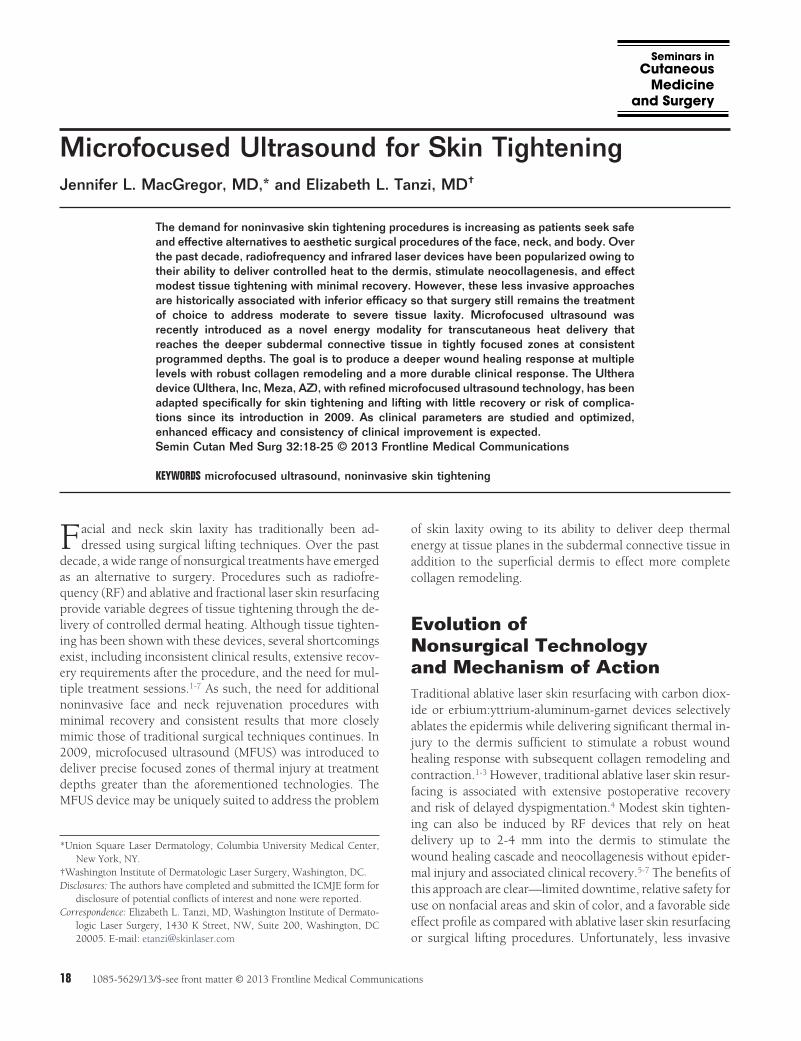
The Ulthera device (Ulthera, Inc, Meza, AZ) has refinedMFUS technology using transducer handpieces uniquely ca-pable of imaging mode (lower energy ultrasound for real-time imaging) and treatment mode (delivery of higher energyultrasound exposures [Fig. 1]). The energy is delivered in astraight 2.5-cm line with TIZs 0.5-5 mm apart at a givendepth within the tissue. Short pulse durations (25-50 ms)and relatively low energy (0.4-1.2 J, range depending on thetransducer) confine the TIZs to their intended depth. Thetransducers are fixed at 7.5 MHz (3 and 4.5 mm focal depths)
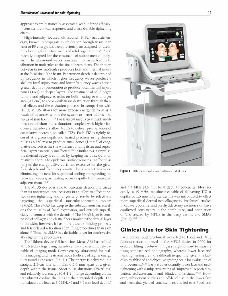
and 4.4 MHz (4.5 mm focal depth) frequencies. Most re-cently, a 19-MHz transducer capable of delivering TIZ atdepths of 1.5 mm into the dermis was introduced to effectmore superficial dermal neocollagenesis. Preclinical studiesin cadaver, porcine, and prerhytidectomy excision skin haveconfirmed consistency in the depth, size, and orientationof TIZ created by MFUS in the deep dermis and SMAS(Fig. 2).12-14,16
Clinical Use for Skin TighteningEarly clinical and preclinical work led to Food and DrugAdministration approval of the MFUS device in 2009 foreyebrow lifting. Eyebrow lifting is straightforward to measureusing standardized photography, whereas lower face andneck tightening are more difficult to quantify, given the lackof an established and objective grading scale for evaluation ofimprovement.17,18 Early studies quantify lower face and necktightening with a subjective rating of “improved” reported bypatient self-assessment and blinded physicians.19,20 How-ever, subsequent studies and off-label use in the lower faceand neck that yielded consistent results led to a Food and
Figure 1 Ulthera microfocused ultrasound device.
Microfocused ultrasound for skin tightening 19
Drug Administration approved indication for “noninvasivelift of lax tissue of the neck and submentum” in 2012.21
Alam et al17 conducted the first clinical study of full-faceand neck MFUS treatment in 35 patients, looking at safetyand efficacy. In standardized photographs, 86% of the pa-tients achieved significant improvement as measured byblinded physician assessment. Photographic measurementsdemonstrated a mean brow lift of 1.7 mm.
Chan et al22 evaluated the safety of MFUS skin tighteningin 49 Chinese patients using an advanced protocol. All pa-tients underwent full-facial and neck treatment without sig-nificant or persistent adverse effects. Suh et al19 evaluated 22Korean patients after full-face treatment and reported 91% ofpatients improved, as rated on a subjective scale where 1 �improved and 2 � much improved at the nasolabial fold andjaw line (1.77 and 1.72 average improvement, respectively).Skin biopsies obtained from 11 study subjects at baseline and2 months after treatment confirmed an increase in reticulardermal collagen and dermal thickening, with elastic fibersappearing more parallel and straighter than pretreatmentspecimens.19 Lee et al20 reported subjective improvement in9 of 10 patients by their own self-assessment, and 8 of 10patients were rated as “improved” by blinded physician as-sessment. Suh et al23 subsequently showed subjective im-provement in most patients treated with a single pass to thelower infraorbital region in 15 patients treated with a 7-MHz3-mm transducer.
Alster and Tanzi24 established the first report of clinicalefficacy in nonfacial areas. Paired sites in 18 women wereevaluated on the arms, knees, or medial thighs where dual-plane treatment with the 4-MHz 4.5-mm-depth and 7-MHz3-mm-depth transducer was compared with single-planetreatment with the 4-MHz 4.5-mm-depth transducer alone.Global assessment scores of skin tightening and lifting weredetermined by 2 blinded physician raters and graded using aquartile grading scale. At the 6-month follow-up visit, statis-tically significant improvement was seen at all 3 body sites,with the arms and knees demonstrating more noticeable im-provement than thighs. Dual-plane treatment yielded addi-tional benefit in smoothing skin texture, an effect potentiallyrelated to more superficial dermal collagen remodeling.When asked to rate their impression of clinical efficacy, 13 of
16 patients reported being “highly satisfied” with the treat-ment.
Sasaki and Tevez21,25 have reported on their extensive ex-perience with the use of MFUS for multiple indications. Us-ing the new 19-MHz 1.5-mm superficial transducer, theytreated 19 patients in the periorbital region with 45 lines oneach side, with another 45 lines using the 7-MHz 3-mm as
Figure 2 Geometry of thermal injury zones in porcine muscle as delivered energy is increased from 2.3 to 7.6 J. Theinverse cone-shaped lesions demonstrate consistent size, depth, and spacing of coagulative necrosis. (Reprinted fromWhite, et al.,12 with permission, Wiley Periodicals.)
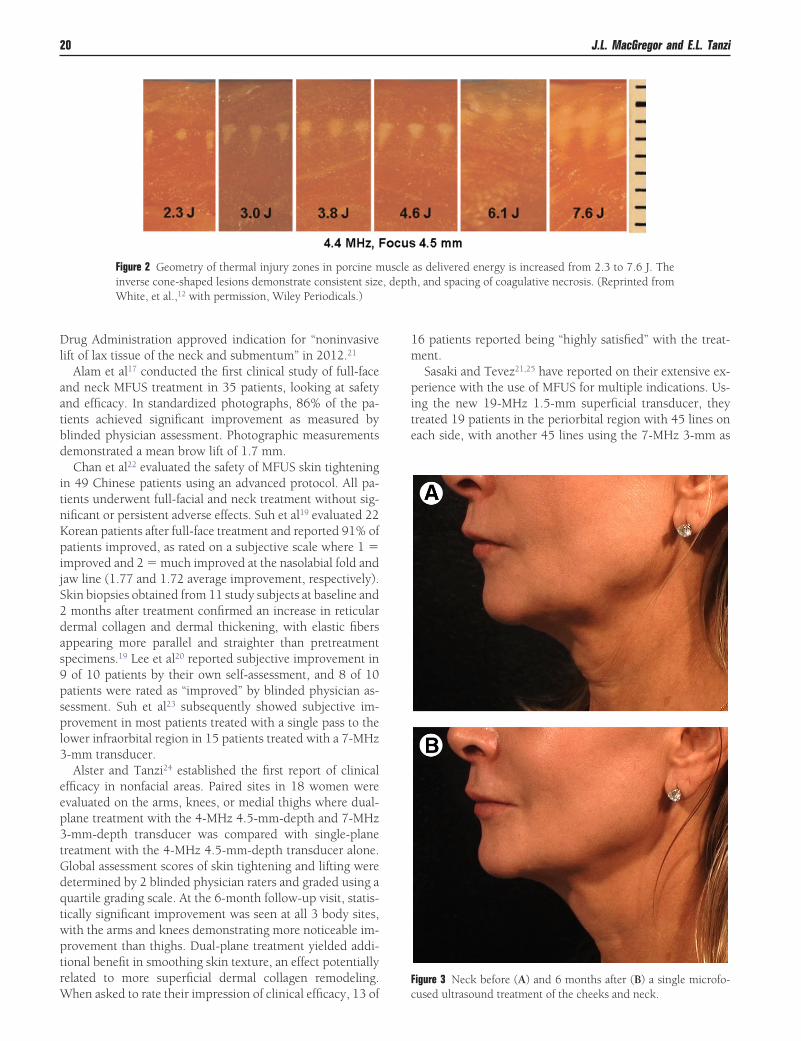
Figure 3 Neck before (A) and 6 months after (B) a single microfo-cused ultrasound treatment of the cheeks and neck.
20 J.L. MacGregor and E.L. Tanzi
the second depth over the orbital rim.25 Brow elevation wasmeasured between 1 and 2 mm in each of 19 patients treated,and periorbital skin tightening was rated as moderate be-tween a 3- and 6-month period. Body sites treated in thisstudy included décolletage (5), brachium (44), periumbilicus(6), inner thigh (1), knee (4), hand (1), and buttocks (2).Treatment protocols varied according to skin thickness at thetarget site. Blinded evaluator assessment scores revealedmoderate improvement in the periorbital area, inner bra-chium, periumbilicus, and knees. Less consistent resultswere achieved in the décolletage, inner thighs, hands, andbuttocks. In a larger series of pilot studies and clinical inves-tigations, the authors compared horizontal and vertical vec-tors in the brow and marionette regions while keeping depthand energy constant.21 Vertical vectors were superior in allsites and energy settings evaluated. They also evaluated a
larger number of patients to confirm that a higher number oflines and joules would yield significantly superior results atall areas treated. In total, 193 patients were included in theinvestigations.21
Recent presentations at scientific meetings have includedadditional data supporting efficacy for MFUS treatment ofwrinkling around the knee,26 tightening of the neck,27 décol-letage,28 and buttock,29 and the potential to treat axillaryhyperhidrosis.30 Future directions of research include its po-tential to induce scar remodeling, which would be particu-larly useful in deep or contracted scars. MFUS has also beenreported to soften silicone and associated scarring of the lip.31
The potential for anti-inflammatory effect and possible use inacneiform disorders is also a current subject of investigation.
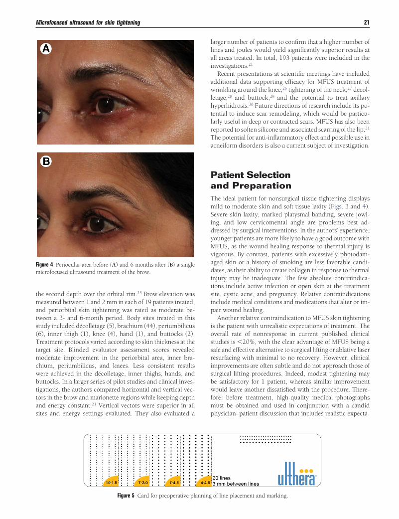
Patient Selectionand PreparationThe ideal patient for nonsurgical tissue tightening displaysmild to moderate skin and soft tissue laxity (Figs. 3 and 4).Severe skin laxity, marked platysmal banding, severe jowl-ing, and low cervicomental angle are problems best ad-dressed by surgical interventions. In the authors’ experience,younger patients are more likely to have a good outcome withMFUS, as the wound healing response to thermal injury isvigorous. By contrast, patients with excessively photodam-aged skin or a history of smoking are less favorable candi-dates, as their ability to create collagen in response to thermalinjury may be inadequate. The few absolute contraindica-tions include active infection or open skin at the treatmentsite, cystic acne, and pregnancy. Relative contraindicationsinclude medical conditions and medications that alter or im-pair wound healing.
Another relative contraindication to MFUS skin tighteningis the patient with unrealistic expectations of treatment. Theoverall rate of nonresponse in current published clinicalstudies is �20%, with the clear advantage of MFUS being asafe and effective alternative to surgical lifting or ablative laserresurfacing with minimal to no recovery. However, clinicalimprovements are often subtle and do not approach those ofsurgical lifting procedures. Indeed, modest tightening maybe satisfactory for 1 patient, whereas similar improvementwould leave another dissatisfied with the procedure. There-fore, before treatment, high-quality medical photographsmust be obtained and used in conjunction with a candidphysician–patient discussion that includes realistic expecta-
Figure 4 Periocular area before (A) and 6 months after (B) a singlemicrofocused ultrasound treatment of the brow.
Figure 5 Card for preoperative planning of line placement and marking.
Microfocused ultrasound for skin tightening 21
tions of improvement, maintenance requirements, limita-tions in achieving the patient’s goal of “lifting” the tissueswithout surgery, and the possibility of no appreciable clinicalimprovement.
As with any heat-based cosmetic procedure, there are vari-able degrees of discomfort associated with MFUS skin tight-ening. Preoperative planning should include a discussion ofthe patient’s historical pain tolerance and response to anxio-lytic and narcotic pain medications. Individual publishedreports of pain in response to the treatment range from mildto severe. Sufficient pain management is critical to an effec-
tive outcome and the overall treatment experience for thepatient. As such, the authors use a combination of oral anx-iolytics (5-10 mg of diazepam) and intramuscular narcotics(50-75 mg of IM meperidine) 20-30 minutes before treat-ment to alleviate discomfort in most patients. Other methodsof pain management have been described, including high-dose nonsteroidal anti-inflammatory drugs, oral or intrave-nous narcotics, topical or local injections of anesthetics, con-scious sedation, and cold techniques.32 The deeper probe andhigher energy delivery is associated with increased pain. Forsuperficial treatment of periocular and perioral rhytides us-
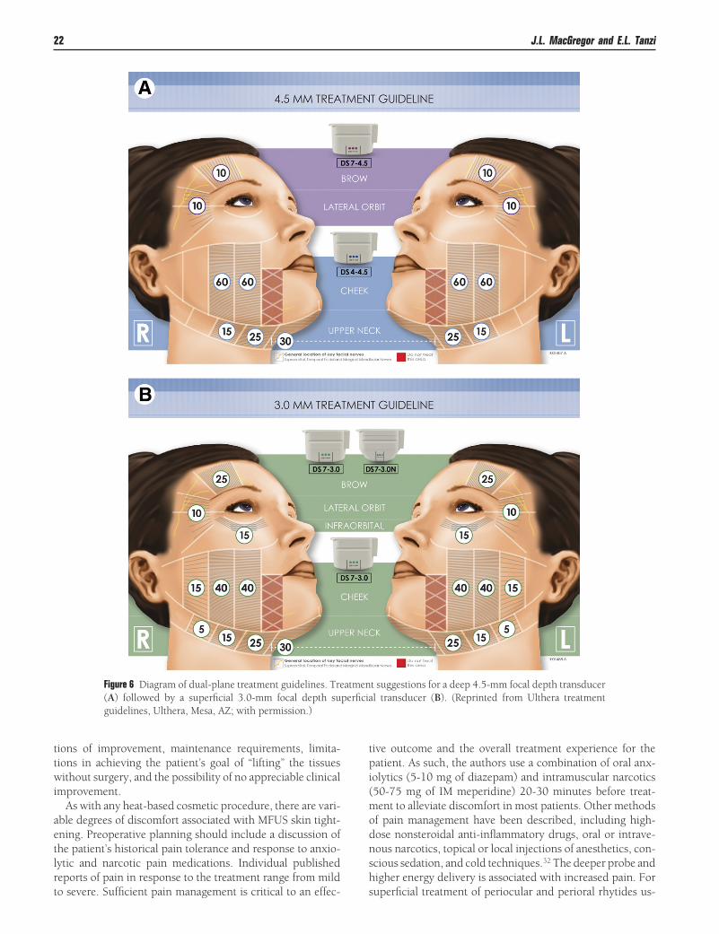
Figure 6 Diagram of dual-plane treatment guidelines. Treatment suggestions for a deep 4.5-mm focal depth transducer(A) followed by a superficial 3.0-mm focal depth superficial transducer (B). (Reprinted from Ulthera treatmentguidelines, Ulthera, Mesa, AZ; with permission.)
22 J.L. MacGregor and E.L. Tanzi
ing the 1.5-mm-depth transducer, topical anesthesia alonemay effectively lessen treatment-associated discomfort.
Operative TechniqueFour transducers are available for transcutaneous treatmentusing the MFUS device. These interchangeable dual-func-tioning transducers are labeled according to their frequencyand focal treatment depth. They include 4-MHz 4.5-mm fo-cal depth (0.75-1.2 J), 7-MHz 4.5-mm focal depth (0.75-1.05 J), 7-MHz 3-mm focal depth (0.4-0.63 J), and 19-MHz1.5-mm focal depth (0.15-0.25 J). In general, the areas withthe thinnest skin, such as the neck and periocular area,should be treated with superficial depth probes; the browand temple should be treated with superficial and deeperprobes; and cheek and submental skin is best treated with thedeepest 4-MHz 4.5-mm probe followed by additional treat-ment with a superficial probe. Multiple treatment protocolsusing single-, double-, and even triple-depth treatmentplanes have been reported, and the parameters continue to berefined in different treatment protocols to enhance efficacy.The technique of layering multiple depths of TIZs through-out the treatment area enhances efficacy in both facial andnonfacial treatment sites.21,24,25
Before treatment, the skin is freshly cleansed, dried, andcleared free of makeup, sunscreen, or products. Each tar-
geted region for treatment is outlined with a planning card todetermine the number of treatment columns required to de-liver energy with minimal overlap (Fig. 5). Ultrasound gel isapplied to the skin, and the probe is placed firmly and gentlyon the target site so the entire transducer is evenly coupled tothe skin surface. Correct technique is confirmed with visual-ization of acoustic coupling as seen on the ultrasound imageson the monitor. Focal depth is visible on the screen in thecorresponding ultrasound image and lined up with the deepdermis to SMAS, depending on the transducer and targetedsite. Treatment lines of ultrasound pulses are manually deliv-ered adjacent and parallel to one another with minimal spac-ing (�3 mm). The overall number of lines placed in a treat-ment area will depend on the size of the treatment area andchosen protocol (Fig. 6). The most advanced protocols callfor the placement of 600-800 lines of ultrasound pulses whentreating the full face. Until additional experience with a largecohort of patients confirms its safety, treatment over softtissue augmentation material and implants should be ap-proached with caution. Because there are no commerciallyavailable eye shields known to prevent propagation of ultra-sound energy over the globe, treatment inside the orbital rimis not possible. The thyroid gland is palpated and markedbefore treatment to avoid inadvertent delivery of ultrasoundpulses over the area.
Postoperative Management,Side Effects, and ComplicationsAfter treatment, ultrasound gel is removed and a bland mois-turizer applied. Patients are instructed to care for their skin asthey normally would with no restrictions on activity. If sys-temic pain management was used, the patient is dischargedwith appropriate transportation. If desired, the patient mayapply cold compresses to the treatment area in the hours afterthe procedure to minimize local edema; however, its use isnot mandatory in all patients, as degrees of swelling aftertreatment are variable.
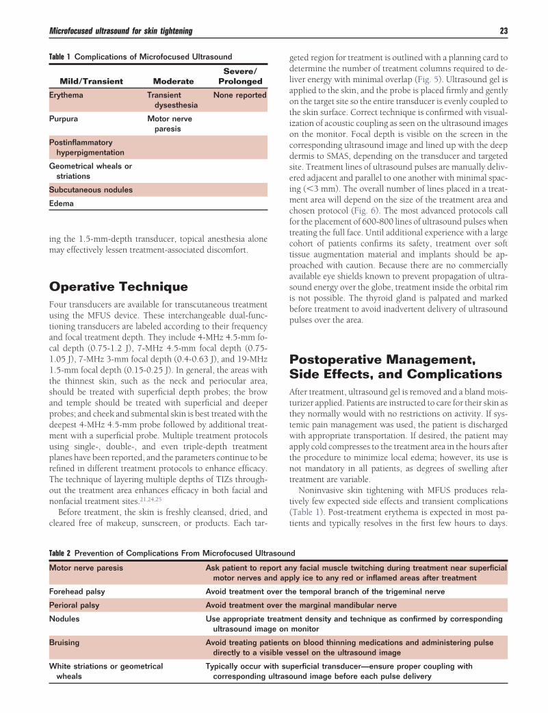
Noninvasive skin tightening with MFUS produces rela-tively few expected side effects and transient complications(Table 1). Post-treatment erythema is expected in most pa-tients and typically resolves in the first few hours to days.
Table 1 Complications of Microfocused Ultrasound
Mild/Transient ModerateSevere/
Prolonged
Erythema Transientdysesthesia
None reported
Purpura Motor nerveparesis
Postinflammatoryhyperpigmentation
Geometrical wheals orstriations
Subcutaneous nodules
Edema
Table 2 Prevention of Complications From Microfocused Ultrasound
Motor nerve paresis Ask patient to report any facial muscle twitching during treatment near superficialmotor nerves and apply ice to any red or inflamed areas after treatment
Forehead palsy Avoid treatment over the temporal branch of the trigeminal nerve
Perioral palsy Avoid treatment over the marginal mandibular nerve
Nodules Use appropriate treatment density and technique as confirmed by correspondingultrasound image on monitor
Bruising Avoid treating patients on blood thinning medications and administering pulsedirectly to a visible vessel on the ultrasound image
White striations or geometricalwheals
Typically occur with superficial transducer—ensure proper coupling withcorresponding ultrasound image before each pulse delivery
Microfocused ultrasound for skin tightening 23
Small areas of purpura may develop and are expected toresolve over 1-2 weeks. Linear or geometrical striations seenafter treatment with the superficial transducer are treatedwith topical corticosteroids and followed for rapid resolu-tion.17,19,21 No permanent textural changes from these lesionshave been reported. Lingering mild to moderate skin tender-ness and edema in the first 1-4 weeks after treatment is com-mon.22,24 Transient postinflammatory pigmentation was ob-served in 2 Chinese patients treated over the brow, but wasmost likely related to placement of the deep 4-MHz 4.5-mmtransducer and was not observed in subsequent treatments.22
Focal areas of numbness on the brow or perioral area canoccur with return of full sensation within several weeks with-out intervention.19,21,22
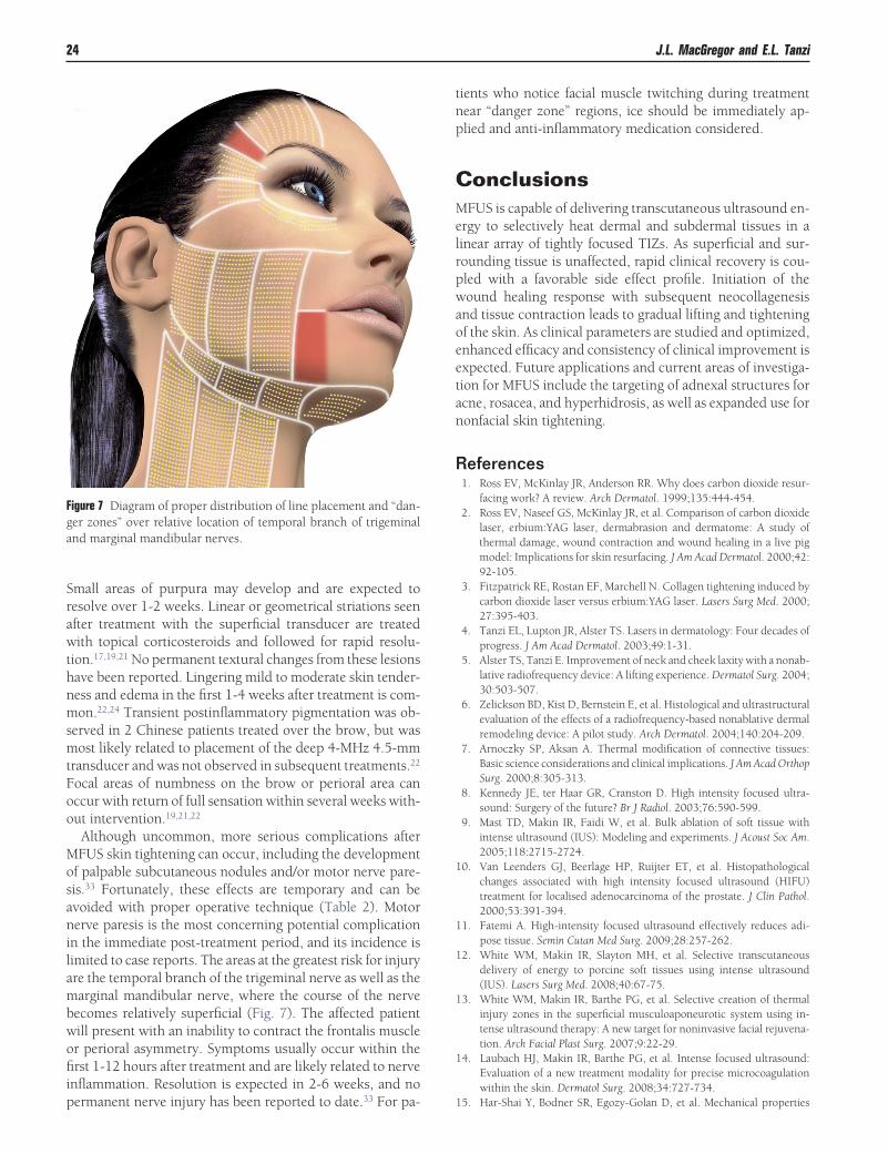
Although uncommon, more serious complications afterMFUS skin tightening can occur, including the developmentof palpable subcutaneous nodules and/or motor nerve pare-sis.33 Fortunately, these effects are temporary and can beavoided with proper operative technique (Table 2). Motornerve paresis is the most concerning potential complicationin the immediate post-treatment period, and its incidence islimited to case reports. The areas at the greatest risk for injuryare the temporal branch of the trigeminal nerve as well as themarginal mandibular nerve, where the course of the nervebecomes relatively superficial (Fig. 7). The affected patientwill present with an inability to contract the frontalis muscleor perioral asymmetry. Symptoms usually occur within thefirst 1-12 hours after treatment and are likely related to nerveinflammation. Resolution is expected in 2-6 weeks, and nopermanent nerve injury has been reported to date.33 For pa-
tients who notice facial muscle twitching during treatmentnear “danger zone” regions, ice should be immediately ap-plied and anti-inflammatory medication considered.
ConclusionsMFUS is capable of delivering transcutaneous ultrasound en-ergy to selectively heat dermal and subdermal tissues in alinear array of tightly focused TIZs. As superficial and sur-rounding tissue is unaffected, rapid clinical recovery is cou-pled with a favorable side effect profile. Initiation of thewound healing response with subsequent neocollagenesisand tissue contraction leads to gradual lifting and tighteningof the skin. As clinical parameters are studied and optimized,enhanced efficacy and consistency of clinical improvement isexpected. Future applications and current areas of investiga-tion for MFUS include the targeting of adnexal structures foracne, rosacea, and hyperhidrosis, as well as expanded use fornonfacial skin tightening.
References1. Ross EV, McKinlay JR, Anderson RR. Why does carbon dioxide resur-
facing work? A review. Arch Dermatol. 1999;135:444-454.2. Ross EV, Naseef GS, McKinlay JR, et al. Comparison of carbon dioxide
laser, erbium:YAG laser, dermabrasion and dermatome: A study ofthermal damage, wound contraction and wound healing in a live pigmodel: Implications for skin resurfacing. J Am Acad Dermatol. 2000;42:92-105.
3. Fitzpatrick RE, Rostan EF, Marchell N. Collagen tightening induced bycarbon dioxide laser versus erbium:YAG laser. Lasers Surg Med. 2000;27:395-403.
4. Tanzi EL, Lupton JR, Alster TS. Lasers in dermatology: Four decades ofprogress. J Am Acad Dermatol. 2003;49:1-31.
5. Alster TS, Tanzi E. Improvement of neck and cheek laxity with a nonab-lative radiofrequency device: A lifting experience. Dermatol Surg. 2004;30:503-507.
6. Zelickson BD, Kist D, Bernstein E, et al. Histological and ultrastructuralevaluation of the effects of a radiofrequency-based nonablative dermalremodeling device: A pilot study. Arch Dermatol. 2004;140:204-209.
7. Arnoczky SP, Aksan A. Thermal modification of connective tissues:Basic science considerations and clinical implications. J Am Acad OrthopSurg. 2000;8:305-313.
8. Kennedy JE, ter Haar GR, Cranston D. High intensity focused ultra-sound: Surgery of the future? Br J Radiol. 2003;76:590-599.
9. Mast TD, Makin IR, Faidi W, et al. Bulk ablation of soft tissue withintense ultrasound (IUS): Modeling and experiments. J Acoust Soc Am.2005;118:2715-2724.
10. Van Leenders GJ, Beerlage HP, Ruijter ET, et al. Histopathologicalchanges associated with high intensity focused ultrasound (HIFU)treatment for localised adenocarcinoma of the prostate. J Clin Pathol.2000;53:391-394.
11. Fatemi A. High-intensity focused ultrasound effectively reduces adi-pose tissue. Semin Cutan Med Surg. 2009;28:257-262.
12. White WM, Makin IR, Slayton MH, et al. Selective transcutaneousdelivery of energy to porcine soft tissues using intense ultrasound(IUS). Lasers Surg Med. 2008;40:67-75.
13. White WM, Makin IR, Barthe PG, et al. Selective creation of thermalinjury zones in the superficial musculoaponeurotic system using in-tense ultrasound therapy: A new target for noninvasive facial rejuvena-tion. Arch Facial Plast Surg. 2007;9:22-29.
14. Laubach HJ, Makin IR, Barthe PG, et al. Intense focused ultrasound:Evaluation of a new treatment modality for precise microcoagulationwithin the skin. Dermatol Surg. 2008;34:727-734.
15. Har-Shai Y, Bodner SR, Egozy-Golan D, et al. Mechanical properties
Figure 7 Diagram of proper distribution of line placement and “dan-ger zones” over relative location of temporal branch of trigeminaland marginal mandibular nerves.
24 J.L. MacGregor and E.L. Tanzi
and microstructure of the superficial musculoaponeurotic system. PlastReconstr Surg. 1996;98:59-70.
16. Gliklich RE, White WM, Slayton MH, et al. Clinical pilot study ofintense ultrasound therapy to deep dermal facial skin and subcutane-ous tissues. Arch Facial Plast Surg. 2007;9:88-95.
17. Alam M, White LE, Martin N, et al. Ultrasound tightening of facial andneck skin: A rater-blinded prospective cohort study. J Am Acad Derma-tol. 2010;62:262-269.
18. Weiss M. Commentary: Noninvasive skin tightening: Ultrasound andother technologies: Where are we in 2011? Dermatol Surg. 2012;38:28-30.
19. Suh DH, Shin MK, Lee SJ, et al. Intense focused ultrasound tighteningin Asian skin: Clinical and pathologic results. Dermatol Surg. 2011;37:1595-1602.
20. Lee HS, Jang WS, Cha YJ, et al. Multiple pass ultrasound tightening ofskin laxity of the lower face and neck. Dermatol Surg. 2012;38:20-27.
21. Sasaki GH, Tevez A. Clinical efficacy and safety of focused-image ul-trasonography: A 2-year experience. Aesthet Surg J. 2012;32:601-612.
22. Chan NP, Shek SY, Yu CS, et al. Safety study of transcutaneous focusedultrasound for non-invasive skin tightening in Asians. Lasers Surg Med.2011;43:366-375.
23. Suh DH, Oh YJ, Lee SJ, et al. Intense focused ultrasound tightening forthe treatment of infraorbital laxity. J Cosmet Laser Ther. 2012;14:290-295.
24. Alster TS, Tanzi EL. Noninvasive lifting of arm, thigh, and knee skinwith transcutaneous intense focused ultrasound. Dermatol Surg. 2012;38:754-759.
25. Sasaki GH, Tevez A. Microfocused ultrasound for nonablative skin and
subdermal tightening to the periorbitum and body sites: Preliminaryreport on eighty-two patients. J Cosmet Dermatol Sci Appl. 2012;2:108-116.
26. Gold MH. Ulthera—A single center, prospective study on the efficacyof the micro-focused ultrasound for the non-invasive treatment of skinwrinkles above the knee. Data Presented at the American Society forDermatologic Surgery Meeting, Atlanta, GA, 2012.
27. Elm KDL, Schram SE, Wallander ID, et al. Evaluation of a high intensityfocused ultrasound system for lifting and tightening of the neck. DataPresented at the American Society for Dermatologic Surgery Meeting,Atlanta, GA, 2012.
28. Fabi SG, Massaki A, Goldman M. Evaluation of the micro-focusedultrasound system for lifting and tightening of the décolletage. DataPresented at the American Society for Dermatologic Surgery Meeting,Atlanta, GA, 2012.
29. Goldberg D, Al-Dujaili Z. Micro-focused ultrasound for lifting andtightening skin laxity of the buttock. Data Presented at the AmericanSociety for Dermatologic Surgery Meeting Atlanta, GA, 2012.
30. Nestor MS. Micro-focused ultrasound for the treatment of axillary hy-perhidrosis. Data Presented at the American Society for DermatologicSurgery Meeting, Atlanta, GA, 2012.
31. Kornstein AN. Ulthera for silicone lip correction. Plast Reconstr Surg.2012;129:1014e-1015e.
32. Brobst RW, Ferguson M, Perkins SW. Ulthera: Initial and six monthresults. Facial Plast Surg Clin North Am. 2012;20:163-176.
33. Missel L. Prevention of potential adverse events associated with use ofUlthera device. Tech Bull, 2011.
Microfocused ultrasound for skin tightening 25