microbiome-modulated metabolites at the interface of … · microbiome modulation of nutritionally...
TRANSCRIPT
of September 8, 2018.This information is current as
Interface of Host ImmunityMicrobiome-Modulated Metabolites at the
ElinavEran Blacher, Maayan Levy, Evgeny Tatirovsky and Eran
http://www.jimmunol.org/content/198/2/572doi: 10.4049/jimmunol.1601247
2017; 198:572-580; ;J Immunol
Referenceshttp://www.jimmunol.org/content/198/2/572.full#ref-list-1
, 16 of which you can access for free at: cites 99 articlesThis article
average*
4 weeks from acceptance to publicationFast Publication! •
Every submission reviewed by practicing scientistsNo Triage! •
from submission to initial decisionRapid Reviews! 30 days* •
Submit online. ?The JIWhy
Subscriptionhttp://jimmunol.org/subscription
is online at: The Journal of ImmunologyInformation about subscribing to
Permissionshttp://www.aai.org/About/Publications/JI/copyright.htmlSubmit copyright permission requests at:
Email Alertshttp://jimmunol.org/alertsReceive free email-alerts when new articles cite this article. Sign up at:
Print ISSN: 0022-1767 Online ISSN: 1550-6606. Immunologists, Inc. All rights reserved.Copyright © 2017 by The American Association of1451 Rockville Pike, Suite 650, Rockville, MD 20852The American Association of Immunologists, Inc.,
is published twice each month byThe Journal of Immunology
by guest on September 8, 2018
http://ww
w.jim
munol.org/
Dow
nloaded from
by guest on September 8, 2018
http://ww
w.jim
munol.org/
Dow
nloaded from
Microbiome-Modulated Metabolites at the Interface ofHost ImmunityEran Blacher,1 Maayan Levy,1 Evgeny Tatirovsky,1 and Eran Elinav
The mammalian gastrointestinal tract and associatedmucosal immune system harbor a large repertoire ofmetabolites of prokaryotic and eukaryotic origin thatplay important roles in eukaryotic development andphysiology. These often bioactive small molecules orig-inate from nutrition- and environmental-related sour-ces, or are endogenously produced and modulated bythe host and its microbiota. A complex network of in-teractions exists between the intestinal mucosal immunesystem and the microbiota. This intimate cross-talk maybe driven by metabolite secretion and signaling, and fea-tures profound influences on host immunity and phys-iology, including the endocrine, metabolic, and nervoussystem function in health and disease. Alterations inmicrobiome-associated metabolite levels and activityare implicated in the pathogenesis of a growing numberof illnesses. In this review we discuss the origin andinfluence of microbiome-modulated metabolites, withan emphasis on immune cell development and func-tion.We further highlight the emerging data potentiallyimplicatingmetabolite misbalance with host-microbiome–associated disease. The Journal of Immunology, 2017,198: 572–580.
The gut microbiome is a microbial ecosystem that hasdiverse effects on physiological host functions, par-ticularly immune development and activity. The
molecular basis of host-microbiome interactions is only justbeginning to be unraveled, and is mediated by both cell to cellinteractions and the production, modification, and sensing ofa large variety of bioactive small molecules, termed metabo-lites. Many gastrointestinal metabolites originate from dietaryor environmental sources. They encounter the gut micro-biome as part of the digestion and intestinal transit process,and link host nutrition with physiology, including immune
development and function. Conversely, nutritional compo-sition tremendously impacts the gut microbial compositionand function (1). Other metabolites are endogenously pro-duced or modified through diverse metabolic processes, bythe host or its microbiota (2). Collectively, it is estimatedthat more than 50% of fecal and urinary metabolites arederived from or modified by the gut microbiome (3).Some microbiome-associated metabolites are bioactive and
affect the host cellular processes including differentiation,migration, proliferation, and apoptosis, thereby featuringpleotropic physiological or pathophysiological effects on theeukaryotic host. A number of metabolites impact mucosal andsystemic immune maturation and function, in steady state andduring disease (4–8). The host has evolved multiple metab-olite sensing platforms, and downstream immune signalingpathways that confer reactivity to microbiome-modulatedmetabolites (9, 10). These sensing platforms are expressedin different combinations in mucosal cellular subsets, such asintestinal epithelial cells, macrophages, dendritic cells (DCs),T cells, and innate lymphoid cells (ILCs), where they playcritical roles in host-microbiome mutualistic cross-talk. Inaddition to local metabolite effects on gastrointestinal mucosalimmune function, many systemically absorbed metabolites mayreach remote organs and modulate the immune responses insterile host regions such as the CNS (11, 12). As such, metab-olites may provide a missing link between the gut microbiomecompositional and functional configuration to its remote effectson host physiology and disease risk in seemingly unrelated sterileorgans. The type, composition, and concentration of metabolitescoupled with the host sensor molecules repertoire orchestrate thenet physiological response at a given physiological context.In this review we provide an overview of gut microbiome-
modulated metabolites, their physiological effects on majorimmune functions, and recent observations possibly linkingmetabolite misbalances with risk of immune-mediated andimmune-associated disease.
Department of Immunology, Weizmann Institute of Science, Rehovot 76100, Israel
1E.B., M.L., and E.T. contributed equally to this work.
ORCIDs: 0000-0003-3207-0153 (E.B.); 0000-0001-6476-9183 (E.T.).
Received for publication July 18, 2016. Accepted for publication October 13, 2016.
E.E. was supported by Y. and R. Ungar, the Abisch Frenkel Foundation for the Promotionof Life Sciences, the Gurwin Family Fund for Scientific Research, the Leona M. and HarryB. Helmsley Charitable Trust, the Crown Endowment Fund for Immunological Research,the estate of J. Gitlitz, the estate of L. Hershkovich, the Benoziyo Endowment Fund for theAdvancement of Science, the Adelis Foundation, J.L. and V. Schwartz, A. and G. Markovitz,A. and C. Adelson, the French National Center for Scientific Research, D.L. Schwarz, theV.R. Schwartz Research Fellow Chair, L. Steinberg, J.N. Halpern, A. Edelheit, grantsfunded by the European Research Council, a Marie Curie Career Integration grant, theGerman–Israeli Foundation for Scientific Research and Development, the Israel Science Foun-dation, the Minerva Foundation, the Rising Tide Foundation, the Helmholtz Association, andthe European Foundation for the Study of Diabetes. E.E. is the incumbent Rina Gudinski
Career Development Chair and is a senior fellow at the Canadian Institute for AdvancedResearch.
Address correspondence and reprint requests to Prof. Eran Elinav, Department of Im-munology, Weizmann Institute of Science, Herzl Street 100, Rehovot 76100, Israel.E-mail address: [email protected]
Abbreviations used in this article: AHR, aryl hydrocarbon receptor; ASD, autistic spectrumdisorder; BBB, blood–brain barrier; DC, dendritic cell; FXR, farnesoid X receptor;HDAC, histone deacetylase; IBD, inflammatory bowel disease; IEC, intestinal epithelialcell; ILC, innate lymphoid cell; NA, nicotinic acid; NAFLD, nonalcoholic fatty liverdisease; NASH, nonalcoholic steatohepatitis; SCFA, short chain fatty acid; SFB, segmentedfilamentous bacteria; TMAO, trimethylamine-N-oxide; Treg, regulatory T cell; Trp, tryp-tophan; WT, wild type.
Copyright� 2017 by TheAmerican Association of Immunologists, Inc. 0022-1767/17/$30.00
www.jimmunol.org/cgi/doi/10.4049/jimmunol.1601247
Journal ofTh e
ImmunologyBrief Reviews
by guest on September 8, 2018
http://ww
w.jim
munol.org/
Dow
nloaded from
Microbiome modulation of the gastrointestinal metaboliteconfiguration
One of the essential functions provided by the gut microbiotaincludes the modulation of gastrointestinal metabolites, in-cluding their synthesis, digestion, fermentation, and secondarymetabolism. Numerous studies have investigated the forcesthat drive the development of the adult microbial populationin the intestine from birth (13, 14), yet the dynamics of themicrobiome impact on the host metabolome remain largelyunknown. The centrality of microbial activity in shaping thegut metabolome is demonstrated by profound metabolitealterations found in germ-free as compared with gnotobioticand colonized animals, including differences in diverse me-tabolite biochemical groups noted in feces, urine, and thesystemic circulation (15–18). Multiple commensal microbialtaxonomies, including Enterobacteriaceae, Enterococus spp.,and Lactobacillus spp., have been shown to influence gastro-intestinal metabolite concentrations (17), by processes span-ning from utilization of amino acids as a nitrogen source, tobyproduct generation of secondary metabolites such as shortchain fatty acids (SCFAs). In the following section we willhighlight key examples of these microbiome effects on dietaryand endogenously generated gut metabolites, and how thesemicrobiome effects contribute to the downstream effectorfunctions of these metabolites on host immunity (Figs. 1, 2).
De novo microbiome production of metabolites. The gut microbiotaproduces a number of metabolites, some of which feature distinctbioactive functions on the eukaryotic host. A prime example ofsuch de-novo synthesized metabolites include SCFAs, which arethe products of microbial fermentation of nondigestible nutritionalfibers, and include acetate (C2), propionate (C3), and butyrate(C4). Gut commensal bacteria, such as those belonging tothe genera Butyrivibrio, Clostridium, and Eubacterium, can
produce SCFAs that locally reach a millimolar concentration (19).Additional fermentation byproducts like succinate and lactate arealso generated during SCFAs production, and are used by themicrobiota for maintenance and survival (20). SCFAs, in turn,mediate a number of important functions for the eukaryotichost, including usage as an energy source by intestinal epithelialcells, and a variety of anti-inflammatory properties in T cells,regulatory T cells (Tregs), neutrophils, and macrophages, wherethey affect migration, cytolytic activity, cytokine production,and epigenetic regulation of gene expression (discussed below).Another example of bioactive de novo microbial-synthesized
metabolites are vitamins, small bioactive nutrients that areextracted from the diet and may be modulated by themicrobiome or de novo produced by intestinal commensals.One such important example involves vitamin K, a centralcofactor regulating the mammalian coagulation cascade (21)and immunity (22). The vitamin K group consists of vitaminK1 (phylloquinone), derived from food, and vitamin K2(menaquinone), which can be also produced from vitamin K1by most of the gut microbiota species, including Enterobactersp., Eubacterium lentum, Veillonella sp. and Bacteroides sp (23).Members of the vitamin K group are absorbed in the smallintestine in a process requiring bile salts (24). The importanceof microbial production of vitamin K to its overall intestinalpool is supported by the fact that broad-spectrum antibiotictreatment induces significantly reduced vitamin K levels (25).Furthermore, a primary vitamin K deficiency animal modelwas not successfully generated using a vitamin K–deficientdiet alone, unless germ-free mice were employed in the process,further supporting the important role of the gut microbiome inits processing (26). However, the transformation of vitamin K1to vitamin K2 may not be solely dependent on the microbiota,as it occurs in gnotobiotic rats (27).
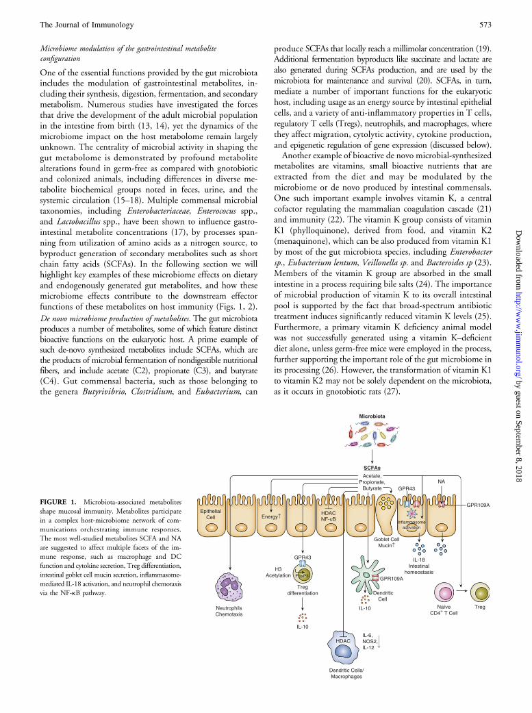
FIGURE 1. Microbiota-associated metabolites
shape mucosal immunity. Metabolites participate
in a complex host-microbiome network of com-
munications orchestrating immune responses.
The most well-studied metabolites SCFA and NA
are suggested to affect multiple facets of the im-
mune response, such as macrophage and DC
function and cytokine secretion, Treg differentiation,
intestinal goblet cell mucin secretion, inflammasome-
mediated IL-18 activation, and neutrophil chemotaxis
via the NF-kB pathway.
The Journal of Immunology 573
by guest on September 8, 2018
http://ww
w.jim
munol.org/
Dow
nloaded from
Other vitamins, such as vitamin B1 (thiamine) (28) and folicacid (vitamin B9) (29), may also be synthesized by somemembers of the microbiome, thereby contributing to theoverall vitamin pull. Vitamin B12 can be produced by bac-teria, e.g., Propionibacterium freudenreichii, Salmonella enter-ica, Listeria innocua, and Lactobacillus reuteri, and may also bedegraded by multiple members of the microbiome (30). Itsbinding to the eukaryotic intrinsic factor as part of the di-gestion process is believed to protect it from microbial deg-radation in the distal small intestine (30).
Microbiome modulation of nutritionally derived metabolites. Mul-tiple bioactive gastrointestinal small molecules originate fromdietary sources, undergo microbial modifications and haverecently been shown to feature important immune functions.One example is the immunomodulatory amino acid trypto-phan (Trp), abundantly found in foods such as milk, eggs, redmeat, and vegetables (e.g., broccoli). Trp is known to undergometabolism by Lactobacilli, giving rise to indole-3-aldehyde,which can bind to the aryl hydrocarbon receptor (AHR),followed by its transport through the epithelial cell layer bya transporter containing angiotensin I converting enzyme 2(9, 31). Deficiency in murine angiotensin I converting enzyme2, which controls the levels of neutral amino acids in the intestine,including Trp, results in higher susceptibility for intestinalinflammation and an altered gut microbial composition(32). Inflammation in this context is transmissible throughtransfer of the microbiota to germ-free mice, whereas dietaryTrp replenishment rescues microbial dysbiosis (32).
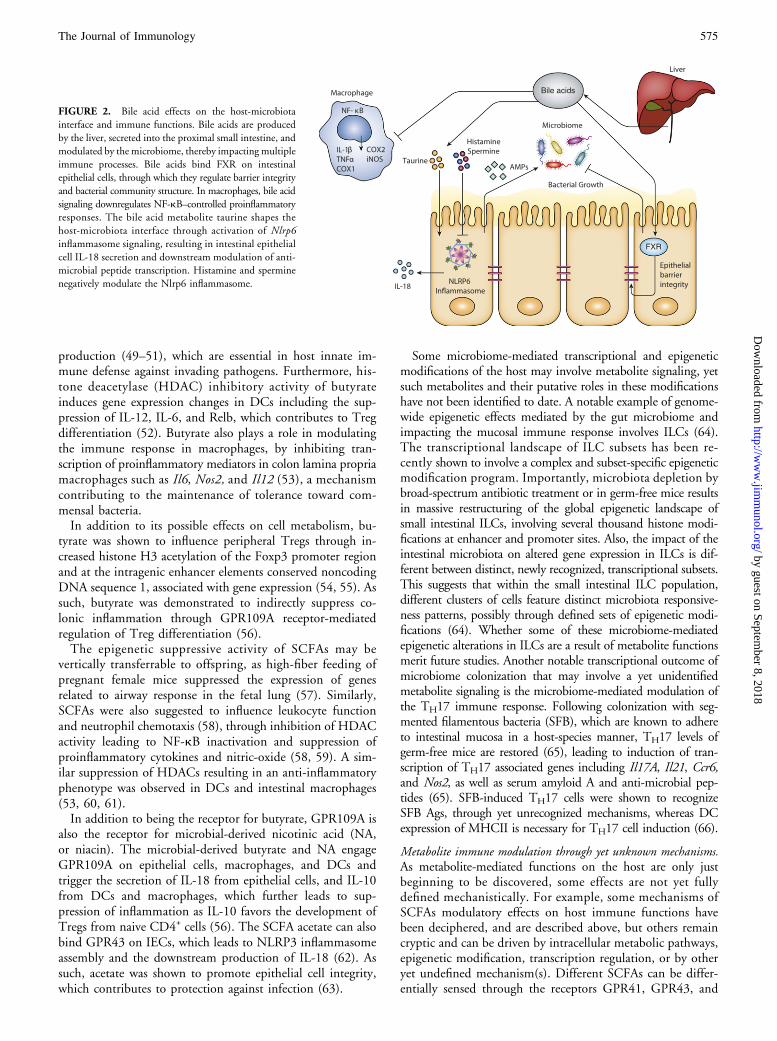
Microbiome modulation of host-derived metabolites. The micro-biome may also modulate metabolites that are produced by theeukaryotic host. An important example of such endogenouslygenerated gastrointestinal metabolites are bile acids, which areproduced from cholesterol by the liver, further conjugated toglycine (in humans) or taurine (in mice) and transported to thegallbladder, common bile duct, and the proximal small in-testine (33, 34). They are considered essential for solubilizingdietary fat and cholesterol, thereby accelerating digestion andabsorption.Secondary microbial-mediated metabolism of primary to
secondary bile acids increases their diversity (35), as exem-plified by germ-free mice having lower levels and reduceddiversity of secondary bile acids as compared with colonizedmice (36). In the intestinal epithelium, many different bileacids, including microbiome-modulated deoxycholic acid andlithocholic acid (37), can bind the G-protein coupled receptorTGR5 as well as the nuclear receptor farnesoid X receptor(FXR). FXR was suggested to regulate epithelial cell integrityand bacterial composition because in the absence of FXR,mice present a disrupted epithelial barrier function and dys-biosis (38). In addition, secondary bile acid signaling mayimpact the host immune response by modulating proin-flammatory genes through NF-kB signaling (39, 40). As such,the addition of an FXR ligand to LPS-treated macrophagesdownregulated the NF-kB–controlled genes IL-1b, TNF-a,COX-1, COX-2, and iNOS. Moreover, Fxr2/2 mice pre-sented a significant exacerbation of colitis symptoms, whichcould be mitigated by treating wild-type (WT) mice with anFXR analog (40). Furthermore, bile acid signaling was re-cently suggested to contribute to the effects of the microbiotaon the metabolic syndrome, which may partially stem from itsimmune modulatory effects (41, 42).
One recently discovered role of microbiome-modulatedmetabolites on the intestinal immune response involves sig-naling through the Nlrp6 inflammasome. The bile acid relatedmetabolite taurine was suggested to shape the host-microbiotainterface through activation of Nlrp6 inflammasome signal-ing, resulting in intestinal epithelial cell IL-18 secretion anddownstream modulation of anti-microbial peptide transcrip-tion (43), thereby impacting the microbiome compositionand risk of auto-inflammation. The microbiome-modulatedmetabolites histamine and spermine, in turn, inhibit theNLRP6 inflammasome signaling, suggesting a mechanism bywhich the combination of bioactive metabolites at givencontexts drive the host-microbiome interface thorough regu-lation of immune sensing and downstream immune modu-lation. Collectively, postbiotic metabolite intervention maytarget a variety of host-related pathways, including those ex-emplified above, representing a promising potential new ther-apeutic modality of microbiome-related metabolic andinflammatory disorders.
Mechanisms of microbiota-modulated metabolite immune regulation
Microbiome-modulated metabolites may impact the hostimmune response through several mechanisms that are onlyjust beginning to be unraveled. In addition to metabolite-induced signaling in immune cell subsets that may trigger acascade of inflammatory changes, recent studies have identifieddirect metabolite-mediated effects on immune cell meta-bolism, often resulting in substantial, previously unappreci-ated, functional outcomes.
Immune cell metabolic reprogramming. One of the recentlycharacterized roles of commensal microbiota is to provideenergy to intestinal epithelial cells (IECs), through fermen-tation of dietary fibers by degradation of undigested complexcarbohydrates into SCFAs. In the absence of the microbiotaand associated SCFAs, germ-free mice feature an altered energymetabolism characterized by preferential fermentation ofglucose into lactate (44), thereby leading to enhanced IECautophagy as a consequence of nutrient starvation (45, 46).Monoinoculation of germ-free mice with the butyrate-producingButyrivibrio fibrisolvens rescued IECs from autophagy andmitochondrial respiration insufficiency (45). Similar to epithe-lial cells, energy metabolism in leukocytes is affected by microbiota-regulated metabolites. T cell responses are dependent onnutrient catabolism, whereas intestinal T cells in germ-free micepresent an immature phenotype characterized by impairedcytolytic activity (47, 48).The molecular mechanisms by which SCFAs, such as buty-
rate, serve as energy sources and consequent functional mod-ulators of intestinal epithelial cells and immune cells arebeginning to be unraveled (45). Butyrate acts as a local substratefor the production of energy through the tricarboxylic acidcycle, ATP generation, and b-oxidation as well as through thesuppression of autophagy in intestinal epithelial cells (45).
Transcriptional and epigenetic modulation of immune-related genes
Another key mechanism by which gut microbiome–modulatedmetabolites influence the immune response involves regula-tion of immune cell transcriptional programming throughimpacting their epigenetic landscape. One such example re-lates to the transcription of mucin-related genes by butyrate,thereby contributing to goblet cell differentiation and mucus
574 BRIEF REVIEWS: METABOLITES, MICROBIOME, AND IMMUNITY
by guest on September 8, 2018
http://ww
w.jim
munol.org/
Dow
nloaded from
production (49–51), which are essential in host innate im-mune defense against invading pathogens. Furthermore, his-tone deacetylase (HDAC) inhibitory activity of butyrateinduces gene expression changes in DCs including the sup-pression of IL-12, IL-6, and Relb, which contributes to Tregdifferentiation (52). Butyrate also plays a role in modulatingthe immune response in macrophages, by inhibiting tran-scription of proinflammatory mediators in colon lamina propriamacrophages such as Il6, Nos2, and Il12 (53), a mechanismcontributing to the maintenance of tolerance toward com-mensal bacteria.In addition to its possible effects on cell metabolism, bu-
tyrate was shown to influence peripheral Tregs through in-creased histone H3 acetylation of the Foxp3 promoter regionand at the intragenic enhancer elements conserved noncodingDNA sequence 1, associated with gene expression (54, 55). Assuch, butyrate was demonstrated to indirectly suppress co-lonic inflammation through GPR109A receptor-mediatedregulation of Treg differentiation (56).The epigenetic suppressive activity of SCFAs may be
vertically transferrable to offspring, as high-fiber feeding ofpregnant female mice suppressed the expression of genesrelated to airway response in the fetal lung (57). Similarly,SCFAs were also suggested to influence leukocyte functionand neutrophil chemotaxis (58), through inhibition of HDACactivity leading to NF-kB inactivation and suppression ofproinflammatory cytokines and nitric-oxide (58, 59). A sim-ilar suppression of HDACs resulting in an anti-inflammatoryphenotype was observed in DCs and intestinal macrophages(53, 60, 61).In addition to being the receptor for butyrate, GPR109A is
also the receptor for microbial-derived nicotinic acid (NA,or niacin). The microbial-derived butyrate and NA engageGPR109A on epithelial cells, macrophages, and DCs andtrigger the secretion of IL-18 from epithelial cells, and IL-10from DCs and macrophages, which further leads to sup-pression of inflammation as IL-10 favors the development ofTregs from naive CD4+ cells (56). The SCFA acetate can alsobind GPR43 on IECs, which leads to NLRP3 inflammasomeassembly and the downstream production of IL-18 (62). Assuch, acetate was shown to promote epithelial cell integrity,which contributes to protection against infection (63).
Some microbiome-mediated transcriptional and epigeneticmodifications of the host may involve metabolite signaling, yetsuch metabolites and their putative roles in these modificationshave not been identified to date. A notable example of genome-wide epigenetic effects mediated by the gut microbiome andimpacting the mucosal immune response involves ILCs (64).The transcriptional landscape of ILC subsets has been re-cently shown to involve a complex and subset-specific epigeneticmodification program. Importantly, microbiota depletion bybroad-spectrum antibiotic treatment or in germ-free mice resultsin massive restructuring of the global epigenetic landscape ofsmall intestinal ILCs, involving several thousand histone modi-fications at enhancer and promoter sites. Also, the impact of theintestinal microbiota on altered gene expression in ILCs is dif-ferent between distinct, newly recognized, transcriptional subsets.This suggests that within the small intestinal ILC population,different clusters of cells feature distinct microbiota responsive-ness patterns, possibly through defined sets of epigenetic modi-fications (64). Whether some of these microbiome-mediatedepigenetic alterations in ILCs are a result of metabolite functionsmerit future studies. Another notable transcriptional outcome ofmicrobiome colonization that may involve a yet unidentifiedmetabolite signaling is the microbiome-mediated modulation ofthe TH17 immune response. Following colonization with seg-mented filamentous bacteria (SFB), which are known to adhereto intestinal mucosa in a host-species manner, TH17 levels ofgerm-free mice are restored (65), leading to induction of tran-scription of TH17 associated genes including Il17A, Il21, Ccr6,and Nos2, as well as serum amyloid A and anti-microbial pep-tides (65). SFB-induced TH17 cells were shown to recognizeSFB Ags, through yet unrecognized mechanisms, whereas DCexpression of MHCII is necessary for TH17 cell induction (66).
Metabolite immune modulation through yet unknown mechanisms.As metabolite-mediated functions on the host are only justbeginning to be discovered, some effects are not yet fullydefined mechanistically. For example, some mechanisms ofSCFAs modulatory effects on host immune functions havebeen deciphered, and are described above, but others remaincryptic and can be driven by intracellular metabolic pathways,epigenetic modification, transcription regulation, or by otheryet undefined mechanism(s). Different SCFAs can be differ-entially sensed through the receptors GPR41, GPR43, and
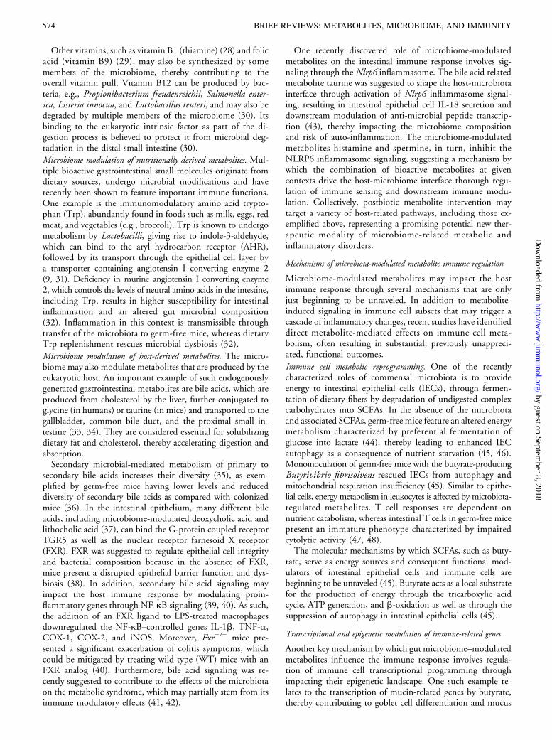
FIGURE 2. Bile acid effects on the host-microbiota
interface and immune functions. Bile acids are produced
by the liver, secreted into the proximal small intestine, and
modulated by the microbiome, thereby impacting multiple
immune processes. Bile acids bind FXR on intestinal
epithelial cells, through which they regulate barrier integrity
and bacterial community structure. In macrophages, bile acid
signaling downregulates NF-kB–controlled proinflammatory
responses. The bile acid metabolite taurine shapes the
host-microbiota interface through activation of Nlrp6inflammasome signaling, resulting in intestinal epithelial
cell IL-18 secretion and downstream modulation of anti-
microbial peptide transcription. Histamine and spermine
negatively modulate the Nlrp6 inflammasome.
The Journal of Immunology 575
by guest on September 8, 2018
http://ww
w.jim
munol.org/
Dow
nloaded from
GPR109A (10). Butyrate is the ligand for GPR109A, expressedby IECs and T cells, where its signaling induces substantial effectson Treg abundance and activity, yet the mechanism of this effectis still not completely understood (56). Likewise, colonic mac-rophages and DCs from mice deficient in GPR109A aredefective in their ability to induce differentiation of naiveT cells into IL-10–expressing Tregs, resulting in a general re-duction in colonic Tregs and increased severity of dextranesodium sulfate–induced colitis, through mechanisms thatare still elusive (56). Germ-free and colonized mice treatedwith SCFAs such as acetate feature reduced inflammation in adextrane sodium sulfate–induced colitis model as well as in aT cell transfer colitis model in a GPR43-dependent manner.Mechanistically, SCFA administration increases the numbersof Treg cells in germ-free mice as well as the secretion of itskey suppressive effector cytokine IL-10, possibly througheffects on immune cell metabolism (54, 67). Furthermore, alack of GPR43 expression in Gpr432/2 mice severely altersSCFA-mediated expansion of colonic Tregs (54). Thus, SCFAsand their GPRs represent one pathway through which thecommensal microbiota regulates the inflammatory response,yet the intracellular and immune-mediated mechanisms of thesemetabolite effects merit further studies. Similar to their localeffect in the intestine, circulating SCFAs dampen the severity ofallergic airway inflammation with reduced levels of IL-4, IL-5,IL-13, and IL-17a in the lung, possibly through increased bonemarrow generation of macrophages and DCs; this and otherpotential mechanisms merit further exploration (60).
Microbiome-modulated metabolites and the risk of disease
Defined compositional and functional microbiome alterationshave been associated with the pathogenesis of commonmultifactorial diseases. These diseases include, among others,
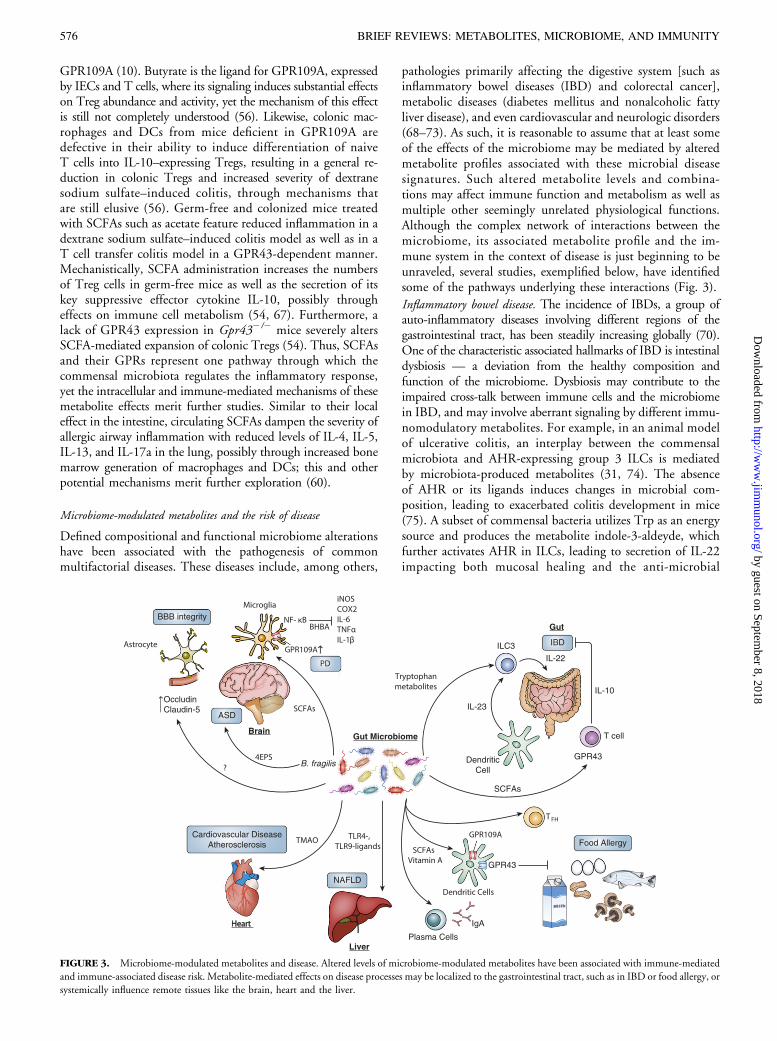
pathologies primarily affecting the digestive system [such asinflammatory bowel diseases (IBD) and colorectal cancer],metabolic diseases (diabetes mellitus and nonalcoholic fattyliver disease), and even cardiovascular and neurologic disorders(68–73). As such, it is reasonable to assume that at least someof the effects of the microbiome may be mediated by alteredmetabolite profiles associated with these microbial diseasesignatures. Such altered metabolite levels and combina-tions may affect immune function and metabolism as well asmultiple other seemingly unrelated physiological functions.Although the complex network of interactions between themicrobiome, its associated metabolite profile and the im-mune system in the context of disease is just beginning to beunraveled, several studies, exemplified below, have identifiedsome of the pathways underlying these interactions (Fig. 3).
Inflammatory bowel disease. The incidence of IBDs, a group ofauto-inflammatory diseases involving different regions of thegastrointestinal tract, has been steadily increasing globally (70).One of the characteristic associated hallmarks of IBD is intestinaldysbiosis — a deviation from the healthy composition andfunction of the microbiome. Dysbiosis may contribute to theimpaired cross-talk between immune cells and the microbiomein IBD, and may involve aberrant signaling by different immu-nomodulatory metabolites. For example, in an animal modelof ulcerative colitis, an interplay between the commensalmicrobiota and AHR-expressing group 3 ILCs is mediatedby microbiota-produced metabolites (31, 74). The absenceof AHR or its ligands induces changes in microbial com-position, leading to exacerbated colitis development in mice(75). A subset of commensal bacteria utilizes Trp as an energysource and produces the metabolite indole-3-aldeyde, whichfurther activates AHR in ILCs, leading to secretion of IL-22impacting both mucosal healing and the anti-microbial
FIGURE 3. Microbiome-modulated metabolites and disease. Altered levels of microbiome-modulated metabolites have been associated with immune-mediated
and immune-associated disease risk. Metabolite-mediated effects on disease processes may be localized to the gastrointestinal tract, such as in IBD or food allergy, or
systemically influence remote tissues like the brain, heart and the liver.
576 BRIEF REVIEWS: METABOLITES, MICROBIOME, AND IMMUNITY
by guest on September 8, 2018
http://ww
w.jim
munol.org/
Dow
nloaded from
peptides repertoire including lipocalin-2, S100A8, and S100A9in mice (31, 74). Another possible metabolite-mediated mechanismof auto-inflammation involves SCFA-mediated inhibition ofHDACs, independently of their receptors GPR41 and GPR43,leading to T cell activation of the mTOR-S6K pathway (76).Acetate supplementation in drinking water was able to mitigateanti-CD3–induced colitis severity in the ileum of WT but notof Il102/2 mice, indicating that this protective effect requiresIL-10 (76). Conversely, oral butyrate treatment enhancedIL-23 levels in DCs and exacerbated colitis symptoms in an IBDanimal model (77). As the effects of SCFAs are complex andsometimes diverse, indirect, and combinatorial, future studieswill have to delineate their human relevance and therapeuticpotential in different combinations and clinical contexts.Another example of the role of the microbiome-immune-
metabolite axis in driving intestinal auto-inflammation is pro-vided by the NLRP6 inflammasome model in mice. As describedabove, a properly functional NLRP6 inflammasome signaling,orchestrated by the microbiome-modulated metabolites taurine,histamine, and spermine (43) in mouse IEC, contributes to ahealthy microbiome population. Conversely, NLRP6-deficientmice feature dysbiosis and associated intestinal auto-inflammation,which is transmittable via fecal transplantation into normalWTmice, thereby inducing exacerbated colitis symptoms, whichare ameliorated by restoration of normal microbiota (43). Mul-tiple other unknown metabolites in mice and in human IBDpatients may impact immune activation and function, therebyaffecting the pathogenesis of different IBD subsets, and meritfurther studies.
Nonalcoholic fatty liver disease. Nonalcoholic fatty liver disease(NAFLD) is the most common hepatic disease in the developedworld, and is tightly associated with other features of metabolicsyndrome including obesity, hyperlipidemia, and adult-onsetdiabetes mellitus. Whereas most affected individuals remainasymptomatic, a significant minority of NAFLD cases developa progressive inflammatory liver disease termed nonalcoholicsteatohepatitis (NASH), ultimately leading to liver dysfunction,cirrhosis, and life-risking complications (78). The microbiomewas suggested in both mouse models and humans to regulatesome manifestations of NAFLD and NASH via its systemiceffects on the immune system (79, 80). In one such animalmodel, inflammasome-deficient mice feature dysbiosis me-diated by an altered metabolite profile (81). When inducedwith NAFLD, these mice developed a context-specificform of dysbiosis characterized by a massive expansion ofPorphyromonadaceae accompanied with enhanced TLR4 andTLR9 ligands influxing into the portal circulation and leadingto massive TNF-a secretion, hepatic inflammation, and theprogression to NASH (82). Interestingly, this microbiome-mediated transition into NASH was abolished by wide-spectrumantibiotic treatment and transferrable by cohabitation ofinflammasome-deficient mice with WT mice. As dysbiosisin this model has been recently linked to microbiome-modulated metabolite misbalance, it hints toward the pos-sibility of an indirect link existing between gastrointestinalmetabolite alterations, dysbiosis, and influx of microbialproducts into the liver leading to inflammatory conseq-uences (43). Likewise, in NAFLD human patients, dysbiosiswas observed with an expansion of Streptococcus, Anaerobacter,Lactobacillus and Escherichia genera as compared with healthysubjects, accompanied by reduction of Alistipes and Prevotella.
Similar to the above animal model, NAFLD patients werefound to have higher levels of TNF-a, IFN-g, and IL-6 anddisrupted microvilli morphology associated with increased gutpermeability (83). These results suggest that dysbiosis may notonly affect the local intestinal inflammatory response, but mayalso systemically influence sterile tissues through metabolitesdriving pathological conditions, inflammatory and metabolicalike. Other manifestations of the metabolic syndrome, suchas obesity and glucose intolerance, were recently suggested toinvolve an altered inflammatory response in tissues such asadipose tissue, the pancreas, and the liver (84). An intriguing,yet unexplored possibility is that some of these inflammatorychanges may be mediated by dysbiosis and its associated alteredmetabolite configuration. Exploring metabolite roles in thesecontexts may enable to identify new therapeutic and diagnosticmodalities to these common idiopathic multifactorial disorders.
Cardiovascular disease. The catastrophic consequences of ath-erosclerosis, myocardial infarction, and cerebrovascular ac-cident still account for the majority of deaths worldwide (85).Atherosclerotic plaques in arterial walls are caused by an imbal-anced lipid homeostasis leading to accumulation of cholesterol–containing low-density lipoprotein particles, accompanied bychronic inflammation (86). In the past decade changes inmicrobiome composition and function have been associatedwith cardiovascular disease pathogenesis. One notable exampleof such an association involves trimethylamine-N-oxide (TMAO)metabolism. TMAO is a metabolite that in humans is generatedby microbial metabolism of dietary phosphatidylcholine orL-carnitine, which are abundant in meat and high-fat diets.Mice fed L-carnitine featured increased levels of TMAOleading to enhanced atherosclerosis. Germ-free mice orantibiotic-treated mice were protected from the disease, therebylinking the gut microbiota, nutrition, and risk for cardio-vascular disease (87). Similar findings were observed in hu-mans, in which a time-dependent increase in plasma TMAO(and other phosphatidylcholine metabolites) was documentedfollowing ingestion of isotopic-labeled food. These increasedmetabolite levels were inhibited by antibiotic treatment, andreappeared after antibiotic withdrawal (88). Increased fastingTMAO plasma levels were significantly associated with majoradverse cardiovascular events such as overall mortality, myo-cardial infarction or stroke. These results demonstrate thepotential central role of this microbiome-derived metabolitein the risk factor for cardiovascular disease (88, 89).
Food allergy. Allergies to nutritional components constitute acommon immune-mediated condition affecting the pediatric andadult population alike. Mucosal DCs contribute to allergy to foodAgs by regulation of Treg differentiation. A high-fiber diet togetherwith vitamin A was recently shown to drive the microbiota towarda configuration that is supportive for tolerogenic CD103+ DCs,which provides tolerance toward food Ags in a mouse model,thereby protecting from food allergy. This protective effectdepends on epithelial GPR43 and immune GPR109A, becausethe protective effect was not observed in Gpr432/2 or inGpr109a2/2 mice. In this model, changes in the microbiotafollowing a high-fiber diet were also associated with enhancedIgA production and T follicular helper response (90), furtherimplicating the microbiome in this food allergy protective effect.
Neurodegenerative disease. Although neurological disorders areincreasingly suggested to feature important immune components,
The Journal of Immunology 577
by guest on September 8, 2018
http://ww
w.jim
munol.org/
Dow
nloaded from
research on neurological immune modulation remains in itsinfancy. Some of the observed immunomodulatory effects on theCNS are mediated by resident immune cell subsets in the CNS,which impact disease progression, and particularly the microglia,the resident myeloid cells of the CNS. Interestingly, microgliawere recently suggested to be affected by metabolites such asSCFAs in a mouse model. SCFAs secreted by the gut microbiotamodulated microglial activation and maturation gene-expressionprofile in the mouse steady state (91). The NA receptorGPR109A was recently found to be expressed by microglia(92) and feature increased expression colocalizing with microglialmarkers in the substantia nigra of Parkinson’s disease patients,possibly participating in disease modulation (92). Likewise,b-hydroxybutyrate treatment given to a Parkinson’s diseaserat model improved motor skills and protected nigro-striatalneurons by reducing neuroinflammation (92). Addition ofb-hydroxybutyrate to mesencephalic neuron-glia culturesreduced the harmful effects of LPS-induced microglialactivation through GPR109A inhibition of NF-kB pathway,resulting in a reduction of proinflammatory enzymes (COX-2,iNOS) and cytokines (IL-6, TNF-a, IL-1b) (93). Butyrate andother SCFAs influx from the gastrointestinal tract through theportal circulation or are absorbed directly into the systemiccirculation through the distal colonic blood supply, and mayaccess the CNS through the blood–brain barrier (BBB). Micefed a fermented-fiber diet presented with a reduced endotoxin-related sickness behavior (94), and interestingly featuredincreased CNS IL-4 mRNA levels probably mediated byenhancement of histone acetylation increasing IL-1RA levels,which inhibits the production of IL-1b proinflammatorycytokine (94). Furthermore, the tight-junction forming proteinsoccludin and claudin-5 were reduced in the brains of germ-free mice, leading to an increased permeability of theirBBB (95). Treatment with butyrate-producing Clostridiumtyrobutyricum bacteria was able to elevate occludin andclaudin-5 levels in germ-free mice brains and to restoretheir BBB permeability to the level of specific pathogen-freemice (95).Interestingly, other neuropsychiatric disorders such as au-
tistic spectrum disorder (ASD) were also suggested to involvemodulation by microbiota-dependent metabolites (72). ASD-prone mice feature dysbiosis, increased gut permeability, andaltered serum metabolites (96). Treatment with Bacteroidesfragilis restored gut permeability pathology and ASD neurologicsymptoms, probably by regulation of 4-ethylphenylsulfate, ametabolite contributing to some ASD-like symptoms. Theseresults suggest that the gut microbiome may influence someCNS neurological functions, including neuro-inflammation,via systemic metabolite regulation (96). Whether some ofthese effects may involve altered activation of the CNS im-mune response, and whether similar effects may be observedin humans remains elusive.
ConclusionsIn this reviewwe highlight some examples by which dietary, hostderived– and microbiota-modulated metabolites may affectnumerous aspects of the host immune response. In many ofthese observations, the mechanisms through which commensalbacteria regulate host immunity remain unclear and meritfuture investigation. Likewise, further investigation is requiredto determine the repertoire, bio-geographical distribution, and
bioactivity of metabolites in the gastrointestinal tract and howit may impact local and systemic inflammatory processes. In-tegration of the accumulating knowledge on microbial com-munity structure in different disease scenarios with thecorresponding changes in the metabolome and its bioactivitymay enable addressing fundamental questions regarding themolecular mechanisms by which the microbiome impactsphysiology, pathophysiology and even its own communityfunction (97). Such studies will likely involve an integration ofadvanced next-generation sequencing related genomics, high-throughput metabolomics, and gnotobiotic experimentationthat is critical in demonstrating causality and differentiatingbetween primary driver microbial impact on disease patho-genesis, and secondary passenger microbial changes. Such in-tegrative studies may potentially yield novel microbiota-baseddiagnostics and therapies of common disease. As such, colo-nization of germ-free animals with microbiota from humans(notwithstanding its inherent technical limitations) has recentlyemerged as a promising way to simplify studying these complexdisorders (98). Humanized germ-free animals may allow betterlong-term elucidation of the roles of specific human-residentbacterial species and their associated metabolites in contribut-ing to disease development and progression.Furthermore, a detailed and comprehensive microbiome
characterization using a combination of 16S rDNA analysis,shotgun metagenomic sequencing of the microbiome meta-genome, and metatranscriptome, coupled with metabolomicsand characterization of the host transcriptome, may be inte-grated into predictive computational modeling that may fa-cilitate early diagnosis, patient stratification, and personalizedtreatment approaches (99). Last, heightened understanding ofthe effects of various metabolites on homeostasis and diseasemay pave the way for their future application as a means ofpostbiotic intervention that may allow the administration ofmetabolite combinations as a preventive or therapeutic mea-sure in microbiome-associated disease, thereby avoiding theneed for dealing with the significant variability in individualhuman microbiome configurations.
AcknowledgmentsWe apologize to those authors whose relevant work could not be included ow-
ing to space constraints. We thank the members of the Elinav laboratory for
discussions.
DisclosuresThe authors have no financial conflicts of interest.
References1. Veldhoen, M., and V. Brucklacher-Waldert. 2012. Dietary influences on intestinal
immunity. Nat. Rev. Immunol. 12: 696–708.2. Lee, W.-J., and K. Hase. 2014. Gut microbiota-generated metabolites in animal
health and disease. Nat. Chem. Biol. 10: 416–424.3. Zheng, X., G. Xie, A. Zhao, L. Zhao, C. Yao, N. H. L. Chiu, Z. Zhou, Y. Bao,
W. Jia, J. K. Nicholson, and W. Jia. 2011. The footprints of gut microbial-
mammalian co-metabolism. J. Proteome Res. 10: 5512–5522.4. Gargaro, M., M. Pirro, R. Romani, T. Zelante, and F. Fallarino. 2016. AhR-
dependent pathways in immune regulation. Am. J. Transplant. 16: 2270–2276.5. Gaudet, R. G., A. Sintsova, C. M. Buckwalter, N. Leung, A. Cochrane, J. Li,
A. D. Cox, J. Moffat, and S. D. Gray-Owen. 2015. Cytosolic detection of the
bacterial metabolite HBP activates TIFA-dependent innate immunity. Science 348:
1251–1255.6. Hall, J. A., J. L. Cannons, J. R. Grainger, L. M. Dos Santos, T. W. Hand, S. Naik,
E. A. Wohlfert, D. B. Chou, G. Oldenhove, M. Robinson, et al. 2011. Essential role
for retinoic acid in the promotion of CD4(+) T cell effector responses via retinoic
acid receptor alpha. Immunity 34: 435–447.
578 BRIEF REVIEWS: METABOLITES, MICROBIOME, AND IMMUNITY
by guest on September 8, 2018
http://ww
w.jim
munol.org/
Dow
nloaded from
7. Lamas, B., M. L. Richard, V. Leducq, H.-P. Pham, M.-L. Michel, G. Da Costa,C. Bridonneau, S. Jegou, T. W. Hoffmann, J. M. Natividad, et al. 2016. CARD9impacts colitis by altering gut microbiota metabolism of tryptophan into aryl hy-drocarbon receptor ligands. Nat. Med. 22: 598–605.
8. Mader, D., M. J. Rabiet, F. Boulay, and A. Peschel. 2010. Formyl peptide receptor-mediated proinflammatory consequences of peptide deformylase inhibition inStaphylococcus aureus. Microbes Infect. 12: 415–419.
9. Thorburn, A. N., L. Macia, and C. R. Mackay. 2014. Diet, metabolites, and“western-lifestyle” inflammatory diseases. Immunity 40: 833–842.
10. Tan, J., C. McKenzie, M. Potamitis, A. N. Thorburn, C. R. Mackay, and L. Macia.2014. The role of short-chain fatty acids in health and disease. Adv. Immunol. 121:91–119.
11. Rothhammer, V., I. D. Mascanfroni, L. Bunse, M. C. Takenaka, J. E. Kenison,L. Mayo, C.-C. Chao, B. Patel, R. Yan, M. Blain, et al. 2016. Type I interferons andmicrobial metabolites of tryptophan modulate astrocyte activity and central nervoussystem inflammation via the aryl hydrocarbon receptor. Nat. Med. 22: 586–597.
12. Butovsky, O., A. E. Talpalar, K. Ben-Yaakov, and M. Schwartz. 2005. Activation ofmicroglia by aggregated b-amyloid or lipopolysaccharide impairs MHC-II expres-sion and renders them cytotoxic whereas IFN-g and IL-4 render them protective.Mol. Cell. Neurosci. 29: 381–393.
13. Koren, O., J. K. Goodrich, T. C. Cullender, A. Spor, K. Laitinen, H. K. Backhed,A. Gonzalez, J. J. Werner, L. T. Angenent, R. Knight, et al. 2012. Host remodelingof the gut microbiome and metabolic changes during pregnancy. Cell 150: 470–480.
14. Ley, R. E., D. A. Peterson, and J. I. Gordon. 2006. Ecological and evolutionaryforces shaping microbial diversity in the human intestine. Cell 124: 837–848.
15. Martin, F. P. J., N. Sprenger, I. Montoliu, S. Rezzi, S. Kochhar, andJ. K. Nicholson. 2010. Dietary modulation of gut functional ecology studied byfecal metabonomics. J. Proteome Res. 9: 5284–5295.
16. Marcobal, A., T. Yusufaly, S. Higginbottom, M. Snyder, J. L. Sonnenburg, andG. I. Mias. 2015. Metabolome progression during early gut microbial colonizationof gnotobiotic mice. Sci. Rep. 5: 11589.
17. Matsumoto, M., R. Kibe, T. Ooga, Y. Aiba, S. Kurihara, E. Sawaki, Y. Koga, andY. Benno. 2012. Impact of intestinal microbiota on intestinal luminal metabolome.Sci. Rep. 2: 233.
18. Nicholls, A. W., R. J. Mortishire-Smith, and J. K. Nicholson. 2003. NMRspectroscopic-based metabonomic studies of urinary metabolite variation in accli-matizing germ-free rats. Chem. Res. Toxicol. 16: 1395–1404.
19. Pryde, S. E., S. H. Duncan, G. L. Hold, C. S. Stewart, and H. J. Flint. 2002. Themicrobiology of butyrate formation in the human colon. FEMS Microbiol. Lett. 217:133–139.
20. Topping, D. L., and P. M. Clifton. 2001. Short-chain fatty acids and human co-lonic function: roles of resistant starch and nonstarch polysaccharides. Physiol. Rev.81: 1031–1064.
21. Card, D. J., R. Gorska, J. Cutler, and D. J. Harrington. 2014. Vitamin K meta-bolism: current knowledge and future research. Mol. Nutr. Food Res. 58: 1590–1600.
22. Xu, Z., Q. Qiu, J. Tian, J. S. Smith, G. M. Conenello, T. Morita, and A. P. Byrnes.2013. Coagulation factor X shields adenovirus type 5 from attack by natural anti-bodies and complement. Nat. Med. 19: 452–457.
23. Biesalski, H. K. 2016. Nutrition meets the microbiome: micronutrients and themicrobiota. Ann. N. Y. Acad. Sci. 1372: 53–64.
24. Beulens, J. W. J., S. L. Booth, E. G. H. M. van den Heuvel, E. Stoecklin, A. Baka,and C. Vermeer. 2013. The role of menaquinones (vitamin K2) in human health.Br. J. Nutr. 110: 1357–1368.
25. Conly, J., and K. Stein. 1994. Reduction of vitamin K2 concentrations in humanliver associated with the use of broad spectrum antimicrobials. Clin. Invest. Med. 17:531–539.
26. Komai, M., H. Shirakawa, and S. Kimura. 1988. Newly developed model for vi-tamin K deficiency in germfree mice. Int. J. Vitam. Nutr. Res. 58: 55–59.
27. Davidson, R. T., A. L. Foley, J. A. Engelke, and J. W. Suttie. 1998. Conversion ofdietary phylloquinone to tissue menaquinone-4 in rats is not dependent on gutbacteria. J. Nutr. 128: 220–223.
28. Champagne, C. P., T. A. Tompkins, N. D. Buckley, and J. M. Green-Johnson.2010. Effect of fermentation by pure and mixed cultures of Streptococcus thermo-philus and Lactobacillus helveticus on isoflavone and B-vitamin content of a fer-mented soy beverage. Food Microbiol. 27: 968–972.
29. Pompei, A., L. Cordisco, A. Amaretti, S. Zanoni, D. Matteuzzi, and M. Rossi.2007. Folate production by bifidobacteria as a potential probiotic property. Appl.Environ. Microbiol. 73: 179–185.
30. Degnan, P. H., M. E. Taga, and A. L. Goodman. 2014. Vitamin B12 as a mod-ulator of gut microbial ecology. Cell Metab. 20: 769–778.
31. Zelante, T., R. G. Iannitti, C. Cunha, A. De Luca, G. Giovannini, G. Pieraccini,R. Zecchi, C. D’Angelo, C. Massi-Benedetti, F. Fallarino, et al. 2013. Tryptophancatabolites from microbiota engage aryl hydrocarbon receptor and balance mucosalreactivity via interleukin-22. Immunity 39: 372–385.
32. Hashimoto, T., T. Perlot, A. Rehman, J. Trichereau, H. Ishiguro, M. Paolino,V. Sigl, T. Hanada, R. Hanada, S. Lipinski, et al. 2012. ACE2 links amino acidmalnutrition to microbial ecology and intestinal inflammation. Nature 487: 477–481.
33. de Aguiar Vallim, T. Q., E. J. Tarling, and P. A. Edwards. 2013. Pleiotropic roles ofbile acids in metabolism. Cell Metab. 17: 657–669.
34. Wahlstrom, A., S. I. Sayin, H.-U. Marschall, and F. Backhed. 2016. Intestinalcrosstalk between bile acids and microbiota and its impact on host metabolism. CellMetab. 24: 41–50.
35. Ridlon, J. M., D.-J. Kang, and P. B. Hylemon. 2006. Bile salt biotransformationsby human intestinal bacteria. J. Lipid Res. 47: 241–259.
36. Swann, J. R., E. J. Want, F. M. Geier, K. Spagou, I. D. Wilson, J. E. Sidaway,J. K. Nicholson, and E. Holmes. 2011. Systemic gut microbial modulation of bileacid metabolism in host tissue compartments. Proc. Natl. Acad. Sci. USA 108(Suppl.1): 4523–4530.
37. Wang, H., J. Chen, K. Hollister, L. C. Sowers, and B. M. Forman. 1999. En-dogenous bile acids are ligands for the nuclear receptor FXR/BAR.Mol. Cell 3: 543–553.
38. Inagaki, T., A. Moschetta, Y.-K. Lee, L. Peng, G. Zhao, M. Downes, R. T. Yu,J. M. Shelton, J. A. Richardson, J. J. Repa, et al. 2006. Regulation of antibacterialdefense in the small intestine by the nuclear bile acid receptor. Proc. Natl. Acad. Sci.USA 103: 3920–3925.
39. Pols, T. W. H., M. Nomura, T. Harach, G. Lo Sasso, M. H. Oosterveer,C. Thomas, G. Rizzo, A. Gioiello, L. Adorini, R. Pellicciari, et al. 2011. TGR5activation inhibits atherosclerosis by reducing macrophage inflammation and lipidloading. Cell Metab. 14: 747–757.
40. Vavassori, P., A. Mencarelli, B. Renga, E. Distrutti, and S. Fiorucci. 2009. The bileacid receptor FXR is a modulator of intestinal innate immunity. J. Immunol. 183:6251–6261.
41. Ridaura, V. K., J. J. Faith, F. E. Rey, J. Cheng, A. E. Duncan, A. L. Kau,N. W. Griffin, V. Lombard, B. Henrissat, J. R. Bain, et al. 2013. Gut microbiotafrom twins discordant for obesity modulate metabolism in mice. Science 341:1241214.
42. Yoshimoto, S., T. M. Loo, K. Atarashi, H. Kanda, S. Sato, S. Oyadomari,Y. Iwakura, K. Oshima, H. Morita, M. Hattori, et al. 2013. Obesity-induced gutmicrobial metabolite promotes liver cancer through senescence secretome. Nature499: 97–101.
43. Levy, M., C. A. Thaiss, D. Zeevi, L. Dohnalova, G. Zilberman-Schapira,J. A. Mahdi, E. David, A. Savidor, T. Korem, Y. Herzig, et al. 2015. Microbiota-modulated metabolites shape the intestinal microenvironment by regulating NLRP6inflammasome signaling. Cell 163: 1428–1443.
44. Donohoe, D. R., A. Wali, B. P. Brylawski, and S. J. Bultman. 2012. Microbialregulation of glucose metabolism and cell-cycle progression in mammalian colo-nocytes. PLoS One 7: e46589.
45. Donohoe, D. R., N. Garge, X. Zhang, W. Sun, T. M. O’Connell, M. K. Bunger,and S. J. Bultman. 2011. The microbiome and butyrate regulate energy metabolismand autophagy in the mammalian colon. Cell Metab. 13: 517–526.
46. Sanderson, I. R. 2004. Short chain fatty acid regulation of signaling genes expressedby the intestinal epithelium. J. Nutr. 134: 2450S–2454S.
47. Buck, M. D., D. O’Sullivan, and E. L. Pearce. 2015. T cell metabolism drivesimmunity. J. Exp. Med. 212: 1345–1360.
48. Chung, H., S. J. Pamp, J. A. Hill, N. K. Surana, S. M. Edelman, E. B. Troy,N. C. Reading, E. J. Villablanca, S. Wang, J. R. Mora, et al. 2012. Gut immunematuration depends on colonization with a host-specific microbiota. Cell 149:1578–1593.
49. Gaudier, E., A. Jarry, H. M. Blottiere, P. de Coppet, M. P. Buisine, J. P. Aubert,C. Laboisse, C. Cherbut, and C. Hoebler. 2004. Butyrate specifically modulatesMUC gene expression in intestinal epithelial goblet cells deprived of glucose. Am. J.Physiol. Gastrointest. Liver Physiol. 287: G1168–G1174.
50. Willemsen, L. E. M., M. A. Koetsier, S. J. van Deventer, and E. A. van Tol. 2003.Short chain fatty acids stimulate epithelial mucin 2 expression through differentialeffects on prostaglandin E(1) and E(2) production by intestinal myofibroblasts. Gut52: 1442–1447.
51. Wrzosek, L., S. Miquel, M.-L. Noordine, S. Bouet, M. Joncquel Chevalier-Curt,V. Robert, C. Philippe, C. Bridonneau, C. Cherbuy, C. Robbe-Masselot, et al.2013. Bacteroides thetaiotaomicron and Faecalibacterium prausnitzii influence theproduction of mucus glycans and the development of goblet cells in the colonicepithelium of a gnotobiotic model rodent. BMC Biol. 11: 61.
52. Arpaia, N., C. Campbell, X. Fan, S. Dikiy, J. van der Veeken, P. deRoos, H. Liu,J. R. Cross, K. Pfeffer, P. J. Coffer, and A. Y. Rudensky. 2013. Metabolites pro-duced by commensal bacteria promote peripheral regulatory T-cell generation.Nature 504: 451–455.
53. Chang, P. V., L. Hao, S. Offermanns, and R. Medzhitov. 2014. The microbialmetabolite butyrate regulates intestinal macrophage function via histone deacetylaseinhibition. Proc. Natl. Acad. Sci. USA 111: 2247–2252.
54. Smith, P. M., M. R. Howitt, N. Panikov, M. Michaud, C. A. Gallini, M. Bohlooly-Y, J. N. Glickman, and W. S. Garrett. 2013. The microbial metabolites, short-chainfatty acids, regulate colonic treg cell homeostasis. Science 341: 569–573.
55. Furusawa, Y., Y. Obata, S. Fukuda, T. A. Endo, G. Nakato, D. Takahashi,Y. Nakanishi, C. Uetake, K. Kato, T. Kato, et al. 2013. Commensal microbe-derived butyrate induces the differentiation of colonic regulatory T cells. Nature504: 446–450.
56. Singh, N., A. Gurav, S. Sivaprakasam, E. Brady, R. Padia, H. Shi, M. Thangaraju,P. D. Prasad, S. Manicassamy, D. H. Munn, et al. 2014. Activation of Gpr109a,receptor for niacin and the commensal metabolite butyrate, suppresses colonic in-flammation and carcinogenesis. Immunity 40: 128–139.
57. Thorburn, A. N., C. I. McKenzie, S. Shen, D. Stanley, L. Macia, L. J. Mason,L. K. Roberts, C. H. Y. Wong, R. Shim, R. Robert, et al. 2015. Evidence thatasthma is a developmental origin disease influenced by maternal diet and bacterialmetabolites. Nat. Commun. 6: 7320.
58. Vinolo, M. A. R., H. G. Rodrigues, E. Hatanaka, F. T. Sato, S. C. Sampaio, andR. Curi. 2011. Suppressive effect of short-chain fatty acids on production ofproinflammatory mediators by neutrophils. J. Nutr. Biochem. 22: 849–855.
59. Usami, M., K. Kishimoto, A. Ohata, M. Miyoshi, M. Aoyama, Y. Fueda, andJ. Kotani. 2008. Butyrate and trichostatin A attenuate nuclear factor kappaB acti-vation and tumor necrosis factor alpha secretion and increase prostaglandin E2secretion in human peripheral blood mononuclear cells. Nutr. Res. 28: 321–328.
The Journal of Immunology 579
by guest on September 8, 2018
http://ww
w.jim
munol.org/
Dow
nloaded from
60. Trompette, A., E. S. Gollwitzer, K. Yadava, A. K. Sichelstiel, N. Sprenger,C. Ngom-Bru, C. Blanchard, T. Junt, L. P. Nicod, N. L. Harris, andB. J. Marsland. 2014. Gut microbiota metabolism of dietary fiber influences allergicairway disease and hematopoiesis. Nat. Med. 20: 159–166.
61. Singh, N., M. Thangaraju, P. D. Prasad, P. M. Martin, N. A. Lambert, T. Boettger,S. Offermanns, and V. Ganapathy. 2010. Blockade of dendritic cell development bybacterial fermentation products butyrate and propionate through a transporter(Slc5a8)-dependent inhibition of histone deacetylases. J. Biol. Chem. 285: 27601–27608.
62. Macia, L., J. Tan, A. T. Vieira, K. Leach, D. Stanley, S. Luong, M. Maruya, C. IanMcKenzie, A. Hijikata, C. Wong, et al. 2015. Metabolite-sensing receptors GPR43and GPR109A facilitate dietary fibre-induced gut homeostasis through regulation ofthe inflammasome. Nat. Commun. 6: 6734.
63. Fukuda, S., H. Toh, K. Hase, K. Oshima, Y. Nakanishi, K. Yoshimura, T. Tobe,J. M. Clarke, D. L. Topping, T. Suzuki, et al. 2011. Bifidobacteria can protect fromenteropathogenic infection through production of acetate. Nature 469: 543–547.
64. Gury-BenAri, M., C. A. Thaiss, N. Serafini, D. R. Winter, A. Giladi, D. Lara-Astiaso, M. Levy, T. M. Salame, A. Weiner, E. David, et al. 2016. The spectrumand regulatory landscape of intestinal innate lymphoid cells are shaped by themicrobiome. Cell 166: 1231–1246.e13.
65. Ivanov, I. I., K. Atarashi, N. Manel, E. L. Brodie, T. Shima, U. Karaoz, D. Wei,K. C. Goldfarb, C. A. Santee, S. V. Lynch, et al. 2009. Induction of intestinal Th17cells by segmented filamentous bacteria. Cell 139: 485–498.
66. Goto, Y., C. Panea, G. Nakato, A. Cebula, C. Lee, M. G. Diez, T. M. Laufer,L. Ignatowicz, and I. I. Ivanov. 2014. Segmented filamentous bacteria antigenspresented by intestinal dendritic cells drive mucosal Th17 cell differentiation. Im-munity 40: 594–607.
67. Maslowski, K. M., A. T. Vieira, A. Ng, J. Kranich, F. Sierro, D. Yu, H. C. Schilter,M. S. Rolph, F. Mackay, D. Artis, et al. 2009. Regulation of inflammatory re-sponses by gut microbiota and chemoattractant receptor GPR43. Nature 461:1282–1286.
68. Giongo, A., K. A. Gano, D. B. Crabb, N. Mukherjee, L. L. Novelo, G. Casella,J. C. Drew, J. Ilonen, M. Knip, H. Hyoty, et al. 2011. Toward defining the au-toimmune microbiome for type 1 diabetes. ISME J. 5: 82–91.
69. Henao-Mejia, J., E. Elinav, C. A. Thaiss, P. Licona-Limon, and R. A. Flavell. 2013.Role of the intestinal microbiome in liver disease. J. Autoimmun. 46: 66–73.
70. Kostic, A. D., R. J. Xavier, and D. Gevers. 2014. The microbiome in inflammatorybowel disease: current status and the future ahead. Gastroenterology 146: 1489–1499.
71. Sears, C. L., and W. S. Garrett. 2014. Microbes, microbiota, and colon cancer. CellHost Microbe 15: 317–328.
72. Parracho, H. M. R. T., M. O. Bingham, G. R. Gibson, and A. L. McCartney. 2005.Differences between the gut microflora of children with autistic spectrum disordersand that of healthy children. J. Med. Microbiol. 54: 987–991.
73. Tang, W. H. W., and S. L. Hazen. 2014. The contributory role of gut microbiota incardiovascular disease. J. Clin. Invest. 124: 4204–4211.
74. Qiu, J., X. Guo, Z.-M. E. Chen, L. He, G. F. Sonnenberg, D. Artis, Y.-X. Fu, andL. Zhou. 2013. Group 3 innate lymphoid cells inhibit T-cell-mediated intestinalinflammation through aryl hydrocarbon receptor signaling and regulation of mi-croflora. Immunity 39: 386–399.
75. Li, Y., S. Innocentin, D. R. Withers, N. A. Roberts, A. R. Gallagher,E. F. Grigorieva, C. Wilhelm, and M. Veldhoen. 2011. Exogenous stimuli maintainintraepithelial lymphocytes via aryl hydrocarbon receptor activation. Cell 147: 629–640.
76. Park, J., M. Kim, S. G. Kang, A. H. Jannasch, B. Cooper, J. Patterson, andC. H. Kim. 2015. Short-chain fatty acids induce both effector and regulatory T cellsby suppression of histone deacetylases and regulation of the mTOR-S6K pathway.Mucosal Immunol. 8: 80–93.
77. Berndt, B. E., M. Zhang, S. Y. Owyang, T. S. Cole, T. W. Wang, J. Luther,N. a. Veniaminova, J. L. Merchant, C. C. Chen, G. B. Huffnagle, and J. Y. Kao.2012. Butyrate increases IL-23 production by stimulated dendritic cells. Am. J.Physiol. Gastrointest. Liver Physiol. 303: G1384–G1392.
78. Angulo, P. 2002. Nonalcoholic fatty liver disease. N. Engl. J. Med. 346: 1221–1231.79. Miele, L., V. Valenza, G. La Torre, M. Montalto, G. Cammarota, R. Ricci,
R. Masciana, A. Forgione, M. L. Gabrieli, G. Perotti, et al. 2009. Increased in-testinal permeability and tight junction alterations in nonalcoholic fatty liver disease.Hepatology 49: 1877–1887.
80. Wigg, A. J., I. C. Roberts-Thomson, R. B. Dymock, P. J. McCarthy, R. H. Grose,and A. G. Cummins. 2001. The role of small intestinal bacterial overgrowth, in-testinal permeability, endotoxaemia, and tumour necrosis factor alpha in thepathogenesis of non-alcoholic steatohepatitis. Gut 48: 206–211.
81. Elinav, E., T. Strowig, A. L. Kau, J. Henao-Mejia, C. A. Thaiss, C. J. Booth,D. R. Peaper, J. Bertin, S. C. Eisenbarth, J. I. Gordon, and R. A. Flavell. 2011.NLRP6 inflammasome regulates colonic microbial ecology and risk for colitis. Cell145: 745–757.
82. Henao-Mejia, J., E. Elinav, C. Jin, L. Hao, W. Z. Mehal, T. Strowig, C. A. Thaiss,A. L. Kau, S. C. Eisenbarth, M. J. Jurczak, et al. 2012. Inflammasome-mediateddysbiosis regulates progression of NAFLD and obesity. Nature 482: 179–185.
83. Jiang, W., N. Wu, X. Wang, Y. Chi, Y. Zhang, X. Qiu, Y. Hu, J. Li, and Y. Liu.2015. Dysbiosis gut microbiota associated with inflammation and impaired mucosalimmune function in intestine of humans with non-alcoholic fatty liver disease. Sci.Rep. 5: 8096.
84. Thaiss, C. A., N. Zmora, M. Levy, and E. Elinav. 2016. The microbiome andinnate immunity. Nature 535: 65–74.
85. Lusis, A. J. 2000. Atherosclerosis. Nature 407: 233–241.86. Moore, K. J., F. J. Sheedy, and E. A. Fisher. 2013. Macrophages in atherosclerosis:
a dynamic balance. Nat. Rev. Immunol. 13: 709–721.87. Koeth, R. A., Z. Wang, B. S. Levison, J. A. Buffa, E. Org, B. T. Sheehy, E. B. Britt,
X. Fu, Y. Wu, L. Li, et al. 2013. Intestinal microbiota metabolism of L-carnitine, anutrient in red meat, promotes atherosclerosis. Nat. Med. 19: 576–585.
88. Tang, W. H. W., Z. Wang, B. S. Levison, R. A. Koeth, E. B. Britt, X. Fu, Y. Wu,and S. L. Hazen. 2013. Intestinal microbial metabolism of phosphatidylcholine andcardiovascular risk. N. Engl. J. Med. 368: 1575–1584.
89. Wang, Z., A. B. Roberts, J. A. Buffa, B. S. Levison, W. Zhu, E. Org, X. Gu,Y. Huang, M. Zamanian-Daryoush, M. K. Culley, et al. 2015. Non-lethal inhibi-tion of gut microbial trimethylamine production for the treatment of atheroscle-rosis. Cell 163: 1585–1595.
90. Tan, J., C. McKenzie, P. J. Vuillermin, G. Goverse, C. G. Vinuesa, R. E. Mebius,L. Macia, and C. R. Mackay. 2016. Dietary fiber and bacterial SCFA enhance oraltolerance and protect against food allergy through diverse cellular pathways. CellRep. 15: 2809–2824.
91. Erny, D., A. L. Hrabe de Angelis, D. Jaitin, P. Wieghofer, O. Staszewski, E. David,H. Keren-Shaul, T. Mahlakoiv, K. Jakobshagen, T. Buch, et al. 2015. Hostmicrobiota constantly control maturation and function of microglia in the CNS.Nat. Neurosci. 18: 965–977.
92. Wakade, C., R. Chong, E. Bradley, B. Thomas, and J. Morgan. 2014. Upregulationof GPR109A in Parkinson’s disease. PLoS One 9: e109818.
93. Fu, S.-P., J.-F. Wang, W.-J. Xue, H.-M. Liu, B. R. Liu, Y.-L. Zeng, S.-N. Li,B.-X. Huang, Q.-K. Lv, W. Wang, and J.-X. Liu. 2015. Anti-inflammatory effectsof BHBA in both in vivo and in vitro Parkinson’s disease models are mediated byGPR109A-dependent mechanisms. J. Neuroinflammation 12: 9.
94. Sherry, C. L., S. S. Kim, R. N. Dilger, L. L. Bauer, M. L. Moon, R. I. Tapping,G. C. Fahey, Jr., K. A. Tappenden, and G. G. Freund. 2010. Sickness behaviorinduced by endotoxin can be mitigated by the dietary soluble fiber, pectin, throughup-regulation of IL-4 and Th2 polarization. Brain Behav. Immun. 24: 631–640.
95. Braniste, V., M. Al-Asmakh, C. Kowal, F. Anuar, A. Abbaspour, M. Toth,A. Korecka, N. Bakocevic, L. G. Ng, P. Kundu, et al. 2014. The gut microbiotainfluences blood-brain barrier permeability in mice. Sci. Transl. Med. 6: 263ra158.
96. Hsiao, E. Y., S. W. McBride, S. Hsien, G. Sharon, E. R. Hyde, T. McCue,J. A. Codelli, J. Chow, S. E. Reisman, J. F. Petrosino, et al. 2013. Microbiotamodulate behavioral and physiological abnormalities associated with neuro-developmental disorders. Cell 155: 1451–1463.
97. Donaldson, G. P., S. M. Lee, and S. K. Mazmanian. 2016. Gut biogeography of thebacterial microbiota. Nat. Rev. Microbiol. 14: 20–32.
98. Charbonneau, M. R., D. O’Donnell, L. V. Blanton, S. M. Totten, J. C. C. Davis,M. J. Barratt, J. Cheng, J. Guruge, M. Talcott, J. R. Bain, et al. 2016. Sialylatedmilk oligosaccharides promote microbiota-dependent growth in models of infantundernutrition. Cell 164: 859–871.
99. Zeevi, D., T. Korem, N. Zmora, D. Israeli, D. Rothschild, A. Weinberger, O. Ben-Yacov, D. Lador, T. Avnit-Sagi, M. Lotan-Pompan, et al. 2015. Personalized nu-trition by prediction of glycemic responses. Cell 163: 1079–1094.
580 BRIEF REVIEWS: METABOLITES, MICROBIOME, AND IMMUNITY
by guest on September 8, 2018
http://ww
w.jim
munol.org/
Dow
nloaded from