microbiological testing of foods, beverages and ...chemicalcenter.com.ar/folletos/sartorius...
TRANSCRIPT
Microbiological Testingof Foods, Beverages and Pharmaceuticals
The Membrane Filter Method
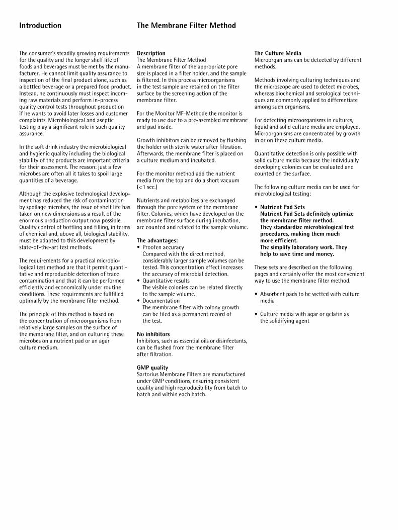
DescriptionThe Membrane Filter MethodA membrane filter of the appropriate pore size is placed in a filter holder, and the sampleis filtered. In this process microorganisms in the test sample are retained on the filtersurface by the screening action of the membrane filter.
For the Monitor MF-Methode the monitor isready to use due to a pre-asembled membraneand pad inside.
Growth inhibitors can be removed by flushingthe holder with sterile water after filtration.Afterwards, the membrane filter is placed on a culture medium and incubated.
For the monitor method add the nutrientmedia from the top and do a short vacuum(<1 sec.)
Nutrients and metabolites are exchangedthrough the pore system of the membrane filter. Colonies, which have developed on themembrane filter surface during incubation, are counted and related to the sample volume.
The advantages:• Proofen accuracy
Compared with the direct method, considerably larger sample volumes can betested. This concentration effect increasesthe accuracy of microbial detection.
• Quantitative resultsThe visible colonies can be related directlyto the sample volume.
• DocumentationThe membrane filter with colony growthcan be filed as a permanent record of the test.
No inhibitors Inhibitors, such as essential oils or disinfectants,can be flushed from the membrane filter after filtration.
GMP quality Sartorius Membrane Filters are manufacturedunder GMP conditions, ensuring consistentquality and high reproducibility from batch tobatch and within each batch.
The Culture MediaMicroorganisms can be detected by differentmethods.
Methods involving culturing techniques andthe microscope are used to detect microbes,whereas biochemical and serological techni-ques are commonly applied to differentiateamong such organisms.
For detecting microorganisms in cultures,liquid and solid culture media are employed.Microorganisms are concentrated by growthin or on these culture media.
Quantitative detection is only possible withsolid culture media because the individuallydeveloping colonies can be evaluated andcounted on the surface.
The following culture media can be used formicrobiological testing:
• Nutrient Pad SetsNutrient Pad Sets definitely optimize the membrane filter method.They standardize microbiological testprocedures, making them much more efficient.The simplify laboratory work. They help to save time and money.
These sets are described on the followingpages and certainly offer the most convenientway to use the membrane filter method.
• Absorbent pads to be wetted with culturemedia
• Culture media with agar or gelatin as the solidifying agent
Introduction
The consumer’s steadily growing requirementsfor the quality and the longer shelf life offoods and beverages must be met by the manu-facturer. He cannot limit quality assurance toinspection of the final product alone, such as a bottled beverage or a prepared food product.Instead, he continuously must inspect incom-ing raw materials and perform in-process quality control tests throughout production if he wants to avoid later losses and customercomplaints. Microbiological and aseptictesting play a significant role in such qualityassurance.
In the soft drink industry the microbiologicaland hygienic quality including the biologicalstability of the products are important criteriafor their assessment. The reason: just a fewmicrobes are often all it takes to spoil largequantities of a beverage.
Although the explosive technological develop-ment has reduced the risk of contamination by spoilage microbes, the issue of shelf life hastaken on new dimensions as a result of theenormous production output now possible.Quality control of bottling and filling, in termsof chemical and, above all, biological stability,must be adapted to this development by state-of-the-art test methods.
The requirements for a practical microbio-logical test method are that it permit quanti-tative and reproducible detection of tracecontamination and that it can be performedefficiently and economically under routineconditions. These requirements are fullfilledoptimally by the membrane filter method.
The principle of this method is based on the concentration of microorganisms fromrelatively large samples on the surface of the membrane filter, and on culturing thesemicrobes on a nutrient pad or an agar culture medium.
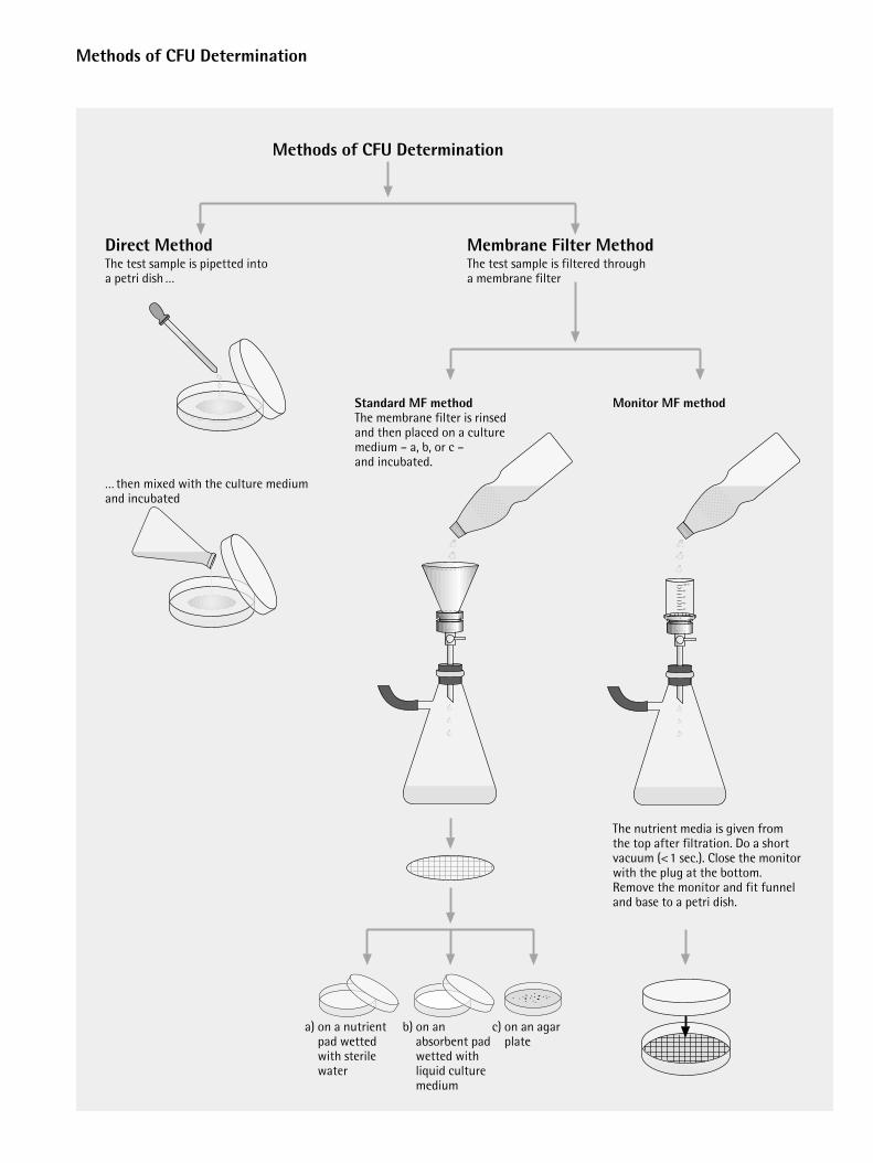
Standard MF methodThe membrane filter is rinsed and then placed on a culture medium – a, b, or c – and incubated.
Membrane Filter MethodThe test sample is filtered through a membrane filter
Methods of CFU Determination
Direct MethodThe test sample is pipetted into a petri dish …
… then mixed with the culture medium and incubated
Monitor MF method
The nutrient media is given from the top after filtration. Do a short vacuum (<1 sec.). Close the monitor with the plug at the bottom. Remove the monitor and fit funnel and base to a petri dish.
Methods of CFU Determination
a) on a nutrientpad wetted with sterilewater
b) on an absorbent padwetted withliquid culturemedium
c) on an agarplate
How to Handle MicroorganismsMicroorganism cultures must always behandled as carefully as if they containedpathogens.
Working with microorganisms is not dangerous if the following safety rules are observed:
Wash your hands thoroughly before and after working in a laboratory.
Do not eat or drink in a laboratory.
Do not touch bacterial matter with yourhands.
Never pipet bacteria suspensions with your mouth. Always use mechanical aids for pipetting (e.g., Peleus ball).
Before and after use, inoculating loops andwires must be sterilized by flaming until theyglow red-hot.
All laboratory equipment which has come incontact with bacteria must be sterilized.
To protect people and animals from conta-gious diseases or poisoning, living cultureshave to be destroyed before cleansing ordisposing of the containers. One method is to coat them thoroughly with disinfectants or to autoclave them in suitable containers.
User Benefits
Sartorius Nutrient Pad Sets have been used successfully in the membrane filter method for20 years. Practical and easy to handle, theyreduce labor and simplify many microbiologi-cal testing procedures.
Nutrient pads are sterile, dehydrated culturemedia. Once they are moistened with 3.0–3.5 mlof sterile and demineralized (or distilled) waterthey are ready to use immediately.
The level of moisture is optimal when anexcess ring of water surrounding the pad is visible.
All Nutrient Pad Set types are supplied with the appropriate membrane filters, which arealso presterilized and individually packaged.The membrane filters tailored to meet the special requirements of microbial detectionare available with 47 mm or 50 mm diameters.
A standard package contains 100 sterilenutrient pads each “preplated” in a petri dish(each bag contains 10 petri dishes) and 100 individually sterile packed membrane filters.
Economical
Eliminates time-consuming and labor- • After wetting with 3,5 ml destilled water intensive preparation of culture media NPS are ready to use: NPS and go(sterilization and cleaning, among others).
Simple to use
Nutrient Pad Sets can also be used in • Everyone can use NPSlaboratories which do not have extensive microbiological equipment. Sterile water for moistening the pads can be prepared easily with a Sartorius Dosing Syringe and an attached Syringe Filter Holder (0,2 µm).
Consistent quality
During manufacture, each type of Nutrient • NPS are validated. In comparison of agar Pad Set is compared with the corresponding which is done within different deviations agar medium with respect to their growth- of amount and height NPS gives always promoting properties. This QA procedure constant resultsensures consistent quality and reproducible results.
Trouble-free storage
Nutrient Pad Sets have a shelf life of • No waste or overproduction9 to at least 24 months at room temperature.
Highly versatile
Nutrient Pad Sets can be modified by additives • Advanced systemin the solution used to wet them; for example, Wort or Orange Serum Nutrient Pads when wetted with 5 % ethanol promote the growth of acetic-acid bacteria.
General Procedure.To obtain reliable results for microbiologicaltests, it is necessary to work under conditionsthat rule out contamination by microorga-nisms which distort such results.
That is why you should work near the flame of a Bunsen burner in a room protected fromdrafts. Before beginning with the actual procedure, spray or wash down your work areawith a disinfectant (e.g., 70% alcohol).
Before use, filter holders, forceps and scissorsshould be sterilized by one of the standardmethods, such as flaming for routine tests.
Nutrient Pad Sets
General Directions
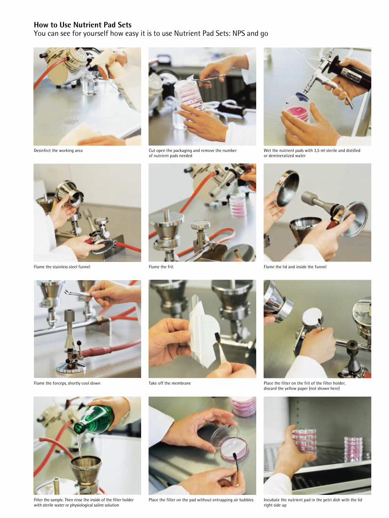
Filter the sample. Then rinse the inside of the filter holderwith sterile water or physiological saline solution
Place the filter on the pad without entrapping air bubbles Incubate the nutrient pad in the petri dish with the lidright side up
Flame the forceps, shortly cool down Take off the membrane Place the filter on the frit of the filter holder, discard the yellow paper (not shown here)
Flame the stainless steel funnel Flame the frit Flame the lid and inside the funnel
Desinfect the working area Cut open the packaging and remove the number of nutrient pads needed
Wet the nutrient pads with 3,5 ml sterile and distilled or demineralized water
How to Use Nutrient Pad SetsYou can see for yourself how easy it is to use Nutrient Pad Sets: NPS and go
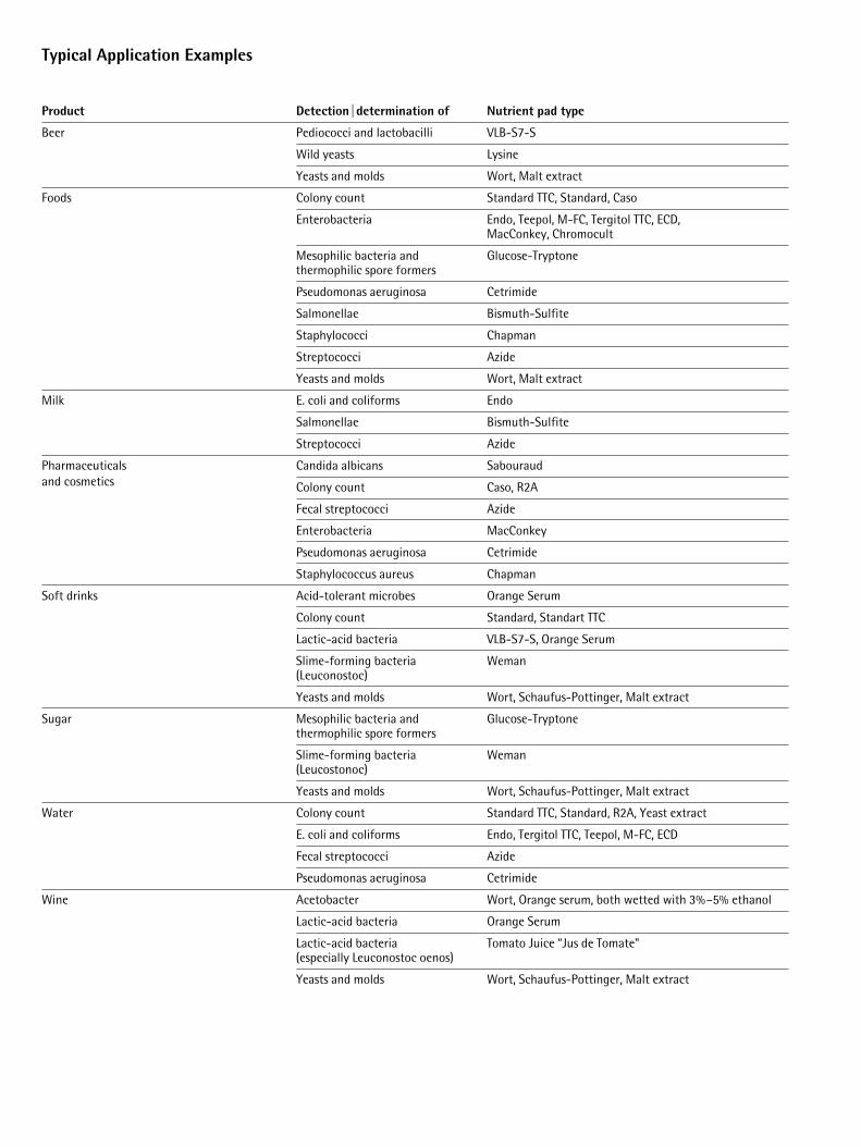
Typical Application Examples
Product Detection|determination of Nutrient pad type
Beer Pediococci and lactobacilli VLB-S7-S
Wild yeasts Lysine
Yeasts and molds Wort, Malt extract
Foods Colony count Standard TTC, Standard, Caso
Enterobacteria Endo, Teepol, M-FC, Tergitol TTC, ECD, MacConkey, Chromocult
Mesophilic bacteria and Glucose-Tryptonethermophilic spore formers
Pseudomonas aeruginosa Cetrimide
Salmonellae Bismuth-Sulfite
Staphylococci Chapman
Streptococci Azide
Yeasts and molds Wort, Malt extract
Milk E. coli and coliforms Endo
Salmonellae Bismuth-Sulfite
Streptococci Azide
Pharmaceuticals Candida albicans Sabouraudand cosmetics Colony count Caso, R2A
Fecal streptococci Azide
Enterobacteria MacConkey
Pseudomonas aeruginosa Cetrimide
Staphylococcus aureus Chapman
Soft drinks Acid-tolerant microbes Orange Serum
Colony count Standard, Standart TTC
Lactic-acid bacteria VLB-S7-S, Orange Serum
Slime-forming bacteria Weman(Leuconostoc)
Yeasts and molds Wort, Schaufus-Pottinger, Malt extract
Sugar Mesophilic bacteria and Glucose-Tryptonethermophilic spore formers
Slime-forming bacteria Weman(Leucostonoc)
Yeasts and molds Wort, Schaufus-Pottinger, Malt extract
Water Colony count Standard TTC, Standard, R2A, Yeast extract
E. coli and coliforms Endo, Tergitol TTC, Teepol, M-FC, ECD
Fecal streptococci Azide
Pseudomonas aeruginosa Cetrimide
Wine Acetobacter Wort, Orange serum, both wetted with 3%–5% ethanol
Lactic-acid bacteria Orange Serum
Lactic-acid bacteria Tomato Juice “Jus de Tomate"(especially Leuconostoc oenos)
Yeasts and molds Wort, Schaufus-Pottinger, Malt extract
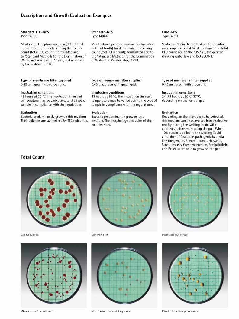
Total Count
Bacillus subtilis
Mixed culture from well water
Escherichia coli Staphylococcus aureus
Mixed culture from process waterMixed culture from drinking water
Description and Growth Evaluation Examples
Standard TTC-NPSType 14055
Meat extract-peptone medium (dehydratednutrient broth) for determining the colonycount (total CFU count); formulated acc. to “Standard Methods for the Examination ofWater and Wastewater”, 1998, and modified by the addition of TTC.
Type of membrane filter supplied0.45 µm; green with green grid.
Incubation conditions48 hours at 30 °C. The incubation time andtemperature may be varied acc. to the type ofsample in compliance with the regulations.
EvaluationBacteria predominantly grow on this medium.Their colonies are stained red by TTC reduction.
Standard-NPSType 14064
Meat extract-peptone medium (dehydratednutrient broth) for determining the colonycount (total CFU count); formulated acc. tothe “Standard Methods for the Examination of Water and Wastewater,” 1998.
Type of membrane filter supplied0.45 µm; green with green grid.
Incubation conditions48 hours at 30 °C. The incubation time andtemperature may be varied acc. to the type ofsample in compliance with the regulations.
EvaluationBacteria predominantly grow on this medium. The morphology and color of theircolonies vary.
Caso-NPSType 14063
Soybean-Casein Digest Medium for isolatingmicroorganisms and for determining the totalCFU count acc. to the “USP 25, the germandrinking water law and ISO 9308-1.”
Type of membrane filter supplied0.45 µm; green with green grid
Incubation conditions 24–72 hours at 30°C–37°C, depending on the test sample
Evaluation Depending on the microbes to be detected,this medium can be converted into a selectiveone by mixing the wetting liquid with additives before moistening the pad. When10% serum is added to the wetting liquid a number of fastidious pathogenic bacterialike the genuses Pneumococcus, Neisseria,Streptococcus, Corynebacterium, Erysipelothrixand Brucella are able to grow on the pad.
Description and Growth Evaluation Examples
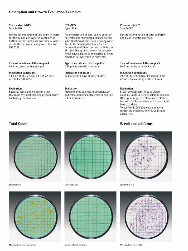
Escherichia coli
Mixed culture from water
R2A-NPSType 14084
For the detection of total colony count ofheterotrophic microorganisms and for thesubcultivation of bacteria in drinking water.Acc. to the Standard Methods for the Examination of Water and Waste Water andEP 1998. The optimal growth for bacteriawhich have adapted to the particular livingconditions of water low in nutrients.
Type of membrane Filter supplied 0.45 µm; green with green grid
Incubation conditions 72 h at 35°C; 5 days at 20°C or 28°C
EvaluationPredominantly colonies of different size and color, predominantly white or colorless <1 mm diameter.
Escherichia coli
Mixed culture from water
Yeast extract-NPS Type 14090
For the determination of CFU count in waterfor the human use, water in container orbottles for the human use and mineral water,acc. to the German drinking water law andISO 6222.
Type of membrane Filter supplied0.45 µm; green with green grid
Incubation conclitions44 ±4 h at 36 ±2°C; 68 ±4 h at 22 ±2°Cacc. to EN ISO 6222.
EvaluationBacteria, yeasts and molds can grow. Due to no dye most colonies, predominantlybacteria, grow colorless.
Escherichia coli
Mixed culture from river water
Total Count E. coli and coliforms
Chromocult-NPSType 14087
For the determination of total coliforms and E.Coli in water and food.
Type of membrane Filter supplied0.45 µm; white with black grid
Incubation conclitions24 h at 36 ±1°C; longer incubation timedisturbs the counting of the colonies.
EvaluationE. Coli develops dark-blue to violett colonies. Coliforme red to salmone colonies.Other gramnegative colonies are colorless,few with ß-Glucuronidase activity are lightblue to turkese.To confirm E. Coli give Kovacs reagent to dark blue colonies. Then E. Coli stains cherry-red.
Description and Growth Evaluation Examples
E. coli and coliforms
Escherichia coli
E. coli and coliforms from river water
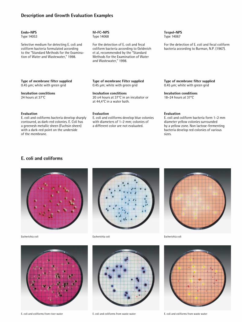
M-FC-NPSType 14068
For the detection of E. coli and fecal coliform bacteria according to Geldreich et al, recommended by the “StandardMethods for the Examination of Water and Wastewater,” 1998.
Type of membrane Filter supplied0.45 µm; white with green grid
Incubation conclitions20 ±4 hours at 37°C in an incubator or at 44,4°C in a water bath.
EvaluationE. coli and coliforms develop blue colonieswith diameters of 1–2 mm; colonies of a different color are not evaluated.
Escherichia coli
E. coli and coliforms from waste water
Escherichia coli
E. coli and coliforms from waste water
Endo-NPS Type 14053
Selective medium for detecting E. coli andcoliform bacteria formulated according to the “Standard Methods for the Examina-tion of Water and Wastewater,” 1998.
Type of membrane filter supplied0.45 µm; white with green grid
Incubation conclitions24 hours at 37°C
EvaluationE. coli and coliforms bacteria develop sharplycontoured, as dark-red colonies. E. Coli has a greenesh metallic sheen (Fuchsin sheen)with a dark-red point on the underside of the membrane.
Teepol-NPSType 14067
For the detection of E. coli and fecal coliformbacteria according to Burman, N.P. (1967).
Type of membrane filter supplied0.45 µm; white with green grid
Incubation conditions18–24 hours at 37°C
EvaluationE. coli and coliform bacteria form 1–2 mmdiameter yellow colonies surrounded by a yellow zone. Non lactose-fermentingbacteria develop red colonies of various sizes.
Description and Growth Evaluation Examples
E. coli and coliforms
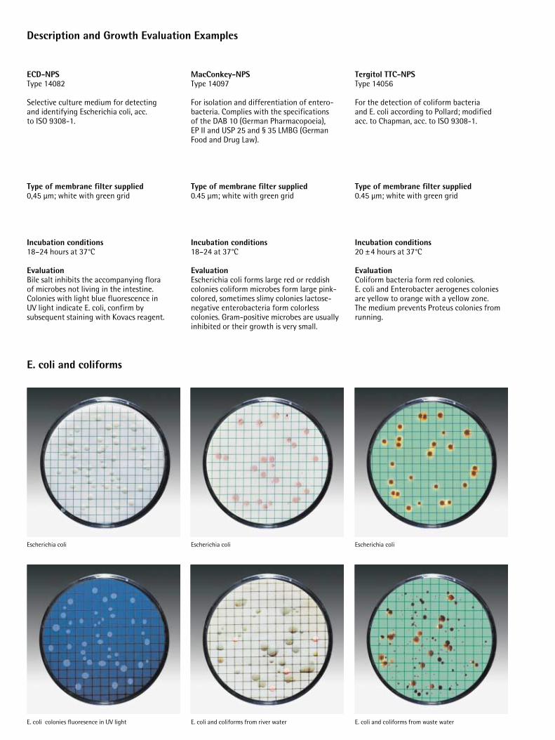
Tergitol TTC-NPSType 14056
For the detection of coliform bacteria and E. coli according to Pollard; modified acc. to Chapman, acc. to ISO 9308-1.
Type of membrane filter supplied0.45 µm; white with green grid
Incubation conditions20 ±4 hours at 37°C
EvaluationColiform bacteria form red colonies. E. coli and Enterobacter aerogenes coloniesare yellow to orange with a yellow zone. The medium prevents Proteus colonies fromrunning.
ECD-NPSType 14082
Selective culture medium for detecting and identifying Escherichia coli, acc. to ISO 9308-1.
Type of membrane filter supplied0,45 µm; white with green grid
Incubation conditions18–24 hours at 37°C
EvaluationBile salt inhibits the accompanying flora of microbes not living in the intestine. Colonies with light blue fluorescence in UV light indicate E. coli, confirm by subsequent staining with Kovacs reagent.
Escherichia coli
E. coli and coliforms from waste water
Escherichia coli
E. coli and coliforms from river water
Escherichia coli
E. coli colonies fluoresence in UV light
MacConkey-NPSType 14097
For isolation and differentiation of entero-bacteria. Complies with the specifications of the DAB 10 (German Pharmacopoeia), EP II and USP 25 and § 35 LMBG (GermanFood and Drug Law).
Type of membrane filter supplied0.45 µm; white with green grid
Incubation conditions 18–24 at 37°C
EvaluationEscherichia coli forms large red or reddishcolonies coliform microbes form large pink-colored, sometimes slimy colonies lactose-negative enterobacteria form colorless colonies. Gram-positive microbes are usuallyinhibited or their growth is very small.

Description and Growth Evaluation Examples
Yeasts and molds
Wort-NPSType 14058
For the detection of yeasts and molds. This culture medium is used in production and quality control testing in the food, pharmaceutical and cosmetics industries,among other applications.
Type of membrane filter supplied0.65 µm; grey with white grid
Incubation conditions2–3 days at 25°C
EvaluationYeasts usually develop smooth white or colored colonies. Molds generally form velvetyor fluffy cotton-like colonies in the earlygrowth phase and may take on various colorsafter conidiospore production.
Sabouraud-NPS Type 14069
For culturing yeasts, molds, acid-tolerant andacidophilic bacteria; also for detecting yeastsand molds in beverages such as fruit juices,sterility testing of pharmaceuticals and forisolating of matopathogenic yeasts and fungi.According to USP 25.
Type of membrane filter supplied0.65 µm; grey with white grid
Incubation conditions2–5 days at 25–30°C
EvaluationYeasts usually develop smooth white or colored colonies. Molds generally form velvety or fluffy, cotton-like colonies in theearly growth phase and may take on variouscolors after conidiospore production.
Schaufus-Pottinger-NPSa) Type 14070b) Type 14072c) Type 14080d) Type 14083
For detection and determination of the total CFU count of yeasts and molds in beverages and sugar according to Schaufusand Pottinger.
Type of membrane filter supplieda) 0.65 µm; white with green gridb) 1.2 µm; white with green gridc) 0,8 µm; grey with white gridd) 0,65 µm; grey with white grid
Incubation conditions2–3 days at 28–30°C
EvaluationMolds develop velvety or fluffy whitish orgreenish colonies which can take on variouscolors after conidiospore production. Yeastand bacteria colonies have smooth surfaces.Acid forming sugar fermenters are whitish to yellow non-acid formers are, by contrast,greenish to blue-green.
Saccharomyces cerevisiae
Yeasts and molds from spoiled beer
Alternaria humicola
Yeasts and molds from cough syrup
Torula lipolytica
Mixed culture from a soft drink
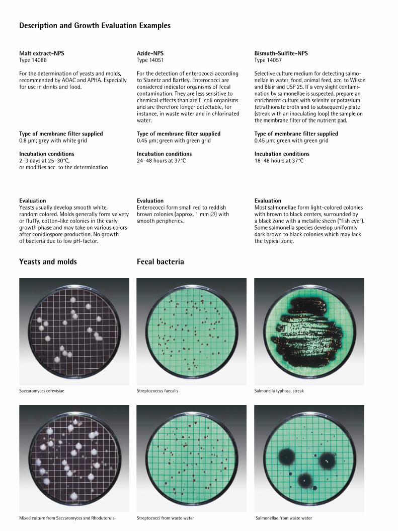
Azide-NPSType 14051
For the detection of enterococci according to Slanetz and Bartley. Enterococci are considered indicator organisms of fecal contamination. They are less sensitive to chemical effects than are E. coli organismsand are therefore longer detectable, forinstance, in waste water and in chlorinatedwater.
Type of membrane filter supplied0.45 µm; green with green grid
Incubation conditions24–48 hours at 37°C
EvaluationEnterococci form small red to reddish brown colonies (approx. 1 mm d) withsmooth peripheries.
Bismuth-Sulfite-NPSType 14057
Selective culture medium for detecting salmo-nellae in water, food, animal feed, acc. to Wilsonand Blair and USP 25. If a very slight contami-nation by salmonellae is suspected, prepare anenrichment culture with selenite or potassiumtetrathionate broth and to subsequentIy plate(streak with an inoculating loop) the sample onthe membrane filter of the nutrient pad.
Type of membrane filter supplied0.45 µm; green with green grid
Incubation conditions18–48 hours at 37°C
EvaluationMost salmonellae form light-colored colonieswith brown to black centers, surrounded by a black zone with a metallic sheen (“fish eye”).Some salmonella species develop uniformlydark brown to black colonies which may lackthe typical zone.
Streptococcus faecalis
Streptococci from waste water
Salmonella typhosa, streak
Salmonellae from waste water
Malt extract-NPSType 14086
For the determination of yeasts and molds,recommended by AOAC and APHA. Especiallyfor use in drinks and food.
Type of membrane filter supplied0.8 µm; grey with white grid
Incubation conditions2–3 days at 25–30°C, or modifies acc. to the determination
EvaluationYeasts usually develop smooth white, random colored. Molds generally form velvetyor fluffy, cotton-like colonies in the earlygrowth phase and may take on various colorsafter conidiospore production. No growth of bacteria due to low pH-factor.
Saccaromyces cerevisiae
Mixed culture from Saccaromyces and Rhodutorula
Description and Growth Evaluation Examples
Yeasts and molds Fecal bacteria
Description and Growth Evaluation Examples
Product Spoiling bacteria
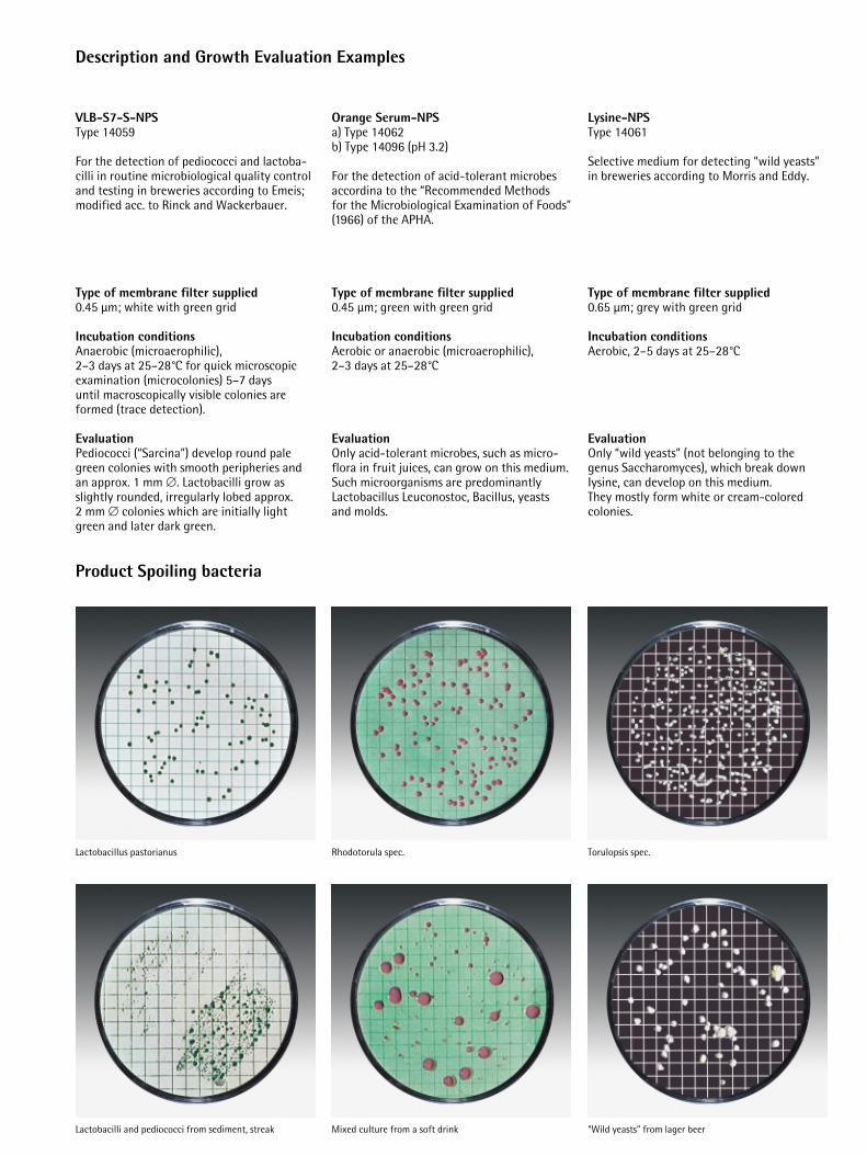
VLB-S7-S-NPSType 14059
For the detection of pediococci and lactoba-cilli in routine microbiological quality controland testing in breweries according to Emeis;modified acc. to Rinck and Wackerbauer.
Type of membrane filter supplied0.45 µm; white with green grid
Incubation conditionsAnaerobic (microaerophilic), 2–3 days at 25–28°C for quick microscopicexamination (microcolonies) 5–7 days until macroscopically visible colonies are formed (trace detection).
EvaluationPediococci (“Sarcina”) develop round palegreen colonies with smooth peripheries andan approx. 1 mm d. Lactobacilli grow asslightly rounded, irregularly lobed approx. 2 mm d colonies which are initially lightgreen and later dark green.
Lactobacillus pastorianus
Lactobacilli and pediococci from sediment, streak
Orange Serum-NPSa) Type 14062b) Type 14096 (pH 3.2)
For the detection of acid-tolerant microbesaccordina to the “Recommended Methods for the Microbiological Examination of Foods”(1966) of the APHA.
Type of membrane filter supplied0.45 µm; green with green grid
Incubation conditionsAerobic or anaerobic (microaerophilic), 2–3 days at 25–28°C
EvaluationOnly acid-tolerant microbes, such as micro-flora in fruit juices, can grow on this medium.Such microorganisms are predominantly Lactobacillus Leuconostoc, Bacillus, yeastsand molds.
Rhodotorula spec.
Mixed culture from a soft drink
Lysine-NPSType 14061
Selective medium for detecting “wild yeasts”in breweries according to Morris and Eddy.
Type of membrane filter supplied0.65 µm; grey with green grid
Incubation conditionsAerobic, 2–5 days at 25–28°C
EvaluationOnly “wild yeasts” (not belonging to thegenus Saccharomyces), which break downIysine, can develop on this medium. They mostly form white or cream-coloredcolonies.
Torulopsis spec.
“Wild yeasts” from lager beer
Description and Growth Evaluation Examples
Product Spoiling bacteria
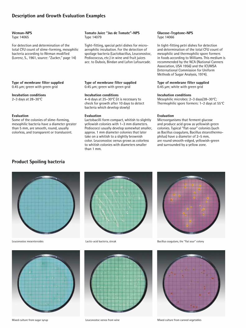
Tomato Juice “Jus de Tomate”-NPSType 14079
Tight-fitting, special petri dishes for micro-aerophilic incubation. For the detection ofspoilage bacteria (Lactobacillus, Leuconostoc,Pediococcus, etc.) in wine and fruit juices acc. to Dubois, Bindan and Lafon Lafourcade.
Type of membrane filter supplied0.45 µm; green with green grid
Incubation conditions 4–6 days at 25–30°C (it is necessary to check for growth after 10 days to detect bacteria which develop slowly)
EvaluationLactobacilli form compact, whitish to slightlyyellowish colonies with 1–3 mm diameters.Pediococci usually develop somewhat smaller,approx. 1 mm diameter colonies that latertake on a whitish to a slightly brownish color. Leuconostoc oenus grows as colorlessto whitish colonies with diameters smallerthan 1 mm.
Lactic-acid bacteria, streak
Leuconostoc oenos from wine
Glucose-Tryptone-NPSType 14066
In tight-fitting petri dishes for detection and determination of the total CFU count ofmesophilic and thermophilic spore formers in foods according to Williams. This medium isrecommended by the NCA (National CannersAssociation, USA 1956) and the ICUMSA(lnternational Commission for UniformMethods of Sugar Analysis, 1974).
Type of membrane filter supplied0.45 µm; white with green grid
Incubation conditionsMesophilic microbes: 2–3 days/28–30°C; Thermophilic spore formers: 1–2 days at 55°C
EvaluationMicroorganisms that ferment glucose and produce acid grow as yellowish green colonies. Typical “flat-sour” colonies (such as Bacillus coagulans, Bacillus stearothermo-philus) have a diameter of 2–5 mm, are round smooth-edged, yellowish-greenand surrounded by a yellow zone.
Weman-NPSType 14065
For detection and determination of the total CFU count of slime-forming, mesophilicbacteria according to Weman modified(Lorenz, S., 1961, source: “Zucker,” page 14)
Type of membrane filter supplied0.45 µm; green with green grid
Incubation conditions2–3 days at 28–30°C
EvaluationSome of the colonies of slime-forming, mesophilic bacteria have a diameter greaterthan 5 mm, are smooth, round, usually colorless, and transparent or translucent.
Bacillus coagulans, the “flat sour” colony
Mixed culture from canned vegetables
Leuconostoc mesenteroides
Mixed culture from sugar syrup
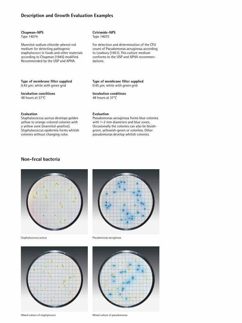
Staphylococcus aureus
Mixed culture of staphylococci
Pseudomonas aeruginosa
Mixed culture of pseudomonas
Description and Growth Evaluation Examples
Non-fecal bacteria
Chapman-NPSType 14074
Mannitol-sodium chloride-phenol redmedium for detecting pathogenic staphylococci in foods and other materialsaccording to Chapman (1945) modified.Recommended by the USP and APHA.
Type of membrane filter supplied0.45 µm; white with green grid
Incubation conclitions48 hours at 37°C
EvaluationStaphylococcus aureus develops golden yellow to orange-colored colonies with a yellow zone (mannitol-positive). Staphylococcus epidermis forms whitish colonies without changing color.
Cetrimide-NPSType 14075
For detection and determination of the CFUcount of Pseudomonas aeruginosa accordingto Lowbury (1951). This culture medium conforms to the USP and APHA recommen-dations.
Type of membrane filter supplied0.45 µm; white with green grid
Incubation conditions48 hours at 37°C
EvaluationPseudomonas aeruginosa forms blue colonieswith 1–2 mm diameters and blue zones. Occasionally the colonies can also be bluish-green, yellowish-green or colorless. Otherpseudomonas develop whitish colonies.
Troubleshooting Guide
Failure to follow the directions may lead to unsatisfactory results listed below:
1. Inhibited growth, dwarf colonies
pad too dry: not enough water used
2. Colonies run
pad too wet, water film on the membranefilter: too much water used. Colonies of motile microbes (such as Bacillusor Proteus) tend to run even though thewater dosage is correct. To prevent this,add NaCI or a similar emulsifier.
3. Contamination from underneath
inhibited colony growth, excess ring of liquid cloudy, often including discoloration of the pad:
a) membrane placed with grid facedown on pad instead of faceup
b) contamination of the water used forrehydration
c) contamination during preparation (by airborne microbes or by contact)
d) microbes rinsed off the membrane filterby incomplete vacuum filtration of the sample or rinse liquid or by tiltingthe prepared petri dish
e) contaminated filter support
f) contaminated forceps
4. Growth on one side only
petri dish slanted in the incubator
5. Too profuse or too sparse growth (optimum microbial number between 20 and 200 per filter)
wrong dilution selected or sample inadequately mixed with the diluent.
6. Non-uniform growth
sample volume less than 5 ml filteredwithout adding sterile water as a diluent or sample volume inadequately mixed with the diluent.
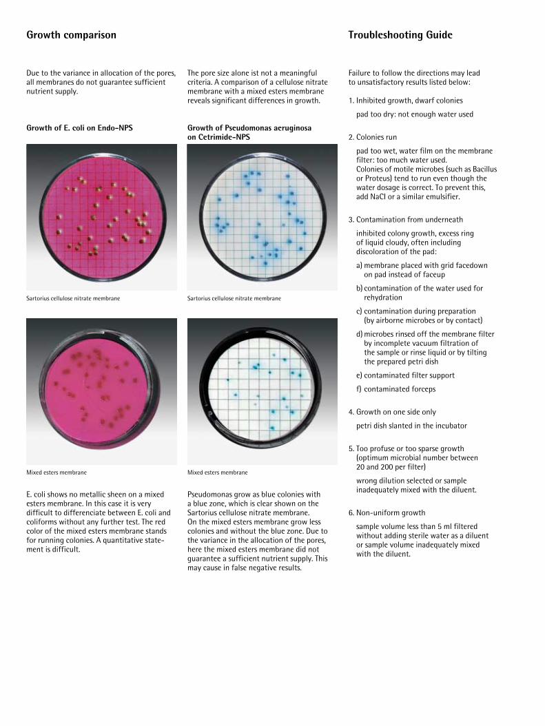
Mixed esters membrane Mixed esters membrane
Sartorius cellulose nitrate membrane
Due to the variance in allocation of the pores,all membranes do not guarantee sufficientnutrient supply.
Growth of E. coli on Endo-NPS
E. coli shows no metallic sheen on a mixedesters membrane. In this case it is very difficult to differenciate between E. coli andcoliforms without any further test. The redcolor of the mixed esters membrane standsfor running colonies. A quantitative state-ment is difficult.
Growth comparison
The pore size alone ist not a meaningful criteria. A comparison of a cellulose nitratemembrane with a mixed esters membranereveals significant differences in growth.
Growth of Pseudomonas aeruginosa on Cetrimide-NPS
Pseudomonas grow as blue colonies with a blue zone, which is clear shown on the Sartorius cellulose nitrate membrane. On the mixed esters membrane grow lesscolonies and without the blue zone. Due tothe variance in the allocation of the pores,here the mixed esters membrane did not guarantee a sufficient nutrient supply. Thismay cause in false negative results.
Sartorius cellulose nitrate membrane
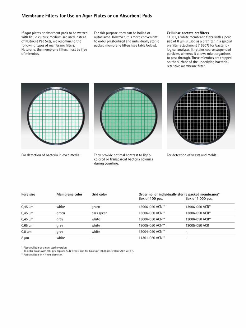
For this purpose, they can be boiled or autoclaved. However, it is more convenient to order presterilized and individually sterilepacked membrane filters (see table below).
Cellulose acetate prefilters 11301, a white membrane filter with a poresize of 8 µm is used as a prefilter in a specialprefilter attachment (16807) for bacterio-logical analyses. It retains coarse suspendedparticles, whereas it allows microorganisms to pass through. These microbes are trappedon the surface of the underlying bacteria-retentive membrane filter.
For detection of bacteria in dyed media. They provide optimal contrast to light-colored or transparent bacteria coloniesduring counting.
For detection of yeasts and molds.
Membrane Filters for Use on Agar Plates or on Absorbent Pads
If agar plates or absorbent pads to be wettedwith liquid culture medium are used insteadof Nutrient Pad Sets, we recommend the following types of membrane filters. Naturally, the membrane filters must be freeof microbes.
Pore size Membrane color Grid color Order no. of individually sterile packed membranes*Box of 100 pcs. Box of 1,000 pcs.
0,45 µm white green 13906-050 ACN** 13906-050 ACR**
0,45 µm green dark green 13806-050 ACN** 13806-050 ACR**
0,45 µm grey white 13006-050 ACN** 13006-050 ACR**
0,65 µm grey white 13005-050 ACN** 13005-050 ACR
0,8 µm grey white 13004-050 ACN** -
8 µm white – 11301-050 ACN** -
* Also available as a non-sterile version.To order boxes with 100 pcs. replace ACN with N and for boxes of 1,000 pcs. replace ACR with R.
** Also available in 47 mm diameter.
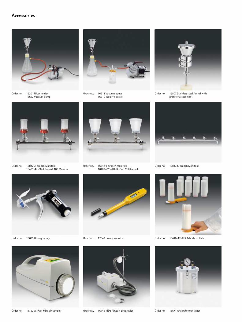
Accessories
Order no. 16757 AirPort MD8 air sampler Order no. 16746 MD8 Airscan air sampler Order no. 16671 Anaerobic container
Order no. 16685 Dosing syringe Order no. 17649 Colony counter Order no. 15410-47-ALR Adsorbent Pads
Order no. 16842 3-branch Manifold16401-47-06-K BioSart 100 Monitor
Order no. 16842 3-branch Manifold16407--25-ALK BioSart 250 Funnel
Order no. 16843 6-branch Manifold
Order no. 16201 Filter holder16692 Vacuum pump
Order no. 16612 Vacuum pump16610 Woulff’s bottle
Order no. 16807 Stainless steel funnel with prefilter attachment
Order no.
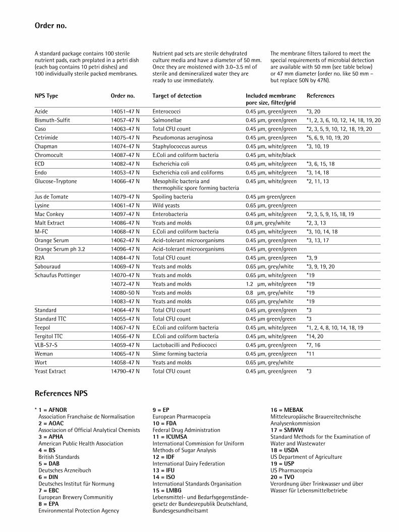
* 1 = AFNORAssociation Franchaise de Normalisation 2 = AOACAssociacion of Official Analytical Chemists3 = APHAAmerican Public Health Association4 = BSBritish Standards5 = DABDeutsches Arzneibuch6 = DINDeutsches Institut für Normung7 = EBCEuropean Brewery Communitiy8 = EPAEnvironmental Protection Agency
9 = EPEuropean Pharmacopeia10 = FDAFederal Drug Administration11 = ICUMSAInternational Commission for Uniform Methods of Sugar Analysis12 = IDFInternational Dairy Federation13 = IFU14 = ISOInternational Standards Organisation15 = LMBGLebensmittel- und Bedarfsgegenstände-gesetz der Bundesrepublik Deutschland,Bundesgesundheitsamt
16 = MEBAKMitteleuropäische Brauereitechnische Analysenkommission17 = SMWWStandard Methods for the Examination of Water and Wastewater18 = USDAUS Department of Agriculture19 = USPUS Pharmacopeia20 = TVOVerordnung über Trinkwasser und über Wasser für Lebensmittelbetriebe
NPS Type Order no. Target of detection Included membrane Referencespore size, filter/grid
Azide 14051-47 N Enterococci 0.45 µm, green/green *3, 20Bismuth-Sulfit 14057-47 N Salmonellae 0.45 µm, green/green *1, 2, 3, 6, 10, 12, 14, 18, 19, 20Caso 14063-47 N Total CFU count 0.45 µm, green/green *2, 3, 5, 9, 10, 12, 18, 19, 20Cetrimide 14075-47 N Pseudomonas aeruginosa 0.45 µm, green/green *5, 6, 9, 10, 19, 20Chapman 14074-47 N Staphylococcus aureus 0.45 µm, white/green *3, 10, 19Chromocult 14087-47 N E.Coli and coliform bacteria 0.45 µm, white/blackECD 14082-47 N Escherichia coli 0.45 µm, white/green *3, 6, 15, 18Endo 14053-47 N Escherichia coli and coliforms 0.45 µm, white/green *3, 14, 18Glucose-Tryptone 14066-47 N Mesophilic bacteria and 0.45 µm, white/green *2, 11, 13
thermophilic spore forming bacteria Jus de Tomate 14079-47 N Spoiling bacteria 0.45 µm green/greenLysine 14061-47 N Wild yeasts 0.65 µm, green/greenMac Conkey 14097-47 N Enterobacteria 0.45 µm, white/green *2, 3, 5, 9, 15, 18, 19Malt Extract 14086-47 N Yeats and molds 0.8 µm, grey/white *2, 3, 13M-FC 14068-47 N E.Coli and coliform bacteria 0.45 µm, white/green *3, 10, 14, 18Orange Serum 14062-47 N Acid-tolerant microorganisms 0.45 µm, green/green *3, 13, 17Orange Serum ph 3.2 14096-47 N Acid-tolerant microorganisms 0.45 µm, green/greenR2A 14084-47 N Total CFU count 0.45 µm, green/green *3, 9Sabouraud 14069-47 N Yeats and molds 0.65 µm, grey/white *3, 9, 19, 20Schaufus Pottinger 14070-47 N Yeats and molds 0.65 µm, white/green *19
14072-47 N Yeats and molds 1.2 µm, white/green *1914080-50 N Yeats and molds 0.8 µm, grey/white *1914083-47 N Yeats and molds 0.65 µm, grey/white *19
Standard 14064-47 N Total CFU count 0.45 µm, green/green *3Standard TTC 14055-47 N Total CFU count 0.45 µm green/green *3Teepol 14067-47 N E.Coli and coliform bacteria 0.45 µm, white/green *1, 2, 4, 8, 10, 14, 18, 19Tergitol TTC 14056-47 N E.Coli and coliform bacteria 0.45 µm, white/green *14, 20VLB-S7-S 14059-47 N Lactobacilli and Pediococci 0.45 µm, green/green *7, 16Weman 14065-47 N Slime forming bacteria 0.45 µm, green/green *11Wort 14058-47 N Yeats and molds 0.65 µm, grey/whiteYeast Extract 14790-47 N Total CFU count 0.45 µm, green/green *3
References NPS
A standard package contains 100 sterilenutrient pads, each preplated in a petri dish(each bag contains 10 petri dishes) and 100 individually sterile packed membranes.
Nutrient pad sets are sterile dehydrated culture media and have a diameter of 50 mm.Once they are moistened with 3.0–3.5 ml ofsterile and demineralized water they areready to use immediately.
The membrane filters tailored to meet thespecial requirements of microbial detectionare available with 50 mm (see table below) or 47 mm diameter (order no. like 50 mm –but replace 50N by 47N).
Specifications subject to change without notice. Printed in Germany on paper that has been bleached without any use of chlorine.W/sart-119a · GPublication No.: SM-4017-e02067Order No.: 85030-503-99
Sartorius AGWeender Landstrasse 94–10837075 Goettingen, Germany
Phone +49.551.308.0 Fax +49.551.308.3289
www.sartorius.com
Sartorius Corporation131 Heartland Boulevard, Edgewood, New York 11717, USA
Phone +1.631.2544274Fax +1.631.2544253Toll-Free +1.800.3687178
Sartorius LimitedLongmead Business Centre Blenheim Road, Epsom, Surrey KT199QN, Great Britain
Phone +44.1372.737100Fax +44.1372.729972
Sartorius S.A.4, rue Emile Baudot91127 Palaiseau, France
Phone +33.1.69192100 Fax +33.1.69200922
Sartorius S.p.AVia dell’Antella, 76/A50011 Antella (FI), Italy
Phone +39.055.634041Fax +39.055.6340526
Sartorius K.K.No. 3 Hoya Building 8–17Kamitakaido 1-chome Suginami-ku Tokyo 168-0074, Japan
Phone +81.3.33295533Fax +81.3.33295543
Sartorius S.A.C/Isabel Colbrand 10-12Edificio Alfa IIIplanta 4, of. 12128050 Madrid, Spain
Phone +34.91.3586100Fax +34.91.3588804