metatarsalgia and lesser metatarsal surgery · metatarsalgia and lesser metatarsal surgery vincent...
TRANSCRIPT

29
Metatarsalgia and Lesser Metatarsal Surgery
VINCENT J. HETHERINGTON
Metatarsalgia is defined as pain localized to the fore- part of the foot. Although the definition is simple enough, it describes only a symptom: metatarsalgia can be a complex clinical problem. Pain under the metatarsals may be a reflection of systemic disease, a result of biomechanical imbalances, or local foot pa- thology.
CLASSIFICATION
Several authors have attempted to classify metatarsal- gia. Helal1 classified metatarsalgia into primary, sec- ondary, and unrelated to weight distribution. Primary metatarsalgia is defined as pain across the area of the metatarsophalangeal joints associated with plantar keratoses. It may be a result of functional problems such as wearing a high-heeled shoe or structural prob- lems associated with, for example, a short first meta- tarsal, limb length discrepancies, or pes cavus. Also included by Helal as primary metatarsalgia were Freiberg's disease, hallux valgus, hallux rigidus, and problems of iatrogenic and traumatic origin. Second- ary metatarsalgia was caused by systemic disease such as rheumatoid arthritis. Metatarsalgias attributed to tar- sal tunnel and vascular insufficiency are those types of metatarsalgia unrelated to load distribution.
Regnauld2 defined metatarsalgia into diffuse, local- ized, subcutaneous soft tissue, and cutaneous metatar- salgia. Diffuse metatarsalgia is an example of an ac- quired mechanical disorder, such as functional disorders, abnormalities of the ankle such as ankle equinus, overload of the metatarsals, and metatarsalgia
from high-heeled shoes. Localized metatarsalgia results from an osseous disorder such as a long meta- tarsal, large metatarsal, or aplasia of a metatarsal. Sub- cutaneous soft metatarsalgia includes pain from inter- metatarsal neuroma, articular synovial cysts, and bursa. Cutaneous metatarsalgia is a result of plantar calluses, verruca, or ulcerations of the skin. Viladot3
describes metatarsalgia in two categories: first, over- load of anterior support as a result of high-heeled shoes, an equinus or cavus foot in which support is by the heel, and the metatarsal heads only, resulting in subluxation of the toes, a change in load, and change in cadence of gait. The second aspect of this classifica- tion is irregular distribution of mechanical load, as evidenced by the following symptoms:
First-ray insufficiency syndrome, examples of which are a short first metatarsal and hypermobility of the first ray. It may also be result iatrogenically follow- ing osteotomy of the metatarsal or Keller resection.
First-ray overload syndrome, which may be a result of chronic overload, plantar flexion of the first meta- tarsal, an index plus, or excessively long first meta- tarsal or first ray.
Central ray insufficiency syndrome, which may arise from congenital, iatrogenic, or neurogenic causes.
Central ray overload syndrome, attributed to long metatarsal or plantar-flexed metatarsals.
Scranton4 described metatarsalgia as primary, sec- ondary, and pain under the forepart of the foot. Pri- mary metatarsalgia included hallux rigidus, a long first ray, Freiberg's disease, hallux valgus, and postsurgical
429

430 HALLUX VALGUS AND FOREFOOT SURGERY
deformities. Secondary metatarsalgia was attributed to rheumatoid arthritis, neurogenic disorders, gout, limb length discrepancies, stress fractures, and sesamoditis. Pain under the forefoot included diagnosis of Mor- ton's neuroma, plantar fasciitis tarsal tunnel syndrome, tumors, Friberg's disease and plantar warts.
It is readily apparent that it is difficult to classify metatarsalgia; however, certain themes run through these classifications. These include evidence of sys- temic involvement, metatarsalgia as result of distur- bances within the foot itself or of the biomechanics and function of the foot, and those unrelated to prob- lems in weight-bearing.
ETIOLOGY
Complaints of pain and parasthesias in the forefoot may also accompany neurologic processes such as nerve impingement syndromes, low back problems, and peripheral neuropathy. Peripheral neuropathy may be associated with diabetes and other systemic diseases. Dyck5 has reported that burning in the fore- foot may be one of the first signs indicating peripheral neuropathy. Rheumatoid arthritis is another form of systemic disease that results in painful disturbances of the forefoot. Metatarsalgia may also occur as a result of vascular insufficiency.
Metatarsalgia may be result from cutaneous soft tis- sue and osseous pathology. Cutaneous causes of meta- tarsalgia include plantar hyperkeratoses, inclusion cysts, foreign-body reactions, porokeratosis, and plan- tar verruca. Manifestations of inherited palmar plantar diskeratoses should also be considered in the differ- ential diagnosis of metatarsalgia associated with plan- tar lesions. Plantar hyperkeratotic lesions presents with a varied clinical picture (Fig. 29-1). They may be diffuse in nature, involving an area plantar to several metatarsal heads, or they may be isolated under dis- crete metatarsal heads or appear as punctuate-type keratotic lesions.
Subotnik6 described two types of plantar calluses, one caused by friction and other caused by shearing forces. Frictional calluses are superficial and nonnu- cleated. It was theorized that they occur as a superfi- cial portion of the skin moves along with the metatar- sal heads against a resistant force. Frictional keratosis may surround the localized shearing lesion. This type
of callus may be more amenable to biomechanical or orthotic treatment. Shearing calluses on the other hand are a result of the skin being stabilized with the supporting surface and the metatarsal heads moving such that shear occurs within the soft tissues on the weight-bearing surface of the foot. Shearing results in fibrosis and scarring of the soft tissues and causes more symptoms than the frictional callus. These types of lesions are believed to result from a structural com- ponent to the deformity and may benefit from surgical intervention. Several types of shearing calluses were identified, including the following6:
Superficial shearing callus Superficial fibrous shearing callus Nucleated shearing callus Fibrous nucleated callus Multinucleated fiberous shearing callus Neurovascular fiber shearing callus.
The degree of fibrous scarring, as well as the progno- sis, worsens as one moves from the superficial shear- ing callus to the fibrous nucleated callus. In the superfi- cial shearing callus there may be a small shallow hyperkeratitic nucleus with a surrounding frictional- type callus. As the condition worsens with the fibrous nucleated shearing callus, a deep fibrous base is present that is easily and well differentiated from the surrounding margins. The multinucleated fibrous shearing callus and the neurovascular shearing callus are uncommon. The neurovascular shearing callus with its extended neurovascular filaments may be con- fused with a plantar verruca. However, the lesion is extremely painful, which helps distinguish it from that of the plantar verruca.
In evaluations of patients with plantar hyperkerato- sis, the existence of an inherited palmer and plantar diskeratosis should be kept in mind. Examples of such conditions are keratosis punctata7 and Unna-Thost8- type palmer plantar keratoderma. Mechanically in- duced plantar hyperkeratotic lesions can be distin- guished histologically from nonmechanical keratoses. As pointed out by Lemont,9 pressure-induced histo- logic changes are not limited to the epidermis but involve the dermis and fatty tissue as well. In contrast, nonmechanical keratoses, even if located on weight- bearing surfaces, usually do not exhibit epidermal, dermal, or fat changes seen in those with mechanical

METATARSALGIA AND LESSER METATARSAL SURGERY 431
Fig. 29-1. Examples of plantar keratotic lesions. (A) Frictional hyperkeratosis in a patient with multiple digital contractions and hallux valgus plantar and dorsal view (flat triangular forefoot syndrome). (B) Nucleated plantar keratosis. (C) Porokeratosis plantaris discreta diagnosis (confirmed by biopsy). (Figure continues.)
C A
B

Fig. 29-1 (Continued). (D) Plantar lesions in a patient with an inherited palmar-plantar dyskeratosis. (E) An atypical presentation of a plantar wart (confirmed by biopsy) in an older patient with neglected general health and foot care.

METATARSALGIA AND LESSER METATARSAL SURGERY 433
causes. The characteristic features in nonmechanically induced keratosis of the sole include a thick stratum cornium with a normal underlying stratum malpighii. The dermis also remains normal.
Foreign-body reactions may also demonstrate small hyperkeratotic lesions as part of the clinical reaction. Inclusion cysts or epidermal inclusion cysts 10 may also be a cause of metatarsalgia. They can usually be pal - pated as cystic lesions and may have an overlying hy - perkeratosis dependent on the location of the lesion itself. Another keratotic lesion, originally described by Taub and Steinberg, 11,12 is the porokeratosis plantaris discreta, which has come under some challenge as to whether it exists as a lesion discrete from other me- chanically induced keratoses. 13 The term porokerato - sis plantaris discreta was originally used to described a plugged sweat duct. The lesion is clinically described as a sharply marginated, cone-shaped, wide-based cal- lus surface of a rubbery co nsistency. The keratotic plug is easily separated by blunt dissection, and an opaque material may be obtained.12
The verruca plantaris or plantar wart is a viral -in- duced lesion that has a tendency to occur in the non- mechanical portions or areas of the foot. The lesion is caused by the human papova virus. Single or multiple lesions may appear on the plantar surface of the foot as well-delineated, circumscribed lesions that are most painful on palpation by lateral pressure. The overlying hyperkeratotic tissue may be hard; it gives way to the softer verrucous tissue. The lesions are characterized by pinpoint bleeding. A plantar verruca on an area of the foot other than the plantar surface may not be associated with an overlying keratosis. As mentioned previously, verruca may occur singly, in small groups, or in a mosaic lesion pattern.
There is a wide variety of treatment for warts, in - cluding applications of liquid nitrogen, electrodesicca- tion and curretage, topical acid preparations, and the carbon dioxide laser.14 Surgical excision may also be performed. The depth of the excision has been re - ported to be to the level of the dermoepidermal junc- tion or as recommended by Lemont and Parekh 15 to the level of the superficial fascia.
Other soft tissue lesions contributing to the devel - opment of the metatarsalgia may include the interme - tatarsal neuroma, intermetatarsal bursitis, 16 and de - generation of the accessory plantar ligament. 17
Displacement of the plantar fat pad, as reported by
Hlavac and Schoenhaus,18 is associated with digital de- formities. Atrophy of the plantar fat pad, which may result after trauma, surgical intervention, or with the natural aging process, may also result in a painful metatarsalgia. Wasting or atrophy of the plantar fat pad interferes with the ability of the foot to absorb shock and dissipate heat, resulting in these energies being transferred to the underlying osseous structures. Treatment of this type of metatarsalgia is best handled by the use of an orthotic device to augment and replace the loss of the dysfunctional area of the skin.
Plantar calluses, especially those of the shearing type, may occur with either structural or mechanical problems in foot functions. Structural considerations for isolated plantar lesions have been divided into stance-phase and propulsive-phase lesions. These for the most part have been described as follows: stance- phase lesions are related to patient history as being lesions more painful with prolonged standing, as op- posed to the propulsive-phase lesions, which the pa- tient may complain cause more pain with active ambu- lation.
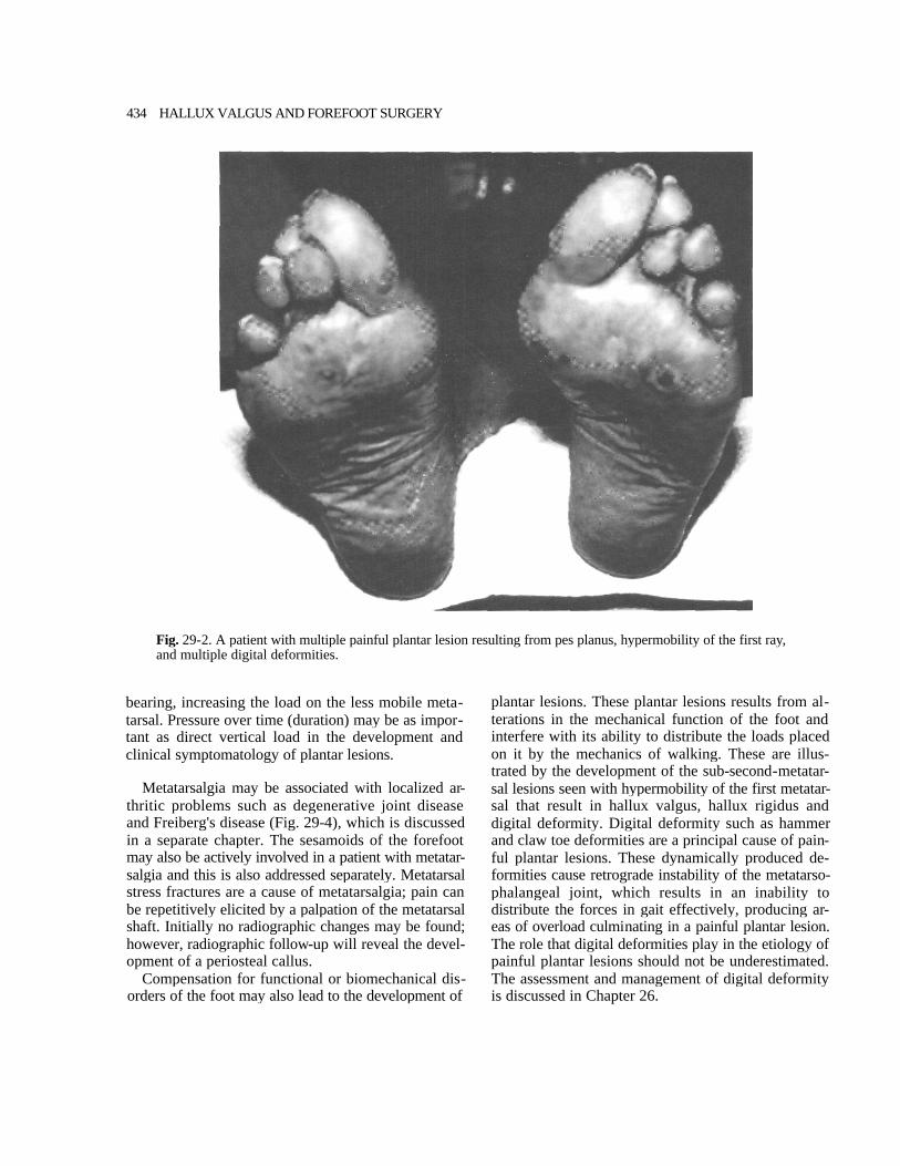
The stance-phase lesion is believed to be caused by a structurally plantar-flexed metatarsal. These may be detected on clinical examination by palpation of the relative position of one metatarsal head to the other while the foot is held in neutral position, and may be identified in axial radiography of the forefoot. The lesion is palpated and observed to be present directly beneath the head of the metatarsal. An elevat ing oste- otomy is the treatment of choice for this type of lesion. The propulsive-phase lesions are clinically located dis- tal to the metatarsal head, and an abnormality in the length of one or more metatarsals may be seen in the dorsoplantar view. This is essentially a focal abnormal- ity of the metatarsal parabola. Other causes of isolated plantar lesions may be an (a) enlarged metatarsal heads, as a result of trauma or degenerative changes, or (b) as secondary to the retrograde instability of the metatarsophalangeal joint associated with a hammer toe or other digital deformity (Figs. 29 -2 and 29-3). Variation and alteration in the location and severity of clinical lesions may result from differences in the rela- tive motion of the lesser metatarsals. Th e degree of mobility available to the second and third metatarsals is limited and increases for the fourth, fifth, and first metatarsals. The degree of mobility may allow the more mobile elements to avoid participation in load-
434 HALLUX VALGUS AND FOREFOOT SURGERY
Fig. 29-2. A patient with multiple painful plantar lesion resulting from pes planus, hypermobility of the first ray, and multiple digital deformities.
bearing, increasing the load on the less mobile meta- tarsal. Pressure over time (duration) may be as impor- tant as direct vertical load in the development and clinical symptomatology of plantar lesions.
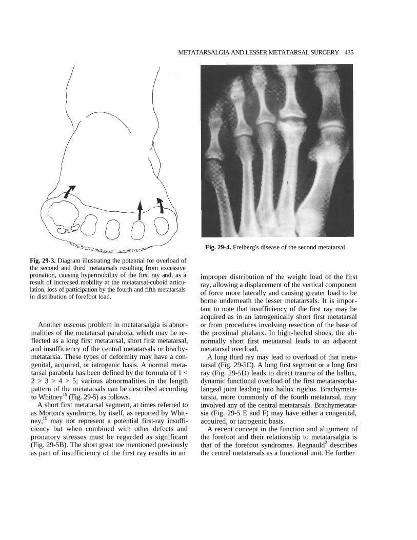
Metatarsalgia may be associated with localized ar- thritic problems such as degenerative joint disease and Freiberg's disease (Fig. 29-4), which is discussed in a separate chapter. The sesamoids of the forefoot may also be actively involved in a patient with metatar- salgia and this is also addressed separately. Metatarsal stress fractures are a cause of metatarsalgia; pain can be repetitively elicited by a palpation of the metatarsal shaft. Initially no radiographic changes may be found; however, radiographic follow-up will reveal the devel- opment of a periosteal callus.
Compensation for functional or biomechanical dis- orders of the foot may also lead to the development of
plantar lesions. These plantar lesions results from al- terations in the mechanical function of the foot and interfere with its ability to distribute the loads placed on it by the mechanics of walking. These are illus- trated by the development of the sub-second-metatar- sal lesions seen with hypermobility of the first metatar- sal that result in hallux valgus, hallux rigidus and digital deformity. Digital deformity such as hammer and claw toe deformities are a principal cause of pain- ful plantar lesions. These dynamically produced de- formities cause retrograde instability of the metatarso- phalangeal joint, which results in an inability to distribute the forces in gait effectively, producing ar- eas of overload culminating in a painful plantar lesion. The role that digital deformities play in the etiology of painful plantar lesions should not be underestimated. The assessment and management of digital deformity is discussed in Chapter 26.
METATARSALGIA AND LESSER METATARSAL SURGERY 435
Fig. 29-4. Freiberg's disease of the second metatarsal.
Fig. 29-3. Diagram illustrating the potential for overload of the second and third metatarsals resulting from excessive pronation, causing hypermobility of the first ray and, as a result of increased mobility at the metatarsal-cuboid articu- lation, loss of participation by the fourth and fifth metatarsals in distribution of forefoot load.
Another osseous problem in metatarsalgia is abnor- malities of the metatarsal parabola, which may be re- flected as a long first metatarsal, short first metatarsal, and insufficiency of the central metatarsals or brachy- metatarsia. These types of deformity may have a con- genital, acquired, or iatrogenic basis. A normal meta- tarsal parabola has been defined by the formula of 1 < 2 > 3 > 4 > 5; various abnormalities in the length pattern of the metatarsals can be described according to Whitney19 (Fig. 29-5) as follows.
A short first metatarsal segment, at times referred to as Morton's syndrome, by itself, as reported by Whit- ney,19 may not represent a potential first-ray insuffi- ciency but when combined with other defects and pronatory stresses must be regarded as significant (Fig. 29-5B). The short great toe mentioned previously as part of insufficiency of the first ray results in an
improper distribution of the weight load of the first ray, allowing a displacement of the vertical component of force more laterally and causing greater load to be borne underneath the lesser metatarsals. It is impor- tant to note that insufficiency of the first ray may be acquired as in an iatrogenically short first metatarsal or from procedures involving resection of the base of the proximal phalanx. In high-heeled shoes, the ab- normally short first metatarsal leads to an adjacent metatarsal overload.
A long third ray may lead to overload of that meta- tarsal (Fig. 29-5C). A long first segment or a long first ray (Fig. 29-5D) leads to direct trauma of the hallux, dynamic functional overload of the first metatarsopha- langeal joint leading into hallux rigidus. Brachymeta- tarsia, more commonly of the fourth metatarsal, may involved any of the central metatarsals. Brachymetatar- sia (Fig. 29-5 E and F) may have either a congenital, acquired, or iatrogenic basis.
A recent concept in the function and alignment of the forefoot and their relationship to metatarsalgia is that of the forefoot syndromes. Regnauld2 describes the central metatarsals as a functional unit. He further

436 HALLUX VALGUS AND FOREFOOT SURGERY
Fig. 29-5. (A) Normal metatarsal alignment (parabola). (B) Short first segment. (C) Long third metatarsal. (Figure continues.)
states that any "disadvantageous configuration" in the alignment of the metatarsal heads cannot be compen- sated for by a intrinsic or extrinsic muscular control. Conversely, however, activity of plantar flexors espe- cially of the first and fifth rays result in a cavus foot and weakness of these plantar flexors causes central meta- tarsal overload. Inability of the first and fifth rays to compensate for forefoot loading, may be regarded as the main influence in protecting the weight-bearing function of the ball of the foot.2 Failure of this mecha- nism is an important cause of painful syndromes of the forefoot (Fig. 29-6). The anterior or frontal plane con- figuration of the Lisfranc's joint is influenced by the load generated by body weight acting through the ta- lus. This is reflected by a change in the convexity of Lisfranc's joint that controls the relative position of the metatarsals and results in four forefoot syndromes:
1. The flat triangular forefoot with frontal protrusion of the second, third, and fourth metatarsals without
equinus of the metatarsals with all metatarsals on the same plane (see Fig. 29-1A);
2. Simple convex forefoot with frontal protrusion of the second, third, and fourth metatarsals associated with the equinus of those metatarsals. The horizon- tal position of the first and fifth metatarsals is nor- mal. One of the more difficult of these forefoot conditions to manage is convex forefoot with insuf- ficiency of the first ray;
3. The convex triangular forefoot, in which the aug- mentation of the frontal convexity of Lisfranc's joint occurs with equinus of the second, third, and fourth metatarsals and divergence of the first and fifth metatarsals;
4. The cavus forefoot, in which metatarsals one or five are lower or relatively shortened in relationship to the central metatarsals.
Regnauld2 recommends management of these disor- ders address realignment of the metatarsal formula to

METATARSALGIA AND LESSER METATARSAL SURGERY 437
D
Fig. 29-5 (Continued). (D) Long first ray. (E) Brachymetatarsia. (F) Brachymetatarsia, second metatarsal.

438 HALLUX VALGUS AND FOREFOOT SURGERY
Fig. 29-6. (A & B) Example of insufficiency of the great toe. The patient underwent implant arthroplasty of the left first metatarsophalangeal joint with a silicone hemi-implant. The right foot was not operated. Postoperatively, a plantar keratosis developed beneath the second metatarsal as a result of the inability of the hallux to participate in weight-bearing because of loss of intrinsic muscle stability of the great toe.
a correct position and preservation of the metatarso- phalangeal joint (Fig. 29-7).
If one considers the alignment of the metatarsals, two axes of function have been described by Bojsen- Moller.20 He described a transverse axis consisting of the first and second metatarsals and an oblique axis consisting of the second through the fifth metatarsals (Fig. 29-8). The transverse was considered to be a high-speed axis and the oblique axis a low-speed axis. These are similar to the axis of propulsion and axis of balance as described by Whitney.19 The second meta- tarsal participates in both axes and represents the key or fulcrum of the forefoot.
Painful conditions of the first-ray, for example, hal- lux valgus and hallux rigidus, will force the patient to use the low-speed or oblique axis. The foot of the patient with hallux valgus is prevented by the deform- ity of the toe and the hypermobility of the first ray from using the long lever arm or transverse axis. The fourth and fifth metatarsals with their greater freedom of mobility than the second or third and can yield dorsally so that the brunt of pressure may be then carried by the second or third metatarsals. Abnormali- ties of metatarsal length may aggravate the situation.
Radiographic assessment of plantar lesions should include dorsoplantar, oblique, forefoot, axial, and lat-
A

METATARSALGIA AND LESSER METATARSAL SURGERY 439
A B
Fig. 29-7. (A & B) Pronated or pes planus foot type, which will develop, depending on the severity and composition of the rearfoot deformity, a flat triangular or convex triangular forefoot. In this instance, a flat triangular forefoot is demonstrated. As a result of the change in the decrease in height of the longitudinal arch, as well as the transverse height of the foot, Lisfranc's joint projects distally, increasing its frontal convexity and resulting in splaying of the forefoot and protrusion and prominence of the central metatarsals. This will be accompanied by digital deformity, hallux valgus, and possibly a tailor's bunion. Treatment may include correction of the first-ray pathology and central metatarsal shaft-head enclavement to restore the metatarsal alignment. At the other end of the spectrum, supination reflected by an increase in the longitudinal arch and transverse height of the foot may result in a simple convex forefoot or cavus forefoot. This foot type is described as frontal protrusion the central metatarsals with equinus of those metatarsals as a result of upward displacement of the cuneiforms, causing overload. This overload will be associated by insufficiency of the first and fifth rays. The cavus forefoot results from continued displacement of the cuneiforms and relative equines of the first and fifth metatarsals. Treatment may include enclavement, osteotomy, or a combination to restore metatarsal alignment with other procedures of the first ray as indicated.2
eral views of the foot. The dorsoplantar or anteropos- terior view allows assessment of metatarsophalangeal joint pathology as well as assessment of metatarsal length patterns. The axial view of the forefoot may identify plantar-flexed metatarsals and plantar promi- nences, and is especially useful in the evaluation of
patients following metatarsal osteotomy or those with transfer lesions. This view has limited value in preop- erative patient assessment for it rarely demonstrates that a plantar-flexed metatarsal. The reason for this is probably that the presence of a true structurally plan- tar-flexed metatarsal is uncommon and most lesions

440 HALLUX VALGUS AND FOREFOOT SURGERY
Fig. 29-8. The transverse and oblique axes of the metatar- sals as described by Bojsen-MÖller.
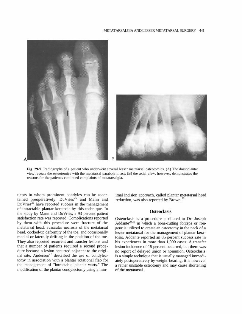
result from biomechanical imbalances often associ- ated with hallux valgus, hallux rigidus, digital deform- ity or other dynamic imbalances. Oblique views can reveal abnormalities in the relationship of lesser meta- tarsals by loss of the normal colinear relationship of the lesser metatarsals. Lateral views may serve as a baseline for future comparisons (Fig. 29-9).
MANAGEMENT
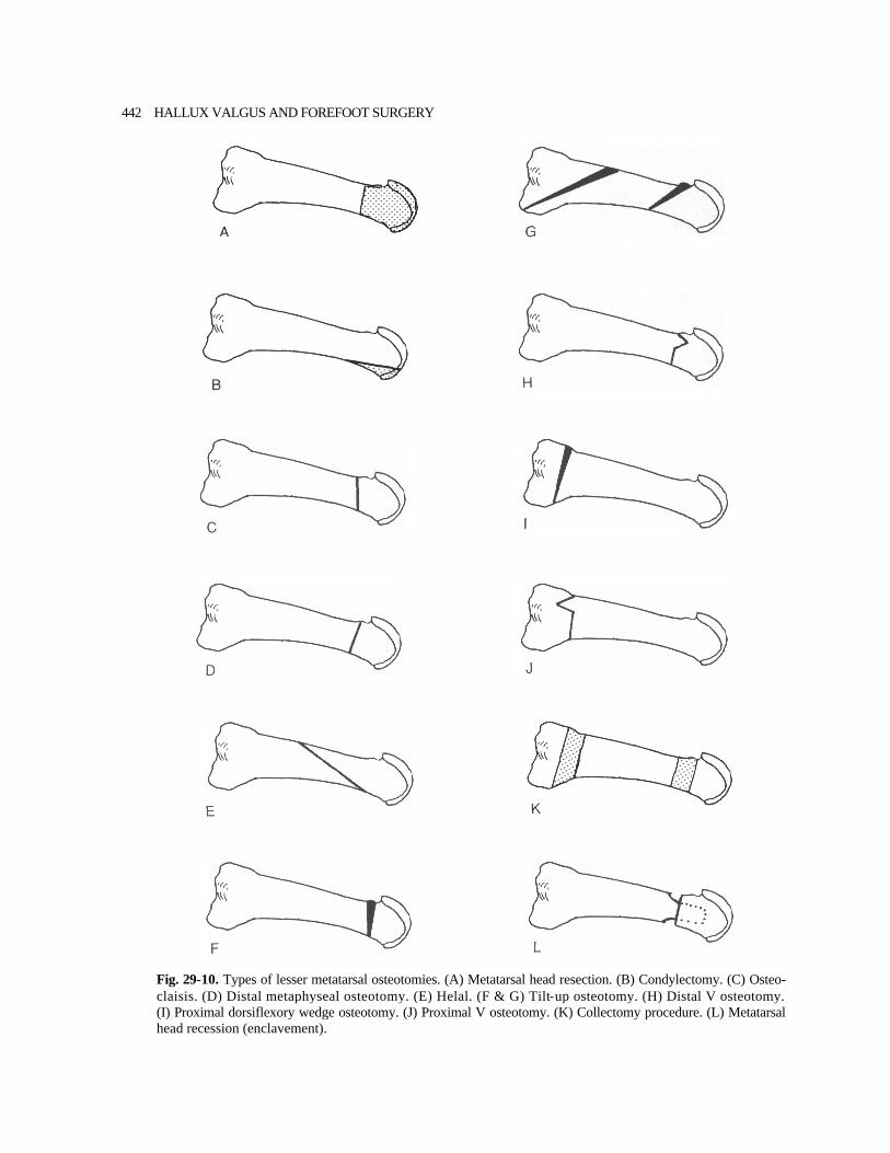
Several procedures have been described for the man- agement of lesser metatarsal lesions (Fig. 29-10). These include the following:
Metatarsal head resection, including both total and partial head resection
Condylectomy Osteoclasis Distal metaphaseal osteotomy Tilt-up osteotomy Distal V osteotomy Proximal wedge osteotomy Proximal V osteotomy Collectomy-type procedures Metatarsal head recession or enclavement.
Metatarsal Head Resection
Metatarsal head resection for the management of plan- tar lesions has been advocated by several authors.21-23
Rutledge and Green21 in 1957 reported on the re- moval of the metatarsal head and one-third of the shaft of the metatarsal. They described their results as excel- lent in a study involving 30 plantar "corns." They de- scribed recession of the toe as a complication and did not observe transfer lesions; 18 months was the long- est follow-up in their series. Perhaps the most aggres- sive form of resection is that of Dickson,24 who advo- cated total ray resection for the management of intractable plantar warts. He reported as results as uni- formly successful in 25 cases.
Metatarsal head resection is not a commonly indi - cated procedure in the management of isolated plan- tar lesions. It is primarily associated with complica- tions of dorsal and posterior retraction of the digit, as well as developments of transfer lesions. Metatarsal head resection is reserved for those cases associated with advanced degenerative disease of the metatarso- phalangeal joint, including prolonged subluxation of the digit, and in certain cases for the management of plantar ulcerations. Metatarsal head resection is indi - cated in the management of tumors of the metatarsal heads. Multiple metatarsal head resections or panme- tatarsal head resections play as an important role in the management of the rheumatoid patient, as well as other patients with chronic disabling arthritic pro- cesses involving all the metatarsophalangeal joints. Isolated metatarsal head resections have less applica- tion.
Condylectomy
Condylectomy, originally described by DuVries,25 in- volves the resection of the plantar condyles of the metatarsal head and is usually reserved for those pa-
TRANSVERSE
METATARSALGIA AND LESSER METATARSAL SURGERY 441
Fig. 29-9. Radiographs of a patient who underwent several lesser metatarsal osteotomies. (A) The dorsoplantar view reveals the osteotomies with the metatarsal parabola intact; (B) the axial view, however, demonstrates the reasons for the patient's continued complaints of metatarsalgia.
tients in whom prominent condyles can be ascer- tained preoperatively. DuVries25 and Mann and DuVries26 have reported success in the management of intractable plantar keratosis by this technique. In the study by Mann and DuVries, a 93 percent patient satisfaction rate was reported. Complications reported by them with this procedure were fracture of the metatarsal head, avascular necrosis of the metatarsal head, cocked-up deformity of the toe, and occasionally medial or laterally drifting in the position of the toe. They also reported recurrent and transfer lesions and that a number of patients required a second proce- dure because a lesion occurred adjacent to the origi- nal site. Anderson27 described the use of condylec- tomy in association with a plantar rotational flap for the management of "intractable plantar warts." The modification of the plantar condylectomy using a min-
imal incision approach, called plantar metatarsal head reduction, was also reported by Brown.28
Osteoclasis
Osteoclasis is a procedure attributed to Dr. Joseph Addante29,30 in which a bone-cutting forceps or ron- geur is utilized to create an osteotomy in the neck of a lesser metatarsal for the management of plantar kera- tosis. Addante reported an 85 percent success rate in his experiences in more than 1,000 cases. A transfer lesion incidence of 15 percent occurred, but there was no report of delayed union or nonunion. Osteoclasis is a simple technique that is usually managed immedi- ately postoperatively by weight-bearing; it is however a rather unstable osteotomy and may cause shortening of the metatarsal.
A
442 HALLUX VALGUS AND FOREFOOT SURGERY
Fig. 29-10. Types of lesser metatarsal osteotomies. (A) Metatarsal head resection. (B) Condylectomy. (C) Osteo- claisis. (D) Distal metaphyseal osteotomy. (E) Helal. (F & G) Tilt-up osteotomy. (H) Distal V osteotomy. (I) Proximal dorsiflexory wedge osteotomy. (J) Proximal V osteotomy. (K) Collectomy procedure. (L) Metatarsal head recession (enclavement).

METATARSALGIA AND LESSER METATARSAL SURGERY 443
Distal Metaphyseal Osteotomies
Distal metaphyseal osteotomies have been reported by several authors. The procedure is usually a trans- verse or oblique osteotomy performed in the distal metaphaseal area of the metatarsal. Variations in tech- nique relate to the angle of orientation of the osteot- omy to the bone itself. Sullivan and O'Donnell31 per- formed the osteotomy in an oblique fashion and reported on a study of 75 patients and 150 procedures. The follow-up range was 4 months to 2.5 years. They reported uniform resolution of the chief complaint and minimal evidence of transfer lesions. The osteot- omy was performed in an oblique fashion from distal- dorsal to plantar-proximal to limit the amount of dor- sal excursion of the metatarsal head. Pedowitz32
performed an osteotomy in the distal metaphaseal area with the osteotomy perpendicular to the support- ing surface. He reported on 69 distal osteotomies with an average follow-up time from 8 months to 8 years (average, 16 months). The overall results were re- ported as 83 percent good, with a good rating consist- ing of no complaints of pain, unrestricted shoe wear, no limitation of activity, and no transfer or asymptom- atic calluses. Six patients had loss of the weight-bear- ing function of the toe on the involved rays and three patients developed transfer lesions. Use of a postoper- ative shoe and immediate weight-bearing was encour- aged.
A modification of the distal metaphaseal osteotomy called the percutaneous metaphyseal osteotomy was described by Smith33; in this technique the metatarsal neck was cut perpendicular to the long axis 2 to 4 mm proximal to the capsular attachments through a percu- taneous approach without visualization.
A modification of the distal metaphaseal reported by Helal34 was initially described to include both distal diaphaseal and metaphaseal bone. The technique as described by Helal is a distal oblique osteotomy in the distal one-half of the metatarsal shaft. The osteotomy is angled 45 degrees plantar and distally. In this proce- dure it is important that the distal fragment slide up- ward but that it is not angled. The prominent dorsal tip of the distal fragment is remodeled. The purpose of the osteotomy was to allow for soft tissue relaxation to realign deformed toes and reposition the metatarsal fat pad. In long-term follow-up, Helal and Greiss35
have reported an overall 88.4 percent success rate.
Primary indication for the procedure is for the man- agement of "pressure metatarsalgia." The procedure is also indicated for the management of patients with rheumatoid arthritis, and it is not uncommon for the procedure to be performed simultaneously on all three metatarsals. Postoperatively the patients are managed by immediate postoperative weight-bearing. Other authors have also reported uniformly success- ful results. Reikeras,36 in a comparison between a proximal wedge resection dorsiflexed osteotomy and a distal oblique osteotomy, reported the distal oblique to be preferred over the more proximal osteotomy.
An osteotomy similar to that of Helal was performed by Turan and Lindgren37 using a distal oblique osteot- omy with internal screw fixation. They thought that the internal fixation in this instance ensured metatarsal head symmetry and provided for more predictable results. Winson38 and others reported that better results were obtained in the Helal procedure when plaster and immobilization was not used postopera- tively. Further details of the Helal osteotomy are de- scribed by a subsequent chapter by Steinbock.
Tilt-Up Osteotomy
One of the more common osteotomies in recent years is the tilt-up osteotomy, which has been described several authors. Although it is more commonly per- formed in the area of the distal metaphysis of the metatarsal, Meisenbach39 in 1916 described this type of osteotomy to be performed 3 cm. from the metatar- sal phalangeal joint. Other authors have referred to the tilt-up osteotomy as a V-type osteotomy (not to be confused with the distal metaphaseal V osteotomy, which is discussed later). This reference is to a wedge osteotomy with a dorsal base and a plantar apex. Wolf40 described this as a V-shaped notch made in the metatarsal down to but not including the plantar cor- tex of the metatarsal shaft. He reported 35 of 42 osteot- omies were completed with excellent results. Four were fair, because of the remaining callous tissue, and 3 were poor because the callus recurred.
Thomas41 also used a tilt-up osteotomy similar to that of Meisenbach and reported a 90 percent success rate as judged by of relief of pain in 73 patients, of whom 39 were rheumatoid; the average follow-up was
444 HALLUX VALGUS AND FOREFOOT SURGERY
4 years. Indications were for painful prominent meta- tarsals with only moderate toe deformity. He recom- mended in his paper that if an adjacent metatarsal feels prominent after osteotomy of the prominent metatar- sal then that metatarsal should be osteotomized as well. Complications included transfer lesions in 4 pa- tients that were treated by osteotomy of the adjacent metatarsal. Kuwada et al.42 described a dorsiflexory distal tilt-up osteotomy for painful plantar lesions. The plantar cortex was left intact, and a rongeur was used to perform the bone cut; in some instances the plantar lesion was excised. They reported no incidence of painful scars, and no transfer lesions, and managed the patient non-weight-bearing for the first 3 weeks post- operative. Berkun et al.43 reported on 14 patients and 25 osteotomies managed by the tilt-up osteotomy. Their overall objective rating was 65 percent excellent, 35 percent good, determined by the appearance of a transfer lesion, and 8 percent poor where there was a painful recurrence and painful transfer lesion. Subjec- tively, all but 1 patient had a good or excellent result; patients satisfaction was 92.5 percent. There was no reports of nonunion, and only minimal bone callous tissue formed. They also noted toe purchase remained active in 23 of 25 digits.
Leventen and Pearson44 described an osteotomy, similar to that of Kuwada et al., as a simple V-shaped osteotomy cut transverse to the shaft with a small longer cut close to the metatarsal head and reported an overall success rate of 86 percent. They noticed that the key to the procedure is early weight-bearing to maintain adequate elevation of the metatarsal head. Dannels45 advocated the use of distal fixated tilt-up osteotomy in the management of preulcers in a dia- betic population. Another osteotomy described by Schwartz et al.,46 is a double oblique lesser metatarsal osteotomy. This osteotomy in the metaphaseal diapha- seal area of the distal metatarsal is performed in an oblique manner and fixated by a simple Kirschner wire (K-wire). For a long metatarsal, the plantar cortex may be osteotomized and shortened. This osteotomy is also readily fixated by screw fixation (interfragmen- tary compression). Jimenez et al.47 described a similar osteotomy called a transverse V-osteotomy, an oblique wedge resection osteotomy that again is amenable to screw fixation and may be performed at the distal metaphysis or as a base osteotomy in the proximal metaphysis.
Distal V Osteotomy
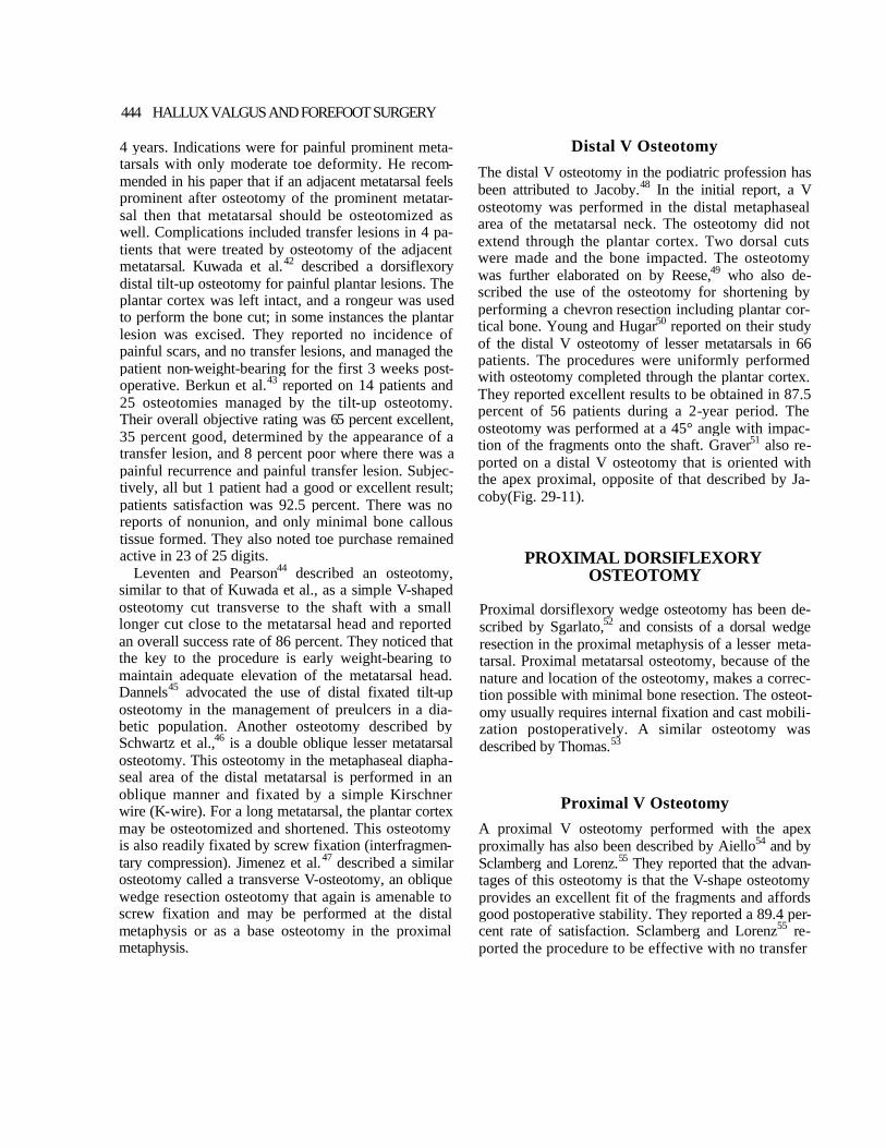
The distal V osteotomy in the podiatric profession has been attributed to Jacoby.48 In the initial report, a V osteotomy was performed in the distal metaphaseal area of the metatarsal neck. The osteotomy did not extend through the plantar cortex. Two dorsal cuts were made and the bone impacted. The osteotomy was further elaborated on by Reese,49 who also de- scribed the use of the osteotomy for shortening by performing a chevron resection including plantar cor- tical bone. Young and Hugar50 reported on their study of the distal V osteotomy of lesser metatarsals in 66 patients. The procedures were uniformly performed with osteotomy completed through the plantar cortex. They reported excellent results to be obtained in 87.5 percent of 56 patients during a 2-year period. The osteotomy was performed at a 45° angle with impac- tion of the fragments onto the shaft. Graver51 also re- ported on a distal V osteotomy that is oriented with the apex proximal, opposite of that described by Ja- coby(Fig. 29-11).
PROXIMAL DORSIFLEXORY OSTEOTOMY
Proximal dorsiflexory wedge osteotomy has been de- scribed by Sgarlato,52 and consists of a dorsal wedge resection in the proximal metaphysis of a lesser meta- tarsal. Proximal metatarsal osteotomy, because of the nature and location of the osteotomy, makes a correc- tion possible with minimal bone resection. The osteot- omy usually requires internal fixation and cast mobili- zation postoperatively. A similar osteotomy was described by Thomas.53
Proximal V Osteotomy
A proximal V osteotomy performed with the apex proximally has also been described by Aiello54 and by Sclamberg and Lorenz.55 They reported that the advan- tages of this osteotomy is that the V-shape osteotomy provides an excellent fit of the fragments and affords good postoperative stability. They reported a 89.4 per- cent rate of satisfaction. Sclamberg and Lorenz55 re- ported the procedure to be effective with no transfer
METATARSALGIA AND LESSER METATARSAL SURGERY 445
B
Fig. 29-11. Distal V osteotomy. (A) Intraoperative view. (B) Postoperative radiographic appearance.
lesions and no subjective complaints voiced by the patients at a 6-month follow-up.
COLLECTOMY
A resection of part of the metatarsal was described by Allen Whitney from the Pennsylvania College of Podi- atric Medicine. Levine56 reported on the use of collec- tomy or cylindrical resection of bone of the metatarsal neck in three cases. The cylindrical resection of proxi- mal metatarsal bone 0.5 cm. in length was described by Spence et al.57 The osteotomy or resection was per- formed by 54 patients (70 metatarsals) with a 6-year follow-up; the results were 89 percent good. Transfers developed in 18 percent; transfers were 23 percent associated with patients undergoing concurrent hallux valgus correction and 12 percent in isolated proce-
dures. The rate of recurrence was reported as 7 per- cent.
In this author's opinion, the two osteotomies more commonly used today are the distal V osteotomy as described by Jacoby and a variation of the tilt-up oste- otomy. The distal V osteotomy is usually performed with an anglation of the arms of the V at 45 to 60 degrees. Minimal dorsal displacement is allowed, and fixation with a K-wire or other simple fixation may be utilized. Weight-bearing in the immediate postopera- tive phase is usually in a postoperative wooden shoe. The advantages of this osteotomy are the ease of per- formance and the frontal and transverse plane stability afforded by the configuration of the osteotomy. Be- cause it is a dorsal displacement osteotomy, if unfix- ated it may exhibit varying degrees of instability in the sagittal plane and result in a floating toe syndrome. Minimal periarticular dissection will reduce the
A

446 HALLUX VALGUS AND FOREFOOT SURGERY
amount of postoperative limitation of motion of the metatarsal phalangeal joint. The transverse V or tilt-up osteotomy allows one to use interfragment com- pression and in most cases no wedge of bone is re- moved.
Studies on the outcomes of surgical correction of intractable plantar keratosis have been reported. Hatcher et al.58 reported on 238 surgical procedures of the central metatarsals with an average follow-up of 2 years and 2 months. The types of procedures evalu- ated in this study were the percutaneous metaphyseal osteotomy, distal V osteotomy, partial metatarsal head resection, total metatarsal head resection, osteoclasis, extension osteotomy, and extension osteoarthrotomy. One hundred patients representing a total of 238 lesser metatarsal osteotomies were studied. Although the paper did not support one procedure over an- other, certain conclusions were drawn. The authors reported a 18 percent rate of recurrence of sympto- matic lesions and an instance of transfer lesions in 39 percent. This included both symptomatic and asymp- totic transfer lesions. That number dropped to 27 per- cent when only symptomatic transfer lesions are in- cluded. The overall success rate for proximal osteotomies was 46 percent, for distal osteotomies, 61 percent, and for joint arthroplastic procedures, partial head or total head resection, it was 53 percent. They did report, however, that based on the studies' criteria for an acceptable result that patients' satisfaction levels were considerably higher than the clinically observed satisfaction level. These authors recommended, in light of the low success rate of proximal osteotomies, that these osteotomies not be recommended as a pri- mary procedure. The authors also believed that the arthroplastic procedure should not be performed un- less pathology at the metatarsophalangeal joint such as arthritic changes is distinctly present. They concluded that distal osteotomies are the preferred procedure based on their findings.
The authors58 reported an increasing number of ac- ceptable results with multiple osteotomies. They indi- cated that consideration should be given to os- teotomizing adjacent metatarsal heads if there is evidence of a lesion beginning adjacent to the primary lesion. The authors also reported better results were obtained for patients of older age and attributed this to the possibility of decreased physical activity in the older age patients.
Hatcher et al. also reported that there was a lack of toe purchase in those cases in which the metatarso- phalangeal joint demonstrated limited plantar flexion postoperatively; however, this problem was not be- lieved to affect the overall outcome of the surgery. They reported the distal V osteotomy to be associated with the greatest lack of plantar flexion and lack of toe purchase when compared with the other osteotomies. Extensor tendon tenotomy did not seem to improve the results or enhance toe purchase postoperatively. The overall decrease in metatarsal length for each in- dividual procedure was also reported. The least amount of shortening was associated with the proxi- mal osteotomies and the greatest amount of shorten- ing with total metatarsal head resection. Almost half their osteotomies were of the second metatarsal, and the remainder was divided between the third and fourth metatarsals. There was no reported difference between the success rates when evaluating the individ- ual metatarsals. Overall the average success rate for lesser metatarsal surgery was 56.5 percent, with pa- tient satisfaction being considerably higher.
In another study by Goforth et al.59 an overall suc- cess rate of 78 percent was reported for 119 osteoto- mies in 94 patients; 74 osteotomies were performed on the second, third, and fourth metatarsals. The distal osteotomies on these metatarsals were nonfixated dis- tal V osteotomies or simple transverse metaphyseal osteotomies. Follow-up time in this study for the pa- tients averaged 16.9 months. The overall results for osteotomies of the second, third, and fourth meta- tarsals were approximately 67 percent success. Re- current or transfer lesions were reported as com- plications.
Young and Hugar50 discussed the evaluation of the V osteotomy for the intractable plantar keratosis. They reported on 56 patients over a 2-year period, and on the basis of their clinical observations and patient sat- isfaction, 75 percent excellent results were obtained. This was defined as resolution of the original lesion with no transfer lesion. Seven patients were reported as good. Three symptomatic transfer lesions and four asymptomatic transfer lesions occurred, and 7 patients were described as having an unacceptable result. Complications included transfer lesions, lack of toe plantar flexion and purchase, and dorsal bone callus associated with the nonfixated osteotomy. They also reported that the diminished range of motion in the

447
A
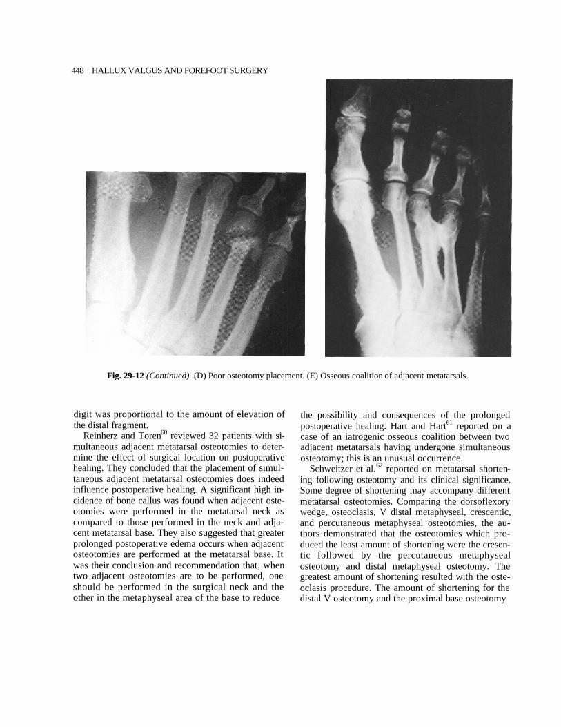
Fig. 29-12. Complications of lesser metatarsal osteotomy. (A) Excessive shortening. (B) Excessive shortening with atrophic nonunion. (C) Poor osteotomy placement. (Figure continues.)
448 HALLUX VALGUS AND FOREFOOT SURGERY
Fig. 29-12 (Continued). (D) Poor osteotomy placement. (E) Osseous coalition of adjacent metatarsals.
digit was proportional to the amount of elevation of the distal fragment.
Reinherz and Toren60 reviewed 32 patients with si- multaneous adjacent metatarsal osteotomies to deter- mine the effect of surgical location on postoperative healing. They concluded that the placement of simul- taneous adjacent metatarsal osteotomies does indeed influence postoperative healing. A significant high in- cidence of bone callus was found when adjacent oste- otomies were performed in the metatarsal neck as compared to those performed in the neck and adja- cent metatarsal base. They also suggested that greater prolonged postoperative edema occurs when adjacent osteotomies are performed at the metatarsal base. It was their conclusion and recommendation that, when two adjacent osteotomies are to be performed, one should be performed in the surgical neck and the other in the metaphyseal area of the base to reduce
the possibility and consequences of the prolonged postoperative healing. Hart and Hart61 reported on a case of an iatrogenic osseous coalition between two adjacent metatarsals having undergone simultaneous osteotomy; this is an unusual occurrence.
Schweitzer et al.62 reported on metatarsal shorten- ing following osteotomy and its clinical significance. Some degree of shortening may accompany different metatarsal osteotomies. Comparing the dorsoflexory wedge, osteoclasis, V distal metaphyseal, crescentic, and percutaneous metaphyseal osteotomies, the au- thors demonstrated that the osteotomies which pro- duced the least amount of shortening were the cresen- tic followed by the percutaneous metaphyseal osteotomy and distal metaphyseal osteotomy. The greatest amount of shortening resulted with the oste- oclasis procedure. The amount of shortening for the distal V osteotomy and the proximal base osteotomy
METATARSALGIA AND LESSER METATARSAL SURGERY 449
were found to be similar, falling between that of the osteoclasis and the cresentic osteotomies. The percu- taneous metaphyseal, distal metaphyseal, and cresen- tic osteotomies revealed less than 0.2 cm. of shorten- ing; the proximal base osteotomy and V osteotomy more than about 0.2 cm. of shortening, and the osteoc- lasis had slightly more than 0.3 cm. McGlamry63 re- ported that the most common cause of the floating toe in a lesser ray is overcorrection following metatarsal osteotomy.
COMPLICATIONS
Complications of lesser metatarsal surgery (Fig. 29-12) include the following64:
1. Recurrence of lesion and symptoms 2. Development of transfer lesions to a adjacent meta-
tarsals because of too much elevation or shortening of the metatarsal
3. Loss of digital function as evidenced by lack of toe purchase and floating toe syndrome seen clinically
4. Loss of metatarsophalangeal joint range of motion as a result of excessive elevation of the metatarsal and of secondary degenerative changes occurring as a result of osteotomy placement or osteone- crosis65
5. Disordered bone healing resulting in the develop- ment of malunion (abnormal alignment of a meta- tarsal head), delayed union, and nonunion
6. Contraction of the metatarsophalangeal joint.
Fixation of osteotomies has always been left to the surgeon's preference. Fixation of lesser metatarsal os- teotomies is readily achieved by a variety of simple fixation techniques including K-wire and absorbable fixation. In cases where a nonfixated osteotomy is an- ticipated, consideration need be given to the type of osteotomy and the compliance of the patient in the immediate postoperative period. Complications are best avoided by careful preoperative planning and as- sessment. Isolated metatarsal and multiple osteoto- mies should be performed only in the presence of the demonstrative criteria.
Prophylactic osteotomies and the management of plantar lesions associated with biomechanical prob- lems such as hallux valgus and hallux rigidus should
be avoided because it does not address the primary etiology and thus only in the most recalcitrant lesions should this be considered as a option. With regards to the surgical management of postoperative transfer le- sions, the goal is to establish normal weight-bearing relationship function and of the metatarsal heads. Management of transfer lesions may require plantar- flexory or lengthening osteotomies with or without bone grafting and, in some instances, osteotomy of the adjacent metatarsals. Panmetatarsal head resection may be indicated as a end-stage salvage procedure. The cause and treatment of metatarsalgia associated with mechanical as well as structural deformities of the foot and leg is an area ripe for continuing study and research as evidenced by the diversity in classifica- tions and treatment methods. Advances in the use of force plate analysis for the distribution of plantar pres- sures combined with biomechanical assessment hold promise for a better understanding of forefoot pathol- ogy in the future.
REFERENCES
1. Thomas N, Nissen KI, Helal B: Disorders of the lesser ray. p. 486. In Helal B, Wilson D (eds): The Foot. Chur- chill Livingstone, New York, 1988
2. Regnauld B: The Foot. Springer-Verlag, Berlin, 1986 3. Viladot A: Metatarsalgia due to biomechanic alterations
of the forefoot. Orthop Clin North Am 4:165, 1973 4. Scranton PE: Metatarsalgia diagnosis and treatment. J
Bone Joint Surg 62:723, 1980 5. Dyck PJ, Low PA, Stevens JC: Burning feet as the only
manifestation of dominantly inherited sensory neuropa- thy. Mayo Clin Proc 58:426, 1983
6. Subotnick S: Observations and discussion of plantar cal- luses. Arch Podiatr Med Foot Surg 1:329, 1974
7. Todoroff DS, Sanders LJ: Keratosis punctata. J Am Podiatr Med Assoc 76:13, 1986
8. Tomassi F, Comerford J, Ransom RA: Unna-Thost-type palmoplantar keratoderma. J Am Podiatr Med Assoc 77:150, 1987
9. Lemont H: Histologic differentiation of mechanical and nonmechanical keratoses of the sole. Clin Dermatol 1:44, 1983
10. Fisher BK, MacPherson M, Epidermoid cyst of the sole. J Am Acad Dermatol 15:1127, 1986
11. Taub J, Steinberg MD: Porokeratosis plantaris discreta; IntJ Dermatol 9:83, 1970
12. Mandojana RM, Katz R, Rodman OG: Porokeratosis plan- taris discreta. Acad Dermatol 10:679, 1984

450 HALLUX VALGUS AND FOREFOOT SURGERY
13. Yanklowitz B, Harkless L: Porokeratosis plantaris dis- creta; a misnomer. J Am Podiatr Med Assoc 80:381, 1990
14. Mancuso JE, Abranmow SP, Dimichino BR, Landsman MJ: Carbon dioxide laser management of plantar verruca : a 6-year follow-up survey. J Foot Surg 30:238, 1991
15. Lemont H, Parekh V: Superficial fascia: an appropriate anatomic boundary for excising warts on the foot. J De- rmatol Surg Oncol 15:710, 1987
16. Bossley CJ, Cairney PC: The intermetatarsal bursa; its significance in Morton's metatarsalgia. J Bone Joint Surg 62:1984, 1980
17. Fitton J, Swinburne L: Degenerative lesions of the acces- sory plantar ligament. Int Orthop 4:295, 1981
18. Hlavac H, Schoenhaus H: The plantar fat pad and some related problems. J Am Podiatr Med Assoc 60:151, 1970
19. Whitney AK: Triplane Taxonomy of Foot and Limb De- formity. Lecture notes, Pennsylvania College of Podiatric Medicine
20. Bojsen-Möller F: Anatomy of the forefoot, normal and pathologic. Clin Orthop Relat Res 142:10, 1979
21. Rutledge A, Green AL: Surgical treatment of plantar corns. US Armed Forces Med J 8:219, 1957
22. Margo MK: Surgical treatment of conditions of the fore- part of the foot. J Bone Joint Surg, 49:1665, 1967
23. Joplin RJ: Surgery of the forefoot in the rheumatoid ar- thritic patient . Surg Clin North Am 49:847, 1969
24. Dickson JA: Surgical treatment of intractable plantar warts. J Bone Joint Surg 30A:757, 1948
25. DuVries HL: New approach to the treatment of intracta- ble verruca plantaris (plantar wart). JAMA 152:1203, 1953
26. Mann A, DuVries L: Intractable plantar keratosis. Orthop Clin North Am 4:67, 1973
27. Anderson R: The treatment of intractable plantar warts. Plast Reconstr Surg 19:384, 1957
28. Brown AR: Painless ambulatory foot surgery – Plantar metatarsal head reduction. Curr Podiatry 26(10):9, 1977
29. Addante B: Metatarsal osteotomy as a surgical approach for elimination of plantar keratosis. J Foot Surg 8:36, 1969
30. Addante JB, Kaufmann D: The metatarsal osteotomy: a ten - year fo l low- up on the second, th i rd , and four th metatarsal osteotomies a nd a new approach to the fifth metatarsal osteotomy. J Foot Surg 16:92, 1977
31. Sullivan JD, O'DonnellJE: The dorsal displacement float- ing metatarsal subcapital osteotomy. J Foot Surg 14:62, 1975
32. Pedowitz WJ: Distal oblique osteotomy for intractable plantar keratosis of the middle three metatarsals. Foot Ankle 9:7, 1988
33. Smith D: Percutaneous metaphyseal osteotomy. Annu J Acad Ambulat Foot Surgeons 1980
34. Helal B: Metatarsal osteotomy for metatarsalgia. J Bone Joint Surg 57:187, 1975
35. Helal B, Greiss M: Telescoping osteotomy for pressure metatarsalgia. J Bone Joint Surg, 66:213, 1984
36. Reikeras O: Metatarsal osteotomy for relief of metatar - salgia. Arch Orthop Traum Surg 101:177, 1983
37. Turan I, Lindgren U: Metatarsal osteotomy using internal fixation with compression screws. J Foot Surg 28:116, 1989
38. Winson IG, Rawlinson J, Broughton NS: Treatment of metatarsalgia by sliding distal metatarsal osteotmy. Foot Ankle 9:2, 1988
39 Meisenbach RO: Painful anterior arch of the foot and operation for its relief by means of raising the arch. Am J Orthop Surg 14:206, 1916
40. Wolf M: Metatarsal osteotomy for relief of painful meta - tarsal. J Bone Joint Surg 55:1750, 1973
41. Thomas WH: Metatarsal osteotomy. Surg Clin 49:879, 1969
42. Kuwada GT, Dockery GL, Schuberth JM: The resistant painful plantar lesion; a surgical approach. J Foot Surg 22:29, 1983
43. Berkun RN, Devincentis A, Goller WL The tilt -up osteot- omy for correction of intractable plantar keratosis. J Foot Surg 2352, 1984
44. Leventen O, Pearson W: Distal metatarsal osteotomy for intractable plantar Keratosis. Foot Ankle 10:247, 1990
45. Dannels EG: A preventive metatarsal osteotomy for heal- ing pre-ulcers in American Indian Diabetes. J Am Podiatr Med Assoc 76:33, 1986
46. Schwartz N, Williams JE, Marcinko DE: Double oblique less metatarsal osteotomy. J Am Podiatr Med Assoc 73:218, 1983
47. Jimenez AL, Martin DE, Phillips AJ: Lesser metatarsalgia evaluation and treatment. Clin Podiatr Med Surg 7:597, 1990
48. Jacob RP: "V" Osteoplasty for correction of intractable plantar keratosis. J Foot Surg 12:10 , 1973
49. Reese HW: Surgical treatment of intractable plantar keratosis. J Foot Surg 12:92, 1973
50. Young E, I lugar W: Evaluation of the V-osteotomy as a procedure to alleviate the intractable planter keratoma. J Foot Surg 19:187, 1980
51. Graver H: Angular metata rsal osteotomy: a preliminary report. J Am Podiatry Assoc 63:96, 1973
52. Sgarlato TE: A compendium of podiatric biomechanics. California College of Podiatric Medicine, San Francisco, 1971
53. Thomas F: Levelling the tread: (elevation of the dropped metatarsal head by metatarsal osteotomy). J Bone Joint Surg 56B:314, 1974
54. Aiello CL: Surgical treatment of metatarsalgia. Int Orthop, 5:107, 1981
55. Sclamberg EL, Lorenz MA: A dorsal wedge "V" osteot- omy for painful plantar callosities. Foot Ankle 4:30,1983
METATARSALGIA AND LESSER METATARSAL SURGERY 451
56. Levine LA: Modern therapeutic approaches to foot prob- lems. Futura Publishing, Mt. Kisco, NY, 1975.
57. Spence KF, O'Connell SJ, KenzoraJE: Proximal metatar- sal segmental resection: a treatment for intractable plan- tar keratoses. Orthopedics 13:741, 1990
58. Hatcher RM, Goller WL, Weil LS: Intractable planter keratoses. J Am Podiatr Med Assoc 68:366, 1978
59. Goforth PW, Karlin JW, DeValentines S, et al: Distal meta- tarsal osteotomy (retrospective study). J Am Podiatr Med Assoc 74:402, 1984
60. Reinherz RP, Toren DJ: Bone healing after adjacent metatarsal osteotomies. J Foot Surg 20:198, 1981
61. Hart DJ, Hart TJ: latrogenic metatarsal coalition: a post- operative complication of adjacent V-osteotomies. J Foot Surg 24:205, 1985
62. Schweitzer DA, Lew J, Morgan J: Central metatarsal
shortening following osteotomy and its clinical signifi- cance. J Am Podiatry Assoc 72:6, 1982
63. McGlamry ED: Floating toe syndrome. J Am Podiatr Med Assoc 72:561, 1982
64. Weinstock RE: Surgical judgement in metatarsal surgery for the elimination of intractable plantar keratoses. J Am Podiatr Med Assoc 65:979, 1975
65. Bayliss NC, Kleneman L: Avascular necrosis of lesser metatarsals heads following forefoot surgery. Foot Ankle 10:124, 1989
