mediastinal tumor
DESCRIPTION
Presented at AW Sjahranie General Hospital under supervision of dr. Syaiful Mukhtar SpB(K)BDTRANSCRIPT
Mediastinal Tumor
Diagnostic, Therapy, Pre-Operative and Post-Operative CareDr. Isa BasukiDepartment of Surgery, AWS General Hospital
Anatomy
▪ The region of the body located between the two pleural spaces
▪ It is derived from the Latin words medius (middle) and stare (to stand) and means literally "standing in the middle.“
▪ The mediastinum is a complex and tightly knit package of structures immediately vital to the life of the individua-the central airways, the heart, and the great vessels.
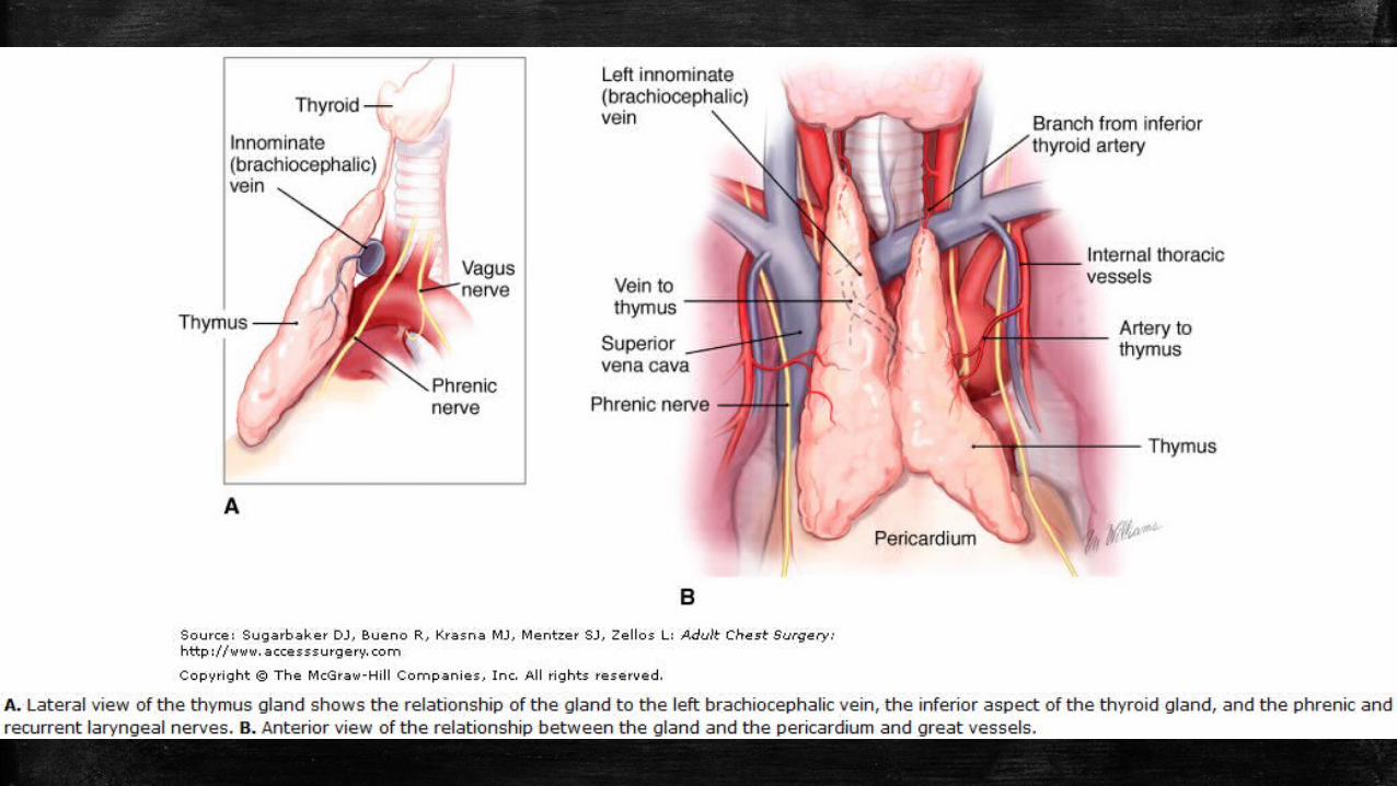
▪ Also contained within the mediastinum are glands, and organs, including the esophagus, thymus, thoracic duct, vagus and phrenic nerves, and lymphatics.
Cont’d
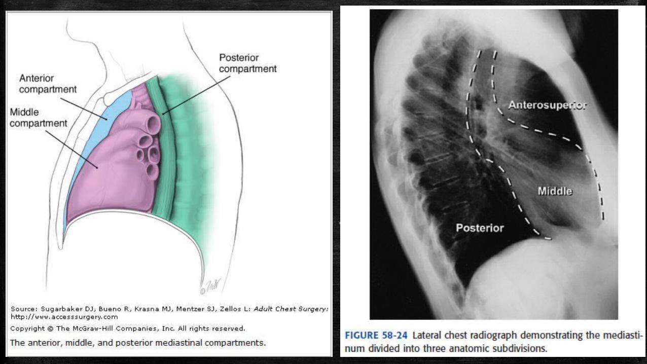
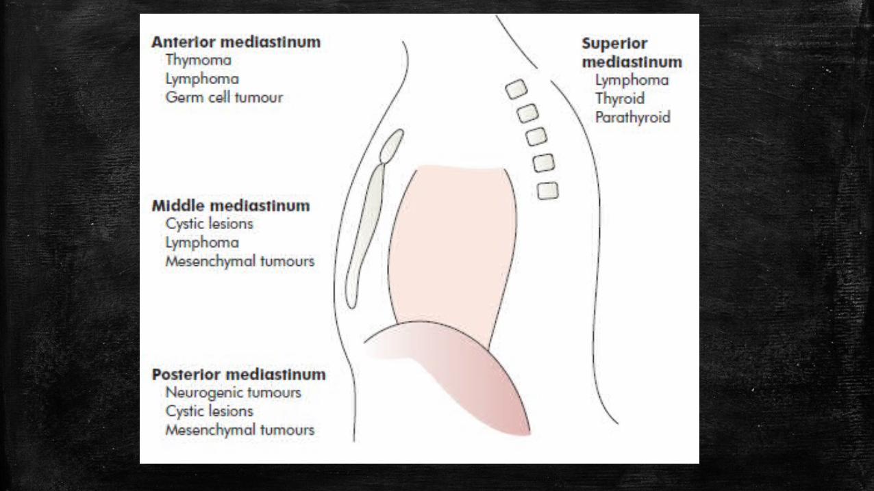
▪ The mediastinum extends from the diaphragm to the thoracic inlet and is divided by anatomists into four regions that are defined by their relationship to the pericardium: – superior, – anterior, – middle, – posterior.
▪ Thoracic surgeons generally divide the mediastinum into just three compartments:– anterior, – middle, – posterior
Cont’d
▪ The mediastinum contains a compact arrangement of vital structures and other tissues abnormalities such as infection, trauma, and neoplasm can have a profound impact and can present with dramatic symptoms
▪ Because the mediastinum is relatively inaccessible to physical examination, imaging studies such as computed tomography (CT) play a particularly important role in the evaluation of suspected pathology.
Mediastinal Lesion
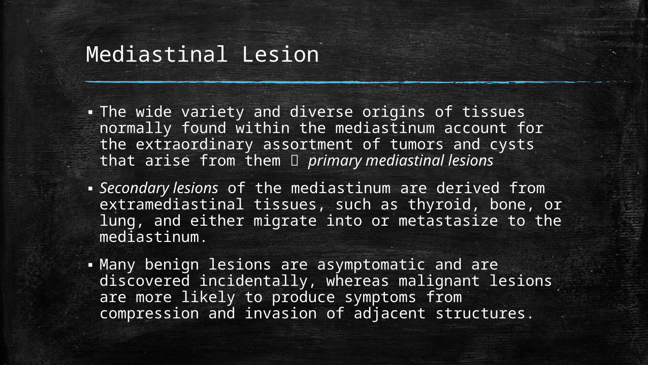
▪ The wide variety and diverse origins of tissues normally found within the mediastinum account for the extraordinary assortment of tumors and cysts that arise from them primary mediastinal lesions
▪ Secondary lesions of the mediastinum are derived from extramediastinal tissues, such as thyroid, bone, or lung, and either migrate into or metastasize to the mediastinum.
▪ Many benign lesions are asymptomatic and are discovered incidentally, whereas malignant lesions are more likely to produce symptoms from compression and invasion of adjacent structures.
Types of Primary Mediastinal Lesion
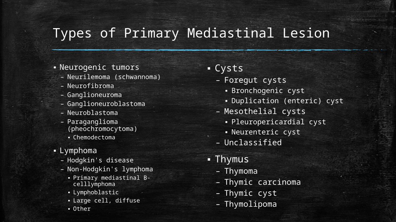
▪ Neurogenic tumors– Neurilemoma (schwannoma)– Neurofibroma– Ganglioneuroma– Ganglioneuroblastoma– Neuroblastoma– Paraganglioma
(pheochromocytoma)▪ Chemodectoma
▪ Lymphoma– Hodgkin's disease– Non-Hodgkin's lymphoma▪ Primary mediastinal B-celllymphoma▪ Lymphoblastic▪ Large cell, diffuse▪ Other
▪ Cysts– Foregut cysts▪ Bronchogenic cyst▪ Duplication (enteric) cyst
– Mesothelial cysts▪ Pleuropericardial cyst▪ Neurenteric cyst
– Unclassified
▪ Thymus– Thymoma– Thymic carcinoma– Thymic cyst– Thymolipoma
Cont’d
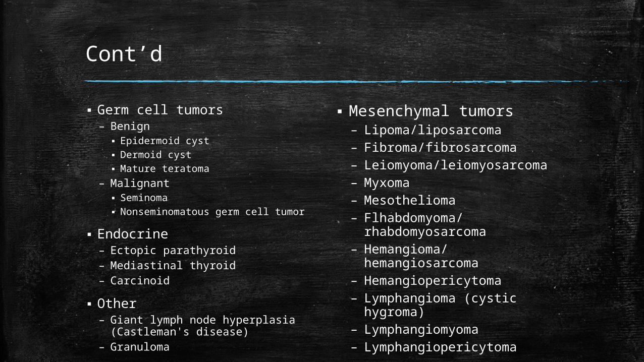
▪ Germ cell tumors– Benign▪ Epidermoid cyst▪ Dermoid cyst▪ Mature teratoma
– Malignant▪ Seminoma▪ Nonseminomatous germ cell tumor
▪ Endocrine– Ectopic parathyroid– Mediastinal thyroid– Carcinoid
▪ Other– Giant lymph node hyperplasia
(Castleman's disease)– Granuloma
▪ Mesenchymal tumors– Lipoma/liposarcoma– Fibroma/fibrosarcoma– Leiomyoma/leiomyosarcoma– Myxoma– Mesothelioma– Flhabdomyoma/
rhabdomyosarcoma– Hemangioma/
hemangiosarcoma– Hemangiopericytoma– Lymphangioma (cystic
hygroma)– Lymphangiomyoma– Lymphangiopericytoma
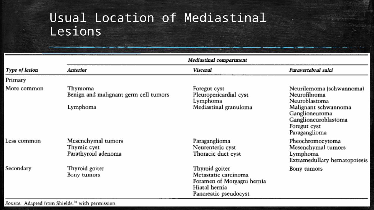
Usual Location of Mediastinal Lesions
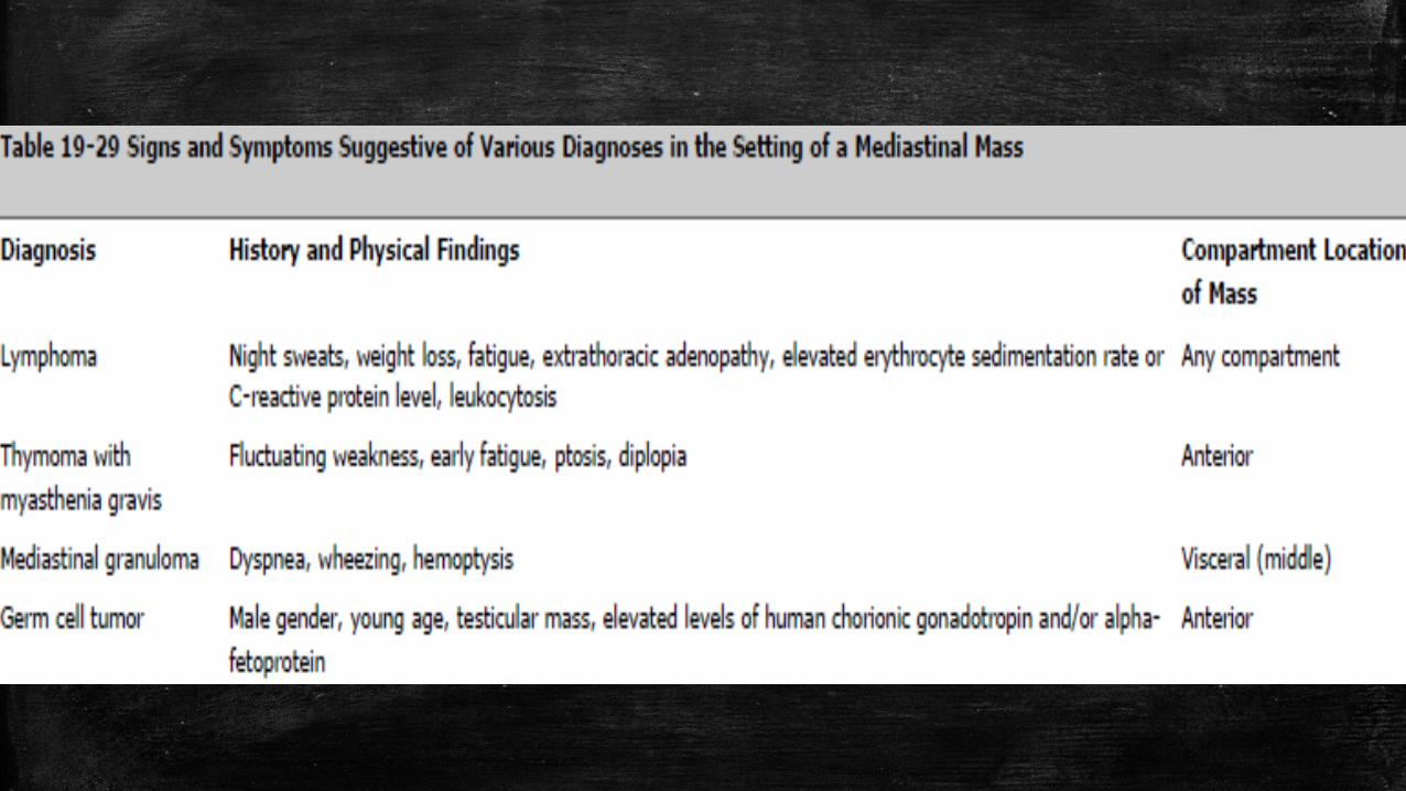
Clinical Presentation
▪ Mediastinal lesions are symptomatic in 50%-75% of patients
▪ Symptoms can be caused by local mass effects, systemic effects of tumorderived hormones and peptides, or infection.
▪ Local effects are dependent on the size and location of the lesion and result from compression of adjacent structures– Examples: cough, stridor, dyspnea, chest pain, and dysphagia
▪ Symptoms are also more common with malignant tumors are more likely to fix, encase, and invade adjacent structures.– Examples: Superior vena cava syndrome, back pain, and neurological
deficits such as Homer's syndrome or phrenic nerve palsy
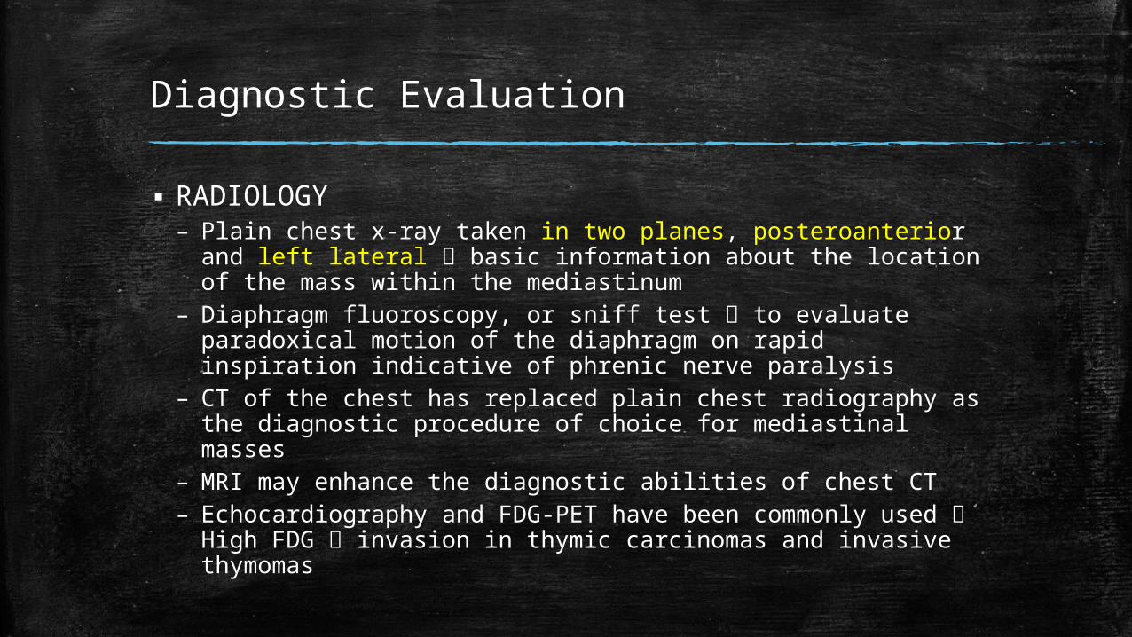
Diagnostic Evaluation
▪ RADIOLOGY– Plain chest x-ray taken in two planes, posteroanterior and left
lateral basic information about the location of the mass within the mediastinum
– Diaphragm fluoroscopy, or sniff test to evaluate paradoxical motion of the diaphragm on rapid inspiration indicative of phrenic nerve paralysis
– CT of the chest has replaced plain chest radiography as the diagnostic procedure of choice for mediastinal masses
– MRI may enhance the diagnostic abilities of chest CT– Echocardiography and FDG-PET have been commonly used
High FDG invasion in thymic carcinomas and invasive thymomas
Cont’d
▪ Histologic– FNA or needle biopsy with CT guidance of a
mediastinal mass may provide sufficient tissue for diagnosis of thymic carcinoma or other defined neoplasms
– Core needle biopsy, mediastinoscopy, or intrathoracic biopsy may be considered for lymphomas in particular, and thymomas and neural tumors
– Electron microscopy may be required for confirmation of specific histologies
Thymoma
▪ The most common neoplasm of the anterosuperior compartment
▪ Peak incidence is in the third through fifth decades
▪ Radiograph: small, well-circumscribed mass or as a bulky lobulated mass confluent with adjacent mediastinal structures
▪ Symptoms: – chest pain, – dyspnea, – hemoptysis, – cough, – superior vena cava syndrome – systemic syndromes caused by immunologic mechanisms
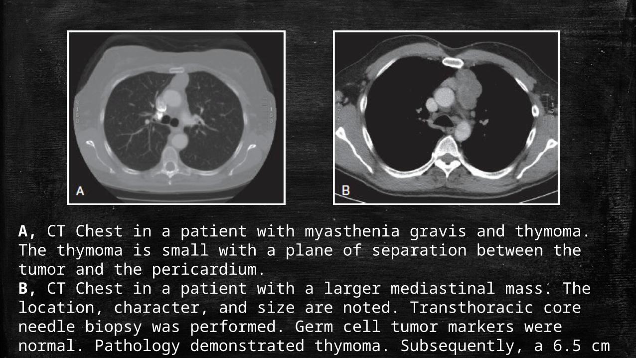
A, CT Chest in a patient with myasthenia gravis and thymoma. The thymoma is small with a plane of separation between the tumor and the pericardium. B, CT Chest in a patient with a larger mediastinal mass. The location, character, and size are noted. Transthoracic core needle biopsy was performed. Germ cell tumor markers were normal. Pathology demonstrated thymoma. Subsequently, a 6.5 cm thymoma was resected. There was no invasion of the pericardium. A complete resection (R0) was accomplished.
Cont’d
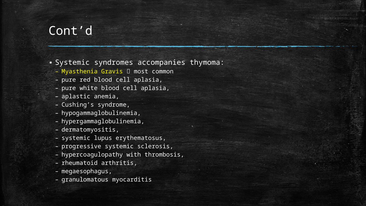
▪ Systemic syndromes accompanies thymoma:– Myasthenia Gravis most common– pure red blood cell aplasia, – pure white blood cell aplasia, – aplastic anemia,– Cushing’s syndrome, – hypogammaglobulinemia, – hypergammaglobulinemia,– dermatomyositis, – systemic lupus erythematosus,– progressive systemic sclerosis, – hypercoagulopathy with thrombosis,– rheumatoid arthritis, – megaesophagus, – granulomatous myocarditis
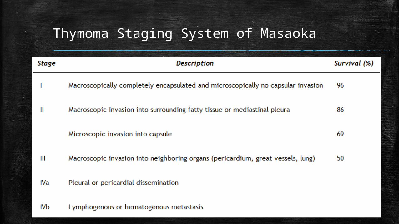
Thymoma Staging System of Masaoka
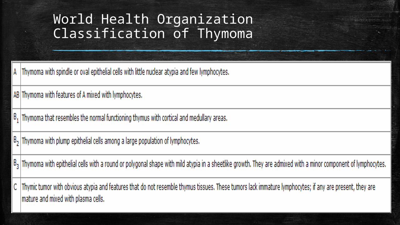
World Health Organization Classification of Thymoma
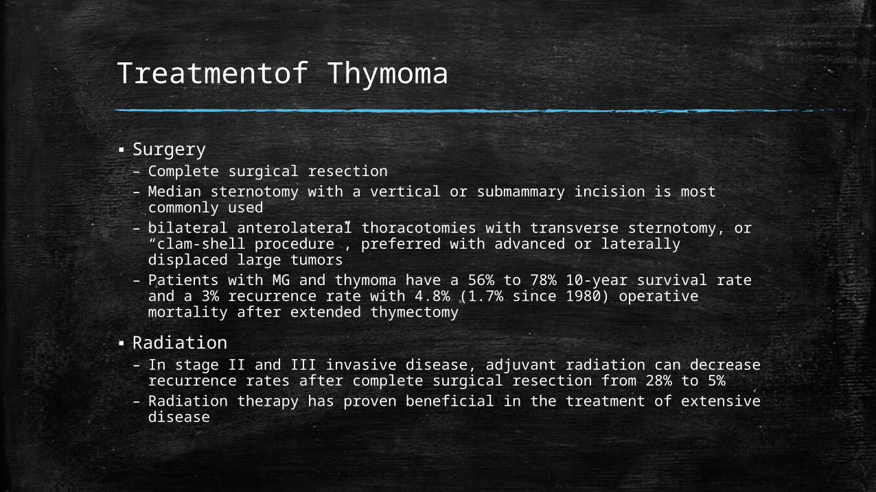
Treatmentof Thymoma
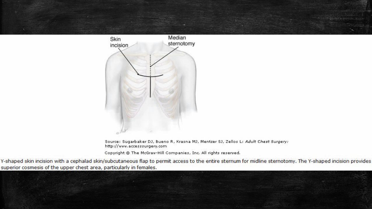
▪ Surgery– Complete surgical resection – Median sternotomy with a vertical or submammary incision is most
commonly used– bilateral anterolateral thoracotomies with transverse sternotomy, or “clam-
shell procedure”, preferred with advanced or laterally displaced large tumors
– Patients with MG and thymoma have a 56% to 78% 10-year survival rate and a 3% recurrence rate with 4.8% (1.7% since 1980) operative mortality after extended thymectomy
▪ Radiation– In stage II and III invasive disease, adjuvant radiation can decrease
recurrence rates after complete surgical resection from 28% to 5%– Radiation therapy has proven beneficial in the treatment of extensive
disease
Cont’d
▪ Systemic Therapy– Steroids have been shown to be active in the management of thymomas– Both single-agent and combination therapy have demonstrated activity in
the adjuvant and neoadjuvant settings– Doxorubicin, cisplatin, ifosfamide, corticosteroids, and cyclophosphamide
all have been used as single-agent therapy
▪ Molecularly Targeted Therapy– Overexpression of c-kit is common in thymic carcinoma– Coamplification of the HER-2/neu topoisomerase 2-alpha gene may
correlate with response to the CAP chemotherapy regimen– antitumor activity has been reported with dasatinib, a small molecule oral,
multitargeted kinase inhibitor of Bcr-Abl and src kinases, ephrin receptor kinases, platelet-derived growth factor receptor, and c-kit, in thymoma
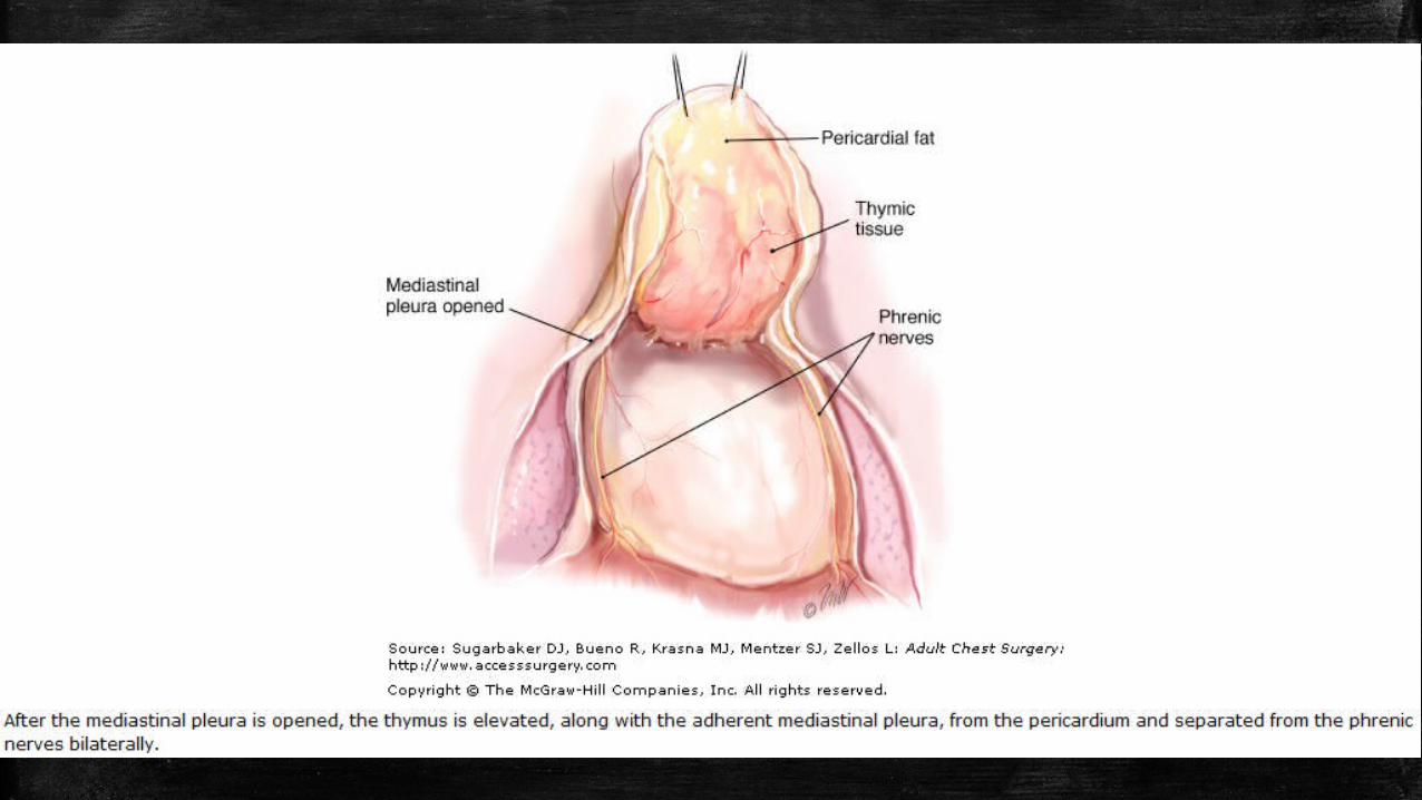
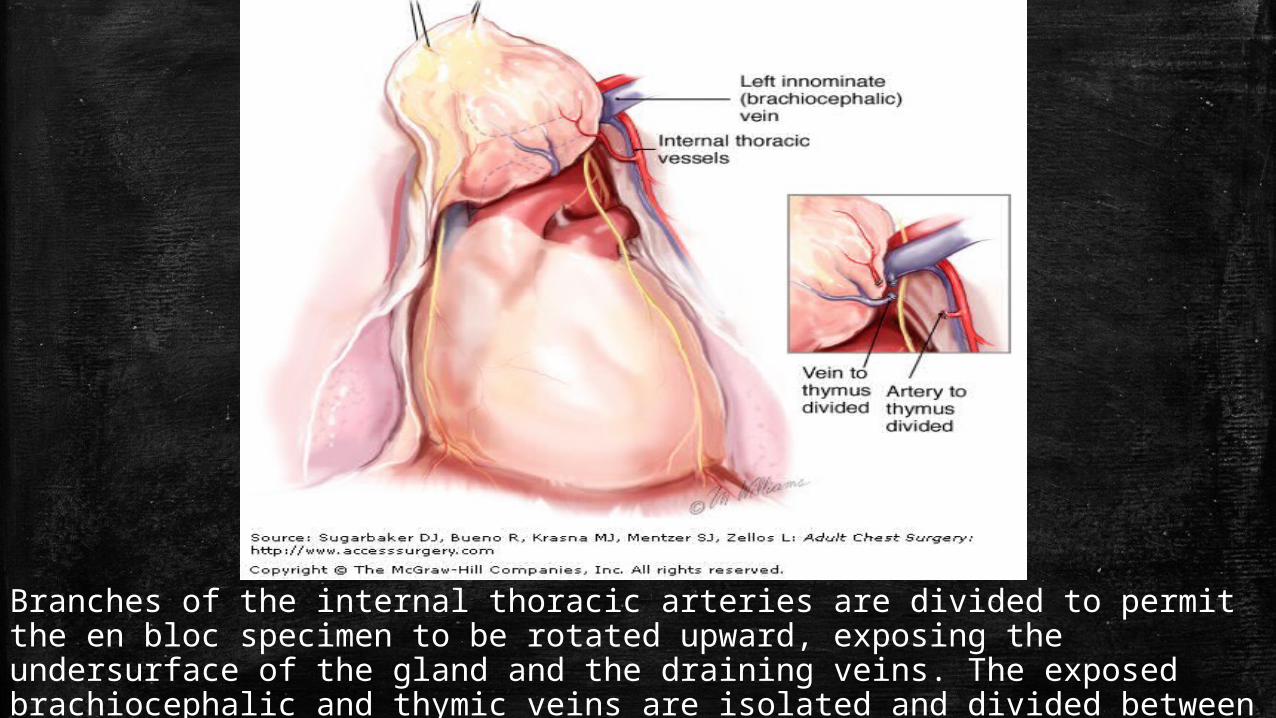
Branches of the internal thoracic arteries are divided to permit the en bloc specimen to be rotated upward, exposing the undersurface of the gland and the draining veins. The exposed brachiocephalic and thymic veins are isolated and divided between ligatures or clips (inset).
Preoperative
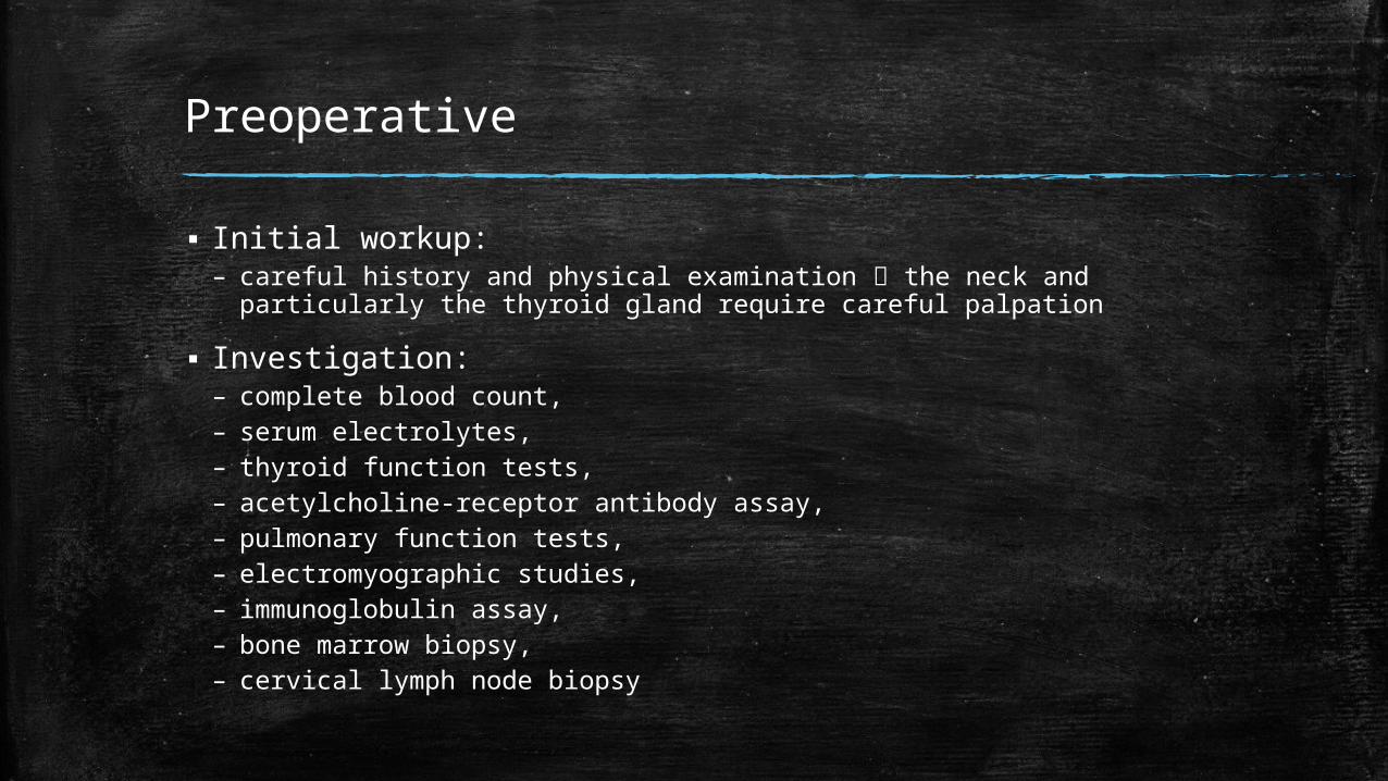
▪ Initial workup: – careful history and physical examination the neck and particularly
the thyroid gland require careful palpation
▪ Investigation:– complete blood count, – serum electrolytes, – thyroid function tests, – acetylcholine-receptor antibody assay, – pulmonary function tests, – electromyographic studies, – immunoglobulin assay, – bone marrow biopsy, – cervical lymph node biopsy
Cont’d
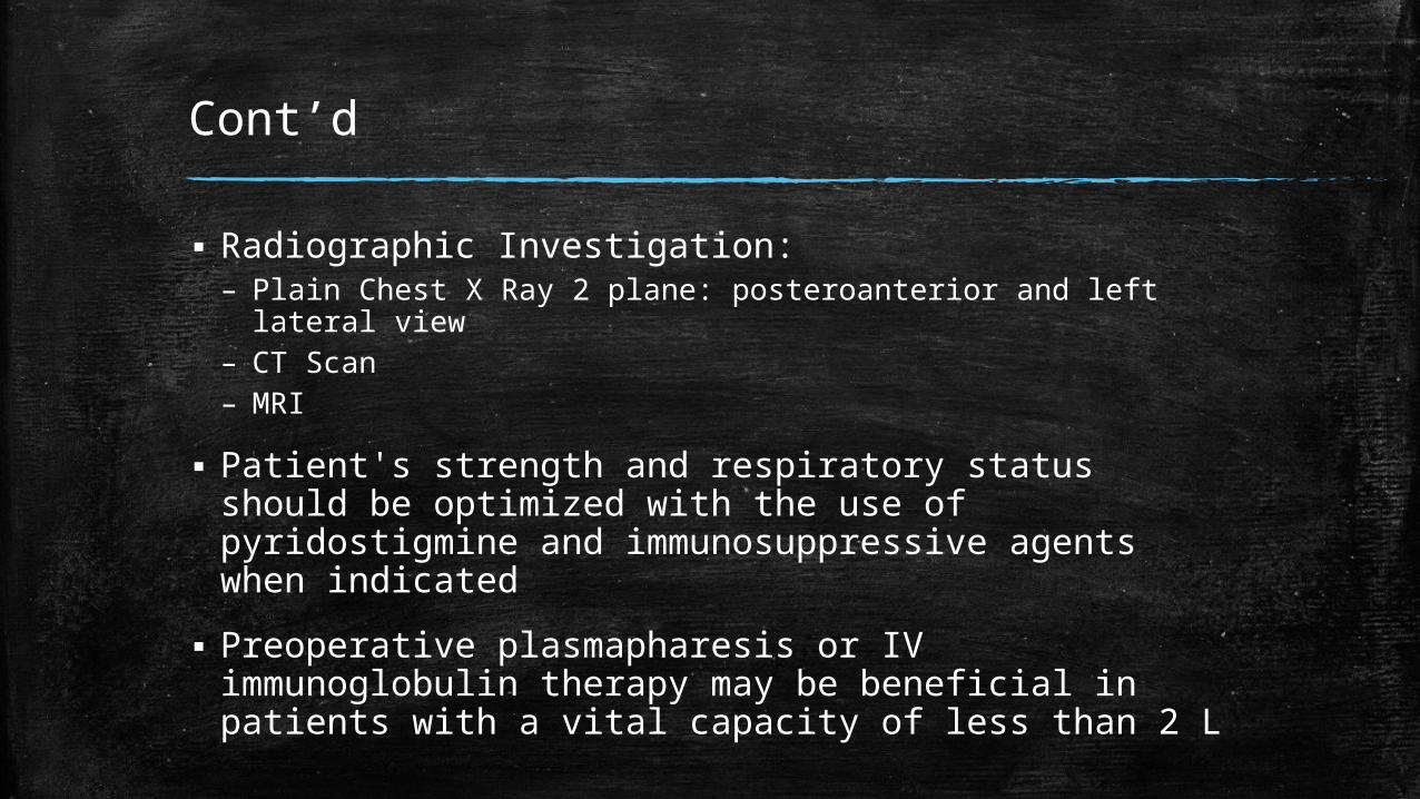
▪ Radiographic Investigation:– Plain Chest X Ray 2 plane: posteroanterior and left lateral view– CT Scan– MRI
▪ Patient's strength and respiratory status should be optimized with the use of pyridostigmine and immunosuppressive agents when indicated
▪ Preoperative plasmapharesis or IV immunoglobulin therapy may be beneficial in patients with a vital capacity of less than 2 L
Postoperative
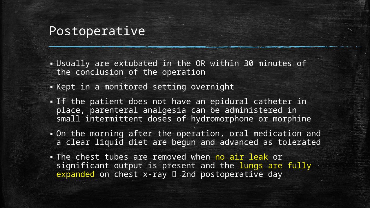
▪ Usually are extubated in the OR within 30 minutes of the conclusion of the operation
▪ Kept in a monitored setting overnight
▪ If the patient does not have an epidural catheter in place, parenteral analgesia can be administered in small intermittent doses of hydromorphone or morphine
▪ On the morning after the operation, oral medication and a clear liquid diet are begun and advanced as tolerated
▪ The chest tubes are removed when no air leak or significant output is present and the lungs are fully expanded on chest x-ray 2nd postoperative day
Cont’d
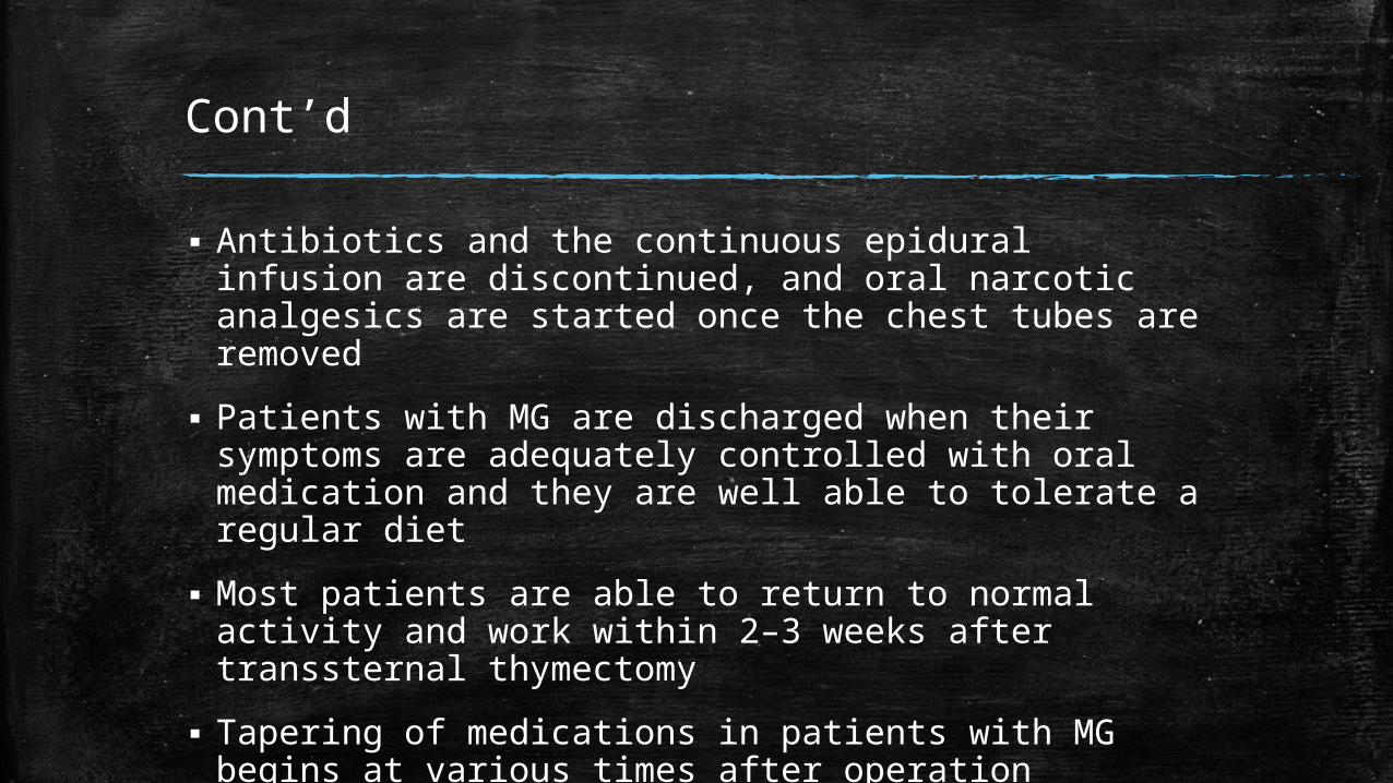
▪ Antibiotics and the continuous epidural infusion are discontinued, and oral narcotic analgesics are started once the chest tubes are removed
▪ Patients with MG are discharged when their symptoms are adequately controlled with oral medication and they are well able to tolerate a regular diet
▪ Most patients are able to return to normal activity and work within 2–3 weeks after transsternal thymectomy
▪ Tapering of medications in patients with MG begins at various times after operation depending on the judgment of the neurologist
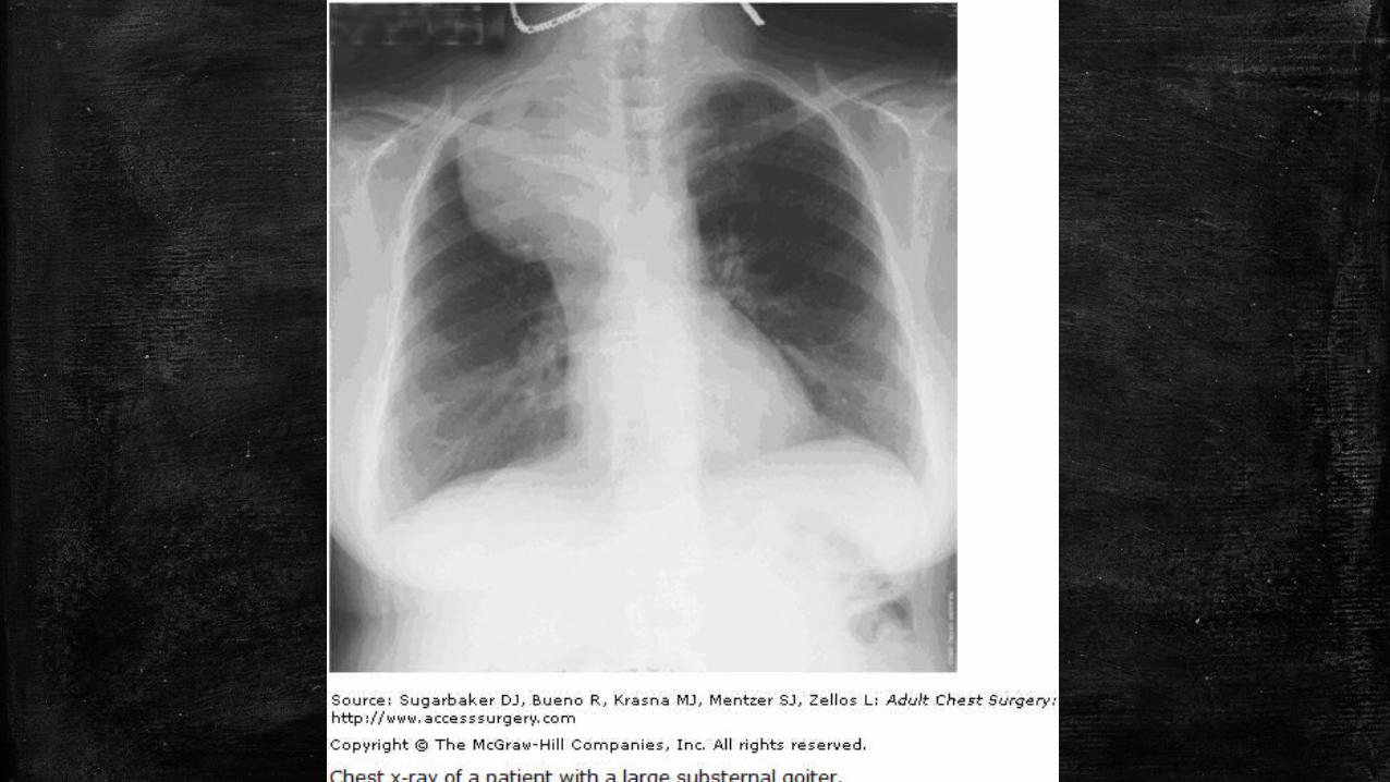
Substernal Goiter
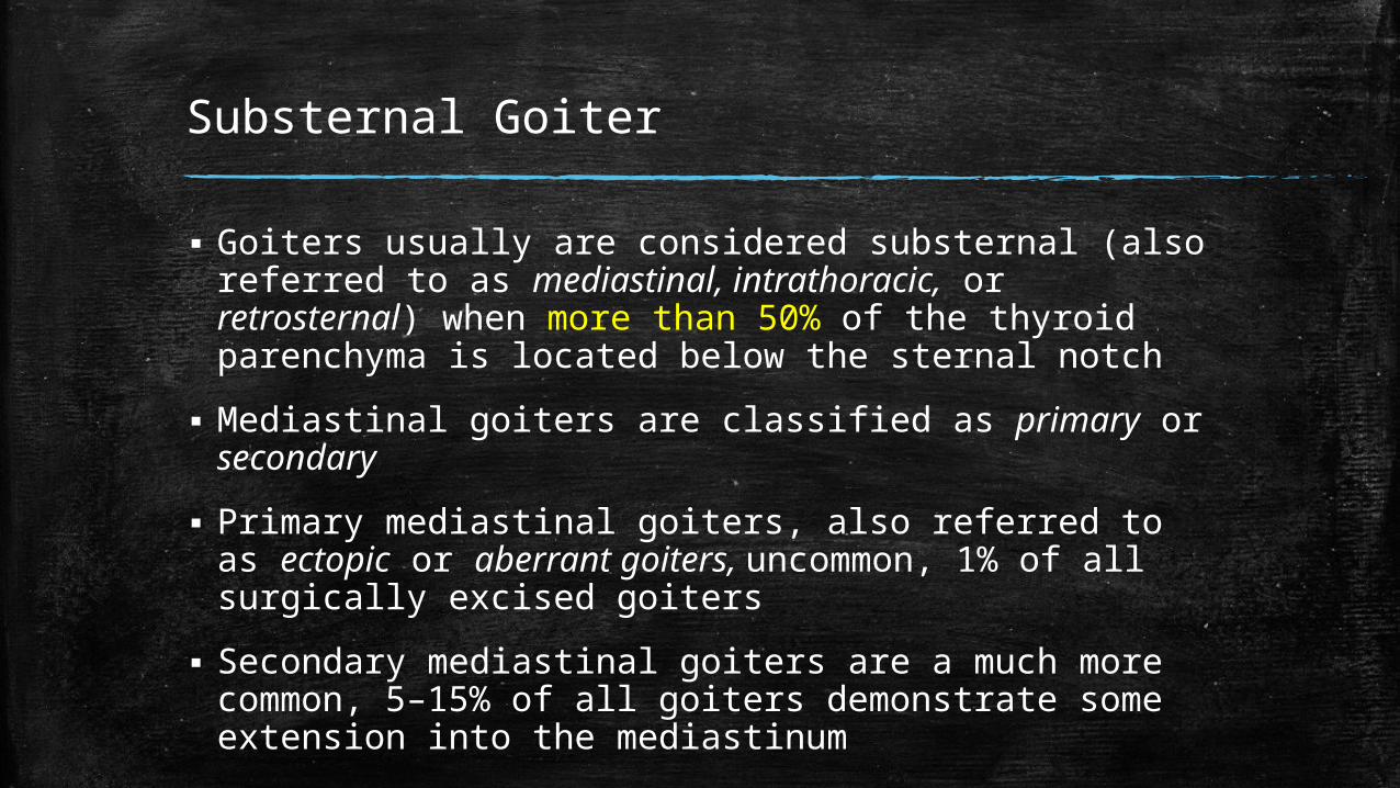
▪ Goiters usually are considered substernal (also referred to as mediastinal, intrathoracic, or retrosternal) when more than 50% of the thyroid parenchyma is located below the sternal notch
▪ Mediastinal goiters are classified as primary or secondary
▪ Primary mediastinal goiters, also referred to as ectopic or aberrant goiters, uncommon, 1% of all surgically excised goiters
▪ Secondary mediastinal goiters are a much more common, 5–15% of all goiters demonstrate some extension into the mediastinum
Symptoms Attributable to Substernal Goiters
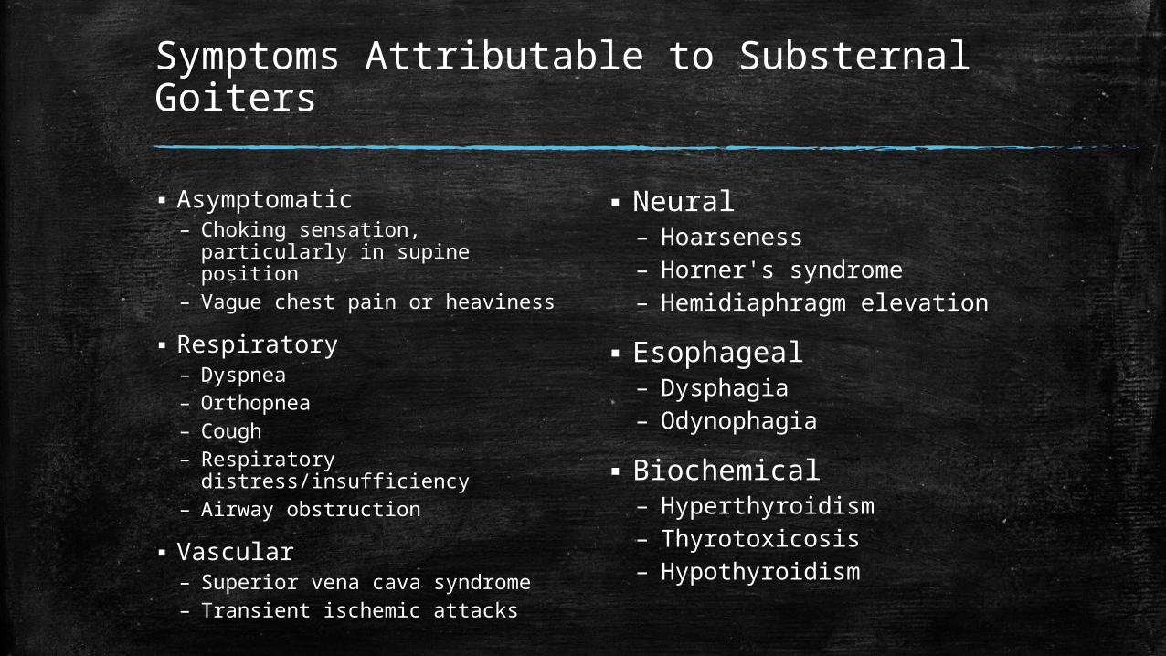
▪ Asymptomatic – Choking sensation, particularly
in supine position– Vague chest pain or heaviness
▪ Respiratory – Dyspnea– Orthopnea– Cough– Respiratory
distress/insufficiency– Airway obstruction
▪ Vascular – Superior vena cava syndrome– Transient ischemic attacks
▪ Neural– Hoarseness– Horner's syndrome– Hemidiaphragm elevation
▪ Esophageal– Dysphagia– Odynophagia
▪ Biochemical – Hyperthyroidism– Thyrotoxicosis– Hypothyroidism
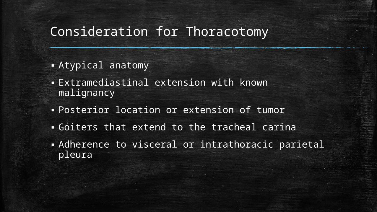
Consideration for Thoracotomy
▪ Atypical anatomy
▪ Extramediastinal extension with known malignancy
▪ Posterior location or extension of tumor
▪ Goiters that extend to the tracheal carina
▪ Adherence to visceral or intrathoracic parietal pleura
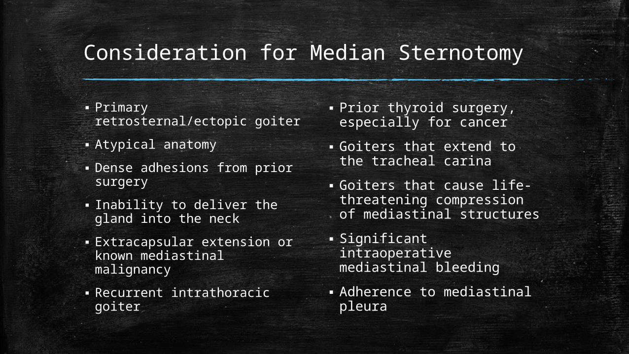
Consideration for Median Sternotomy
▪ Primary retrosternal/ectopic goiter
▪ Atypical anatomy
▪ Dense adhesions from prior surgery
▪ Inability to deliver the gland into the neck
▪ Extracapsular extension or known mediastinal malignancy
▪ Recurrent intrathoracic goiter
▪ Prior thyroid surgery, especially for cancer
▪ Goiters that extend to the tracheal carina
▪ Goiters that cause life-threatening compression of mediastinal structures
▪ Significant intraoperative mediastinal bleeding
▪ Adherence to mediastinal pleura
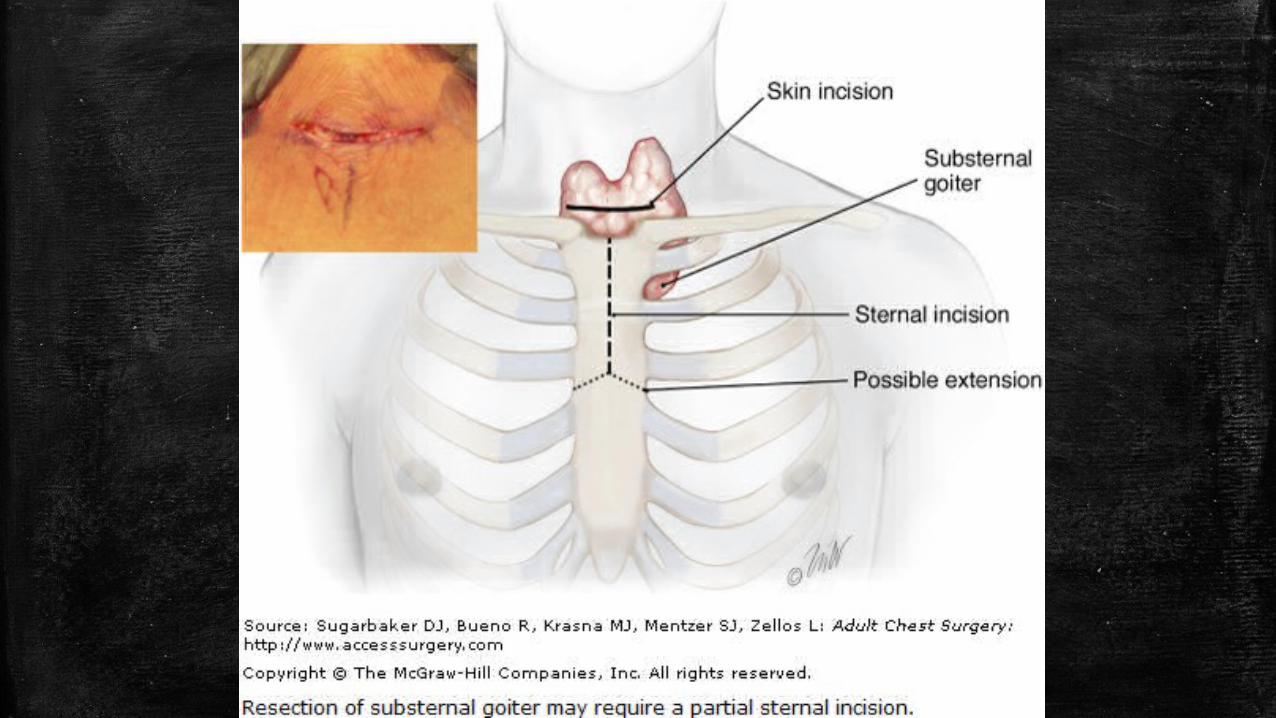
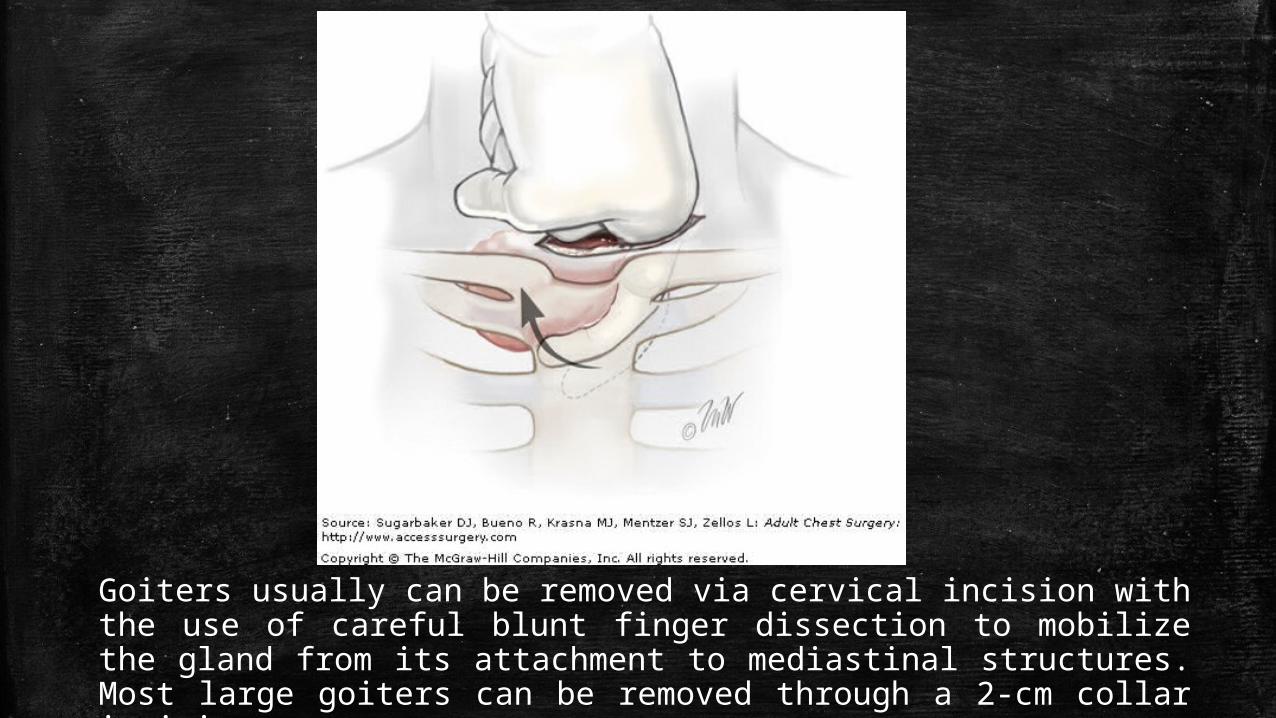
Goiters usually can be removed via cervical incision with the use of careful blunt finger dissection to mobilize the gland from its attachment to mediastinal structures. Most large goiters can be removed through a 2-cm collar incision.
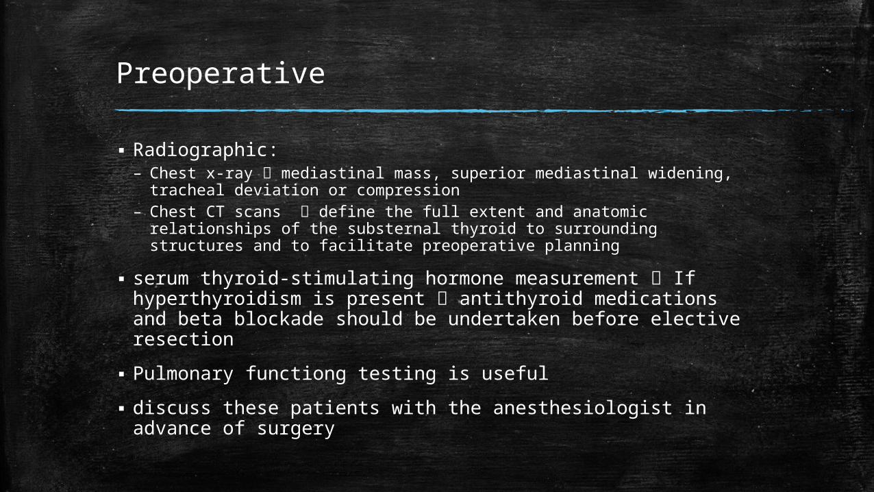
Preoperative
▪ Radiographic:– Chest x-ray mediastinal mass, superior mediastinal widening,
tracheal deviation or compression– Chest CT scans define the full extent and anatomic relationships of
the substernal thyroid to surrounding structures and to facilitate preoperative planning
▪ serum thyroid-stimulating hormone measurement If hyperthyroidism is present antithyroid medications and beta blockade should be undertaken before elective resection
▪ Pulmonary functiong testing is useful
▪ discuss these patients with the anesthesiologist in advance of surgery
Postoperative
▪ Length of stay for an uncomplicated procedure is overnight
▪ patients can be discharged uneventfully with calcium or calcitriol supplementation
▪ If a thoracotomy or sternotomy is required, length of stay is increased
▪ major complications injury to the trachea, parathyroid glands, or recurrent laryngeal nerves
▪ The need for tracheostomy is rare
Germ Cell Tumors
▪ Arise from primordial germ cells that fail to complete the migration from the urogenital ridge and rest in the mediastinum
▪ Anterosuperior mediastinum is the most common extragonadal primary site
▪ The current recommendations for evaluating the testes of a patient with mediastinal germ cell tumor are:– careful physical examination– ultrasonography of the testes.
▪ Biopsy is reserved for positive findings
Teratomas
▪ The most common mediastinal germ cell neoplasms
▪ Usually located in the anterosuperior mediastinum
▪ Composed of multiple tissue elements derived from the three primitive embryonic layers
▪ The peak incidence is in the second and third decades of life
▪ Radiographic evidence of normal tissue (e.g., well-formed teeth or globular calcifications, a fatty mass) in an abnormal location can be considered specific
▪ The teratodermoid (dermoid) cyst is the simplest form of a teratoma and composed of derivatives of the epidermal layer, including dermal and epidermal glands, hair, and sebaceous material

Cont’d
▪ Diagnosis and therapy rely on surgical excision
▪ For benign tumors that are so large or with involvement of adjacent mediastinal structures so that complete resection is impossible partial resection (debulking) can lead to the resolution of symptoms, frequently without relapse
▪ Malignant teratomas chemotherapy and radiation therapy, combined with surgical excision
▪ Overall prognosis is poor for malignant teratomas
Malignant Nonteratomatous Germ Cell Tumors
▪ Occur predominantly in the anterosuperior mediastinum with a marked male predominance
▪ Usually in the third and fourth decades of life
▪ Symptoms: chest pain, cough, dyspnea, and hemoptysis
▪ The superior vena cava syndrome occurs commonly
▪ Diagnostic imaging: A large anterior mediastinal mass
▪ CT and MRI are helpful to define the extent of disease and involvement of mediastinal structures
Cont’d
▪ Serologic measurements (α-fetoprotein and β-hCG) useful for:– differentiating seminomas from nonseminomas tumors, – assessing response to therapy,– diagnosing relapse or failure of therapy
▪ Seminomas rarely produce β-hCG and never produce α-fetoprotein
▪ More than 90% of nonseminomas secrete one or both of these hormones
▪ seminomas are radiosensitive and nonseminomas are relatively radiosensitive
Seminomas
▪ Constitute 50% of malignant germ cell tumors
▪ Usually remain intrathoracic
▪ Symptoms are related to the mechanical effects of the tumor on adjacent mediastinal and pulmonary structures
▪ Superior Vena Cava syndrome occurs in 10% to 20% of patients.
▪ Sensitive to irradiation and chemotherapy
▪ Treatment consists of systemic and local therapy:– chemotherapy with salvage surgery– combined chemoradiotherapy
Cont’d
▪ Radiation therapy may be considered for early-stage disease, but is not recommended for regional disease
▪ Platinum-based chemotherapy is common
▪ Occasionally, excision is possible without injury to vital structures and can be recommended
▪ When complete resection is possible, the use of adjuvant therapy is unnecessary
Nonseminomatous Tumors
▪ Malignant nonseminomatous germ cell tumors include:– choriocarcinomas, – embryonal cell carcinomas,– immature teratomas, – teratomas with malignant components,– endodermal cell (yolk sac) tumors
▪ Occur mostly in men in their third or fourth decade
▪ Diagnostic imaging: large anterior mediastinal mass with frequent extension to the lung, chest wall, and mediastinal structures
Cont’d
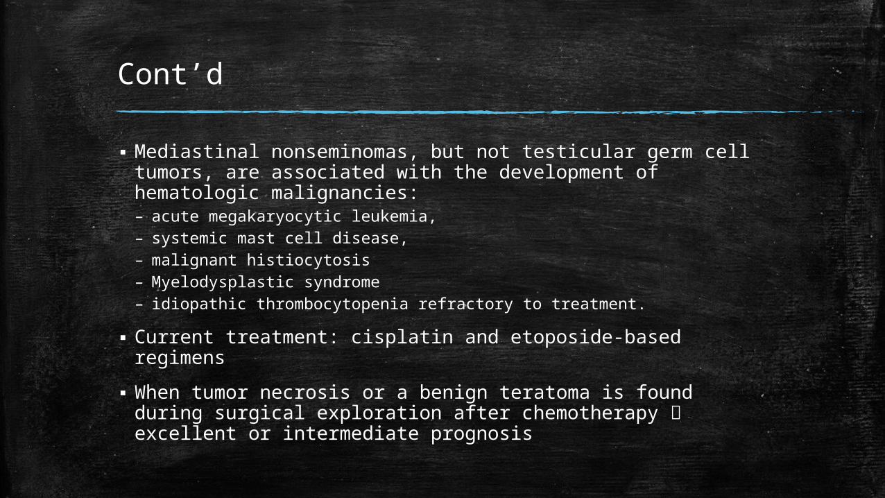
▪ Mediastinal nonseminomas, but not testicular germ cell tumors, are associated with the development of hematologic malignancies:– acute megakaryocytic leukemia,– systemic mast cell disease, – malignant histiocytosis– Myelodysplastic syndrome – idiopathic thrombocytopenia refractory to treatment.
▪ Current treatment: cisplatin and etoposide-based regimens
▪ When tumor necrosis or a benign teratoma is found during surgical exploration after chemotherapy excellent or intermediate prognosis
Lymphomas
▪ Hodgkin’s and non-Hodgkin’s lymphoma are distinct clinical entities with overlapping features
▪ Symptoms: chest pain, cough, dyspnea, hoarseness, and superior vena caval syndrome
▪ Nonspecific systemic symptoms: fever and chills, weight loss, and anorexia
▪ Symptoms characteristic of Hodgkin’s lymphoma chest pain after consumption of alcohol and the cyclic fevers
Cont’d
▪ Surgeon’s primary role is to provide sufficient tissue for diagnosis and to assist in pathologic staging.
▪ Thoracoscopy, mediastinoscopy, or mediastinotomy and, rarely, thoracotomy or median sternotomy may be necessary to obtain sufficient tissue
▪ Lymphoblastic lymphoma occurs predominantly in children, adolescents, and young adults and represents 60% of cases of mediastinal non-Hodgkin’s lymphoma.
Neurogenic Tumors
▪ Usually located in the posterior mediastinum
▪ Originate from: – The sympathetic ganglia (ganglioma, ganglioneuroblastoma, and
neuroblastoma)– intercostal nerves (neurofibroma, neurilemoma, and neurosarcoma)– paraganglia cells (paraganglioma)
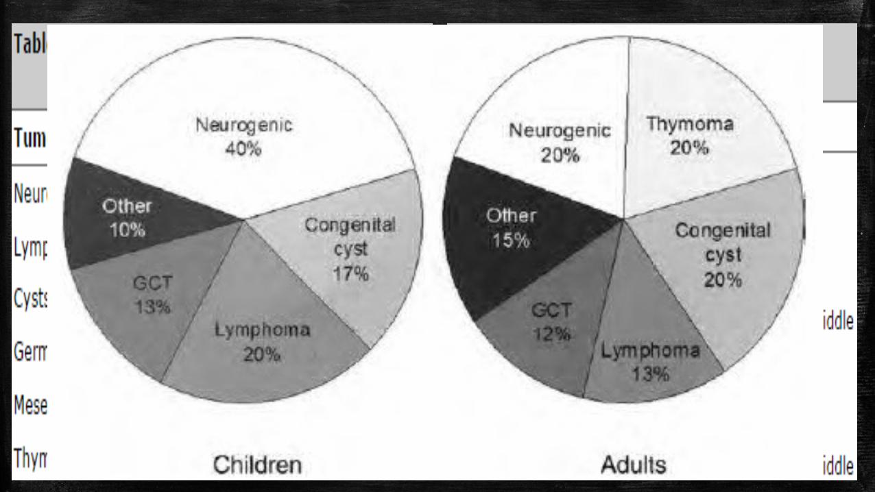
▪ Peak incidence occurs in adults but it make up a proportionally greater percentage of mediastinal masses in children
▪ Most neurogenic tumors in adults are benign but a greater percentage of neurogenic tumors are malignant in children.
Schwannoma / Neurilimoma
▪ The most common neurogenic tumor
▪ Originates from perineural Schwann cells
▪ benign, slow-growing neoplasms, frequently arise from a spinal nerve root, but can involve any thoracic nerve, well circumscribed and have a defined capsule
▪ Peak incidence: third through fifth decades of life
▪ Many are asymptomatic
▪ Pain occur from compression or invasion of intercostal nerve, bone, and chest wall
Cont’d
▪ cough and dyspnea are caused by compression of the tracheobronchial tree
▪ Pancoast syndrome and Horner’s syndrome result from involvement of t
▪ 10% of neurogenic tumors have extensions into the spinal columnhe brachial and the cervical sympathetic chain dumbbell tumors
▪ MRI scan to evaluate the presence and extent of the tumor and its relationship to the neural foramen and intraspinal space
▪ During resection, the intraspinal component should be removed first via a posterior laminectomy minimizes the potential for spinal column hematoma, cord ischemia, and paralysis
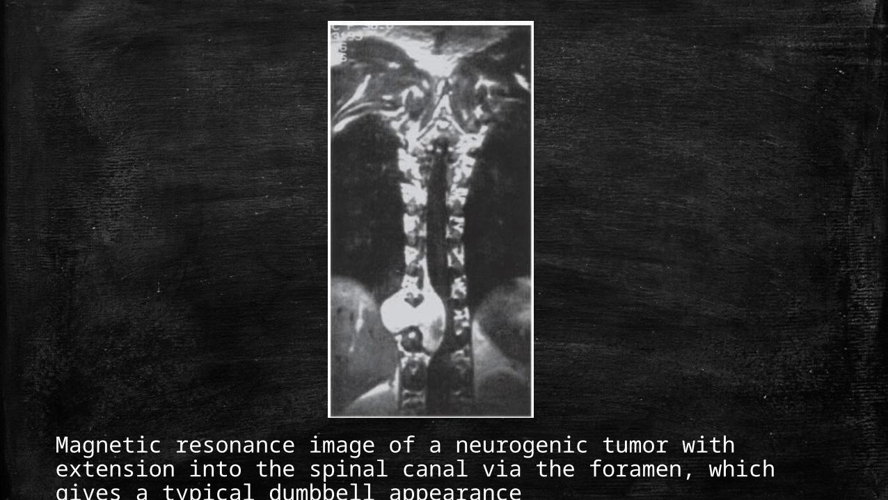
Magnetic resonance image of a neurogenic tumor with extension into the spinal canal via the foramen, which gives a typical dumbbell appearance
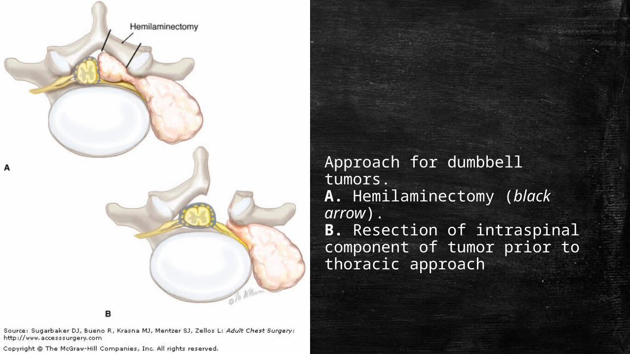
Approach for dumbbell tumors. A. Hemilaminectomy (black arrow). B. Resection of intraspinal component of tumor prior to thoracic approach
Neuroblastomas
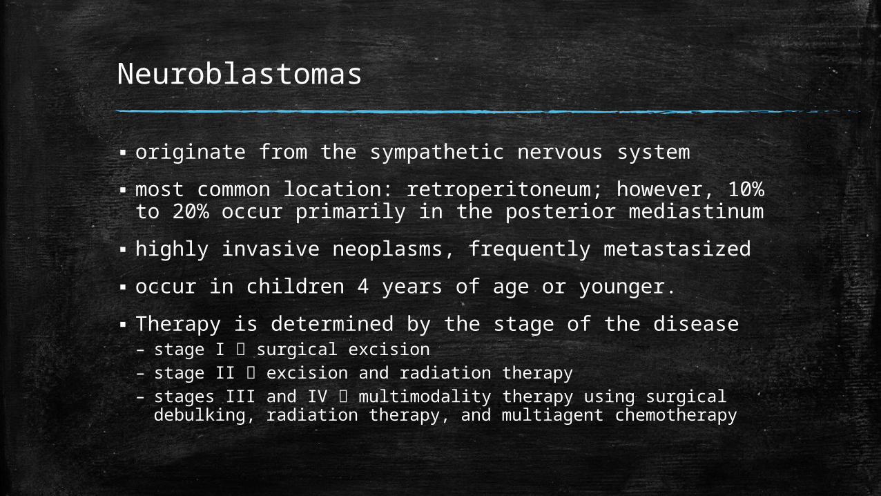
▪ originate from the sympathetic nervous system
▪ most common location: retroperitoneum; however, 10% to 20% occur primarily in the posterior mediastinum
▪ highly invasive neoplasms, frequently metastasized
▪ occur in children 4 years of age or younger.
▪ Therapy is determined by the stage of the disease– stage I surgical excision– stage II excision and radiation therapy– stages III and IV multimodality therapy using surgical
debulking, radiation therapy, and multiagent chemotherapy
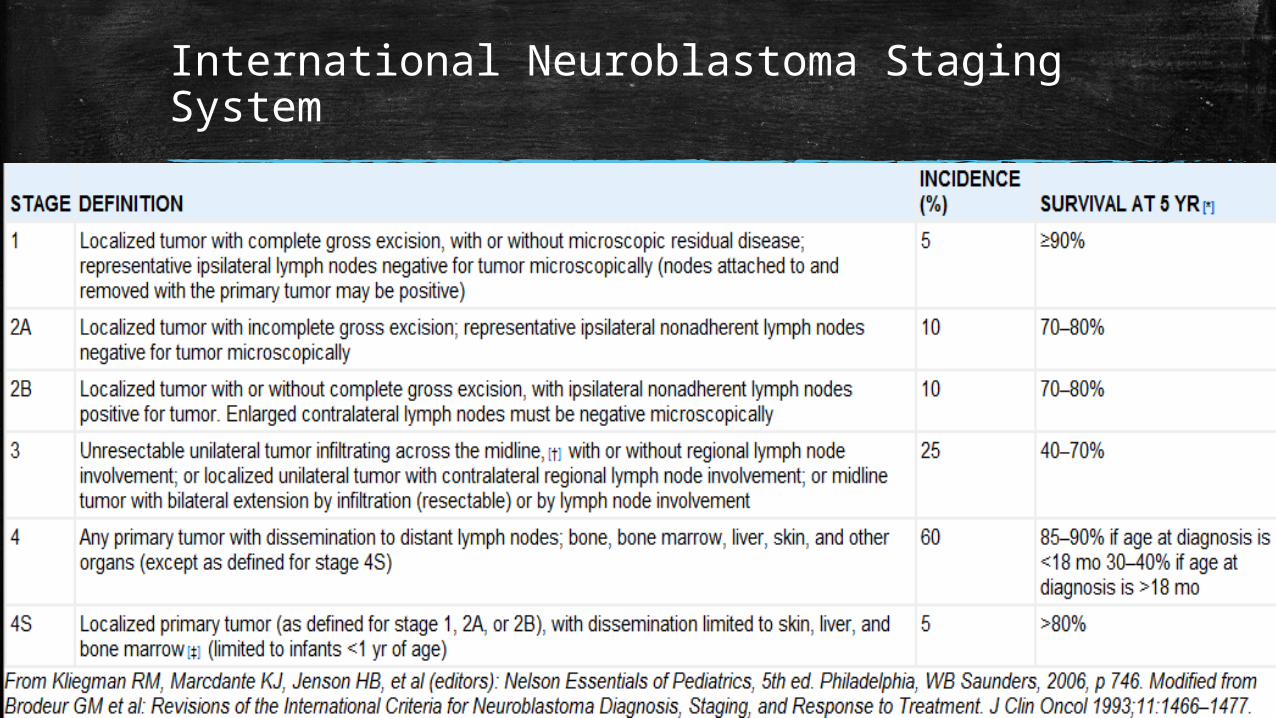
International Neuroblastoma Staging System
Ganglion Tumors
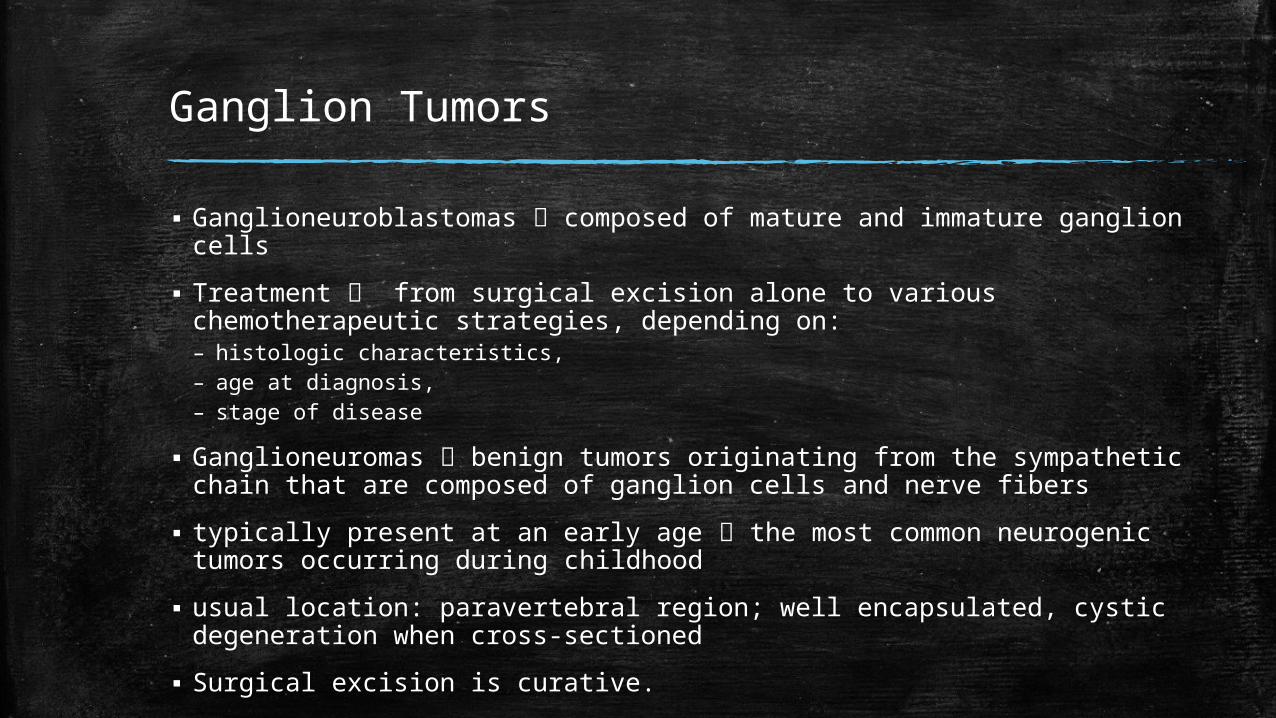
▪ Ganglioneuroblastomas composed of mature and immature ganglion cells
▪ Treatment from surgical excision alone to various chemotherapeutic strategies, depending on: – histologic characteristics, – age at diagnosis, – stage of disease
▪ Ganglioneuromas benign tumors originating from the sympathetic chain that are composed of ganglion cells and nerve fibers
▪ typically present at an early age the most common neurogenic tumors occurring during childhood
▪ usual location: paravertebral region; well encapsulated, cystic degeneration when cross-sectioned
▪ Surgical excision is curative.
Preoperative
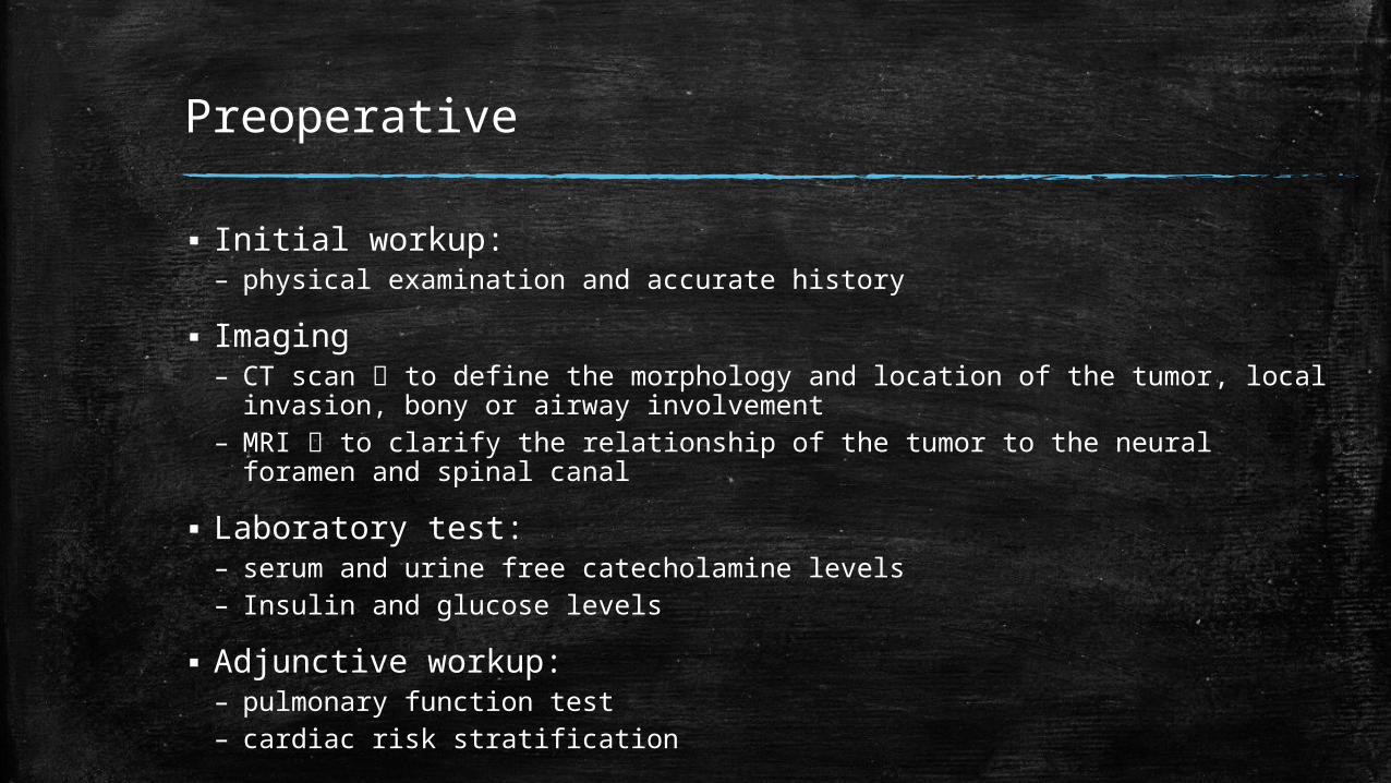
▪ Initial workup:– physical examination and accurate history
▪ Imaging– CT scan to define the morphology and location of the tumor, local invasion,
bony or airway involvement– MRI to clarify the relationship of the tumor to the neural foramen and spinal
canal
▪ Laboratory test:– serum and urine free catecholamine levels – Insulin and glucose levels
▪ Adjunctive workup:– pulmonary function test– cardiac risk stratification
Postoperative
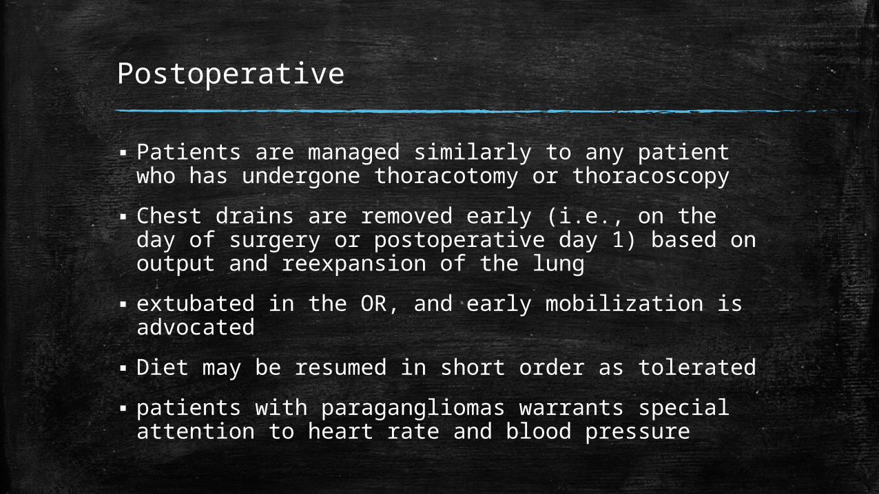
▪ Patients are managed similarly to any patient who has undergone thoracotomy or thoracoscopy
▪ Chest drains are removed early (i.e., on the day of surgery or postoperative day 1) based on output and reexpansion of the lung
▪ extubated in the OR, and early mobilization is advocated
▪ Diet may be resumed in short order as tolerated
▪ patients with paragangliomas warrants special attention to heart rate and blood pressure
References
1. Sugarbaker D, Bueno R, Krasna M, Mentzer S, Zellos L. Adult Chest Surgery. McGraw Hill Professional; 2009.
2. DeVita VT, Lawrence TS, Rosenberg SA. DeVita, Hellman, and Rosenberg’s Cancer: Principles & Practice of Oncology. Lippincott Williams & Wilkins; 2008.
3. Jr CMT, Beauchamp RD, Evers BM, Mattox KL. Sabiston Textbook of Surgery: Expert Consult Premium Edition: Enhanced Online Features. 19th ed. Elsevier Health Sciences; 2012.
4. Brunicardi F, Andersen D, Billiar T, Dunn D, Hunter J, Matthews J, et al. Schwartz’s Principles of Surgery. 9th ed. McGraw-Hill Education; 2009.
5. Norton JA, Barie PS, Bollinger RR, Chang AE, M.D SFL, M.D SJM, et al. Surgery: Basic Science and Clinical Evidence. Springer; 2009.

THANK YOU