mechanisms of wound repair in crayfish - … 6: 125-137, 2009 issn 1824-307x review mechanisms of...
TRANSCRIPT
ISJ 6: 125-137, 2009 ISSN 1824-307X
REVIEW
Mechanisms of wound repair in crayfish X Vafopoulou Biology Department, York University, 4700 Keele Street, Toronto M3J 1P3, Ontario, Canada
Accepted September 8, 2009
Abstract This review describes the complexity of events involved with repair to integumentary wounds and
their regulation using the crayfish as a model system. Injuries to integument precipitate a cascade of cellular events that lead to rapid healing of the wound, regeneration of damaged tissues and repair of the integument. The first step in this cascade is hemolymph clotting and subsequent melanization, events documented thoroughly elsewhere and not discussed here. Wound healing and repair in crayfish involves the action of two physiological systems, the immune system and the neuroendocrine system regulating synthesis of the steroid molting hormones, ecdysteroids. Injury promotes a swift rise in hemolymph ecdysteroids to a low, sustained plateau, followed by a premolt peak and molting. The plateau is essential for wound healing since its principal targets are the circulating cells of the immune system, the hemocytes, and healthy epidermal cells and fibrocytes. Massive migration of these cells occurs under the wound and their concerted efforts under ecdysteroid control are paramount to wound healing and repair. These cells are likely engaged in physiological and biochemical activities that promote cell communication and cell to cell adhesion, removal of dead and harmful material and production of molecules essential to tissue regeneration. Key Words: Crustacea; immune system; ecdysteroids; hemocytes; epidermis; regeneration
Introduction
Structural integrity in crustaceans is maintained by a rigid exoskeleton. The exoskeleton is also the main defense barrier against invasions by microbes and parasites. When this barrier is breached by mechanical injuries or erosion due to chemicals or bacteria, the animal becomes vulnerable to systemic infections. It is therefore vital for survival that injuries to integument are healed promptly and the damaged integument is repaired quickly. This review addresses events and mechanisms by which these responses are brought about. Injury triggers a dramatic awakening of the machinery that directs restoration and repair of damaged tissues the details of which are discussed in the following section. Wound healing is a complex and orderly process involving the coordinated behavior of different cell types. It relies heavily on rapid immune responses by the hemocytes which seal and protect the wound site and massive movements of cells which regenerate the damaged tissues. The initial event is coagulation of hemolymph components that ___________________________________________________________________________
Corresponding author: Xanthe Vafopoulou Biology Department York University 4700 Keele Street, Toronto M3J 1P3, Ontario, Canada E-mail: [email protected]
seal off wounds temporarily and thus contain hemolymph loss and trap microbes (see for review Lee and Söderhäll, 2002; Cerenius and Söderhäll, 2004). Ruptured hemocytes release clotting enzymes that coagulate hemolymph and enzymes that activate the pro-phenoloxidase (proPO) cascade which leads to melanisation and ensures trapping of foreign and damaged cell material. Additional immune responses are also triggered that include phagocytosis, cytotoxicity, nodule formation and encapsulation which kill invading microbes and remove cellular and matrix debris. Concurrently with hemocyte recruitment, epidermis regeneration is triggered and when this process is completed the newly formed epidermal layer deposits a new cuticle that seals the wound permanently and restores the damaged integument to its original condition.
The phenomena of wound healing and repair raise fundamental biological issues concerning the origin of cues that initiate the cascade of this process. Cues may originate locally to act from one cell population to another or may be humoral in nature. For example the cues that trigger hemolymph coagulation are usually local. In the case of bacterial infection, local signals present on the surface of microbes initiate hemolymph clotting (see for review Sritunyalucksana and Söderhäll, 2000; Theopold et al., 2002, 2004). By contrast, in
125
the case of physical wounding the cues are largely unknown. It is hypothesized that, as in vertebrates (e.g., Gallucci and Matzinger, 2001), unidentified “danger signals” are released from the damaged cells that trigger the clotting reaction. Danger signals may include cellular components of damaged cells such as nucleotides, reactive oxygen intermediates or other intracellular proteins, which activate the hemolymph clotting cascade (review by Theopold et al., 2002). Another example of local signals derives from the study of limb regeneration in crabs following limb autotomy; regenerating limb buds release and respond to growth factor(s) that promote local growth (Hopkins et al., 1979, 1999; Hopkins, 2001).
Cues for the cascade of wound healing and repair may also be provided distantly, specifically from the central neuroendocrine system. The primary candidate for promoting tissue regeneration is the steroid molting hormones of crustaceans, ecdysteroids. For example, wound healing and repair depends on the presence of low levels of ecdysteroids following carapace injury in crabs (Halcrow and Steel, 1992) and crayfish (Vafopoulou et al., 2007) or limb autotomy in crabs (McCarthy and Skinner, 1977).
Freshwater crayfish provide an excellent model system to advance knowledge on the mechanisms of wound healing and repair. Crayfish are of commercial and ecological importance. They are easy and inexpensive to rear in large numbers under laboratory conditions for experimentation in a simple aquatic environment. Key components of the innate immunity system have been established in the crayfish (e.g., Söderhäll et al., 1994; Söderhäll and Cerenius, 1998; Wang et al., 2001a; Lee and Söderhäll, 2001, 2002) and much is known about the morphology and development of the animal (references in Vogt, 2008). In the present review, we will discuss the current state of understanding of wound healing and tissue regeneration in the freshwater crayfish Procambarus clarkii and the underlying mechanism of hormonal control that brings about these changes. Wound healing and repair in crayfish
We have found a direct physiological link
between the immune system and the system that controls molting during wound healing and repair in the crayfish. Specifically, we found in wounded animals immune responses affect the molting system and molting responses affect the immune system. Interaction of these two systems is crucial in the ability and speed by which a crayfish heal wounds to the integument. The two physiological systems and their involvement in wound healing are described in details below.
The first system to respond to injuries is the immune system. Many aspects of the crustacean immune system have been elucidated in crayfish, making it a valuable animal to study the involvement of the immune system in wound healing. The immune system is centered on the circulating hemocytes and involves mechanisms to recognize and destroy non-self material and heal wounds. There are two components in innate immunity in
crustaceans, the humoral and cellular components, both of which are activated upon immune challenges resulting from wounds to integument. Cellular defenses include hemocyte-mediated responses such as phagocytosis, nodulation and encapsulation. Humoral defences include antimicrobial peptides, coagulation and melanization of the hemolymph and production of reactive intermediates of oxygen and nitrite (see Lee and Söderhäll, 2002; Lee et al., 2004). Most of the bioactive peptides are produced by the hemocytes. Among these bioactive compounds, the proPO-activating system is the best studied and plays a pivotal role in innate immunity. In crayfish, proPO and the enzymes responsible for its conversion to its active form phenoloxidase (PO) reside as zymogens in the granules of granular and semigranular hemocytes, the contents of which are released by exocytosis into the hemolymph under challenges by pathogens or cell damage because of wounding (Smith and Söderhäll, 1991; Wang et al., 2001a). This conversion ultimately results in the formation of melanin and the generation of a number of potent bioactive products, which assist phagocytosis, cell to cell adhesion and melanization (see Söderhäll et al., 1994; Söderhäll and Cerenius, 1998; see for review Lee and Söderhäll, 2002; Cerenius and Söderhäll, 2004). ProPO has been cloned in crayfish (Aspán et al., 1995). Other important components of immune responses have also been extensively studied in crayfish. For example, components of the clotting and melanization system have been characterized such as a transglutaminase essential for hemolymph clotting (Hall et al., 1999; Wang et al., 2001b) and an inhibitory protein that halts melanization (Söderhäll et al., 2009); antimicrobial peptides such as astacidin and crustin (Jiravanichpaisal et al., 2007); hemocyanin which acts as a phenoloxidase in crayfish and a precursor to an antibacterial peptide (Lee et al., 2004); peroxinectin, a multifunctional peptide critical for cell to cell adhesion (Johansson and Söderhäll, 1988), encapsulation (Kobayashi et al., 1990) and degranulation (Johansson and Söderhäll, 1989); a pattern recognition peptide (Lee and Söderhäll, 2001; see for review Sritunyalucksana and Söderhäll, 2000; Lee and Söderhäll, 2002; Cerenius and Söderhäll, 2004).
Study of the cascade of events in wound healing depends on recognition of the morphological and functional characteristics of hemocytes. Hemocytes are classified according to their morphology, cytochemistry and function (Smith and Söderhäll, 1983a; see for review Johansson et al., 2000) and are recognized as hyaline, semigranular and granular hemocytes based on a classification system established by Smith and Söderhäll (1983a). Based on this system, hemocytes have been characterized in several crayfish such as Astacus astacus (Smith and Söderhäll, 1983a), Procambarus zonangulus (Cárdenas et al., 2000), Pacifastacus leniusculus (Wang et al., 2001a), Procambarus clarkii (Vafopoulou et al., 2007) and Astacus leptodactylus (Giulianini et al., 2007). In general, all three classes and subclasses of hemocytes, are recognized in all crustacean species (e.g., Vázquez et al., 1997; Johansson et al., 2000; Sung and Sun,
126
2002; Battison et al., 2003; Martin et al., 2003; Liu et al., 2007; Zhan et al., 2008). Hemocytes have a defined division of labor in innate immunity. Hyaline hemocytes are critical for hemolymph clotting (Hall and Söderhäll, 1994; Wang et al., 2001a; reviews by Theopold et al., 2002, 2004) and phagocytosis (Smith and Söderhäll, 1983b). The principal role of semigranular hemocytes is encapsulation (Kobayashi et al., 1990) and cytotoxicity (Söderhäll et al., 1985), whereas granular hemocytes are the primary source of the proPO-activating system (Johansson and Söderhäll, 1985; see for review Sritunyalucksana and Söderhäll, 2000).
The second system in crayfish that responds to injury is the neuroendocrine system that controls molting (Vafopoulou et al., 2007). Molting is the cyclical formation of a new exoskeleton and the shedding of the old one. Growth and molting in crustaceans is regulated by ecdysteroids (such ecdysone and 20-hydroxyecdysone) the concentration of which displays a characteristic sequence of increases and decreases over a molt cycle (Steel and Vafopoulou, 1989). Ecdysteroids are synthesized and secreted by the Y-organs under the negative control of the neuropeptide molt-inhibiting hormone (MIH). MIH is synthesized by the X-organ in the brain and released from the sinus gland in the eyestalk (see for review Webster, 1998). When present, it prevents the secretion of ecdysteroids from the Y organ and holds crustaceans in intermolt (e.g., Snyder and Chang, 1986; see for review Chang et al., 2001). New cuticle is secreted by the epidermis under the action of ecdysteroids; this period in the molt cycle is called ‘premolt’ and it culminates in shedding of the old exoskeleton (ecdysis) and hardening of the new one in the post-ecdysial period known as ‘postmolt’. During the remainder of a molt cycle, animals are devoid of ecdysteroids and not preparing to molt; this period is called ‘intermolt’.
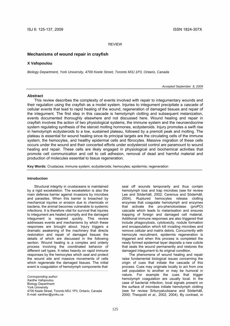
We have found that injuries to the crayfish integument during intermolt induce precocious production of ecdysteroids, measured by radioimmunoassay (RIA), as shown in Fig. 1 (details in Vafopoulou et al., 2007). These injuries cause initially a small but significant elevation in the concentration of hemolymph ecdysteroids to a low, sustained plateau (10-12 days after injury) (Fig. 1A, B; dark triangles). The plateau is then followed after about 2-3 weeks by a sharp increase to the peak values characteristic of premolt. Ecdysis occurs at about 2 months later in crayfish. Uninjured, healthy controls show only a slight increase in hemolymph ecdysteroid levels throughout the premolt period of wounded animals (Fig. 1A, B; open triangles), and they never form a premolt peak. Positive control animals which are subjected to removal of both eyestalks but no exocuticular injury exhibit no plateau phase but rather commence the steep premolt increase promptly after ablation (Fig. 1A, B; open circles). Bilateral eyestalk removal is known to precipitate an immediate premolt, because it removes the source of the neuropeptide molt-inhibiting hormone (MIH). Therefore, both eyestalkless and wounded animals are induced to enter premolt but in wounded animals the premolt peak is delayed relative to eyestalkless animals by the duration of the plateau phase.
Fig. 1 Induction of premolt and molt in intermolt crayfish determined by changes in hemolymph ecdysteroid titre using a RIA. Time zero indicates time of treatment. (A) Bilateral eyestalk ablation (removal of molt inhibiting hormone) (positive control; open circles, orange line). Premolt peak is reached at about 35 days and animals undergo ecdysis at about 50 days after treatment. Dark triangles (light green line) represent wound to the integument. Premolt peak is delayed compared to eyestalk ablated animals by about two weeks and animals undergo ecdysis at about 55 days after treatment. This delay is due to a plateau phase of low ecdysteroid level at days 10-12 after wounding. Dark circles (brown line) represent integument wound plus bacteria infection. Premolt peak is reached at about day 55 after treatment. A plateau phase (days 0-20) is further prolonged when compared to line b (dark triangles) by about a week. Open triangles (dark green line) represent untreated, intact crayfish (negative control). These animals do not reach a premolt peak during the experiment and do not undergo ecdysis. (B) Enlarged view of the plateau phase of ecdysteroid titre (days 0-15 underlined with a light blue line on the X-axis of panel (A). (Modified from Vafopoulou et al., 2007).
127
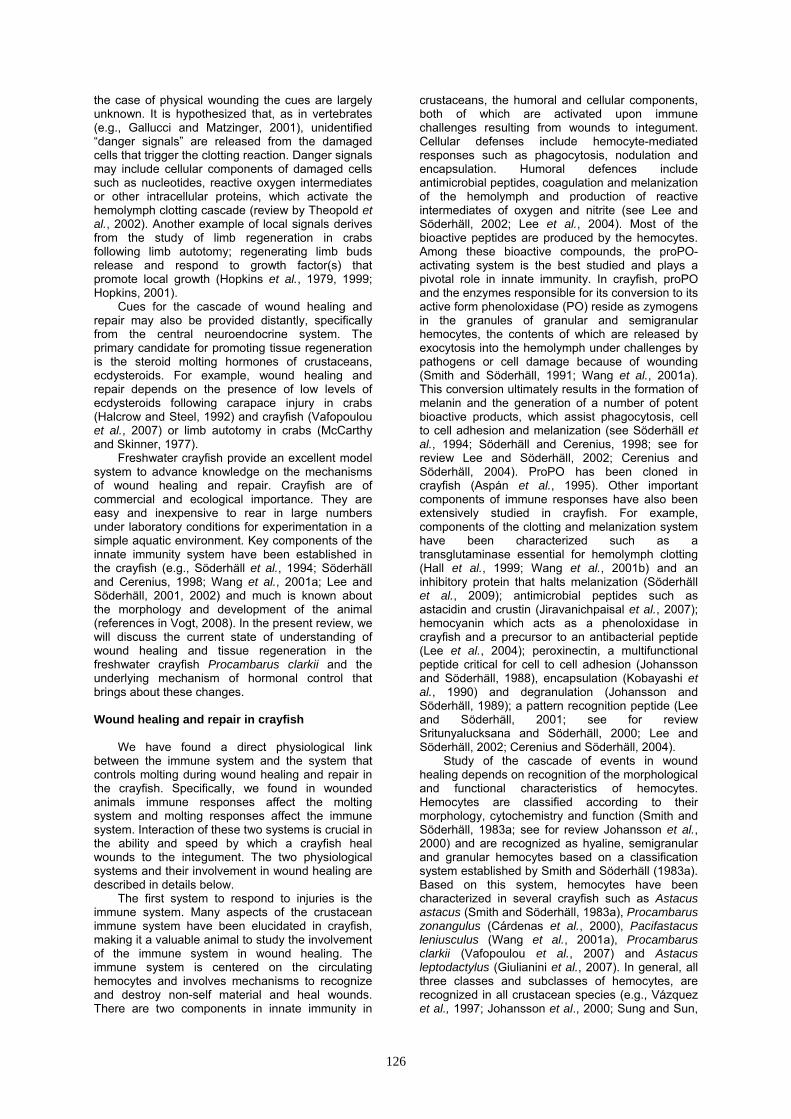
Fig. 2 Digital images of cells under the wound using confocal laser scanning microscopy and fluorescent immunohistochemistry on whole tissue mounts. Tissues were stained with an antibody against EcR. Ecdysteroid-responsive cells are revealed by EcR fluorescence which appears as yellow-green in the nuclei. Cellular material and fibres of coagulated hemolymph without EcR are shown as olive green or red. Optical sections are 1 μm thick. Image A was taken immediately after wounding. Images B-I were taken at 2 h after wounding. (A) Hyaline hemocytes (short arrows) and strands of coagulated hemolymph fibres (long arrow). (B) Aggregation of large number of ecdysone-responsive hyaline hemocytes (stack of 9 optical sections from a z-series). (C) Granular hemocytes (arrows) interspersed among hyaline hemocytes (stack of 12 optical sections from a z-series). Red, spherical cytoplasmic inclusions represent granules. (D-F) Enlarged views of the three types of hemocytes in crayfish: (D) Hyaline hemocytes; (E) Semigranular hemocytes; arrow shows small size cytoplasmic granules; (F) Granular hemocytes; arrow shows large size cytoplasmic granules. (G-I) High magnification of a single nucleus from hyaline hemocyte double-labelled with anti-EcR (G, green) and a fluorescent nucleic acid dye, propidium iodide (I, red) showing co-localization of EcR with chromatin fibres in the nucleus (H, shows merged image of G and F; co-localization is shown as yellow-green). This configuration of distribution of EcR fluorescence in the nuclei of ecdysone-responsive cells is suggested to represent the active state of EcR engaged in gene transcription (Vafopoulou et al., 2005; Vafopoulou and Steel, 2006). Bar = 10 μm.
The plateau phase of ecdysteroids following injury is necessary for wound healing, epidermal regeneration and initiation of the process of repair of the wound on the integument. This plateau represents the main difference in the premolt processes between eyestalk ablated animals and wounded animals. Successful restoration of the damaged integument during molting for wounded crayfish depends on previous regeneration of the damaged epidermis layer; a fully restored epidermis is required to secrete a new cuticle. During the
plateau phase, we found the ecdysteroid receptor (EcR) localized in the nuclei of various types of cells involved in wound healing and repair using fluorescent antibodies to EcR, immunohistochemistry (IHC) and confocal laser scanning microscopy (Fig. 2) (details in Vafopoulou et al., 2007). Cellular responses to ecdysteroids are mediated by EcR. EcR belongs to the nuclear receptor superfamily that includes receptors of hormones like estrogen, progesterone, thyroid hormone, vitamin D and others. EcR acts as ligand-
128
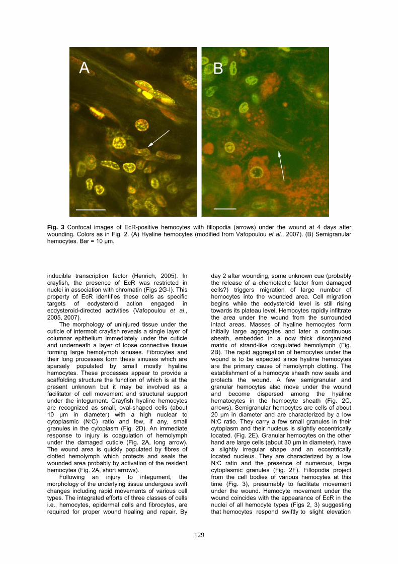
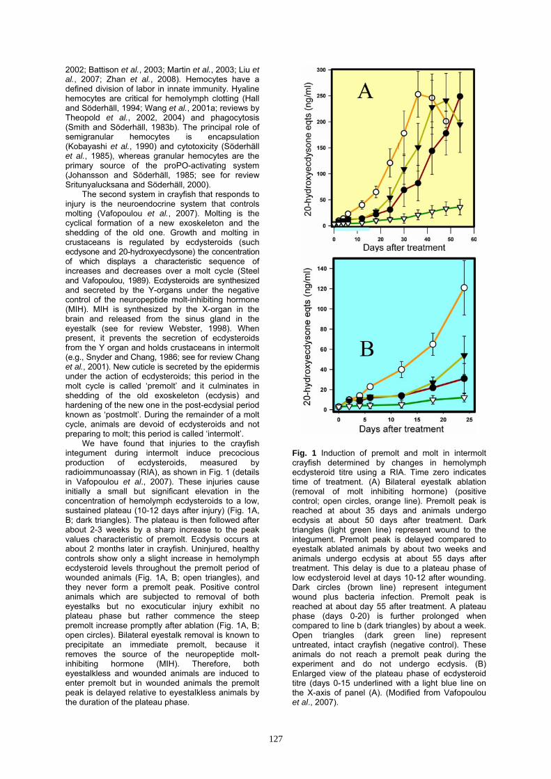
Fig. 3 Confocal images of EcR-positive hemocytes with fillopodia (arrows) under the wound at 4 days after wounding. Colors as in Fig. 2. (A) Hyaline hemocytes (modified from Vafopoulou et al., 2007). (B) Semigranular hemocytes. Bar = 10 μm. inducible transcription factor (Henrich, 2005). In crayfish, the presence of EcR was restricted in nuclei in association with chromatin (Figs 2G-I). This property of EcR identifies these cells as specific targets of ecdysteroid action engaged in ecdysteroid-directed activities (Vafopoulou et al., 2005, 2007).
The morphology of uninjured tissue under the cuticle of intermolt crayfish reveals a single layer of columnar epithelium immediately under the cuticle and underneath a layer of loose connective tissue forming large hemolymph sinuses. Fibrocytes and their long processes form these sinuses which are sparsely populated by small mostly hyaline hemocytes. These processes appear to provide a scaffolding structure the function of which is at the present unknown but it may be involved as a facilitator of cell movement and structural support under the integument. Crayfish hyaline hemocytes are recognized as small, oval-shaped cells (about 10 μm in diameter) with a high nuclear to cytoplasmic (N:C) ratio and few, if any, small granules in the cytoplasm (Fig. 2D). An immediate response to injury is coagulation of hemolymph under the damaged cuticle (Fig. 2A, long arrow). The wound area is quickly populated by fibres of clotted hemolymph which protects and seals the wounded area probably by activation of the resident hemocytes (Fig. 2A, short arrows).
Following an injury to integument, the morphology of the underlying tissue undergoes swift changes including rapid movements of various cell types. The integrated efforts of three classes of cells i.e., hemocytes, epidermal cells and fibrocytes, are required for proper wound healing and repair. By
day 2 after wounding, some unknown cue (probably the release of a chemotactic factor from damaged cells?) triggers migration of large number of hemocytes into the wounded area. Cell migration begins while the ecdysteroid level is still rising towards its plateau level. Hemocytes rapidly infiltrate the area under the wound from the surrounded intact areas. Masses of hyaline hemocytes form initially large aggregates and later a continuous sheath, embedded in a now thick disorganized matrix of strand-like coagulated hemolymph (Fig. 2B). The rapid aggregation of hemocytes under the wound is to be expected since hyaline hemocytes are the primary cause of hemolymph clotting. The establishment of a hemocyte sheath now seals and protects the wound. A few semigranular and granular hemocytes also move under the wound and become dispersed among the hyaline hematocytes in the hemocyte sheath (Fig. 2C, arrows). Semigranular hemocytes are cells of about 20 μm in diameter and are characterized by a low N:C ratio. They carry a few small granules in their cytoplasm and their nucleus is slightly eccentrically located. (Fig. 2E). Granular hemocytes on the other hand are large cells (about 30 μm in diameter), have a slightly irregular shape and an eccentrically located nucleus. They are characterized by a low N:C ratio and the presence of numerous, large cytoplasmic granules (Fig. 2F). Fillopodia project from the cell bodies of various hemocytes at this time (Fig. 3), presumably to facilitate movement under the wound. Hemocyte movement under the wound coincides with the appearance of EcR in the nuclei of all hemocyte types (Figs 2, 3) suggesting that hemocytes respond swiftly to slight elevation
129
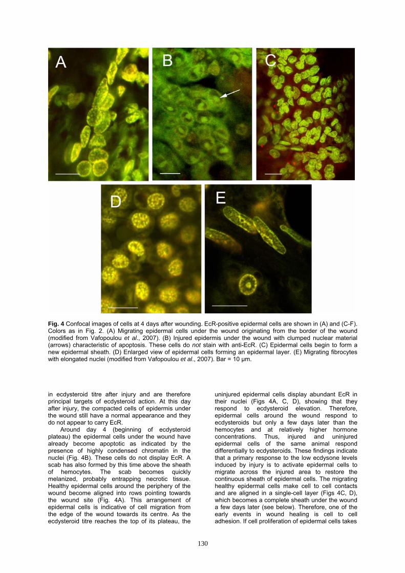
Fig. 4 Confocal images of cells at 4 days after wounding. EcR-positive epidermal cells are shown in (A) and (C-F). Colors as in Fig. 2. (A) Migrating epidermal cells under the wound originating from the border of the wound (modified from Vafopoulou et al., 2007). (B) Injured epidermis under the wound with clumped nuclear material (arrows) characteristic of apoptosis. These cells do not stain with anti-EcR. (C) Epidermal cells begin to form a new epidermal sheath. (D) Enlarged view of epidermal cells forming an epidermal layer. (E) Migrating fibrocytes with elongated nuclei (modified from Vafopoulou et al., 2007). Bar = 10 μm. in ecdysteroid titre after injury and are therefore principal targets of ecdysteroid action. At this day after injury, the compacted cells of epidermis under the wound still have a normal appearance and they do not appear to carry EcR.
Around day 4 (beginning of ecdysteroid plateau) the epidermal cells under the wound have already become apoptotic as indicated by the presence of highly condensed chromatin in the nuclei (Fig. 4B). These cells do not display EcR. A scab has also formed by this time above the sheath of hemocytes. The scab becomes quickly melanized, probably entrapping necrotic tissue. Healthy epidermal cells around the periphery of the wound become aligned into rows pointing towards the wound site (Fig. 4A). This arrangement of epidermal cells is indicative of cell migration from the edge of the wound towards its centre. As the ecdysteroid titre reaches the top of its plateau, the
uninjured epidermal cells display abundant EcR in their nuclei (Figs 4A, C, D), showing that they respond to ecdysteroid elevation. Therefore, epidermal cells around the wound respond to ecdysteroids but only a few days later than the hemocytes and at relatively higher hormone concentrations. Thus, injured and uninjured epidermal cells of the same animal respond differentially to ecdysteroids. These findings indicate that a primary response to the low ecdysone levels induced by injury is to activate epidermal cells to migrate across the injured area to restore the continuous sheath of epidermal cells. The migrating healthy epidermal cells make cell to cell contacts and are aligned in a single-cell layer (Figs 4C, D), which becomes a complete sheath under the wound a few days later (see below). Therefore, one of the early events in wound healing is cell to cell adhesion. If cell proliferation of epidermal cells takes
130
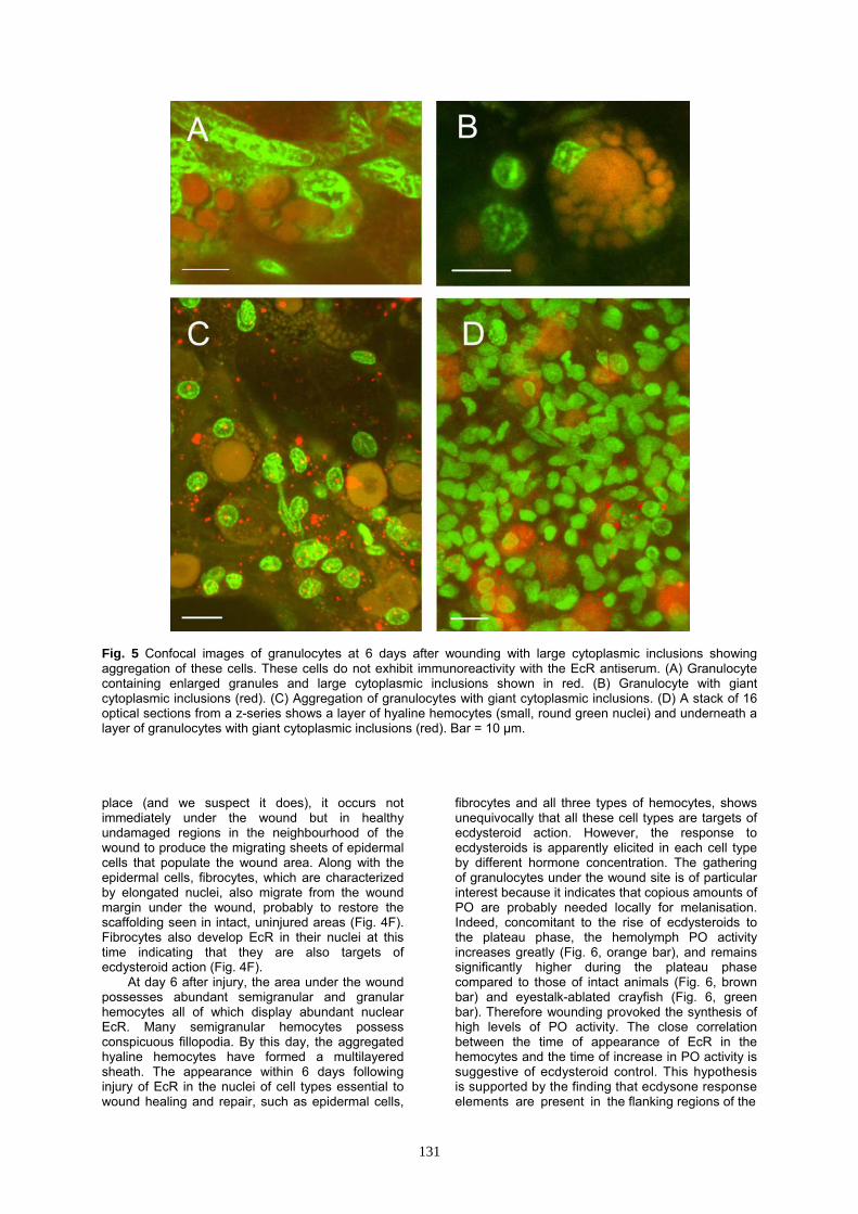
Fig. 5 Confocal images of granulocytes at 6 days after wounding with large cytoplasmic inclusions showing aggregation of these cells. These cells do not exhibit immunoreactivity with the EcR antiserum. (A) Granulocyte containing enlarged granules and large cytoplasmic inclusions shown in red. (B) Granulocyte with giant cytoplasmic inclusions (red). (C) Aggregation of granulocytes with giant cytoplasmic inclusions. (D) A stack of 16 optical sections from a z-series shows a layer of hyaline hemocytes (small, round green nuclei) and underneath a layer of granulocytes with giant cytoplasmic inclusions (red). Bar = 10 μm. place (and we suspect it does), it occurs not immediately under the wound but in healthy undamaged regions in the neighbourhood of the wound to produce the migrating sheets of epidermal cells that populate the wound area. Along with the epidermal cells, fibrocytes, which are characterized by elongated nuclei, also migrate from the wound margin under the wound, probably to restore the scaffolding seen in intact, uninjured areas (Fig. 4F). Fibrocytes also develop EcR in their nuclei at this time indicating that they are also targets of ecdysteroid action (Fig. 4F).
At day 6 after injury, the area under the wound possesses abundant semigranular and granular hemocytes all of which display abundant nuclear EcR. Many semigranular hemocytes possess conspicuous fillopodia. By this day, the aggregated hyaline hemocytes have formed a multilayered sheath. The appearance within 6 days following injury of EcR in the nuclei of cell types essential to wound healing and repair, such as epidermal cells,
fibrocytes and all three types of hemocytes, shows unequivocally that all these cell types are targets of ecdysteroid action. However, the response to ecdysteroids is apparently elicited in each cell type by different hormone concentration. The gathering of granulocytes under the wound site is of particular interest because it indicates that copious amounts of PO are probably needed locally for melanisation. Indeed, concomitant to the rise of ecdysteroids to the plateau phase, the hemolymph PO activity increases greatly (Fig. 6, orange bar), and remains significantly higher during the plateau phase compared to those of intact animals (Fig. 6, brown bar) and eyestalk-ablated crayfish (Fig. 6, green bar). Therefore wounding provoked the synthesis of high levels of PO activity. The close correlation between the time of appearance of EcR in the hemocytes and the time of increase in PO activity is suggestive of ecdysteroid control. This hypothesis is supported by the finding that ecdysone response elements are present in the flanking regions of the
131
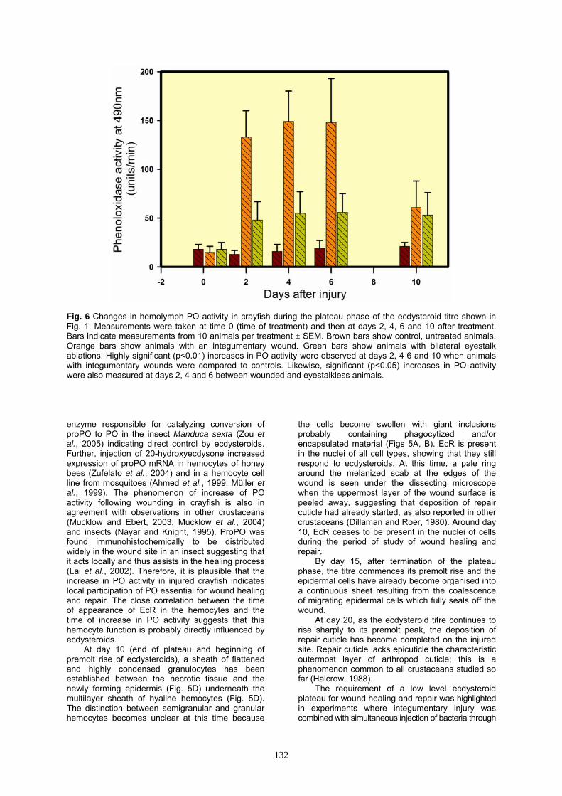
Fig. 6 Changes in hemolymph PO activity in crayfish during the plateau phase of the ecdysteroid titre shown in Fig. 1. Measurements were taken at time 0 (time of treatment) and then at days 2, 4, 6 and 10 after treatment. Bars indicate measurements from 10 animals per treatment ± SEM. Brown bars show control, untreated animals. Orange bars show animals with an integumentary wound. Green bars show animals with bilateral eyestalk ablations. Highly significant (p<0.01) increases in PO activity were observed at days 2, 4 6 and 10 when animals with integumentary wounds were compared to controls. Likewise, significant (p<0.05) increases in PO activity were also measured at days 2, 4 and 6 between wounded and eyestalkless animals. enzyme responsible for catalyzing conversion of proPO to PO in the insect Manduca sexta (Zou et al., 2005) indicating direct control by ecdysteroids. Further, injection of 20-hydroxyecdysone increased expression of proPO mRNA in hemocytes of honey bees (Zufelato et al., 2004) and in a hemocyte cell line from mosquitoes (Ahmed et al., 1999; Müller et al., 1999). The phenomenon of increase of PO activity following wounding in crayfish is also in agreement with observations in other crustaceans (Mucklow and Ebert, 2003; Mucklow et al., 2004) and insects (Nayar and Knight, 1995). ProPO was found immunohistochemically to be distributed widely in the wound site in an insect suggesting that it acts locally and thus assists in the healing process (Lai et al., 2002). Therefore, it is plausible that the increase in PO activity in injured crayfish indicates local participation of PO essential for wound healing and repair. The close correlation between the time of appearance of EcR in the hemocytes and the time of increase in PO activity suggests that this hemocyte function is probably directly influenced by ecdysteroids.
At day 10 (end of plateau and beginning of premolt rise of ecdysteroids), a sheath of flattened and highly condensed granulocytes has been established between the necrotic tissue and the newly forming epidermis (Fig. 5D) underneath the multilayer sheath of hyaline hemocytes (Fig. 5D). The distinction between semigranular and granular hemocytes becomes unclear at this time because
the cells become swollen with giant inclusions probably containing phagocytized and/or encapsulated material (Figs 5A, B). EcR is present in the nuclei of all cell types, showing that they still respond to ecdysteroids. At this time, a pale ring around the melanized scab at the edges of the wound is seen under the dissecting microscope when the uppermost layer of the wound surface is peeled away, suggesting that deposition of repair cuticle had already started, as also reported in other crustaceans (Dillaman and Roer, 1980). Around day 10, EcR ceases to be present in the nuclei of cells during the period of study of wound healing and repair.
By day 15, after termination of the plateau phase, the titre commences its premolt rise and the epidermal cells have already become organised into a continuous sheet resulting from the coalescence of migrating epidermal cells which fully seals off the wound.
At day 20, as the ecdysteroid titre continues to rise sharply to its premolt peak, the deposition of repair cuticle has become completed on the injured site. Repair cuticle lacks epicuticle the characteristic outermost layer of arthropod cuticle; this is a phenomenon common to all crustaceans studied so far (Halcrow, 1988).
The requirement of a low level ecdysteroid plateau for wound healing and repair was highlighted in experiments where integumentary injury was combined with simultaneous injection of bacteria through
132
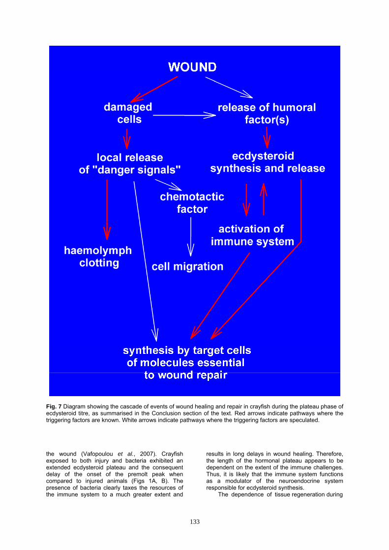
Fig. 7 Diagram showing the cascade of events of wound healing and repair in crayfish during the plateau phase of ecdysteroid titre, as summarised in the Conclusion section of the text. Red arrows indicate pathways where the triggering factors are known. White arrows indicate pathways where the triggering factors are speculated. the wound (Vafopoulou et al., 2007). Crayfish exposed to both injury and bacteria exhibited an extended ecdysteroid plateau and the consequent delay of the onset of the premolt peak when compared to injured animals (Figs 1A, B). The presence of bacteria clearly taxes the resources of the immune system to a much greater extent and
results in long delays in wound healing. Therefore, the length of the hormonal plateau appears to be dependent on the extent of the immune challenges. Thus, it is likely that the immune system functions as a modulator of the neuroendocrine system responsible for ecdysteroid synthesis.
The dependence of tissue regeneration during
133
wound healing and repair on low levels of ecdysteroids can be generalized for decapod Crustacean and probably for other arthropods. Earlier studies in crabs suffering limb autotomy demonstrated that limb regeneration depends on low ecdysteroid levels (see for review Skinner, 1985; Hopkins, 2001) and results in EcR expression in whole limb regenerates (Durica et al., 1996; 1999; 2002; Chung et al., 1998). In insects, regeneration of imaginal discs also requires low ecdysteroid level and is inhibited by high (Madhaven and Schneiderman, 1969; Kunieda et al., 1997).
Why are ecdysteroids needed for wound healing? The work with crayfish shows that hemocytes are principal targets of ecdysteroids and wounding results in elevation of PO activity. These findings suggest that the nature of hemocyte responsiveness to ecdysteroids is activation of immune responses. This conclusion is supported by the fact that expression of several immune parameters in other decapod crustaceans is correlated with molt stage (Cheng et al., 2003; Liu et al., 2004, 2006; Kuballa and Elizur, 2008; Ho et al., 2009; Yeh et al., 2009) or with changes in ecdysteroid level following eyestalk ablation (Sainz-Hernández et al., 2008). All this reinforces the hypothesis that immune responses in Crustacea are regulated by ecdysteroids during wound healing. Even though the area of ecdysteroid control of immune responses in crustaceans is largely unexplored, there is considerable evidence from insect systems supporting this hypothesis. Hemocytes failed to respond to bacterial challenge in the absence of ecdysteroids (Ahmed et al., 1999; Müller et al., 1999). Molt-dependent variations in immune parameters (Yamamoto et al., 2001; Zufelato et al., 2004; Meylaers et al., 2006; Eleftherianos et al., 2008) and positive correlations between ecdysteroids and immune responses have been observed in insects (Meister and Richards, 1996; Dimarcq et al., 1997; Dimopoulos et al., 1997; Lee et al., 2002; Sorrentino et al., 2002; Korayem et al., 2004; Aye et al., 2008; Flatt et al., 2008). Most interesting is the finding that ecdysteroid induction of antimicrobial peptides in the fruit fly Drosophila melanogaster required the presence of EcR (Flatt et al., 2008). Therefore, it appears that the situation of ecdysteroid control of immune responses unites arthropods with vertebrates in which steroid hormones, their nuclear receptors and other members of the nuclear receptor family regulate adaptive and innate immunity (see for review Flatt et al., 2008).
Another cellular aspect of ecdysteroid influence in wound healing in crayfish appears to be the massive migration of various cell types under a wound, notably hemocytes, epidermal cells and fibrocytes. Again convincing supporting evidence derives from insect studies where it has been shown that cell migration during morphogenesis is regulated by ecdysteroids (Hackney et al., 2007; Jang et al., 2009; see for review Montell, 2001).
A less understood aspect of wound healing process is the cue(s) that activate the crayfish neuroendocrine system to synthesize and release ecdysteroids following an integumentary injury. We speculate that these cues may be supplied by the
wound itself as is the case with limb regeneration in crabs. An inhibitory factor similar to molt-inhibiting hormone is released by the regenerating limb bud that inhibits proecdysial growth of limb buds and delays molting by acting on the central neuroendocrine system (see for review Mykles, 2001; Yu et al., 2002). Conclusions
In summary, integumentary wounds damage
the underlying tissues and set in motion a cascade of events that lead to regeneration and repair of the damaged area. In the event of a wound, both the immune and neuroendocrine systems (ecdysteroid synthesis) are activated to repair the wound rapidly and prevent hemolymph and tissue loss and infection. A schematic summary of the events involved is shown in Fig. 7. Several triggering factors in this cascade are known (red arrows) and have been described above in detail, whereas the existence of many others is speculative (white arrows) and invites further research. In this scheme, a wound damages cells that lie under the cuticle and these cells leak a variety of cellular components which may trigger several reactions. They may act chemotactically and trigger cell migration towards and under the wound from neighboring healthy areas, e.g., hemocytes, epidermis and fibrocytes, and they may also activate synthesis of specific proteins by these cell types. Or they may act on the neuroendocrine system and induce synthesis of ecdysteroids. Likewise, the act of wounding itself may activate the neuroendocrine system by some unknown mechanism to initiate synthesis of ecdysteroids. Factors released from damaged hemocytes are known to stimulate hemolymph clotting. Migration of cells may also be triggered by ecdysteroids. Ecdysteroids released into the hemolymph target primarily the immune system, e.g., the hemocytes, thereby appearing to regulate immune responses. Conversely, the immune system may also transmit feedback signals to the molting system that modulates ecdysteroid synthesis. Ecdysteroids act also upon epidermal cells and fibrocytes to synthesize the necessary building blocks that restore the damaged area.
Acknowledgements We are grateful to CCH Steel for his
constructive comments in the writing of this review. Supported by Natural Science and Engineering Research Council of Canada Discovery Grant 6069. References Ahmed A, Martin D, Manetti AGO, Han S-J, Lee W-
J, Mathiopoulos KD, et al. Genomic structure and ecdysone regulation of the prophenoloxidase 1 gene in the malaria vector Anopheles gambiae. Proc. Natl. Acad. Sci. USA 96: 14795-14800, 1999.
Aspán A, Huang T, Cerenius L, Söderhäll K. cDNA cloning of prophenoloxidase from the freshwater crayfish Pacifastacus leniusculus and its activation. Proc. Natl. Acad. Sci. USA 92: 939-943, 1995.
134
Aye TT, Shim JK, Rhee IK, Lee KY. Upregulation of the immune protein gene hemolin in the epidermis during the wandering larval stage of the Indian meal moth, Plodia interpunctella. J. Insect Physiol. 54: 1301-1305, 2008.
Battison A, Cawthorn R, Horney B. Classification of Homarus americanus hemocytes and the use of differential counts infected with Aerococcus viridans var homari (Gaffkemia). J. Invert. Pathol. 84: 177-197, 2003.
Cárdenas W, Jenkins JA, Dankert JR. A flow cytometric approach to the study of crustacean cellular immunity. J. Invert. Pathol. 76: 112-119, 2000.
Cerenius L, Söderhäll K. The prophenoloxidase-activating system in invertebrates. Immunol. Rev. 198: 116-126, 2004.
Chang ES, Chang SA, Mulder EP. Hormones in the lives of crustaceans: an overview. Amer. Zool. 41: 1090-1097, 2001.
Cheng WT, Juang FM, Li JT, Lin MC, Liu CH, Chen JC. The immune response of the giant freshwater prawn Macrobrachium rosenbergii and its susceptibility to Lactococcus garvieae in relation to the molt stage. Aquaculture 218: 33-45, 2003.
Chung AC-K, Durica DS, Clifton SW, Roe BA, Hopkins PM. Cloning of crustacean ecdysteroid receptor and retinoid-X receptor gene homologs and elevation of retinoid-X receptor mRNA by retinoic acid. Mol. Cell. Endocrinol. 139: 209-227, 1998.
Dillaman MD, Roer RD. Carapace repair in the green crab, Carcinus maenas (L.). J. Morphol. 163: 135-155, 1980.
Dimarcq J-L, Imler J-L, Lanot R, Ezekowitz RAB, Hoffmann JA, Janeway CA, et al. Treatment of l(2)mbn Drosophila tumorous blood cells with the steroid hormone ecdysone amplifies the inducibility of antimicrobial peptide gene expression. Insect Biochem. Molec. Biol. 27: 877-886, 1997.
Dimopoulos G, Richman A, Müller HM, Kafatos FC. Molecular immune responses of the mosquito Anopheles gambiae to bacteria and malaria parasites. Proc. Natl. Acad. Sci. USA 94: 11508-11513, 1997.
Durica DS, Hopkins PM. Expression of the genes encoding the ecdysteroid and retinoid receptors in regenerating limb tissues from the fiddler crab, Uca pugilator. Gene 171: 237-241, 1996.
Durica DS, Chung A C–K, Hopkins PM. Characterization of EcR and RXR gene homologs and receptor expression during the molt cycle in the crab, Uca pugilator. Amer. Zool. 39: 758-773, 1999.
Durica DS, Wu X, Anilkumar G, Hopkins PM, Chung AC-K. Characterization of crab EcR and RXR homologs and expression during limb regeneration and oocyte maturation. Mol. Cell. Endocrinol. 189: 59-76, 2002.
Eleftherianos I, Baldwin H, French-Constant RH, Reynolds SE. Developmental modulation of immunity: Changes within the feeding period of the fifth larval stage in the defence reactions of Manduca sexta to infection by Photorhabdus. J. Insect Physiol. 54: 309-318, 2008.
Flatt T, Heyland A, Rus F, Porpiglia E, Sherlock C, Yamamoto R et al. Hormonal regulation of the humoral innate immune response in Drosophila melanogaster. J. Exp. Biol. 211: 2712-2724, 2008.
Gallucci S, Matzinger P. Danger signals: SOS to the immune system. Curr. Opin. Immunol. 13: 114-119, 2001.
Giulianini PG, Biert M, Lorenzon S, Battistella S, Ferrero EA. Ultrastructural and functional characterization of circulating hemocytes from the freshwater crayfish Astacus leptodactylus: Cell types and their role after in vivo artificial non-self challenge. Micron 38: 49-57, 2007.
Hackney JF, Pucci C, Naes E, Dobens L. Ras signalling modulates activity of the ecdysone receptor EcR during cell migration in the Drosophila ovary. Dev. Dynamics 236: 1213-1226, 2007.
Halcrow K. 1988. Absence of epicuticle from the repair cuticle produced by four malacostracan crustaceans. J. Crust. Biol. 8: 346-354, 1988.
Halcrow K, Steel CGH. Cuticular repair in the mature male snowcrab, Chionocetes opilio: Relation to ecdysteroids. Can. J. Zool. 70: 314-319, 1992.
Hall M, Söderhäll K. Crayfish α-macroglobulin as a substrate for transglutaminases. Comp. Biochem. Physiol. 108B: 65-72, 1994.
Hall M, Wang R, Antwerpen RK, Sottrup-Jensen L, Söderhäll K. The crayfish plasma clotting: a vitellogenin-related protein responsible for clot formation in crustacean blood. Proc. Natl. Acad. Sci. USA 96: 1965-1970, 1999.
Henrich V. The ecdysteroid receptor (EcR). In: Gilbert LI, Iatrou K, Gill S (eds), Comprehensive molecular insect science, Elsevier Science, Oxford, UK, vol. 3, pp 243-285, 2005.
Ho PY, Cheng CH, Cheng WT. Identification and cloning of the alpha 2-macroglobulin of giant freshwater prawn Macrobrachium rosenbergii and its expression in relation with the molt stage and bacteria infection. Fish Shellfish Immunol. 26: 459-466, 2009.
Hopkins PM. Limb regeneration in the fiddler crab, Uca pugilator: Hormonal and growth factor control. Amer. Zool. 41: 389-398, 2001.
Hopkins PM, Bliss DE, Sheehan SW, Boyer JR. Limb growth-controlling factors in the crab Gecarcinus lateralis, with special reference to the limb growth-inhibiting factor. Gen. Comp. Endocrinol. 39: 192-207, 1979.
Hopkins PM, Chung C-K, Durica DS. Limb regeneration in the fiddler crab, Uca pugilator: Histological, physiological and molecular considerations. Amer. Zool. 39: 513-617, 1999.
Jang ACC, Chang YC, Bai JW, Montell C. Border-cell migration requires integration and temporal signals by the BTB protein Abrupt. Nat. Cell Biol. 11: 569-U100, 2009.
Jiravanichpaisal P, Lee SY, Kim YA, Andren T, Söderhäll I. Antibacterial peptides in hemocytes and hematopoietic tissue from freshwater crayfish Pacifastacus leniusculus: Characterization and expression pattern. Dev. Comp. Immunol. 31: 441-455, 2007.
135
Johansson MW, Söderhäll K. Isolation and purification of a cell adhesion factor from crayfish blood cells. J. Cell Biol. 106: 1795-1803, 1988.
Johansson, M.W., Söderhäll K. A cell adhesion factor from crayfish hemocytes has degranulating activity towards crayfish granular cells. Insect Biochem. 19: 183-190, 1989.
Johansson MW, Söderhäll K. Exocytosis of the phenoloxidase activating system from crayfish hemocytes. J. Comp. Physiol. 156B: 175-191, 1985.
Johansson MW, Keyser P, Sritunyalucksana K, Söderhäll K. Crustacean hemocytes and hematopoiesis. Aquaculture 191: 45-52, 2000.
Kobayashi M, Johansson MW, Söderhäll K. The 76 kD cell-adhesion factor from crayfish hemocytes promotes encapsulation in vitro. Cell Tissue Res. 260: 13-18, 1990.
Korayem AM, Fabbri M, Takahashi K, Scherfer C, Lindgren M, Schmidt O, et al. A Drosophila salivary gland mucin is also expressed in immune cells: evidence for a function in coagulation and the entrapment of bacteria. Insect Biochem. Mol. Boil. 34: 1297-1304, 2004.
Kuballa AV, Elizur A. Differential expression profiling of components associated with exoskeletal hardening in crustaceans. BMC Genomics 9: 575, 2008.
Kunieda T, Kurat S, Natori S. Regeneration of Sarcophaga imaginal discs in vitro: Implication of 20-hydroxyecdysone. Dev. Biol. 183: 86-94, 1997.
Lai SC, Chen CC, Hou RF. Immunolocalization of prophenoloxidase in the process of wound healing in the mosquito Armigeres subalbatus (Diptera: Culicidae). J. Med. Entomol. 39: 266-274, 2002.
Lee K-Y, Horodyski FM, Valaitis AP, Denlinger DL. Molecular characterization of the insect immune protein hemolin and its high induction during embryonic diapause in the gypsy moth, Lymantria dispar. Insect Biochem. Mol. Boil. 32: 1457-1467, 2002.
Lee SY, Söderhäll K. Characterization of a pattern recognition protein, a masquerade-like protein, in the freshwater crayfish, Pacifastacus leniusculus. J. Immunol. 166: 7319-7326, 2001.
Lee SY, Söderhäll K. Early events in crustacean innate immunity. Fish Shellfish Immunol. 12: 421-437, 2002.
Lee SY, Lee BL, Söderhäll K. Processing of crayfish hemocyanin subunits into phenoloxidase. Biochem. Biophys. Res. Commun. 322: 490-496, 2004.
Liu YC, Li FH, Wang B, Dong B, Zhang QL, Luan W, et al. A transglutaminase from Chinese shrimp (Fenneropenaeus chinensis), full-length cDNA cloning, tissue localization and expression profiles after challenge. Fish Shellfish Immunol. 22: 576-588, 2007.
Liu C-H, Tseng D-Y, Lai C-Y, Cheng W, Kuo C-M. Molecular cloning and characterization of prophenoloxidase cDNA from hemocytes of the giant freshwater prawn, Macrobrachium rosenbergii, and its transcription in relation with
the molt stage. Fish Shellfish Immunol. 21, 60-69, 2006.
Liu CH, Yeh ST, Cheng SY, Chen JC. The immune response of the white shrimp Litopenaeus vannamei and its susceptibility to Vibrio infection in relation with the molt cycle. Fish Shellfish Immunol. 16: 151-161, 2004.
Madhavan K, Schneiderman HA. Hormonal control of imaginal disc regeneration in Galleria mellonella (Lepidoptera). Biol. Bull. 137: 321-331, 1969.
Martin GG, Castro C, Moy N, Rubin N. N-acetyl-D-glucosamine in crustacean hemocytes; possible functions and usefulness in hemocyte classification. Invert. Biol. 122: 265-270, 2003.
Meister M, Richards G. Ecdysone and insect immunity: the maturation of the inducibility of the diptericin gene in Drosophila larvae. Insect Biochem. Mol. Biol. 26: 155-160, 1966.
McCarthy JF, Skinner DM. Proecdysial changes in serum ecdysone titers, gastrolith formation and limb regeneration following molt induction by limb autotomy and /or eyestalk removal in the land crab, Gecarcinus lateralis. Gen. Comp. Endocrinol. 33: 278-292, 1977.
Meylaers K, Freitak D, Schoofs L. Immunocompetence of Galleria mellonella: Sex- and stage-specific differences and the physiological cost of mounting an immune response during metamorphosis. J. Insect Physiol. 53: 146-156, 2007.
Montell DJ. Command and control: regulatory pathways controlling invasive behaviour of the border cells. Mech. Dev. 105: 19-25, 2001.
Mucklow PT, Ebert D. Physiology of immunity in the water flea Daphnia magna: environmental and genetic aspects of phenoloxidase activity. Physiol. Biochem. Zool. 76: 836-842, 2003.
Mucklow TP, Vizoso DB, Jensen KH, Refardt D, Ebert D. Variation in phenoloxidase activity and its relation to parasite resistance within and between populations of Daphnia magna. Proc. R. Soc. London B 271: 1175-1183, 2004.
Müller HM, Dimopoulos G, Blass C, Kafatos FC. A hemocyte-like cell line established from the malaria vector Anopheles gambiae expresses six prophenoloxidase genes. J. Biol. Chem. 274: 11727-11735, 1999.
Mykles DL. Interactions between limb regeneration and molting in decapod crustaceans. Amer. Zool. 41: 399-406, 2001.
Nayar JK, Knight JW. Wounding increases intracellular encapsulation (melanization) of developing Brugia malayi (Nematoda: Filarioidea) larvae in thoracic muscles of Anopheles quadrimaculatus. Comp. Biochem. Physiol. 112A: 553-557, 1995.
Sainz-Hernández JC, Racotta IS, Dumas S, Hernández-López J. Effect of unilateral and bilateral eyestalk ablation in Litopenaeus vannamei male and female on several metabolic and immunologic variables. Aquaculture 283: 188-193, 2008.
Skinner DM. Molting and regeneration. In: Bliss DE, Mantel LH (eds), The Biology of Crustacea, Academic Press, New York, USA, vol 9, pp 43-146, 1985.
136
Smith VJ Söderhäll K. β-1,3 glucan activation of crustacean hemocytes in vitro and in vivo. Biol. Bull. 164: 299-314, 1983a.
Smith VJ Söderhäll K. Induction of degranulation and lysis of hemocytes in the freshwater crayfish, Astacus astacus by components of the prophenoloxidase activating system in vitro. Cell Tissue Res. 233: 295-303, 1983b.
Smith VJ Söderhäll K. A comparison of phenoloxidase activity in the blood of marine-invertebrates. Dev. Comp. Immunol. 15: 251-261, 1991.
Snyder MJ, Chang ES. Effects of sinus gland extracts on larval molting and ecdysteroid titers of the American lobster, Homarus americanus. Biol. Bull. 170: 244-254, 1986.
Söderhäll K, Cerenius L. Role of the prophenoloxidase-activating system in invertebrate immunity. Curr. Opin. Immunol. 10: 23-28, 1998.
Söderhäll K, Cerenius L, Johansson MW. The prophenoloxidase activating system and its role in invertebrate defence. Annals NY Acad. Sci. 712, 155-161, 1994.
Söderhäll K, Wingren A, Johansson MW, Bertheussen K. The cytotoxic reaction of hemocytes from the freshwater crayfish, Astacus astacus. Cell Immunol. 94, 326-332, 1985.
Söderhäll I, Wu C, Novotny M, Lee BL, Söderhäll K. A novel protein acts as a negative regulator of prophenoloxidase activation and melanisation in the freshwater crayfish Pacifastacus leniusculus. J. Biol. Chem. 284: 6301-6310, 2009.
Sorrentino RP, Carton Y, Govind S. Cellular immune response to parasite infection in the Drosophila lymph gland is developmentally regulated. Dev. Biol. 243: 65-80, 2002.
Sritunyalucksana K, Söderhäll K. The proPO and clotting system in crustaceans. Aquaculture 191: 53-69, 2000.
Steel CGH, Vafopoulou X. Ecdysone titre profiles during growth and development of arthropods. In: Koolman J (ed), Ecdysone: from chemistry to mode of action, Georg Thieme Verlag, Stuttgart, Germany, pp 221-231, 1989.
Sung HH, Sun R. Use of monoclonal antibodies to classify hemocyte subpopulation of tiger shrimp (Penaeus monodon). J. Crustacean Biol. 22: 337-344, 2002.
Theopold U, Li D, Fabbri M, Scherfer C, Schmidt O. The coagulation of insect haemolymph. Cell. Mol. Life Sci. 59: 363-372, 2002.
Theopold U, Schmidt O, Söderhäll K, Dushay MS. Coagulation in arthropods: defence, wound closure and healing. Trends Immunol. 25: 289-294, 2004.
Vafopoulou X, Laufer H, Steel CGH. Spatial and temporal distribution of the ecdysteroid receptor (EcR) in haemocytes and epidermal cells during wound healing in the crayfish, Procambarus clarkii. Gen. Comp. Endocrinol. 152: 359-370, 2007.
Vafopoulou X, Steel CGH. Ecdysteroid hormone nuclear receptor (EcR) exhibits circadian
cycling in certain tissues, but not others, during development in Rhodnius prolixus (Hemiptera). Cell Tissue Res. 323: 443-455, 2006.
Vafopoulou X, Steel CGH, Terry KL. Ecdysteroid receptor (EcR) shows marked differences in temporal patterns between tissues during larval-adult development in Rhodnius prolixus: correlations with haemolymph ecdysteroid titres. J. Insect Physiol. 51: 27-38, 2005.
Vázquez L, Pérez A, Millán D, Agundis C, Martin G, Cooper RL, et al. Morphology of hemocytes from the freshwater prawn Macrobrachium rosenbergii. J. Morphol. 234: 147-153, 1997.
Vogt G. The marbled crayfish: a new model organism for research on development, epigenetics and evolutionary biology. J. Zool. 276: 1-13, 2008.
Wang R, Lee SY, Cerenius L, Söderhäll K. Properties of the prophenoloxidase activating enzyme of the freshwater crayfish, Pacifastacus leniusculus. Eur. J. Biochem. 268: 895-902, 2001a.
Wang R, Liang Z, Hall M, Söderhäll K. A transglutaminase involved in the coagulation system of the freshwater crayfish, Pacifastacus leniusculus. Tissue localisation and cDNA cloning. Fish Shellfish Immunol. 11: 623-637, 2001b.
Webster SG. Neuropeptides inhibiting growth and reproduction in crustaceans. In: Coast GM, Webster SG (ed), Recent advances in arthropod endocrinology, Cambridge University Press, Cambridge, UK, pp 33-52, 1998.
Yamamoto K, Fujii H, Kusakabe T, Koga K, Aso Y, Ishiguro M. Change in phenoloxidase and its precursor during silkworm (a80 strain) development. J. Fac. Agric. Kyushu Univ. 45: 487-493, 2001.
Yeh MS, Lai CY, Liu CH, Kuo CM, Cheng W. A second proPO present in white shrimp Litopenaeus vannamei and expression of the proPOs during a Vibrio alginolyticus injection, molt stage. Fish Shellfish Immunol. 26: 49-55, 2009.
Yu X, Chang ES, Mykles DL. Characterization of limb autotomy factor-proecdysis (LAFpro), isolated from limb regenerates, that suspends molting in the land crab Gecarcinus lateralis. Biol. Bull. 202: 204-212, 2002.
Zhan WB, Wei XM, Xing J, Zhang ZD. Characterization of monoclonal antibodies to haemocyte types of the shrimp, Fenneropenaeus chinensis. Crustaceana 81: 931-942, 2008.
Zou Z, Wang Y, Jiang H. Manduca sexta prophenoloxidase activating proteinase-1 (PAP-1) gene: Organization, expression, and regulation by immune and hormonal signals. Insect Biochem. Mol. Biol. 35: 627-636, 2005.
Zufelato MS, Lourenco AP, Simŏes ZLO, Jorge JA, Bitondi MMG. Phenoloxidase activity in Apis mellifera honey bee pupae, and ecdysteroid-dependent expression of the prophenoloxidase mRNA. Insect Biochem. Mol. Biol. 34: 1257-1268, 2004.
137