mechanisms of neurogenesis in the normal and injured … · 13 review mechanisms of neurogenesis in...
TRANSCRIPT
13
REVIEWMechanisms of Neurogenesis in the Normal and
Injured Adult BrainMasato Sawada and Kazunobu Sawamoto
Department of Developmental and Regenerative Biology, Nagoya City University Graduate School of Medical Sciences, Aichi, Japan
(Received for publication on March 28, 2012)(Accepted for publication on May 18, 2012)
Even in the adult brain, new neurons are continuously generated from endogenous neural stem cells that reside in two restricted regions: the subventricular zone (SVZ) of the lateral ventricle and the den-tate gyrus of the hippocampus. These new neurons are integrated into the mature neuronal circuitry and become involved in various functions, thereby contributing to structural and functional plasticity in the adult brain. In this review, we summarize our recent findings on the regulatory mechanisms of SVZ neurogenesis under physiological and pathological conditions in various animal models. Some of these findings were presented in the Kitazato Prize Lecture at Keio University School of Medicine in 2011. (doi: 10.2302/kjm.2012-0005-RE; Keio J Med 62 (1) : 13–28, March 2013)
Keywords: adult neurogenesis, subventricular zone
Introduction
Until recently, it was commonly believed that the adult brain does not produce new neurons. However, it is now well accepted that neural stem cells continue to reside in the subventricular zone (SVZ) of the lateral ventricle and dentate gyrus (DG) of the hippocampus, and that they produce functional neurons in the adult brain throughout life. In the SVZ, which is the largest neurogenic region in the adult brain, neural stem cells continuously generate new neurons via transit-amplifying neural progenitors. The new neurons form chain-like cell aggregates and mi-grate toward the olfactory bulb (OB) through the rostral migratory stream (RMS), a specialized route for migrat-ing neurons. After arriving at the OB, the new neurons differentiate into olfactory interneurons, which are im-plicated in olfactory processing. Recent studies demon-strated that this neurogenic potential in the adult brain is widely conserved from nonmammalian vertebrates to primates, including humans. Interestingly, SVZ neuro-genesis can be activated by various injuries and neuro-logical diseases, suggesting that endogenous neural stem cells in the SVZ may be used for brain repair in lower ani-
mals, and might be exploited for therapeutic strategies in humans. In this review, we introduce our recent findings on SVZ neurogenesis in the physiological and pathologi-cal conditions of various animals.
Neural Stem Cells in the Subventricular Zone
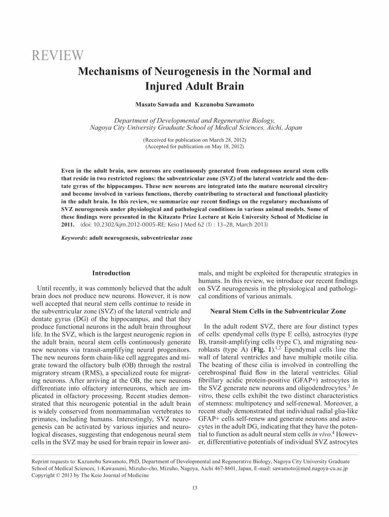
In the adult rodent SVZ, there are four distinct types of cells: ependymal cells (type E cells), astrocytes (type B), transit-amplifying cells (type C), and migrating neu-roblasts (type A) (Fig. 1).1,2 Ependymal cells line the wall of lateral ventricles and have multiple motile cilia. The beating of these cilia is involved in controlling the cerebrospinal fluid flow in the lateral ventricles. Glial fibrillary acidic protein-positive (GFAP+) astrocytes in the SVZ generate new neurons and oligodendrocytes.3 In vitro, these cells exhibit the two distinct characteristics of stemness: multipotency and self-renewal. Moreover, a recent study demonstrated that individual radial glia-like GFAP+ cells self-renew and generate neurons and astro-cytes in the adult DG, indicating that they have the poten-tial to function as adult neural stem cells in vivo.4 Howev-er, differentiative potentials of individual SVZ astrocytes
Reprint requests to: Kazunobu Sawamoto, PhD, Department of Developmental and Regenerative Biology, Nagoya City University Graduate School of Medical Sciences, 1-Kawasumi, Mizuho-cho, Mizuho, Nagoya, Aichi 467-8601, Japan, E-mail: [email protected] © 2013 by The Keio Journal of Medicine
Sawada M and Sawamoto K: Neurogenesis in the Adult Brain14
in vivo have yet to be clarified. In addition, the precise identification and classification of neural stem cells in the adult SVZ are still difficult because of the lack of an adult neural stem cell-specific marker. Like embryonic neural stem cells, GFAP+ astrocytes in the adult SVZ express neural stem cell markers, including Musashi, LeX/ssea-1, Sox2, Id, and Nestin.1,5–9 GFAP+ astrocytes in the SVZ proliferate very slowly and generate rapidly dividing transit-amplifying progenitors that generate migrating neuroblasts. In addition, blood vessels in the SVZ that are in contact with these cells play important roles in the control of neurogenesis.10–13
Several extracellular signals and their downstream pathways cooperatively regulate cell proliferation and differentiation in the SVZ. Because SVZ cells are packed into narrow regions lining the lateral ventricles and are in close contact with each other, cell–cell interaction signals could affect their proliferation state. For example, cell-surface carbohydrates could regulate the intercellular
interactions of adult neural stem cells in the SVZ. The carbohydrate-binding protein Galectin-1, a key regula-tor of various stem cells, is secreted by the majority of GFAP+ astrocytes in the adult SVZ.14 Cell proliferation in the SVZ is reduced by galectin-1 deficiency and acti-vated by the infusion of exogenous Galectin-1, indicating that Galectin-1 positively regulates the proliferation of neural stem cells in the SVZ. The Eph-ephrin system is also important for intercellular interaction. The receptor tyrosine kinases EphA/B are expressed in the SVZ and regulate cell proliferation, although their downstream signaling molecules are currently unknown.15
Notch signaling is another key factor in many cell–cell interaction systems. During embryonic brain develop-ment, Notch is the most critical regulator of neural stem-cell maintenance. Ligand binding to Notch induces cleav-age of the Notch intracellular domain, which translocates into the nucleus and induces the expression of transcrip-tional repressor proteins such as those encoded by the
Fig. 1 Neurogenesis in the adult subventricular zone.In the subventricular zone (SVZ) of the lateral ventricle, there are four distinct types of cells: ependymal cells (purple), astrocytes (blue), transit-amplifying cells (green), and neuroblasts (red). SVZ astrocytes contact the lateral ventricle and act as neural stem cells that generate neural progenitors called transit-amplifying cells. The transit-amplifying cells actively proliferate and generate neuro-blasts. Blood vessels surrounding these cells have important roles in regulating neurogenesis in the SVZ. Neuroblasts generated in the SVZ migrate toward the olfactory bulb (OB) through the rostral migratory stream (RMS). These cells form chain aggregates, which are surrounded by tunnels composed of astrocytic processes. After arriving at the OB, the chain-forming neuroblasts are dissociated into individual cells and migrate radially inside the OB. Most of the neuroblasts differentiate into granule cells (pink) and form synapses with mitral/tufted cells (yellow), which are projection neurons in the OB. A small portion of the neuroblasts continue their radial mi-gration to the glomeruli located in the superficial layer of the OB, where they differentiate into periglomerular cells (orange) and form synapses with both olfactory sensory neurons (gray) and the mitral/tufted cells.
15Keio J Med 2013; 62 (1): 13–28
Hes gene family, which repress the activities of proneural genes. One Notch ligand, Delta-like homologue 1 (Dlk1), is expressed by GFAP+ astrocytes in the postnatal SVZ and is required for the maintenance of neural stem cells.16 Even in the adult brain, Notch signaling activity is ob-served in GFAP+ astrocytes in the SVZ. The conditional knockout of Rbpj, a Notch-signaling component, in the Nestin-expressing cell lineage causes the complete loss of neural stem cells in the SVZ, indicating that Notch signaling is also important for the maintenance of neural stem cells in the adult SVZ.17 Interestingly, Notch sig-naling in neural stem cells in the SVZ is modulated by other extrinsic factors. For example, enhanced epidermal growth factor receptor (EGFR) signaling reduces both Notch1 expression and the number of GFAP+ neural stem cells in the SVZ, and it increases the number of transit-amplifying cells, suggesting that interplay between the Notch and EGFR pathways regulates the balance of the number of neural stem/progenitor cells.18 In addition, pigment epithelium-derived factor, which is secreted from ependymal cells and blood vessels in the SVZ, en-hances the Notch-dependent transcription of target genes, including Hes1 and Egfr, in GLAST+Sox2+ neural stem cells with low Notch activity, suggesting that a vascular modulation of neural stem cells in the adult SVZ is medi-ated by Notch signaling.19,20
Because neural stem cells are widely distributed throughout the SVZ and generate various types of OB neurons, it is possible that they have distinct regional identities. The precise regional labeling of neural stem cells by adenovirus injection revealed that neuronal fates in the OB are pre-determined by the region of the SVZ inhabited by their precursors.21 Sonic hedgehog (Shh) signaling, which is the determinant for the ventral axis in early neural development, is restricted to the ventral re-gion of the adult SVZ.22,23 Shh signaling also determines the ventral specification of neural stem cells, which gen-erate specific neuronal progeny in the OB.24 Bone mor-phogenic protein (BMP) signaling, which is the deter-minant of the dorsal axis in the embryonic brain, is also involved in neurogenesis in the adult SVZ,25 although whether this signaling determines the dorsal specifica-tion of neural stem cells is currently unknown. Thus, in addition to Notch signaling, the regional specification by dorsoventral determinants such as Shh and BMP signal-ing could regulate the proliferation of neural stem cells and contribute to the diversity of neuronal progeny.
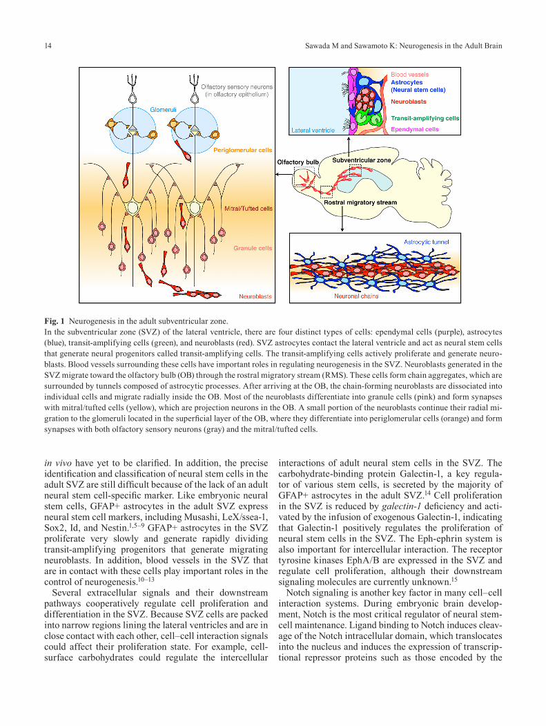
Wnt proteins are important soluble regulators of mam-malian neural development and have two distinct down-stream pathways. The canonical Wnt pathway is medi-ated by β-catenin, which acts as both a cytoskeletal component and a transcription factor. In the absence of Wnt stimulation, β-catenin is phosphorylated by casein kinase I and GSK3β and is constantly degraded by the ubiquitin–proteasome system. However, Wnt-mediated inhibition of GSK3β blocks this degradation of β-catenin;
the unphosphorylated β-catenin then translocates into the nucleus, where it promotes the transcription of develop-mentally important genes. On the other hand, the non-canonical Wnt pathway is mediated by Wnt/planar cell polarity (PCP) signaling, which mainly regulates cyto-skeletal dynamics and cell polarity through c-Jun N-ter-minal kinase (JNK) and Rho GTPase. However, the roles of these distinct Wnt pathways in the adult SVZ were completely unknown until recently.
Using Axin2-d2EGFP mice, in which the activation of β-catenin signaling can be monitored in vivo, we found that canonical Wnt/β-catenin signaling is activated in as-trocytes and transit-amplifying cells in the adult SVZ.26 The injection of a retrovirus carrying a constitutively ac-tive form of β-catenin or a highly selective GSK3β in-hibitor, both of which activate Wnt/β-catenin signaling, increased the proliferation of transit-amplifying cells, which ultimately led to an increased number of new neurons in the OB. These results indicated that Wnt/β-catenin signaling positively regulates the proliferation of neuronal progenitors in the adult SVZ (Fig. 2).
Diversin, the mammalian homolog of the Drosophila PCP protein Diego, is a component of both canonical and non-canonical Wnt signaling (Fig. 2). We found that Diversin is expressed by neuroblasts in the neonatal and adult SVZ and DG.27 In the adult SVZ, retrovirus-medi-ated overexpression of full-length Diversin increased the proliferation of neuroblasts. A mutant Diversin lacking the ankyrin repeat domain, which is important for acti-vating the Wnt/PCP pathway, did not have this activity, suggesting that the Wnt/PCP pathway is necessary for the proliferation of neuroblasts. Interestingly, phosphorylat-ed JNK, a downstream component of the Wnt/PCP path-way, could be observed in neuroblasts undergoing mito-sis. Taken together, these results suggest that the Wnt/PCP pathway is active in the proliferation of neuroblasts (Fig. 2).
Thus, the two Wnt pathways have distinct functions in regulating the proliferation of transit-amplifying cells and neuroblasts in the SVZ (Fig. 2). Canonical Wnt/β-catenin signaling controls the proliferation of transit-amplifying cells, whereas the non-canonical Wnt/PCP pathway regu-lates the proliferation of neuroblasts. Since Diversin is involved in both the canonical and non-canonical Wnt pathways, it could act as a molecular switch for the tran-sition from neuronal progenitors to migrating neuroblasts in the SVZ.
Neuronal Migration in the Rostral Migratory Stream
Neuroblasts generated from neural stem cells migrate at high speed from the SVZ toward the OB (Fig. 1). These cells form elongated cell aggregates called “chains.”28 In the chains, each neuroblast migrates along its neighbors, indicating that neuroblasts use other neuroblasts as a scaffold for efficient migration. In the lateral wall of the

Sawada M and Sawamoto K: Neurogenesis in the Adult Brain16
lateral ventricle in the adult brain, an extensive network of neuronal chains can be observed, which converge into bundles of chains when entering the RMS, a restricted pathway for migrating neuroblasts.29 In the RMS, the chain-forming migrating neuroblasts are surrounded by tunnels formed by astrocytic processes. The chain forma-tion of migrating neuroblasts and surrounding glial tubes are two distinct features of the adult RMS (Fig. 1).30
Several proteins that regulate chain formation have
been identified. For example, mice lacking the cell-surface receptors ErbB4 and ApoER2 show defective chain formation and migration in the RMS, resulting in decreased numbers of newly arriving neurons in the OB.31,32 Extracellular matrix-related molecules such as α6β1-integrin and ADAM2 protease are also involved in neuroblast chain formation.33–35 These findings raise the possibility that various extracellular stimuli actively regulate the chain formation of neuroblasts.
Fig. 2 Regulation of SVZ neurogenesis by Wnt signaling.Wnt proteins have two distinct downstream signaling pathways. The canonical Wnt pathway is mediated by β-catenin. In the absence of Wnt stimulation, β-catenin is constantly phosphorylated by casein kinase I/GSK3β complex and degraded by the ubiquitin-prote-asome system. This process is inhibited by Wnt-mediated inhibition of GSK3β; the unphosphorylated β-catenin then accumulates in the cytosol and translocates into the nucleus. In the adult SVZ, Wnt/β-catenin signaling is activated in the astrocytes and transit-ampli-fying cells and regulates their proliferation. The non-canonical Wnt pathway is mediated by Wnt/planar cell polarity (PCP) signaling, which regulates cytoskeletal dynamics and cellular polarity through JNK and Rho family small GTPases. Diversin is a component of both the canonical and non-canonical Wnt signaling pathways. The Diversin-mediated Wnt/PCP pathway regulates the proliferation of neuroblasts in the adult SVZ.
17Keio J Med 2013; 62 (1): 13–28
To migrate at high speed within the neuronal chains, the chain-forming neuroblasts need to slide along each other efficiently. Polysialylated neural cell adhesion molecule (PSA-NCAM), a cell adhesion molecule with a negatively charged polysialic acid (PSA) moiety, is highly expressed on the cell surface of chain-forming neuroblasts in the RMS. NCAM gene deletion or enzymatic digestion of the PSA moiety cause migrating neuroblasts to accumulate in the RMS without any defect in chain formation.36,37 The negative charge of the PSA moiety might increase the cellular motility by decreasing the adhesion between neuroblasts, leading to smooth chain migration in the RMS.38
Since chain-forming neuroblasts must migrate over a long distance from the SVZ to the OB, the determina-tion of directionality is important for their appropriate migration. We demonstrated that the direction of migrat-ing neuroblasts in the SVZ is determined by the repulsive factor Slit,39 which is reported to control axonal guid-ance in the developing brain by regulating the cytoskel-eton.40,41 Several studies have shown that Slit is secreted from the septum and choroid plexus and can repel migrat-ing neuroblasts generated in the SVZ.42–44 However, the mechanism by which the Slit proteins reach the migrating neuroblasts in the SVZ was not known.
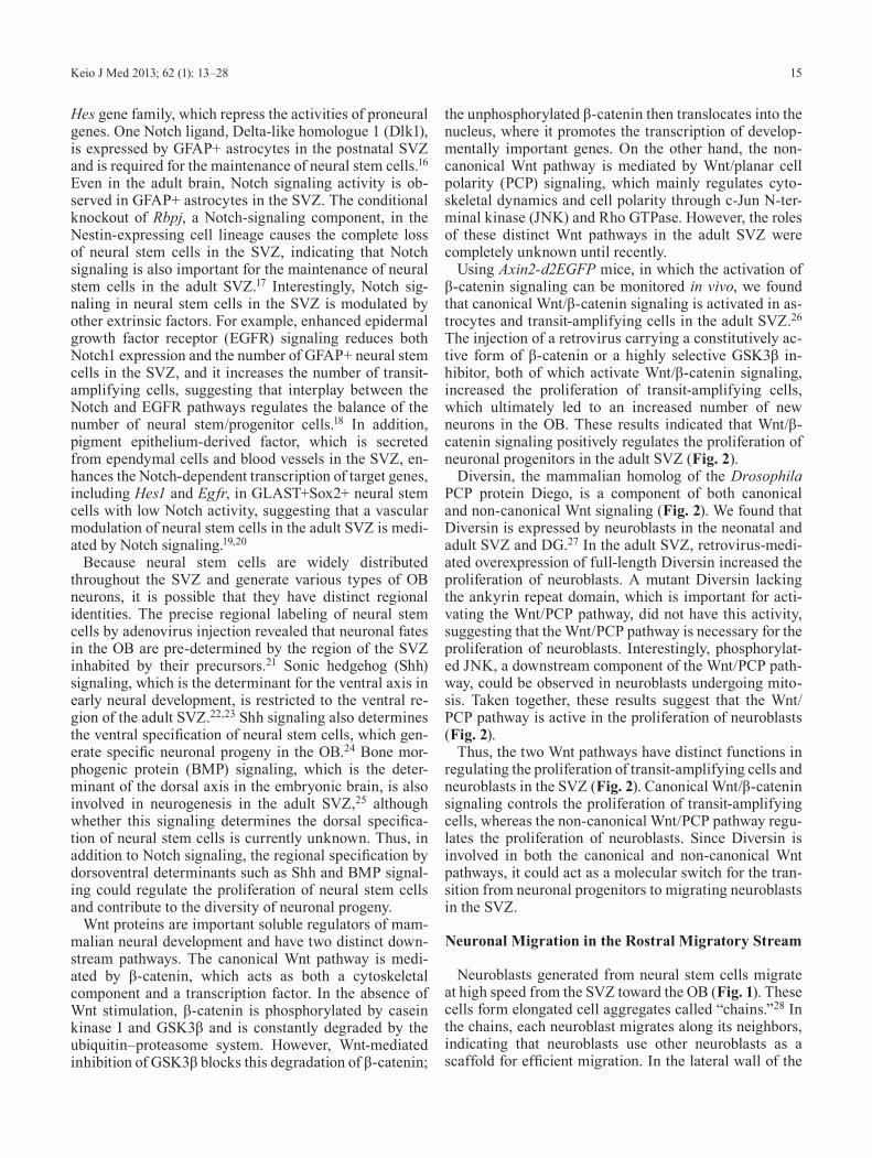
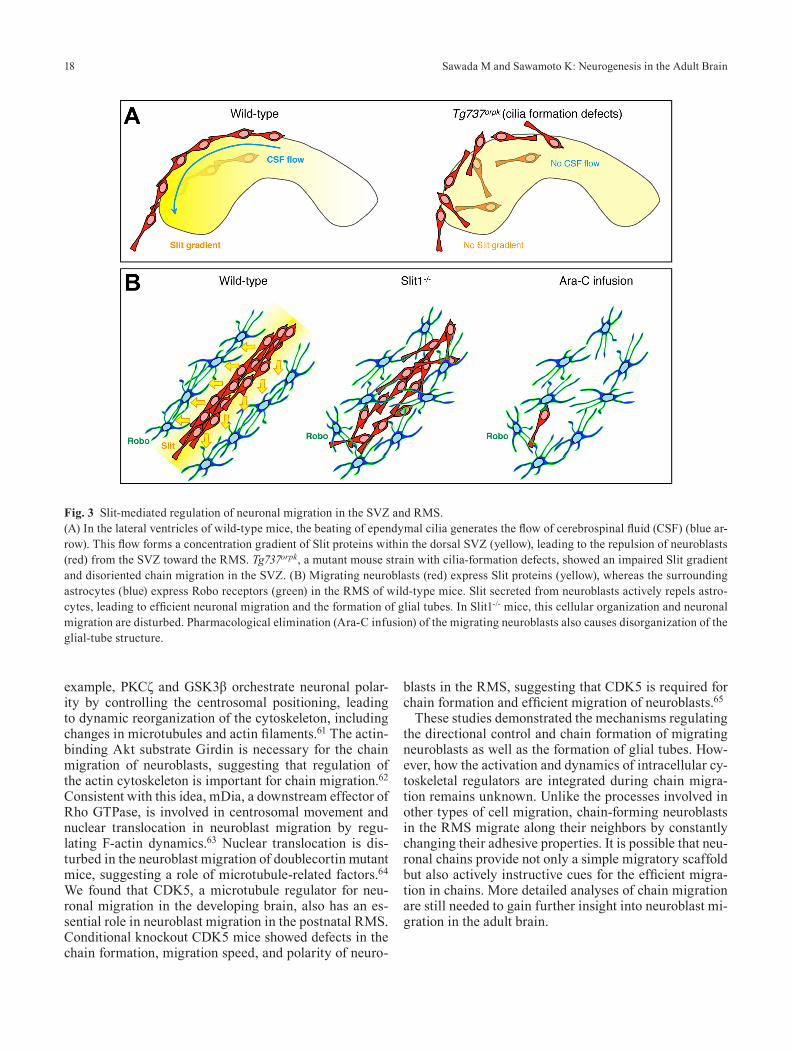
We found that migrating neuroblasts in the SVZ follow the direction of cerebrospinal fluid (CSF) flow created by the beating of ependymal cilia.39 Exogenous Slit protein injected into the lateral ventricle formed a concentration gradient within the dorsal SVZ, with the highest signal in the caudal region and the weakest in the rostral area, in parallel with the direction of CSF flow (Fig. 3A). The Tg737orpk mouse, a mutant strain with cilia-formation de-fects, showed an impaired Slit gradient and disoriented chain migration in the SVZ (Fig. 3A). These results sug-gest that the concentration gradient of Slit in the SVZ created by the beating of ependymal cilia determines the direction of migrating neuroblasts.
Our findings indicated that the beating of ependymal cilia plays an important role in neuronal migration in the SVZ. During the early postnatal stage, the develop-ing ependymal cells acquire two types of cellular polar-ity, translational and rotational polarity.45,46 The anterior migration of basal bodies (translational polarity) and coordinated ciliary beating (rotational polarity) in the ependymal cells are both important for appropriate cili-ary beating and the formation of CSF flow. We showed that non-muscle myosin II and the Wnt/PCP pathway distinctly regulate the processes responsible for generat-ing translational and rotational polarity, respectively.47,48 These results suggest that the distribution and orientation of basal bodies are regulated by distinct mechanisms, and that both contribute to the control of CSF flow.
Although repulsive mechanisms could be crucial for neuroblast movement from the SVZ toward the RMS, such mechanisms seemed less effective for cells migrat-
ing in the RMS toward the OB because of the long dis-tances involved. Instead, attractant factors released from the OB could be involved in directionality in the RMS. The secreted protein prokineticin 2 (PK2) is expressed in the granule and glomerular layer of the OB, whereas PK2 receptor (Prokr2) is expressed in the neuroblasts migrat-ing in the RMS.49,50 PK2 knockout and Prokr2 mutant mice show an accumulation of migrating neuroblasts in the rostral RMS, indicating that PK2 is a possible attrac-tant for neuroblasts toward the OB.49,50 Other growth/tro-phic factors such as hepatocyte growth factor, brain-de-rived neurotrophic factor (BDNF), glial cell line-derived neurotrophic factor, and Netrin-1 also show attractant ac-tivity for neuroblasts in vitro, although in vivo evidence for their involvement remains to be demonstrated.51–54
In the RMS, neuroblasts move inside the glial tube, which is reported to support their long-distance cell mi-gration through intercellular molecular interactions. For example, the inhibitory neurotransmitter gamma-amino-butyric acid (GABA) secreted by migrating neuroblasts is taken up by the RMS astrocytes, leading to negative regulation of the migration speed.55 Conversely, gluta-mate released by RMS astrocytes activates N-methyl-d-aspartate receptor in the migrating neuroblasts, which promotes their survival before they enter the synaptic network in the OB.56 RMS astrocytes also interact with blood vessels inside the glial tube and capture BDNF se-creted from endothelial cells, which leads to the vascula-ture-associated migration of neuroblasts in the RMS.57,58 It was reported that Slit1 and its receptors Robo2/3, which mediate the repellant properties of Slit1, are expressed by migrating neuroblasts in the RMS, suggesting that Slit1 has a cell-autonomous role in the RMS.59 We showed that Robo2 is strongly expressed by tunnel-forming astrocytes in the RMS. Slit1-secreting migrating neuroblasts repel the Robo-expressing astrocytes to actively regulate the astrocytes’ morphology, leading to efficient neuroblast migration.60 In the RMS of Slit1-/- mice, cellular organi-zation and neuronal migration are disturbed (Fig. 3B). To examine whether neuroblasts regulate the formation of the glial tube, we eliminated migrating neuroblasts phar-macologically by Ara-C treatment. After the elimination of migrating neuroblasts, the glial-tube structure became disorganized (Fig. 3B). These results indicated that Slit–Robo-mediated repulsive signaling is required for both the efficient migration of neuroblasts and the formation of the glial tube in the RMS.
Extracellular cues, such as Slit proteins, dynamically reorganize the cytoskeleton of each migrating neuroblast through various intracellular pathways. Migrating neu-roblasts have a bipolar shape with a long leading process and a short trailing process. Because neuroblasts migrate in a saltatory manner, with repeated steps of leading-pro-cess extension followed by movement of the cell soma, it is necessary to maintain the cell polarity and coopera-tively regulate the cytoskeleton of each neuroblast. For
Sawada M and Sawamoto K: Neurogenesis in the Adult Brain18
example, PKCζ and GSK3β orchestrate neuronal polar-ity by controlling the centrosomal positioning, leading to dynamic reorganization of the cytoskeleton, including changes in microtubules and actin filaments.61 The actin-binding Akt substrate Girdin is necessary for the chain migration of neuroblasts, suggesting that regulation of the actin cytoskeleton is important for chain migration.62 Consistent with this idea, mDia, a downstream effector of Rho GTPase, is involved in centrosomal movement and nuclear translocation in neuroblast migration by regu-lating F-actin dynamics.63 Nuclear translocation is dis-turbed in the neuroblast migration of doublecortin mutant mice, suggesting a role of microtubule-related factors.64 We found that CDK5, a microtubule regulator for neu-ronal migration in the developing brain, also has an es-sential role in neuroblast migration in the postnatal RMS. Conditional knockout CDK5 mice showed defects in the chain formation, migration speed, and polarity of neuro-
blasts in the RMS, suggesting that CDK5 is required for chain formation and efficient migration of neuroblasts.65
These studies demonstrated the mechanisms regulating the directional control and chain formation of migrating neuroblasts as well as the formation of glial tubes. How-ever, how the activation and dynamics of intracellular cy-toskeletal regulators are integrated during chain migra-tion remains unknown. Unlike the processes involved in other types of cell migration, chain-forming neuroblasts in the RMS migrate along their neighbors by constantly changing their adhesive properties. It is possible that neu-ronal chains provide not only a simple migratory scaffold but also actively instructive cues for the efficient migra-tion in chains. More detailed analyses of chain migration are still needed to gain further insight into neuroblast mi-gration in the adult brain.
Fig. 3 Slit-mediated regulation of neuronal migration in the SVZ and RMS.(A) In the lateral ventricles of wild-type mice, the beating of ependymal cilia generates the flow of cerebrospinal fluid (CSF) (blue ar-row). This flow forms a concentration gradient of Slit proteins within the dorsal SVZ (yellow), leading to the repulsion of neuroblasts (red) from the SVZ toward the RMS. Tg737orpk, a mutant mouse strain with cilia-formation defects, showed an impaired Slit gradient and disoriented chain migration in the SVZ. (B) Migrating neuroblasts (red) express Slit proteins (yellow), whereas the surrounding astrocytes (blue) express Robo receptors (green) in the RMS of wild-type mice. Slit secreted from neuroblasts actively repels astro-cytes, leading to efficient neuronal migration and the formation of glial tubes. In Slit1-/- mice, this cellular organization and neuronal migration are disturbed. Pharmacological elimination (Ara-C infusion) of the migrating neuroblasts also causes disorganization of the glial-tube structure.
19Keio J Med 2013; 62 (1): 13–28
Neuronal Maturation and Turnover in the Olfactory Bulb
After arriving at the OB, the chain-forming migrat-ing neuroblasts dissociate into individual cells and start to migrate radially within the OB (Fig. 1). In the transi-tion from chain migration to radial migration, the neu-rons must detach from the neuronal chains at the core of the OB. PK2 has a role in the detachment of cells from the chains within the OB in addition to its role in the attraction of neuroblasts in the RMS toward the OB.49 Reelin, which is important for neuronal positioning in the developing brain, is secreted by mitral cells in the adult OB and promotes chain dissociation.66 Interestingly, the expression of Tenascin-R, another detachment signaling molecule, is regulated by olfactory input, suggesting that activity-dependent detachment from the neuronal chains occurs.67
Most new neurons differentiate into granule cells in the granule cell layer (Fig. 1). In addition, a small portion of the new neurons continue their radial migration and reach olfactory glomeruli located in the superficial layer of the OB, where they differentiate into periglomerular cells (Fig. 1). These two types of adult-born neurons are GABAergic interneurons and are constantly replaced throughout life.68–70 These neurons are reported to have various olfactory functions, such as in olfactory learning and odor discrimination.71–75
The maturation process of new neurons in the OB is dynamically regulated by both intrinsic and extrinsic factors. Several studies using knockout mice suggest that transcriptional factors such as Pax6 and Sp8 are required for the genesis of distinct subtypes of olfactory interneu-rons.76–79 On the other hand, extrinsic factors including olfactory input from olfactory sensory neurons and cen-trifugal inputs from higher brain centers are also impor-tant for the maturation of new neurons in the OB.
Since the OB is a primary processing center for ol-factory stimulations, it directly receives olfactory input transmitted by olfactory sensory neurons in the olfac-tory epithelium. Olfactory input is a critical factor for the survival of new neurons in the OB. Several studies using anosmic mice,80 olfactory deprivation,81–83 and various odorants84,85 revealed that olfactory input promotes the survival of newly arrived neurons in the OB. A recent study revealed that new granule cells are eliminated dur-ing the postprandial period, and this elimination is en-hanced by olfactory deprivation.86 Thus, olfactory input regulates the integration process of newly arrived neu-rons in the OB.
In addition, the OB receives various kinds of centrifu-gal input from higher brain centers. Several neurotrans-mitter systems such as noradrenergic input and seroto-nergic input are reported to innervate within the OB.87 Acetylcholine (ACh) is an important neurotransmitter in learning and memory formation. The granule cell layer
and glomerular layer of the OB are extensively innervated with basal forebrain cholinergic neurons, suggesting that cholinergic input may affect mature or immature inter-neurons in the OB. We found that both PSA-NCAM-ex-pressing migrating neuroblasts and mature granule cells express multiple types of ACh receptors and often make contact with cholinergic fibers in the OB.88 Inhibiting the cholinergic system by injecting the acetylcholinesterase inhibitor Donepezil increased the survival of new neu-rons in the OB without affecting cell proliferation in the SVZ. These results suggested that the survival of new neurons in the OB is partly regulated by the activity of the centrifugal cholinergic system, which can be affected by various physiological and pathological aspects of an animal. Thus, the survival of new neurons is regulated by the combination of peripheral and centrifugal inputs, which can be dynamically affected by the conditions, be-haviors, and environment of an animal.
The surviving new neurons are functionally integrated into mature circuits in the adult OB. During this pro-cess, the new neurons show high synaptic and dendritic plasticity, which is promoted by olfactory input.89,90 A recent study demonstrated that the synaptic plasticity of new neurons lasts for several months, contributing to the plasticity of OB circuits.91 At the same time, older interneurons in the OB are eliminated by apoptotic cell death. Thus, olfactory interneurons are continuously re-placed throughout life, and this process can be regulated by olfactory input. Although this neuronal turnover was thought to underlie plasticity and stability in the adult OB, the relationship between lost and replaced cells was not fully understood, largely due to the difficulty of mon-itoring individual neurons over time in live adult animals.
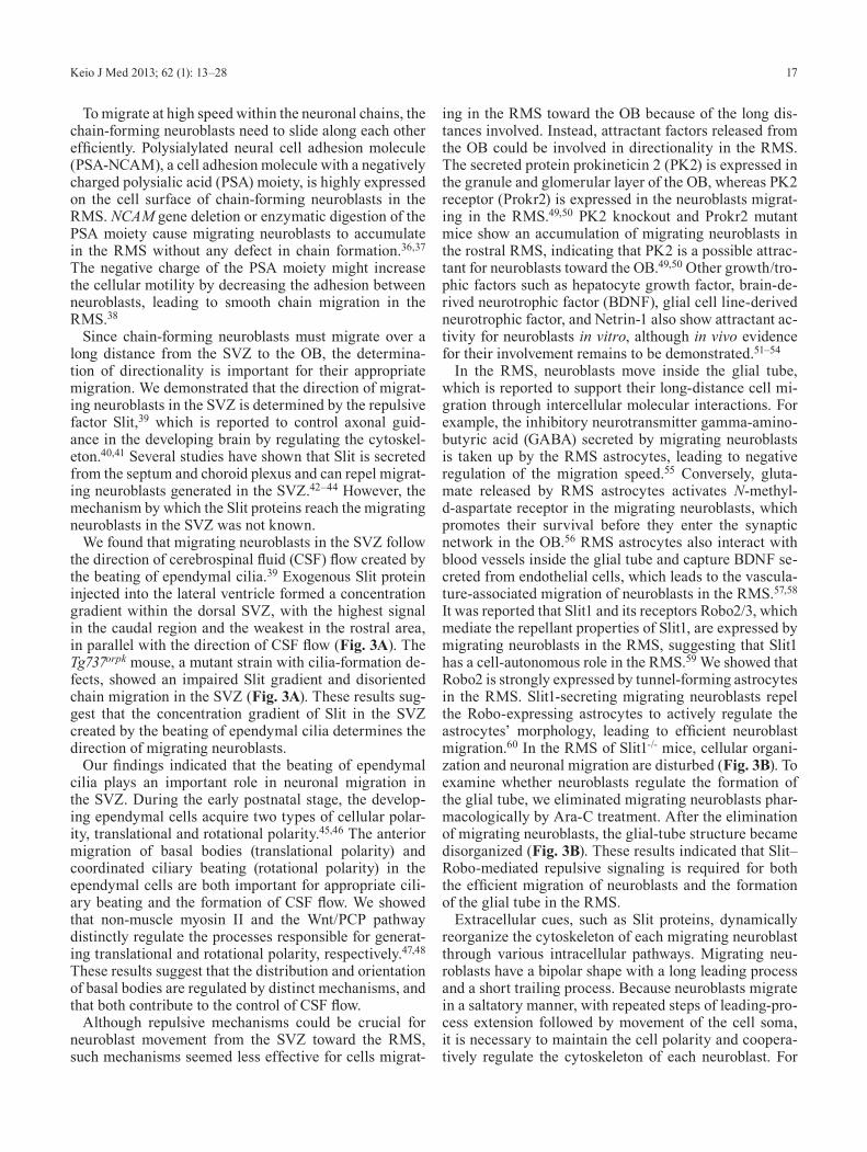
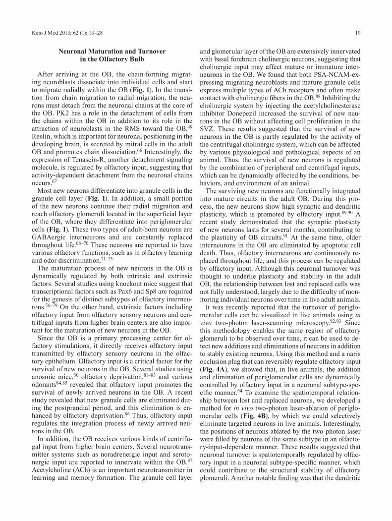
It was recently reported that the turnover of periglo-merular cells can be visualized in live animals using in vivo two-photon laser-scanning microscopy.92,93 Since this methodology enables the same region of olfactory glomeruli to be observed over time, it can be used to de-tect new additions and eliminations of neurons in addition to stably existing neurons. Using this method and a naris occlusion plug that can reversibly regulate olfactory input (Fig. 4A), we showed that, in live animals, the addition and elimination of periglomerular cells are dynamically controlled by olfactory input in a neuronal subtype-spe-cific manner.94 To examine the spatiotemporal relation-ship between lost and replaced neurons, we developed a method for in vivo two-photon laser-ablation of periglo-merular cells (Fig. 4B), by which we could selectively eliminate targeted neurons in live animals. Interestingly, the positions of neurons ablated by the two-photon laser were filled by neurons of the same subtype in an olfacto-ry-input-dependent manner. These results suggested that neuronal turnover is spatiotemporally regulated by olfac-tory input in a neuronal subtype-specific manner, which could contribute to the structural stability of olfactory glomeruli. Another notable finding was that the dendritic
Sawada M and Sawamoto K: Neurogenesis in the Adult Brain20
directions of the lost and replaced neurons at the same po-sitions were different, indicating that these neurons main-tain their dendritic plasticity (Fig. 4C). Taken together, we propose that the activity-dependent reiterated use of the same positions by new neurons enables both active remodeling of neuronal circuits by cellular turnover and maintenance of the glomerular structure.
Several studies have shown that the survival of new neurons in the OB is regulated by olfactory input; howev-er, the mechanisms regulating neuronal turnover are just beginning to be clarified. A recent study showed that de-pleted granule cells are compensated for by new cells of the same neuronal subtype in a local area of the granule cell layer.95 Thus, it is possible that adult-born olfactory interneurons show subtype-specific turnover patterns.94 The turnover of olfactory interneurons is thought to be important for both the maintenance and the plasticity of
olfactory functions. Further analysis of the spatiotempo-ral regulation of neuronal turnover will be needed to un-derstand how neuronal turnover contributes to structural and functional plasticity and/or stability in the adult OB.
Neurogenesis in the Adult Injured Brain
Recent studies suggest that neurogenesis in the adult brain can be affected by various pathological conditions. Experimental models of brain injury (such as trauma, hy-poxia, and ischemia) and of neurodegenerative diseases (including Alzheimer’s disease, Parkinson’s disease, and Huntington’s disease) are reported to stimulate neurogen-esis in the SVZ of the adult rodent brain.96 Of these dis-ease models, middle cerebral artery occlusion (MCAO) is widely used for investigating neurogenesis in the SVZ of the injured brain. In the MCAO model, the striatum
Fig. 4 Olfactory input-dependent spatial regulation of neuronal turnover in the adult olfactory bulb (modified from Sawada et al., J. Neurosci., 2011).94
(A) Fluorescent protein-labeled periglomerular cells (PGCs, green) could be observed over time in live animals under two-photon laser-scanning microscopy (2PLSM). To reversibly regulate the olfactory input, a naris occlusion plug was used. (B) Two-photon laser-ablation enables the selective elimination of targeted PGCs (red asterisks). Twenty-eight days after laser-ablation, newly added PGCs could be observed (yellow arrows and circles). The positions that lost PGCs (pink circles) by laser-ablation were filled by new PGCs (black asterisks) in an olfactory input-dependent manner. (C) Maximum projection images of lost and replaced PGCs shown in (B). In the replaced positions, the dendritic directions (yellow open arrowheads) of lost and replaced PGCs were different. Numbers indicate PGCs whose positions were stable during the imaging period. Scale bar: 10 µm.
21Keio J Med 2013; 62 (1): 13–28
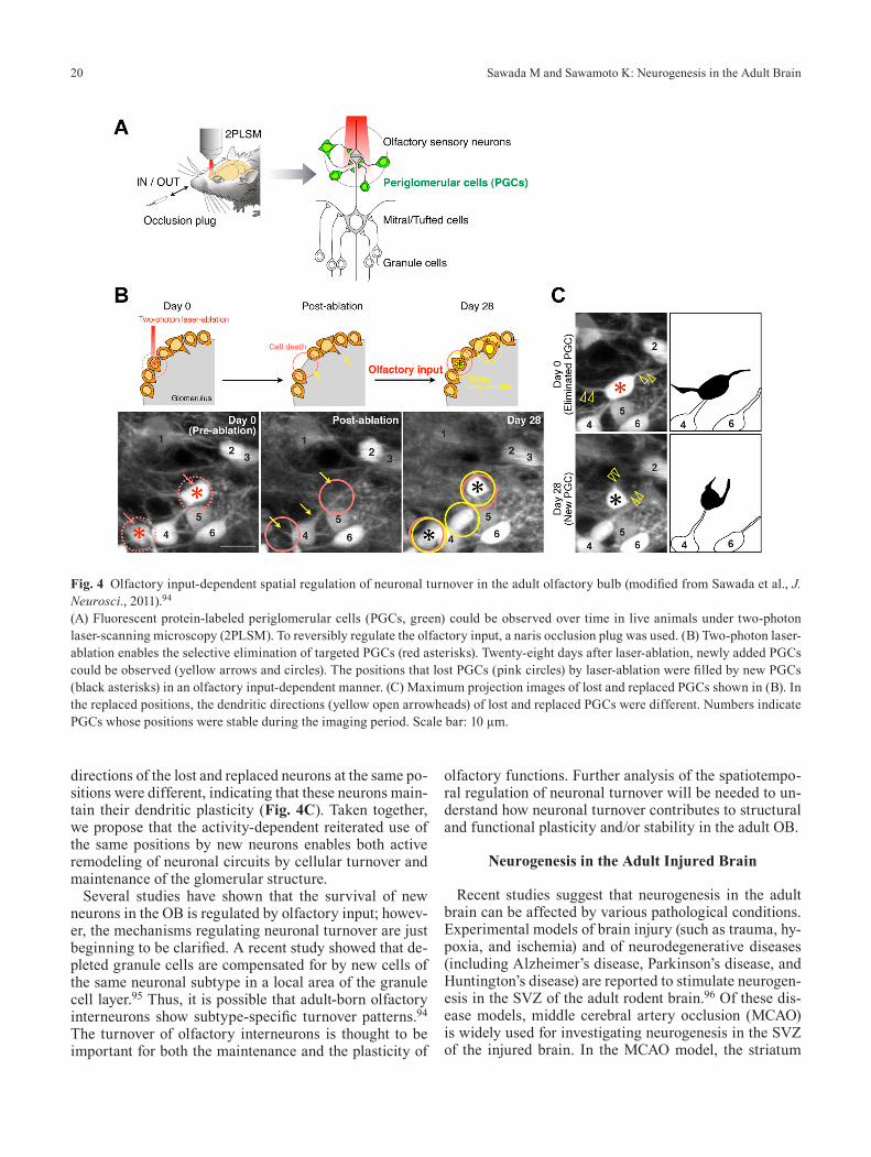
and dorsolateral cerebral cortex are damaged, and mature neurons in these regions undergo cell death (Fig. 5A). After MCAO, neurogenesis in the adult SVZ is upregulat-ed, followed by the appearance of many neuroblasts ex-pressing Dcx in the vicinity of the injured area, called the ischemic penumbra (Fig. 5B, C). However, only a small portion of these cells differentiate into mature neurons in the striatum.97,98 We have focused on this experimental model and investigated the regenerative potential of SVZ cells after injury.
The first reactive response after MCAO is an increased proliferation of SVZ cells (Fig. 5B). Several reports sug-gest that various chemokines and growth factors, includ-ing EGF, are upregulated in the damaged striatum99 and vascular endothelial cells,100 which could stimulate the proliferation of SVZ cells after MCAO. However, it was unknown what kind of cells could respond to EGF in the SVZ. We determined that EGFR is expressed by transit-amplifying type C cells in the SVZ after MCAO.101 In addition, these cells dynamically expand their number in response to EGF infusion. EGF infusion increased the number of neuroblasts in the damaged area, suggesting that neuronal replacement in the injured striatum might be enhanced by the EGF-induced expansion of transit-amplifying cells in the SVZ.
The second reactive response is the appearance of Dcx+ cells in the ischemic penumbra (Fig. 5C). These Dcx+ cells show a migratory morphology, suggesting
that they are migrating from somewhere toward the dam-aged area.97 Several angiogenic and inflammatory fac-tors such as angiopoietin-1 and SDF-1 are involved in this migration.102–104 However, the origin of these Dcx+ cells remained unknown. We demonstrated using a virus in-fection-mediated fate-mapping technique that these Dcx+ cells are derived from GFAP+ astrocytes in the SVZ and migrate toward the damaged regions.105 In addition, some of the migrating Dcx+ neuroblasts form chains and close-ly associate with blood vessels and astrocytic processes. These results raised the possibility that in the injured stri-atum, migrating cells use the blood vessels as a scaffold for their efficient migration. Using time-lapse recording in slice culture, we demonstrated that SVZ-derived neu-roblasts migrate along blood vessels toward the damaged striatum.106 Furthermore, the migration speed of these neuroblasts was decreased by the pharmacological inhi-bition of CXCR4, suggesting that chemokine signaling is involved in this vasophilic migration.
In contrast to the adult RMS, which provides a special-ized, permissive environment for cell migration, neu-roblasts in the damaged striatum must migrate through complex neuronal circuits. Furthermore, the activation of reactive astrocytes and microglia, and the subsequent formation of a glial scar, could worsen the conditions for migrating neuroblasts to reach the damaged area. More-over, many questions remain to be answered. Why do neuroblasts form chains and migrate along the blood ves-
Fig. 5 Neurogenesis in the injured adult brain.(A) Experimental ischemic model in which middle cerebral artery occlusion (MCAO) causes infarction in the striatum and cerebral cortex (brown). (B) After MCAO, the proliferation of SVZ cells (green) is activated. Angiogenesis also occurs surrounding the dam-aged region (red). (C) Dcx+ neuroblasts (red) derived from SVZ astrocytes migrate along both newly formed and preexisting blood vessels toward the damaged region. In some cases, these migrating neuroblasts form chains in the striatum. Despite the appearance of many migrating neuroblasts in the damaged striatum, only a small number of them survive and differentiate into mature neurons (yellow).
Sawada M and Sawamoto K: Neurogenesis in the Adult Brain22
sels in the damaged brain? What mechanisms direct the migrating neuroblasts toward the damaged area? How do neurons migrate through the complex, disadvantageous environment? Understanding the mechanisms regulat-ing the interactions between migrating neuroblasts and their surrounding microenvironment may provide new insights for novel therapies involving the regeneration of lost neuronal circuits using endogenous neural stem cells in the adult brain.96,107–110
Adult Neurogenesis in Various Species
Most of the studies on adult neurogenesis cited above used rodents. However, neurogenesis is observed in the adult brain of many vertebrates, from fish to humans.
A comparable neural stem cell niche in the adult brain is widely conserved at the ventricular wall1,111–115 (Fig. 6), where embryonic neural stem cells are located in the developing brain. Neural stem cells in the adult human brain are a promising endogenous cell source for brain regeneration after various injuries and diseases. Thus, comparative studies of neurogenesis in the adult brain among different species are needed to better understand the features and regulatory mechanisms of adult neural stem cells.
We have focused on zebrafish as a nonmammalian ver-tebrate model in which genetic and pharmacological ma-nipulations are available. Recent studies suggested that, as in rodents, neuronal progenitors are generated in the telencephalic ventricular zone (VZ) and migrate toward
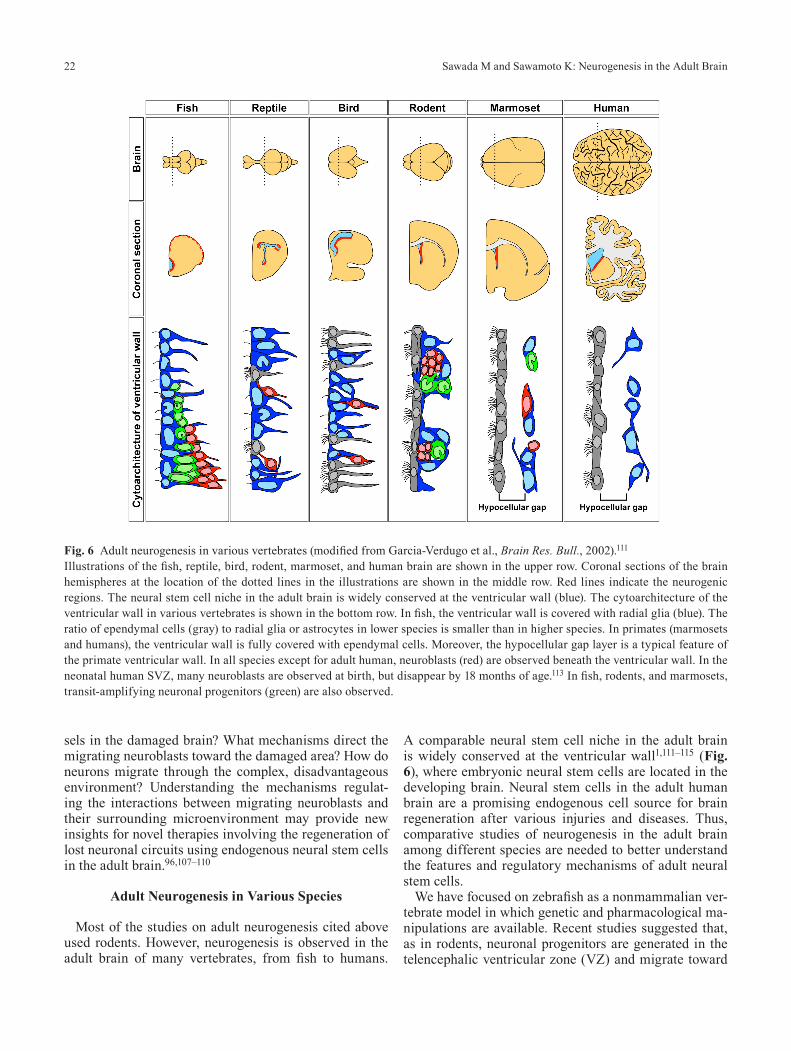
Fig. 6 Adult neurogenesis in various vertebrates (modified from Garcia-Verdugo et al., Brain Res. Bull., 2002).111
Illustrations of the fish, reptile, bird, rodent, marmoset, and human brain are shown in the upper row. Coronal sections of the brain hemispheres at the location of the dotted lines in the illustrations are shown in the middle row. Red lines indicate the neurogenic regions. The neural stem cell niche in the adult brain is widely conserved at the ventricular wall (blue). The cytoarchitecture of the ventricular wall in various vertebrates is shown in the bottom row. In fish, the ventricular wall is covered with radial glia (blue). The ratio of ependymal cells (gray) to radial glia or astrocytes in lower species is smaller than in higher species. In primates (marmosets and humans), the ventricular wall is fully covered with ependymal cells. Moreover, the hypocellular gap layer is a typical feature of the primate ventricular wall. In all species except for adult human, neuroblasts (red) are observed beneath the ventricular wall. In the neonatal human SVZ, many neuroblasts are observed at birth, but disappear by 18 months of age.113 In fish, rodents, and marmosets, transit-amplifying neuronal progenitors (green) are also observed.
23Keio J Med 2013; 62 (1): 13–28
the OB in the adult zebrafish.116–119 However, the cyto-architecture of the VZ–OB pathway was unknown. We demonstrated that the telencephalic VZ of the zebrafish consists of three types of cells: GFAP+ radial glial cells, PCNA+ proliferative cells, and PSA-NCAM+ neuroblasts (Fig. 6).112 Moreover, a continuous migratory stream of PSA-NCAM+ neuronal progenitors extends from the VZ to the OB, but the neurons do not move through glial tun-nels. Time-lapse imaging using Tg(ngn1:gfp) fish demon-strated that neuronal progenitors migrate rostrally along blood vessels in the RMS toward the OB, indicating that the PSA-NCAM+ stream is the functional route for the migration of neuronal progenitors in the adult zebrafish brain. In the rodent RMS, the chain-forming new neurons also migrate along blood vessels,57,58 suggesting the ex-istence of common mechanisms for vasophilic migration. Thus, these results reveal similarities and differences in the ventricular neurogenic niche between zebrafish and rodent brains.
Compared with rodents, zebrafish have a higher regen-erative potential after brain injury. We established a trau-matic brain injury (TBI) model in the adult zebrafish and demonstrated that the glutamatergic neurons lost from the dorsolateral telencephalon after TBI are regenerated from VZ-derived neuronal progenitors.120 This process was abolished by the pharmacological inhibition of Notch signaling, indicating that Notch signaling is required for neuronal repair. Thus, the zebrafish is a useful and pow-erful model for genetic and pharmacological screening to investigate the mechanisms regulating adult neurogen-esis under physiological and pathological conditions.
To understand evolutionarily conserved neurogenesis in the adult brain, we need to examine the primate brain in addition to that of nonmammalian vertebrates and ro-dents. The SVZ–OB pathway has been reported in the Old World primates Macaca mulatta and Macaca fas-cicularis.121–123 However, there are several limitations associated with the use of these macaque monkeys for studying adult neurogenesis, including their low avail-ability, slow sexual maturation, and large body size. In contrast, the common marmoset, Callithrix jacchus, a New World primate, has advantages in terms of its small size (300–500 g at maturity) and stable supply because of its availability from breeding colonies. In addition, sev-eral neurological disease models and transgenic animals are already available ,124,125 indicating that the common marmoset is useful for studying adult neurogenesis.
We recently reported the cellular composition and or-ganization of the SVZ and RMS in the common mar-moset.115 Interestingly, the SVZ–OB pathway in the marmoset is more similar to that of humans than to that of rodents. Like the adult human SVZ,114 the adult mar-moset SVZ consists of three distinct layers: the ependy-mal, hypocellular gap, and astrocytic ribbon layers (Fig. 6).115 Moreover, although an extensive network of PSA-NCAM+ elongated cell aggregates can be observed in the
SVZ, only individually migrating neuroblasts are found in the olfactory tract and OB. These findings indicate that the SVZ–OB pathway in the adult marmoset has several characteristics in common with that in humans. Howev-er, the anterior SVZ contains some putative progenitors similar to the transit-amplifying cells in rodents, but this feature is not conserved in the human brain (Fig. 6). No-tably, in the neonatal (postnatal day 0–1) marmoset, the SVZ contains chains of migrating neuroblasts that con-tinue toward the OB.115 This active neonatal neurogenesis and rodent-like chain migration was also recently report-ed in the neonatal human SVZ, in which many Dcx+ neu-roblasts exist in the layer corresponding to the hypocel-lular gap of the adult stage.113 Thus, again, the SVZ–OB pathway in marmosets is very similar to that in humans.
The neurogenic potential of the SVZ–OB pathway dif-fers among animal species, probably because of differ-ences in how much the animal depends on olfaction for its survival. In primates such as humans and marmosets, which depend on the visual system more than the olfac-tory system, new neurons generated in the SVZ may migrate to different locations and have different func-tions. Indeed, the human brain has an additional migra-tory stream of neurons that branches off from the RMS and ends in the ventromedial prefrontal cortex, raising the possibility that new neurons derived from SVZ neu-ral stem cells are involved in functions other than olfac-tion in higher primates.113 Further characterization of the fates of the neural stem/progenitor cells in the primate SVZ using marmoset models of neurological disorders should contribute to novel strategies for brain repair us-ing endogenous neural stem cells.
Conclusion
Since the discovery of neural stem cells in the adult brain, many features of adult neurogenesis have been described.2,96,126–129 We now know that neurogenesis in the adult brain consists of complex biological events in-cluding the genesis, migration, differentiation, and main-tenance of new neurons (Fig. 1). Although a variety of molecular and cellular mechanisms have been shown to regulate each aspect of adult neurogenesis, it is important to integrate these findings to obtain a comprehensive un-derstanding of this complex system. Adult neurogenesis could enable the dynamic remodeling of mature neuronal circuits by adding new neurons, contributing to struc-tural and functional plasticity in the adult brain. Adult neurogenesis could also have an important role in the in-jured brain, in regenerating the lost neurons from adult neural stem cells. This endogenous potential needs to be further investigated in future studies. Importantly, adult neurogenesis is regulated not only by developmental ge-netic programs but also by environmental and internal changes in animals. Thus, adult neurogenesis could be a fundamental mechanism for experience-dependent plas-
Sawada M and Sawamoto K: Neurogenesis in the Adult Brain24
ticity, which is the most important feature of the adult brain. We hope that future studies on the physiological and pathological conditions of various animals will im-prove our understanding of the biological functions and significance of neurogenesis in the adult brain.
Acknowledgments
This article summarizes our studies presented by K.S. in the Kitazato Prize lecture (2011) at Keio University School of Medicine. We apologize to all those whose work we could not cite because of space limitations. We thank all our collaborators for their contributions to origi-nal research, and past and current members of Sawamoto laboratory for their encouragement and discussions. M.S. is a Research Fellow of the Japan Society for the Pro-motion of Science. This work was supported by the Keio University Medical Science Fund, by the Funding Pro-gram for the Next Generation World-Leading Research-ers, a Grant-in-Aid for Young Scientists (S), by the Inter-national Human Frontier Science Program Organization, and by the Project for Realization of Regenerative Medi-cine from MEXT.
References
1. Doetsch F, Garcia-Verdugo JM, Alvarez-Buylla A: Cellular com-position and three-dimensional organization of the subventricular germinal zone in the adult mammalian brain. J Neurosci 1997; 17: 5046–5061. [Medline]
2. Ihrie RA, Alvarez-Buylla A: Lake-front property: a unique ger-minal niche by the lateral ventricles of the adult brain. Neuron 2011; 70: 674–686. [Medline] [CrossRef]
3. Menn B, Garcia-Verdugo JM, Yaschine C, Gonzalez-Perez O, Rowitch D, Alvarez-Buylla A: Origin of oligodendrocytes in the subventricular zone of the adult brain. J Neurosci 2006; 26: 7907–7918. [Medline] [CrossRef]
4. Bonaguidi MA, Wheeler MA, Shapiro JS, Stadel RP, Sun GJ, Ming GL, Song H: In vivo clonal analysis reveals self-renewing and multipotent adult neural stem cell characteristics. Cell 2011; 145: 1142–1155. [Medline] [CrossRef]
5. Brazel CY, Limke TL, Osborne JK, Miura T, Cai J, Pevny L, Rao MS: Sox2 expression defines a heterogeneous population of neurosphere-forming cells in the adult murine brain. Aging Cell 2005; 4: 197–207. [Medline] [CrossRef]
6. Capela A, Temple S: LeX/ssea-1 is expressed by adult mouse CNS stem cells, identifying them as nonependymal. Neuron 2002; 35: 865–875. [Medline] [CrossRef]
7. Ellis P, Fagan BM, Magness ST, Hutton S, Taranova O, Hayashi S, McMahon A, Rao M, Pevny L: SOX2, a persistent marker for multipotential neural stem cells derived from embryonic stem cells, the embryo or the adult. Dev Neurosci 2004; 26: 148–165. [Medline] [CrossRef]
8. Kaneko Y, Sakakibara S, Imai T, Suzuki A, Nakamura Y, Sawa-moto K, Ogawa Y, Toyama Y, Miyata T, Okano H: Musashi1: an evolutionally conserved marker for CNS progenitor cells includ-ing neural stem cells. Dev Neurosci 2000; 22: 139–153. [Medline] [CrossRef]
9. Nam HS, Benezra R: High levels of Id1 expression define B1 type adult neural stem cells. Cell Stem Cell 2009; 5: 515–526. [Med-line] [CrossRef]
10. Kokovay E, Goderie S, Wang Y, Lotz S, Lin G, Sun Y, Roysam B, Shen Q, Temple S: Adult SVZ lineage cells home to and leave the vascular niche via differential responses to SDF1/CXCR4 signal-ing. Cell Stem Cell 2010; 7: 163–173. [Medline] [CrossRef]
11. Mirzadeh Z, Merkle FT, Soriano-Navarro M, Garcia-Verdugo JM, Alvarez-Buylla A: Neural stem cells confer unique pinwheel architecture to the ventricular surface in neurogenic regions of the adult brain. Cell Stem Cell 2008; 3: 265–278. [Medline] [Cross-Ref]
12. Shen Q, Wang Y, Kokovay E, Lin G, Chuang SM, Goderie SK, Roysam B, Temple S: Adult SVZ stem cells lie in a vascular niche: a quantitative analysis of niche cell-cell interactions. Cell Stem Cell 2008; 3: 289–300. [Medline] [CrossRef]
13. Tavazoie M, Van der Veken L, Silva-Vargas V, Louissaint M, Col-onna L, Zaidi B, Garcia-Verdugo JM, Doetsch F: A specialized vascular niche for adult neural stem cells. Cell Stem Cell 2008; 3: 279–288. [Medline] [CrossRef]
14. Sakaguchi M, Shingo T, Shimazaki T, Okano HJ, Shiwa M, Ishi-bashi S, Oguro H, Ninomiya M, Kadoya T, Horie H, Shibuya A, Mizusawa H, Poirier F, Nakauchi H, Sawamoto K, Okano H: A carbohydrate-binding protein, Galectin-1, promotes proliferation of adult neural stem cells. Proc Natl Acad Sci USA 2006; 103: 7112–7117. [Medline] [CrossRef]
15. Conover JC, Doetsch F, Garcia-Verdugo JM, Gale NW, Yanco-poulos GD, Alvarez-Buylla A: Disruption of Eph/ephrin signal-ing affects migration and proliferation in the adult subventricular zone. Nat Neurosci 2000; 3: 1091–1097. [Medline] [CrossRef]
16. Ferrón SR, Charalambous M, Radford E, McEwen K, Wildner H, Hind E, Morante-Redolat JM, Laborda J, Guillemot F, Bauer SR, Fariñas I, Ferguson-Smith AC: Postnatal loss of Dlk1 imprinting in stem cells and niche astrocytes regulates neurogenesis. Nature 2011; 475: 381–385. [Medline] [CrossRef]
17. Imayoshi I, Sakamoto M, Yamaguchi M, Mori K, Kageyama R: Essential roles of Notch signaling in maintenance of neural stem cells in developing and adult brains. J Neurosci 2010; 30: 3489–3498. [Medline] [CrossRef]
18. Aguirre A, Rubio ME, Gallo V: Notch and EGFR pathway inter-action regulates neural stem cell number and self-renewal. Nature 2010; 467: 323–327. [Medline] [CrossRef]
19. Andreu-Agulló C, Morante-Redolat JM, Delgado AC, Fariñas I: Vascular niche factor PEDF modulates Notch-dependent stem-ness in the adult subependymal zone. Nat Neurosci 2009; 12: 1514–1523. [Medline] [CrossRef]
20. Ramírez-Castillejo C, Sánchez-Sánchez F, Andreu-Agulló C, Ferrón SR, Aroca-Aguilar JD, Sánchez P, Mira H, Escribano J, Fariñas I: Pigment epithelium-derived factor is a niche signal for neural stem cell renewal. Nat Neurosci 2006; 9: 331–339. [Med-line] [CrossRef]
21. Merkle FT, Mirzadeh Z, Alvarez-Buylla A: Mosaic organization of neural stem cells in the adult brain. Science 2007; 317: 381–384. [Medline] [CrossRef]
22. Balordi F, Fishell G: Mosaic removal of hedgehog signaling in the adult SVZ reveals that the residual wild-type stem cells have a limited capacity for self-renewal. J Neurosci 2007; 27: 14248–14259. [Medline] [CrossRef]
23. Palma V, Lim DA, Dahmane N, Sánchez P, Brionne TC, Herzberg CD, Gitton Y, Carleton A, Alvarez-Buylla A, Ruiz i Altaba A: Sonic hedgehog controls stem cell behavior in the postnatal and adult brain. Development 2005; 132: 335–344. [Medline] [Cross-Ref]
24. Ihrie RA, Shah JK, Harwell CC, Levine JH, Guinto CD, Lezam-eta M, Kriegstein AR, Alvarez-Buylla A: Persistent sonic hedge-hog signaling in adult brain determines neural stem cell positional identity. Neuron 2011; 71: 250–262. [Medline] [CrossRef]
25. Colak D, Mori T, Brill MS, Pfeifer A, Falk S, Deng C, Monteiro R, Mummery C, Sommer L, Götz M: Adult neurogenesis requires Smad4-mediated bone morphogenic protein signaling in stem
25Keio J Med 2013; 62 (1): 13–28
cells. J Neurosci 2008; 28: 434–446. [Medline] [CrossRef] 26. Adachi K, Mirzadeh Z, Sakaguchi M, Yamashita T, Nikolcheva T,
Gotoh Y, Peltz G, Gong L, Kawase T, Alvarez-Buylla A, Okano H, Sawamoto K: Beta-catenin signaling promotes proliferation of progenitor cells in the adult mouse subventricular zone. Stem Cells 2007; 25: 2827–2836. [Medline] [CrossRef]
27. Ikeda M, Hirota Y, Sakaguchi M, Yamada O, Kida YS, Ogura T, Otsuka T, Okano H, Sawamoto K: Expression and proliferation-promoting role of Diversin in the neuronally committed precursor cells migrating in the adult mouse brain. Stem Cells 2010; 28: 2017–2026. [Medline] [CrossRef]
28. Wichterle H, Garcia-Verdugo JM, Alvarez-Buylla A: Direct evi-dence for homotypic, glia-independent neuronal migration. Neu-ron 1997; 18: 779–791. [Medline] [CrossRef]
29. Doetsch F, Alvarez-Buylla A: Network of tangential pathways for neuronal migration in adult mammalian brain. Proc Natl Acad Sci USA 1996; 93: 14895–14900. [Medline] [CrossRef]
30. Sawada M, Huang S, Hirota Y, Kaneko N, Sawamoto K: Neuronal migration in the adult brain. In: Seki T, Sawamoto K, Parent JM, Alvarez-Buylla A, eds, Neurogenesis in the Adult Brain I, Neuro-biology, Tokyo, Springer, 2011; 337–355.
31. Andrade N, Komnenovic V, Blake SM, Jossin Y, Howell B, Goffi-net A, Schneider WJ, Nimpf J: ApoER2/VLDL receptor and Dab1 in the rostral migratory stream function in postnatal neuronal mi-gration independently of Reelin. Proc Natl Acad Sci USA 2007; 104: 8508–8513. [Medline] [CrossRef]
32. Anton ES, Ghashghaei HT, Weber JL, McCann C, Fischer TM, Cheung ID, Gassmann M, Messing A, Klein R, Schwab MH, Lloyd KC, Lai C: Receptor tyrosine kinase ErbB4 modulates neu-roblast migration and placement in the adult forebrain. Nat Neu-rosci 2004; 7: 1319–1328. [Medline] [CrossRef]
33. Belvindrah R, Hankel S, Walker J, Patton BL, Muller U: Beta1 integrins control the formation of cell chains in the adult rostral migratory stream. J Neurosci 2007; 27: 2704–2717. [Medline] [CrossRef]
34. Emsley JG, Hagg T: Alpha6beta1 integrin directs migration of neuronal precursors in adult mouse forebrain. Exp Neurol 2003; 183: 273–285. [Medline] [CrossRef]
35. Murase S, Cho C, White JM, Horwitz AF: ADAM2 promotes migration of neuroblasts in the rostral migratory stream to the olfactory bulb. Eur J Neurosci 2008; 27: 1585–1595. [Medline] [CrossRef]
36. Cremer H, Lange R, Christoph A, Plomann M, Vopper G, Roes J, Brown R, Baldwin S, Kraemer P, Scheff S, et al: Inactivation of the N-CAM gene in mice results in size reduction of the olfactory bulb and deficits in spatial learning. Nature 1994; 367: 455–459. [Medline] [CrossRef]
37. Ono K, Tomasiewicz H, Magnuson T, Rutishauser U: N-CAM mutation inhibits tangential neuronal migration and is pheno-copied by enzymatic removal of polysialic acid. Neuron 1994; 13: 595–609. [Medline] [CrossRef]
38. Gascon E, Vutskits L, Kiss JZ: The role of PSA-NCAM in adult neurogenesis. Adv Exp Med Biol 2010; 663: 127–136. [Medline] [CrossRef]
39. Sawamoto K, Wichterle H, Gonzalez-Perez O, Cholfin JA, Ya-mada M, Spassky N, Murcia NS, Garcia-Verdugo JM, Marin O, Rubenstein JL, Tessier-Lavigne M, Okano H, Alvarez-Buylla A: New neurons follow the flow of cerebrospinal fluid in the adult brain. Science 2006; 311: 629–632. [Medline] [CrossRef]
40. Brose K, Bland KS, Wang KH, Arnott D, Henzel W, Goodman CS, Tessier-Lavigne M, Kidd T: Slit proteins bind Robo receptors and have an evolutionarily conserved role in repulsive axon guid-ance. Cell 1999; 96: 795–806. [Medline] [CrossRef]
41. Li HS, Chen JH, Wu W, Fagaly T, Zhou L, Yuan W, Dupuis S, Jiang ZH, Nash W, Gick C, Ornitz DM, Wu JY, Rao Y: Vertebrate Slit, a secreted ligand for the transmembrane protein roundabout, is a repellent for olfactory bulb axons. Cell 1999; 96: 807–818.
[Medline] [CrossRef] 42. Hu H: Chemorepulsion of neuronal migration by Slit2 in the
developing mammalian forebrain. Neuron 1999; 23: 703–711. [Medline] [CrossRef]
43. Hu H, Rutishauser U: A septum-derived chemorepulsive factor for migrating olfactory interneuron precursors. Neuron 1996; 16: 933–940. [Medline] [CrossRef]
44. Wu W, Wong K, Chen J, Jiang Z, Dupuis S, Wu JY, Rao Y: Di-rectional guidance of neuronal migration in the olfactory system by the protein Slit. Nature 1999; 400: 331–336. [Medline] [Cross-Ref]
45. Mirzadeh Z, Han YG, Soriano-Navarro M, Garcia-Verdugo JM, Alvarez-Buylla A: Cilia organize ependymal planar polarity. J Neurosci 2010; 30: 2600–2610. [Medline] [CrossRef]
46. Planar polarity of ependymal cilia. Differentiation; research in biological diversity 2011.
47. Guirao B, Meunier A, Mortaud S, Aguilar A, Corsi JM, Strehl L, Hirota Y, Desoeuvre A, Boutin C, Han YG, Mirzadeh Z, Cremer H, Montcouquiol M, Sawamoto K, Spassky N: Coupling between hydrodynamic forces and planar cell polarity orients mammalian motile cilia. Nat Cell Biol 2010; 12: 341–350. [Medline] [Cross-Ref]
48. Hirota Y, Meunier A, Huang S, Shimozawa T, Yamada O, Kida YS, Inoue M, Ito T, Kato H, Sakaguchi M, Sunabori T, Naka-ya MA, Nonaka S, Ogura T, Higuchi H, Okano H, Spassky N, Sawamoto K: Planar polarity of multiciliated ependymal cells involves the anterior migration of basal bodies regulated by non-muscle myosin II. Development 2010; 137: 3037–3046. [Medline] [CrossRef]
49. Ng KL, Li JD, Cheng MY, Leslie FM, Lee AG, Zhou QY: Depen-dence of olfactory bulb neurogenesis on prokineticin 2 signaling. Science 2005; 308: 1923–1927. [Medline] [CrossRef]
50. Puverel S, Nakatani H, Parras C, Soussi-Yanicostas N: Proki-neticin receptor 2 expression identifies migrating neuroblasts and their subventricular zone transient-amplifying progenitors in adult mice. J Comp Neurol 2009; 512: 232–242. [Medline] [CrossRef]
51. Chiaramello S, Dalmasso G, Bezin L, Marcel D, Jourdan F, Per-etto P, Fasolo A, De Marchis S: BDNF/ TrkB interaction regulates migration of SVZ precursor cells via PI3-K and MAP-K signal-ling pathways. Eur J Neurosci 2007; 26: 1780–1790. [Medline] [CrossRef]
52. Garzotto D, Giacobini P, Crepaldi T, Fasolo A, De Marchis S: He-patocyte growth factor regulates migration of olfactory interneu-ron precursors in the rostral migratory stream through Met-Grb2 coupling. J Neurosci 2008; 28: 5901–5909. [Medline] [CrossRef]
53. Murase S, Horwitz AF: Deleted in colorectal carcinoma and dif-ferentially expressed integrins mediate the directional migration of neural precursors in the rostral migratory stream. J Neurosci 2002; 22: 3568–3579. [Medline]
54. Paratcha G, Ibanez CF, Ledda F: GDNF is a chemoattractant fac-tor for neuronal precursor cells in the rostral migratory stream. Mol Cell Neurosci 2006; 31: 505–514. [Medline] [CrossRef]
55. Bolteus AJ, Bordey A: GABA release and uptake regulate neu-ronal precursor migration in the postnatal subventricular zone. J Neurosci 2004; 24: 7623–7631. [Medline] [CrossRef]
56. Platel JC, Dave KA, Gordon V, Lacar B, Rubio ME, Bordey A: NMDA receptors activated by subventricular zone astrocytic glu-tamate are critical for neuroblast survival prior to entering a syn-aptic network. Neuron 2010; 65: 859–872. [Medline] [CrossRef]
57. Bozoyan L, Khlghatyan J, Saghatelyan A: Astrocytes control the development of the migration-promoting vasculature scaffold in the postnatal brain via VEGF signaling. J Neurosci 2012; 32: 1687–1704. [Medline] [CrossRef]
58. Snapyan M, Lemasson M, Brill MS, Blais M, Massouh M, Ninkovic J, Gravel C, Berthod F, Götz M, Barker PA, Parent A, Saghatelyan A: Vasculature guides migrating neuronal precur-
Sawada M and Sawamoto K: Neurogenesis in the Adult Brain26
sors in the adult mammalian forebrain via brain-derived neuro-trophic factor signaling. J Neurosci 2009; 29: 4172–4188. [Med-line] [CrossRef]
59. Nguyen-Ba-Charvet KT, Picard-Riera N, Tessier-Lavigne M, Baron-Van Evercooren A, Sotelo C, Chedotal A: Multiple roles for slits in the control of cell migration in the rostral migratory stream. J Neurosci 2004; 24: 1497–1506. [Medline] [CrossRef]
60. Kaneko N, Marin O, Koike M, Hirota Y, Uchiyama Y, Wu JY, Lu Q, Tessier-Lavigne M, Alvarez-Buylla A, Okano H, Rubenstein JL, Sawamoto K: New neurons clear the path of astrocytic pro-cesses for their rapid migration in the adult brain. Neuron 2010; 67: 213–223. [Medline] [CrossRef]
61. Higginbotham H, Tanaka T, Brinkman BC, Gleeson JG: GSK3be-ta and PKCzeta function in centrosome localization and process stabilization during Slit-mediated neuronal repolarization. Mol Cell Neurosci 2006; 32: 118–132. [Medline] [CrossRef]
62. Wang Y, Kaneko N, Asai N, Enomoto A, Isotani-Sakakibara M, Kato T, Asai M, Murakumo Y, Ota H, Hikita T, Namba T, Ku-roda K, Kaibuchi K, Ming GL, Song H, Sawamoto K, Takahashi M: Girdin is an intrinsic regulator of neuroblast chain migration in the rostral migratory stream of the postnatal brain. J Neurosci 2011; 31: 8109–8122. [Medline] [CrossRef]
63. Shinohara R, Thumkeo D, Kamijo H, Kaneko N, Sawamoto K, Watanabe K, Takebayashi H, Kiyonari H, Ishizaki T, Furuyashiki T, Narumiya S: A role for mDia, a Rho-regulated actin nucleator, in tangential migration of interneuron precursors. Nat Neurosci 2012; 15: 373–380. [Medline] [CrossRef]
64. Koizumi H, Higginbotham H, Poon T, Tanaka T, Brinkman BC, Gleeson JG: Doublecortin maintains bipolar shape and nuclear translocation during migration in the adult forebrain. Nat Neuro-sci 2006; 9: 779–786. [Medline] [CrossRef]
65. Hirota Y, Ohshima T, Kaneko N, Ikeda M, Iwasato T, Kulkarni AB, Mikoshiba K, Okano H, Sawamoto K: Cyclin-dependent kinase 5 is required for control of neuroblast migration in the postnatal subventricular zone. J Neurosci 2007; 27: 12829–12838. [Medline] [CrossRef]
66. Hack I, Bancila M, Loulier K, Carroll P, Cremer H: Reelin is a detachment signal in tangential chain-migration during postnatal neurogenesis. Nat Neurosci 2002; 5: 939–945. [Medline] [Cross-Ref]
67. Saghatelyan A, de Chevigny A, Schachner M, Lledo PM: Tenas-cin-R mediates activity-dependent recruitment of neuroblasts in the adult mouse forebrain. Nat Neurosci 2004; 7: 347–356. [Med-line] [CrossRef]
68. Imayoshi I, Sakamoto M, Ohtsuka T, Takao K, Miyakawa T, Ya-maguchi M, Mori K, Ikeda T, Itohara S, Kageyama R: Roles of continuous neurogenesis in the structural and functional integrity of the adult forebrain. Nat Neurosci 2008; 11: 1153–1161. [Med-line] [CrossRef]
69. Lagace DC, Whitman MC, Noonan MA, Ables JL, DeCarolis NA, Arguello AA, Donovan MH, Fischer SJ, Farnbauch LA, Beech RD, DiLeone RJ, Greer CA, Mandyam CD, Eisch AJ. : Dynamic contribution of nestin-expressing stem cells to adult neurogenesis. J Neurosci 2007; 27: 12623–12629. [Medline] [CrossRef]
70. Ninkovic J, Mori T, Gotz M: Distinct modes of neuron addition in adult mouse neurogenesis. J Neurosci 2007; 27: 10906–10911. [Medline] [CrossRef]
71. Breton-Provencher V, Lemasson M, Peralta MR 3rd, Saghatelyan A: Interneurons produced in adulthood are required for the nor-mal functioning of the olfactory bulb network and for the execu-tion of selected olfactory behaviors. J Neurosci 2009; 29: 15245–15257. [Medline] [CrossRef]
72. Gheusi G, Cremer H, McLean H, Chazal G, Vincent JD, Lledo PM: Importance of newly generated neurons in the adult olfactory bulb for odor discrimination. Proc Natl Acad Sci USA 2000; 97: 1823–1828. [Medline] [CrossRef]
73. Mak GK, Enwere EK, Gregg C, Pakarainen T, Poutanen M, Huhtaniemi I, Weiss S: Male pheromone-stimulated neurogenesis in the adult female brain: possible role in mating behavior. Nat Neurosci 2007; 10: 1003–1011. [Medline] [CrossRef]
74. Moreno MM, Linster C, Escanilla O, Sacquet J, Didier A, Man-dairon N: Olfactory perceptual learning requires adult neurogen-esis. Proc Natl Acad Sci USA 2009; 106: 17980–17985. [Medline] [CrossRef]
75. Sakamoto M, Imayoshi I, Ohtsuka T, Yamaguchi M, Mori K, Kageyama R: Continuous neurogenesis in the adult forebrain is required for innate olfactory responses. Proc Natl Acad Sci USA 2011; 108: 8479–8484. [Medline] [CrossRef]
76. Hack MA, Saghatelyan A, de Chevigny A, Pfeifer A, Ashery-Padan R, Lledo PM, Gotz M: Neuronal fate determinants of adult olfactory bulb neurogenesis. Nat Neurosci 2005; 8: 865–872. [Medline]
77. Kohwi M, Osumi N, Rubenstein JL, Alvarez-Buylla A: Pax6 is required for making specific subpopulations of granule and peri-glomerular neurons in the olfactory bulb. J Neurosci 2005; 25: 6997–7003. [Medline] [CrossRef]
78. Waclaw RR, Allen ZJ 2nd, Bell SM, Erdelyi F, Szabo G, Potter SS, Campbell K: The zinc finger transcription factor Sp8 regu-lates the generation and diversity of olfactory bulb interneurons. Neuron 2006; 49: 503–516. [Medline] [CrossRef]
79. Li X, Sun C, Lin C, Ma T, Madhavan MC, Campbell K, Yang Z: The transcription factor Sp8 is required for the production of parvalbumin-expressing interneurons in the olfactory bulb. J Neurosci 2011; 31: 8450–8455. [Medline] [CrossRef]
80. Petreanu L, Alvarez-Buylla A: Maturation and death of adult-born olfactory bulb granule neurons: role of olfaction. J Neurosci 2002; 22: 6106–6113. [Medline]
81. Cummings DM, Henning HE, Brunjes PC: Olfactory bulb recov-ery after early sensory deprivation. J Neurosci 1997; 17: 7433–7440. [Medline]
82. Mandairon N, Sacquet J, Jourdan F, Didier A: Long-term fate and distribution of newborn cells in the adult mouse olfactory bulb: Influences of olfactory deprivation. Neuroscience 2006; 141: 443–451. [Medline] [CrossRef]
83. Yamaguchi M, Mori K: Critical period for sensory experience-dependent survival of newly generated granule cells in the adult mouse olfactory bulb. Proc Natl Acad Sci USA 2005; 102: 9697–9702. [Medline] [CrossRef]
84. Alonso M, Viollet C, Gabellec MM, Meas-Yedid V, Olivo-Marin JC, Lledo PM: Olfactory discrimination learning increases the survival of adult-born neurons in the olfactory bulb. J Neurosci 2006; 26: 10508–10513. [Medline] [CrossRef]
85. Rochefort C, Gheusi G, Vincent JD, Lledo PM: Enriched odor exposure increases the number of newborn neurons in the adult olfactory bulb and improves odor memory. J Neurosci 2002; 22: 2679–2689. [Medline]
86. Yokoyama TK, Mochimaru D, Murata K, Manabe H, Kobayaka-wa K, Kobayakawa R, Sakano H, Mori K, Yamaguchi M: Elimi-nation of adult-born neurons in the olfactory bulb is promoted during the postprandial period. Neuron 2011; 71: 883–897. [Med-line] [CrossRef]
87. Shepherd GM, Chen WR, Greer CA: Olfactory bulb. In: Shepherd GM, ed, The Synaptic Organization of the Brain, New York, Ox-ford, 2004, 165-216.
88. Role of the cholinergic system in regulating survival of newborn neurons in the adult mouse dentate gyrus and olfactory bulb. Genes to cells: devoted to molecular and cellular mechanisms 2006; 11: 1145-1159.
89. Livneh Y, Feinstein N, Klein M, Mizrahi A: Sensory input en-hances synaptogenesis of adult-born neurons. J Neurosci 2009; 29: 86–97. [Medline] [CrossRef]
90. Nissant A, Bardy C, Katagiri H, Murray K, Lledo PM: Adult neu-rogenesis promotes synaptic plasticity in the olfactory bulb. Nat

27Keio J Med 2013; 62 (1): 13–28
Neurosci 2009; 12: 728–730. [Medline] [CrossRef] 91. Livneh Y, Mizrahi A: Experience-dependent plasticity of mature
adult-born neurons. Nat Neurosci 2011; 15: 26–28. [Medline] [CrossRef]
92. Adam Y, Mizrahi A: Long-term imaging reveals dynamic chang-es in the neuronal composition of the glomerular layer. J Neurosci 2011; 31: 7967–7973. [Medline] [CrossRef]
93. Mizrahi A, Lu J, Irving R, Feng G, Katz LC: In vivo imaging of juxtaglomerular neuron turnover in the mouse olfactory bulb. Proc Natl Acad Sci USA 2006; 103: 1912–1917. [Medline] [Cross-Ref]
94. Sawada M, Kaneko N, Inada H, Wake H, Kato Y, Yanagawa Y, Kobayashi K, Nemoto T, Nabekura J, Sawamoto K: Sensory input regulates spatial and subtype-specific patterns of neuronal turn-over in the adult olfactory bulb. J Neurosci 2011; 31: 11587–11596. [Medline] [CrossRef]
95. Murata K, Imai M, Nakanishi S, Watanabe D, Pastan I, Kobayashi K, Nihira T, Mochizuki H, Yamada S, Mori K, Yamaguchi M: Compensation of depleted neuronal subsets by new neurons in a local area of the adult olfactory bulb. J Neurosci 2011; 31: 10540–10557. [Medline] [CrossRef]
96. Kaneko N, Sawamoto K: Adult neurogenesis and its alteration under pathological conditions. Neurosci Res 2009; 63: 155–164. [Medline] [CrossRef]
97. Arvidsson A, Collin T, Kirik D, Kokaia Z, Lindvall O: Neuronal replacement from endogenous precursors in the adult brain after stroke. Nat Med 2002; 8: 963–970. [Medline] [CrossRef]
98. Parent JM, Vexler ZS, Gong C, Derugin N, Ferriero DM: Rat forebrain neurogenesis and striatal neuron replacement after fo-cal stroke. Ann Neurol 2002; 52: 802–813. [Medline] [CrossRef]
99. Hayashi T, Noshita N, Sugawara T, Chan PH: Temporal profile of angiogenesis and expression of related genes in the brain after ischemia. J Cereb Blood Flow Metab 2003; 23: 166–180. [Med-line] [CrossRef]
100. Shen Q, Goderie SK, Jin L, Karanth N, Sun Y, Abramova N, Vin-cent P, Pumiglia K, Temple S: Endothelial cells stimulate self-renewal and expand neurogenesis of neural stem cells. Science 2004; 304: 1338–1340. [Medline] [CrossRef]
101. Ninomiya M, Yamashita T, Araki N, Okano H, Sawamoto K: En-hanced neurogenesis in the ischemic striatum following EGF-in-duced expansion of transit-amplifying cells in the subventricular zone. Neurosci Lett 2006; 403: 63–67. [Medline] [CrossRef]
102. Imitola J, Raddassi K, Park KI, Mueller FJ, Nieto M, Teng YD, Frenkel D, Li J, Sidman RL, Walsh CA, Snyder EY, Khoury SJ: Directed migration of neural stem cells to sites of CNS injury by the stromal cell-derived factor 1alpha/CXC chemokine receptor 4 pathway. Proc Natl Acad Sci USA 2004; 101: 18117–18122. [Med-line] [CrossRef]
103. Ohab JJ, Fleming S, Blesch A, Carmichael ST: A neurovascular niche for neurogenesis after stroke. J Neurosci 2006; 26: 13007–13016. [Medline] [CrossRef]
104. Thored P, Arvidsson A, Cacci E, Ahlenius H, Kallur T, Darsa-lia V, Ekdahl CT, Kokaia Z, Lindvall O: Persistent production of neurons from adult brain stem cells during recovery after stroke. Stem Cells 2006; 24: 739–747. [Medline] [CrossRef]
105. Yamashita T, Ninomiya M, Hernandez Acosta P, García-Verdugo JM, Sunabori T, Sakaguchi M, Adachi K, Kojima T, Hirota Y, Ka-wase T, Araki N, Abe K, Okano H, Sawamoto K: Subventricular zone-derived neuroblasts migrate and differentiate into mature neurons in the post-stroke adult striatum. J Neurosci 2006; 26: 6627–6636. [Medline] [CrossRef]
106. Kojima T, Hirota Y, Ema M, Takahashi S, Miyoshi I, Okano H, Sawamoto K: Subventricular zone-derived neural progenitor cells migrate along a blood vessel scaffold toward the post-stroke stria-tum. Stem Cells 2010; 28: 545–554. [Medline]
107. Kaneko N, Kako E, Sawamoto K: Prospects and limitations of using endogenous neural stem cells for brain regeneration. Genes
2011; 2: 107–130. [CrossRef] 108. Nakaguchi K, Masuda H, Kaneko N, Sawamoto K: Strategies
for regenerating striatal neurons in the adult brain by using en-dogenous neural stem cells. Neurol Res Int 2011; 2011: 898012. [Medline]
109. Okano H, Sakaguchi M, Ohki K, Suzuki N, Sawamoto K: Re-generation of the central nervous system using endogenous re-pair mechanisms. J Neurochem 2007; 102: 1459–1465. [Medline] [CrossRef]
110. Okano H, Sawamoto K: Neural stem cells: involvement in adult neurogenesis and CNS repair. Philos Trans R Soc Lond B Biol Sci 2008; 363: 2111–2122. [Medline] [CrossRef]
111. Garcia-Verdugo JM, Ferron S, Flames N, Collado L, Desfilis E, Font E: The proliferative ventricular zone in adult vertebrates: a comparative study using reptiles, birds, and mammals. Brain Res Bull 2002; 57: 765–775. [Medline] [CrossRef]
112. Kishimoto N, Alfaro-Cervello C, Shimizu K, Asakawa K, Ura-saki A, Nonaka S, Kawakami K, Garcia-Verdugo JM, Sawamoto K: Migration of neuronal precursors from the telencephalic ven-tricular zone into the olfactory bulb in adult zebrafish. J Comp Neurol 2011; 519: 3549–3565. [Medline] [CrossRef]
113. Sanai N, Nguyen T, Ihrie RA, Mirzadeh Z, Tsai HH, Wong M, Gupta N, Berger MS, Huang E, Garcia-Verdugo JM, Rowitch DH, Alvarez-Buylla A: Corridors of migrating neurons in the human brain and their decline during infancy. Nature 2011; 478: 382–386. [Medline] [CrossRef]
114. Sanai N, Tramontin AD, Quiñones-Hinojosa A, Barbaro NM, Gupta N, Kunwar S, Lawton MT, McDermott MW, Parsa AT, Manuel-García Verdugo J, Berger MS, Alvarez-Buylla A: Unique astrocyte ribbon in adult human brain contains neural stem cells but lacks chain migration. Nature 2004; 427: 740–744. [Medline] [CrossRef]
115. Sawamoto K, Hirota Y, Alfaro-Cervello C, Soriano-Navarro M, He X, Hayakawa-Yano Y, Yamada M, Hikishima K, Tabata H, Iwanami A, Nakajima K, Toyama Y, Itoh T, Alvarez-Buylla A, Garcia-Verdugo JM, Okano H: Cellular composition and organi-zation of the subventricular zone and rostral migratory stream in the adult and neonatal common marmoset brain. J Comp Neurol 2011; 519: 690–713. [Medline] [CrossRef]
116. Grandel H, Kaslin J, Ganz J, Wenzel I, Brand M: Neural stem cells and neurogenesis in the adult zebrafish brain: origin, pro-liferation dynamics, migration and cell fate. Dev Biol 2006; 295: 263–277. [Medline] [CrossRef]
117. Adolf B, Chapouton P, Lam CS, Topp S, Tannhauser B, Strahle U, Gotz M, Bally-Cuif L: Conserved and acquired features of adult neurogenesis in the zebrafish telencephalon. Dev Biol 2006; 295: 278–293. [Medline] [CrossRef]
118. Pellegrini E, Mouriec K, Anglade I, Menuet A, Le Page Y, Gueg-uen MM, Marmignon MH, Brion F, Pakdel F, Kah O: Identifica-tion of aromatase-positive radial glial cells as progenitor cells in the ventricular layer of the forebrain in zebrafish. J Comp Neurol 2007; 501: 150–167. [Medline] [CrossRef]
119. 2009; 238: 475-486. 120. Neuronal regeneration in a zebrafish model of adult brain injury.
Disease Models and Mechanisms 2011. 121. Gil-Perotin S, Duran-Moreno M, Belzunegui S, Luquin MR, Gar-
cia-Verdugo JM: Ultrastructure of the subventricular zone in Ma-caca fascicularis and evidence of a mouse-like migratory stream. J Comp Neurol 2009; 514: 533–554. [Medline] [CrossRef]
122. Kornack DR, Rakic P: The generation, migration, and differen-tiation of olfactory neurons in the adult primate brain. Proc Natl Acad Sci USA 2001; 98: 4752–4757. [Medline] [CrossRef]
123. Pencea V, Bingaman KD, Freedman LJ, Luskin MB: Neurogen-esis in the subventricular zone and rostral migratory stream of the neonatal and adult primate forebrain. Exp Neurol 2001; 172: 1–16. [Medline] [CrossRef]
Sawada M and Sawamoto K: Neurogenesis in the Adult Brain28
124. Mansfield K: Marmoset models commonly used in biomedical re-search. Comp Med 2003; 53: 383–392. [Medline]
125. Sasaki E, Suemizu H, Shimada A, Hanazawa K, Oiwa R, Kamio-ka M, Tomioka I, Sotomaru Y, Hirakawa R, Eto T, Shiozawa S, Maeda T, Ito M, Ito R, Kito C, Yagihashi C, Kawai K, Miyoshi H, Tanioka Y, Tamaoki N, Habu S, Okano H, Nomura T: Generation of transgenic non-human primates with germline transmission. Nature 2009; 459: 523–527. [Medline] [CrossRef]
126. Kriegstein A, Alvarez-Buylla A: The glial nature of embryonic and adult neural stem cells. Annu Rev Neurosci 2009; 32: 149–
184. [Medline] [CrossRef] 127. Lledo PM, Alonso M, Grubb MS: Adult neurogenesis and func-
tional plasticity in neuronal circuits. Nat Rev Neurosci 2006; 7: 179–193. [Medline] [CrossRef]
128. Ming GL, Song H: Adult neurogenesis in the mammalian brain: significant answers and significant questions. Neuron 2011; 70: 687–702. [Medline] [CrossRef]
129. Zhao C, Deng W, Gage FH: Mechanisms and functional implica-tions of adult neurogenesis. Cell 2008; 132: 645–660. [Medline] [CrossRef]