mechanisms of intestinal inflammation · possible mechanism for impaired migration to inflamed...
TRANSCRIPT
AbstractsPoster Abstracts
Falk Workshop
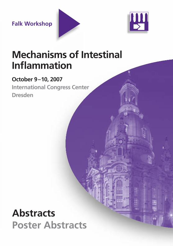
Mechanisms of IntestinalInflammationOctober 9–10, 2007International Congress CenterDresden

© 2007 Falk Foundation e.VAll rights reserved.
Abstracts of Invited Lectures Poster Abstracts Falk Workshop MECHANISMS OF INTESTINAL INFLAMMATION
Dresden (Germany) October 9–10, 2007 Scientific Organization: R.S. Blumberg, Boston (USA) M.F. Neurath, Mainz (Germany) S. Schreiber, Kiel (Germany) W. Strober, Bethesda (USA) M. Zeitz, Berlin (Germany)
3
CONTENTS Page Session I Genetic factors in IBD
Chair: J.P. Hugot, Paris D.P. Jewell, Oxford
New genetic defects in IBD and their relation to epithelial barrier function S. Schreiber, Kiel 15 – 16
Genetic polymorphisms in the IL-23R locus and susceptibility to IBD J. Cho, New Haven 17 – 18
Distinct defensin deficiencies in small intestinal and colonic Crohn’s disease J. Wehkamp, Stuttgart 19
The molecular basis of NOD2 susceptibility mutations in Crohn’s disease W. Strober, Bethesda 20 – 22
Session II Cytokine abnormalities underlying inflammatory bowel disease
Chair: P.J. Mannon, Bethesda A. Radbruch, Berlin
IL-12 family members in experimental colitis M.F. Neurath, Mainz 25
Cytokines mediating the induction and resolution of chronic colitis and the induction of colitis-associated fibrosis S. Fichtner-Feigl, Regensburg 26 – 27
RORγt and IL-17 responses D. Littman, New York 28
4
The role of IL-13 and the IL-13Rα2 in experimental and human ulcerative colitis I.J. Fuss, Bethesda 29
Session III Intestinal microflora and its role in IBD
Chair: C.O. Elson, Birmingham R.B. Sartor, Chapel Hill
Molecular analysis of the intestinal microflora in IBD G.W. Tannock, Dunedin 33
Identification of the predominant antigenic epitopes in intestinal flora in IBD R. Duchmann, Berlin 34
Intestinal microflora and immunoregulation M. Boirivant, Rome 35
TLR responses and their role in intestinal inflammation S. Rakoff-Nahoum, New Haven 36
Session IV Epithelial barrier function in inflammatory bowel disease
Chair: L. Mayer, New York D.K. Podolsky, Boston
Barrierprotective function of intestinal epithelial TLR2 E. Cario, Essen 39
Interleukin-13 and epithelial cell function J.D. Schulzke, Berlin 40
IKK/NF-κB signalling in intestinal epithelial cells controls immune homeostasis in the gut M. Pasparakis, Cologne 41
5
Transcription factor XBP1 regulates Paneth cell function and the inflammatory tone of the intestinal epithelium A. Kaser, A.-H. Lee, C. Lefebvre, J.N. Glickman, H. Tilg, E.E.S. Nieuwenhuis, D.E. Higgings, J.D. Rioux, L.H. Glimcher, R.S. Blumberg, Innsbruck, Hall/Tirol, Boston 42
Session V Regulatory defects in IBD
Chair: J. Hühn, Berlin T.T. MacDonald, London
TGF-β1 and Smad7 in the regulation of IBD G. Monteleone, Rome 45 – 46
Regulatory T cells induce CD4+CD25-Fox3- T cells or are self-induced to become Th17 cells in the absence of exogenous TGF-β L. Xu, A. Kitani, I. Fuss, W. Strober, Bethesda 47
SHP-1-dependent T cell inhibition by CEACAM1 isoforms expressing a long cytoplasmic tail domain T. Nagaishi, Z. Chen, L. Chen, L. Pao, A. Nakajima, H. Iijima, B. Neel, R.S. Blumberg, Boston 48
Immune regulation in the intestine: A balancing act between effector and regulatory T cells F. Powrie, J. Coombes, S. Hue, A. Izcue, K. Maloy, K. Siddiqui, Oxford 49
List of Speakers, Moderators and Scientific Organizers 51 – 53
7
Poster Abstracts 1. The influence of endotoxin and intestinal dysbiosis on cellular reactions in liver
S. Abdulkhakov, A.P. Kiassov, A.S. Sozinov (Kazan, R) 2. Pathogenic mechanisms of anemia of chronic disease in inflammatory bowel
disease D. Badea, M. Badea, A. Genunche-Dumitrescu (Craiova, RO)
3. Platelets abnormality in inflammatory bowel disease
M. Badea, D. Badea, A. Genunche-Dumitrescu (Craiova, RO) 4. Interferon gamma primes flagellin responses in gut epithelial cells through the
modulation of MyD88 expression C. Bannon, P. Davies, A. Collett, G. Warhurst (Manchester, Salford, GB)
5. The caspase-1 inhibitor pralnacasan and the PDE IV inhibitor rolipram exert
additive efficacy in CD45Rbhigh transfer colitis C. Bauer, N. Landauer, C. Mayer, M. Dauer, R. Kiefl, H.A. Lehr, S. Endres, A. Eigler (Munich, D; Lausanne, CH)
6. Epithelial NEMO deficiency disrupts the intestinal barrier and leads to severe
chronic intestinal inflammation in mice C. Becker, A. Nenci, A. Wullaert, R. Gareus, G. van Loo, S. Danese, M. Huth, A. Nikolaev, C. Neufert, B. Madison, D. Gumucio, M.F. Neurath, M. Pasparakis (Mainz, Cologne, D; Monterotondo, Milan, I; Ann Arbor, USA)
7. Bacterially-mediated activation of hypoxia inducible factor-1 (HIF-1) in intestinal
epithelial cells involves the deneddylation of cullin-2 G.O. Canny, A. Neish, H. Wu, L. Bry, S.P. Colgan (Lausanne, CH; Atlanta, Boston, Denver, USA)
8. Early epithelial responses that precede increased gut permeability during colitis
development in mdr1a(-/-) mice A. Collett, N.B. Higgs, G. Warhurst (Salford, Manchester, GB)
9. Immunohistochemical evaluation of Fhit protein expression in cases of
inflammatory bowel disease J. Czyzewska, K. Guzinska-Ustymowicz, A. Kemona, A. Pryczynicz, H. Kemona, A. Chetnik, R. Bandurski, M. Gryko, A. Kuklinski, D. Cepowicz, B. Kedra (Bialystok, PL)
10. Fecal calprotectin as a diagnostic and monitoring marker in Crohn's disease
P. Eder, K. Stawczyk, I. Krela-Kazmierczak, K. Linke (Poznan, PL) 11. Profibrotic IL-13 negatively regulates chronic inflammation during intestinal
fibrosis via glycogen synthase kinase-3beta S. Fichtner-Feigl, E. Geissler, H.-J. Schlitt (Regensburg, D)
8
12. Regulation and stability of the homing receptor integrin α4/β7 on CD4+ T cells S. Floess, B. Szilagyi, K. Schlawe, A. Hamann, J. Hühn (Berlin, D)
13. Upregulated expression of CD14, Toll-like receptor 2 (TLR2) amd TLR4 in
biopsy samples of patients with inflammatory bowel diseases L. Frolova, P. Drastich, P. Rossmann, K. Klimesova, H. Tlaskalova-Hogenova (Prague, CZ)
14. The serum level of cytokines (IL-1, IL-6) and TNF-alpha in patients with
inflammatory bowel disease A. Genunche-Dumitrescu, P. Mitrut, D. Badea, M. Badea (Craiova, RO)
15. Prostaglandin E receptor subtype EP1 (PTGER1): A new candidate gene for
IBD A. Geremia, J. Beckly, L. Hancock, F. Cummings, R. Cooney, S. Pathan, D.P. Jewell (Oxford, GB)
16. Whipple's disease: Diagnosis and long-term follow-up. 30-years experience
V. Gerova, S. Stoinov (Sofia, BG) 17. Role of tumor-associated macrophages in colorectal cancer
M.V. Gulubova, T. Vlaykova, Y. Yovchev, I. Vassilev, S. Stratiev (Stara Zagora, BG)
18. Prenatal development of interstitial cells of Cajal in human intestine
A.A. Gumerova, A.P. Kiassov, M.S. Kaligin (Kazan, R) 19. Influence of diet on the susceptibility to endotoxin shock
T. Hrncir, T. Hudcovic, H. Kozakova, R. Stepankova, M. Schwarzer, L. Frolova, M. Kverka, H. Tlaskalova-Hogenova (Novy Hradek, Prague, CZ)
20. Effects of probiotic strain Escherichia coli Nissle 1917 on the development of
intestinal inflammation induced in gnotobiotic models T. Hudcovic, R. Stepankova, H. Kozakova, T. Hrncir, M. Schwarzer, H. Tlaskalova-Hogenova (Novy Hradek, CZ)
21. Biological parameters in inflammatory bowel diseases in children
I. Ignys, N. Kobelska-Dubiel, H. Krauss, W. Cichy, M. Kobelski (Poznan, PL) 22. Adipocytes and preadipocytes - Phagocytes within the mesenteric fat?
J. Ihbe, A. Batra, R. Glauben, T. Stroh, I. Fedke, M. Zeitz, B. Siegmund (Berlin, D)
23. Cyclooxygenase-1 as well as cyclooxygenase-2 contributes anaphylaxis-
induced alterations in intestinal motility M. Kadowaki, T. Yamamoto, N. Kurokawa, M. Kadowaki (Toyama, Osaka, J)
24. Cholinergic anti-inflammatory pathway through α7-nicotinic acetylcholine
receptors in the colon reduces oxazolone-induced colitis in mouse M. Kadowaki, M. Yoshida, K. Fujiwara, T. Yamamoto (Toyama, J)
9
25. Pathogenesis and bacteriology in inflammatory bowel disease K. Kalinova (Stara Zagora, BG)
26. Immune modulation after oral antigen administration
K. Kapp, J. Maul, A. Wenzel, J. Preiss, P. Mundt, M. Zeitz, R. Duchmann (Berlin, D)
27. Inflammation-related colon cancer development in conventional and germ-free
mice K. Klimesova, P. Rossmann, M. Kverka, L. Frolova, H. Tlaskalova-Hogenova (Prague, CZ)
28. Tumor necrosis factor-alpha antibodies in Crohn's disease and investigation of
interleukin 10 V. Kupcová, Z. Zelinkova, L. Turecky, S. Ilavska, E. Jahnová (Bratislava, SK)
29. Peroral administration of mycobacterial HSP60 and HSP70 prevents severe
forms of DSS-induced intestinal inflammation in Balb/c mice M. Kverka, Z. Zákostelská, R. van der Zee, W. van Eden, H. Tlaskalova-Hogenova (Prague, CZ; Utrecht, NL)
30. TNBS-induced colitis models in SJL and BALB/c mice
T. Lagerweij, N. Worms, R. Verbeek, L. Nagelkerken (Leiden, NL) 31. Elucidating the pathogenic role of Th17 cells in an adoptive transfer model of
chronic intestinal inflammation M. Leppkes, C. Becker, S. Wirtz, C. Neufert, A. Nikolaev, M.F. Neurath (Mainz, D)
32. Role of transforming growth factor β1 (TGF-β1) gene polymorphism in chronic
inflammatory bowel diseases of the digestive system in children A. Liberek, J. Jakobkiewicz-Banecka, A. Kloska, J. Swiderska, A. Szlagatys-Sidorkiewicz, A. Marek, P. Landowski, K. Marek, G. Luczak, Z. Kmiec, G. Wegrzyn, M. Korzon (Gdansk, PL)
33. Transforming growth factor beta1 (TGF-beta1) and clinical course of chronic
inflammatory bowel disease (IBD) in children A. Liberek, A. Marek, A. Szlagatys-Sidorkiewicz, D. Kartanowicz, P. Landowski, G. Luczak, P. Wierzbicki, M. Stanislawowski, K. Marek, Z. Kmiec, M. Korzon (Gdansk, PL)
34. Histological and immunohistochemical analysis of vascular changes in
idiopathic bowel diseases P. Majewski, J. Zurawski, W. Salwa-Zurawska (Poznan, PL)
35. Behavior of Crohn's disease - Analysis according to Vienna classification and
the need for surgery M. Manuc, D. Manuc, C. Pop, C. Lamatic, C. Preda, L. Tugui, C. Vasilescu, M. Diculescu (Bucharest, RO)
10
36. Dextran sulphate sodium-induced colitis induces small intestinal changes in villus height and crypt depth that may be regulated by dipeptidyl peptidase activity S. Mashtoub, R. Yazbeck, C. Abbott, A. Wendland, M. Leach, R. Jennings, H.-U. Demuth, G.S. Howarth (Adelaide, AUS; Halle, D)
37. Decreased expression of CCR4 on CD4+CD25high regulatory T cells as a
possible mechanism for impaired migration to inflamed mucosa in Crohn's disease J. Maul, S. Pförtner, R. Geffers, K. Kapp, J. Buer, M. Zeitz, R. Duchmann (Berlin, Braunschweig, Hannover, D)
38. Clinical significance of anti-neutrophil cytoplasmic antibodies (ANCA) in Crohn's
disease with liver determination P. Mitrut, A. Genunche-Dumitrescu (Craiova, RO)
39. Visfatin - A novel member of the adipocytokine family is involved in the
immunopathogenesis of inflammatory bowel disease A.R. Moschen, A. Kaser, M. Theurl, B. Mosheimer, H. Niederegger, P.L. Moser, H. Tilg (Innsbruck, A)
40. Increased transmucosal uptake of E. coli in collagenous colitis is not reversed
by budesonide A. Münch, J.D. Söderholm, A. Öst, M. Ström (Linköping, Täby, S)
41. Increased colonic dendritic cells in acute colitis - Key mediators of inflammation
and immunity? S.C. Ng, S. Plamondon, H. Omar Al-Hassi, M.A. Kamm, S.C. Knight, A.J. Stagg (Middlesex, London, GB)
42. Enhanced IL-10 and IL-12/p40 production by intestinal dendritic cells in acute
ulcerative colitis S.C. Ng, S. Plamondon, M.A. Kamm, S.C. Knight, A.J. Stagg (Middlesex, London, GB)
43. Adipokine-dependent T cell polarization - Modulating intestinal inflammation
B. Okur, A. Batra, R. Glauben, T. Stroh, I. Fedke, M. Zeitz, B. Siegmund (Berlin, D)
44. Effects of ursodeoxycholic acid treatment on colon cancer cell proliferation
R. Peiro-Jordan, S. Subramanian, M.L. Hanski, J. Behrens, M. Zeitz, C. Hanski (Berlin, Erlangen, D)
45. Oxidative damage of plasma proteins and activity of antioxidative enzymes in
children with inflammatory bowel disease S. Popadiuk, A. Szlagatys-Sidorkiewicz, P. Landowski, J. Renke, B. Kaminska, M. Korzon, M. Wozniak (Gdansk, PL)
11
46. Lactobacillus plantarum precolonization of BALB/c mice stimulates the immune system, but doesn't protect against the inflammation induced by DSS treatment M. Schwarzer, B. Cukrowska, U. Witoslaw, T. Hudcovic, T. Hrncir, R. Stepankova, H. Tlaskalova-Hogenova, H. Kozakova (Novy Hradek, CZ; Warsaw, PL)
47. Stay with friends: Commensal microflora drives expansion of Foxp3+ Tregs in
gut-associated lymphoid tissue C. Siewert, S. Cording, M.M. Heimesaat, O. Liesenfeld, S. Bereswill, C. Loddenkemper, G. Loh, M. Blaut, A. Hamann, J. Hühn (Berlin, Potsdam-Rehbrücke, D)
48. The impact of HDAC inhibition on intestinal inflammation and its mechanistic
background E. Sonnenberg, R. Glauben, P. Mascagni, M. Zeitz, B. Siegmund (Berlin, D; Cinisello, I)
49. Expression and localization of vascular endothelial growth factor (VEGF) and its
receptor in patients with ulcerative colitis A. Stadnicki, D. Frysz-Naglak, E.l. Klimacka-Nawrot, C. Kruszniewska, U. Mazurek, I. Stadnicka, B. Fryz, M. Kajor (Sosnowiec, Katowice, PL)
50. Morphological characteristics of colitis developing in SCID mice reconstituted
with CD4+CD45RBhigh T cells and influence of intestinal bacteria R. Stepankova, F. Powrie, O. Kofronova, H. Kozakova, T. Hudcovic, T. Hrncir, H. Tlaskalova-Hogenova (Novy Hradek, Prague, CZ; Oxford, GB)
51. Glutathione peroxidase (Gpx2) and aquaporin 8 (Aqp8) as new markers for
colonic inflammation in experimental colitis and IBD: An important role for H2O2? A.A. te Velde, I. Pronk, F. de Kort, P.C.F. Stokkers (Amsterdam, NL)
52. Morphological features of ulcerative colitis in childhood
E.G. Tsimbalova, A.S. Potapov, L.L. Kapuller (Moscow, R) 53. Increased expression of VEGF and CD146 in patients with inflammatory bowel
disease G. Tsiolakidou, I.E. Koutroubakis, M. Tzardi, E.A. Kouroumalis (Heraklion, GR)
54. RAGE-/- mice are more susceptible to DSS-induced colitis
M.I. Verstege, M.A. van Zoelen, F.J. ten Kate, C. Verseyden, P.C. Stokkers, A.A. te Velde (Amsterdam, NL)
55. The role of TNF signaling in spontaneous colitis development in mice lacking
NEMO specifically in intestinal epithelial cell K. Vlantis, A. Wullaert, M. Pasparakis (Cologne, D)
56. Inflammatory infiltration in the invasive margin and tumor-infiltrating VEGF-
positive inflammatory cells may predict colorectal cancer outcome T. Vlaykova, M.V. Gulubova (Stara Zagora, BG)
12
57. Influence of cannabinoid 1 receptor agonist and antagonist on the development of stress-induced gastric ulcers Z. Warzecha, A. Dembinski, P. Ceranowicz, M. Dembinski, J. Cieszkowski, W.W. Pawlik, P. Konturek (Cracow, PL; Erlangen, D)
58. Ghrelin accelerates the healing of chronic experimental duodenal ulcers. Role
of growth hormone (GH) and IGF-1 Z. Warzecha, A. Dembinski, P. Ceranowicz, M. Dembinski, J. Cieszkowski, W.W. Pawlik, P. Konturek (Cracow, PL; Erlangen, D)
59. Enteral nutrition therapy modified serum growth factors concentrations in
children with inflammatory bowel diseases - Preliminary report A. Wedrychowicz, S. Pieczarkowski, K. Przybyszewska, U. Jedynak-Wasowicz, K. Fyderek (Cracow, PL)
60. Important role of the transcription factor NFATc2 in the pathogenesis of
ulcerative colitis B. Weigmann, R. Atreya, H.A. Lehr, P.R. Galle, M.F. Neurath (Mainz, D; Lausanne, CH)
61. Intestinal induction of CD8+Foxp3+ T cell to maintain homeostasis and regulate
tolerance and inflammation A.M. Westendorf, J. Buer (Braunschweig, D)
62. Depressiveness and mucosal proinflammatory cytokines are associated in
patients with ulcerative colitis and pouchitis R. Wilhelm-Schwenk, C. Schmidt, A. Stallmach, W. Häuser (Saarbrücken, Jena, D)
63. IL-27 contributes to protective colonic immune response in experimental colitis
S. Wirtz, I. Tubbe, C. Becker, B. Weigmann, H.A. Lehr, M. Birkenbach, P.R. Galle, R.S. Blumberg, M.F. Neurath (Mainz, D; Lausanne, CH; Norfolk, Boston, USA)
64. Role of the intestinal microflora in the development of colon inflammation in
mice lacking NEMO in intestinal epithelial cells A. Wullaert, K. Vlantis, M. Pasparakis (Cologne, D)
65. Inhibition of dipeptidyl peptidase activity increases circulating glucagon-like
peptide-2 and partially ameliorates neutrophil infiltration during experimental colitis in mice R. Yazbeck, G.S. Howarth, H.-U. Demuth, J.J. Holst, C.A. Abbott (Adelaide, AUS; Halle, D; Copenhagen, DK)
66. Anti-inflammatory effects of bacterial components tested in Raw 264.7 and
J774A.1 macrophage cell lines Z. Zakostelska, M. Kverka, D. Sokol, P. Jelen, R. van der Zee, W. van Eden, H. Tlaskalova-Hogenova (Prague, CZ; Utrecht, NL)
13
Session I
Genetic factors in IBD
15
New genetic defects in IBD and their relation to epithelial barrier function Stefan Schreiber Christian-Albrechts-University, Kiel Clustering of inflammatory bowel disease in large families and the observation of an increased concordance between monozygotic twins suggests heritable components in these disorders. The high concordance in monozygotic twins (> 55%), which is not seen in dizygotic twins (< 5%) points to strong contribution of genetic susceptibility to the overall risk for disease. IBD represents a “complex disease” and may involve a large number of interacting disease genes. Crohn’s disease has become an example for the successful molecular exploration of a polygenic etiology. Crohn’s disease was not known before 1920. Incidence has increased since now leading to a lifetime prevalence of up to 0.5% in Western industrialized countries. The current hypotheses propose unknown trigger factors in the life style of Western industrialized nations that interact with a polygenic susceptibility. It appears that increased expression and production of TNF and an enhanced state of activation of the NFκB system are main drivers of the mucosal inflammatory reaction. The exploration of inflammatory pathophysiology of Crohn’s disease using full genome, cDNA and oligonucleotide based arrays, respectively, has generated large sets of genes that are differentially expressed between inflamed mucosa and normal controls. While this may lead to new targets for a pathophysiology oriented therapy, it appears, however, that the dissection of the inflammatory pathophysiology does not allow to identify the multifactorial etiology of the disease. Genome-wide linkage analysis has demonstrated eight confirmed susceptibility regions with the one on chromosome 16 being most consistent between different populations. In 2001 three coding variations in the CARD 15 gene were identified that are highly associated with development of the disease. All variants affect a part of the gene that codes for the leucin rich part of the protein that appears to be involved in bacteria induced activation of NFkB in macrophages and epithelial cells. Interestingly, the three disease-associated SNPs are never found on the same haplotype. In compound heterozygotes or homozygotes they result in a RR of > 35 to develop Crohn’s disease as an adult. A particular subphenotype with localization of the disease in the ileocecal region is highly associated with the variants in the CARD 15 gene. Variations in the CARD 15 gene do not fully explain the linkage finding in the pericentromeric region of chromosome 16. After stratification for CARD 15 variants, the broad linkage peak is reduced to two more defined peaks on 16p and 16q, respectively. While the exploration of these regions has led to several association signals that are subject to further fine mapping a further disease gene progress has been greater in the other linkage regions (i. e. on chromosomes 10 and 5, respectively). DLG-5 is the example of a low-risk susceptibility gene with a modest associated odds ratio (1.2–1.5). Interestingly, the association signal appears to be confined to young males. SLC22A4/5 which encode the kation-transporters OCTN1 and 2 have been suggested to represent
16
the disease gene in the 200+ κB haplotype block on chromosome 5q31. MDR1 has also been implicated as a disease gene in IBD. Although the human association studies have resulted in highly controversial findings a knockout mouse with a colitis phenotype makes MDR1 likely as a low risk susceptibility gene. With the advent of high-density, genome wide association studies enormous progress has been made to discover the remaining disease genes. Recently a 330k Illumina scan has been published identifying IL-23R as a further disease gene. We used a genome wide candidate gene approach (with approx. 20.000 cSNPs) to identify ATG16L1 as a further disease gene. Both genes were confirmed and a further regulatory SNP involving PTGER4 was annotated by a Belgian genome wide scan. By the time of presentation three further genome wide SNP scans in Crohn’s disease will most likely have entered the public domain. The further exploration of Crohn’s disease (and other inflammatory conditions of barrier organs) will have to annotate genetic risk maps that are completed with amazing speed. With the limited possibilities for interpretation through in silico information it appears that epithelial cells are an important primary player in early pathogenic events. Disturbed bacterial defense but also an altered handover between innate and adaptive immunity are likely mechanisms by which genetic susceptibility translates into a dysregulated adaptive immunity. The creation of a medical systems biology of disease will lead to new models and eventually new therapies.
17
Genetic polymorphisms in the IL-23R locus and susceptibility to IBD Judy Cho, M.D. Yale University School of Medicine, New Haven, CT 06520, USA Genome-wide association studies represent a major new advance in identifying common genetic variation contributing to common, multigenic disorders such as IBD. In European ancestry cohorts, genome-wide association studies have identified a number of novel, well-established gene associations. In Crohn’s disease (CD), in addition to the NOD2 associations, the strongest association is observed in the interleukin 23 receptor subunit (IL23R) gene region on chromosome 1p31. Multiple independent signals throughout this region are observed, indicating that multiple susceptibility alleles reside in this region. One likely susceptibility allele is the Arg381Gln variant in the intracellular domain of IL23R, where protection against developing CD or ulcerative colitis (UC) is conferred by carriage of the less common glutamine allele. In addition, multiple, independent non-coding association signals are observed throughout the IL23R gene region. It may be speculated that these non-coding variants affect IL23R expression and/or alternative splicing. Some support for distinct IL23R association patterns between UC and CD has been reported. The IL-23 receptor complex is comprised of the IL23R gene and IL12RB1 (chromosome 19p13), with the latter being a receptor subunit common to the functional IL-12 receptor. The IL-23 cytokine is comprised of p19 and p40 subunits. Modest evidence for CD association has been reported in the p40 gene region (chromosome 5q33) in a large CD cohort. The functional IL-12 receptor is comprised of IL12RB1 and IL12RB2, with the latter gene being located immediately centromeric to the IL23R gene on chromosome 1p31. The regulated expression of IL-12 and IL23 receptor complexes contributes to the functional expression of Th1 and Th17 CD4+ subsets, respectively. In humans, differentiation of naïve CD4+ cells occurs in response to activation in the presence of IL-1 and IL-6. IL-23 appears to be dispensable for the initial differentiation of Th17 cells, but likely plays a crucial role in their perpetuation. In multiple murine models of IBD, an intact IL-23 pathway is required for full expression of intestinal inflammation, suggesting that IL23R protective alleles are associated with decreased IL-23 pathway function compared to IL23R risk allele carriage. However, comparative studies of IL23R region susceptibility alleles have yet to be reported. Interestingly, similar associations in IL23R and p40 have been reported in psoriasis, and may partially account for the association between psoriasis and IBD. In contrast, no association of IL23R variants has been observed in rheumatoid arthritis or systemic lupus erythematosis, despite the role of the IL-23 pathway in murine models of these disorders. While the IL23R gene associations have been definitively observed and replicated in European ancestry IBD, no association has been observed Asian IBD, highlighting the potentially distinct mechanisms of disease pathogenesis between various population cohorts.
18
In addition to the modest CD association in the p40 region, there is some support for the concept that multiple genes along the IL-23 pathway may similarly be associated in IBD. A large U.K. CD cohort demonstrated significant evidence for association to PTPN2 (protein tyrosine phosphatase) and modest evidence for association to STAT3. STAT3 is a downstream signaling target of IL-23 signaling and PTPN2 may play a role in de-phosphorylating activated STAT3. PTPN2 has been significantly associated with Type I diabetes mellitus, further highlighting the genetic overlap between distinct inflammatory disorders. Taken together, an emerging model of IBD pathogenesis revolves around two major themes. First, multiple susceptibility alleles along the IL-23 pathway likely contribute to disease, with the IL23R susceptibility alleles playing a major role. These susceptibility alleles contribute in multiple inflammatory disorders in addition to IBD. The second major theme in IBD highlights the more unique contribution of altered host intracellular processing of intestinal bacteria, as highlighted by the CD associations to NOD2, ATG16L1 (autophagy gene) and IRGM (immunity-related GTPase family, M) genes.
19
Distinct defensin deficiencies in small intestinal and colonic Crohn’s disease J. Wehkamp Dr. Margarete Fischer-Bosch Institut für Klinische Pharmakologie, Stuttgart, Germany Different clinical localizations of Crohn's disease are associated with different deficiencies in epithelial and leukocyte antimicrobial peptide expression. As compared with ulcerative colitis, Crohn's disease of the colon is characterized by an impaired induction of beta defensins, and antimicrobial antiproteases elafin and SLPI, as well as the cathelicidin LL37. The attenuated induction of beta defensins is linked to fewer gene copy numbers in this locus, which is associated with colonic but not ileal Crohn's disease. In contrast, ileal Crohn's disease patients are characterized by a reduced antibacterial activity and a specific reduction of ileal Paneth cell defensins. This decrease is independent of the grade of histological inflammation and cannot be found in inflammation controls. Thus, some of these defects can be explained either by direct or indirect genetic mechanisms and appear to be primary. Unlike ulcerative colitis, ileal and colonic Crohn's disease are characterized by localized deficiencies of antibacterial peptides. Understanding the precise molecular mechanisms of the defective antibacterial barrier function might provide new therapeutic directions.
20
The molecular basis of NOD2 susceptibility mutations in Crohn’s disease Warren Strober Mucosal Immunity Section, Laboratory of Host Defenses, NIAID, NIH, Bethesda, USA The discovery a half-decade ago that some 15% of patients with Crohn’s disease bear a homozygous or compound heterozygous mutation in the gene that encodes NOD2 (the CARD15 gene) has opened a new and unquestionably important window on the pathogenesis of this disease (1, 2). If we can understand how this mutation creates susceptibility in some patients with Crohn’s disease we can establish an invaluable paradigm for the causation of disease in all patients. NOD2 an intra-cytoplasmic member of the family of proteins now known as the NLR proteins (3). These proteins are usually composed of a central NOD domain (nucleotide oligomerization domain) flanked on its C-terminal side by a LRR domain (leucine-rich repeat domain) that is capable of recognizing microbial components and on its N-terminal side by a CARD or pyrin domain that interacts with downstream molecules to bring about effector function. NOD2 has been shown to recognize muramyl dipeptide (MDP), a component of peptidoglycan (PGN), the latter itself a component of the bacterial wall of virtually all bacteria. Upon interaction with its ligand, MDP, NOD2 is thought to undergo a conformational change that allows it to interact with a downstream adaptor molecule known as RICK (RIP2). RICK, in turn, induces the polyubiquitination of NEMO (Iκκγ) the key scaffolding protein of NF-κB and thus initiates NF-κB activation and its down-stream panoply of inflammatory cytokines, including IL-12 (4). On the one hand, the ability of NOD2 to recognize a more or less ubiquitous bacterial component positions this molecule to mediate an inflammatory response and thus play a role in the induction of Crohn’s inflammation. On the other hand, if a mutation in NOD2 results in loss of the ability of NOD2 to activate NF-κB, (as has in fact been shown) then the mutation would be expected to lead to decreased NF-κB activation and decreased inflammation rather then increased NF-κB activation and inflammation that in fact characterizes Crohn’s disease. A possible solution to this conundrum was provided several years ago with the demonstration that antigen-presenting cells (APCs) from NOD2-deficient mice exhibit increased IL-12p70 synthesis when stimulated by the molecule that give rise to MDP, peptidoglycan (acting through TLR2) and that addition of MDP to cultures of APCs from NOD2-intact mice led to decreased IL-12p70 responses (5). The logical conclusion from these findings was that NOD2 activation by MDP ordinarily results in down-modulation of responses to TLR2 ligands. This finding was tied to the NOD2 mutation in Crohn’s disease by studies showing that transfection of NOD2-deficient APCs with a wild-type NOD2 plasmid led to correction of the IL-12p70 response whereas transfection of the same cells with a mutated NOD2 plasmid did not lead to such correction. In further buttressing the concept that NOD2 has a regulatory function with respect to TLR2 ligands, we established a colitis model NOD2 deficient mice based on the ability of a recombinant E.coli organism expressing OVA peptide (ECOVA organisms) to
21
induce inflammation in mice that have T cells that recognize and react to OVA peptide (6). Using this model we showed that NOD2-deficient mice but not NOD2 intact mice administered T cells that react to OVA peptide develop a transient but intense colitis when exposed to intra-rectal administration of ECOVA. In addition, this colitis was linked to the TLR2 response by the fact that mice deficient in both NOD2 and TLR2 no longer develop disease when exposed to ECOVA. The results obtained from this model led to the conclusion that NOD2 mutations lead to colitis because they establish a milieu characterized by an exuberant “innate” IL-12p70 response. However, inflammation does not occur unless a second defect is present that leads to increased reactivity with one or more antigens in the intestinal microflora. The concept that a major function of NOD2 is a negative regulatory function has not been universally accepted, in part because this concept must be reconciled with other studies showing that NOD2 has positive effects on cytokine/chemokine synthesis in under some activation conditions. To throw fresh light on this controversy we turned to the study of mice expressing increased amounts of NOD2, reasoning that if the negative regulatory function of NOD2 was a reality, it should be increased in such mice. In an initial set of studies we focused on mice that bear a NOD2 transgene under a MHC class II promoter so that within the hematopoietic cell compartment its expression is limited to APCs. We found that APCs from mice bearing the transgene and therefore over-expressing NOD2, but not litter-mate control mice, mount greatly reduced IL-12p70 responses when stimulated by peptidoglycan. Furthermore, APC responses of the transgenic mice to Pam3CysK4, a TLR2 ligand that does not contain MDP, was equavalent to that in litter-mate control mice, but such normal responses were subject to much more intense down-regulation by addition of MDP to the culture than seen in control mice cultures. Taking these findings to an in vivo arena we then went on to show that mice bearing a NOD2 transgene were almost totally resistant to the induction of peptidoglycan-induced colitis, an intense and usually fatal colitis occurring in certain mouse strains upon intra-rectal instillation of peptidoglycan. Similarly, the transgenic mice developed far less severe TNBS-colitis than their normal litter-mates when subject to intra-rectal instillation of TNBS. Thus, the reduced APC IL-12p70 response to peptidoglycan in vitro was translated into a increased resistance to induced colitis wholly or partially driven by peptidoglycan. In further studies we evaluated mice expressing increased NOD2 as a result of in vivo administration of plasmids encapsulated in a viral envelope that ensures excellent entry into cells in live mice. In this case, the NOD2 plasmids delivered in this manner gave rise to either a normal (unmutated NOD2 ) or a NOD2 bearing a mutation like that in Crohn’s disease. The striking finding here was the administration of the plasmid encoding intact NOD2 led to complete resistance to the induction of TNBS-colitis, whereas the administration of the plasmid encoding a mutated NOD2 had only a minor effect on the induction of TNBS-colitis. These studies offer striking confirmation of the original view that holds that NOD2 has negative regulatory function. In addition, they show for the first time that provision of NOD2 can protect from the development of colitis and may therefore be a novel way of treating Crohn’s disease.
22
References: 1. Hugot, J.P., M. Chamaillard, H. Zouali, S. Lesage, J.P. Cezard, J. Belaiche,
S. Almer, C. Tysk, C.A. O'Morain, M. Gassull, V. Binder, Y. Finkel, A. Cortot, R. Modigliani, P. Laurent-Puig, C. Gower-Rousseau, J. Macry, J.F. Colombel, M. Sahbatou, and G. Thomas. 2001. Association of NOD2 leucine-rich repeat variants with susceptibility to Crohn's disease. Nature 411: 599–603.
2. Ogura, Y., D.K. Bonen, N. Inohara, D.L. Nicolae, F.F. Chen, R. Ramos,
H. Britton, T. Moran, R. Karaliuskas, R.H. Duerr, J.P. Achkar, S.R. Brant, T.M. Bayless, B.S. Kirschner, S.B. Hanauer, G. Nunez, and J.H. Cho. 2001. A frameshift mutation in NOD2 associated with susceptibility to Crohn's disease. Nature 411: 603–606.
3. Strober, W., P.J. Murray, A. Kitani, and T. Watanabe. 2006. Signalling pathways and
molecular interactions of NOD1 and NOD2. Nat Rev Immunol 6: 9–20. 4. Abbott, D.W., A. Wilkins, J.M. Asara, and L.C. Cantley. 2004. The Crohn's disease
protein, NOD2, requires RIP2 in order to induce ubiquitinylation of a novel site on NEMO. Curr Biol 14: 2217–2227.
5. Watanabe, T., A. Kitani, P.J. Murray, and W. Strober. 2004. NOD2 is a negative
regulator of Toll-like receptor 2-mediated T helper type 1 responses. Nat Immunol 5: 800–808.
6. Watanabe, T., A. Kitani, P.J.Murray, Y. Wakatsuki, I.J. Fuss, and W. Strober. 2006.
Nucleotide binding oligomerization domain 2 deficiency leads to dysregulated TLR2 signaling and induction of antigen-specific colitis. Immunity 25: 473–485.
23
Session II
Cytokine abnormalities underlying inflammatory bowel disease
25
IL-12 family members in experimental colitis Prof. Dr. Markus F. Neurath I. Medical Clinic, University of Mainz, Germany Inflammatory bowel diseases (IBD: Crohn’s disease and ulcerative colitis) are relapsing inflammations of the gastrointestinal tract not due to specific pathogens. Although the precise etiology of the diseases is still unknown, recent data from animal model strongly suggest that predisposing genetic factors, barrier defects and bacterial antigens lead to an unbalanced activation of the mucosal immune system that in turn causes chronic intestinal inflammation. There has been a growing interest in understanding the role of cytokines and cytokine signaling events in IBD models in recent years. T-bet and STAT4 expressing Th1 cells, GATA-3 expressing T cells producing IL-13 and THIL-17 cells are key effector cell populations with major relevance for the design of novel therapeutic approaches for IBD. Furthermore, IL-12 family cytokines such as IL-23 and IL-27 appear to play a prominent role in modulating the activity of effector T cells in experimental colitis. The role of IL-23 is underlined by the recent findings on IL-23R mutations in IBD patients. Finally, various proinflammatory cytokines and transcription factors in the gut have been shown to regulate the development and progression of colitis associated colon cancer in murine models. These data provide a rationale for selective targeting of cytokines and transcription factors in IBD. Such targeting has the potential advantage of targeting the activity of various cell types simultaneously rather than of a single cell type. In any case, the findings in animal models of chronic intestinal inflammation have provided new insights into the pathogenesis of IBD and are important for the development of novel immunotherapeutic approaches.
26
Cytokines mediating the induction and resolution of chronic colitis and the induction of colitis-associated fibrosis S. Fichtner-Feigl Chirurgische Universitätsklinik Regensburg, Germany To investigate the immunologic events underlying the evolution of a chronic colitis we analyzed the colitis occurring in BALB/c mice administered weekly doses of intra-rectal trinitrobenzene sulfonic acid (TNBS). Mice treated in this way initially develop intense colitis associated with severe weight loss and considerable mortality. However, about three weeks after the initiation of TNBS-colitis, the colitis moderates and, while the mice do not exhibit the weight gain of control mice, they regain their lost weight. This period of moderate colitis lasts about four weeks and then, about seven weeks after initiation of the TNBS-colitis, the colitis gradually subsides and is all but gone at 10–12 weeks after initiation of the TNBS-colitis (despite the continued administration of weekly TNBS). Importantly, the termination phase of this inflammatory cycle is accompanied by the development of fibrotic cycle. Thus, four-five weeks after initiation of TNBS-colitis, the mice develop steadily increasing fibrosis of the colonic lamina propria that persists through the period of subsiding inflammation. To understand the immunologic basis of this complex series of events we determined the cytokines produced by lamina propria cells during the various stages of the inflammation. We found that the initial intense inflammation was driven by a Th1 response characterized by the production of IL-12p70 and IFN-γ. This cytokine response subsided after three weeks and corresponding to the onset of a more moderate inflammation and was then replaced by a gradually increasing IL-23/IL-25 response accompanied, after one or two weeks, by the appearance of IL-17. The Th17 response thus formed plateaued at 7–9 weeks and then declined in concert with the subsidence of the inflammation and the appearance of IL-10. In further studies we showed that the appearance of the IL-17 response was accompanied by cytokines normally seen during a Th2 response, particularly IL-13. Production of this latter cytokine rose steadily to a plateau level at 8–9 weeks and then persisted at this level even when the inflammation was subsiding. In vitro stimulation studies suggested that such IL-13 production was dependent on IL-23 and IL-25, but not on IL-12p70. We then showed that IL-13 production results in the induction of a novel IL-13 receptor formerly thought to function only as a decoy receptor, IL-13Rα2, and that this receptor was critical to the production of TGF-β1 and the onset of fibrosis. Thus, if IL-13 signaling through this receptor is blocked by administration of soluble IL-13Rα2-Fc, or by administration of IL-13Rα2-specific siRNA, TGF-β1 is not produced and fibrosis does not occur. It should be noted, however, that while inhibition of IL-13 signaling did not affect the inflammation or the production Th17 cytokines during the phase of moderate inflammation mediated by these cytokines, it did affect the phase of gradual subsidence of inflammation: if IL-13 signaling is blocked moderate inflammation continues.
27
In a final series of studies, we therefore addressed the mechanism by which IL-13 production leads to control of Th17 inflammation in this model. Recent studies have shown that TLR signaling, a process necessary for the maintenance of mucosal inflammation is regulated by glycogen synthase kinase 3 beta (GSK3β). This kinase, when present in an active (unphosphorylated) state, facilitates TLR-mediated NF-κB activation and inflammatory cytokine production; at the same time, active GSK3β inhibits anti-inflammatory IL-10 production through down-regulation of CREB. On the other hand, this pattern is reversed when GSK3β is present in an inactive (phosphorylated) state. To prove that GSK3β is involved in the regulation of inflammation in chronic TNBS colitis we showed that inhibition of GSK3β phosphorylation by the synthetic inhibitor SB216763 and therefore activation of GSK3β, prevented subsidence of inflammation in the terminal phase of chronic TNBS-colitis; in addition, such inhibition leads to down-regulation of IL-10 production that accompanies such subsidence. Finally, we showed that IL-13 induces GSK3β phosphorylation and that blockade of IL-13 signaling results in maintenance of GSK3β in an active, unphosphorylated state. In summary, these findings suggest that chronic inflammation is orchestrated by a succession of cytokines that ultimately result in IL-13 production and subsequent resolution of the inflammation and the induction of fibrosis.
28
RORγt and IL-17 responses Dan R. Littman Howard Hughes Medical Institute and the Skirball Institute of Biomolecular Medicine, New York University School of Medicine, New York, NY 10016, USA T helper cells differentiate into lineages with distinct effector functions in response to the diverse cytokines induced following infection or tissue damage. In addition to Th1 and Th2 cells, T helper cells that secrete IL-17, IL-22, and other pro-inflammatory cytokines (Th17 cells) were recently described. Cells of this lineage have key roles in mouse models of autoimmunity, and they are induced by a combination of TGF-β and IL-6, while their maintenance and expansion requires IL-23. Induction of Th17 cells is dependent on the orphan nuclear receptor RORγt, which is expressed in response to either TGF-β or IL-6. We have found that Foxp3, which is also induced upon treatment with TGF-β alone and, to a lesser extent, by a combination of TGF-β and IL-6, represses RORγt-induced expression of IL-17, and this involves a direct interaction of the two transcription factors. The decision of a naïve T helper cell to differentiate into a Foxp3+ regulatory T cell versus a Th17 cell thus appears to rely, at least in part, on the balance of Foxp3 and RORγt expression. IL-6 treatment also results in the induction of IL-21 and IL-23R. As a consequence, IL-21 and IL-23 can also inhibit Foxp3 gene expression while synergizing with TGF-β to elevate the level of IL-17. Following its induction in response to IL-6 in antigen-stimulated naïve CD4+ T cells, IL-21 signals through its receptor to induce expression of more IL-21, and thus functions in a positive regulatory autocrine loop. Induction of IL-21 mRNA in response to either IL-6 or IL-21 requires activation of STAT3, but is independent of RORγt. However, induction of IL-23R in response to IL-6 occurs only if IL-21R signaling is intact and if both STAT3 and RORγt are present. Adjuvant immunization in mice lacking IL-21R results in compromised Th17 responses. The IL-21 and IL-23R-dependent induction of IL-17 is also dependent on the presence of RORγt. This nuclear receptor thus regulates a large part of the differentiation program of Th17 cells in response to diverse cytokine receptor signals. T cells expressing RORγt and IL-17 have been found constitutively only at mucosal surfaces. Lack of RORγt rendered T cells defective in induction of inflammatory bowel disease and other mouse models of autoimmunity. Together, our results suggest that RORγt may be an attractive therapeutic target in autoimmune diseases.
29
The role of IL-13 and the IL-13Rα2 in experimental and human ulcerative colitis I.J. Fuss Mucosal Immunity Section, Laboratory of Clinical Investigation, National Institutes of Health, Bethesda, MD, USA In the past few years, a great deal of insight has been obtained in the pathogenesis of ulcerative colitis. In prior studies of the animal model oxazolone colitis, a colitis model that shares some features of ulcerative colitis, it was demonstrated that the occurrence of inflammation was dependent upon invariant NK T cells, which secreted increased amounts of IL-13. In related human studies, it was subsequently found that UC patients also produce significantly greater amounts of IL-13 from LPMC than CD or normal control patient populations. It was observed, however, that this cytokine secretion profile arose from natural killer (NK) T cells which did not bear an invariant TCR. Most importantly, these non-invariant NK T cells were found to be directly cytotoxic for a HT-29 epithelial cell line. Thus, these studies provided a possible basis for the pathogenic potential of IL-13 and NK T cells in the induction of inflammation observed in experimental models and that of human ulcerative colitis. More recently, the pathways involved in IL-13 signaling have been further elucidated. It has been demonstrated that a receptor formerly thought to function only as a decoy receptor, IL-13Rα2, can indeed lead to activation of downstream inflammatory transcription factors. Given these results, the present studies wished to determine the relationship of this receptor to the occurrence of inflammation in oxazolone and human ulcerative colitis. In initial studies, it was found that UC patients express a higher percentage of peripheral blood T cells that bear the dual markers for CD161 (NK T) and IL-13Rα2 as compared to Crohn’s disease or normal control patient population. In correlative studies of the lamina propria, an increased expression of the IL-13Rα2 receptor was found on cytospin preparations from LPMC of UC patients as compared to other patient populations. The functionality of this receptor was demonstrated in depletion studies in which cells bearing this receptor were eliminated by the use of exotoxin coupled to a highly affinity binding IL-13Rα2 molecule. Polyclonal stimulation of cells after treatment with the former IL-13 exotoxin demonstrated a marked reduction in IL-13 secretion. To examine the pathogenicity of the IL-13Rα2 bearing cells these studies were extended to the oxazolone model of colitis. Treatment of mice in vivo with the IL-13 exotoxin led to both amelioration of intestinal inflammation and decreased IL-13 secretion. These studies therefore provide evidence that a highly selective population of NK T cells bearing the IL-13Rα2 receptor can be responsible for the production of IL-13 and may underlie the pathogenesis of experimental and human ulcerative colitis.
31
Session III
Intestinal microflora and its role in IBD
33
Molecular analysis of the intestinal microflora in IBD Gerald W. Tannock University of Otago School of Medicine, Dunedin, New Zealand Numerous microbial populations, mostly bacterial, that interact to form a community of considerable biomass, biodiversity and stability, inhabit the large bowel. The microbial community (microflora/microbiota) digests complex polymers derived from the host’s food (dietary fibre) and from mucus. Fermentation of the hydrolysis products produces short chain fatty acids, gases, phenols, indoles and amines as major products. The bowel community, whole or in part, may act as a surrogate pathogen in IBD. Continuous challenge of the mucosal immune system by microbial antigens as a result of abnormal epithelial permeability may cause the chronic immune inflammation observed in IBD. Nucleic acid-based (molecular) analytical methods have been used to monitor the composition of the microbial community. This has been necessary because most of its members have not yet been cultivated under laboratory conditions. Although these methods have assisted in defining and comparing microbial communities in general terms, the phylogenetic information is coarse (broad microbial groups) and outcomes of investigations are confounded by inter-individual and perhaps international differences in bowel communities, a polluted phylogenetic database, and biases inherent in sampling (subjects and specimens) and analytical procedures. It may be more useful to first identify the microbial antigens that drive the chronic immune inflammatory conditions characteristic of IBD rather than to continue phylogenetic comparisons of the composition of bowel communities. Community genome analysis (metagenomics) coupled with culture-based studies could form the basis of these future investigations.
34
Identification of the predominant antigenic epitopes in intestinal flora in IBD R. Duchmann Medizinische Klinik I, Charité – Universitätsmedizin, Campus Benjamin Franklin, Berlin, Germany It has been shown that the normal intestinal flora is necessary to develop intestinal inflammation in animal models. However due to the complexity of the intestinal flora it has been difficult to design experimental approaches to investigate the potential stimulatory bacterial antigen(s) involved. In humans, several studies indicated a potential association of E. coli with IBD. In addition, we have shown that T cell clones of IBD patients cross react towards different enteric bacterial species and thus likely respond to conserved bacterial antigens. We therefore hypothesized that highly conserved E. coli proteins might be a reasonable candidate to screen for abnormal T cell responses in IBD. The most conserved protein functions are represented in all three biological kingdoms, Archaea, Prokarya, and Eukarya. Thus, we first chose a set of E. coli proteins hypothetically inherited by the Last Universal Common Ancestor (LUCA) of the three kingdoms. As a second set we identified additional conserved proteins between E. coli and humans that were not included in the LUCA set of proteins. In general, these highly conserved proteins are not represented in Archaea and therefore not included in the LUCA protein. These proteins are referred to as E. coli-Human-Homologues (ECHH). We then used high-throughput techniques for cloning, expression and purification under native conditions of a set of 271 ECHH and LUCA proteins represented in E. coli suitable for downstream cellular immunological assays.
35
Intestinal microflora and immunoregulation Monica Boirivant Immune-mediated Disease Section, Department of Infectious, Parasitic and Immune-mediated Diseases, Istituto Superiore di Sanità, Rome, Italy The commensal organisms of the gut microflora, separated from the mucosal lymphoid tissue by the epithelial cell layer play an important role in host defense by inhibiting the colonization of the mucosal surface by pathogenic organisms. However, if and when they penetrate the epithelial cell layer they are themselves able to cause inflammation. It is important to note that epithelial barrier function is not so efficient that it excludes any exposure of mucosal immune elements to antigens in the commensal microflora. Subversion of epithelial barrier function may, at least in part, be necessitated by the need of the mucosal system to develop immunological tolerance toward antigens in the commensal microflora. We investigated this hypothesis by asking if a transient increase in mucosal permeability induced in SJL/J mice by exposure to rectal administration of ethanol or an agent (AT1002) that specifically affects tight junction integrity would have any effect in the generation of regulatory cells which in turn are able to influence the occurrence and severity of a subsequent TNBS-colitis. We found that both types of treatment, while itself inducing a mild and self-limited inflammatory response, led to a state of resistance to the induction of TNBS-colitis. In studies addressing the mechanism of this resistance, we found that the transient disruption of barrier function led to the appearance of CD4+CD25+Foxp3+ regulatory cells as well as a population of CD4+ cells expressing a latent form of TGF-β (Latency-Associated Peptide [LAP]). Development of these cells was strictly dependent on the presence of an intact commensal microflora and protection was dependent on the presence of these cells. Thus, increased but limited exposure of the mucosal immune system to the commensal microflora leads to increased elaboration of regulatory T cells and the latter then render the organism resistant to colitis. By extension, it seems likely that limited exposure of the mucosal immune system to the microflora is an important mechanism of tolerance induction and gut homeostasis.
36
TLR responses and their role in intestinal inflammation S. Rakoff-Nahoum Yale Univesity School of Medicine, Section of Immunobiology, New Haven, CT, USA Our studies have focused on understanding the mechanisms of interactions between the indigenous intestinal flora interacts and the mammalian host in both physiological and non-physiological conditions. In particular, we have focused on the role of innate microbial pattern recognition by toll-like receptors. Toll-like receptors (TLRs) play a crucial role in host defense against microbial infection. The microbial ligands recognized by TLRs are not unique to pathogens, however, and are produced by both pathogenic and commensal microorganisms. In the course of our studies we have found that commensal bacteria are recognized by TLRs under normal steady-state conditions and this interaction plays a crucial role in the maintenance of intestinal epithelial homeostasis. Furthermore, we have found that activation of TLRs by commensal microflora is critical for the protection against gut injury and associated mortality. These findings have revealed a novel function of TLRs-control of intestinal epithelial homeostasis and protection from injury-and provide a new perspective on the evolution of host-microbial interactions. We have extended these observations regarding the role of TLR signaling in intestinal inflammatory signaling and tissue repair to the study of tumorigenesis. We have found that the signaling through the adaptor protein MyD88 has a critical role in spontaneous tumor development in mice with heterozygous mutation in the adenomatous polyposis coli (APC) gene. In addition, MyD88-dependent signaling controls the expression of several key modifier genes of intestinal tumorigenesis and has a critical role in both spontaneous and carcinogen-induced tumor development. Inflammatory bowel disease (IBD) is thought to result from a dysregulated interaction between the host immune system and its commensal microflora. Heterogeneity of disease susceptibility in humans and rodents suggest that multiple mechanisms are responsible for the etiology of IBD. In particular, deficiencies in anti-inflammatory and immune-suppressive mechanisms play an important role in the development of IBD. However, it is unknown how the indigenous microflora stimulates the immune system and how this response is regulated. To address these questions, we investigated the role of Toll-like receptor (TLR) signaling in the development of spontaneous, commensal-dependent colitis in interleukin (IL)-2- and IL-10-deficient mice. We report that colitis was dependent on TLR signaling in IL-10-/- mice. In contrast, IL-2-/- mice developed intestinal inflammation in the absence of TLR signaling pathways. These results demonstrate a differential role of innate immune recognition by TLRs in the development of commensal-dependent colitis.
37
Session IV Epithelial barrier function in
inflammatory bowel disease
39
Barrierprotective function of intestinal epithelial TLR2 Priv.-Doz. Dr. med. Elke Cario Universitätsklinikum Essen, Klinik für Gastroenterologie & Hepatologie, Essen, Germany, E-mail: [email protected] The intestinal epithelium serves as an essential defensive barrier of the mucosal immune system that forms a bipolar interface between the diverse populations of microbes of the lumen and subjacent immune cells present in the lamina propria. The intestinal epithelial cell (IEC) barrier plays an important role in maintaining mucosal immune homeostasis. Dysregulated IEC barrier function appears to trigger and perpetuate inflammation in IBD. IEC maintain close contact with each other through the formation of tight junctions (TJ). Alterations in TJs have been attributed to the increased intestinal permeability seen in IBD. Commensal bacteria may modulate key epithelial cell functions that help maintain TJ-associated intestinal epithelial barrier integrity against injury. Toll-like receptors (TLR) represent a class of transmembrane pattern recognition receptors – essential for microbial recognition and control of immune responses. TLR2, a member of the TLR family, which is expressed by IEC, recognizes conserved molecular patterns associated with both Gram-negative and Gram-positive commensals, including lipopeptides/-proteins, peptidoglycan, lipoteichoic acid and zymosan. We have recently demonstrated that TLR2 deficiency predisposes to stress-induced injury of TJ-modulated barrier function leading to perpetuation of mucosal inflammation and apoptosis. However, oral treatment of colitis with a synthetic TLR2 ligand significantly suppresses mucosal inflammation by efficiently protecting TJ-associated integrity of the intestinal epithelium in vivo. TLR2-induced TJ modulation strongly interrelates with promotion of intestinal epithelial cell survival through the PI3K/Akt pathway. TLR2 activation directly enhances transepithelial resistance through TJ redistribution via protein kinase C in an in-vitro IEC model. In conclusion, cell-specific targeting of TLR2 could possibly help in the design of novel adjuvant therapeutic means to enhance intestinal epithelial barrier function to protect the underlying mucosa.
40
Interleukin-13 and epithelial cell function J.D. Schulzke Charité Centre 10, Campus Benjamin Franklin, Charité, D-12200 Berlin, Germany In inflammatory bowel disease (IBD), intestinal barrier function is seriously impaired. This is especially the case for ulcerative colitis (UC), which is characterized by a Th2 immune response with interleukin-4 and -13 as important pro-inflammatory effector cytokines. So far, Th2 cytokines were not directly identified as epithelial barrier affecting principles. Thus, we aimed to characterize Th2-cytokine influences on the large intestinal epithelium. For this purpose, lamina propria mononuclear cells (LPMC) were stimulated and IL-13 measured by ELISA. LPMC from ulcerative colitis patients produced large amounts of IL-13, much more than from controls or CD patients. IL-13/IL-4 receptors were analyzed by RT-PCR and immunofluorescence and IL-13Rα1 and IL-4Rα receptors were found to be present in HT-29/B6 cells and colonic epithelial cells of control and UC patients. Functional IL-13 and IL-4 effects were studied on HT-29/B6 colonic epithelial cells in Ussing-chambers and by the conductance scanning-technique. IL-13 had a dose-dependent effect on transepithelial resistance of HT-29/B6 monolayers, while IL-4 had no effect. This was due to an increased number of apoptotic cells by a factor of 5.6 as detected by TUNEL assays and a threefold increased expression of the pore-forming tight junction protein claudin-2 in Western blots combined with immunofluorescence confocal laser scanning microscopy (LSM) to detect tight junction proteins. Apoptosis and the increase in claudin-2 expression equally contributed to the change in epithelial barrier function. Furthermore, epithelial restitution velocity after scraping off part of the monolayer was shown to be decreased by 30% after IL-13 treatment. Finally, mucosal biopsies from UC patients were compared to cultured cells for these features and parallel changes were observed in human samples with a pronounced increase in claudin-2 expression. In conclusion, interleukin-13 could be identified as important effector cytokine in the Th2-cytokine response of ulcerative colitis which can hamper epithelial barrier function by stimulating epithelial apoptotic rate, affecting tight junction protein expression regulation and retarding epithelial restitution velocity.
41
IKK/NF-κB signalling in intestinal epithelial cells controls immune homeostasis in the gut Manolis Pasparakis Institute for Genetics, University of Cologne, Zülpicherstr. 47, D-50674 Cologne, Germany Deregulation of immune responses in the gut causes inflammatory bowel disease (IBD). The gut epithelium has an important function in the maintenance of intestinal immune homeostasis, by preventing the contact of luminal bacteria with immune cells through the formation of a physical barrier and the expression of antimicrobial peptides. The transcription factor NF-κB has been implicated in the regulation of this function of intestinal epithelial cells (IECs). NF-κB activation is mediated by the IκB kinase (IKK) complex, consisting of the IKK1 and IKK2 catalytic subunits and the NEMO/IKKγ regulatory protein. We employ conditional targeting of IKK subunits to investigate the role of IKK/NF-κB signalling in the gut. We showed that efficient inhibition of NF-κB in IECs, achieved by IEC-specific deletion either of NEMO or of both IKK1 and IKK2, caused severe chronic intestinal inflammation in mice. IEC-specific deletion of either IKK1 or IKK2 alone did not cause colitis showing that the two IκB kinases share a redundant function in IECs that is critical to protect the gut from chronic inflammation. These findings demonstrated that a primary NF-κB signalling defect in intestinal epithelial cells disrupts immune homeostasis in the gastrointestinal tract causing an IBD-like phenotype. Mice with IEC-restricted NEMO deficiency (NEMOIEC-KO) showed increased apoptosis of IECs, disruption of the epithelial barrier and subsequent translocation of bacteria into the mucosa. Concurrently, a chronic inflammatory response developed in the colon of these mice, initially dominated by innate immune cells but later also involving T lymphocytes. Genetic deficiency of MyD88, an essential adapter for TLR-induced signalling, prevented the development of colon inflammation in NEMOIEC-KO mice, suggesting that TLR-dependent interaction of bacteria with innate immune cells is important for the development of inflammation in these mice. Moreover, TNF receptor l deficiency inhibited the development of colonic inflammation in NEMOIEC-KO mice, arguing that TNF signalling plays a critical role in disease development. In order to evaluate the causative role of the microflora in this novel mouse model of IBD, we are raising NEMOIEC-KO mice in a germ-free environment and are also evaluating the therapeutic potential of antibiotic treatment. Also, current experiments aim to dissect the cell-specific function of TLR and TNFRI signalling, using reciprocal bone marrow transfer and/or conditional targeting of distinct mediators of these signalling cascades such as MyD88, FADD and TRADD. Finally, to address the role of T lymphocytes, NEMOIEC-KO mice are bred into a RAG1-deficient background.
42
Transcription factor XBP1 regulates Paneth cell function and the inflammatory tone of the intestinal epithelium Arthur Kaser*, Ann-Hwee Lee, Céline Lefebvre, Jonathan N. Glickman, Herbert Tilg, Edward E.S. Nieuwenhuis, Darren E. Higgins, John D. Rioux, Laurie H. Glimcher, Richard S. Blumberg *Universitätsklinik Innsbruck, Klinische Abteilung für Gastroenterologie und Hepatologie, Innsbruck, Austria Background: A single layer of intestinal epithelial cells (IEC) is the structure in immediate contact with the commensal microbiota and provides an immunologically functional barrier between luminal microbes and the hematopoietic system. Paneth cells (PC), at the crypt base, contain anti-bacterial peptides, α-defensins (cryptdins). A subset of CD is linked to mutations in NOD2/CARD15, in association with reduced expression of α-defensins. The unfolded protein response (UPR) is activated upon stress in the endoplasmic reticulum (ER), and is required for efficient protein production in highly secretory cells. Methods: We hypothesized that ER stress regulates PC and IEC function. To address this, we generated two IEC specific (using Villin-Cre) knock-out mouse models of X box binding protein-1 (XBP1), a key transcription factor of the ER stress response. Results: Constitutive genetic deletion of XBP1 in IEC led to development of spontaneous enteritis with features characteristic of IBD, including crypt abscesses. IEC from these mice showed increased grp78 expression, a marker of ER stress. XBP1 deletion resulted in the absence of PC, associated with a virtual absence of cryptdin and lysozyme mRNA and protein expression, and decreased antimicrobial activity as demonstrated by 2-log higher Listeria monocytogenes c.f.u. in the faeces of XBP1-/- mice upon oral infection. Using a second conditional IEC knock-out mouse of XBP1 with Cre activation by tamoxifen, we demonstrated apoptotic death of PC as the underlying mechanism for the absence of PC. ER stress results in increased JNK phosphorylation, due to the interaction of IRE1, the endoribonuclease activating XBP1, and TRAF2. We therefore assessed JNK activation in IEC and found JNK phosphorylation in XBP1-/-, but not XBP1+/+ epithelium, demonstrating an inflammatory tone of IEC. Consistent with this, XBP1-silenced MODE-K cells secreted increased CXCL1 after flagellin or TNF treatment, which was dependent on increased JNK phosphorylation. XBP1-/- mice were more sensitive to dextran sodium sulphate colitis. Conclusion: We demonstrate the ER stress response as being of central importance for IEC and PC function. XBP1 function integrates the two aspects considered central for IBD; regulation of the intestinal microbiota (through effects on PC), and an inflammatory tone of the mucosal immune system. This leads to spontaneous enteritis upon selective deletion solely in the intestinal epithelium.
43
Session V
Regulatory defects in IBD
45
TGF-β1 and Smad7 in the regulation of IBD Giovanni Monteleone Internal Medicine Department, University Tor Vergata of Rome, Italy E-mail: [email protected] Crohn's disease (CD) and ulcerative colitis (UC), the major forms of inflammatory bowel disease (IBD) in humans, result from the interaction of genetic and environmental factors that ultimately promote an immunopathologic process leading to chronic inflammation. This immunopathologic process consists of an aberrant local immune response to components of the bacterial microflora, either due to abnormally strong effector cell activity or to normal effector cell activity that is poorly controlled by counter-regulatory mechanisms. One such counter-regulatory mechanism involves the synthesis of TGF-β1, a cytokine capable of exerting a number of negative effects on immune cells. In line with this, defects either in the production or activity of TGF-β1 have been associated with the development and/or progression of intestinal inflammation in experimental models of IBD. Studies of the inflamed tissues of IBD patients have documented a disruption of TGF-β1 signaling marked by a block in the phosphorylation of the activated TGF-β receptor-associated Smad3, despite TGF-β1 and its receptor are expressed at high levels. This is due to high levels of an inhibitory Smad, Smad7. Indeed, in vitro inhibition of Smad7 with a Smad7 anti-sense oligonucleotide led to restoration of TGF-β1/Smad3 signaling, thus resulting in a marked suppression of inflammatory cytokines, such as TNF-α and IFN-γ. Analysis of TGF-β1 activity in the gut also revealed that TGF-β1 enhances IkBα gene transcription with the downstream effect of suppressing NF-κB activation and NF-κB-dependent gene expression in normal intestinal mucosal cells. In contrast, TGF-β1 neither enhanced IkBα nor inhibited the prominent NF-κB activation in IBD LPMC. Again, these findings were dependent on the high Smad7, as down-regulation of Smad7 by the antisense oligonucleotide strategy led to a significant up-regulation of IkBα and suppression of NF-κB. In IBD Smad7 is not transcriptionally regulated, but its increase is due to post-transcriptional acetylation and stabilization by p300, which prevents Smad7 ubiquitination and degradation in the proteosome. The functional relevance of Smad7 to block the TGF-β1-mediated counter-regulation of gut inflammation was confirmed by studies in experimental models of IBD, such as the trinitrobenzene sulfonic acid (TNBS)-mediated Th1-type colitis, which shows immunological similarities with CD, and the oxazolone-induced Th2-type colitis which has histologic features resembling UC. In inflamed tissues of mice with either the TNBS- or oxazolone-colitis, TGF-β1-associated p-Smad3 was very low, despite active TGF-β1 was produced in excess. This was associated with high Smad7. In vivo administration of Smad7 antisense oligonucleotides into mice with colitis restored TGF-β1 signaling, thereby decreasing the synthesis of inflammatory molecules and the inflammatory lesions. Overall, data support the role of Smad7 in the maintenance of intestinal inflammation, and suggest that blocking Smad7 can be a new and promising way to dampen the ongoing mucosal inflammation in patients with IBD.
46
References: 1. Monteleone G, Kumberova A, Croft NM, McKenzie C, Steer HW, MacDonald TT.
Blocking Smad7 restores TGF-beta1 signaling in chronic inflammatory bowel disease. J Clin Invest 2001; 108: 601–609.
2. Monteleone G, Mann J, Monteleone I, Vavassori P, Bremner R, Fantini M,
Del Vecchio Blanco G, Tersigni R, Alessandroni L, Mann D, Pallone F, MacDonald TT. A failure of transforming growth factor-β1 negative regulation maintains sustained NF-κB activation in gut inflammation. J Biol Chem 2004; 279: 3925–3932.
3. Monteleone G, Pallone F, MacDonald TT. Smad7 in TGF-β-mediated negative
regulation of gut inflammation. Trends Immunol 2004; 25: 513–517. 4. Monteleone G, Del Vecchio Blanco G, Monteleone I, Fina D, Caruso R, Gioia V,
Ballerini S, Federici G, Bernardini S, Pallone F, MacDonald TT. Post-transcriptional regulation of Smad7 in the gut of patients with inflammatory bowel disease. Gastroenterology 2005; 129: 1420–1429.
5. Boirivant M, Pallone F, Di Giacinto C, Fina D, Monteleone I, Marinaro M,
Caruso R, Colantoni A, Palmieri G, Sanchez M, Strober W, Macdonald TT, Monteleone G. Inhibition of Smad7 with a specific antisense oligonucleotide facilitates TGF-beta1-mediated suppression of colitis. Gastroenterology 2006; 131: 1786–1798.
47
Regulatory T cells induce CD4+CD25-Foxp3- T cells or are self-induced to become Th17 cells in the absence of exogenous TGF-β LiLi Xu, Atsushi Kitani, Ivan Fuss and Warren Strober Mucosal Immunity Section, Laboratory of Host Defenses, National Institute of Allergy and Infectious Diseases, National Institutes of Health, Bethesda, MD 20892, USA Recent studies have shown that TGF-β together with IL-6 induce the differentiation of IL-17-producing T cells (Th17) T cells. We therefore examined if CD4+CD25+Foxp3+ regulatory T cells (Tregs), i. e., cells previously shown to produce TGF-β, serve as Th17 inducers. Co-culture of fresh GFP+ (CD4+) T cells with GFP- (CD4+) T cells obtained by flow cytometric cell sorting led to the appearance of a low number of IL-17-positive cells at a 1:1 cell (GFP+/GFP-) ratio (1.8%) and a somewhat increased number of these cells at a 2:1 ratio (5.76%), while co-culture of activated CD4+Foxp3+ T cells with fresh CD4+Foxp3- T cells led to a cell population containing about 21% IL-17-producing cells. The induction of IL-17-producing cells by activated CD4+CD25+Foxp3+ T cells is TGF-β-dependent, because we found that upon activation purified CD25+ T cells (or sorted GFP+ T cells obtained from Foxp3-GFP knock-in mice) produce high amount of soluble TGF-β and the addition of TGF-βRI (ALK5) inhibitor dramatically decreased the induction of IL-17-positive cells. More importantly, upon activation, CD4+CD25+Foxp3+(GFP+) T cells themselves differentiate into Th17 cells in the presence of IL-6 (and in the absence of exogenous TGF-β). Furthermore, we found in preliminary studies that mice develop a more severe TNBS-colitis accompanied by increased IL-17 production when existing colitis is accompanied by administration of regulatory T cells. These results indicate that CD4+CD25+Foxp3+ regulatory T cells can function as inducers of Th17 cells and can differentiate into Th17 cells. They thus have important implications to our understanding of regulatory T cell function and their possible therapeutic use.
48
SHP-1-dependent T cell inhibition by CEACAM1 isoforms expressing a long cytoplasmic tail domain Takashi Nagaishi, Zhangguo Chen, Lanfen Chen, Lily Pao, Atsushi Nakajima, Hideki Iijima, Benjamin Neel, Richard S. Blumberg Brigham and Women’s Hospital, Harvard Medical School, Boston, MA, USA Regulation of aggressor T cells is a major mechanism of imposing control on organ-specific inflammation. T cell inhibition can be exerted by populations of regulatory T and B cells through their secretion and/or cell surface expression of cytokines such as IL-10 and TGF. In addition, through the expression of a variety of CD28-related molecules, T cells can also receive co-inhibitory signals that limit their function. Non-CD28 related molecules also exist which may have analogous negative regulatory functions. One such candidate molecule is Carcinoembryonic antigen (CEA)-related cell adhesion molecule 1 (CEACAM1, CD66a or biliary glycoprotein). We have therefore defined the role of mouse and human CEACAM1 isoforms expressing a long (L) cytoplasmic (cyt) tail as potential co-inhibitory molecules. CEACAM1 is primarily an activation-induced molecule on the cell surface of mouse and human T cells. Transfection of T cell lines and primary naïve, CD4+ T cells has shown inhibition of T cell activation (cytotoxicity, proliferation, cytokine production) in response to T cell receptor/CD3 complex stimuli by CEACAM1-L splice variants. This inhibition is dependent upon two immunoreceptor tyrosine based inhibitory motifs in the cyt tail, the recruitment of Src homology phosphatase domain-1 (SHP-1) and inhibition of ZAP-70 phosphorylation. Heterophilic and homophilic ligation of CEACAM1 in vivo, or overexpression of CEACAM1-L by retrovirus or transgenic overexpression, inhibits both inflammatory bowel disease and rheumatoid arthritis models. This in vivo inhibition is also dependent upon the ITIM domains and SHP-1. T cell specific deletion of CEACAM1 results in T cell hyper-responsiveness as would be predicted by a co-inhibitory molecule. These studies, taken together, indicate that CEACAM1 variants expressing a long cyt tail have the general property of inhibiting T cell function and organ-specific inflammation such as inflammatory bowel disease.
49
Immune regulation in the intestine: A balancing act between effector and regulatory T cells Fiona Powrie, Janine Coombes, Sophie Hue, Ana Izcue, Kevin Maloy and Karima Siddiqui Sir William Dunn School of Pathology, University of Oxford, UK The immune system in the intestine is delicately balanced to ensure host protective immunity to invading pathogens in the absence of sustained effector cell responses to harmless commensal bacteria and food antigens. In inflammatory bowel disease (IBD), a chronic debilitating inflammatory disease of the gastrointestinal tract, there is a breakdown in intestinal homeostasis resulting in aberrant inflammatory responses to intestinal bacteria. Mouse models of IBD have shown that intestinal inflammation can develop as a consequence of excessive effector responses or impaired regulatory T cell activity. Both innate and adaptive immune mechanisms can cause colitis with elevations of the pro-inflammatory cytokines, IL-6, IFN-gamma, IL-12/IL-23p40 and TNF-alpha. Early studies suggested IL-12 and Th1 cells play a pivotal role in orchestrating intestinal inflammation, however new data from this laboratory has revealed that it is IL-23 and not IL-12 that is crucial for severe intestinal immune pathology. This has been established for T cell dependent and independent colitis; the latter providing the first evidence that IL-23 plays a key role in the innate immune response independent of its effects on T cells. Amelioration of T cell dependent colitis that is observed in T cell-restored RAG-/-IL-23-/- is accompanied by an increased frequency of Foxp3+ cells in mucosal sites. Neutralization of IL-10 or TGF-beta enhances colitis in RAG-/-IL-23-/- mice following T cell transfer. These results suggest that IL-23 contributes to intestinal inflammation not only by coordinating an innate inflammatory cascade but also by inhibiting IL-10 and TGF-beta-dependent immune suppressive pathways that control the intestinal immune response. CD4+CD25+ TR cells prevent and importantly cure intestinal inflammation induced by both adaptive and innate immune responses by mechanisms involving the immune suppressive cytokines IL-10 and TGF-beta and the negative regulator of T cell activation CTLA4. Foxp3+ regulatory T cells (TR) can develop their regulatory activity in the thymus, but there is also evidence for development of Foxp3+ TR from naïve precursors in the periphery. Recent studies have shown that TGF-beta can promote TR cell development in culture but little is known about the cellular and molecular mechanisms that mediate this pathway under more physiological conditions. In recent studies we have shown that following antigen activation in the intestine naïve T cells acquire expression of Foxp3. Furthermore, we identify a population of CD103+ mesenteric lymph node DC that induce the development of Foxp3+ TR cells expressing gut homing receptors. Importantly, promotion of TR responses by CD103+ DC is dependent on TGF-beta and the dietary metabolite, retinoic acid. These results newly identify retinoic acid as a co-factor in TR cell generation, providing a mechanism via which functionally specialised GALT DC can extend the repertoire of TR cells focussed on the intestine.
51
List of Speakers, Moderators and Scientific Organizers R.S. Blumberg, M.D. Professor of Medicine Harvard Medical School Brigham and Women's Hospital GI Division 75 Francis Street Boston, MA 02115 USA Dr. M. Boirivant Istituto Superiore di Sanita Dipartimento Immunologia Viale Regina Elena, 299 I-00161 Rome Italy PD Dr. E. Cario Gastroenterologie/Hepatologie Universitätsklinikum Essen Hufelandstr. 55 D-45147 Essen Germany J. Cho, M.D. Yale University School of Medicine Department of Internal Medicine 333 Cedar Street New Haven, CT 06520 USA Prof. Dr. R. Duchmann Medizinische Klinik I Charité Universitätsmedizin Campus Benjamin Franklin (CBF) Hindenburgdamm 30 D-12203 Berlin Germany
C.O. Elson, M.D. Professor of Medicine Univ. of Alabama at Birmingham Division of Gastroenterology ZRB 636 103 19th Street South Birmingham, AL 35294-0005 USA Dr. S. Fichtner-Feigl Chirurgie Klinikum der Universität Regensburg D-93042 Regensburg Germany I.J. Fuss, M.D. National Institutes of Health NIAID, Bldg. 10, Room 11N242 Mucosal Immunity Section Bethesda, MD 20892 USA Dr. J.-P. Hugot Hôpital Robert Debré INSERM U 843 48, Bd. Sérurier F-75019 Paris France Prof. Dr. J. Hühn Deutsches Rheuma-Forschungszentrum Charitéplatz 1 D-10117 Berlin Germany Prof. Dr. D.P. Jewell University of Oxford The Radcliffe Infirmary Gastroenterology Unit Nuffield Department of Medicine Woodstock Road Oxford OX2 6HE Great Britain
52
Dr. A. Kaser Universitätsklinik Innsbruck Klinische Abteilung für Gastroenterologie und Hepatologie Anichstr. 35 A-6020 Innsbruck Austria A. Kitani, M.D. National Institutes of Health NIAID, Bldg. 10, Room 11N242 Mucosal Immunity Section Bethesda, MD 20892 USA D. Littman, M.D. New York University Medical Center Skirball Institute of Biomolecular Medicine 530 First Avenue New York, NY 10016 USA Prof. Dr. T.T. MacDonald Barts & The London/Queen Mary's School of Medicine & Dentistry Centre for Infectious Disease Institute of Cell & Molecular Sciences 4 Newark Street, Whitechapel London E1 2AT Great Britain P.J. Mannon, M.D. National Institutes of Health LCI/NIAID, Bldg. 10, Room 11N238 Mucosal Immunity Section Bethesda, MD 20892 USA L. Mayer, M.D. Professor of Medicine Mount Sinai School of Medicine Clinical Immunology, Rm 11-20 1425 Madison Ave. New York, NY 10029 USA
Prof. Dr. G. Monteleone Università di Roma Cattedra di Gastroenterologia Dipartimento di Medicina Interna Viale Montpellier, 1 I-00133 Rome Italy Prof. Dr. M.F. Neurath Innere Medizin I Klinikum der Universität Langenbeckstr. 1 D-55131 Mainz Germany Prof. Dr. M. Pasparakis Genetik Universität Köln Zülpicher Str. 47 D-50674 Köln Germany D.K. Podolsky, M.D. Professor of Medicine Massachusetts General Hospital School of Medicine GI Unit 55 Fruit Street Boston, MA 02114-2696 USA Prof. Dr. F. Powrie University of Oxford Sir William Dunn School of Pathology South Parks Road Oxford OX1 3RE Great Britain Dr. A. Radbruch Deutsches Rheuma-Forschungszentrum Charitéplatz 1 D-10117 Berlin Germany
53
S. Rakoff-Nahoum, M.D. Yale University School of Medicine Section of Immunobiology CAB S669 333 Cedar Street New Haven, CT 06520 USA R.B. Sartor, M.D. Professor of Medicine University of North Carolina School of Medicine Microbiology & Immunology 778, Burnett Womack Building Chapel Hill, NC 27599-7080 USA Prof. Dr. S. Schreiber Allgemeine Innere Medizin und Klinische Molekularbiologie Christian-Albrechts-Universität Schittenhelmstr. 12 D-24105 Kiel Germany Prof. Dr. J.D. Schulzke Medizinische Klinik I Charité Universitätsmedizin Campus Benjamin Franklin (CBF) Hindenburgdamm 30 D-12203 Berlin Germany W. Strober, M.D. National Institutes of Health NIAID, Bldg. 10, CRC, Rm 5-3940 Bethesda, MD 20892 USA Dr. G.W. Tannock University of Otago School of Medicine Department of Microbiology P.O. Box 56 Dunedin New Zealand
Dr. J. Wehkamp Innere Medizin I Robert-Bosch-Krankenhaus Auerbachstr. 110 D-70376 Stuttgart Germany Prof. Dr. M. Zeitz Medizinische Klinik I Charité Universitätsmedizin Campus Benjamin Franklin (CBF) Hindenburgdamm 30 12203 Berlin Germany
POSTER ABSTRACTS Poster Numbers 1 – 66
Author Index to Poster Abstracts
1
The influence of endotoxin and intestinal dysbiosis on cellular reactions in liver S. Abdulkhakov, A. Kiassov, A. Sozinov Kazan State Medical University, Kazan, Russia The aim was to study the role of endotoxin in liver injury. I. Lipopolisaccharide of E. coli was injected to rats in different dosages (0.5–50 mg/kg). Liver specimens were obtained 0.5, 1, 3, 6, 12, 24, 48, 72 h, 1 and 2 weeks after the injection of endotoxin. II. Canamycin was given 1.4 mg/100 g p.o. for 2 weeks to induce intestinal dysbiosis which was proved by microbiology. Liver tissue was examined 24 h after the last dosage of canamycin and 2 weeks later. III. Carbon tetrachloride was injected 0.1 mg/100 g i.m. twice a week for 4 weeks. Liver tissue, mesenteric lymph nodes, portal and systemic blood and contents of intestines and colon were studied microbiologically. Liver tissue was examined 4 weeks after carbon tetrachloride injection. Paraffin liver sections were stained with antibodies to desmin, alpha-Smooth Muscle Actin, and cytokeratin 19. Endotoxin injection and experimental dysbiosis lead to hepatiс stellate cells activation without transdifferentiation to myofibroblasts and induce hepatocyte proliferation. 4 weeks of carbon tetrachloride injection result in hepatic stellate cell activation and transdifferentiation to myofibroblasts thus leading to the development of liver fibrosis. We observed development of intestinal dysbiosis, bacterial translocation to mesenteric lymph nodes and liver tissue (proved by bacteriological examination) both after carbon tetrachloride and canamycin courses. Latter proves close interaction among the liver and intestines. In all cases there were increased number of bile ducts in portal tracts, and cytokeratin 19-positive cells were found in liver parenchyma; this can be the result of activation of regional stem cell compartment.
2
Pathogenic mechanisms of anemia of chronic disease in inflammatory bowel disease Daniela Badea, M. Badea, Amelia Genunche UMF Craiova, Romania Introduction: The pathogenesis of the anemia in inflammatory bowel disease (IBD) is complex and is primarily associated with the anemia of chronic disease (ACD). The anemia is typically normochromic, normocytic, and primarily reflects a reduction in red blood cell (RBC) production by the bone marrow, but also a mild shortening of RBC survival. The pathogenesis are linked to the interleukin (IL)-6, IL-1 beta, tumor necrosis factor-alpha (TNF-alpha), interferon gamma, and transforming growth factor (TGF)-beta. Methods: We have evaluated a number of 42 patients with IBD during a period of 5 years. It was determined the Ht value, Hb, eritocytes indices, the number of reticulocyte, the total capacity of iron bounding (TIBC), the latent capacity of iron bounding (LIBC), the transferine saturation (TS), the feritine, fibrinogen, erythrocyte sedimentation rate, C-reactive protein (CRP), seric protein electrophoresis Results: Out of these ones, 18 of the patients with IBD presented anemia. In 61.11% anemic patients (Chi2 = 3.51, p < 0.055), the intestinal disease was active. The medium Hb value in this lot of patients was of 8.3 ± 1.6 g/dl and Ht value was of 28 ± 3.5%. The median value of eritocytes indices was: VEM 87 ± 13 CHEM 29.45 ± 5.3 HEM 30.14 ± 4.65, reticulocyte count 0.9% ± 0.5. In patients with anemia CRP used as surrogate for IL6 seric level is frequently elevated above (8.5 mg/dl) Chi2 = 4.65, p 0.53 versus CRP level in patients without anemia. Serum iron concentration 40 ± 7.2 and TIBC = 250 ± 82 mg/dl are both low in this lot. Discussion/Conclusion: The anemia follows frequently IBD in 37.75% of patients. The ACD is mediated by the inflammatory cytokines and hecidin decrease intestinal iron absorption and release of iron from macrophages
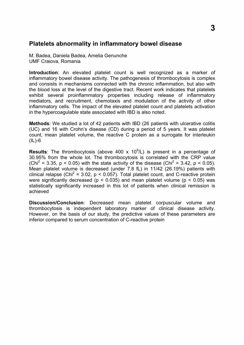
3
Platelets abnormality in inflammatory bowel disease M. Badea, Daniela Badea, Amelia Genunche UMF Craiova, Romania Introduction: An elevated platelet count is well recognized as a marker of inflammatory bowel disease activity. The pathogenesis of thrombocytosis is complex and consists in mechanisms connected with the chronic inflammation, but also with the blood loss at the level of the digestive tract. Recent work indicates that platelets exhibit several proinflammatory properties including release of inflammatory mediators, and recruitment, chemotaxis and modulation of the activity of other inflammatory cells. The impact of the elevated platelet count and platelets activation in the hypercoagulable state associated with IBD is also noted. Methods: We studied a lot of 42 patients with IBD (26 patients with ulcerative colitis (UC) and 16 with Crohn’s disease (CD) during a period of 5 years. It was platelet count, mean platelet volume, the reactive C protein as a surrogate for interleukin (IL)-6 Results: The thrombocytosis (above 400 x 109/L) is present in a percentage of 30.95% from the whole lot. The thrombocytosis is correlated with the CRP value (Chi2 = 3.35, p < 0.05) with the state activity of the disease (Chi2 = 3.42, p < 0.05). Mean platelet volume is decreased (under 7.8 fL) in 11/42 (26.19%) patients with clinical relapse (Chi2 = 3.02, p < 0.057). Total platelet count, and C-reactive protein were significantly decreased (p < 0.035) and mean platelet volume (p < 0.05) was statistically significantly increased in this lot of patients when clinical remission is achieved Discussion/Conclusion: Decreased mean platelet corpuscular volume and thrombocytosis is independent laboratory marker of clinical disease activity. However, on the basis of our study, the predictive values of these parameters are inferior compared to serum concentration of C-reactive protein
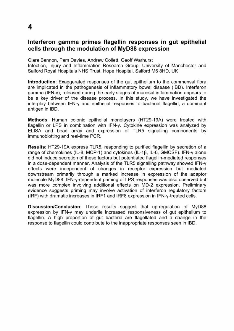
4
Interferon gamma primes flagellin responses in gut epithelial cells through the modulation of MyD88 expression Ciara Bannon, Pam Davies, Andrew Collett, Geoff Warhurst Infection, Injury and Inflammation Research Group, University of Manchester and Salford Royal Hospitals NHS Trust, Hope Hospital, Salford M6 8HD, UK Introduction: Exaggerated responses of the gut epithelium to the commensal flora are implicated in the pathogenesis of inflammatory bowel disease (IBD). Interferon gamma (IFN-γ), released during the early stages of mucosal inflammation appears to be a key driver of the disease process. In this study, we have investigated the interplay between IFN-γ and epithelial responses to bacterial flagellin, a dominant antigen in IBD. Methods: Human colonic epithelial monolayers (HT29-19A) were treated with flagellin or LPS in combination with IFN-γ. Cytokine expression was analyzed by ELISA and bead array and expression of TLR5 signalling components by immunoblotting and real-time PCR. Results: HT29-19A express TLR5, responding to purified flagellin by secretion of a range of chemokines (IL-8, MCP-1) and cytokines (IL-1β, IL-6, GMCSF). IFN-γ alone did not induce secretion of these factors but potentiated flagellin-mediated responses in a dose-dependent manner. Analysis of the TLR5 signalling pathway showed IFN-γ effects were independent of changes in receptor expression but mediated downstream primarily through a marked increase in expression of the adaptor molecule MyD88. IFN-γ-dependent priming of LPS responses was also observed but was more complex involving additional effects on MD-2 expression. Preliminary evidence suggests priming may involve activation of interferon regulatory factors (IRF) with dramatic increases in IRF1 and IRF8 expression in IFN-γ-treated cells. Discussion/Conclusion: These results suggest that up-regulation of MyD88 expression by IFN-γ may underlie increased responsiveness of gut epithelium to flagellin. A high proportion of gut bacteria are flagellated and a change in the response to flagellin could contribute to the inappropriate responses seen in IBD.
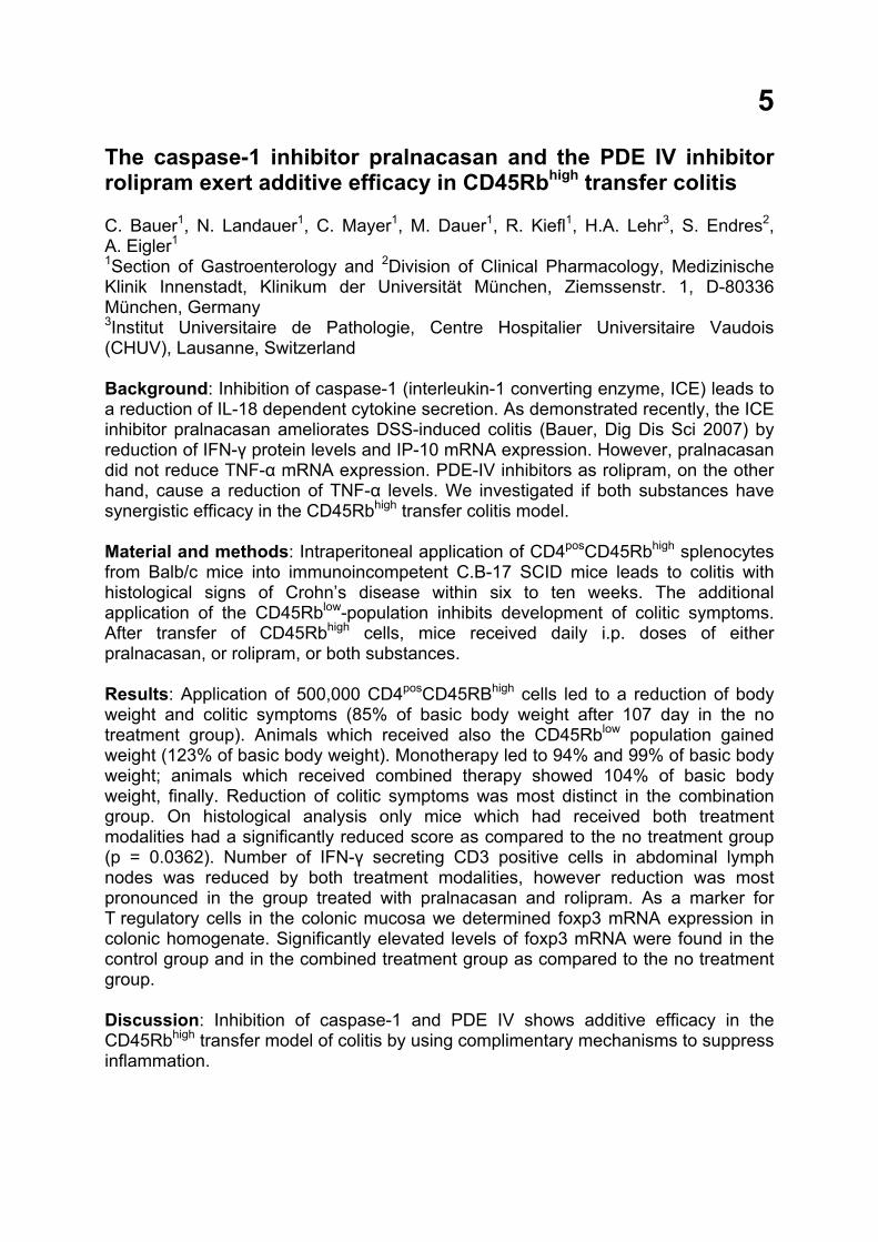
5
The caspase-1 inhibitor pralnacasan and the PDE IV inhibitor rolipram exert additive efficacy in CD45Rbhigh transfer colitis C. Bauer1, N. Landauer1, C. Mayer1, M. Dauer1, R. Kiefl1, H.A. Lehr3, S. Endres2, A. Eigler1 1Section of Gastroenterology and 2Division of Clinical Pharmacology, Medizinische Klinik Innenstadt, Klinikum der Universität München, Ziemssenstr. 1, D-80336 München, Germany 3Institut Universitaire de Pathologie, Centre Hospitalier Universitaire Vaudois (CHUV), Lausanne, Switzerland Background: Inhibition of caspase-1 (interleukin-1 converting enzyme, ICE) leads to a reduction of IL-18 dependent cytokine secretion. As demonstrated recently, the ICE inhibitor pralnacasan ameliorates DSS-induced colitis (Bauer, Dig Dis Sci 2007) by reduction of IFN-γ protein levels and IP-10 mRNA expression. However, pralnacasan did not reduce TNF-α mRNA expression. PDE-IV inhibitors as rolipram, on the other hand, cause a reduction of TNF-α levels. We investigated if both substances have synergistic efficacy in the CD45Rbhigh transfer colitis model. Material and methods: Intraperitoneal application of CD4posCD45Rbhigh splenocytes from Balb/c mice into immunoincompetent C.B-17 SCID mice leads to colitis with histological signs of Crohn’s disease within six to ten weeks. The additional application of the CD45Rblow-population inhibits development of colitic symptoms. After transfer of CD45Rbhigh cells, mice received daily i.p. doses of either pralnacasan, or rolipram, or both substances. Results: Application of 500,000 CD4posCD45RBhigh cells led to a reduction of body weight and colitic symptoms (85% of basic body weight after 107 day in the no treatment group). Animals which received also the CD45Rblow population gained weight (123% of basic body weight). Monotherapy led to 94% and 99% of basic body weight; animals which received combined therapy showed 104% of basic body weight, finally. Reduction of colitic symptoms was most distinct in the combination group. On histological analysis only mice which had received both treatment modalities had a significantly reduced score as compared to the no treatment group (p = 0.0362). Number of IFN-γ secreting CD3 positive cells in abdominal lymph nodes was reduced by both treatment modalities, however reduction was most pronounced in the group treated with pralnacasan and rolipram. As a marker for T regulatory cells in the colonic mucosa we determined foxp3 mRNA expression in colonic homogenate. Significantly elevated levels of foxp3 mRNA were found in the control group and in the combined treatment group as compared to the no treatment group. Discussion: Inhibition of caspase-1 and PDE IV shows additive efficacy in the CD45Rbhigh transfer model of colitis by using complimentary mechanisms to suppress inflammation.
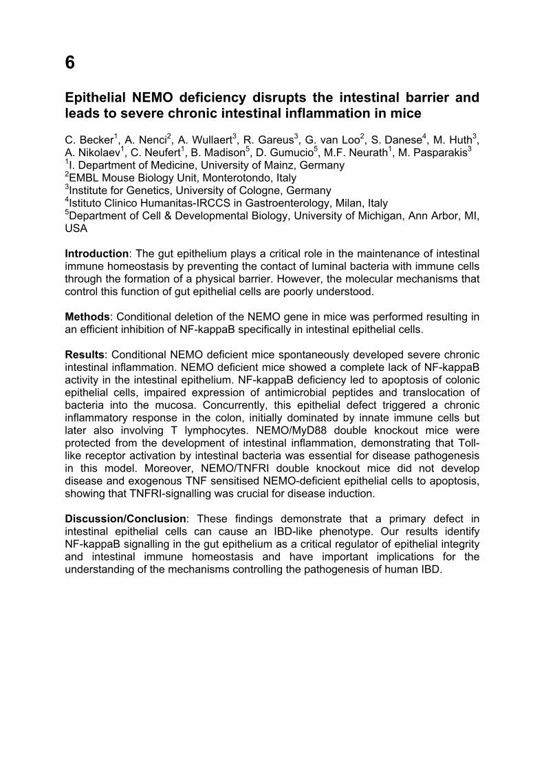
6
Epithelial NEMO deficiency disrupts the intestinal barrier and leads to severe chronic intestinal inflammation in mice C. Becker1, A. Nenci2, A. Wullaert3, R. Gareus3, G. van Loo2, S. Danese4, M. Huth3, A. Nikolaev1, C. Neufert1, B. Madison5, D. Gumucio5, M.F. Neurath1, M. Pasparakis3 1I. Department of Medicine, University of Mainz, Germany 2EMBL Mouse Biology Unit, Monterotondo, Italy 3Institute for Genetics, University of Cologne, Germany 4Istituto Clinico Humanitas-IRCCS in Gastroenterology, Milan, Italy 5Department of Cell & Developmental Biology, University of Michigan, Ann Arbor, MI, USA Introduction: The gut epithelium plays a critical role in the maintenance of intestinal immune homeostasis by preventing the contact of luminal bacteria with immune cells through the formation of a physical barrier. However, the molecular mechanisms that control this function of gut epithelial cells are poorly understood. Methods: Conditional deletion of the NEMO gene in mice was performed resulting in an efficient inhibition of NF-kappaB specifically in intestinal epithelial cells. Results: Conditional NEMO deficient mice spontaneously developed severe chronic intestinal inflammation. NEMO deficient mice showed a complete lack of NF-kappaB activity in the intestinal epithelium. NF-kappaB deficiency led to apoptosis of colonic epithelial cells, impaired expression of antimicrobial peptides and translocation of bacteria into the mucosa. Concurrently, this epithelial defect triggered a chronic inflammatory response in the colon, initially dominated by innate immune cells but later also involving T lymphocytes. NEMO/MyD88 double knockout mice were protected from the development of intestinal inflammation, demonstrating that Toll-like receptor activation by intestinal bacteria was essential for disease pathogenesis in this model. Moreover, NEMO/TNFRI double knockout mice did not develop disease and exogenous TNF sensitised NEMO-deficient epithelial cells to apoptosis, showing that TNFRI-signalling was crucial for disease induction. Discussion/Conclusion: These findings demonstrate that a primary defect in intestinal epithelial cells can cause an IBD-like phenotype. Our results identify NF-kappaB signalling in the gut epithelium as a critical regulator of epithelial integrity and intestinal immune homeostasis and have important implications for the understanding of the mechanisms controlling the pathogenesis of human IBD.

7
Bacterially-mediated activation of hypoxia inducible factor-1 (HIF-1) in intestinal epithelial cells involves the deneddylation of cullin-2 Geraldine O. Canny1,3, Andrew Neish2, Huixia Wu2, Lynn Bry3, Sean P. Colgan4 1Central University Hospital of Vaud (CHUV), Lausanne, Switzerland 2Emory University School of Medicine, Atlanta, GA, USA 3Brigham and Women’s Hospital, Boston, MA, USA 4University of Colorado Health Sciences Center, Denver, CO, USA Introduction: Bacteria influence intracellular signaling processes in mucosal epithelial cells, resulting in infection or homeostasis for the host. HIF-1, a heterodimeric transcription factor comprising a stable subunit (HIF-1beta/ARNT) and a labile alpha subunit, mediates the regulation of a number of genes including vascular endothelial growth factor (VEGF), an important molecule in angiogenesis. Interestingly, HIF-1 may play a protective role in the intestine. Bacteria can activate HIF-1 in epithelia but the underlying mechanism is unclear. Methods and results: We demonstrate that both Gram negative (Salmonella typhimurium) and Gram positive (Lactobacillus acidophilus) bacteria, which activate HIF-1, cause rapid deneddylation of cullin (cul)-2 in Caco-2 cells in a (multiplicity of infection) MOI-dependent manner. This requires contact of bacteria with epithelial cells. The COP9 signalosome, a nuclear complex composed of eight subunits, is considered a major mechanism of deneddylation in mammalian cells. By transfecting siRNA, we demonstrate that the activity of CSN5, a component of the COP9 signalosome, is necessary for this bacterially-mediated deneddylation and for HIF-1 activation, as assessed by western blot. In an extension of these studies, using epithelial-enriched mucosal scrapings from germ free mice and from mice colonized with Lactobacillus acidophilus in an ileal loop model, we observe that bacteria also effect cul-2 deneddylation in vivo. Discussion/Conclusion: This is the first report detailing the mechanism whereby bacteria activate HIF-1. Our results provide an insight into mechanisms which potentially benefit the host, or which when consistently activated, may represent a molecular link between chronic infection and the development of cancer.
8
Early epithelial responses that precede increased gut permeability during colitis development in mdr1a(-/-) mice Andrew Collett, Norman B. Higgs and Geoffrey Warhurst Infection, Injury and Inflammation Research Group, University of Manchester and Salford Royal Hospitals NHS Trust, Hope Hospital, Salford M6 8HD, UK Introduction: The early molecular changes preceding the onset of mucosal inflammation in colitis and their temporal relationship with gut permeability remain poorly defined. This study has investigated functional and transcriptomic changes in mdr1a(-/-) mice which lack an intestinal transport protein, P-glycoprotein and develop colitis spontaneously when exposed to normal enteric flora. Methods: Mdr1a(-/-) mice housed in specific pathogen-free conditions were compared to congenic controls. Mucosal permeability and secretion of immune factors were analyzed in ex vivo colon. Gene expression in colonic mucosal and epithelial preparations was analyzed by microarray and real-time PCR. Colonic epithelial responsiveness to bacterial antigens was measured in short term culture. Results: Colon from 4–5 week mdr1a(-/-) mice was histologically normal with the same permeability as controls, however, they had increased chemokine secretion accompanied by a change in the expression of a small number of genes. The majority of up-regulated genes was gamma-interferon (IFN-γ) responsive and associated with bacterial recognition and the ubiquitin-proteasome system. There was also a marked down-regulation of the anti-inflammatory genes PAP/RegIIIγ. These changes preceded increases in colonic permeability, which was associated with older (12–16 week) mdr1a(-/-) displaying molecular and functional evidence of active inflammation. Colonocytes isolated from 4–5 week mdr1a(-/-) exhibited similar transcriptomic changes, and higher spontaneous chemokine secretion and increased responsiveness to LPS. Discussion/Conclusion: This study provides new insight into the early sequence of events that initiate mucosal inflammation, before a general increase in colonic permeability, in this “barrier” defect model of colitis and demonstrates the importance of P-glycoprotein in regulating interactions with the commensal microflora.
9
Immunohistochemical evaluation of Fhit protein expression in cases of inflammatory bowel disease Jolanta Czyzewska1, Katarzyna Guzinska-Ustymowicz2, Andrzej Kemona2, Anna Pryczynicz2, Halina Kemona1, Adam Chetnik4, Roman Bandurski3, Mariusz Gryko3, Adam Kukliński3, Dariusz Cepowicz3, Bogusław Kędra3 Departament of Clinical Laboratory Diagnostics1, Departament of General Pathomorphology2, Department of General Surgery and Gastroenterology3 Medical University of Bialystok, III Department of Internal Medicine and Gastroenterology Śniedecki District Hospital4 in Bialystok, Poland Background/Aim: Reduced or absent expression of Fhit protein was observed in many types of neoplasm, but mechanism of this protein is still unclear. The aim of this study was to evaluate the expression of Fhit in ulcerative colitis and Crohn’s disease. Methods: Study was performed on 38 cases patients with inflammatory bowel disease (18 cases of Crohn’s disease and 20 cases of ulcerative colitis). Standard, immunohistochemical technique was adopted to detect the expression of Fhit protein (Rabbit polyclonal to FHIT [ab15287], Abcam Ltd., UK) in evaluated samples. Results: We observe the cytoplasmic immunostaining pattern in all investigated cases. In 2/18 cases with Crohn’s and 7/20 with ulcerative colitis expression for Fhit protein was observed. Where in 4 cases of 7 positive, high expression of Fhit was observed in dysplastic cells. Conclusion: We can concluded that expression of Fhit protein is reduced in inflamed colonic epithelium but in dysplastic cells is increasing. Correspondence: Jolanta Czyzewska Department of Clinical Laboratory Diagnostics Medical University of Bialystok ul. Waszyngtona 15a PL-15-274 Bialystok Poland E-mail: [email protected]
10
Fecal calprotectin as a diagnostic and monitoring marker in Crohn’s disease Piotr Eder, Kamila Stawczyk, Iwona Krela-Kaźmierczak, Krzysztof Linke Chair and Department of Gastroenterology, Human Nutrition and Internal Medicine, University of Medical Sciences in Poznań, Poland Introduction: Calprotectin (MRP8/14) is a leukocyte protein involved in inflammation processes. The aim of this study was to assess the diagnostic and monitoring value of the fecal calprotectin concentration measurement in Crohn’s disease (CD). Methods: Stool samples, collected from 31 patients with CD and 11 patients (control group) with irritable bowel syndrome (IBS), were frozen and stored. After thawing 100 mg aliquot was extracted using sample preparation tubes. Calprotectin concentration was estimated twice for each sample by ELISA immunoenzymatic test. There were blood tests performed, including blood cell count, ESR (erythrocyte sedimentation rate) and CRP (C-reactive protein) concentration. Disease activity was evaluated using CDAI (Crohn’s Disease Activity Index). Results: Fecal calprotectin concentration in patients with CD and IBS was 32.01 ± 22.58 mg/l and 14.73 ± 4.58 mg/l, respectively (p < 0.0003). 16.01 mg/l has a 67.7% sensitivity and a 66.7% specificity for distinguishing between CD and IBS. There were statistically important correlation (p < 0.05) between MRP8/14 concentration and CRP. There were positive correlation between calprotectin concentration and CDAI, but it was not statistically important. Discussion/Conclusion: Assessment of fecal calprotectin concentration is a good method in differential diagnosis between CD and IBS. MRP8/14 can play an important role in pathogenesis of chronic inflammation in the intestines. CDAI is a subjective method in evaluation of the clinical activity of CD, what explains the poor correlation with fecal MRP8/14 concentration. High concentration of fecal calprotectin in patients with quiescent CD (CDAI < 150) suggests that even in those patients there is an active inflammation present in the gastrointestinal tract.
11
Profibrotic IL-13 negatively regulates chronic inflammation during intestinal fibrosis via glycogen synthase kinase-3beta Stefan Fichtner-Feigl, Edward Geissler, Hans-Jürgen Schlitt Department of Surgery, Regensburg University, Germany Introduction: Chronic inflammatory diseases often result in extensive tissue fibrosis and its associated untoward effects on organ function. Once organ fibrosis, including loss of organ function, has developed the underlying chronic inflammatory response often subsides. Methods: To investigate the immunopathogenesis of this phenomenon we analyzed the chronic colitis after late-developing fibrosis occurring in BALB/c mice administered weekly doses of intra-rectal trinitrobenzene sulfonic acid (TNBS). Results: Previously, we showed that in this model the chronic inflammation is driven by IL-23 and IL-17, whereas the cytokines leading towards fibrosis are IL-13 and TGF-beta1. We found that after full development of colonic fibrosis in this model the expression levels of IL-23 and IL-17 are gradually decreasing and inflammatory characteristics are diminished in the colon. This decrease in inflammatory cytokine expression was dependent on IL-13 mediated effects. We could show that GSK-3beta is a central effector molecule in this anti-inflammatory process. IL-13 leads to increased phosphorylation (decreased kinase activity) of GSK-3beta. Consequently, the innate immune response is guided away from the production of pro-inflammatory cytokines towards the production of anti-inflammatory IL-10 by decreased NF-kappaB and increased CREB activation. Ultimately, IL-10 and its signalling through STAT3 are responsible for mediating anti-inflammatory effects after development of intestinal fibrosis. Discussion/Conclusion: Therefore, besides profibrotic effects in IBD, IL-13 can diminish the chronic inflammatory response after fibrotic organ dysfunction and GSK-3beta is the central molecule to regulate innate immune responses in IBD.
12
Regulation and stability of the homing receptor integrin α4β7 on CD4+ T cells Stefan Floess, Balint Szilagyi, Kerstin Schlawe, Alf Hamann and Jochen Hühn Charité University Medicine Berlin, Exp. Rheumatology, c/o DRFZ, Charitéplatz 1, 10117 Berlin, Germany Migration of CD4+ T cells into inflamed gut-associated lymphoid tissue is enabled by upregulation of the homing receptor integrin α4β7. Recently, Iwata and coworkers identified the vitamin A metabolite retinoic acid (RA) as inducer of this homing receptor. To clarify whether a stable integrin α4β7 expression occur in CD4+ T cells, ex vivo isolated α4β7 + CD4+ memory T cells were stimulated in the presence of IL-12 or RA as well as organ-specific dendritic cells. In contrast to what has been reported for CD8+ cells, the expression of the integrin α4β7 on CD4+ memory T cells seem to be stable, since neither the absence of RA nor the presence of peripheral lymph node DC led to a decrease of this homing receptor. The existence of a CpG island within the murine integrin α4 promoter prompted us to investigate whether the stability of the integrin is regulated by epigenetic mechanisms. We analyzed the methylation status of four regions within the α4 locus in naïve α4β7dimCD4+ T cells as well as in memory and RA-induced α4β7highCD4+ T cells. The bisulphite sequencing method revealed no change in the methylation pattern after a switch from low to high α4β7 expression. Thus, the region between the transcriptional start site and exon 4 of α4 appears not to be regulated via DNA modification. However, this result does not exclude such a regulation in other regions. Further experiments will show if other epigenetic mechanisms like modification of the histone code contribute to a stable integrin α4β7 expression.
13
Upregulated expression of CD14, Toll-like receptor 2 (TLR2) and TLR4 in biopsy samples of patients with inflammatory bowel diseases Lenka Frolova1, Pavel Drastich2, Pavel Rossmann1, Klara Klimesova1, Helena Tlaskalova-Hogenova1 1Department of Immunology and Gnotobiology, Institute of Microbiology, Academy of Sciences of the Czech Republic, Videnska 1083, CZ-142 20 Prague 4, Czech Republic 2Institute of Clinical and Experimental Medicine, Videnska 1958/9, CZ-142 21 Prague 4, Czech Republic Introduction: It is generally accepted that dysregulation of the intestinal immune response to gut microbiota is involved in pathogenetic mechanisms of IBD. Pattern recognition receptors – PRRs (e. g. TLRs, CD14) on innate immunity cells recognise bacterial components and are involved in the regulation of the inflammatory processes. The aim of our study was to characterize the expression of TLR2 and TLR4 and their transmembrane coreceptor CD14 in intestinal mucosa obtained from different parts of intestine from patients with ulcerative colitis (UC) and Crohn’s disease (CD) in comparison with non-inflamed gut mucosa from controls. Methods: The biopsy samples were obtained by colonoscopy, frozen in liquid nitrogen. Cryostat sections were analyzed by immunohistochemistry using polyclonal and monoclonal antibodies specific for TLR2, TLR4 and CD14. Results: Immunohistochemistry showed a significant increase in the TLR2 expression in the terminal ileum of patients with UC as compared with controls. In contrast, no difference between the expression of TLR2 and TLR4 was found in patients with CD and controls. CD14 expression was upregulated in the terminal ileum of CD patients and in the cecum and rectum of UC patients. Statisticaly significant upregulation of TLR4 expression was found in the rectum of UC patients compared to healthy controls. Conclusion: Hence, dysregulation of TLR2, TLR4 and CD14 expression in different parts of intestinal mucosa might be crucial in IBD pathogenesis.
14
The serum level of cytokines (IL-1, IL-6) and TNF-alpha in patients with inflammatory bowel disease Amelia Genunche-Dumitrescu, P. Mitrut, Daniela Badea, M. Badea University of Medicine and Pharmacy, Clinical Hospital of Emergency, Craiova, Romania Introduction: The aim of our study was to determine serum levels of cytokines IL-1, IL-6 and the level of tumor necrosis factor alpha (TNF-alpha) in group of patients with inflammatory bowel disease (IBD). Methods: We studied 42 patients with IBD (28 females/14 males). According to the clinical picture, laboratory examinations, colonoscopy with histopathological examinations, 26 patients had UC and 16 patients had CD. We determined TNF-alpha in serum by ELISA test and cytokines, in inflammatory activity and in remission of IBD. Statistical analysis was performed using the Wilcoxon and Anova tests and Person linear correlation. Results: IL-1 was significantly higher in UC than CD and correlates with inflammatory process activity. IL-6 in UC was lower than in CD and also correlates with inflammatory process activity. TNF-alpha presented serum levels 3 to 4 times higher during the active periods of the disease. TNF-alpha levels were significantly higher in UC than CD patients. The serum cytokine values of patients with IBD, who received steroid therapy, were lower than patients who received 5-ASA. We observed correlation between values of cytokines, and the disease duration, clinical activity and localisation of disease. Discussion/Conclusion: The serum cytokine values were correlated with the activity of disease and therapy characters. TNF-alpha is an important cytokine which play an important role in immunopathogenesis of autoimmune diseases.
15
Prostaglandin E receptor subtype EP1 (PTGER1): A new candidate gene for IBD Alessandra Geremia1, John Beckly1, Laura Hancock1,2, Fraser Cummings1, Rachel Cooney1, Saad Pathan1, Derek Jewell1 1Gastroenterology , University of Oxford, Oxford, UK 2Colorectal Surgery, University of Oxford, Oxford, UK Introduction: Inflammatory bowel disease (IBD), encompassing Crohn's disease (CD) and Ulcerative Colitis (UC), is a chronic idiopathic inflammatory disorder of the gastrointestinal tract. The area of linkage on chromosome 19p13 (IBD6) has been identified by a genome-wide scan. PTGER1 lies within the IBD6 locus and encodes for the EP1 receptor of Prostaglandin E2 (PGE2). PGE2 has recently been involved in dendritic cell maturation and IL-23 induction. For functional and topographic reasons PTGER1 represents a good candidate gene for IBD. Methods: Single nucleotide polymorphisms (SNPs) within PTGER1 were selected using the public databases and genotyped in 1278 cases (608 CD and 670 UC patients) and 1131 controls. All study subjects were unrelated UK non-Jewish white Caucasians. The SNPs were genotyped using the MALDI-TOF homogenous mass extendTM platform. Results: An association was found between the minor allele of the SNP rs2241360 and both the combined IBD (p = 0.01) and CD phenotypes (p = 0.01). Stratification of the CD cohort by known variants showed that the rs2241360 minor allele is rarely observed in patients carrying any of the three susceptibility NOD2 variants (p = 0.05). On sub-phenotype analysis there was also an association with non-stricturing behavior of the disease (p = 0.04). Discussion/Conclusion: An association was found between CD and a SNP within the PTGER1 gene. This polymorphism, due to its location in an intron-exon boundary, might be implicated in splicing regulation. Further studies are needed to elucidate if this is the causal variant or if it is in linkage disequilibrium with the functional SNP elsewhere in the gene or in a neighbouring region.
16
Whipple’s disease: Diagnosis and long-term follow-up. 30-years experience V. Gerova, S. Stoinov University Hospital “Queen Joanna”, Clinical Center of Gastroenterology, Sofia, Bulgaria Introduction: Whipple’s disease (WD) is a rare, chronic, multisystemic infectious disease. Our aim was to evaluate the clinical, diagnostic, therapeutic aspects and long-term outcome in patients with WD. Methods: The study included 9 patients (6 male, 3 female; mean age 40.3 years) with WD, diagnosed in our Center between 1976–2006 by histopathologic demonstration of PAS-positive macrophages in jejunal biopsies. The patients were treated with antibiotics and were followed up regularly. Results: The most common intestinal symptoms were chronic diarrhea (8/9), weight loss and malabsorption (8/9), preceded (a mean of 9 years) or accompanied by extraintestinal symptoms as arthritis (8/9), enlargement of the lymph nodes (5/9), low grade fever (4/9). All patients had histologicaly abnormal small bowel biopsies with evidence of PAS-positive macrophages, lymphangiectasis and ultrastructural data for bacterial structures in the mucosa. Two weeks treatment with penicillin + sreptomycin or cefalosporins (third generation), followed by tetracycline or trimethoprim-sulfamethoxazole (1 year) resulted in clinical and morphological remission. The recurrence was observed in 2 patients after the end of the treatment, successfully treated with the same therapy by which the first remission was achieved. Until now all patients are in clinical and morphological remission (for 3 of them more than 20 years). Conclusion: WD may have fatal consequences when the diagnosis is delayed. Early diagnosing and prolonged antibacterial therapy made the clinical outcome better.
17
Role of tumor-associated macrophages in colorectal cancer M. Gulubova1, T. Vlaykova2, Y. Yovchev3, I. Vassilev1, S. Stratiev1 1Dept. of General and Clinical Pathology, 2Dept. of Chemistry and Biochemistry, 3Dept. of General Surgery, Medical Faculty, Trakia University, Stara Zagora, 11 Armeiska Str, Stara Zagora, BG-6000 Bulgaria, E-mail: [email protected] Introduction: Tumor-associated macrophages (TAMs) have ambivalent functions. TAMs have been shown to play a key role in tumor angiogenesis, which modulates the tumor growth and invasion. It was shown that the number of TAMs in the tumor stroma or in tumor nests correlated with more favorable prognosis for the patients with colorectal cancer. The aim of our study was to determine the location and number of TAMs in primary colorectal cancer and to explore the prognostic significance of TAMs for overall survival of the patients. Methods: We investigated 137 (84 male and 53 female) patients, operated for colorectal cancer from 2000 to 2003 year and followed-up them until the 30th of June 2006. The median survival period was 31.9 months, ranging from 0.5 to 105.2 months. TAMs were evaluated by immunohistochemistry in the invasive margin of the tumors and in the tumor nests. Results: TAMs were found to be more diffusely distributed along the invasive margin of the tumor sections. In some tumors CD68+ TAMs were present in tumor nests. The number of CD68+ TAMs in the invasive margin varied from 15.37 cells/mm2 to 296.33 cells/mm2 (median of 44.62 cells/mm2). The number of CD68+ TAMs in tumor nests was significantly lower (p = 0.0004, Wilcoxon signed rank test) and ranged from 1.5 to 63.95 cells/mm2 (median of 22.58 cells/mm2). There was no significant association between the TAMs number and microvessel density, inflammatory infiltrate and grade of differentiation of the tumors. Based on the median values of TAMs patients were dichotomized into two groups with severe and moderate macrophage infiltration. Patients with severe TAM infiltration either in the invasive margin or in the tumor nests had more favourable prognosis than those with moderate TAM infiltration (p = 0.003 and p < 0.0001, respectively for TAMs in the invasive margin and in the tumor nests, Log-rank test). Discussion/Conclusion: These results suggest that the aggregation of TAMs within tumor nests as well as the TAM infiltration in the invasive margin of tumors has a beneficial effect on the host in term of augmented cytotoxicity and antigen presentation. Key words: colorectal cancer, tumor-associated macrophages, CD68, prognosis
18
Prenatal development of interstitial cells of Cajal in human intestine Gumerova A.A., Kiassov A.P., Kaligin M.S. Kazan Medical Unversity, Kazan, Russia Interstitial cells of Cajal (ICC) play an important role in the control of gastrointestinal motility (they are pacemaker cells) and in inflammation-induced motor disturbances. However development of this cellular type in human prenatal ontogenesis and formation of interactions between them and intestinal smooth-muscle cells (SMC) practically are not studied. The purpose of our research was immunohistochemical investigation of development of intestine ICC and SMC during early human ontogenesis. Embryos and fetuses (3–33 weeks of gestation) have been received after legal medical abortions and miscarriage. We used antibodies to С-kit (marker of ICC) and to marker of SMC α-Smooth-Muscle-Actin (α-SMA). The first C-kit cells appeared in nervous tube on 3-th week of gestation. Since this moment and up to 5-th week it was possible to see migration С-kit+ cells into surrounding primary gut mesenchyma from nervous tube that specifies on nervous origin of these cells. Since 5-th week single ICC cells were observed around of stomach, since 6-th week – around intestine. The first α-SMA+ cells were visible in foregut also on 5-th week of gestation. Thus, pacemaker cells appear in intestine wall approximately at the same time that differentiation of its SMC begins. Probably ICC initiates differentiation of mesenchymal cells of intestine wall into SMC. Since 8-th week of gestation we observed two-layer muscular coat in intestine and ICC in its intermuscular nervous plexus. We conclude that to the termination embryonal period in intestine are developed two layers of muscular environment and intermuscular plexus formed by ICC.
19
Influence of diet on the susceptibility to endotoxin shock T. Hrncir, T. Hudcovic, H. Kozakova, R. Stepankova, M. Schwarzer, L. Frolova, M. Kverka, H. Tlaskalova-Hogenova Institute of Microbiology, v.v.i., Academy of Sciences of the Czech Republic, Department of Immunology and Gnotobiology, Prague and Novy Hradek, Czech Republic Introduction: Endotoxin shock is the most severe complication of sepsis and is triggered by lipopolysaccharide (LPS) or endotoxin, a structural component of gram negative bacteria. To disclose a role of intestinal bacterial flora in endotoxin shock we use a model of germ-free Balb/c and SCID mice. In this study we show that diet may have a strong influence on the outcomes of experiments in particular when conducted under germ-free conditions. Methods: Balb/c and SCID mice were bred under germ-free conditions and fed ad libitum with either semi-purified diet (low LPS content) or standard diet (high LPS content). To test the susceptibility to endotoxin shock mice were challenged with LPS and serum concentrations of pro-inflammatory cytokines including TNFa and IL-6 were examined by ELISA. To test the susceptibility to LPS in vitro resident peritoneal macrophages, spleen cells and mesenteric lymph node cells from germ-free Balb/c and SCID mice were stimulated with different concentrations of LPS and production of pro-inflammatory cytokines and proliferative response were measured. Results: We have observed significantly lower serum concentrations of pro-inflammatory cytokines in both germ-free Balb/c and SCID mice fed with a semi-purified diet (low LPS content). We have not found any statistically important difference in the susceptibility to LPS in vitro. Discussion/Conclusion: Our data show that the LPS content in diets may influence the susceptibility of germ-free mice to endotoxin shock in vivo. Hence, diet must be perceived as a critical factor when planning LPS challenge experiments in germ-free animals.
20
Effects of probiotic strain Escherichia coli Nissle 1917 on the development of intestinal inflammation induced in gnotobiotic models T. Hudcovic, R. Štěpánková, H. Kozáková, T. Hrnčíř, M. Schwarzer, H. Tlaskalová-Hogenová Department of Immunology and Gnotobiology, Institute of Microbiology, Academy of Sciences of the Czech Republic, Nový Hrádek, Czech Republic Aim: The participation of the intestinal microflora, mainly role of E. coli strains, in the development of the acute and chronic intestinal inflammation (model of ulcerative colitis) was studied in mice. Methods: Experimental colitis was evoked by an administration of 2.5% dextran sodium sulphate (DSS) in drinking water (7 days). Germ-free mice, mice monoassociated by (E. coli Nissle 1917 and E. coli O6K13) and conventional mice of SCID and BALB/c strains were used in our experiments. One week prior to DSS exposure the conventional mice received daily E. coli Nissle 1917 by intragastrical tubing or intrarectally and bacteria were continuously given during DSS drinking. Colon morphology and mucin production were evaluated. Results: Mice monoassociated with E. coli O6K13 developed intestinal inflammation in colon whereas colonization with E. coli Nissle 1917 strain protected mice against inflammation. Intragastrically administrated E. coli Nissle had only a mild effect on the intestinal inflammation, whereas mice treated per rectum remained healthy. In this group, the level of pro-inflammatory cytokine TNF-alpha and IL-6 were reduced markedly in colon descendens compared with controls. Conclusion: We conclude that E. coli Nissle 1917 colonization protects mice against intestinal inflammation induced by DSS treatment.
21
Biological parameters in inflammatory bowel diseases in children Ignyś Iwona, Kobelska-Dubiel Natalia, Krauss Hanna, Cichy Wojciech, Kobelski Mikołaj I Chair of Pediatrics, Department of Pediatric Gastroenterology and Metabolic Diseases, Poznan University of Medical Sciences, Poznan, Poland Introduction: Inflammatory bowel diseases in children are chronic intestinal disorders of unknown etiology and relapsing course. In assessment of disease activity CRP and ESR are used. Fecal calprotectin has been proposed as non-invasive marker in distinguishing between organic and functional disorders of gastrointestinal tract, for selection of children needing invasive diagnostic procedures and for the differential diagnosis and monitoring of IBD children. Methods: 60 children aged 7–18 were involved in the study: 35 with ulcerative colitis, 9 with Crohn’s disease and 16 with nonspecific colitis. Disease activity was assessed using Truelove-Witts Scale in ulcerative colitis and Pediatric Crohn’s Disease Activity Index by Hyams in Crohn’s disease (both scales modified by Ryżko and Woynarowski). CRP and ESR levels were assessed in blood samples and FC concentration was measured in stool samples. Results: Mean fecal calprotectin, CRP and ESR values were significantly increased in children with disease exacerbation. Disease activity assessment according to clinical indices revealed the highest values in exacerbation of inflammatory process. The highest values of all measured parameters were observed in children with Crohn’s disease, especially in exacerbation of the inflammatory process. A positive correlation was observed between disease activity indices and CRP, ESR and FC concentration. The differences were statistically significant (p < 0.05). Discussion/Conclusion: Biological parameters of inflammatory process such as CRP, ESR and FC appear to be sensitive markers in detecting the relapse of the disease and assessment of disease activity in IBD children. They seem to be useful additive tools in clinical observation and management.
22
Adipocytes and preadipocytes – Phagocytes within the mesenteric fat? Jakob Ihbe, Arvind Batra, Rainer Glauben, Thorsten Stroh, Inka Fedke, Martin Zeitz, Britta Siegmund Charité Universitätsmedizin Berlin, CBF, Medizinische Klinik I, Berlin, Germany Hypertrophy of mesenteric fat is a characteristic finding in Crohn’s disease, but the underlying mechanisms and its relevance are still unknown. However, in the recent years it became evident that pre- and adipocytes are potent producers of various pro-inflammatory mediators including leptin and IL-6. In addition recent data from our own group indicate a leptin-dependent expression and functionality of Toll-like receptors on preadipocytes and adipocytes of murine mesenteric fat which might be of particular importance since colonic inflammation favors bacterial translocation into the mesenteric tissue. To gain further information about the immunological properties of mesenteric preadipocytes, phagocytic activity and the surface expression of co-stimulatory molecules on leptin- and leptin-receptor-deficient as well as WT preadipocytes was characterized. Murine preadipocyte cell lines originally generated from the mesenteric fat of WT, leptin- (ob/ob) or leptin-receptor-deficient (db/db) mice were characterized by flow cytometry for phagocytic activity and expression of B7-1, B7-2, CD11b, CD11c, MHCII and MOMA-2. All cell lines were potent in phagocyting latex beads, S. aureus and ovalbumin, solely leptin receptor-deficient cells were less competent in the uptake of opsonized S. aureus. MHCII, B7-2, CD11b and CD11c were barely detectable, but B7-1 and MOMA-2 staining were evident on 2-5% of cells from all cell lines. In summary mesenteric preadipocytes exert phagocytic activity and express co-stimulatory molecules at basal levels. Hence, the link between adipose tissue and immune cells might be tighter than originally expected, suggesting that the mesenteric fat hypertrophy seen in Crohn’s disease may be of immunological relevance.
23
Cyclooxygenase-1 as well as cyclooxygenase-2 contributes anaphylaxis-induced alterations in intestinal motility Hiroko Kadowaki1, Takeshi Yamamoto2, Nobuo Kurokawa1, Makoto Kadowaki2 1Osaka University Hospital, Department of Pharmacy, Osaka, Japan 2Division of Gastrointestinal Pathophysiology, Institute of Natural Medicine, University of Toyama, Japan Introduction: A number of studies have shown that mast cells activated by various luminal antigens including enterobacterial antigens and food antigens are involved in the pathogenesis of ulcerative colitis (UC). In particular, various inflammatory mediators released from activated mast cells are considered to play a key role. The aim of the present study was to determine the mediators responsible for mast cells-mediated anaphylaxis in the rat colon. Methods: Wistar rat was sensitized by intraperitoneal injection of ovalbumin. Fourteen days later the contractility of isolated proximal colon was studied in organ bath to evaluate anaphylaxis in response to the antigen. Results: Antigen challenge induced contraction in the colon of sensitized rat. Mast cell involvement was indicated by a marked reduction in the antigen-induced contraction in the presence of mast cell stabilizer doxantrazole (10 µM). The contraction was resistant to prostaglandin D2 receptor antagonist BW A868C (10 µM) and substance-P receptor antagonist L-732,138 (20 µM). In contrast, non-selective cyclooxygenase (COX) inhibitor, indomethacin (1 µM) significantly reduced the antigen-induced anaphylaxis by 61%. Therefore, we evaluated selective COX-2 inhibitor NS398 (10 µM), which significantly inhibited the anaphylaxis by 50%. Furthermore, COX-1 inhibitor piroxicam (10 µM) and FR122047 (10 µM) reduced the anaphylaxis by 58% and 50%, respectively. In addition, immunohistochemical studies and real-time PCR studies demonstrated that COX-1 and COX-2 mRNAs and proteins were expressed in the colon and were upregulated by anaphylaxis. Discussion/Conclusion: Until recently, COX-1 was considered the constitutive isoform. Nevertheless, not only COX-2 but also COX-1 may play an important role in the pathogenesis of UC.
24
Cholinergic anti-inflammatory pathway through α7-nicotinic acetylcholine receptors in the colon reduces oxazolone-induced colitis in mouse Makoto Kadowaki, Minako Yoshida, Kanae Fujiwara, Takeshi Yamamoto Division of Gastrointestinal Pathophysiology, Institute of Natural Medicine, University of Toyama, Japan Introduction: It has been reported that the cholinergic anti-inflammatory pathway is controlled by the vagus nerve, and inhibits local cytokine release. Epidemiologic reports suggest that smoking may improve the symptoms of ulcerative colitis (UC). The purpose of the present study was to investigate the pathophysiological role of vagus nerve in Th2-mediated oxazolone (OXZ)-induced colitis. Methods: OXZ was injected into the colon of BALB/c mice (Th2 dominant strain). OXZ colitis was assessed in the colon with the disease activity score (DAS), pathological colonic damage score (CDS) by macroscopic evaluation and MPO. Results: The central stimulation of vagus nerves by 2-deoxy-d-glucose (100 mg/kg ip) significantly improved DAS, CDS and MPO in OXZ colitis. In addition, nicotine significantly alleviated the OXZ colitis in a dose-dependent fashion (0.32–3.2 mg/kg sc). Hexamethonium and α7-nicotinic receptor (nAChR) antagonist methyllycaconitine significantly inhibited the therapeutic effects of nicotine in OXZ colitis. The systemic immunity in OXZ colitis was found to be under Th2-polarized condition. On the other hand, transcript levels of IFN-γ, IL-4, IL-5 and IL-10 significantly increased in the colon of OXZ colitis, which were significantly down-regulated by nicotine. FITC-α-BTx-bindings to α7-nAChR were observed in the mucosa of the colon, which were upregulated in the colon of OXZ colitis. Furthermore, double staining with FITC-α-BTx and anti-CD4 mAb demonstrated that some α7 nAChRs lay adjacent to CD4+ T cells but didn’t co-localize with each other in the colon of OXZ colitis. Discussion/Conclusion: The vagal anti-inflammatory pathway acts through α7-nAChR in the mucosa of the colon to alleviate colonic inflammation.
25
Pathogenesis and bacteriology in inflammatory bowel disease Krasimira Kalinova University Hospital, Stara Zagora, Bulgaria Introduction: Ulcerative colitis (UC) and Crohn's disease (CD), the primary constituents of inflammatory bowel disease (IBD), are precipitated by a complex interaction of environmental, microbiological, genetic, and immunoregulatory factors. Aims: To investigate and characterise the predominant composition of the mucosa-associated intestinal microflora in colonoscopic biopsy specimens of patients with newly diagnosed IBD. Methods: Mucosa-associated bacteria were quantified and isolated from biopsy specimens of the ileum, caecum and rectum obtained at colonoscopy in 16 patients with Crohn's disease, 8 with ulcerative colitis, 4 with indeterminate colitis, and 10 with lymphonodular hyperplasia of the distal ileum and in 6 controls. Isolation and characterisation were carried out by conventional culture techniques for aerobic and facultative-anaerobic microorganisms, and analysis for the detection of anaerobic bacterial groups or species. Results: A higher number of mucosa-associated aerobic and facultative-anaerobic bacteria were found in biopsy specimens of children with IBD than in controls. An overall decrease in some bacterial species or groups belonging to the normal anaerobic intestinal flora was suggested by molecular approaches; in particular, occurrence of Bacteroides vulgates was low in Crohn's disease, ulcerative colitis and indeterminate colitis specimens. In human IBD, inflammation is present in parts of the gut containing the highest bacterial concentrations. Moreover, the terminal ileum, caecum and rectum are areas of relative stasis, providing prolonged mucosal contact with luminal contents. In Crohn disease, concentrations of Bacteroides, Eubacteria and Peptostreptococcus are increased, whereas Bifidobacteria numbers are significantly reduced. Furthermore, in ulcerative colitis, concentrations of facultative anaerobic bacteria are increased. The arrival of new molecular techniques qualifying and quantifying the complex intestinal flora has induced a revival of interest in this microflora. Therapeutic approaches geared towards changing the environment at the mucosal border have been attempted by the use of elemental diets, total parenteral nutrition, surgical diversion of the faecal stream and antibiotics. Discussion/Conclusion: Epidemiological, clinical and experimental evidence support an association between IBD and a large number of seemingly unrelated environmental factors, which include smoking, diet, drugs, geographical and social status, stress, microbial agents, intestinal permeability and appendectomy. Data supporting the involvement of each of these factors in predisposing to, triggering or modulating the course or outcome of IBD vary from strong to tenuous. Smoking and the enteric bacterial flora are the ones for which the most solid evidence is currently available. Smoking increases the risk of Crohn's disease (CD) and worsens its clinical course, but has a protective effect in ulcerative colitis (UC). Presence of enteric bacteria is indispensable to develop gut inflammation in most animal models
of IBD, and modulation of the quantity or quality of the flora can be beneficial in patients with IBD. Surprisingly, evidence for a major role of the diet in inducing or modifying IBD is limited, while that for nonsteroidal anti-inflammatory drugs is more convincing than for oral contraceptives. Northern geographic location and a high social, economical, educational or occupational status increase the risk of IBD, an observation fitting the hygiene hypothesis for allergic and autoimmune diseases. Stress is also associated with IBD, but more as a modifier than an inducing factor. Finally, an increased intestinal permeability may increase the risk for developing CD, whereas an appendectomy lowers the risk of developing UC.
26
Immune modulation after oral antigen administration Kerstin Kapp, Jochen Maul, Arlett Wenzel, Jan Preiss, Pamela Mundt, Martin Zeitz and Rainer Duchmann Medizinische Klinik I; Charité – Universitätsmedizin Berlin; Campus Benjamin Franklin; Hindenburgdamm 30, D-12200 Berlin, Germany Oral exposure to antigen has several potential outcomes, including the induction of systemic tolerance, systemic priming and/or the induction of local mucosal immune responses. Using the neo-antigen KLH we analyzed how mucosal antigen exposure alters the antigen-specific T- and B-cell response induced by a subsequent parenteral immunization. The kinetic of KLH-specific T-cell responses, the cytokine pattern and the expression of homing receptors was analyzed on single cell level. KLH-specific antibodies were measured by ELISA. Oral antigen administration itself induces a weak antigen-specific T-cell response and primes the immune system in a way that the T-cell response following a subsequent parenteral immunization develops much faster. It also shifts the cytokine pattern of antigen-specific T-helper cells induced by a subsequent parenteral immunization to significant more IL-4- and less IFN-γ-producing cells. After the second parenteral boost we also observed significant higher amounts of IL-10- and less IL-2-producing cells. T-helper cells expressing the skin-homing marker CLA were reduced. In parallel, the B-cell response after parenteral immunization followed a faster kinetic and significant more KLH-specific antibodies could be detected in the serum. Mucosal exposure of KLH can modulate T- and B-cell responses after a subsequent parenteral immunization. Especially the observed shift in the cytokine pattern of antigen-specific T cells and the amplification of KLH-specific B-cell responses are of interest for vaccination strategies and the treatment of chronic inflammatory diseases.
27
Inflammation-related colon cancer development in conventional and germ-free mice K. Klimesova, P. Rossmann, M. Kverka, L. Frolova, H. Tlaskalova-Hogenova Institute of Microbiology, Academy of Sciences of the Czech Republic, Videnska 1083, CZ-142 20 Prague, Czech Republic Introduction: Chronic inflammation (e. g. inflammatory bowel disease) is linked with higher risk of cancer. Intestinal microflora also seems to play an important role in inflammation. The aim of our study was to describe inflammation-related colon carcinogenesis under conventional and germ-free conditions. Methods: Male Balb/c mice received a single subcutaneous injection of azoxymethane (AOM; 10 mg/kg body weight). One week later colitis was induced by supplying 3% dextran sodium sulfate (DSS) in drinking water continuously for up to 5 days. Macroscopic and microscopic changes of the colon mucosa were evaluated in specimens taken from proximal and distal colon and rectum in weeks 6, 12, 18 and 24. Beta-catenin and inducible nitric oxid synthase (iNOS) were determined immunohistochemically. Results: Chronic colitis, low or high grade dysplasia or adenocarcinoma was visible in histological sections since the week 6. Following week 12 the incidence of macroscopically visible lesions reached nearly 100% and histopathology findings were more extensive (the incidence of colonic adenocarcinoma, always situated in the descendent portion and in rectum, in weeks 6, 12, 18 and 24 was 30%, 75%, 89% and 100%, respectively). Strong nuclear and cytoplasmic expression of beta-catenin was observed in high-grade dysplasia and adenocarcinoma cells. iNOS level in the cytoplasm of neoplasia cells was higher than in controls. Discussion/Conclusion: Our study provides interesting data that may help in identification of etiological agents and in developing novel preventive and therapeutic approaches.
28
Tumor necrosis factor-alpha antibodies in Crohn’s disease and investigation of interleukin 10 V. Kupčová1, Z. Zelinková1, L. Turecký1, S. Ilavská2, E. Jahnová2 Medical Faculty, Comenius University1, Slovak Health Care University2 Bratislava, Slovakia Increased serum levels of interleukin 10 (IL-10) were observed in patients with active Crohn’s disease (CD) and ulcerative colitis, suggesting that IL-10 acts as a naturally occurring damper in the acute inflammatory process of inflammatory bowel disease. Thus, the administration of anti-TNF antibody might be associated with changes in both, proinflammatory and regulatory parts of the immune system. In an attempt to assess the pattern of immunoregulatory cytokine response in CD patients treated with anti-TNF antibody, serum levels of IL-10 were measured together with clinical and laboratory parameters of disease activity. Clinical activity (in 14 patients with active, moderate to severe Crohn’s disease), serum IL-10, basic haematological and biochemical parameters (blood count, prothrombin time, renal and hepatic functions) were assessed. All parameters were obtained before treatment in Month 0 (Mo 0) and in Month 1 and 5 (Mo 1, Mo 5) after treatment. Clinical activity was assessed by Crohn’s disease activity index (CDAI). Serum IL-10 was measured by a commercially available kit Quantikine® (R&D Systems). Patients received 5 mg per kg of anti-TNF antibody (infliximab) in intravenous infusion. Clinical improvement was observed in 12 patients with a decrease in median CDAI from 228 (163–294) before treatment to 98.5 (56–160) in Mo 1; two patients did not respond. According to the clinical response in Mo 1, patients were divided into two groups: Group 1 (7 patients) with a decrease in CDAI of 50% and more; in this group the median of CDAI before treatment was 240 (169–294) diminishing in Mo 1 to 81 (56–125); and the group 2 (7 patients) with a drop of CDAI less than 50%; the median of CDAI in this group was 265 (163–300) before treatment and after 1 month it decreased to 145 (114–294). During the further clinical follow-up, patients in the group 1 remained stable with CDAI of 82 (28–216) in Mo 5, while in the group 2 the clinical activity raised to 203 (108–318) in Mo 5 that did not differ significantly from the clinical activity before treatment. IL-10 levels before treatment ranged from 3.62 pg/ml to 6.08 pg/ml with a median of 4.44 pg/ml in 13 patients; in one case the IL-10 levels were elevated up to 22.72 pg/ml. During the further follow-up, there was a significant decrease in IL-10 levels in the group 1 (p < 0.05) in Mo 1 and IL-10 levels remained decreased compared to values before treatment in Mo 5 (Table 1). On the other hand, in the group 2 a significant increase in IL-10 levels (p < 0.05) was observed in Mo 1, without significant changes in Mo 5. We conclude that the pattern of IL-10 response might play a role in determining the response to anti-TNF therapy. This work was supported by VEGA grant 1/4277/07
29
Peroral administration of mycobacterial HSP60 and HSP70 prevents severe forms of DSS-induced intestinal inflammation in Balb/c mice M. Kverka1, Z. Zákostelská1, R. van der Zee2, W. van Eden2 and H. Tlaskalová-Hogenová1 1Institute of Microbiology Academy of Sciences of the Czech Republic, Prague, Czech Republic 2Department of Infectious Diseases and Immunology, Faculty of Veterinary Medicine, Utrecht University, The Netherlands Introduction: Heat shock proteins (HSP) can be used as therapeutic agents to prevent or arrest the inflammatory damage in both experimental arthritis and experimental diabetes. In this study we test the efficacy of perorally administered mycobacterial HSP60 and HSP70 in the acute experimental colitis prevention. Methods: BALB/c mice first received HSP60 and HSP70 (30 micrograms once a week) in the presence of soybean trypsin inhibitor for 4 weeks, then colitis was induced by administrating 3% dextran sodium sulfate for one week. Inflammation was assessed by disease activity score, colon shortening and histological scoring. Results: We have found that repeated oral application of HSP60 or HSP70 significantly decreases the severity of subsequent acute colitis in all tested parameters. Disease was milder in HSP treated mice (1.73 ± 0.78, p < 0.01 for HSP60 and 1.87 ± 0.88, p < 0.01; for HSP70) compared to the control mice (3.13 ± 0.69). The colon was significantly longer in HSP treated mice (6.99 ± 0.39 cm, p < 0.01 for HSP60 and 6.83 ± 0.57 cm, p < 0.05; for HSP70) compared to the control mice (6.22 ± 0.42 cm). The colonic architecture in HSPs treated mice was improved (1.31 ± 0.51, p < 0.01 for HSP60 and 1.31 ± 0.45, p < 0.01; for HSP70) compared to the control mice (1.86 ± 0.56). Discussion/Conclusion: The current study demonstrates that peroral administration of HSP60 and HSP70 attenuates colonic inflammation. This may be a novel therapeutic option for IBDs. More studies are needed to elicit the mechanism that lies behind this effect.
30
TNBS-induced colitis models in SJL and BALB/c mice T. Lagerweij#, N. Worms#, R. Verbeek#, L. Nagelkerken# #TNO Quality of Life, Leiden, The Netherlands Introduction: The development of therapies for inflammatory bowel disease requires preclinical models which sufficiently represent disease in patients. One of these is the TNBS-induced colitis model, which shows a clear T cell involvement, but is usually associated with unacceptable mortality. Methods: We studied colitis in the mice that were sensitized by application of TNBS on the skin, followed by one (SJL) or three (BALB/c) weekly rectal challenges. Mice were treated with budesonide or sulfasalazine. Tissue samples were analyzed by light microscopy and immunohistology. Results: In the acute model in SJL mice, inflammation is associated with inflammation, characterized by severe necrosis, mucosal damage, loss of crypt architecture and transmural inflammation with granulocytes and mononuclear cells. The concentration of TNF-alpha in tissue extracts was increased, and inflammation was associated with a strong increase of serum amyloid A. This model is partially sensitive to treatment with budesonide, sulfasalazine, and several experimental compounds. As opposed to the acute model, bowel inflammation in BALB/c mice shows limited mucosal damage despite numerous inflammatory infiltrates. The infiltrates contain abundant CD4+ T cells, CD11b+ macrophages, neutrophils and eosinophils. Daily rectal treatment with budesonide resulted in a significant inhibition of inflammation. Discussion/Conclusion: Although the acute model in SJL mice has the disadvantage of a short treatment period it is sensitive to several therapeutics. The model in BALB/c mice has the advantage of being less acute with an extended duration of three weeks, allowing prolonged treatment with candidate drugs.
31
Elucidating the pathogenic role of Th17 cells in an adoptive transfer model of chronic intestinal inflammation M. Leppkes, C. Becker, S. Wirtz, C. Neufert, A. Nikolaev, M.F. Neurath Laboratory of Immunology, I Medical Clinic, Johannes Gutenberg University of Mainz, Mainz, Germany Introduction: Recent studies have underlined the presence of IL-17 producing CD4+ T helper cells (Th17) in mouse models of chronic and autoimmune inflammation such as experimental autoimmune encephalitis (EAE) or collagen-induced arthritis (CIA). As supernatants of CD4+ T helper cells isolated from colitis bearing mice reveal the extensive production of IL-17, we evaluated the pathogenic role of the Th17 subset in a colitis model by the adoptive transfer of CD4+CD25- cells into immunocompromised hosts. Methods: Colitis was induced by the transfer of MACS separated CD4+CD25- cells isolated from the spleen. These cells were injected intraperitoneally into Rag-/- mice. We monitored disease development by endoscopic inflammation score and analyzed the colon concerning immunohistochemistry, mRNA profile and cytokine production of CD4+ T cells. Results: We found elevated mRNA levels of known Th17 cytokines (IL-17A, IL-17F, IL-22) in the inflamed colon. We reasoned that Th17 cells might play an important role in the pathogenesis of colitis. In the following, we were able to identify a significant proportion of Th17 cells after reisolation of CD4+ cells from adoptively transferred mice by flow cytometry. To study the functional relevance of T-cell derived IL-17, inhibition of this factor was assessed in the adoptive transfer model. Interestingly, inhibition of IL-17 did not lead to a protection in this model of disease concerning endoscopic and histopathological scoring. Discussion/Conclusion: Our data imply that either other factors derived from the Th17 subset are more important in disease development or that Th17 cells are of minor importance concerning the morphological component of this inflammation.
32
Role of transforming growth factor β1 (TGF-β1) gene polymorphism in chronic inflammatory diseases of the digestive system in children A. Liberek1, J. Jakobkiewicz-Banecka2, A. Kloska2, J. Swiderska2, A. Szlagatys-Sidorkiewicz1, A. Marek1, P. Landowski1, K. Marek1, G. Luczak1, Z. Kmiec3, G. Wegrzyn2, M. Korzon1 1Department of Paediatrics, Paediatric Gastroenterology and Oncology, Medical University of Gdansk, Gdansk, Poland 2Laboratory of Molecular Biology, University of Gdansk, Gdansk, Poland 3Department of Histology, Medical University of Gdansk, Gdansk, Poland There is some evidence that the clinical course and natural history of some chronic inflammatory diseases may be linked to the polymorphism of the gene encoding TGF-β1. The aim of the study was to analyze the relationship between TGF-β1 gene polymorphism and the clinical course of chronic inflammatory diseases of the digestive system in children Material and methods: 243 children were included into the study: 25 with chronic hepatitis, 115 with inflammatory bowel disease (IBD) (ulcerative colitis – 70, Crohn’s disease – 45) and 103 healthy controls. The TGF-β1 gene T869C and G915C polymorphisms in exon 1 and G-800A and C-509T polymorphisms in the promoter region were analyzed by restriction fragments lengths polymorphism (RFLP) method. The plasma concentration of TGF-β1 was evaluated using commercial ELISA kit. In the intestinal mucosa in patients with IBD tissue TGF-β1 content was evaluated by using immunohistochemical assay and TGF-β1 gene expression was evaluated by real time RT-PCR method. Results and conclusions: We found no statistically significant differences in frequencies in any of the analyzed polymorphisms between patients and the control group. There was no statistically significant correlation between distributions of any these polymorphisms and type of the disease and their clinical and histological activity. No differences in plasma TGF-β1 concentration and tissue TGF-β1 expression with respect to gene polymorphism were found. These preliminary data indicate that TGF-β1 genotype does not appear to play a role in clinical course of chronic inflammatory diseases of the digestive system in children.
33
Transforming growth factor beta1 (TGF-beta1) and clinical course of chronic inflammatory bowel disease (IBD) in children A. Liberek, A. Marek, A. Szlagatys-Sidorkiewicz, D. Kartanowicz, P. Landowski, G. Luczak, P. Wierzbicki, M. Stanislawowski, K. Marek, Z. Kmiec, M. Korzon Department of Paediatrics, Paediatric Gastroenterology and Oncology, Medical University of Gdansk, Gdansk, Poland The role of TGF-beta1 in chronic inflammatory diseases of the digestive tract has not been fully elucidated. The aim of the study was to analyze the relationship between TGF-beta1 expression and clinical course of IBD in children. Patients and methods: Study was conducted in 115 patients with IBD (aged 12.36 ± 4.86 years) including 70 patients with ulcerative colitis (UC), 45 with Crohn’s disease (CD) and 42 in the control group (aged 15.45 ± 3.5 years). The plasma TGF-beta1 was evaluated using commercial ELISA kit and concentration of tissue TGF-beta1 content in intestinal mucosa using immunohistochemical staining. Tissue TGF-beta1 gene expression was evaluated by real time RT-PCR method. Results: In IBD group plasma concentration of TGF-beta1 was 7.26 ± 8.78 ng/ml (UC – 7.68 ± 9.82 ng/ml, CD – 6.61 ± 6.94 ng/ml), in control group was 6.96 ± 11.6 ng/ml. There were no significant differences between these groups. There was significant positive correlation between plasma TGF-beta1 concentration and some inflammation markers like white cell count but not CRP. The plasma TGF-beta1 and its gene expression in intestinal mucosa biopsy tissue were significantly higher during clinical activity of the disease when compared to the same patients in remission. In active disease TGF-beta1 gene expression, but not protein expression evaluated by immunohistochemistry was significantly higher in specimens taken from the inflammatory changes in comparison to unchanged mucosa. Conclusions: Our data suggest that TGF-beta1 plays an essential role in the pathogenesis of IBD. Importantly, it appears to act locally in the intestinal mucosa, but also it may be involved in systemic response.
34
Histological and immunohistochemical analysis of vascular changes in idiopathic bowel diseases Majewski Przemyslaw, Zurawski Jakub, Salwa-Zurawska Wieslawa Department of Clinical Pathomorphology, Poznan University of Medical Sciences, Poland Introduction: In typical cases of ulcerative colitis (UC) and in Crohn’s disease (CD) the macroscopic and histological patterns are characteristics, but such typical lesions occur rarely and pathomorphologist may have difficulties in differentiation. That’s why additional morphological features helpful in diagnosis are still necessary. The attention in this work was primarily focused on vascular changes. Objectives: The aim of the investigation was an attempt at assessing the role of vessels in morphological changes of the intestinal wall affected by UC and CD, as well as determining the character of vascular changes and comparing them in both the afore-mentioned diseases. Material and methods: The investigations included archival surgical materials originating from 42 patients with UC and 30 individuals with CD. A histological analysis was performed, along with an immunohistochemical assessment (reactions with antibodies against factor VIII, Ulex Europeus lectin, antigen CD34 and adhesion molecules ICAM-1 and VCAM-1). The results were analyzed statistically. Results: It was found that most important differences in the pattern of vessels appeared in the mucous membrane. In the cases of UC there are capillary vessels with sinusoidally widened lumens, when in CD vessels have slit-like lumens and commonly lumens are inconspicious. The number of vessels in the tunica mucosa was higher in ulcerative colitis. This difference is statistically significant. In the other layers of the bowel wall the number of vessels is not different. Important consideration is that in both diseases occur inflammatory infiltrates surrounding vessels in all layers of bowel wall. They are more intense in CD. So, vascular changes have the leading role in both entities. In the next part of these investigations the immunohistochemical reactions characteristic for vessels were made. These reactions help to visualize vessels and make possible counting their number. Antigen CD34, ICAM-1 and VCAM-1 characterize the activity of endothelial cells. These factors help in the process of leucocytes diapedesis from the peripheral blood out side the vascular wall and formation if inflammatory infiltrates. The role of individual vascular markers in both diseases is different. In UC there is higher expression of ICAM-1, CD34 and UEA1, when in CD VCAM-1 and FIII are more expressed. This feature may be useful in diagnostically difficult cases. Conclusions: The study confirmed that some histological differences, especially these involving the condition of vessels situated in the mucosa, as well as differences in the expression of immunohistochemical markers may be helpful in differentiating between the two diseases, and mostly in evaluating surgical materials.
35
Behavior of Crohn’s disease – Analysis according to Vienna classification and the need for surgery M. Manuc, D. Manuc, C. Pop, C. Lamatic, C. Preda, L. Tugui, C. Vasilescu*, M. Diculescu National Center of Gastroenterology and Hepatology, *Clinic of Surgery, Fundeni Clinical Institute, Bucharest, Romania Background: Crohn’s disease is a chronic transmural inflammation that may involve any part of the GI tract, leading to severe complications, requiring a complex medical and surgical treatment. Surgery is a very important tool for the treatment of CD, around 60% of patients requiring surgery during lifetime. Aims: To evaluate the applicability of Vienna classification in relation with clinical particularities and the needed for surgery. Materials and methods: A retrospective study was conducted on 455 patients present in our data base with diagnosis of CD during the last 10 years. We evaluated epidemiological, clinical and endoscopical data and the indications for surgery. Results: Gender ratio was 235 F/220 M. The disease began before the age of 40 in (A1) in 244 patients, and after 40 years (A2) in 201 patients. The severity of the disease according to CDAI score was severe in 11 subjects, moderate in 109 subjects, mild in 335 subjects. The behavior was inflammatory (B1) in 334 patients, structuring (B2) in 70 patients and penetrating (B3) in 51 patients. Location of lesions was ileal (L1) in 68 patients, colonic (L2) in 271 patients, ileocolonic (L3) in 113, and upper GI tract (L4) in 3 patients. The prevalence of surgery in the studied group was 26.81%. Indications for surgery were: intraabdominal abcessess 26 subjects, perianal fistulae 16 subjects, perianal abcesses 11 subjects, intraabdominal fistulae 6 subjects, enterocutaneous fistulae in 7 subjects, peritonitis in 17 subjects, occlusive strictures in 38 subjects, inflammatory tumors in 12 subjects, toxic megacolon in 6 subjects, other causes in 5 subjects. Surgery was positively correlated (p < 0.05) with the stages A1, L3, B2, B3 and higher CDAI scores. Conclusion: The milder evolution of the disease seems to be a specific feature in our geographical area. The large proportion of patients belonging to colonic extension and inflammatory behavior could explain the lower frequency of surgical interventions in our group comparing to other studies.
36
Dextran sulphate sodium-induced colitis induces small intestinal changes in villus height and crypt depth that may be regulated by dipeptidyl peptidase activity Suzanne Mashtoub1,2, Roger Yazbeck2,3, Catherine A Abbott3, Annika Wendland1,2, Miranda Leach1,2, Rachel Jennings1,2, Hans-Ulrich Demuth4, Gordon S Howarth1,2,3,5 1Disciplines of Physiology, School of Molecular and Biomedical Sciences and the Department of Paediatrics, The University of Adelaide, SA, Australia 2Centre for Paediatric and Adolescent Gastroenterology, Children, Youth and Women’s Health Services, Adelaide, Australia 3School of Biological Sciences, Flinders University, Adelaide, Australia 4Probiodrug AG, Halle, Germany 5Discipline of Agricultural and Animal Science, School of Agriculture, Food and Wine, The University of Adelaide, South Australia, Australia Introduction: The dextran sulphate sodium (DSS) model of colitis is highly reflective of human ulcerative colitis (UC) and is commonly used to assess novel therapies. Little is known about the small intestinal (SI) effects of DSS. The aim of the current study was to determine the effects of DSS colitis on SI histology in wild-type (WT) and DPIV knock-out (DPIV-/-) mice receiving a DP inhibitor. Methods: Groups of DPIV-/- and WT mice were orally gavaged twice daily with saline or the DP inhibitor p32/98. Mice simultaneously consumed 2% DSS in drinking water for 6-days to induce colitis. Disease activity was monitored throughout the experimental period. Sections of jejunum were assessed histologically for measurements of crypt depth and villus height. Results: At day 6 of DSS colitis, percentage duodenal and SI weight was significantly higher in DPIV-/- + p32/98 mice (p < 0.05) when compared to all other treatment groups. At day 6, jejunal crypt depth was significantly increased in DPIV-/- + p32/98 (44.71 ± 1.20 µm) compared to WT + saline (38.53 ± 1.50 µm), DPIV-/- + saline (39.64 ± 1.20 µm) and WT + p32/98 (40.81 ± 1.76 µm). Villus height was significantly increased in all treatment groups at day 6 compared to healthy controls (p < 0.05). However, villus height was significantly greater in WT + p32/98 mice (347.96 ± 8.80 µm) compared to WT + saline (314.23 ± 11.26 µm), DPIV-/- + saline (319.86 ± 7.29 µm) and DPIV-/- + p32/98 (314.68 ± 10.35 µm, p < 0.05) mice. Conclusions: This study suggests a compensatory villus hyperplasia in response to the colonic damage associated with DSS colitis. Further, this study suggests that members of the dipeptidyl peptidase family, via differential mechanisms may be involved in the observed increase in crypt depth and villus height.
37
Decreased expression of CCR4 on CD4+CD25high regulatory T cells as a possible mechanism for impaired migration to inflamed mucosa in Crohn’s disease Jochen Maul1, Susanne Pförtner2, Robert Geffers2, Kerstin Kapp1, Jan Buer2,3, Martin Zeitz1, Rainer Duchmann1 1Medical Clinic I, Charité-University Medicine Berlin, Campus Benjamin Franklin, Berlin, Germany 2Mucosal Immunity Research Group, Helmholtz Centre for Infection Research, Braunschweig, Germany 3Institute of Medical Microbiology, Hanover Medical School, Hannover, Germany Background: Regulatory T cells (Treg) prevent and treat established colitis in animal models. Although previous findings show that CD4+CD25highFOXP3+ Treg from patients with Crohn’s disease (CD) display normal suppressive function to allogeneic antigens in vitro, Treg function may be impaired in vivo. Aims: To elucidate possible mechanisms for impaired Treg function in CD. Materials and methods: Treg and naive T cells were isolated from peripheral blood of patients with active (aCD; n = 3) and inactive CD (iCD; n = 3) and healthy controls (HC; n = 11) using MACS. Hybridisation to a self-developed microarray (Human TReg Chip) was performed and differential expression was analyzed by SAM. Regulation of candidate genes was confirmed by FACS analysis with a different group of HC (n = 5) and CD patients (n = 9). Results: 39 genes are significantly up-regulated when comparing Treg from HC to aCD and 25 genes comparing HC to iCD. Transcripts for CCR4 were among the strongest expressed and showed an up-regulation of 5.0-fold (HC vs. aCD) and 6,4fold (HC vs. iCD). FACS analysis showed no significant difference in the percentage of CCR4+ Treg comparing HC to CD (57.9 ± 11.3 vs. 54.4 ± 12.7), but comparison of normalized mean fluorescence intensity (nMFI; MFI CCR4+/MFI CCR4-) showed a significant decrease of nMFI in CD (15.8 ± 3.7 vs. 10.4 ± 3.4; p = 0.019). Conclusions: CCR4 is a possible target gene for Treg pathobiology in CD. Since the chemokines TARC and MDC – both ligands for CCR4 – are expressed in inflamed mucosa, the decreased expression of CCR4 points to an impaired mucosal Treg migration in CD.
38
Clinical significance of anti-neutrophil cytoplasmic antibodies (ANCA) in Crohn’s disease with liver determination Mitrut Paul, M.D., Ph.D., Genunche Amelia, M.D., Ph.D., M.D. UMF Craiova, Emergency District Hospital of Craiova – The II-nd Medical Clinic – Romania Introduction: Nodular cell proliferation, known as “granulomas”, also develops in the liver tissue and the antibodies present in Crohn’s disease may also react with the surface of the bile ducts. Anti-neutrophil cytoplasmic antibodies (ANCA) have been detected in ulcerative colitis, Crohn’s disease, autoimmune hepatitis and primary sclerosing cholangitis (PSC). Aim of study: In this study we analyzed the prevalence of ANCA in Crohn’s disease associated with liver disease. Patients and methods: Serum samples were collected from all patients known at the Medical Clinic II, Emergency District Hospital of Craiova with Crohn’s disease (n = 86; 52 male, 34 female; median ages 49 years; range: 22–69 years). The research protocol contained a clinical biological and a complete imagistic evaluation of the liver and port system. The diagnostic of Crohn’s disease was made in all cases by colonoscopy and biopsy examination. Detection of ANCA by indirect imunofluorescence was performed on ethanol fixed granulocytes. Results and discussions: The liver determination was present at 24 patients with the following etiological spectrum: primary sclerosing cholangitis 4 cases, pericholangitis 6 cases, primary biliary cirrhosis 2 cases, steatofibrosis 6 cases, colangiocarcinom 2 cases and liver cirrhosis 4 cases. In these cases the diagnostic was confirmed by biopsy and histological examination and RMN cholangiography. ANCA were present at 41 cases with Crohn’s disease. In this cases ANCA were present at 18 patients and in forms without liver determination at 23 patients. In primary sclerosing cholangitis (CSP) ANCA was detected in all the cases. Conclusions: The liver determination in Crohn’s disease is frequently. Specifically was primary sclerosing cholangitis, pericholangitis, steatofibrosis and liver cirrhosis. ANCA as detected by indirect imunofluorescence seem to be associated with a severe course of Crohn’s disease. In the forms with hepatic determination, primordially primary sclerosing cholangitis, ANCA is more increased.
39
Visfatin – A novel member of the adipocytokine family is involved in the immunopathogenesis of inflammatory bowel disease A.R. Moschen1, A. Kaser1, M. Theurl2, B. Mosheimer2, H. Niederegger3, P.L. Moser4, H. Tilg1 1Department of Medicine, Clinical Division of Gastroenterology and Hepatology and Christian Doppler Research Laboratory for Gut Inflammation, 2Department of General Internal Medicine, 3Innsbruck Biocentre, Division of Experimental Pathophysiology and Immunology, 4Institute of Pathology, Innsbruck Medical University, Innsbruck, Austria Background: Cytokines derived from the adipose tissue – so called adipocytokines – have emerged as important immunologic mediators that are potent modulators of inflammation. Some of these adipocytokines such as adiponectin have potent anti-inflammatory capacities whereas others like leptin act as pro-inflammatory cytokines. In the present study we focused on the role of the recently identified adipocytokine visfatin in the immunopathogenesis of inflammatory bowel disease (IBD). Methods: We assayed serum samples of 56 IBD patients (Crohn’s disease, n = 30; ulcerative colitis, n = 26) and 37 healthy controls for visfatin using a specific enzyme immunoassay. Visfatin mRNA in involved, non-involved, and control colonic biopsy specimens was quantitated by qPCR. Cellular sources were determined by confocal microscopy. In vitro, the effect of recombinant visfatin was tested on monocytes, macrophages, and dendritic cells (DCs). In vivo, the effect of recombinant visfatin was tested in Balb/c mice. Results: Serum visfatin concentrations were significantly elevated in IBD patients compared to healthy controls. Colonic visfatin mRNA expression was significantly up-regulated in involved colonic biopsy specimens of both CD and UC patients compared to control subjects. Determined by confocal microscopy, visfatin expression was detected in macrophages (CD163+) and dendritic cells (DC-Sign+) of the submucosa. Notably, visfatin was found in colonic epithelial cells (CK18+) and mesenteric adipocytes. In vitro, recombinant visfatin induced the production of IL-1β, TNFα and especially IL-6. It increased the surface expression of CD54, CD40, and CD80. Moreover, visfatin- stimulated monocytes showed augmented FITC-dextran uptake and an enhanced capacity to induce alloproliferative responses. Notably, in vivo treatment with recombinant visfatin resulted in elevated circulating levels of IL-6 that mainly originated from the small intestine. Conclusions: Taken together, our data demonstrate that visfatin is upregulated in human IBD. Recombinant visfatin shows considerably pro-inflammatory capacity in vivo and in vitro and thus might be considered as a novel pro-inflammatory adipocytokine in IBD.
40
Increased transmucosal uptake of E. coli in collagenous colitis is not reversed by budesonide Andreas Münch*, Johan Dabrosin Söderholm**, Åke Öst***; Magnus Ström* *Div. of Gastroenterology and Hepatology, Dept. of Molecular and Clinical Medicine, Faculty of Health Science, Linköpings University, Sweden **Dept. of Biomedicine and Surgery, Faculty of Health Science, Linköpings University, Sweden ***Medilab, Täby, Sweden Introduction: Collagenous colitis is increasingly recognized as a common diarrheal disorder of inflammatory origin. Intestinal inflammation is generally associated with increased mucosal permeability, but little is known on barrier function in microscopic colitis. Our aim was to investigate mucosal barrier function in collagenous colitis before and after budesonide treatment. Methods: The study included 19 patients with collagenous colits (8 in clinical remission, 11 with active disease, and 8 of these again after 6 weeks budesonide treatment) and 8 controls. Bowel movements were registered one week before endoscopy and mean stool frequency/day/week was calculated. Endoscopic biopsies from the sigmoid colon were mounted in modified Ussing chambers and assessed for short circuit current (Isc) transepithelial resistance (TER) and transmucosal passage of chemically killed non-pathogenic E. coli. Results: Bacterial uptake was increased in patients with remission 2.3% (1.6–6.0) and in patients with active disease 4.6% [2.5–5.8] (median [IQR]), compared to controls 0.7% [0.1–1.1]; (p = 0.002 and p = 0.001, respectively). The bacterial uptake was still increased after budesonide treatment, 2.9% [1.5–3.8], compared to controls (p = 0.006). Patients with active disease also had significantly decreased TER after 120 minutes -9.7 Ωcm2 [(-13)–(-4.3)] compared to controls -5.2 Ωcm2 [(-7.2)–(-3.1)] (p = 0.03) or patients in remission -4.8 Ωcm2 [(-8.0)–(-1.2)] (p = 0.04). Budesonide treatment decreased stool frequency to 1.9 [1.3–2.2] compared to 3.8 [3.7–4.2] before treatment (p = 0.01), but there were no significant changes in histology. Discussion/Conclusion: Collagenous colitis presents with significantly increased bacterial uptake and impaired tight junction permeability. The increased bacterial uptake was independent of disease activity and budesonide treatment. Thus, short-term treatment with budesonide improves clinical symptoms but does not normalize the barrier dysfunction.
41
Increased colonic dendritic cells in acute colitis – Key mediators of inflammation and immunity? S.C. Ng, S. Plamondon, H. Omar Al-Hassi, M.A. Kamm, S.C. Knight, A.J. Stagg Antigen Presentation Research Group, Imperial College London, St Mark’s Hospital, UK Introduction: Dendritic cells (DC) are heterogeneous, comprising phenotypically and functionally distinct subpopulations. In the intestine, DC sample the luminal microbiota, and help to maintain local immune homeostasis. Altered DC function in inflammatory bowel disease is likely to contribute to the dysregulated recognition of bacteria that drives inflammation but data in human disease are limited. We aimed to identify changes in DC associated with acute ulcerative colitis (UC). Methods: Colonic DC from patients with active UC (36) and controls (16) were identified by multi-colour flow cytometry as HLA-DR+lin-/dim cells (lin: anti-CD3, CD14, CD16, CD19 and CD34). The proportion, number and cell surface phenotype, including Toll-like receptors (TLR-2 and TLR-4) of CD11c+ and CD11c- DC were assessed. Results: More colonic DC were obtained from UC patients than from controls (mean 431 versus 132 per mg; p < 0.005). Most additional DC in inflamed tissue were CD11c-, putative plasmacytoid, DC (373 versus 61 per mg; p < 0.001). Unlike CD11c+ myeloid DC, CD11c- DC from UC patients did not express TLR-2 and TLR-4, and few expressed CD40 (26% CD11c- DC versus 72% CD11c+ DC) or CD86 (7% CD11c- DC versus 58% CD11c+ DC). Upon repeat sampling, reduction in macroscopic inflammation was associated with decreased CD11c- DC. CD11c- DC lacked expression of classic blood plasmacytoid DC markers (CD123, BDCA-2, BDCA-4) and plasma cell marker (CD138). Discussion/Conclusion: Active UC patients have increased colonic DC, most of which are CD11c- cells with a phenotype distinct from that of blood plasmacytoid DC. These cells may contribute to local tissue damage in UC, and their potential as therapeutic targets merits further characterization.
42
Enhanced IL-10 and IL-12/p40 production by intestinal dendritic cells in acute ulcerative colitis S.C. Ng, S. Plamondon, M.A. Kamm, S.C. Knight, A.J. Stagg Antigen Presentation Research Group, Imperial College London, St. Mark’s Hospital, UK Introduction: Intestinal dendritic cells (DC) interact with the luminal flora and play a key role in maintaining immune homeostasis. Unbalanced DC cytokine production contributes to dysregulated recognition of bacteria that drives inflammation in inflammatory bowel diseases. The roles of individual cytokines in acute intestinal inflammation remain unclear. We assessed spontaneous intracellular cytokine production by ex vivo colonic DC and lymphocytes in acute ulcerative colitis (UC). Methods: Rectal biopsies were obtained from active UC patients (n = 30) and controls (n = 16). DC subsets, CD11c+ HLA-DR+ lineage- myeloid DCs and CD11c- HLA-DR+ lineage- plasmacytoid DC were identified by multi-colour flow cytometry of cells extracted from collagenase digested tissue. ‘Spontaneous’ interleukin [IL]-10, IL-12p40, IL-6, and IL-13 production by lamina propria DC was measured by intracellular staining of permeabilised cells in the absence of exogenous stimulation. Results: In acute UC, a significantly greater proportion of colonic CD11c+ DC produced IL-10 and IL-12p40 than did equivalent cells from control tissue (p < 0.05). In contrast, production of IL-6 and IL-13 by CD11c+ DC from UC patients did not differ from that of control DC. Compared with CD11c+ DC and lymphocytes, fewer CD11c- DC from UC patients and controls, displayed production of the cytokines examined. Oral corticosteroids was associated with increase IL-10 production by colonic CD11c+ DC (p < 0.05), and reduction in IL-12p40 by both colonic CD11c+ and CD11c- DC in UC (p < 0.05). Discussion/Conclusion: Acute UC is associated with enhanced colonic DC production of regulatory cytokine, IL-10 and pro-inflammatory cytokine, IL-12p40. Oral corticosteroids suppress IL-12p40 and potentiate IL-10 secretion by colonic DC. These findings suggest local modulation of DC cytokine contributes to intestinal immune homeostasis and provide novel mechanisms of glucocorticoid immunosuppressive functions in the gut.
43
Adipokine-dependent T cell polarization – Modulating intestinal inflammation Besir Okur, Arvind Batra, Rainer Glauben, Thorsten Stroh, Inka Fedke, Martin Zeitz, Britta Siegmund Charité Campus Benjamin Franklin, Medizinische Klinik 1, Berlin, Germany The adipokines leptin as well as adiponectin have been well described as key mediators in intestinal inflammation. Leptin was initially described to favor polarization of naïve T cells towards a Th1 phenotype. With the present study the effect of leptin on T cell polarization and antigen presentation was further characterized. T cell polarization was induced in an antigen-specific (DO11.10) and –unspecific (CD3/CD28) system in the presence or absence of leptin or leptin signaling. Under antigen-unspecific conditions, leptin increased the amount of Th1 cells under non-polarizing and polarizing conditions, but additionally decreased the amount of IL-4+ cells under Th2-polarizing conditions. In contrast, in the antigen-specific system leptin suppressed Th1 and Th2 polarization. While leptin clearly modulated the T cell response, the presence or absence of leptin in the antigen presenting cells did not influence T cell polarization. Even though our data suggest, that the role of leptin in T cell polarization is far more intricately balanced than originally expected, they clearly support its role in T cell polarization. Hence increased local levels of this adipokine, which have been described in Crohn’s disease where a hypertrophy of the mesenteric fat usually accompanies the intestinal inflammation, might affect the responsiveness and polarization of local T cells.
44
Effects of ursodeoxycholic acid treatment on colon cancer cell proliferation Peiro-Jordan R.1, Subramanian S.1, Hanski M.L.1, Behrens J.2, Zeitz M.1, Hanski C.1 1Charité Campus Benjamin Franklin, Berlin, Germany 2Friedrich-Alexander-Universität Erlangen-Nürnberg, Erlangen, Germany Introduction: Ursodeoxycholic acid (UDCA) treatment of patients with primary sclerosing cholangitis appears to reduce the risk of colon cancer. UDCA prevents colon cancer in animal models; the mechanism of the chemopreventive action is still unknown. We investigated the effects of UDCA on proliferation of colon cancer cells in vitro. Methods: Twelve established human carcinoma cell lines were treated with UDCA (0–400 µM) for 3 days (short-term) or 12 days (long-term). The short-term effect on proliferation was determined by MTT test and bromodeoxyuridine (BrdU) incorporation, the long-term by clonogenic assay. The cell cycle was studied by FACS using nonsynchronized and nocodazol-synchronized cells. Cell cycle marker expression was analyzed by western blot and senescence by β-galactosidase staining. The effects on the wnt pathway were studied in an isogenic pair of wnt-proficient and wnt-deficient cell lines. Results: UDCA inhibited the proliferation of seven cell lines by 50–84% and of five by 6–31%. Continuous treatment inhibited clonogenic survival. The inhibition of proliferation correlated with the decrease of BrdU incorporation and prolongation of the S-phase. G1 arrest and apoptosis were low or absent. UDCA decreased Rb and p130 phosphorylation and upregulated the expression of p53 and p21 proteins in majority of the cell lines. No signs of senescence and no effect of the wnt pathway status on UDCA response were observed. Discussion/Conclusion: Our results indicate that UDCA decreases the proliferation of colon cancer cells by slowing down the cell cycle and that the decrease of phosphorylation of the proteins Rb and p130 may be involved in this process.
45
Oxidative damage of plasma proteins and activity of antioxidative enzymes in children with inflammatory bowel disease S. Popadiuk1, A. Szlagatys-Sidorkiewicz1, P. Landowski1, J. Renke1, B. Kaminska1, M. Korzon1, M. Wozniak2 1Department of Pediatrics, Pediatric Gastroenterology and Oncology, Medical University of Gdansk, Gdansk, Poland 2Department of Medical Chemistry, University of Gdansk, Poland Introduction: Pathogenesis of IBD has not been fully discovered so far. Growing number of evidence implicate the involvement of reactive oxygen species in chronic inflammation. Plasma protein carbonyls (PCC) are sensitive marker of the intensity of protein oxidation. The aim of the study was to asses the intensity of plasma oxidative damage as a result of oxidative stress and activity of antioxidative enzymes in blood of children with IBD. Material and methods: Fifty-two children with IBD were included in the study: 30 with ulcerative colitis (UC) and 22 with Crohn’s disease (CD). Control group consisted of 61 healthy children. Plasma protein carbonyls (PCC) and erythrocytes superoxide dismutase (SOD) and glutathione peroxidase (GPx) activity was measured in all cases. Results: Increased PPC was found in children with IBD (1.11 ± 0.43 mmol/mg protein) as compared to controls (0.86 ± 0.20) (p < 0.001). SOD activity in children with IBD (2418 ± 783 U/g Hb) was higher as compared to controls (1904 ± 840 U/gHb) (p < 0.001). Decreased activity of GPx was found in children with IBD (17.6 ± 12.2 U/gHb) as compared to controls (25.7 ± 12.7) (p < 0.001). Conclusions: Increased plasma protein oxidative damage and antioxidative enzymes imbalance are present in patients with inflammatory bowel disease.
46
Lactobacillus plantarum precolonization of BALB/c mice stimulates the immune system, but doesn’t protect against the inflammation induced by DSS treatment Martin Schwarzer1, Bożena Cukrowska2, Urszula Witosław3, Tomas Hudcovic1, Tomas Hrncir1, Renata Stepankova1, Helena Tlaskalova-Hogenova1, Hana Kozakova1 1Inst. Microbiol., Acad. Sci. of the Czech Rep., Novy Hradek, Czech Republic 2Dept. Pathol., The Children’s Memorial Health Inst., Warsaw, Poland 3Warsaw Agricultural University, Warsaw, Poland Introduction: We investigated whether precolonization of BALB/c mice with Lactobacillus plantarum affects the development of the experimental ulcerative colitis. Methods: Mice were born to ex-germ-free mothers monocolonized with L. plantarum. At the age of two months, monoassociated mice and age-matched germ-free controls were transferred in conventional conditions. One month later, mice received 3% DSS in drinking water for one week to develop acute enterocolitis. Clinical symptoms were evaluated. Impairment of the colon was detected histologicaly. Immunohistochemical detection of TLR2,4,5 and 9 was done on colon dissections. The level of TNF-alpha and IL-6 was determined in colon descendens by qPCR and in cultivation medium of colon descendens by ELISA. TNF-alpha, IL-6, INF-gamma and IL-10 were measured in supernatants of cultivated splenocytes by ELISA. Results: Bleeding, rectal prolapses and decreased colon length were found in control mice, these clinical symptoms were not observed in L. plantarum-precolonized mice. Expression of TLR4 was found only in crypts of L. plantarum-precolonized mice. The level of IL-6 in colon descendens was lower in L. plantarum-precolonized mice. Cultivated splenocytes of L. plantarum-precolonized mice restimulated in vitro with L. plantarum secreted significantly increased amounts of TNF-alpha, IL-6, INF-gamma and IL-10 as compared with control mice. Discussion/Conclusion: We conclude that precolonization of mice with L. plantarum stimulates their immune system, but doesn’t prevent development of DSS-induced inflammation. Grant 2B6053 of Ministry of Education, Youth and Sports.
47
Stay with friends: Commensal microflora drives expansion of Foxp3+ Tregs in gut-associated lymphoid tissue Christiane Siewert1, Sascha Cording1, Markus M. Heimesaat2, Oliver Liesenfeld2, Stefan Bereswill2, Christoph Loddenkemper3, Gunnar Loh4, Michael Blaut4, Alf Hamann1 and Jochen Hühn1 1Experimentelle Rheumatologie, Charité Universitätsmedizin Berlin, Campus Mitte, Berlin, Germany 2Institut für Mikrobiologie und Hygiene, Charité Universitätsmedizin Berlin, Campus Benjamin Franklin, Berlin, Germany 3Institut für Pathologie, Charité Universitätsmedizin Berlin, Campus Benjamin Franklin, Berlin, Germany 4Deutsches Institut für Ernährungsforschung (DIFE), Potsdam-Rehbrücke, Germany Introduction: Colitis models have provided compelling evidence for a protective role of Foxp3+CD25+CD4+ regulatory T cells (Treg) in intestinal homeostasis. Foxp3+ Tregs have been described as thymus-derived cells, however, more recent studies demonstrate a significant peripheral turnover. Here we investigated the proliferation of Foxp3+ Tregs in gut-associated lymphoid tissues (GALT) and whether this process is driven by commensal microflora. Methods: Germ-free and antibiotics-treated mice were analyzed for frequencies and numbers of Foxp3+CD4+ T cells. In addition, in vivo proliferation of Foxp3+ Tregs in different lymphoid compartments was determined by BrdU-incorporation. Results: In germ-free mice we observed a decreased number of Foxp3+CD4+ T cells in the GALT. Similarly, depletion of the commensal microflora by antibiotic treatment led to a reduction of CD4+Foxp3+ cell numbers in the GALT, and, moreover, in spleen and peripheral lymph nodes. A significantly reduced frequency of cycling BrdU+Foxp3+ Tregs was found in the GALT. Interestingly, homeostatic proliferation was not compromised in mice deficient for TLR2, the main Toll-like receptor of Tregs Discussion/Conclusion: Stimuli from the commensal microflora of the gut contribute to the generation or maintenance of Tregs in the mucosal compartments and affect Treg numbers systemically. Lack of TLR2-mediated effects suggests that other signals, such as stimulation of Tregs by bacterial antigens, are driving Treg expansion in the mucosal system. These data show that microbial stimuli critically influence homeostasis of naturally occurring Foxp3+CD25+CD4+ Tregs and that these cells, which protect against intestinal inflammation, might not exclusively consist of self-reactive T cells.
48
The impact of HDAC inhibition on intestinal inflammation and its mechanistic background Elena Sonnenberg*, Rainer Glauben*, P. Mascagni#, M. Zeitz*, B. Siegmund* *Charité Berlin CBF; Berlin, Germany, #Italfarmaco, Cinisello, Italy Histone deacetylase (HDAC) inhibitors have been initially described for their anti-proliferative effect. Recent data from our group provide evidence for a strong anti-inflammatory potency of HDAC inhibitors in various models of experimental colitis. To further evaluate the mechanisms involved, Th1- or Th2-polarized T helper cells were generated in the presence or absence of HDAC inhibitors. Cytokine profiles were analyzed and acetylation patterns of the respective promotor regions were characterized. Here a dose-dependent suppression of pro-inflammatory cytokines (IFNγ and TNFα) was associated with an increase in acetylation of histone 3 in T-helper cells. No difference in the anti-inflammatory cytokine profile (IL-4 and IL-10) was observed during treatment with HDAC inhibitors. Interestingly, all relevant cytokine promotor regions were hyperacetylated, suggesting modulating mechanisms beyond the particular acetylation pattern. Furthermore, to determine the relevance of acetylation/deactetylation during the process of T cell development and activation, the expression pattern of the HDAC 1-11 in naïve CD4-positive T cells compared to Th1- or Th2 cells was analyzed via realtime PCR. All tested HDAC are expressed on T helper cells and there is a clear downregulation of HDAC mRNA during the polarization process. The present study indicates that HDAC inhibition in T helper cells exerts anti-inflammatory effects. Furthermore, our data suggest, that histone deacetylases modulate CD4 T cell development and activation. In conclusion, histone modifications represent a novel target for the regulation of inflammatory responses.
49
Expression and localization of vascular endothelial growth factor (VEGF) and its receptor in patients with ulcerative colitis. A. Stadnicki1,2, D. Frysz-Naglak2, E. Klimacka-Nawrot1, C. Kruszniewska3, U. Mazurek3, I. Stadnicka4, B. Fryz4, M. Kajor5 1Department of Basic Biomedical Sciences, Medical University of Silesia, Katowice, Poland 2Department of Gastroenterology, St Barbara’s Hospital, Sosnowiec, Poland 3Department of Molecular Biology, Medical University of Silesia, Katowice, Poland 4Department of Neurology, District Hospital, Sosnowiec, Poland 5Department of Pathology, Medical University of Silesia, Katowice, Poland Vascular endothelial grow factor (VEGF) is a multifunctional cytokine, that plays a role in homeostasis of intestinal mucosa and during angiogenesis by activating the specific receptor FLT-1. In the present study we investigate the expression of genes encoding VEGF and FLT-1 as well as protein level of VEGF and its receptor in colonic tissue of patients with ulcerative colitis (UC). Methods: Biopsy samples from 14 patients with active UC phase, 12 with inactive UC phase and 12 normal controls were taken during colonoscopy. The expression of genes encoding VEGF and FLT-1 was quantitatively assessed by real time QRT-PCR reaction using (TaqMan). The localization of both VEGF and FLT-1 protein in intestinal tissue was estimated by immunohistochemistry. The level of VEGF protein and FlT-1 protein in colonic samples were measured by optical density analysis. Results: We found a significant (p < 0.001) increase of VEGF genes expression in patients with active UC (1715 ± 1493 number copies of mRNAµg/total RNA) as compared with controls (696 ± 679). The expression of genes encoding receptor FLT-1 was significantly higher (p < 0.05) in patients with active UC as compared with controls. However no significant difference in VEGF genes expression was observed in inactive UC patients (1091 ± 558) as compared with control subjects. VEGF and FLT-1 proteins were colocalized in enterocytes and as well as endothelium of mucosa and muscularis layer of intestine. The specific staining reaction measured by optical density for VEGF protein (0.079 ± 0.045) as well as for FlT-1 protein (0.082 ± 0.041) was significantly higher (p < 0.001) in active UC as compared with normal controls (0.042 ± 0.12 and 0.050 ± 0.16 respectively). Conclusion: The increase of genes expression as well as protein levels for VEGF and its receptor in active inflammatory colonic tissue indicate a significance of angiogenesis in active ulcerative colitis.
50
Morphological characteristics of colitis developing in SCID mice reconstituted with CD4+CD45RBhigh T cells and influence of intestinal bacteria R. Stepankova1, F. Powrie3, O. Kofronova2, H. Kozakova1, T. Hudcovic1, T. Hrncir1, H. Tlaskalova-Hogenova1 1Department of Immunology and Gnotobiology and 2Laboratory of Electron-Microscopy, Institute of Microbiology, Prague, Czech Republic, 3Sir William Dunn School of Pathology, University of Oxford, UK The subpopulation of T cells CD4+CD45RBhigh was purified from spleen of conventional BALB/c mice by magnetic separation (MACS) and cells were transferred i.p. into immunodeficient SCID mice. Recipient germfree SCID mice were colonized by defined cocktail of bacteria isolated from SPF (Specific Pathogen Free) mice (Oxford Laboratory). Other recipient SCID mice were monoassociated with SFB (Segmented Filamentous Bacteria) bacteria; control SCID mice were either germfree or conventional. 8–12 weeks after the cell transfer morphological changes in colon and terminal ileum were evaluated by histology (H&E, PAS-Schiff) and scanning electron microscopy. Immunohistological analysis of expression of α-defensin and tight junctions (TJs) structural proteins (ZO-1 and Claudin-1) on ileal mucosa was performed. Fluorescence in situ hybridization (FISH) using 16S rRNA oligonucleotide probes was performed to detect bacteria attached to denuded mucosal layer. Colitis was present only in SCID recipients colonized with defined SPF cocktail microflora plus SFB bacteria. Monoassociation of SCID mice with SFB did not lead to intestinal inflammation. We found decreased production of mucus by goblet cells as an important feature of intestinal inflammation. Simultaneously impairment of tight junctions detected by changes in ZO-1 and Claudin-1 expression was found. We did not detect α defensin expression on inflammed ileal mucosa. Supported by European Union Grant No. QLGI-199-00050 and by the Grant Agency of the Czech Republic 303/04/0849
51
Glutathione peroxidase2 (Gpx2) and aquaporin 8 (Aqp8) as new markers for colonic inflammation in experimental colitis and IBD: An important role for H2O2? A.A. te Velde1, I. Pronk1, F. de Kort1, P.C.F. Stokkers2 1Center for Experimental and Molecular Medicine, Academic Medical Center, Amsterdam, The Netherlands 2Department of Gastroenterology, Academic Medical Center, Amsterdam, The Netherlands Introduction: Different mouse models of inflammatory bowel diseases (IBD) demonstrate various aspects of the pathophysiology of IBD. We looked for overlapping gene expression profiles in three different mouse models of experimental colitis and analyzed if these overlapping genes are of help to find new genes that could be used as general markers in human IBD. Methods: Using Agilent mouse TOX oligonucleotide microarrays, we analyzed the gene expression profiles in three widely used models of experimental colitis: TNBS, DSS and CD4+CD45RBhigh transfer and looked for overlapping gene expression in these models. Overlapping genes were analyzed using Lightcycler in biopsy material from human IBD. Results: Compared to control mice, in DSS, TNBS and the CD45RB transfer colitis mice five known genes: expi, gpx2, mcpt1, retnlb and sulf2 and two unknown genes were upregulated in all three models and two genes: aqp8 and klk5 were down regulated. In human CD and CU biopsies one of the up regulated Gpx2 and one of the down regulated Aqp8 genes in the mouse models were also differentially expressed in affected colonic tissue of patients with IB. Discussion/Conclusion: Experimental mouse models are suitable models for the search of new markers for human IBD. Since both Gpx2 and Aqp8 are involved in H2O2 metabolism (Gpx2 as a radical scavenger while Aqp8 facilitates its diffusion) up regulation of Gpx2 and down regulation of Aqp8 could be a mechanism to defend against severe oxidative stress and indicate that H2O2 is a universal mediator in the inflammatory process in the colon.
52
Morphological features of ulcerative colitis in childhood E.G. Tsimbalova, A.S. Potapov, L.L. Kapuller Department of Gastroenterology, Research Center of Children’s Health, Moscow, Russia Summary: Histological investigations of multiple colonic mucosal biopsy specimens are gold standards in ulcerative colitis diagnostic. Children (especially very young children) have specific histological features. Aim: To study morphological features of mucosal biopsy specimens of UC in children. Methods: We observe 32 children (13 boys and 19 girls) with UC from 2 month to 7 years old. All the children have been covered by endoscope research and histological investigations of multiple colonic mucosal biopsy specimens. Results: The analysis of the morphological research data it was observe that children with UC more frequently have total colitis – 14 children (43%). Chronic inflammation of the left side of colon we found in 10 children (31%), and rectum was abnormalities were in 8 children with UC. We discovered, that ulcer and atrophy of mucosal was seen only in 20% cases, and dominated proliferate inflammation: crypt architectural abnormalities, plasma cells in the lamina propria, cryptitis, crypt abscesses. Increase of crypt architectural distortion and plasma cell infiltrates in distal direction was observed among all children. Cecum had abnormal histological structure in 34% cases, in left side of colon only 5% of children have normal mucosa and the rectum inflammation was in all 100% of children. High active destruction increase from cecum to rectum (from 45% to 80% cases correspondingly). Only 2 children in our investigation have distal Paneth cell metaplasia. Conclusion: Proliferate inflammation process dominate in children with UC. The differences from the adults who usually have alteration destruction and Paneth cell metaplasia. Rectum inflammation was found in all the children with IBD.
53
Increased expression of VEGF and CD146 in patients with inflammatory bowel disease G. Tsiolakidou, I.E. Koutroubakis, M. Tzardi, E.A. Kouroumalis Departments of Gastroenterology and Pathology University Hospital Heraklion Crete, Greece Introduction: Angiogenesis has been suggested as an integral part of inflammatory bowel disease (IBD). Vascular endothelial growth factor (VEGF) has long been considered to play a central, specific role in angiogenesis. Endothelial junction adhesion molecules, such as CD34 and CD146, have recently been suggested to play a potent role in angiogenesis. We investigated the expression of tissue VEGF, CD34 and CD146 in the inflamed mucosa of patients with active IBD compared with no inflamed mucosa of healthy controls. Methods: 42 IBD patients (23 ulcerative colitis [UC] 19 Crohn’s disease [CD]) and 10 healthy controls were included in the study. In colonoscopically obtained biopsies, CD34, CD146 and VEGF expression were evaluated by immunohistochemestry. Results: VEGF was detected in the mucosa of all groups, and its amount was significantly higher in both CD and UC compared with controls (p < 0.05). Immunohistochemical staining for CD146 in the inflamed mucosa was significantly higher in both CD and UC compared with controls (p = 0.03). A trend of higher CD34 expression in CD and UC compared with controls was also found but the difference among the three groups was not statistically significant (p = 0.09). No significant correlations between the three examined markers were found. IBD patients with early disease (diagnosis < 2 years) had similar levels of these markers with IBD patients with late disease (diagnosis > 2 years). Discussion/Conclusion: Inflamed mucosa of patients with active CD and UC showed a markedly enhanced expression of VEGF and CD146, than normal mucosa of controls. The up-regulated expression of VEGF and CD146 indicates an important role of angiogenesis in the pathogenesis of IBD.
54
RAGE-/- mice are more susceptible to DSS-induced colitis Marleen I. Verstege, MSc, Marieke A. van Zoelen, MD, Fiebo J. ten Kate, M.D., Ph.D., Caroline Verseyden, Pieter C. Stokkers, M.D., Ph.D., Anje A. te Velde, Ph.D. Academic Medical Centre, Amsterdam, The Netherlands Introduction: Evidence is increasing that a defect in the NF-κB pathway plays an important role in the pathogenesis of CD. One of the receptors that can activate the NF-κB pathway is RAGE and it has already been shown that the administration of soluble RAGE inhibits inflammation in experimental colitis models. We postulated that RAGE-/- mice would develop less severe DSS-induced colitis. Methods: colitis was induced by the administration of DSS in the drinking water of ten wild type (wt) and ten RAGE-/- mice for one week. The weight of the mice and disease activity were recorded daily. After sacrificing the mice the weight and length of the colon and histological score of the colon were determined. Results: After five days the weight of the RAGE-/- mice became significantly decreased compared to the wt mice (day 5: p = 0.015, day 6: p = 0.0011, day 7: p = 0.0007). At the day of sacrifice, the disease activity was significantly increased in the RAGE-/- mice (p = 0.0011). Moreover, colonic length of the RAGE-/- mice was significantly decreased (wt: 7.0 ± 0.6 cm, RAGE-/-: 5.9 ± 0.6 cm; p = 0.0015), whereas colonic weight was significantly increased compared to the wt mice (wt: 202.5 ± 24.6 g, RAGE-/-: 242.3 ± 23.1 g; p = 0.0021). Histological analyses revealed that the RAGE-/- mice demonstrated significantly increased inflammation in the colon compared to the wt mice (p = 0.019). Discussion/Conclusion: RAGE-/- mice are more susceptible to DSS-induced colitis than the wt mice.
55
The role of TNF signaling in spontaneous colitis development in mice lacking NEMO specifically in intestinal epithelial cell K. Vlantis, A. Wullaert and M. Pasparakis Department of Mouse Genetics and Inflammation, Institute of Genetics, University of Cologne, Cologne, Germany Introduction: The intestinal epithelium plays an essential role in the maintenance of a healthy immune homeostasis in the gut. Previous results showed that NF-κB signaling in intestinal epithelial cells (IEC) is important for the maintenance of intestinal epithelial integrity. Specific inactivation of the gene encoding NF-κB Essential Modulator (NEMO/IKKγ) in mouse intestinal epithelium (NEMOIEC-KO mice) using Cre/loxP mediated gene targeting caused the development of severe spontaneous intestinal inflammation. NEMO deficiency in IECs resulted in complete inhibition of canonical NF-κB signaling rendering these cells more vulnerable to TNF-mediated apoptosis. Moreover, NEMOIEC-KO mice that are also deficient for TNF receptor l did not develop spontaneous colonic inflammation, arguing that TNF signaling plays an essential role in disease development. It was therefore hypothesized that TNF-induced apoptosis of NEMO-deficient IECs might be a crucial event in disease induction by leading to the disruption of the epithelial barrier, thus allowing commensal bacteria to translocate to the lamina propria triggering an inflammatory response. Methods: To address the cell specific function of TNF signaling in the development of colitis in NEMOIEC-KO mice we use genetically modified mice allowing the conditional manipulation of TNF signaling. To investigate the role of apoptotic TNFRI signaling in IECs we employ mice carrying a loxP-flanked allele of FADD, an adapter molecule essential for death receptor induced apoptosis. FADDFL mice are crossed with NEMOIEC-KO mice to generate mice lacking both NEMO and FADD in IECs. The analysis of double FADD/NEMOIEC-KO mice will reveal whether apoptotic TNFRI signaling in IECs is essential for the development of colitis in these mice. Results: The results from the analysis of double FADD/NEMOIEC-KO mice will be presented on the poster
56
Inflammatory infiltration in the invasive margin and tumor-infiltrating VEGF-positive inflammatory cells may predict colorectal cancer outcome T. Vlaykova1, M. Gulubova2 1Dept. of Chemistry and Biochemistry, 2Dept. of General and Clinical Pathology, Medical Faculty, Trakia University, 11 Armeiska Str, BG-6000 Stara Zagora, Bulgaria Introduction: Most of the human colorectal cancer (CRC) tissues are infiltrated by various immune/inflammatory cells. Although there are numerous reports demonstrating the relationship between the prognosis of CRC and severity of tumor inflammatory infiltrate, some recent studies failed to confirm this relation and thus the evidence is inconclusive. In this respect, in the current work we aimed to evaluate the level of tumor inflammatory infiltration, to correlate it with other clinicopathological characteristics and to define its potential prognostic significance in colorectal cancer. Methods: The level of tumor inflammatory infiltration was determined by routine histopathological methods and immunohistochemistry in a retrospective series of 141 biopsies from patients with primary colorectal cancers. Results: Patients without detectable or with moderate inflammatory infiltration in the invasive margin had significantly shorter overall survival (median of 15.5 months and 27.5 months, respectively), compared to those with severe inflammatory response in (105.2 months, p = 0.0003, Log rank test). The absence or moderate immune host response significantly correlated with some other unfavourable prognostic markers such as depth of invasion (p = 0.047, χ2-test), presence of regional lymph node metastases (p = 0.021, χ2-test), and presence of blood emboli, lymph vessel- or perineural invasion (VELIPI, p = 0.002, χ2-test), but did not associate significantly with presence of distant metastases (p = 0.081), age (≥ 65 years, p = 0.135), high microvessel density (MVD, p = 0.175), high number of tryptase-positive mast cells (MC-Try, p = 0.615), and high GST-pi expression (p = 0.220). In the multivariate proportional hazard analysis the inflammatory infiltration in the invasive margin retained its significance as prognostic marker (p = 0.019) together with MVD, MC-Try and GST-pi. Interestingly, among the tumor glands there were inflammatory cells positive for VEGF and/or GST-pi in 24 out of 127 tumor specimens. Patients with tumor tissues infiltrated by VEGF-positive inflammatory cells appeared to have, although not significantly, more favourable prognosis than those without VEGF-positive cells (50% vs. 35% 5-years survival rate, p = 0.251, Log rank test). Discussion/Conclusion: The severe inflammatory infiltration in invasive margin, which is easily assessed by routine histopathological examination on haematoxilyn-eosine stained tumor tissue slides, could be considered as a favourable independent prognostic factor for overall survival after surgical therapy of patients with primary colorectal cancers. Key words: colorectal cancer, inflammatory infiltration, prognostic factors
57
Influence of cannabinoid 1 receptor agonist and antagonist on the development of stress-induced gastric ulcers Z. Warzecha1, A. Dembinski1, P. Ceranowicz1, M. Dembinski2, J. Cieszkowski1, W.W. Pawlik1, P.C. Konturek3 1Department of Physiology and 2II Department of Surgery, Jagiellonian University Medical College, Kraków, Poland; 3Department of Internal Medicine I, University of Erlangen-Nuremberg, Erlangen, Germany Introduction: Recent studies have shown that stimulation of cannabinoid 1 (CB1) receptor reduces the ischemic myocardial necrosis area and inhibits gastric and intestinal secretion, whereas CB1 receptor antagonist prolongs the survival of rats with taurocholate-induced acute pancreatitis. Aim of the present study was to check whether the administration of CB1 receptor agonist or antagonist affects the development of stress-induced gastric ulcers. Methods: Gastric lesions were induced by water immersion and restrain stress (WRS) in rats. A natural ligand for CB1 receptor, anandamide (Ki = 61 nM) was administrated twice, before and during WRS at the dose of 0.3, 1.5 or 3 µmol/kg. A synthetic CB1 receptor antagonist, AM 251 (ALEXIS® Biochemicals) (Ki = 7.49 nM) was administrated i.p. at the dose of 4 µmol/kg alone or in combination with anandamide at the dose of 1.5 µmol/kg. Results: Administration of anandamide dose-dependently reduced gastric lesions and this effect was associated with increase in gastric mucosal blood flow and mucosal DNA synthesis. Also, administration of anandamide reduced serum level of pro-inflammatory interleukin-1β. Treatment with AM 251 aggravated gastric damage and reversed protective effect of anandamide administration. These effects were associated with a reduction in gastric blood flow, and a decrease in proliferation gastric mucosa cells. Serum level of pro-inflammatory interleukin-1β was increased. Conclusions: Activation of CB1 receptors protects the gastric mucosa against stress-induced gastric ulcers. This effect seems to be related to improvement of gastric blood flow, an increase in mucosal cell proliferation and reduction in inflammatory process.
58
Ghrelin accelerates the healing of chronic experimental duodenal ulcers. Role of growth hormone (GH) and IGF-1 Z. Warzecha1, A. Dembinski1, P. Ceranowicz1, M. Dembinski2, J. Cieszkowski1, W.W. Pawlik1, P.C. Konturek3 1Department of Physiology and 2II Department of Surgery, Jagiellonian University Medical College, Kraków, Poland; 3Department of Internal Medicine I, University of Erlangen-Nuremberg, Erlangen, Germany Introduction: Recent study has shown that administration of ghrelin exhibits gastroprotective effect. However, the role of ghrelin in duodenal ulcer healing is unknown. Aim of the present study was to examine the influence of ghrelin administration on the healing of chronic duodenal ulcers. Methods: Two weeks after hypohysectomy, chronic duodenal ulcers were induced by 75% acetic acid applied to the serosal surface of duodenal wall. After induction of duodenal ulcers, animals were treated for 6 or 10 days intraperitoneally twice a day with saline or ghrelin at the doses: 4, 8 or 16 nmol/kg/dose. Results: In animals with intact pituitary, treatment with ghrelin increased food intake and serum level of ghrelin, GH and IGF-1. These effects were accompanied by the increase in mucosal cell proliferation, duodenal blood flow and acceleration of healing rate of duodena ulceration. Ghrelin at the dose of 8 nmol/kg exhibited maximal beneficial effect. After hypophysectomy, the healing of ulcers was delayed. This was accompanied by a significant increase in serum level of endogenous ghrelin and decrease in food intake and serum concentration of GH and IGF-1. Also, duodenal blood flow and mucosal DNA synthesis were reduced. In hypophysectomized rats, administration of exogenous ghrelin was without effect on serum level of GH and IGF-1, and did not affect the healing rate of chronic duodenal ulcers. Conclusion: Treatment with ghrelin accelerates healing of chronic duodenal ulcers by the increase in gastric mucosal cell proliferation and this effect is dependent on the release of endogenous GH and IGF-1.
59
Enteral nutrition therapy modified serum growth factors concentrations in children with inflammatory bowel diseases – Preliminary report A. Wedrychowicz, S. Pieczarkowski, K. Przybyszewska, U. Jedynak-Wasowicz, K. Fyderek Dept. of Pediatrics, Gastroenterology and Nutrition, Polish-American Children’s Hospital, Jagiellonian University, Krakow, Poland Introduction: Enteral nutrition is effective method of the therapy in achieving clinical remission in inflammatory bowel disease (IBD). However, the mechanism of action of this therapy is still poorly understood. The aim of our study was to assess the influence of the enteral nutrition treatment on the vascular endothelial growth factor (VEGF) and transforming growth factor 1 (TGF-1) concentrations in serum in children with IBD. Methods: There were 22 children with IBD (12 boys, 10 girls, mean age: 14.4 yo, range: 6.5–18 yo) and 12 healthy controls included into the study. The Crohn’s disease group (CD) consisted of 12 patients and ulcerative colitis group (UC) consisted of 10 patients. VEGF and TGF-1 concentrations were assessed before starting and after 2 and 4 weeks of enteral nutrition therapy using ELISA immunoassays. Results: We found increased VEGF concentrations in CD group (464.1 pg/ml) compared to UC group (343.5 pg/ml) and controls (307.3 pg/ml, p < 0.05) before starting enteral nutrition, VEGF concentrations decreased during enteral nutrition therapy both in CD and UC groups. TGF-1 concentrations were increased before starting enteral nutrition in UC group (33.0 ng/ml) compared to CD group (29.6 ng/ml) and controls (30.1 ng/ml, p < 0.05). TGF-1 concentrations increased after 4 weeks of enteral nutrition in CD group (32.2 ng/ml) and decreased in UC group (25.7 ng/ml). TGF-1 correlated with protein and calories daily intake in CD group (R = 0.97; p < 0.05). CD children faster than UC achieved disease remission and their weight gain was higher (6.5% vs. 4%). Discussion/Conclusion: Different effectiveness of the enteral nutrition therapy in achieving remission in CD and UC may be a result of a modification of the growth factors production.
60
Important role of the transcription factor NFATc2 in the pathogenesis of ulcerative colitis Benno Weigmann1, Raja Atreya1, Hans A. Lehr2, Peter R. Galle1 and Markus F. Neurath1 1I. Med. Clinic, Johannes Gutenberg-University Mainz, Germany 2Institute of Pathology, Centre Hospitalier Universitaire Vaudois, Lausanne, Switzerland Introduction: The cytokine production of CD4+ T-cells plays an important role in the pathogenesis of inflammatory bowel disease (IBD) with its entities Crohn’s disease (CD) and ulcerative colitis (UC). Although various data indicate a pathologic Th2 cytokine expression in UC, the molecular mechanisms underlying this altered cytokine production is still unknown. Transcription factors of the NFAT (nuclear factor of activated T-cells) family seem to have a pivotal role in disease pathogenesis, as they modulate the cytokine production of T-cells. The aim of this study was to further analyze this novel signal transduction pathway and its pathogenic significance in UC. Methods and results: Isolated Lamina propria mononuclear cells (LPMC) from the mucosa of patients with IBD were examined regarding their rate of apoptosis induction upon treatment with Cyclosporin A (CsA). Cytometric analysis of annexin V/propidium iodide stained cells indicated an induction of apoptosis upon CsA administration in UC compared to LPMC's from CD and control patients. Further studies revealed that CD4+ T-cells were the main cell population that underwent apoptosis. ELISAs of supernatants from cultivated LPMC revealed a significant inhibition of IFN-γ and IL-13 after CsA treatment, while IL-2 production was not suppressed. Next LPMC's from intestinal biopsies of patients suffering from UC and CD were isolated and fluorescence-stained with NFATc2 antibodies. A significantly higher expression of NFATc2 was found in UC and CD tissue compared to control specimen. Finally, in the oxazolone induced colitis model, NFATc2 deficient mice were significantly protected against the development of intestinal inflammation compared to control mice. This could be demonstrated by loss of weight, histological score and miniendoscopy. TUNEL staining of cyrosections of inflammed colonic tissue displayed a higher apoptotic rate in NFATc2 deficient mice. Discussion/Conclusion: Our data identify a central regulatory role of NFATc2 in the pathogenesis of UC. The examination of this novel signal transduction pathway has important implications for the development of new therapeutic strategies.
61
Intestinal induction of CD8+ Foxp3+ T cell to maintain homeostasis and regulate tolerance and inflammation Astrid M. Westendorf & Jan Buer Department of Mucosal Immunity, Helmholtz-Centre for Infection Research, Braunschweig, Germany Introduction: Little data exist regarding mechanisms of mucosal T cell reactivity or tolerance against the variety of antigens in the intestine. In contrast to antigen specific CD4+ regulatory T cells, the generation and function of immunomodulatory antigen-specific CD8+ T cells is less well defined. Methods: To dissect the immunologic mechanisms of CD8+ T cell function in the mucosa, reactivity to a self-antigen expressed in intestinal epithelium of mice bearing a MHC class-I-restricted T-cell-receptor specific for this antigen was studied. Results: Here, we demonstrate that intestinal self-antigen expression leads to peripheral induction of antigen-specific CD8+ Foxp3+ T cells rather than the induction of cytotoxic CD8+ effector T cells and intestinal pathology. This induction is restricted to the mesenteric lymph node and the lamina propria. Antigen-experienced CD8+ T cells in this transgenic mouse model are characterized by significantly upregulation of CD103, CD83, GPR83 and granzyme A/B expression, molecules also expressed on regulatory CD4+ T cell subsets. Despite the fact that naïve and antigen-experienced CD8+ T cell exhibit the same proliferative capacity in vitro, CD8+ T cells from this transgenic mouse model produce much less IFN-gamma and no TNF-alpha after in vitro stimulation in comparison to naïve CD8+ T cells. Furthermore, whereas naïve CD8+ T cells further aggravate CD4+ T cells proliferation in an inhibition assay CD8+ T cells isolated from this transgenic mouse model slightly reduce antigen-specific CD4+ T cell proliferation in vitro. Discussion/Conclusion: In summary, we demonstrate that self-antigen expression of intestinal epithelial cells is sufficient to induce CD8+ regulatory T cells which maintain intestinal homeostasis by down-modulating effector functions of T cells.
62
Depressiveness and mucosal proinflammatory cytokines are associated in patients with ulcerative colitis and pouchitis Ruth Wilhelm-Schwenk1, Carsten Schmidt2, Andreas Stallmach2, Winfried Häuser1 1Department Internal Medicine I, Klinikum Saarbrücken, Saarbrücken, Germany 2Department of Internal Medicine I, Friedrich-Schiller Universität Jena, Germany Introduction: The role of psychological factors in ileal pouch-anal anastomosis (IPAA) after restorative proctocolectomy in patients with ulcerative colitis (UC) had not been studied up to now. In UC-patients with a preserved colon studies demonstrated that depressiveness increased the risk of flare ups and induced mucosal inflammation. Therefore we hypothesized that in UC-patients with pouchitis a correlation between mucosal proinflammatory cytokines and depressiveness can be found. Methods: We compared 18 UC-patients with pouchitis and 10 UC-patients in remission. Mucosal biopsies were taken from the areas with maximal inflammation in the pouch or from the posterior wall of the pouch if the pouch had a normal endoscopic appearance. Disease acivity was assessed by the Pouch Disease Activity Index. The expression of mucosal proinflammatory gene transcrips (Myeloid related protein 14 [MRP-14], interleukin-8 [IL-8], interleukin-1β [IL-1β] und matrix metalloproteinase 1 [MMP1]) was quantified by real-time polymerase chain reaction. Depressiveness was assessed by the Hospital Anxiety and Depression Scale. Partial correlation coefficients controlling for age and body mass index between depressiveness and cytokine transcripts were calculated. Results: The depressiveness-scores of the patients with pouchitis were significantly associated with MRP-14 (r = 0.54; p = 0.02), IL-8 (r = 0.54; p = 0.02), IL-1β (r = 0.49; p = 0.03) and MMP-1 (r = 0.53; p = 0.02). There were no significant correlations between the depressiveness- scores of patients in remission and cytokine-transcripts of the pouch. Discussion/Conclusion: Depressiveness might trigger mucosal inflammation in UC-patients with IPAA and thus contribute to the manifestation of pouchitis.
63
IL-27 contributes to protective colonic immune response in experimental colitis Stefan J. Wirtz1, Ingrid Tubbe1, Christoph Becker1, Benno Weigmann1, Hans A. Lehr2, Mark Birkenbach3, Peter R. Galle1, Richard S. Blumberg4 and Markus F. Neurath1 1University of Mainz, I. Medical Department, Mainz, Germany 2Centre Hospitalier Universitaire Vaudois, Lausanne, Switzerland 3Department of Pathology and Anatomy, Eastern Virginia Medical School, Norfolk, VA, USA 4Brigham and Women’s Hospital, Harvard Medical School, Boston, MA, USA IL-27 is a recently discovered cytokine that is primarily produced by activated macrophages and dendritic cells (DC). Initial in vitro studies have described the IL-27/IL-27R pathway as a promoter of TH1 differentiation. However, it has also been shown that IL-27 has important immunoregulatory functions in vivo by modulating the generation of proinflammatory TH17 cells. Transcripts of IL-27 subunits EBI3/p28 were shown to be upregulated in IBD. Thus, we evaluated the contribution of IL-27 to the development of intestinal inflammation in experimental colitis. Initial qPCR and ELISA studies showed that IL-27-EBI3 was expressed in epithelial cells, whereas IL-27-p28 was barely detectable in the non-inflamed colonic tissue. In contrast, there was a strong upregulation of both subunits in the gut of mice with DSS colitis. Immunohistochemistry of cross sections and isolated LPMC showed that there was a strong accumulation of DC in colitis that produce IL-27. In order to analyze if the presence of IL-27 in the inflamed gut is somehow associated with colitis development/colitis severity, we analyzed EBI3-/- mice in the acute DSS-colitis model. 10 days after DSS treatment both EBI3-/- mice and EbI3+/+ mice lost weight, however wasting disease was significantly more prominent in the EBI3-/- group. Miniature endoscopy and histology demonstrated that the EBI3-/- mice have indeed more severe colitis than the controls. MPO and CD11c staining showed that colitis in EBI3-/- mice is reflected by increased mucosal presence of granulocytes and DC. These results suggest that IL-27 contributes to protective colonic immune responses in experimental colitis.
64
Role of the intestinal microflora in the development of colon inflammation in mice lacking NEMO in intestinal epithelial cells Andy Wullaert, Katerina Vlantis, Manolis Pasparakis Department of Mouse Genetics and Inflammation, Institute for Genetics, University of Cologne, Germany Introduction: Although the intestine contains billions of bacteria that can be recognized by Toll-like Receptors (TLRs), the mucosal immune system stays hyporesponsive towards the gut microflora. Intestinal epithelial cells (IEC) form a physical barrier between the gut lumen and the mucosa, which prevents the interaction of microflora with mucosal immune cells. Disruption of the epithelial barrier and subsequent immune responses to the microflora are thought to be key factors in the development of Inflammatory Bowel Diseases (IBD). Recently, activation of the transcription factor NF-κB was shown to be essential for maintaining the integrity of the intestinal epithelial barrier. Activation of NF-κB is crucially dependent on NEMO, which is an essential component of the IKK complex. Mice with deletion of NEMO specifically in intestinal epithelial cells (NEMOIEC-KO) develop severe inflammation in the colon. NEMOIEC-KO mice show increased apoptosis of intestinal epithelial cells, disruption of the epithelial barrier and subsequent translocation of bacteria into the mucosa. Genetic deficiency of MyD88, an essential adapter for TLR-induced signalling, rescues NEMOIEC-KO mice from colonic inflammation. Thus, interaction of bacteria with innate immune cells triggering TLR signalling appears to be important for the development of inflammation in these mice. Methods: In order to evaluate the causative role of the microflora in this novel mouse model of IBD, we are raising NEMOIEC-KO mice in a germ-free environment. Moreover, we are evaluating the therapeutic potential of antibiotic treatment in NEMOIEC-KO mice. Furthermore, to address whether TLR-signalling in hematopoietic cells is critical for the induction of colitis in NEMOIEC-KO mice we are performing reciprocal bone marrow transfer experiments from MyD88 knockout mice into NEMOIEC-KO mice. The analysis of these mice will elucidate the role of bacteria and the cellular specificity of TLR signalling in the development of intestinal inflammation in NEMOIEC-KO mice. Results: The results of this study will be presented on the poster.
65
Inhibition of dipeptidyl peptidase activity increases circulating glucagon-like peptide-2 and partially ameliorates neutrophil infiltration during experimental colitis in mice Roger Yazbeck1,2, Gordon S. Howarth1,2,3, Hans-Ulrich Demuth4, Jens Juul Holst5 and Catherine A. Abbott1 1School of Biological Sciences, Flinders University, Adelaide, Australia 2Centre for Paediatric and Adolescent Gastroenterology, Children, Youth and Women’s Health Services, Adelaide, Australia 3Discipline of Agricultural and Animal Science, School of Agriculture, Food and Wine, The University of Adelaide, South Australia, Australia 4Probiodrug AG, Halle, Germany 5Department of Medical Physiology, University of Copenhagen the Panum Institute, Copenhagen N, Denmark Introduction: Glucagon-like peptide (GLP)-21-33 is a potent, intestinotrophic factor. Previous studies in our laboratory have shown inhibition of dipeptidyl peptidase (DP) activity, including DPIV, DP8 and DP9 to partially attenuate the effects of dextran sulfate sodium (DSS) colitis in mice. To determine the mechanisms behind this, we investigated whether DPIV knockout mice (DPIV-/-) and wild-type (WT) mice treated with DP inhibitor, have an increased GLP-21-33 bioavailability. Methods: WT and DPIV-/- mice (n = 12) consumed 2% DSS in drinking water for 6 days to induce colitis. Mice were treated with saline or a DP inhibitor, either p59/99 or p32/98. Disease severity was assessed daily using a disease activity index (DAI). Measurements of GLP-21-33 were made using a radioimmunoassay. Neutrophil infiltration was assessed by myeloperoxidase (MPO) assay. Results: DAI was lower in WT + p59/99 (2.8 ± 0.8) and WT + p32/98 mice (2.5 ± 0.8) compared to WT + saline mice (4.4 ± 0.7, p < 0.05). After 6 days DSS, circulating concentrations of GLP-21-33 were significantly increased in DPIV-/- + saline mice (46.5 ± 14.2 pM) and WT + p59/99 mice (62.1 ± 13.7 pM) compared to WT + saline mice (28.8 ± 3.8 pM, p < 0.05). MPO activity was significantly increased after 6 days DSS in WT + saline mice (6.9 ± 1.7 U/mg protein) from day 0 (1.1 ± 0.2 U/mg protein, p < 0.01). MPO activity was not significantly increased in DPIV-/- + saline mice (2.3 ± 0.6 U/mg protein) compared to day 0 (1.2 ± 0.3 U/mg protein). Conclusions: Inhibition of DP activity resulted in increased circulating GLP-21-33 after 6 days of DSS. Furthermore, during DSS colitis, neutrophil infiltration is partially attenuated in DPIV-/- mice compared to WT mice. This study provides further evidence for DP inhibition as a potential treatment strategy for IBD.
66
Anti-inflammatory effects of bacterial components tested in Raw 264.7 and J774A.1 macrophage cell lines Z. Zakostelska1, M. Kverka1, D. Sokol1, P. Jelen1, R. van der Zee2, W. van Eden2, H. Tlaskalova-Hogenova1 1Institute of Microbiology, Academy of Sciences of the Czech Republic, Prague, Czech Republic 2Department of Infectious Diseases and Immunology, Faculty of Veterinary Medicine, Utrecht University, The Netherlands Introduction: Inflammatory bowel disease (IBD) results from a dysregulated inflammatory response of the host to intestinal microbes. The mechanism of this action remains unclear. Our previous studies confirmed that the lysates of Lactobacillus casei DN 114001, Bacteroides disastonis and its fraction and mycobacterial heat shock proteins (HSP) 60 and HSP70 mitigate the severity of experimental colitis in mice. The aim of the present study was to investigate whether these bacterial components have an influence on macrophages which play an important role in mediating chronic inflammation. Methods: Macrophage cell lines Raw 264.7 and J774A.1 were cultivated with or without LPS and various levels of HSP or bacterial sonicates. After 24 hours supernatants were collected and levels of TNF-α, IL-10, IL-6 (ELISA) and NO (Griess reaction) were measured. Results: We observed that the bacterial components do not change the viability of cells and downregulate TNF-α, IL-6 and NO production in LPS activated macrophages. We found also a significantly higher production of IL-10 in macrophages stimulated by HSP60 and LPS or by HSP60 only in comparision to macrophages stimulated by bacterial sonicates. Discussion/Conclusion: These results suggest that modulation of macrophages by orally applied bacterial components can change a cytokine milieu in the gut, favoring tolerance induction as one of the possible protective mechanisms.
Author Index to Poster Abstracts (Name - Poster Number) Abbott, C. 36, 65Abdulchakov, S. 1Atreya, R. 60 Badea, D. 2, 3, 14Badea, M. 2, 3, 14Bandurski, R. 9Bannon, C. 4Batra, A. 22, 43Bauer, C. 5Becker, C. 6, 31, 63Beckly, J. 15Behrens, J. 44Bereswill, S. 47Birkenbach, M. 63Blaut, M. 47Blumberg, R.S. 63Bry, L. 7Buer, J. 37, 61 Canny, G.O. 7Cepowicz, D. 9Ceranowicz, P. 57, 58Chetnik, A. 9Cichy, W. 21Cieszkowski, J. 57, 58Colgan, S.P. 7Collett, A. 4, 8Cooney, R. 15Cording, S. 47Cukrowska, B. 46Cummings, F. 15Czyzewska, J. 9 Danese, S. 6Dauer, M. 5Davies, P. 4Dembinski, A. 57, 58Dembinski, M. 57, 58Demuth, H.-U. 36, 65Diculescu, M. 35Drastich, P. 13Duchmann, R. 26, 37 Eder, P. 10Eigler, A. 5Endres, S. 5
Fedke, I. 22, 43Fichtner-Feigl, S. 11Floess, S. 12Frolova, L. 13, 19, 27Frysz-Naglak, D. 49Fryz, B. 49Fujiwara, K. 24Fyderek, K. 59 Galle, P.R. 60, 63Gareus, R. 6Geffers, R. 37Geissler, E. 11Genunche-Dumitrescu, A. 2, 3, 14, 38Geremia, A. 15Gerova, V. 16Glauben, R. 22, 43, 48Gryko, M. 9Gulubova, M.V. 17, 56Gumerova, A.A. 18Gumucio, D. 6Guzinska-Ustymowicz, K. 9 Hamann, A. 12, 47Hancock, L. 15Hanski, C. 44Hanski, M.L. 44Häuser, W. 62Heimesaat, M.M. 47Higgs, N.B. 8Holst, J.J. 65Howarth, G.S. 36, 65Hrncir, T. 19, 20, 46, 50Hudcovic, T. 19, 20, 46, 50Hühn, J. 12, 47Huth, M. 6 Ignys, I. 21Ihbe, J. 22Ilavska, S. 28 Jahnová, E. 28Jakobkiewicz-Banecka, J. 32Jedynak-Wasowicz, U. 59Jelen, P. 66Jennings, R. 36Jewell, D.P. 15
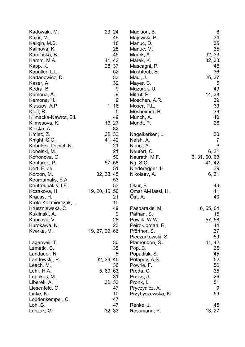
Kadowaki, M. 23, 24Kajor, M. 49Kaligin, M.S. 18Kalinova, K. 25Kaminska, B. 45Kamm, M.A. 41, 42Kapp, K. 26, 37Kapuller, L.L. 52Kartanowicz, D. 33Kaser, A. 39Kedra, B. 9Kemona, A. 9Kemona, H. 9Kiassov, A.P. 1, 18Kiefl, R. 5Klimacka-Nawrot, E.l. 49Klimesova, K. 13, 27Kloska, A. 32Kmiec, Z. 32, 33Knight, S.C. 41, 42Kobelska-Dubiel, N. 21Kobelski, M. 21Kofronova, O. 50Konturek, P. 57, 58Kort, F. de 51Korzon, M. 32, 33, 45Kouroumalis, E.A. 53Koutroubakis, I.E. 53Kozakova, H. 19, 20, 46, 50Krauss, H. 21Krela-Kazmierczak, I. 10Kruszniewska, C. 49Kuklinski, A. 9Kupcová, V. 28Kurokawa, N. 23Kverka, M. 19, 27, 29, 66 Lagerweij, T. 30Lamatic, C. 35Landauer, N. 5Landowski, P. 32, 33, 45Leach, M. 36Lehr, H.A. 5, 60, 63Leppkes, M. 31Liberek, A. 32, 33Liesenfeld, O. 47Linke, K. 10Loddenkemper, C. 47Loh, G. 47Luczak, G. 32, 33
Madison, B. 6Majewski, P. 34Manuc, D. 35Manuc, M. 35Marek, A. 32, 33Marek, K. 32, 33Mascagni, P. 48Mashtoub, S. 36Maul, J. 26, 37Mayer, C. 5Mazurek, U. 49Mitrut, P. 14, 38Moschen, A.R. 39Moser, P.L. 39Mosheimer, B. 39Münch, A. 40Mundt, P. 26 Nagelkerken, L. 30Neish, A. 7Nenci, A. 6Neufert, C. 6, 31Neurath, M.F. 6, 31, 60, 63Ng, S.C. 41, 42Niederegger, H. 39Nikolaev, A. 6, 31 Okur, B. 43Omar Al-Hassi, H. 41Öst, A. 40 Pasparakis, M. 6, 55, 64Pathan, S. 15Pawlik, W.W. 57, 58Peiro-Jordan, R. 44Pförtner, S. 37Pieczarkowski, S. 59Plamondon, S. 41, 42Pop, C. 35Popadiuk, S. 45Potapov, A.S. 52Powrie, F. 50Preda, C. 35Preiss, J. 26Pronk, I. 51Pryczynicz, A. 9Przybyszewska, K. 59 Renke, J. 45Rossmann, P. 13, 27
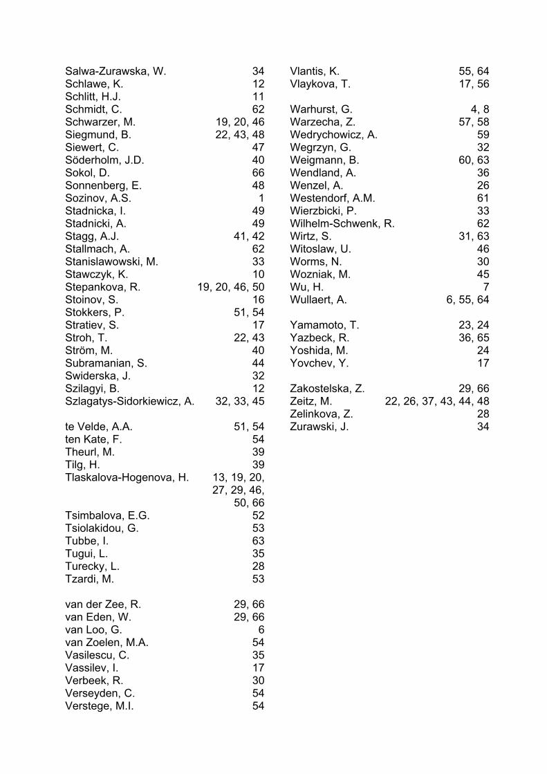
Salwa-Zurawska, W. 34Schlawe, K. 12Schlitt, H.J. 11Schmidt, C. 62Schwarzer, M. 19, 20, 46Siegmund, B. 22, 43, 48Siewert, C. 47Söderholm, J.D. 40Sokol, D. 66Sonnenberg, E. 48Sozinov, A.S. 1Stadnicka, I. 49Stadnicki, A. 49Stagg, A.J. 41, 42Stallmach, A. 62Stanislawowski, M. 33Stawczyk, K. 10Stepankova, R. 19, 20, 46, 50Stoinov, S. 16Stokkers, P. 51, 54Stratiev, S. 17Stroh, T. 22, 43Ström, M. 40Subramanian, S. 44Swiderska, J. 32Szilagyi, B. 12Szlagatys-Sidorkiewicz, A. 32, 33, 45 te Velde, A.A. 51, 54ten Kate, F. 54Theurl, M. 39Tilg, H. 39Tlaskalova-Hogenova, H. 13, 19, 20,
27, 29, 46, 50, 66
Tsimbalova, E.G. 52Tsiolakidou, G. 53Tubbe, I. 63Tugui, L. 35Turecky, L. 28Tzardi, M. 53 van der Zee, R. 29, 66van Eden, W. 29, 66van Loo, G. 6van Zoelen, M.A. 54Vasilescu, C. 35Vassilev, I. 17Verbeek, R. 30Verseyden, C. 54Verstege, M.I. 54
Vlantis, K. 55, 64Vlaykova, T. 17, 56 Warhurst, G. 4, 8Warzecha, Z. 57, 58Wedrychowicz, A. 59Wegrzyn, G. 32Weigmann, B. 60, 63Wendland, A. 36Wenzel, A. 26Westendorf, A.M. 61Wierzbicki, P. 33Wilhelm-Schwenk, R. 62Wirtz, S. 31, 63Witoslaw, U. 46Worms, N. 30Wozniak, M. 45Wu, H. 7Wullaert, A. 6, 55, 64 Yamamoto, T. 23, 24Yazbeck, R. 36, 65Yoshida, M. 24Yovchev, Y. 17 Zakostelska, Z. 29, 66Zeitz, M. 22, 26, 37, 43, 44, 48Zelinkova, Z. 28Zurawski, J. 34
Where medicineand pharmaceuticals meet –
a tried and trusted link
Falk FoundationDr. Falk Pharma
We help
by providing innovative medicines for bowel and liver diseases.
We promote knowledge
from 1967 onwards, there have been more than 200 internationalFalk Symposia and Workshops with over 100,000 participants frommore than 100 countries
1 million doctors and patients have attended over 11,000Falk educational meetings during the last 30 years
our Literature and Patient Information Service –including on-line ordering through our website.
www.falkfoundation.de www.drfalkpharma.de
Leinenweberstr. 5 79108 Freiburg Germany Tel +49 (0) 761/1514-0 Fax +49 (0) 761/1514-321 E-Mail [email protected]