maternal social stress during pregnancy alters immune function and immune cell numbers in adult male...
TRANSCRIPT
y 185 (2007) 95–102www.elsevier.com/locate/jneuroim
Journal of Neuroimmunolog
Maternal social stress during pregnancy alters immune functionand immune cell numbers in adult male Long–Evans
rat offspring during stressful life-events
Alexander A. Götz a,⁎, Sabrina Wittlinger a, Volker Stefanski a,b
a Department of Animal Physiology, University of Bayreuth, Universitätsstraße 30, D-95440 Bayreuth, Germanyb Leibniz Institute for Zoo and Wildlife Research, Alfred-Kowalke-Straße 17, D-10315 Berlin, Germany
Received 1 December 2006; received in revised form 25 January 2007; accepted 29 January 2007
Abstract
The impact of social confrontations on distribution and function of blood immune cells in adult male rat offspring from stressed and non-stressed pregnancies was studied. Repeated 2 h resident–intruder confrontations were performed on ten consecutive days using a protective cage.Prenatally stressed intruder males (PSI) had a generally lower number of neutrophiles, monocytes, T and NK cells and reduced lymphocyteproliferation in whole blood cultures than prenatally non-stressed control intruders (PCI). Differences also existed in the temporal dynamics ofimmunological changes. On confrontation day 1, stress-induced reductions in lymphocyte and monocyte numbers but increased granulocytecounts were observed in both groups. However, only PCI showed a partial recovery of T cell and monocyte numbers on confrontation day 10 and afull restoration in all immune cell numbers 5 days post-confrontation. Thus, the immunological response to a psychosocial stressor in adult rats canbe modified by the mothers’ exposure to stress during pregnancy.© 2007 Elsevier B.V. All rights reserved.
Keywords: Prenatal; Pregnancy; Psychosocial stress; Offspring; Immune cells; Corticosterone
1. Introduction
Prenatal stress can influence the neuroendocrine system andthe behaviour of mammals (Austin et al., 2005; Kapoor et al.,2006; Kofman, 2002; Owen et al., 2005). Although theoffspring from stressed and unstressed pregnancies differ insome behavioural and endocrine aspects under normal lifeconditions, most of the differences become evident when theanimals are exposed to challenging or stressful conditions. Forexample, offspring of stressed pregnancies have a higher HPAaxis activity under immobilisation or exposure to novelenvironment as compared to offspring from unstressed mothers(Barbazanges et al., 1996; Maccari et al., 1995; Vallee et al.,1999; Vallee et al., 1996; Weinstock, 1997). Moreover,prenatally stressed males are often more anxious in behaviouraltests such as the elevated plus maze (Estanislau and Morato,2005; Poltyrev et al., 1996; Vallee et al., 1997; Welberg et al.,
⁎ Corresponding author. Tel.: +49 921 55 2468; fax: +49 921 55 2794.E-mail address: [email protected] (A.A. Götz).
0165-5728/$ - see front matter © 2007 Elsevier B.V. All rights reserved.doi:10.1016/j.jneuroim.2007.01.019
2001), although this effect may be reversed depending on thenature of maternal stressor (Götz and Stefanski, 2007). Prenatalstress has also been linked to decreased pup survival (Lordiet al., 2000; Patin et al., 2002; Pratt and Lisk, 1989; Tuchschereret al., 2002) and increased susceptibility to infection and diseasein later life (Bailey et al., 2004; Herrenkohl, 1986; Kapoor et al.,2006). Despite of its pivotal importance for the health ofindividuals, only few studies so far have investigated the effectsof prenatal stress on the immune system (Coe et al., 1996; Götzand Stefanski, 2007; Kay et al., 1998; Klein and Rager, 1995;Llorente et al., 2002; Sobrian et al., 1997). The outcome of thesestudies, however, was not always uniform: for example,Llorente et al., (2002) found reduced percentages of bloodCD8+ T cells and increased percentages of granulocytes in adultmale rat offspring of mothers exposed to “hanging stress”during pregnancy, whereas (Kay et al., 1998) found nodifferences in the percentages of lymphocyte subsets in theoffspring from mothers of stressed (noise and light) andunstressed pregnancies. Some studies (Kay et al., 1998;Tuchscherer et al., 2002) also indicate lower proliferation
96 A.A. Götz et al. / Journal of Neuroimmunology 185 (2007) 95–102
rates of lymphocytes in response to mitogen stimulation inprenatally stressed offspring. With respect to the offsprings’immune system, only one study (Götz and Stefanski, 2007) hasinvestigated the effect of a maternal social stressor (repeatedsocial confrontations). In this recent report, we found lowernumbers of CD4+ and CD8+ T lymphocytes and decreasedproliferative capacities to pokeweed mitogen stimulation in theblood of prenatally stressed as compared to non-stressed maleoffspring.
With one exception (Llorente et al., 2002), only basal levelsof immune measures were investigated in offspring of stressedpregnancies. Thus, in the present study we investigated thepattern of immunological changes in offspring from stressedand control mothers in response to a social stressor in later life.Maternal stress was induced by using an established psycho-social stress model in female rats (Stefanski et al., 2005). Adultoffspring from stressed and control mothers were theninvestigated in social confrontations which have been shownto result in marked endocrine and immunological changes inadult laboratory rats (Koolhaas et al., 1997; Raab et al., 1986;Stefanski, 2000; Stefanski and Engler, 1998; Stefanski andEngler, 1999).
2. Materials and methods
2.1. Animals and standard housing conditions
Adult male–female pairs of Long–Evans rats, descendantsof an outbred stock originally obtained from Harlan Winkel-mann (Borchen, Germany), were kept in standard polycarbon-ate cages (26×42×15 cm). The animals had ad libitum accessto rat standard diet and water, and were kept under controlledconditions on a 12:12-h light/dark cycle (lights off at 01:00 h).Temperature was 20 ±1 °C, and the relative humidityapproximately 50%. Cages were cleaned weekly, but nocleaning was done during the confrontation period (see Section2.3). All procedures were approved by the local authority forlaboratory animal care and use (Regierung von Mittelfranken,Ansbach, Germany).
2.2. Maternal stress (female confrontation)
Maternal stress was induced by using a resident–intruderconfrontation procedure. Confrontations were conducted in thedark period by transferring a pregnant intruder female into thehome cage of an unfamiliar resident female in a separate room.Except during the confrontations, resident females were housedwith a male partner. Female–female confrontation resulted indyadic aggressive interactions and in social defeat of theintruder female, but biting and wounding was absent. Intruderfemales were confronted daily for 2 h (continuous coexistence)for a period of 2 months, using a daily rotation system withdifferent unfamiliar resident females (each resident female wasused at least every other day). For further details see Götz andStefanski (2007), Stefanski and Gruner (2006) and Stefanski etal. (2005). Experimental females were randomly assigned tomaternal stress or control group and were 3–4 months old at the
start of the confrontations (equalling the start of their secondpregnancy). Control females remained undisturbed in theirhome cages. In all experiments litter of the third pregnancy wereused.
2.3. Prenatally stressed offspring and experimental design
After delivery no further confrontations were conducted andthe male mating partner of each pair was removed from thestandard housing cage to assure undisturbed weaning conditionsfor offspring from stressed (prenatal stress, PS) and controlfemales (prenatal control, PC). The young were kept with theirmothers until weaning (postnatal day 21). One or two male–female pairs from each litter were used in the present study andkept in standard cages as described before. Experimentalprocedures were performed on 20 prenatal control males,descendants from 16 control mothers, and 24 prenatally stressedmales, descendants from 19 females stressed during pregnancy,at an age of 5–6 months (weighing approximately 450 g). Thepresent report is part of a long-term survey on the effects ofprenatal stress on the adult male progeny in Long–Evans rats(see also Götz and Stefanski, 2007).
2.4. Social stress procedure of offspring (resident–intruder-confrontation using protective cage)
For the induction of social defeat stress an experimental male(= intruder) was placed in the home cage (100×50×70 cm) ofan adult male resident rat (for methodological details of theconfrontation procedure see (Engler and Stefanski, 2003;Stefanski et al., 2003)). In all confrontations, adult maleoffspring from stressed mothers (PS-intruders (PSI) and controlmothers (PC-intruders (PCI)) were attacked and subdued by therespective residents within the first 10 min. Social defeat wasdefined successful if the intruder showed defensive behaviouras indicated by submissive body posture (lying motionless onthe back with ventral surface exposed to the opponent) or flightbehaviour (direct movement away from the opponent at runningpace). In contrast to continuous confrontations with directphysical contact between the two opponents, in the presentstudy the intruder was separated from the resident bytransferring it to a protective wire mesh cage (50×15×30 cm)located within the resident's enclosure immediately after socialdefeat. This procedure assured that the intruder was protectedfrom repeated attacks and potential injury, while it was stillexposed to visual, acoustic and olfactory cues from the resident.The intruder was kept for two hours in the protective cagebefore returning to its female partner (see Section 2.1). Thesocial defeat procedure was conducted on ten consecutive daysusing a daily rotation system and started during the middle ofthe active (dark) period between 06:00 h and 08:00 h.
2.5. Blood sampling
Blood samples were always taken between 08:00 and10:00 h from male offspring (1) 5 days prior to the start ofconfrontations (base value), (2) immediately after the first day
97A.A. Götz et al. / Journal of Neuroimmunology 185 (2007) 95–102
(confrontation day 1), (3) after the 10th day (confrontation day10) in protective cage and (4) 5 days after the final confrontation(post5). Blood was taken from PCI and PSI from the tail veinwithout anaesthesia (Stefanski, 1998). About 0.5 ml of bloodwas collected in several tubes (uncoated tubes for serumcorticosterone (CORT) measurement, K2-EDTA-coated tubesfor FACS-analysis and heparinized tubes for activity tests). TheCORT sample was usually collected within 1–3 min, and within4 min for immune parameters from entering the experimentalroom. For determination of serum corticosterone concentra-tions, serum was separated by centrifugation and stored at−20 °C until further analysis. Blood for immunologicalmeasurements was used within 2 h after blood sampling.
2.6. Corticosterone measurement
Total serum corticosterone concentration was measuredusing a standard radioimmunoassay (Foster and Dunn, 1974).The specific antibody was kindly provided by the Institute ofPharmacology, University of Heidelberg, Germany. Antibodycross-reactivity with other relevant steroids was 4.4% (cortisol),30% (deoxy-corticosterone) and 5.5% (testosterone). The sen-sitivity of the corticosterone radioimmunoassay was 5 ng/ml.The intraasssay coefficient of variation was 0.4% and interassaycoefficient of variation was 9.9%. Data of animals from whichnot all four CORT samples were obtained in a 3 minute timelimit were discarded from analysis.
2.7. Leukocyte counts and subpopulations
Total leukocyte counts were determined on an automatic cellcounter (Z2, Beckman-Coulter, Miami, FL). For leukocytephenotyping, blood aliquots were incubated for 20 min at roomtemperature with FITC-conjugated anti-rat myeloid cells (clone
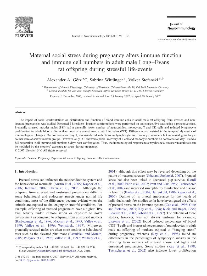
Fig. 1. Serum corticosterone concentration of prenatal control intruders (PCI)(n=18; hatched bars) and prenatally stressed intruder males (PSI males) (n=23;grey bars) 5 days before (base value), on day one (day 1) and day ten (day 10) ofconfrontation as well as 5 days post-confrontation (post5). RM-ANOVA:postnatal confrontation: F3,39=35.919, Pb0.001; postnatal confrontation×pre-natal stress: F3,39=0.845, P=0.472; prenatal stress: F1,39=0.287, P=0.595.Post-hoc: paired t-test (within group differences): #different from any other datapoint, at least Pb0.05.
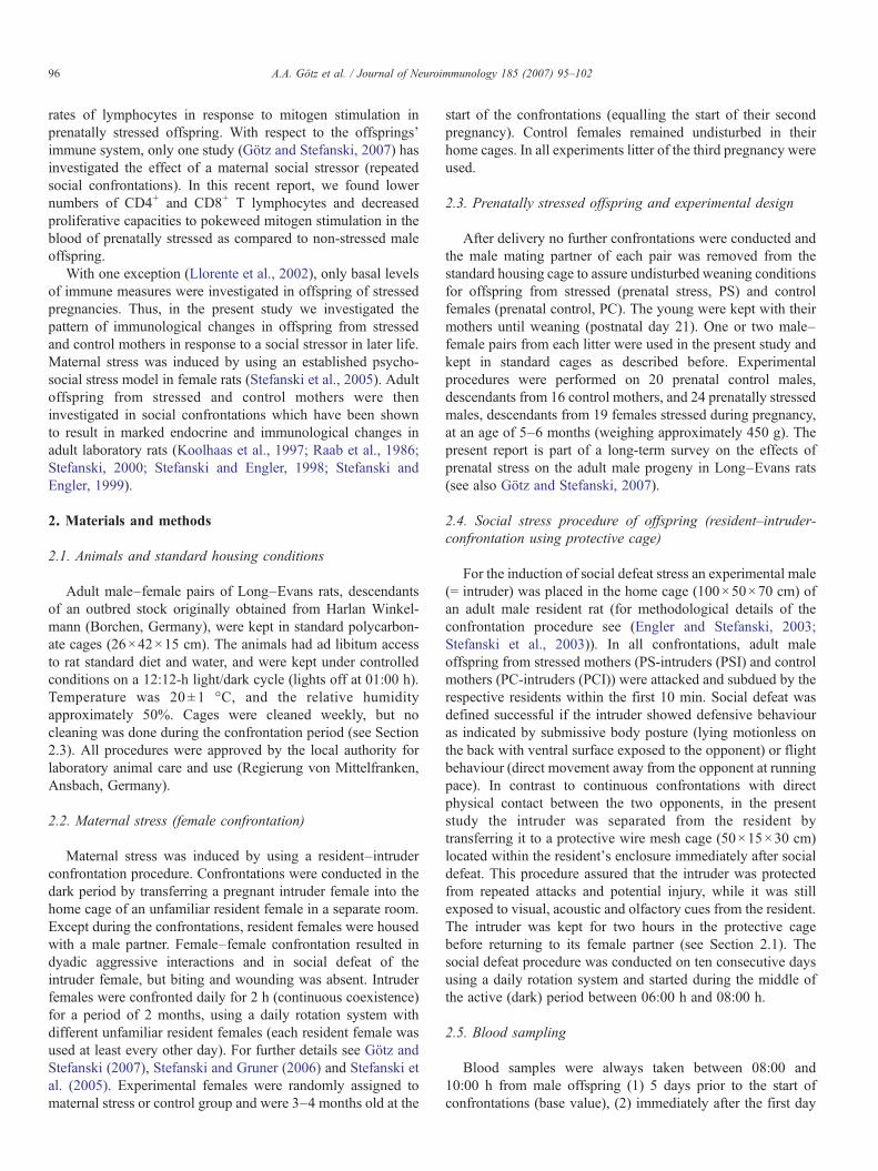
Fig. 2. Lymphocyte (LYM), Monocyte (MON) and Granulocyte (GRAN) countsof PCI (n=20; hatched bars) and PSI males (n=24; grey bars) 5 days before(base value), on day one and day ten of confrontation as well as 5 days post-confrontation (post5). RM-ANOVA: Lymphocytes: postnatal confrontation:F3,42 = 141.658, P b0.001; postnatal confrontation × prenatal stress:F3,42=2.865, P=0.039; prenatal stress: F1,42=6.735, P=0.013; Monocytes:postnatal confrontation: F3,42=21.738, Pb0.001; postnatal confrontation×pre-natal stress: F3,42=0.632, P=0.596; prenatal stress: F1,42=5.976, P=0.019;Granulocytes: postnatal confrontation: F3,42 =3.801, P=0.012; postnatalconfrontation×prenatal stress: F3,42 = 0.877, P=0.455; prenatal stress:F1,42=6.320, P=0.016. Post-hoc analysis: unpaired t-test (between groups):⁎Pb0.05; ⁎Pb0.01; paired t-test (within group): 1significantly different frombase value: Pb0.05, 4significantly different from post5: Pb0.05, #significantlydifferent from any other data point: Pb0.05.
98 A.A. Götz et al. / Journal of Neuroimmunology 185 (2007) 95–102
ED9), PE-conjugated anti-rat CD45/LCA (clone OX-1), FITC-conjugated anti-rat CD4 (clone OX-38), FITC-conjugated anti-rat CD8b (clone 341), FITC-conjugated anti-rat NKR-P1A(clone 10/78), FITC-conjugated anti-rat CD45RA (clone OX-33)or PE-conjugated anti-rat CD3 (clone G4.18). Monoclonalantibodies were obtained from BD Pharmingen (Heidelberg,Germany) and Serotec (Düsseldorf, Germany). Antibodylabeling was performed following the standard protocols andusing FACS lysing solution (BD Immunocytometry Systems,Heidelberg, Germany) supplemented by PBS (Dulbecco's PBSwithout calcium and magnesium, 2% FBS, 0.1% NaN3). Tenthousand cells from each sample were analysed on a FACSCa-libur flow cytometer (BD Immunocytometry Systems, Heidel-berg, Germany) using the software Attractors and CellQuest.
Lymphocytes, monocytes and neutrophil granulocytes wereidentified by their forward and side scatter characteristics and their
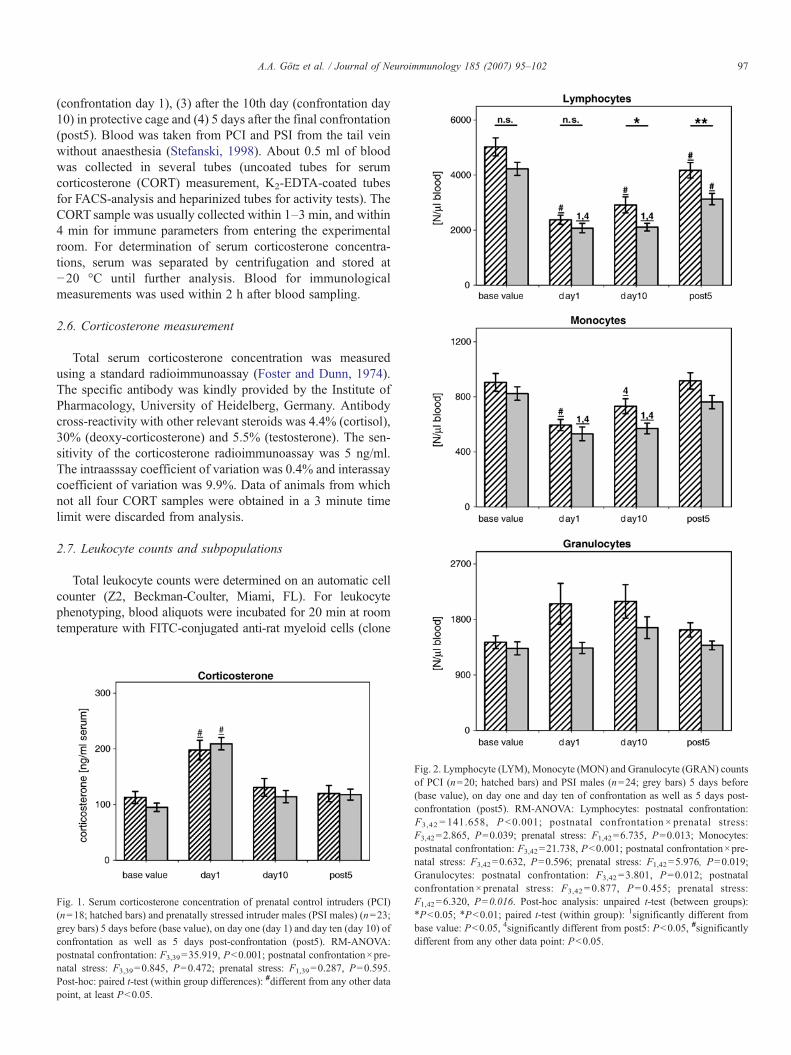
Fig. 3. T cell (CD4+ and CD8+), B cell and Natural killer (NK) cell counts of PCI (n=day one and day ten of confrontation as well as 5 days post-confrontation (post5).postnatal confrontation×prenatal stress: F3,42=2.380, P=0.073; prenatal stress: FPb0.001; postnatal confrontation×prenatal stress: F3,42=1.964, P=0.123; prenatalPb0.001; postnatal confrontation×prenatal stress: F3,42=1.767, P=0.157; prenatalPb0.001; postnatal confrontation×prenatal stress: F3,42=1.128, P=0.340; prenatgroups) and paired t-test (within group); for details see Fig. 2.
differences in CD45 and ED9 expression. Lymphocyte sub-populations were identified by a combination of lineage specificsurface markers: CD3+/CD4+ (T helper cells), CD3+/CD8b+
(cytotoxic Tcells), CD3−/NKR-P1A+ (NK cells), CD45RA+ (Bcells). Monocytes (CD3−/CD4+) were excluded from thelymphocyte gate.
2.8. Proliferative response
Whole blood mitogenic stimulation assays were performendto determine the proliferative capacity of lymphocytes usingConcanavalin A (ConA,) (predominantly stimulating T cells)and pokeweed mitogen (PWM) (stimulating B and T cells)(both Sigma, Deisenhofen, Germany). In brief, heparinizedblood was diluted 1:10 with RPMI-10 (RPMI-1640 supple-mented with 10% FCS and 50 μg/ml gentamicin). One hundred
20; hatched bars) and PSI males (n=24; grey bars) 5 days before (base value), onRM-ANOVA: CD4+ T cells: postnatal confrontation: F3,42=82.483, Pb0.001;
1,42=6.883, P=0.012; CD8+ T cells: postnatal confrontation: F3,42=71.692,
stress: F1,42=4.418, P=0.042; B cells: postnatal confrontation: F3,42=184.038,stress: F1,42=1.071, P=0.307; NK cells: postnatal confrontation: F3,42=6.428,al stress: F1,42=6.567, P=0.014. Post-hoc analysis: unpaired t-test (between
99A.A. Götz et al. / Journal of Neuroimmunology 185 (2007) 95–102
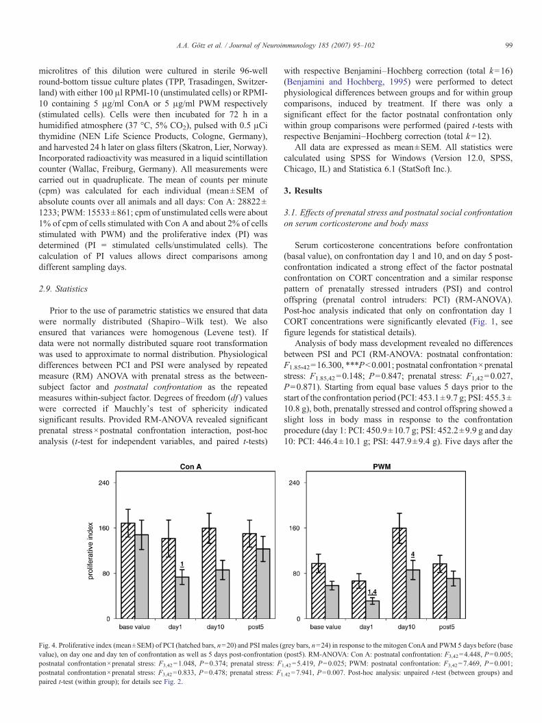
microlitres of this dilution were cultured in sterile 96-wellround-bottom tissue culture plates (TPP, Trasadingen, Switzer-land) with either 100 μl RPMI-10 (unstimulated cells) or RPMI-10 containing 5 μg/ml ConA or 5 μg/ml PWM respectively(stimulated cells). Cells were then incubated for 72 h in ahumidified atmosphere (37 °C, 5% CO2), pulsed with 0.5 μCithymidine (NEN Life Science Products, Cologne, Germany),and harvested 24 h later on glass filters (Skatron, Lier, Norway).Incorporated radioactivity was measured in a liquid scintillationcounter (Wallac, Freiburg, Germany). All measurements werecarried out in quadruplicate. The mean of counts per minute(cpm) was calculated for each individual (mean±SEM ofabsolute counts over all animals and all days: Con A: 28822±1233; PWM: 15533±861; cpm of unstimulated cells were about1% of cpm of cells stimulated with Con A and about 2% of cellsstimulated with PWM) and the proliferative index (PI) wasdetermined (PI = stimulated cells/unstimulated cells). Thecalculation of PI values allows direct comparisons amongdifferent sampling days.
2.9. Statistics
Prior to the use of parametric statistics we ensured that datawere normally distributed (Shapiro–Wilk test). We alsoensured that variances were homogenous (Levene test). Ifdata were not normally distributed square root transformationwas used to approximate to normal distribution. Physiologicaldifferences between PCI and PSI were analysed by repeatedmeasure (RM) ANOVA with prenatal stress as the between-subject factor and postnatal confrontation as the repeatedmeasures within-subject factor. Degrees of freedom (df ) valueswere corrected if Mauchly's test of sphericity indicatedsignificant results. Provided RM-ANOVA revealed significantprenatal stress×postnatal confrontation interaction, post-hocanalysis (t-test for independent variables, and paired t-tests)
Fig. 4. Proliferative index (mean±SEM) of PCI (hatched bars, n=20) and PSI males (value), on day one and day ten of confrontation as well as 5 days post-confrontationpostnatal confrontation×prenatal stress: F3,42=1.048, P=0.374; prenatal stress: F1
postnatal confrontation×prenatal stress: F3,42=0.833, P=0.478; prenatal stress: F1
paired t-test (within group); for details see Fig. 2.
with respective Benjamini–Hochberg correction (total k=16)(Benjamini and Hochberg, 1995) were performed to detectphysiological differences between groups and for within groupcomparisons, induced by treatment. If there was only asignificant effect for the factor postnatal confrontation onlywithin group comparisons were performed (paired t-tests withrespective Benjamini–Hochberg correction (total k=12).
All data are expressed as mean±SEM. All statistics werecalculated using SPSS for Windows (Version 12.0, SPSS,Chicago, IL) and Statistica 6.1 (StatSoft Inc.).
3. Results
3.1. Effects of prenatal stress and postnatal social confrontationon serum corticosterone and body mass
Serum corticosterone concentrations before confrontation(basal value), on confrontation day 1 and 10, and on day 5 post-confrontation indicated a strong effect of the factor postnatalconfrontation on CORT concentration and a similar responsepattern of prenatally stressed intruders (PSI) and controloffspring (prenatal control intruders: PCI) (RM-ANOVA).Post-hoc analysis indicated that only on confrontation day 1CORT concentrations were significantly elevated (Fig. 1, seefigure legends for statistical details).
Analysis of body mass development revealed no differencesbetween PSI and PCI (RM-ANOVA: postnatal confrontation:F1.85,42=16.300, ⁎⁎⁎Pb0.001; postnatal confrontation×prenatalstress: F1.85,42=0.148; P=0.847; prenatal stress: F1,42=0.027,P=0.871). Starting from equal base values 5 days prior to thestart of the confrontation period (PCI: 453.1±9.7 g; PSI: 455.3±10.8 g), both, prenatally stressed and control offspring showed aslight loss in body mass in response to the confrontationprocedure (day 1: PCI: 450.9±10.7 g; PSI: 452.2±9.9 g and day10: PCI: 446.4±10.1 g; PSI: 447.9±9.4 g). Five days after the
grey bars, n=24) in response to the mitogen ConA and PWM 5 days before (base(post5). RM-ANOVA: Con A: postnatal confrontation: F3,42=4.448, P=0.005;
,42=5.419, P=0.025; PWM: postnatal confrontation: F3,42=7.469, P=0.001;
.42=7.941, P=0.007. Post-hoc analysis: unpaired t-test (between groups) and
100 A.A. Götz et al. / Journal of Neuroimmunology 185 (2007) 95–102
end of the confrontation period both groups had recovered inbodymass andwere heavier than before the confrontation period(PCI: 460.7±9.8 g; PSI: 464.7±9.3 g).
3.2. Effects of prenatal stress and postnatal social confrontationon number of blood immune cells
RM-ANOVA showed that postnatal confrontation had asignificant impact on all immune cells investigated in bothintruder groups (PCI and PSI) (Fig. 2, see figure legends forstatistical details). The confrontation resulted in decreasednumbers of lymphocytes (LYM) and monocytes (MON) butincreased numbers of neutrophile granulocytes (GRA). Thedecrease of lymphocyte number was a combined result ofsignificant reductions of the number of CD4+ and CD8+ T cells,B cells, and to a lower extent NK cells (Fig. 3, see figurelegends for statistical details).
In addition, several significant differences existed betweenthe PCI and PSI males. The numbers of monocytes,granulocytes, T cells, and NK cells were generally lower inPSI males than in PCI males during the whole investigationperiod. Moreover, interaction between the factors postnatalconfrontation×prenatal stress indicates differences in thetemporal dynamics of lymphocyte and especially T cellnumbers. Detailed post-hoc analysis indicated similarly reducedCD4+ and CD8+ T cell numbers at confrontation day 1 in PCIand PSI males. However, only the PCI group showed partiallyrecovered T cell numbers on day 10 and fully restored numberspost-confrontation.
3.3. Effects of prenatal stress and postnatal social confrontationon function of blood immune cells
Postnatal confrontation significantly affected the capabilityof lymphocytes to proliferate in response to mitogen stimulation(ConA and PWM). In addition, RM-ANOVA indicatedsignificant group differences. The proliferative capacity toboth mitogens was generally lower in PSI males than in PCImales during the whole investigation period (Fig. 4, see figurelegends for statistical details).
4. Discussion
This study demonstrates that exposure to prenatal stressmodifies the magnitude and the pattern of social stress-inducedimmune system changes in the adult offspring. First, however,we focus on the general effects of confrontation that wereevident in both offspring groups. Social confrontation caused astrong increase in corticosterone concentrations in the intrudermales on confrontation day 1 but not on day 10. Together withthe observation that confronted males showed only a slight lossin body mass after ten consecutive days of confrontation, onewould conclude that the males adapt to the repeated socialstressor. However, the immunological data point to a sustainedeffect of social stress on the immune system and thereforesuggest that the immune system ranges among the biologicalsystems very sensitive to stress. The pattern of immunological
changes in protective cage intruders in the present report wascharacterized by reduced numbers and function of lymphocytesand an increased number of neutrophiles. This pattern is inaccordance with previous chronic social stress studies onmammals (Engler et al., 2004; Raab et al., 1986; Stefanski andEngler, 1998; Stefanski et al., 1996; von Holst, 1998) and mayindicate a possible shift in the balance of the immune systemfrom specific towards unspecific immune system dominance inthe peripheral blood. Because the protective cage drasticallyreduces the possibility of undesired wounding and subsequentinflammatory processes involving the immune system it appearsto be an advantageous system to investigate stress-inducedimmune alterations in social confrontations.
In response to social confrontation, PSI differed from PCImales in two main aspects. First, with the exception of B cellnumbers, PSI males had a lower number and function of allimmune cells investigated. We recently reported that suchdifferences exist under non-stress (basal) conditions (Götz andStefanski, 2007) and now demonstrate that this prenatal stressassociated pattern is also evident in confrontations and extendsinto the post-confrontational period. It should be noted that thelower proliferative response in PSI males is probably not aconsequence of reduced lymphocyte numbers in the blood,because the proliferative index takes into account differences inlymphocyte numbers. Importantly, PSI and PCI males showed adifferent recovery pattern during confrontation. In contrast to PCImales, PSI males showed neither a partial recovery of Tlymphocyte and monocyte numbers during confrontation nor afull recovery after the end of the confrontations. This indicates thatstress-induced alterations are not quickly reversible in PSI males.
The lower overall levels of several immune measures in PSImales could mirror a displacement of setpoints for numbers andfunction of immune cells in the peripheral blood. Such adisplacement may be a consequence of an altered receptordensity on immune or/and endothelial cells caused bydifferential exposure to stress hormones such as corticosteroneduring intrauterine development. Differences in receptorsensitivity could be a possible explanation why immune cellsin PSI males react more strongly to a similar concentration of aspecific hormone such as corticosterone. Although prenatalstress-associated changes of hormone receptors on immune andendothelial cells have not been investigated yet, it has beenshown that prenatal stress alters glucocorticoid type I and type IIreceptor density in other body regions such as the hippocampus(Henry et al., 1994; Maccari et al., 1995). With respect to thedifferential recovery pattern of immune cell numbers in PCI andPSI males, differences in the stress perception in between couldalso play a role. Although there was no difference in CORTconcentrations, other stress-related hormones such as catecho-lamines might be more strongly increased in the PSI intruders.Future investigations should thus include activity measures ofthe sympathetico-adrenalmedullary system.
Stress-induced immune changes of a magnitude as observedin the present study could be of health relevance as they mayimpair antibody production (Fleshner et al., 1995) and increasetumor metastasis (Stefanski and Ben-Eliyahu, 1996). BecausePSI males had lower number and function of immune cells, one
101A.A. Götz et al. / Journal of Neuroimmunology 185 (2007) 95–102
might assume a higher risk for health in this group, especiallyunder stressful condition. However, this assumption must betaken with caution as the results are limited to the peripheralblood. In future investigations the study of lymphatic organs, invivo challenge of specific immune subsystems, and the use ofanimal disease models could help to better evaluate thebiological relevance of the immunological findings. Neverthe-less, the fact that a relatively naturalistic mild maternal socialstressor has the potential to influence the immunologicalreaction of male offspring under a relatively mild stressfulcondition in adulthood is an important finding: it leads to theconclusion that maternal stress effects on offspring can easily beoverlooked or underestimated when testing offspring immunity.The present findings are therefore of relevance for normalpregnancies in mammals including humans, where moderatedisturbances induced by social conflicts occur in daily life.
The study of Lorente (Llorente et al., 2002) is of specialinterest for the present report, because prenatally stressedoffspring was investigated under challenge condition inadulthood (repeated immobilisation for 10 days). Similar tothe present study, the authors showed that prenatal stressreduced basal lymphocyte numbers in the blood of adultoffspring. However, the effects of immobilisation stress wereonly minor, and a reduced percentage of blood CD8+ T cellswas observed in the control rather than in the prenatal stressoffspring. Several methodological differences such as thematernal stress procedure (“hanging stress”) or the use of adifferent rat strain (Sprague–Dawley rats) may have contributedto the differential outcome. We would rate the nature of thestressor (challenge) among the most important factors, as thephysiological stress response can substantially differ betweenphysical and social stressors (Sgoifo et al., 1996).
In prenatal stress studies, pups can be raised either with theirnative mothers or with foster dams. We have chosen the firstcondition because this is the most naturalistic raising conditionfrom a biological point of view (see also Götz and Stefanski,2007). But although the period in utero was probably mostrelevant for the observed differences between PSI and PCImales, one cannot fully exclude that postnatal maternal nurtureand care also differed between stressed and control mothers.Thus, it would be worthwhile to include cross-fostered pups insubsequent studies. Regardless of the possible contribution ofpostnatal environment however, the consequences for theoffspring later in life were purely based on the exposure ofthe mother to social stress during pregnancy.
In conclusion our results demonstrate for the first time thatmaternal social stress during pregnancy affects the reactionpattern of the immune system to social stress in the adultoffspring in rats.
Acknowledgements
The present work was supported by the German ResearchFoundation (DFG) (project number: STE 633/5–1). We thankHeiko Rödel for his helpful advice in statistical analysis, andAndrea Berger and Inge Zerenner-Fritzsche for their excellenttechnical assistance.
References
Austin, M.-P., Leader, L.R., Reilly, N., 2005. Prenatal stress, the hypothalamic-pituitary-adrenal axis, and fetal and infant neurobehaviour. Early Hum. Dev.81, 917–926.
Bailey, M.T., Lubach, G.R., Coe, C.L., 2004. Prenatal stress alters bacterialcolonization of the gut in infant monkeys. J. Pediatr. Gastroenterol. Nutr.414–421.
Barbazanges, A., Piazza, P.V., Le Moal, M., Maccari, S., 1996. Maternalglucocorticoid secretion mediates long-term effects of prenatal stress.J. Neurosci. 16, 3943–3949.
Benjamini, Y., Hochberg, Y., 1995. Controlling the false discovery rate: apractical and powerful approach to multiple testing. J. R. Stat. Soc. B 57,289–300.
Coe, C.L., Lubach, G.R., Karaszewski, J.W., Ershler, W.B., 1996. Prenatalendocrine activation alters postnatal cellular immunity in infant monkeys.Brain Behav. Immun. 10, 221–234.
Engler, H., Stefanski, V., 2003. Social stress and T cell maturation in male rats:transient and persistent alterations in thymic function. Psychoneuroendo-crinology 28, 951–969.
Engler, H., Dawils, L., Hoves, S., Kurth, S., Stevenson, J.R., Schauenstein, K.,Stefanski, V., 2004. Effects of social stress on blood leukocyte distribution:the role of alpha- and beta-adrenergic mechanisms. J. Neuroimmunol. 156,153–162.
Estanislau, C., Morato, S., 2005. Prenatal stress produces more behavioralalterations than maternal separation in the elevated plus-maze and in theelevated T-maze. Behav. Brain Res. 163, 70–77.
Fleshner, M., Hermann, J., Lockwood, L.L., Laudenslager, M.L., Watkins, L.R.,Maier, S.F., 1995. Stressed rats fail to expand the CD45RC+CD4+ (Th1-like)T cell subset in response to KLH: possible involvement of IFN-gamma. BrainBehav. Immun. 9, 101–112.
Foster, L.B., Dunn, R.T., 1974. Single-antibody technique for radioimmunoas-say of cortisol in unextracted serum or plasma. Clin. Chem. 20, 365–368.
Götz, A., Stefanski, V., 2007. Psychosocial maternal stress during pregnancyaffects serum corticosterone, blood immune parameters and anxietybehaviour in adult male rat offspring. Physiol. Behav. 90, 108–115.
Henry, C., Kabbaj, M., Simon, H., Le Moal, M., Maccari, S., 1994. Prenatalstress increases the hypothalamo-pituitary-adrenal axis response in youngand adult rats. J. Neuroendocrinol. 6, 341–345.
Herrenkohl, L.R., 1986. Prenatal stress disrupts reproductive behavior andphysiology in offspring. Ann. N YAcad Sci. 474, 120–128.
Kapoor, A., Dunn, E., Kostaki, A., Andrews, M.H., Matthews, S.G., 2006. Fetalprogramming of hypothalamo-pituitary-adrenal function: prenatal stress andglucocorticoids. J. Physiol. 572, 31–44.
Kay, G., Tarcic, N., Poltyrev, T., Weinstock, M., 1998. Prenatal stress depressesimmune function in rats. Physiol. Behav. 63, 397–402.
Klein, S.L., Rager, D.R., 1995. Prenatal stress alters immune function in theoffspring of rats. Dev. Psychobiol. 28, 321–326.
Kofman, O., 2002. The role of prenatal stress in the etiology of developmentalbehavioural disorders. Neurosci. Biobehav. Rev. 26, 457–470.
Koolhaas, J.M., Meerlo, P., De Boer, S.F., Strubbe, J.H., Bohus, B., 1997. Thetemporal dynamics of the stress response. Neurosci. Biobehav. Rev. 21,775–782.
Llorente, E., Brito, M.L., Machado, P., Gonzalez, M.C., 2002. Effect of prenatalstress on the hormonal response to acute and chronic stress and on immuneparameters in the offspring. J. Physiol. Biochem. 58, 143–149.
Lordi, B., Patin, V., Protais, P., Mellier, D., Caston, J., 2000. Chronic stress inpregnant rats: effects on growth rate, anxiety and memory capabilities of theoffspring. Int. J. Psychophysiol. 37, 195–205.
Maccari, S., Piazza, P.V., Kabbaj, M., Barbazanges, A., Simon, H., Le Moal, M.,1995. Adoption reverses the long-term impairment in glucocorticoidfeedback induced by prenatal stress. J. Neurosci. 15, 110–116.
Owen, D., Andrews, M.H., Matthews, S.G., 2005. Maternal adversity,glucocorticoids and programming of neuroendocrine function and behav-iour. Neurosci. Biobehav. Rev. 29, 209–226.
Patin, V., Lordi, B., Vincent, A., Thoumas, J.L., Vaudry, H., Caston, J., 2002.Effects of prenatal stress on maternal behavior in the rat. Brain Res. Dev.Brain Res. 139, 1–8.
102 A.A. Götz et al. / Journal of Neuroimmunology 185 (2007) 95–102
Poltyrev, T., Keshet, G.I., Kay, G., Weinstock, M., 1996. Role of experimentalconditions in determining differences in exploratory behavior of prenatallystressed rats. Dev. Psychobiol. 29, 453–462.
Pratt, N.C., Lisk, R.D., 1989. Effects of social stress during early pregnancy onlitter size and sex ratio in the golden hamster (Mesocricetus auratus).J. Reprod. Fertil. 87, 763–769.
Raab, A., Dantzer, R., Michaud, B., Mormede, P., Taghzouti, K., Simon, H., LeMoal, M., 1986. Behavioural, physiological and immunological conse-quences of social status and aggression in chronically coexisting resident–intruder dyads of male rats. Physiol. Behav. 36, 223–228.
Sgoifo, A., de Boer, S.F., Haller, J., Koolhaas, J.M., 1996. Individual differencesin plasma catecholamine and corticosterone stress responses of wild-typerats: relationship with aggression. Physiol. Behav. 60, 1403–1407.
Sobrian, S.K., Vaughn, V.T., Ashe,W.K., Markovic, B., Djuric, V., Jankovic, B.D.,1997. Gestational exposure to loud noise alters the development and postnatalresponsiveness of humoral and cellular components of the immune system inoffspring. Environ. Res. 73, 227–241.
Stefanski, V., 1998. Social stress in loser rats: opposite immunological effects insubmissive and subdominant males. Physiol. Behav. 63, 605–613.
Stefanski, V., 2000. Social stress in laboratory rats: hormonal responses andimmune cell distribution. Psychoneuroendocrinology 25, 389–406.
Stefanski, V., Ben-Eliyahu, S., 1996. Social confrontation and tumor metastasisin rats: defeat and beta-adrenergic mechanisms. Physiol. Behav. 60,277–282.
Stefanski, V., Engler, H., 1998. Effects of acute and chronic social stress onblood cellular immunity in rats. Physiol. Behav. 64, 733–741.
Stefanski, V., Engler, H., 1999. Social stress, dominance and blood cellularimmunity. J. Neuroimmunol. 94, 144–152.
Stefanski, V., Gruner, S., 2006. Gender difference in basal and stress levels ofperipheral blood leukocytes in laboratory rats. Brain Behav. Immun. 20,369–377.
Stefanski, V., Solomon, G.F., Kling, A.S., Thomas, J., Plaeger, S., 1996. Impactof social confrontation on rat CD4 Tcells bearing different CD45R isoforms.Brain Behav. Immun. 10, 364–379.
Stefanski, V., Peschel, A., Reber, S., 2003. Social stress affects migration ofblood T cells into lymphoid organs. J. Neuroimmunol. 138, 17–24.
Stefanski, V., Raabe, C., Schulte, M., 2005. Pregnancy and social stress infemale rats: influences on blood leukocytes and corticosterone concentra-tions. J. Neuroimmunol. 162, 81–88.
Tuchscherer, M., Kanitz, E., Otten, W., Tuchscherer, A., 2002. Effects ofprenatal stress on cellular and humoral immune responses in neonatal pigs.Vet. Immunol. Immunopathol. 86, 195–203.
Vallee, M., Mayo, W., Maccari, S., Le Moal, M., Simon, H., 1996. Long-termeffects of prenatal stress and handling on metabolic parameters: relationshipto corticosterone secretion response. Brain Res. 712, 287–292.
Vallee, M., Mayo, W., Dellu, F., Le Moal, M., Simon, H., Maccari, S., 1997.Prenatal stress induces high anxiety and postnatal handling induces lowanxiety in adult offspring: correlation with stress-induced corticosteronesecretion. J. Neurosci. 17, 2626–2636.
Vallee, M., MacCari, S., Dellu, F., Simon, H., Le Moal, M., Mayo, W., 1999.Long-term effects of prenatal stress and postnatal handling on age-relatedglucocorticoid secretion and cognitive performance: a longitudinal study inthe rat. Eur. J. Neurosci. 11, 2906–2916.
von Holst, D., 1998. The concept of stress and its relevance for animal behavior.Adv. Stud. Behav. 27, 1–131.
Weinstock, M., 1997. Does prenatal stress impair coping and regulation ofhypothalamic-pituitary-adrenal axis? Neurosci. Biobehav. Rev. 21, 1–10.
Welberg, L.A., Seckl, J.R., Holmes, M.C., 2001. Prenatal glucocorticoidprogramming of brain corticosteroid receptors and corticotrophin-releasinghormone: possible implications for behaviour. Neuroscience 104, 71–79.