maternal obesity and increased risk for autism and developmental delay among very preterm infants
TRANSCRIPT
ORIGINAL ARTICLE
Maternal obesity and increased risk for autism anddevelopmental delay among very preterm infantsLC Reynolds1,2, TE Inder2,3,4, JJ Neil3,4, RG Pineda2,5 and CE Rogers1,2
OBJECTIVE: Thirty-five percent of women of child-bearing age are obese, and there is evidence that maternal obesity may increasethe risk for adverse neurodevelopmental outcome. However, research regarding obesity and neurodevelopment among childrenborn preterm is limited. This study aimed to determine associations between maternal obesity and neurodevelopment in verypreterm children at age 2 years.STUDY DESIGN: Maternal/infant dyads (n= 62) born ⩽ 30 weeks gestation were enrolled in a prospective cohort study at a level-IIIneonatal intensive care unit. Mothers were classified as obese or non-obese based on pre-pregnancy body mass index. Infantsunderwent magnetic resonance imaging at term equivalent and developmental testing at age 2. Maternal obesity was investigatedfor associations with neurodevelopment.RESULT: Maternal obesity was associated with positive screen for autism (odds ratio = 9.88, P= 0.002) and lower compositelanguage scores (β=− 9.36, (confidence interval =− 15.11, − 3.61), P= 0.002).CONCLUSION: Maternal obesity was associated with adverse neurodevelopmental outcome at age 2 in this cohort of very pretermchildren. This study requires replication, but may support targeted surveillance of infants born to women with maternal obesity.
Journal of Perinatology (2014) 34, 688–692; doi:10.1038/jp.2014.80; published online 8 May 2014
INTRODUCTIONIn the United States, the rate of obesity is steadily rising.1,2 Amongwomen of child-bearing age, approximately 64% are overweightand 35% are obese.3 Maternal obesity is a predictor of adversehealth conditions in pregnancy, such as pre-eclampsia, gestationaldiabetes, inflammation and increased risk for maternal death.4
Subsequently, the hospital costs associated with childbirth forwomen with maternal obesity is an average of $4000 more thanamong women without obesity.5 Maternal obesity not only affectsmaternal health and healthcare expenditure, but it is an importantpredictor for infant and childhood outcomes. There is evidencethat maternal obesity may increase the risk for poor neurodeve-lopmental outcomes in term-born infants. The associationbetween maternal obesity and impaired cognitive developmentin early childhood has been well defined.6,7,8 Research findingshave supported that maternal metabolic conditions, includingmaternal obesity, are associated with increased risk for autism,developmental delay and impaired language skills.9 In addition,maternal obesity in animal models has been associated withabnormal brain development, including impaired hippocampalgrowth, impaired hippocampus progenitor cell division andneuronal production10 and inflammation.11 It has been hypothe-sized that inflammation of the fetal brain could be related toinflammatory processes associated with maternal obesity,11 whichmay be a possible mechanism for adverse neurodevelopmentaloutcomes observed in infants of mothers with a high body massindex (BMI). Although there is burgeoning research investigatingthe effects of maternal obesity in full-term infants, evidence forthe relationship between maternal obesity and outcome inpreterm children is limited. The results of a single study support
that maternal obesity may predict adverse cognitive outcomes invery preterm (VPT) infants;12 however, the percentage of womenin this study with maternal obesity was slightly below the nationalaverage. Given that preterm infants are at a high risk for earlybrain injury and adverse neurodevelopmental outcomes, includ-ing autism,13 it is important to investigate the associationsbetween maternal obesity and adverse outcomes in a pretermpopulation who may be at increased risk. Understanding maternalfactors that can result in high risk of neurodevelopmentalimpairment can improve surveillance measures to enable earlyintervention service activation, and provide opportunities formaternal education and interventions that can promote healthand well being to both mothers and children.The aim of the study was to investigate the associations between
maternal obesity and neurodevelopmental outcomes in preterminfants. We hypothesized that VPT infants born to women withmaternal obesity would have a higher risk of screening positive forautism and have poorer neurodevelopment at age 2 years.
METHODSThis was a prospective cohort study contained in an overarching study thatinvestigated brain development and outcomes in preterm infants enrolledfrom 2007 to 2010. Study participants (n= 62 mother/infant dyads) were asubset of the overarching study who had a calculable maternal BMI andincluded infants who returned for developmental testing at age 2 years.Participants were born ⩽ 30 weeks estimated gestational age, were free ofcongenital anomalies, and were enrolled within the first 72 h after birth.The study took place in a level-III, 75-bed neonatal intensive care unit inthe Midwestern United States, which serves a large population of urbanpatients with diverse socioeconomic status.
1Department of Psychiatry, Washington University School of Medicine, St Louis, MO, USA; 2Department of Pediatrics, Washington University School of Medicine, St Louis, MO,USA; 3Department of Neurology, Washington University School of Medicine, St Louis, MO, USA; 4Department of Radiology, Washington University School of Medicine, St Louis,MO, USA and 5Program in Occupational Therapy, Washington University School of Medicine, St Louis, MO, USA. Correspondence: Dr LC Reynolds, Department of Pediatrics,Washington University School of Medicine, Campus Box 8116 660 S. Euclid Avenue, St Louis, MO, 63110, USA.E-mail: [email protected] 19 November 2013; revised 10 March 2014; accepted 12 March 2014; published online 8 May 2014
Journal of Perinatology (2014) 34, 688–692© 2014 Nature America, Inc. All rights reserved 0743-8346/14
www.nature.com/jp
This study was approved by the Human Research Protection Office atWashington University.Infants were enrolled following informed parental consent. Perinatal infant
factors and maternal factors were collected from the medical chart. Magneticresonance imaging (MRI) was undertaken at term equivalent age. At 2 yearsof age, the infants returned for developmental follow-up testing. Data wereused to investigate the associations between maternal obesity and outcome.Additional analyses investigated associations between maternal obesity andperinatal factors, in addition to maternal obesity and brain structure at term.
Independent variables–maternal BMI and weight gainBody mass index and weight gain. Maternal height, pre-pregnancy weightand pregnancy weight gain were collected from the medical chart. Pre-pregnancy BMI was calculated from height and weight during the firsttrimester, classified into obese (BMI⩾ 30) and non-obese (BMIo30) andanalyzed as a dichotomous variable. Weight gain was adjusted for bylength of gestation. It was calculated by dividing the pregnancy weightgain by total weeks of gestation for a measure of pounds per week.
Outcome measuresPerinatal infant and maternal factors. Baseline infant medical factorscollected included gestational age at birth, gender, race, birthweight,occipito-frontal head circumference, weight-for-height/length standarddeviation score, days of ventilation (categorized according to quartiles),length of hospitalization, Critical Risk Index for Babies score14 and absence/presence of patent ductus arteriosus, retinopathy of prematurity, necrotiz-ing enterocolitis and sepsis. The aforementioned factors were collecteddue to known associations with brain development and outcomes.15–25
Factors collected from the maternal medical record included absence orpresence of gestational diabetes, hypertension and pre-eclampsia. Inaddition, the following measures were collected to create a compositesocial risk score: family structure, education of primary caregiver,occupation of primary income, employment status of primary incomeearner, insurance status and maternal age at birth.
Brain injury. At term equivalent age, participants were escorted to an MRIsuite, where MRI was conducted without sedation. MRI findings werecombined with routine cranial ultrasound. A single neuroradiologist,blinded to infant or maternal health factors, interpreted the MRI images.
Qualitative assessment of brain structure. A standardized scoring evalua-tion of brain growth and development was applied.26 In addition tospecific types of injury, a dichotomous variable was used to control forbrain injury in the statistical model. For this control variable, the presenceof brain injury was defined as having either grade III–IV intraventricularhemorrhage, cystic periventricular leukomalacia or cerebellar hemorrhage.
Brain metrics. Regional brain measures, including bifrontal, biparietal andtranscerebellar diameters, ventricular size and interhemispheric distancewere obtained.27
Diffusion. Regions of interest were manually placed using fractionalanisotropy, mean diffusivity and red-green-blue maps to identify anatomicstructures on three contiguous slices for each infant scan in order tominimize through-slice partial volume averaging, which may contribute toinaccurate diffusion measurements. Anatomic structures sampled includedthe anterior limb of the internal capsule, posterior limb of the internalcapsule, optic radiations, frontal lobe, corpus callosum, cingulum bundle,centrum semiovale, superior and inferior temporal lobe, orbitofrontalcortex. These were sampled for mean diffusivity, fractional anisotropy, axialdiffusivity and radial diffusivity using ANALYZE 10.0 software (MayoFoundation, Rochester, MN, USA).
Volumetry. Volumetry was conducted with Advanced Normalization Toolssoftware (University of Pennsylvania, Philadelphia, PA, USA), a multi-platform automated tissue segmentation software package, using meth-odologies previously described.28,29 Images were registered to a neonatalatlas, and tissue types were delineated using tissue probability maps.30 TheAdvanced Normalization Tools software-segmented MRI volumes werethen manually corrected using ITK-Snap software (University of Pennsylva-nia, Philadelphia, PA, USA). The analyst was blinded to patient group.
Surface-based analysis. A subset of the cohort was selected for having nosignificant brain injury and having the best quality scans to conduct
surface-based analysis. This subset consisted of 13 infants of obesemothers and 23 infants of non-obese mothers. While infants were notmatched, analyses confirmed that there were no significant differences ingestational age, birthweight, gender or Critical Risk Index for Babies score14
among infants of obese mothers compared with infants of motherswithout obesity. A semi-automated algorithm, LIGASE,31 was applied andsegmentations were manually edited using CARET software (http://brainvis.wustl.edu) to generate three-dimensional cortical mid-thicknesssurface reconstructions. Surfaces were then registered to the PALS-term12version 2 atlas, and sulcal depth analyses between the groups wereperformed using established methods.32
Primary outcome measuresModified checklist for autism in toddlers. The Modified Checklist for Autismin Toddlers (M-CHAT)33 is a parent-report screening tool developed toidentify children 12–36 months of age who are at risk for autism spectrumdisorders. The sensitivity and specificity, for the M-CHAT identifyingindividuals who screen positive and are later diagnosed with autism, are0.70 (95% confidence interval (CI) = 0.54–0.83) and 0.38 (95% CI = 0.14–0.68), respectively.34 The M-CHAT was completed by the primary caregiverat age 2 years.35 Pass/fail scores on the M-CHAT were analyzed.
Bayley Scales of Infant and Toddler Development, 3rd edition. The BayleyScales of Infant and Toddler Development is an assessment of language,motor and cognitive development,36 and was administered at age 2 yearsby a trained rater. The sensitivity and specificity for the Bayley-III are 58%and 100%, respectively.37 Composite scores for language, motor andcognitive, along with their subscales of expressive and receptive languageand gross and fine motor were analyzed.
Statistical management and data analysisData were analyzed using SPSS 20 (IBM Corporation, Armonk, NY, USA,1989, 2011). Multivariate linear mixed models and logistic regression wereused to determine associations between maternal obesity and (1) neonatalbrain structure measures, and (2) short-term neurodevelopmental outcome,while controlling for potential confounding factors (two-sided analysis,α=0.01). Statistical models accounted for multiple gestation with use of afamily ID variable that accounted for the clustering between twins/tripletsin the analysis. Gender and gestational age at birth were controlled for in allperinatal analyses. Gender and postmenstrual age at scan were entered ascovariates in all models assessing neonatal brain measures. Gestational ageat birth and markers of sociodemographics were controlled for in thecognitive, motor and language analyses at age 2 years by using a compositeSocial Risk Score in the model.38 This score was modified to includeinsurance status rather than language spoken in the home.
Additional analyses. In obese mothers, independent samples t-tests wereused for exploratory analyses to understand perinatal and maternal factordifferences in children who failed versus passed the M-CHAT. Brainmeasures at term equivalent were analyzed as secondary outcome mea-sures to assess whether alterations in brain structure at term equivalentwere associated with maternal obesity and potential mediators of adverseneurodevelopmental outcomes at age 2 years. In addition, group differ-ences between obese and non-obese mothers in regards to hypertensionand pre-eclampsia were analyzed using χ2 analysis.
RESULTSThere were 62 infants and 49 mothers (due to multiple gestationpregnancies) included in the final analysis. See Tables 1 and 2 forinfant and maternal cohort characteristics.
Neurodevelopmental outcome at age 2 yearsThe results of the analyses between maternal BMI and neurode-velopmental outcome at age 2 are presented in Table 3. Maternalobesity was associated with lower Bayley-III composite language(β=− 9.36, (CI = − 15.11, − 3.612, P= 0.002) with notable effect onexpressive language (β=− 2.06, (CI = − 3.13, − .98), P= o0.001).The mean composite language score for infants born to non-obese mothers and obese mothers was 92.02 (s.d. = 9.22) and82.08 (s.d. = 10.79), respectively. There was a strong trend for
Maternal obesity and outcome in preterm infantsLC Reynolds et al
689
© 2014 Nature America, Inc. Journal of Perinatology (2014), 688 – 692
associations between reduced motor (β=− 7.88, (CI =− 14.55,− 1.21), P= 0.02) scores most notably in gross motor (β= 1.76,(CI =− 3.10, − .43), P= 0.01) scores. The mean composite motorscore for infants born to non-obese mothers and obese motherswas 86.10 (s.d. = 10.03) and 76.48 (s.d. = 13.18), respectively.Maternal obesity was also associated with failures on the M-CHAT(ODDS RATIO= 9.88, (CI = 0.88, 3.70), P= 0.002). 11.9% of non-obese mothers had a child fail the M-CHAT compared with 58.8%of obese mothers. Cognitive and fine motor scores were notassociated with maternal obesity.
Maternal obesity and M-CHAT resultsIn addition, analyses evaluating differences between obesemothers whose children had failed or passed the M-CHATindicated a difference in gestational age at birth between thetwo groups, t(2.36) = 22, P= 0.03. These results suggest that obese
mothers of children who failed the M-CHAT had given birth earlierin their pregnancy (mean= 26.08 weeks; s.d. = 1.50) than mothersof children who passed the M-CHAT (mean= 27.67; s.d. = 1.78). Asnoted in the above multivariate analyses (see Table 3), maternalobesity remained significant when adjusted for the gestationalage at birth.
Neonatal brain structureBrain measures of injury, metrics, volumetry and surfaces at termequivalent age were not associated with maternal obesity.Maternal obesity was associated with fractional anisotropydiffusion in the region of the right inferior temporal lobe(P= 0.005); however, it was no longer statistically significant afterBonferroni correction (α= 0.004) was applied to the analysis tocorrect for multiple comparisons. Maternal obesity was notassociated with any other diffusion measures.
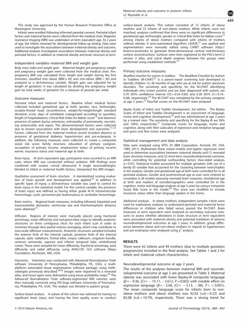
Table 1. Infant cohort characteristics
Infant Whole cohort (n= 62) Born to obese mothers (n= 24) Born to non-obese mothers (n=38) P-value a
Gestational age at birth, weeks, M (s.d.) 26.74 (1.64) 26.88 (1.8) 26.66 (1.55) 0.62SexMale, N % 38 (61.3) 8 (33.3) 16 (42.1) 0.50
RaceWhite, non-Hispanic, N % 32 (51.6) 11 (45.8) 21 (55.3) 0.73African American, N % 26 (41.9) 12 (50) 14 (36.8)Other, N % 4 (6.5) 1 (4.2) 3 (7.9)
Length of stay (days), M (s.d.) 85.19 (24.19) 80.09 (27.03) 88.97 (21.53) 0.19Brain injury, N % 12 (20.7) 4 (18.2) 8 (22.2) 0.72Sepsis, N % 17 (31.5) 5 (21.7) 12 (38.7) 0.13Patent ductus arteriosus, N % 28 (51.9) 13 (56.5) 15 (48.4) 0.56Necrotizing enterocolitis, N % 2 (3.7) 1 (4.2) 1 (3.2) 0.83
aP-value investigating differences in perinatal factors among obese and non-obese mothers using independent samples t-tests and χ2 analyses.
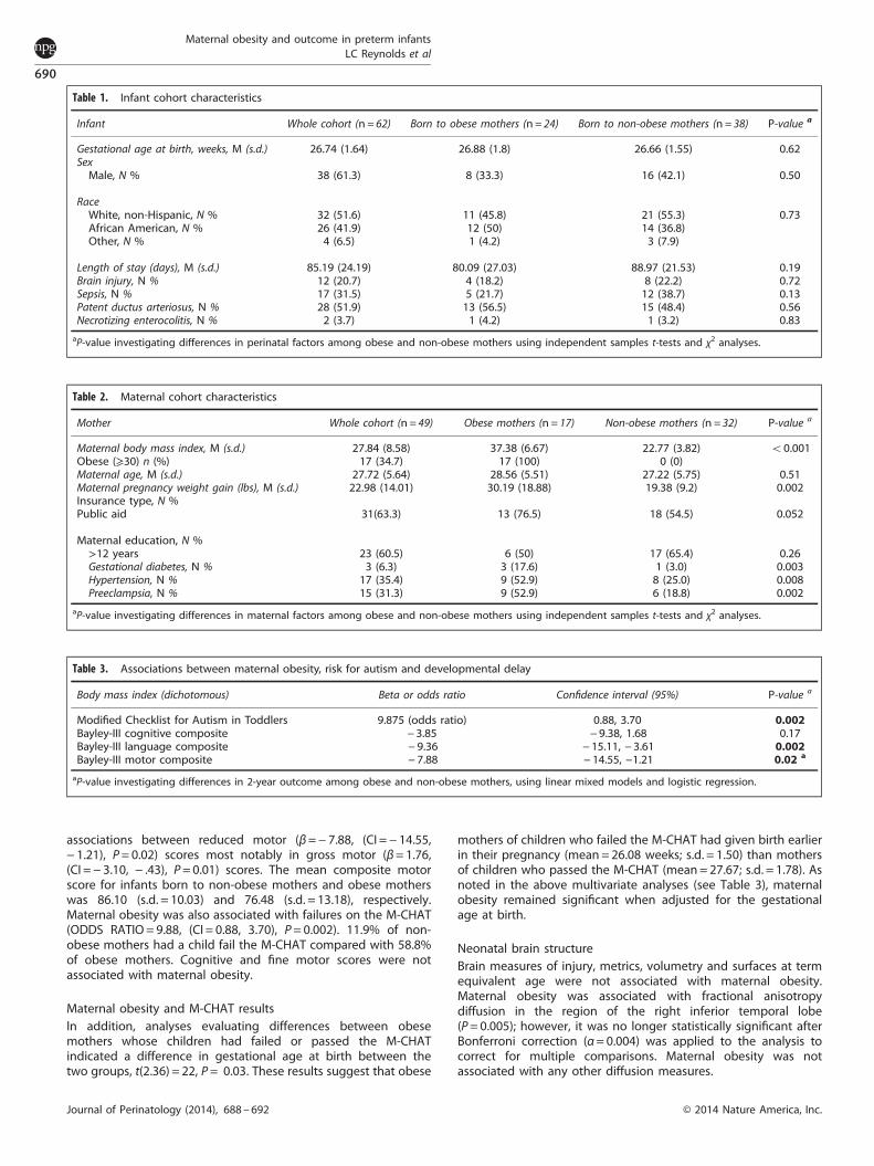
Table 2. Maternal cohort characteristics
Mother Whole cohort (n=49) Obese mothers (n= 17) Non-obese mothers (n=32) P-value a
Maternal body mass index, M (s.d.) 27.84 (8.58) 37.38 (6.67) 22.77 (3.82) o0.001Obese (⩾30) n (%) 17 (34.7) 17 (100) 0 (0)Maternal age, M (s.d.) 27.72 (5.64) 28.56 (5.51) 27.22 (5.75) 0.51Maternal pregnancy weight gain (lbs), M (s.d.) 22.98 (14.01) 30.19 (18.88) 19.38 (9.2) 0.002Insurance type, N %Public aid 31(63.3) 13 (76.5) 18 (54.5) 0.052
Maternal education, N %>12 years 23 (60.5) 6 (50) 17 (65.4) 0.26Gestational diabetes, N % 3 (6.3) 3 (17.6) 1 (3.0) 0.003Hypertension, N % 17 (35.4) 9 (52.9) 8 (25.0) 0.008Preeclampsia, N % 15 (31.3) 9 (52.9) 6 (18.8) 0.002
aP-value investigating differences in maternal factors among obese and non-obese mothers using independent samples t-tests and χ2 analyses.
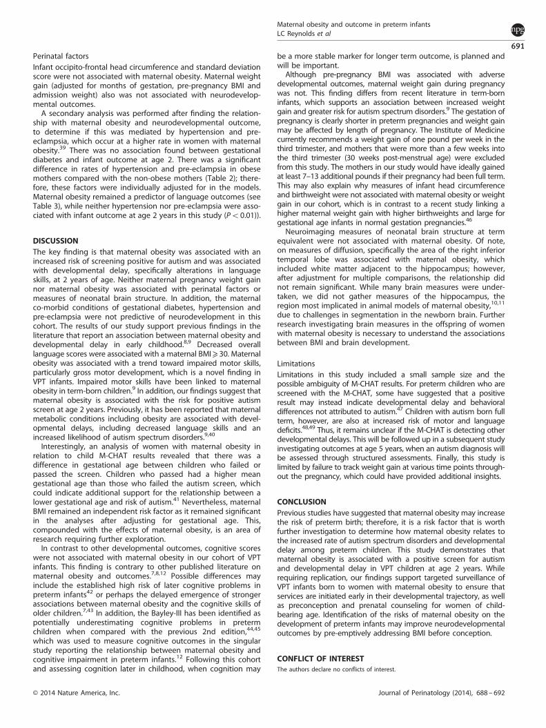
Table 3. Associations between maternal obesity, risk for autism and developmental delay
Body mass index (dichotomous) Beta or odds ratio Confidence interval (95%) P-value a
Modified Checklist for Autism in Toddlers 9.875 (odds ratio) 0.88, 3.70 0.002Bayley-III cognitive composite − 3.85 − 9.38, 1.68 0.17Bayley-III language composite − 9.36 − 15.11, − 3.61 0.002Bayley-III motor composite − 7.88 − 14.55, −1.21 0.02 a
aP-value investigating differences in 2-year outcome among obese and non-obese mothers, using linear mixed models and logistic regression.
Maternal obesity and outcome in preterm infantsLC Reynolds et al
690
Journal of Perinatology (2014), 688 – 692 © 2014 Nature America, Inc.
Perinatal factorsInfant occipito-frontal head circumference and standard deviationscore were not associated with maternal obesity. Maternal weightgain (adjusted for months of gestation, pre-pregnancy BMI andadmission weight) also was not associated with neurodevelop-mental outcomes.A secondary analysis was performed after finding the relation-
ship with maternal obesity and neurodevelopmental outcome,to determine if this was mediated by hypertension and pre-eclampsia, which occur at a higher rate in women with maternalobesity.39 There was no association found between gestationaldiabetes and infant outcome at age 2. There was a significantdifference in rates of hypertension and pre-eclampsia in obesemothers compared with the non-obese mothers (Table 2); there-fore, these factors were individually adjusted for in the models.Maternal obesity remained a predictor of language outcomes (seeTable 3), while neither hypertension nor pre-eclampsia were asso-ciated with infant outcome at age 2 years in this study (Po0.01)).
DISCUSSIONThe key finding is that maternal obesity was associated with anincreased risk of screening positive for autism and was associatedwith developmental delay, specifically alterations in languageskills, at 2 years of age. Neither maternal pregnancy weight gainnor maternal obesity was associated with perinatal factors ormeasures of neonatal brain structure. In addition, the maternalco-morbid conditions of gestational diabetes, hypertension andpre-eclampsia were not predictive of neurodevelopment in thiscohort. The results of our study support previous findings in theliterature that report an association between maternal obesity anddevelopmental delay in early childhood.8,9 Decreased overalllanguage scores were associated with a maternal BMI⩾30. Maternalobesity was associated with a trend toward impaired motor skills,particularly gross motor development, which is a novel finding inVPT infants. Impaired motor skills have been linked to maternalobesity in term-born children.9 In addition, our findings suggest thatmaternal obesity is associated with the risk for positive autismscreen at age 2 years. Previously, it has been reported that maternalmetabolic conditions including obesity are associated with devel-opmental delays, including decreased language skills and anincreased likelihood of autism spectrum disorders.9,40
Interestingly, an analysis of women with maternal obesity inrelation to child M-CHAT results revealed that there was adifference in gestational age between children who failed orpassed the screen. Children who passed had a higher meangestational age than those who failed the autism screen, whichcould indicate additional support for the relationship between alower gestational age and risk of autism.41 Nevertheless, maternalBMI remained an independent risk factor as it remained significantin the analyses after adjusting for gestational age. This,compounded with the effects of maternal obesity, is an area ofresearch requiring further exploration.In contrast to other developmental outcomes, cognitive scores
were not associated with maternal obesity in our cohort of VPTinfants. This finding is contrary to other published literature onmaternal obesity and outcomes.7,8,12 Possible differences mayinclude the established high risk of later cognitive problems inpreterm infants42 or perhaps the delayed emergence of strongerassociations between maternal obesity and the cognitive skills ofolder children.7,43 In addition, the Bayley-III has been identified aspotentially underestimating cognitive problems in pretermchildren when compared with the previous 2nd edition,44,45
which was used to measure cognitive outcomes in the singularstudy reporting the relationship between maternal obesity andcognitive impairment in preterm infants.12 Following this cohortand assessing cognition later in childhood, when cognition may
be a more stable marker for longer term outcome, is planned andwill be important.Although pre-pregnancy BMI was associated with adverse
developmental outcomes, maternal weight gain during pregnancywas not. This finding differs from recent literature in term-borninfants, which supports an association between increased weightgain and greater risk for autism spectrum disorders.9 The gestation ofpregnancy is clearly shorter in preterm pregnancies and weight gainmay be affected by length of pregnancy. The Institute of Medicinecurrently recommends a weight gain of one pound per week in thethird trimester, and mothers that were more than a few weeks intothe third trimester (30 weeks post-menstrual age) were excludedfrom this study. The mothers in our study would have ideally gainedat least 7–13 additional pounds if their pregnancy had been full term.This may also explain why measures of infant head circumferenceand birthweight were not associated with maternal obesity or weightgain in our cohort, which is in contrast to a recent study linking ahigher maternal weight gain with higher birthweights and large forgestational age infants in normal gestation pregnancies.46
Neuroimaging measures of neonatal brain structure at termequivalent were not associated with maternal obesity. Of note,on measures of diffusion, specifically the area of the right inferiortemporal lobe was associated with maternal obesity, whichincluded white matter adjacent to the hippocampus; however,after adjustment for multiple comparisons, the relationship didnot remain significant. While many brain measures were under-taken, we did not gather measures of the hippocampus, theregion most implicated in animal models of maternal obesity,10,11
due to challenges in segmentation in the newborn brain. Furtherresearch investigating brain measures in the offspring of womenwith maternal obesity is necessary to understand the associationsbetween BMI and brain development.
LimitationsLimitations in this study included a small sample size and thepossible ambiguity of M-CHAT results. For preterm children who arescreened with the M-CHAT, some have suggested that a positiveresult may instead indicate developmental delay and behavioraldifferences not attributed to autism.47 Children with autism born fullterm, however, are also at increased risk of motor and languagedeficits.48,49 Thus, it remains unclear if the M-CHAT is detecting otherdevelopmental delays. This will be followed up in a subsequent studyinvestigating outcomes at age 5 years, when an autism diagnosis willbe assessed through structured assessments. Finally, this study islimited by failure to track weight gain at various time points through-out the pregnancy, which could have provided additional insights.
CONCLUSIONPrevious studies have suggested that maternal obesity may increasethe risk of preterm birth; therefore, it is a risk factor that is worthfurther investigation to determine how maternal obesity relates tothe increased rate of autism spectrum disorders and developmentaldelay among preterm children. This study demonstrates thatmaternal obesity is associated with a positive screen for autismand developmental delay in VPT children at age 2 years. Whilerequiring replication, our findings support targeted surveillance ofVPT infants born to women with maternal obesity to ensure thatservices are initiated early in their developmental trajectory, as wellas preconception and prenatal counseling for women of child-bearing age. Identification of the risks of maternal obesity on thedevelopment of preterm infants may improve neurodevelopmentaloutcomes by pre-emptively addressing BMI before conception.
CONFLICT OF INTERESTThe authors declare no conflicts of interest.
Maternal obesity and outcome in preterm infantsLC Reynolds et al
691
© 2014 Nature America, Inc. Journal of Perinatology (2014), 688 – 692
ACKNOWLEDGEMENTSWe wish to acknowledge Karen Lukas RN (Washington University School ofMedicine), Anthony Barton (Washington University School of Medicine), JessicaConners (Washington University School of Medicine), Dimitrios Alexopolous MS(Washington University School of Medicine), Joe Ackerman, Jr (Washington UniversitySchool of Medicine), Claudine Vavasseur MD (National Maternity Hospital, DublinIreland) and Han Tjoeng MD (University of Hawaii) who obtained informed consentsand conducted patient-oriented responsibilities to support the success of this project.We also wish to thank all the families whose infants participated in this study. Thisproject was supported by the National Institute of Health (ROI HD 057098), the DorisDuke Foundation, the Washington University Intellectual and DevelopmentalDisabilities Research Center (NIH/NICHD P30 HD062171), and the UL1 TR000448,sub award KL2 TR000450 from the National Center for Advancing TranslationalSciences.
REFERENCES1 Jeffery RW, Utter J. The changing environment and population obesity in the
United States. Obesity Res 2012; 11(S10): 12S–22S.2 Nodine PM, Hastings-Tolsma M. Maternal obesity: improving pregnancy out-
comes. MCN: Am J Matern Child Nurs 2012; 37(2): 110.3 Flegal KM, Carroll MD, Kit BK, Ogden CL. Prevalence of obesity and trends in the
distribution of body mass index among US adults, 1999-2010. JAMA 2012; 307(5):491–497.
4 Catalano P. The impact of gestational diabetes and maternal obesity on themother and her offspring. J Dev Orig Health Dis 2010; 1(04): 208–215.
5 Trasande L, Lee M, Yinghua L, Weitzman M, Savitz D. Incremental charges, costs,and length of stay associated with obesity as a secondary diagnosis amongpregnant women. Med care 2009; 47(10): 1046–1052.
6 Van Lieshout R, Taylor V, Boyle M. Pre‐pregnancy and pregnancy obesity andneurodevelopmental outcomes in offspring: a systematic review. Obes Rev 2011;12(5): e548–e559.
7 Basatemur E, Gardiner J, Williams C, Melhuish EC, Barnes J, Sutcliffe A. Relationshipbetween maternal body mass index and child cognition: evidence from the UKMillennium Cohort Study. Paediatrics 2013; 131(1): 56–63.
8 Hinkle SN, Sharma AJ, Schieve LA, Ramakrishnan U, Swan DW, Stein AD. Reliabilityof gestational weight gain reported postpartum: a comparison to the birth cer-tificate. Matern Child Health J 2012; 17(4): 756–765.
9 Krakowiak P, Walker CK, Bremer AA, Baker AS, Ozonoff S, Hansen RL et al. Maternalmetabolic conditions and risk for autism and other neurodevelopmental dis-orders. Pediatrics 2012; 129(5): e1121–e1128.
10 Tozuka Y, Wada E, Wada K. Diet-induced obesity in female mice leads to perox-idized lipid accumulations and impairment of hippocampal neurogenesis duringthe early life of their offspring. FASEB J 2009; 23(6): 1920–1934.
11 Bilbo SD, Tsang V. Enduring consequences of maternal obesity for brain inflam-mation and behavior of offspring. FASEB J 2010; 24(6): 2104–2115.
12 Helderman JB, O’Shea TM, Kuban KCK, Allred EN, Hecht JL, Dammann O et al.Antenatal antecedents of cognitive impairment at 24 months in extremely lowgestational age newborns. Pediatrics 2012; 129(3): 494–502.
13 Hertz-Picciotto I, Delwiche L. The rise in autism and the role of age at diagnosis.Epidemiology 2009; 20(1): 84–90.
14 Cockburn F, Cooke R. The crib (clinical risk index for babies) score: a tool forassessing initial neonatal risk. Lancet 1993; 342(8865)):193–198.
15 Anderson P, Doyle LW. Neurobehavioral outcomes of school-age children bornextremely low birth weight or very preterm in the 1990 s. JAMA 2003; 289(24):3264–3272.
16 Cheong JL, Hunt RW, Anderson PJ, Howard K, Thompson DK, Wang HX et al. Headgrowth in preterm infants: correlation with magnetic resonance imaging andneurodevelopmental outcome. Pediatrics 2008; 121(6): e1534–e1540.
17 Hamrick SE, Hansmann G. Patent ductus arteriosus of the preterm infant. Pedia-trics 2010; 125(5): 1020–1030.
18 Hintz SR, Kendrick DE, Vohr BR, Poole WK, Higgins RD. Gender differences inneurodevelopmental outcomes among extremely preterm, extremely‐low‐birth-weight infants. Acta Paediatr 2007; 95(10): 1239–1248.
19 Hintz SR, Kendrick DE, Wilson-Costello DE, Das A, Bell EF, Vohr BR et al. Early-childhood neurodevelopmental outcomes are not improving for infants bornato 25 weeks' gestational age. Pediatrics 2011; 127(1): 62–70.
20 Klinger G, Levy I, Sirota L, Boyko V, Lerner-Geva L, Reichman B. Outcome of early-onset sepsis in a national cohort of very low birth weight infants. Pediatrics 2010;125(4): e736–e740.
21 Leversen KT, Sommerfelt K, Rønnestad A, Kaaresen PI, Farstad T, Skranes J et al.Prediction of neurodevelopmental and sensory outcome at 5 years in Norwegianchildren born extremely preterm. Pediatrics 2011; 127(3): e630–e638.
22 Patole S, Deshpande G. Effect of Necrotizing Enterocolitis on Growth andDevelopment in Preterm Neonates. Handbook of Growth and Growth Monitoringin Health and Disease: Springer, Elsevier, 2012, p 567–583.
23 Pietz J, Peter J, Graf R, Rauterberg-Ruland I, Rupp A, Sontheimer D et al. Physicalgrowth and neurodevelopmental outcome of nonhandicapped low-risk childrenborn preterm. Early Hum Dev 2004; 79(2): 131–143.
24 Reagan PB, Salsberry PJ. Race and ethnic differences in determinants of preterm birthin the USA: broadening the social context. Soc Sci Med 2005; 60(10): 2217–2228.
25 Stoll BJ, Hansen NI, Bell EF, Shankaran S, Laptook AR, Walsh MC et al. Neonataloutcomes of extremely preterm infants from the NICHD Neonatal ResearchNetwork. Pediatrics 2010; 126(3): 443–456.
26 Kidokoro H, Neil J, Inder T. A new MRI assessment tool to define brain abnor-malities in very preterm infants at term. Am J Neuroradiol 34(11): 2208–2214.
27 Nguyen The Tich S, Anderson PJ, Shimony JS, Hunt RW, Doyle LW, Inder TE. Anovel quantitative simple brain metric using MR imaging for preterm infants.AJNR Am J Neuroradiol 2009; 30(1): 125–131.
28 Klein A, Ghosh SS, Avants B, Yeo BT, Fischl B, Ardekani B et al. Evaluation ofvolume-based and surface-based brain image registration methods. NeuroImage2010; 51(1): 214–220.
29 Avants BB, Tustison NJ, Wu J, Cook PA, Gee JC. An open source multivariateframework for n-tissue segmentation with evaluation on public data. Neu-roinformatics 2011; 9(4): 381–400.
30 Avants BB, Tustison NJ, Song G, Cook PA, Klein A, Gee JC. A reproducible eva-luation of ANTs similarity metric performance in brain image registration. Neu-roImage 2011; 54(3): 2033–2044.
31 Hill J, Dierker D, Neil J, Inder T, Knutsen A, Harwell J et al. A surface-based analysisof hemispheric asymmetries and folding of cerebral cortex in term-born humaninfants. J Neuroscience 2010; 30(6): 2268–2276.
32 Hill J, Dierker D, Neil J, Inder T, Knutsen A, Harwell J et al. A surface-based analysisof hemispheric asymmetries and folding of cerebral cortex in term-born humaninfants. J Neurosci 2010; 30(6): 2268–2276.
33 Robins DL, Fein D, Barton ML, Green JA. The Modified checklist for autism intoddlers: an initial study investigating the early detection of autism and pervasivedevelopmental disorders. J Autism Dev Disord 2001; 31(2): 131–144.
34 Snow AV, Lecavalier L. Sensitivity and specificity of the Modified Checklist for Autismin Toddlers and the Social Communication Questionnaire in preschoolers suspectedof having pervasive developmental disorders. Autism 2008; 12(6): 627–644.
35 Carter A, Briggs-Gowan M. Infant Toddler Social and Emotional Assessment (ITSEA) .San Antonio, TX: Psychological Corporation, Harcourt Assessment, 2006.
36 Bayley N. Bayley Scales of Infant and Toddler Development® 3rd Edition (Bayley-III®)1969.
37 Moore T, Johnson S, Haider S, Hennessy E, Marlow N. Relationship between testscores using the second and third editions of the Bayley Scales in extremelypreterm children. J Pediatr 2012; 160(4): 553–558.
38 Spittle AJ, Anderson PJ, Lee KJ, Ferretti C, Eeles A, Orton J et al. Preventive care athome for very preterm infants improves infant and caregiver outcomes at 2 years.Pediatrics 2010; 126(1): e171–e178.
39 O’Brien TE, Ray JG, Chan W-S. Maternal body mass index and the risk of pre-eclampsia: a systematic overview. Epidemiology 2003; 14(3): 368–374.
40 Neggers YH, Goldenberg RL, Ramey SL, Cliver SP. Maternal prepregnancy bodymass index and psychomotor development in children. Acta Obstet Gynecol Scand2003; 82(3): 235–240.
41 Leavey A, Zwaigenbaum L, Heavner K, Burstyn I. Gestational age at birth and riskof autism spectrum disorders in alberta, Canada. J Pediatr 2012; 162(2): 361–368.
42 Laptook AR, O'Shea TM, Shankaran S, Bhaskar B. Adverse neurodevelopmentaloutcomes among extremely low birth weight infants with a normal head ultra-sound: prevalence and antecedents. Pediatrics 2005; 115(3): 673–680.
43 Tanda R, Salsberry PJ, Reagan PB, Fang MZ. The impact of prepregnancy obesityon children’s cognitive test scores. Matern Child Health J 2012; 1–8.
44 Anderson PJ, De Luca CR, Hutchinson E, Roberts G, Doyle LW. Underestimation ofdevelopmental delay by the new Bayley-III Scale. Arch Pediatr Adolesc Med 2010;164(4): 352.
45 Moore T, Johnson S, Haider S, Hennessy E, Marlow N. Relationship between testscores using the second and third editions of the Bayley scales in extremelypreterm children. J Pediatr 2011; 160(4): 553–558.
46 Norman JE, Reynolds R. Symposium I: consequences of obesity and overweightduring pregnancy the consequences of obesity and excess weight gain inpregnancy. Proc Nutr Soc 2011; 70(4): 450–456.
47 Moore T, Johnson S, Hennessy E, Marlow N. Screening for autism in extremelypreterm infants: problems in interpretation. Dev Med Child Neurol 2012; 54(6):514–520.
48 Ming X, Brimacombe M, Wagner GC. Prevalence of motor impairment in autismspectrum disorders. Brain Dev 2007; 29(9): 565–570.
49 Rapin I, Dunn M. Update on the language disorders of individuals on the autisticspectrum. Brain Dev 2003; 25(3): 166–172.
Maternal obesity and outcome in preterm infantsLC Reynolds et al
692
Journal of Perinatology (2014), 688 – 692 © 2014 Nature America, Inc.