maternal cocaine exposure alters mesolimbic dopaminergic function in rat offspring
TRANSCRIPT
Ž .European Journal of Pharmacology 345 1998 175–180
Short communication
Maternal cocaine exposure alters mesolimbic dopaminergic function inrat offspring
Arcangela Giustino a,), Vincenzo Cuomo a, Charles A. Marsden b
a Department of Pharmacology and Human Physiology, Medical School, UniÕersity of Bari, Policlinico, Piazza G. Cesare, 70124 Bari, Italyb School of Biomedical Sciences, Nottingham UniÕersity, Nottingham, UK
Received 11 December 1997; revised 26 January 1998; accepted 30 January 1998
Abstract
Ž .Hooded Lister female rats were treated with either saline or cocaine 20 mgrkg s.c. from gestational day 10 every other day untilŽ .weaning postnatal day 25 . In vivo microdialysis has shown that maternal cocaine exposure significantly decreases basal extracellular
Ž .concentrations of dopamine in the nucleus accumbens of young-adult offspring 4 weeks after cessation of cocaine treatment . Moreover,q Ž qthe increase in extracellular dopamine levels induced by a challenge dose of K intracerebral 60 mM K artificial cerebrospinal fluid
Ž . . Ž .aCSF infusion or cocaine 15 mgrkg i.p. was significantly attenuated in rats exposed to cocaine during perinatal life with respect tocontrols. The alterations in mesolimbic dopamine transmission observed in these experiments might underlie behavioral abnormalitiesinduced in rat offspring by maternal exposure to cocaine at dose levels which do not produce gross malformations andror overtneurotoxic effects. q 1998 Elsevier Science B.V.
Keywords: Cocaine; Perinatal exposure; Microdialysis; Nucleus accumbens; Dopamine; Rat
1. Introduction
Recent findings have shown that a number of neurobe-havioral effects due to prenatal or postnatal administrationof cocaine in rats could be linked to an impairment of
Žcentral dopamine transmission Bowman et al., 1997;Heyser et al., 1994; Keller et al., 1994; Meyer et al., 1992;
.Peris et al., 1992; Seidler et al., 1994; Spear et al., 1989 ,thus suggesting that the developing dopamine system is aprimary target for this drug of abuse.
Only few studies have investigated the effects of perina-Ž .tal combined intrauterine and early postnatal cocaine
exposure in rats. The status of rat nervous system develop-ment over the first 2 postnatal weeks corresponds roughlyto the last trimester of human neural development, andtherefore combined prenatal and early postnatal exposureto cocaine may be relevant to gestational human exposureŽ .Dobbing and Sands, 1979 .
The available literature has shown that perinatal expo-sure to cocaine alters rat functional brainstem developmentŽ .Salamy et al., 1992 and decreases the number of sponta-
) Corresponding author. Tel.: q39-80-5478448; fax: q39-80-5478444;e-mail: cuomo@cimedoc. uniba.it
neously active midbrain dopamine neurons in rat pupsŽ .Wang and Pitts, 1994 .
Moreover, our recent findings have demonstrated thatperinatal exposure to cocaine impairs visual discriminationin young-adult male rats subjected to a novel exploration
Ž .object test Giustino et al., 1996 . Since mesolimbicdopamine system plays an important role in the physio-
Žlogical mediation of this behavioral response Hooks and.Kalivas, 1995 , we hypothesized that the alterations in the
exploratory activity observed in young-adult rats exposedperinatally to cocaine could be due to an impairment ofdopamine transmission. Furthermore, perinatal exposure tococaine has been shown to attenuate acute cocaine-induced
Ž .hyperactivity in an open field test Giustino et al., 1996 .Therefore, the aim of these experiments was to extend
our previous investigations and to explore whether thebehavioral alterations caused by maternal exposure to co-caine could be related to changes in mesolimbic dopaminefunction. In particular, in vivo microdialysis was used tomeasure extracellular dopamine concentrations in the nu-cleus accumbens of freely-moving young-adult male ratswhose mothers were treated with cocaine during bothgestation and lactation. Moreover, the influence of perina-tal exposure to cocaine on the increase in extracellular
0014-2999r98r$19.00 q 1998 Elsevier Science B.V. All rights reserved.Ž .PII S0014-2999 98 00091-0
( )A. Giustino et al.rEuropean Journal of Pharmacology 345 1998 175–180176
dopamine levels elicited by acute intraperitoneal cocaineinjection or intracerebral high Kq infusion in the nucleusaccumbens of young-adult male rats was also assessed.
2. Materials and methods
2.1. Animals
All experiments were performed under UK Home Of-fice regulations and project licence number 40r01089.
ŽHooded Lister rats Nottingham University breeding.colony weighing 200–230 g were housed at constant
room temperature, exposed to a light cycle of 12 hrdayŽ .07:00–19:00 for 2 weeks before the experiment. Singlefemales were placed with a male rat in the afternoon.Mating was confirmed by the presence of copulatory plugon the following morning which was designated gesta-tional day 1. On gestational day 10, dams were weighedand randomly assigned to two groups treated with saline orcocaine, respectively. From gestational day 10 to postnatalday 25, dams were injected with 0.9% sodium chloride
Ž .solution vehicle or with 20 mgrkg s.c. of cocaine hydro-chloride every other day between 09:00 and 11:00. Thistreatment schedule was chosen on the basis of our recentdata showing that this exposure to cocaine does not inducematernal toxicity and gross malformations andror overt
Žsigns of neurotoxicity in rat offspring Giustino et al.,.1996 . The day of the birth was defined as postnatal day 0.
Pups were weaned on postnatal day 25 and male rats wereused for neurochemical studies that were performed 4weeks after weaning. One rat per litter from differentlitters per treatment group was used.
ŽIn agreement with our previous findings Giustino et al.,.1996 , perinatal cocaine exposure did not affect dam weight
gain, litter size at birth, litter weight, and male pup weightŽ .gain data not shown .
2.2. Microdialysis and analytical procedure
Microdialysis experiments were carried out according tothe technique previously used by Saulskaya and MarsdenŽ .1995 .
Concentric microdialysis probes were prepared usingdialysis membrane made of acrylonitrile-sodium methallyl
Žsulphonate Hospal, UK, KDa 20; 300 mm o.d; 220 mm.i.d. .
ŽRats were anaesthetized with halothane 1.5–2% in a.50:50 O rNO mixture and placed in a stereotaxic frame.2 2
Ž .Dialysis probes length: 2 mm were implanted in the rightnucleus accumbens according to the following coordinates:APs1.6, MLs0.9, from bregma and Vs7.8 mm from
Žthe dura with the incisor bar set at y3.3 mm Paxinos and.Watson, 1986 . The probe was fixed to the skull using two
stainless steel screws and acrylic dental cement. The inletof the probe, since the beginning of the implantation up to
the end of the experiment, was attached to a liquid swivelŽ .Harvard Apparatus, South Natick, MA , positioned overthe cage to allow free movement of the rat during theexperiment. The swivel was continuously connected to a
Ž .microinfusion pump CMAr100 CMA Microdialysis setŽ .at 1 mlrmin, perfusing artificial cerebrospinal fluid aCSF
containing 140 mM NaCl, 3 mM KCl, 2.5 mM CaCl , 12
mM MgCl , 1.2 Na HPO , 0.27 mM NaH PO , 7.2 M2 2 4 2 4
glucose, pHs7. Animals were allowed 24 h to recoverfrom surgery with food and water available ad libitum.
The day of the experiment, after the probe had beenflowing for 1 h, samples were collected into polyethylene
Ž . Ž .vials at 20-min Experiment 1 or 10-min Experiment 2intervals.
The position of the microdialysis probe was verified byhistological procedures at the end of each experiment.Only rats in which the probe was exactly located in thetarget area were considered in the results.
Dialysate samples were immediately injected onto ahigh-performance liquid chromatography-electrochemicaldetection system. This system consisted of an isocratic
Ž . Žpump Gilson , a Spherisorb column 3 mm, ODS, 10 cm.length, 2 mm i.d. , a Rheodyne injector with a 10-ml
Ž .injector loop, and an electrochemical detector BAS LC 3equipped with a glassy carbon electrode set at q0.7 Vversus AgrAgCl reference electrode.
The mobile phase containing 0.15 M NaH PO 2 H O,2 4 2
1 mM EDTA, 0.5 mM sodium octane sulfonate and 10%methanol was adjusted to pHs3 using phosphoric acidand pumped through the system at 0.2 mlrmin. The
Ž .detection limit signal to noise ratios3 was 10 fmolr10ml.
2.2.1. Experiment 1
( )2.2.1.1. Effect of cocaine challenge 15 mgrkg i.p. onextracellular dopamine concentrations. Once a stable basal
Ždopamine output was obtained no more than 10% differ-.ences between three consecutive samples rats were given
Ž .a challenge dose of cocaine 15 mgrkg i.p. . 10 ml of eachŽ .dialysate sample 20 ml were injected into the high
performance liquid chromatography system and dopamineŽ .levels fmolr10 ml were measured. The remaining 10 ml
of each sample were used for measurements of otherŽ .neurotransmitters data not reported in the present study .
2.2.2. Experiment 2
2.2.2.1. Effect of intracerebral 60 mM K q aCSF infusionon extracellular dopamine concentrations. Once a stable
Žbasal dopamine output was obtained no more than 10%.differences between three consecutive samples rats were
given a 60 mM Kq aCSF infusion for 50 min. DialysateŽ .samples 10 ml were injected into the high performance
liquid chromatography system and dopamine levelsŽ .fmolr10 ml were measured.
( )A. Giustino et al.rEuropean Journal of Pharmacology 345 1998 175–180 177
2.3. Statistical analysis
All data were expressed as mean"SEM.Statistical analysis of basal dopamine concentrations
was performed using two-way analysis of varianceŽ . ŽANOVA for repeated measures three consecutive sam-ples collected before cocaine challenge or high Kq infu-
.sion, respectively with treatment as the between-subjectfactor and time as the within-subject factor. Actual and
Žabsolute dopamine increases with respect to the last basal. qvalue induced by acute cocaine or high K infusion were
evaluated by two-way ANOVAs for repeated measures,respectively. Tukey’s test was used to perform individualwithin-group and between-groups comparisons.
3. Results
3.1. Experiment 1
3.1.1. Effects of cocaine challenge on extracellulardopamine concentrations in the nucleus accumbens ofperinatally saline- and cocaine-exposed rats
The results are reported in Fig. 1. A two-way ANOVAfor repeated measures of basal dopamine concentrationsŽthree consecutive samples collected before cocaine chal-
. Ž .lenge showed the following differences: i between treat-Ž Ž . . Ž .ments F 1,11 s8.24; P-0.01 ; ii between times
Ž Ž . . Ž .F 2,22 s0.37; n.s. ; iii between treatments= timesŽ Ž . .F 2,22 s1.89; n.s. . These results indicate that perinataltreatment with cocaine significantly decreases basal
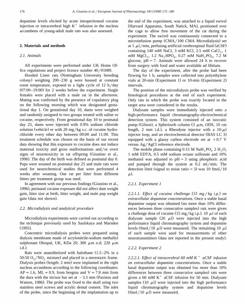
Ž .Fig. 1. The effects of cocaine challenge 15 mgrkg i.p. on extracellularŽ .dopamine DA concentrations in the nucleus accumbens of rats exposed
Ž . Ž .perinatally to saline Ø ns6 , or cocaine l ns7 . Data were ex-Ž .pressed as the mean"SEM. Significant differences Tukey’s test : ),
ŽP -0.05; )), P -0.01 vs. last basal sample actual dopamine concen-. Ž . Ž .trations . Absolute dopamine increases mean fmol"SEM s i saline-
Ž .exposed rats: 41.2"11.6; 90.8"11.3; 67.7"4.6; 63.5"3.3; iicocaine-exposed rats: 20"3.7; 22.8"4.2; 17.6"4.6; 18.9"3.6. Signifi-
Ž .cant differences between absolute dopamine increases Tukey’s test : 8,P -0.01 vs. perinatal cocaine.
dopamine levels in the nucleus accumbens of young-adultrats. Since differences between times and between treat-ments= times were not significant, post-hoc tests for indi-vidual comparisons were not performed.
A two-way ANOVA for repeated measures of changesŽ .in extracellular dopamine concentrations actual values
Želicited by a challenge dose of cocaine last basal value.and four consecutive samples after cocaine challenge gave
Ž . Ž Ž .the following differences: i between treatments F 1,11. Ž . Ž Ž .s23.31; P-0.0005 ; ii between times F 4,44 s38.0;
. Ž . Ž Ž .P-0.0001 ; iii between treatments= times F 4,44 s.14.08; P-0.0001 .
Ž .Within-group comparisons Tukey’s test showed thatacute cocaine administration induces a significant increase
Ž .in extracellular dopamine concentrations actual valueswith respect to the last basal sample in both saline- andcocaine-exposed rats.
In order to exclude that the altered responsiveness tococaine challenge exhibited by perinatally cocaine-exposedrats with respect to control animals could reflect differ-ences in basal extracellular dopamine concentrations, atwo-way ANOVA for repeated measures of absolute extra-cellular dopamine increases induced by cocaine challengewith respect to the last basal sample was performed.
Ž .This analysis showed the following differences: i be-Ž Ž . . Ž .tween treatments F 1,11 s39.45; P-0.0001 ; ii be-
Ž Ž . . Ž .tween times F 3,33 s12.04; P-0.0001 ; iii betweenŽ Ž . .treatments= times F 3,33 s9.63; P-0.0001 .
Ž .Between-groups comparisons Tukey’s test indicatedthat cocaine-induced increase in extracellular dopamine
Ž .levels absolute increase was significantly attenuated inperinatally cocaine-exposed rats with respect to saline-treated animals.
Finally, in order to get information about the mecha-nisms underlying the decreased responsivity in cocainetreated rats, a two-way ANOVA for repeated measures of
Ž .log-transformed data actual values was performed. ThisŽ .analysis gave the following differences: i between treat-
Ž Ž . . Ž .ments F 1,11 s24.57; P-0.0004 ; ii between timesŽ Ž . . Ž .F 4,44 s41.7; P-0.0001 ; iii between treatments=
Ž Ž . .times F 4,44 s4.53; P-0.005 .
3.2. Experiment 2
3.2.1. Effects of intracerebral 60 mM K q aCSF infusionon extracellular dopamine concentrations in the nucleusaccumbens of perinatally saline- and cocaine-exposed rats
The results are reported in Fig. 2. A two-way ANOVAfor repeated measures of basal dopamine concentrationsŽ qthree consecutive samples collected before 60 mM K
. Ž .aCSF infusion showed the following differences: i be-Ž Ž . . Ž .tween treatments F 1,10 s7.88; P-0.01 ; ii between
Ž Ž . . Ž .times F 2,20 s2.52; n.s. ; iii between treatments=Ž Ž . .times F 2,20 s2.52; n.s. . These results confirm those
obtained in the Experiment 1 showing that perinatal treat-
( )A. Giustino et al.rEuropean Journal of Pharmacology 345 1998 175–180178
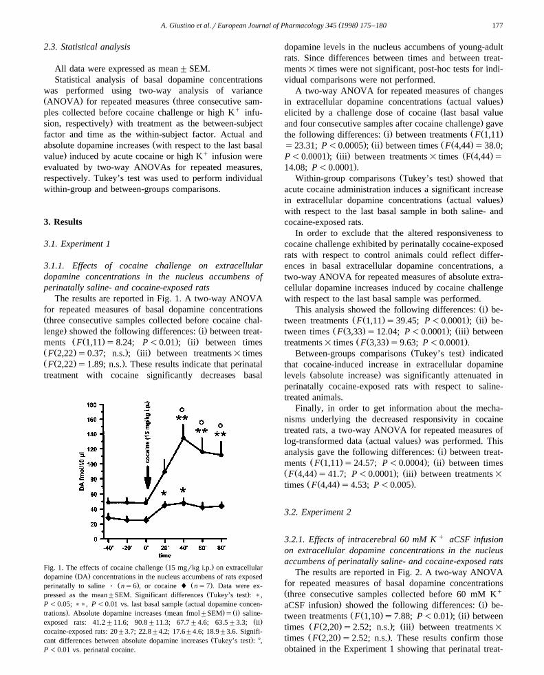
Fig. 2. The effects of 60 mM Kq aCSF infusion on extracellular DAconcentrations in the nucleus accumbens of rats exposed perinatally to
Ž . Ž .saline Ø ns6 , or cocaine l ns6 . Data were expressed as theŽ .mean"SEM. Significant differences Tukey’s test : ), P -0.05, )),
Ž .P -0.01 vs. last basal sample actual dopamine concentrations . AbsoluteŽ . Ž .dopamine increases mean fmol"SEM s i saline-exposed rats: 5.7"
Ž .4.4; 37.2"7; 75.2"12; 91.8"13.8; 96"15; ii cocaine-exposed rats:7.4"3.2; 23.2"6; 51"5.5; 53.7"5; 54.3"5.4. Significant differences
Ž .between absolute dopamine increases Tukey’s test : 8, P -0.05 vs.perinatal cocaine.
ment with cocaine significantly reduces basal dopaminelevels in the nucleus accumbens of young-adult rats. Sincedifferences between times and between treatments= timeswere not significant, post-hoc tests for individual compar-isons were not performed.
A two-way ANOVA for repeated measures of changesŽ .in extracellular dopamine concentrations actual values
q Želicited by high K infusion last basal value and fiveconsecutive samples after the beginning of high Kq infu-
. Ž .sion gave the following differences: i between treat-Ž Ž . . Ž .ments F 1,10 s8.67; P-0.02 ; ii between times
Ž Ž . . Ž .F 5,50 s67.5; P-0.0001 ; iii between treatments=Ž Ž . .times F 5,50 s5.09; P-0.001 .
Ž .Within-group comparisons Tukey’s test showed that60 mM Kq aCSF infusion induces a significant increase in
Ž .extracellular dopamine concentrations actual values withrespect to the last basal sample in both saline- andcocaine-exposed rats.
A two-way ANOVA for repeated measures of absoluteextracellular dopamine increases induced by 60 mM Kq
aCSF infusion with respect to the last basal sample showedŽ . Ž Ž .the following differences: i between treatments F 1,10
. Ž . Ž Ž .s4.85; P-0.05 ; ii between times F 4,40 s64.08;. Ž . Ž Ž .P-0.0001 ; iii between treatments= times F 4,40 s
.5.63; P-0.001 .Ž .Between-groups comparisons Tukey’s test showed that
60 mM Kq aCSF-induced increase in extracellularŽ .dopamine levels absolute increase was significantly at-
tenuated in perinatally cocaine-exposed animals with re-spect to controls.
Finally, a two-way ANOVA for repeated measures ofŽ .log-transformed data actual values gave the following
Ž . Ž Ž .differences: i between treatments F 1,10 s13.10; P-. Ž . Ž Ž . .0.005 ; ii between times F 5,50 s117.78; P-0.0001 ;
Ž . Ž Ž . .iii between treatments= times F 5,50 s1.59; n.s. .
4. Discussion
The results of the present study indicate that maternalŽexposure to cocaine from gestational day 10 until wean-
.ing reduces mesolimbic dopamine function in young-adultmale rats. Lack of significant changes in maternal and pupweight gain suggests that nutritional deficiency was not afactor in the cocaine offspring outcome.
In particular, in vivo microdialysis experiments haveshown a significant decrease in extracellular concentra-tions of dopamine in the nucleus accumbens of rats perina-tally exposed to this drug of abuse. These changes havebeen observed 4 weeks after the cessation of cocainetreatment.
Ž .Our recent findings Giustino et al., 1996 have shownŽan impairment in visual discrimination novel exploration
.object test in young-adult male rats exposed perinatally tothe same cocaine treatment schedule used in the presentstudy. Considering the role of the mesolimbic dopaminesystem in the physiological mediation of this behaviorŽ .Hooks and Kalivas, 1995 , the decrease in basal extracel-lular dopamine levels occurring into the nucleus accum-bens of young-adult rats exposed to cocaine during perina-tal life might account, at least in part, for the alterations intheir response to novelty.
Ž .Previous studies Wang and Pitts, 1994 have shownthat perinatal cocaine exposure decreases the number ofspontaneously active midbrain dopamine neurons in neona-tal rats. Moreover, adult rats prenatally exposed to cocainehave been found to exhibit fewer spontaneously activedopamine neurons in the substantia nigra pars compactaand the ventral tegmental area with respect to control
Ž .animals Minabe et al., 1992 . According to Heyser et al.Ž .1994 , the decrease in neuronal activity in the dopaminesystem shown by these electrophysiological studies is con-sistent with behavioral changes suggesting a possible im-pairment in dopamine functioning in animals treated with
Žcocaine during development Spear et al., 1989; Meyer et.al., 1992 .
In this study, we have also found that perinatal cocainereduces high Kq-evoked dopamine output in the nucleusaccumbens of young-adult rats. These changes, that furtherconfirm a decreased activity of the dopamine system dueto developmental exposure to cocaine, are in agreement
Ž .with the results obtained by Wang et al. 1995 showingthat prenatal cocaine treatment reduces mesocortical Kq-
( )A. Giustino et al.rEuropean Journal of Pharmacology 345 1998 175–180 179
w3 x Ževoked H dopamine release in rabbits via changes sensi-.tization in terminal presynaptic dopamine autoreceptors.
It is of interest to point out that these preclinicalobservations are consistent with the results of humanstudies demonstrating that neonatal cerebrospinal fluidconcentrations of homovanillic acid are significantly lower
Ž .after gestational cocaine exposure Needleman et al., 1993 .Moreover, the present neurochemical experiments indi-
cate that developmental cocaine exposure affects biochem-ical responsivity to acute cocaine in young-adult rats. Inparticular, the increase in extracellular dopamine levelsinduced by a challenge dose of cocaine in the nucleusaccumbens of rats exposed to cocaine during both preg-nancy and lactation was significantly attenuated with re-spect to control animals. These data parallel those obtainedin previous behavioral experiments showing that cocaine-induced hyperactivity in young-adult rats was significantly
Žreduced by maternal treatment with this drug Giustino et.al., 1996 .
In order to get information about the mechanism under-lying the reduced biochemical responsiveness in the groupexposed to cocaine during perinatal life, repeated measuresANOVAs used to make the conclusions in Figs. 1 and 2were reexamined using log-transformed data.
The results showed that ANOVA of log-transformedŽ .data Experiment 1, cocaine challenge still showed a
significant interaction between treatments and time,whereas the interaction between treatments and time disap-
Žpeared in the ANOVA dealing with Experiment 2 highq .K challenge .The results, indicating that the same proportional change
Ž q .occurs in basal and stimulated high K infusion condi-tions, suggest that perinatal exposure to cocaine may in-duce some alterations affecting both basal and Kq stimu-lated release, such as a loss of dopaminergic innervation ora reduced vesicular dopamine concentration.
On the other hand, the lack of the same proportionalchange in basal and stimulated conditions following co-caine challenge could be indicative of an additional loss ofa specific mechanism sensitive to cocaine stimulation inrats perinatally exposed to cocaine.
The results of the present study suggest that the effectsof prolonged cocaine exposure on the developing dopaminesystem may differ from those found in mature animals. Infact, previous findings have shown that repeated treatmentwith cocaine during adulthood leads to an enhanced effect
Žof a cocaine challenge on dopamine release Kalivas and.Duffy, 1990; Pettit et al., 1990 . Increases of both bio-
chemical and behavioral responses to cocaine after a previ-ous administration of this drug is a commonly observed
Žphenomenon referred to as sensitization Zahniser and.Peris, 1992 . However, conflicting findings exist regarding
the influence of developmental exposure to cocaine onlater biochemical and behavioral responsiveness to a co-caine challenge. Increases or decreases in the responsivityto cocaine have been reported in rats exposed to this drug
Žduring gestation Ferrari and Riley, 1994; Heyser et al.,1992; Keller et al., 1994, 1996; Meyer et al., 1992; Miller
.and Seidler, 1994; Peris et al., 1992; Sobrian et al., 1990 .The discrepancy in the results may be explained by severalvariables, such as the period of developmental exposure,the dose, the time of postnatal assessment, and the sex ofthe subject. Moreover, neonatal cocaine treatment does notseem to affect the responsiveness to acute administration
Ž .of cocaine Barron et al., 1994; Meyer and Yacht, 1993 .In this regard, recent studies using early postnatal cocaineexposure have failed to elicit long-lasting sensitizationunless the chronic treatment is initiated near weaning ageŽ .see the work of Bowman et al., 1997, for references .
The neurochemical alterations observed in the presentstudy could be partly attributable to a decrease in oxygenavailability produced by developmental exposure to co-caine.
ŽIn fact, it has been previously shown Woods et al.,.1987 that cocaine induces dose-dependent ischemia in
uterine circulation. According to Weese-Mayer et al.Ž .1994 , this ischemia results in periods of brain hypoxiawhich could negatively affect neuronal development. Inparticular, these authors have demonstrated that prenatalcocaine exposure enhances the vulnerability of thedopamine system to the stress of hypoxia, possibly throughalterations in neurotrophic activity. Our recent findings,confirming that the developing brain is extremely vulnera-
Ž .ble to hypoxia De Salvia et al., 1995 , have also shownthat relatively mild reduction in oxygen availability duringdevelopment attenuates amphetamine-induced increase inextracellular dopamine concentrations in the nucleus ac-
Ž .cumbens of rat offspring unpublished data .Interestingly, cocaine-exposed human neonates exhibit
two distinctive neurobehavioral syndromes. It has beenhypothesized that a hyperexcitable syndrome is attributableto direct actions of cocaine on the fetal nervous system,whereas a depressive syndrome is secondary to hypoxemia
Ž .and intrauterine growth retardation Lester et al., 1991 .In summary, perinatal cocaine exposure alters mesolim-
bic dopamine function in young-adult rats at dose levelswhich do not produce maternal toxicity, gross morphologi-cal defects andror overt signs of neurotoxicity in theoffspring.
The neurochemical changes induced by perinatal co-Žcaine exposure in the young-adult offspring decrease in
basal extracellular dopamine levels, attenuation of cocaine-and high Kq-evoked increase in extracellular dopamine
.levels in the nucleus accumbens might underlie behavioralalterations observed in rats exposed perinatally to this drug
Ž .of abuse Giustino et al., 1996 .
Acknowledgements
This work has been partially supported by MURST andby the Italian Ministry of Health.
( )A. Giustino et al.rEuropean Journal of Pharmacology 345 1998 175–180180
References
Barron, S., Kaiser, D.H., Hansen, L.S., 1994. The effects of neonatalcocaine exposure on two measures of balance and coordination.Neurotoxicol. Teratol. 16, 401–409.
Bowman, B.P., Blatt, B., Kuhn, C.M., 1997. Ontogeny of the behavioralresponse to dopamine agonists after chronic cocaine. Psychopharma-cology 129, 121–127.
De Salvia, M.A., Cagiano, R., Carratu, M.R., Di Giovanni, V., Trabace,`L., Cuomo, V., 1995. Irreversible impairment of active avoidancebehavior in rats prenatally exposed to mild concentration of carbonmonoxide. Psychopharmacology 122, 66–71.
Dobbing, J., Sands, J., 1979. Comparative aspects of the brain growthspurt. Early Hum. Dev. 3, 79–83.
Ferrari, C.M., Riley, A.L., 1994. Effects of prenatal cocaine on theacquisition of cocaine induced taste aversions. Neurotoxicol. Teratol.16, 17–23.
Giustino, A., Beckett, S., Ballard, T., Cuomo, V., Marsden, C.A., 1996.Perinatal cocaine reduces responsiveness to cocaine and causes alter-ations in exploratory behavior and visual discrimination in young-adultrats. Brain Res. 728, 149–156.
Heyser, C.J., Goodwin, G.A., Moody, C.A., Spear, L.P., 1992. Prenatalcocaine exposure attenuates cocaine-induced odor preference in infantrats. Pharmacol. Biochem. Behav. 42, 169–173.
Heyser, C.J., Rajachandran, L., Spear, N.E., Spear, L.P., 1994. Respon-siveness to cocaine challenge in adult rats following prenatal exposureto cocaine. Psychopharmacology 116, 45–55.
Hooks, M.S., Kalivas, P.W., 1995. The role of meso-accumbens pallidalcircuitry in novelty-induced behavioral activation. Neuroscience 64,587–597.
Kalivas, P.W., Duffy, P., 1990. Effect of acute and daily cocaine treat-ment on extracellular dopamine in the nucleus accumbens. Synapse 5,48–58.
Keller, R.W., Maisonneuve, I.M., Nuccio, D.M., Carlson, J.N., Glick,S.D., 1994. Effects of prenatal cocaine exposure on the nigrostriataldopamine system: an in vivo microdialysis study in the rat. Brain Res.634, 266–274.
Keller, R.W., Johnson, K.S., Snyder-Keller, A.M., Carlson, J.N., Glick,S.D., 1996. Effects of prenatal cocaine exposure on the mesocorticol-imbic dopamine system: an in vivo microdialysis study in the rat.Brain Res. 742, 71–79.
Lester, B.M., Corwin, M.J., Sepkosky, C., Seifer, R., Peucker, M.,McLaughlin, S., Golub, H.L., 1991. Neurobehavioral syndromes incocaine-exposed newborn infants. Child Dev. 62, 694–705.
Meyer, J.S., Yacht, A.C., 1993. Lack of behavioral sensitization torepeated cocaine administration from postnatal days 1 to 10. Int. J.Neurosci. 72, 107–113.
Meyer, J.S., Sherlock, J.D., MacDonald, N.R., 1992. Effect of prenatalcocaine on behavioral responses to a cocaine challenge on postnatalday 11. Neurotoxicol. Teratol. 14, 183–189.
Miller, D.B., Seidler, F.J., 1994. Prenatal cocaine eliminates the sex-de-
pendent differences in activation observed in adult rats after cocainechallenge. Brain Res. Bull. 33, 179–182.
Minabe, Y., Ashby, C.R. Jr., Heyser, C., Spear, L.P., Wang, R.Y., 1992.The effects of prenatal cocaine exposure on spontaneously activemidbrain dopamine neurons in postnatal male rats: an electrophysio-logical study. Brain Res. 586, 152–156.
Needleman, R., Zuckerman, B.S., Anderson, G., Mirochmick, M., Cohen,D.J., 1993. CSF monoamine precursors and metabolites in humanneonates following in utero cocaine exposure. Pediatrics 92, 55–60.
Ž .Paxinos, G., Watson, C. Eds. , 1986. The Rat Brain in StereotaxicCoordinates. Academic Press, New York.
Peris, J., Coleman-Hardee, M., Millard, W.J., 1992. Cocaine in uteroenhances the behavioral response to cocaine in adult rats. Pharmacol.Biochem. Behav. 42, 509–515.
Pettit, H.O., Pan, H.T., Parson, L.H., Justice Jr., J.B., 1990. Extracellularconcentrations of cocaine and dopamine are enhanced during chroniccocaine administration. J. Neurochem. 55, 798–804.
Salamy, A., Dark, K., Salfi, M., Shah, S., Peeke, H.V.S., 1992. Perinatalcocaine exposure and functional brainstem development in the rat.Brain. Res. 596, 307–310.
Saulskaya, N., Marsden, C.A., 1995. Extracellular glutamate in thenucleus accumbens during a conditioned emotional response in therat. Brain Res. 698, 114–120.
Seidler, F.J., Temple, S.W., McCook, E.C., Slotkin, T.A., 1994. Cocaineinhibits central noradrenergic and dopaminergic activity during thecritical developmental period in which catecholamines influence celldevelopment. Dev. Brain Res. 85, 48–53.
Sobrian, S.K., Burton, L.E., Robinson, N.L., Ashe, W.K., Hutchinson, J.,Stokes, D.L., Turner, L.M., 1990. Neurobehavioural and immunologi-cal effects of prenatal cocaine exposure in rat. Pharmacol. Biochem.Behav. 35, 617–629.
Spear, L.P., Kirstein, C.L., Frambes, N.A., 1989. Cocaine effects on thedeveloping central nervous system: behavioral, psychopharmacologi-cal and neurochemical studies. Ann. N.Y. Acad. Sci. 562, 290–307.
Wang, L., Pitts, D.K., 1994. Perinatal cocaine exposure decreases thenumber of spontaneously active midbrain dopamine neurons in neona-tal rats. Synapse 17, 275–277.
Wang, H.Y., Yeung, J.M., Friedman, E., 1995. Prenatal cocaine exposureselectively reduces mesocortical dopamine release. J. Pharmacol. Exp.Ther. 273, 1211–1215.
Weese-Mayer, D.E., Silvestri, J.M., Lin, D., Buhrfiend, C.M., Ptak, L.R.,Lo, E.S., Carvey, P.M., 1994. Hypoxia after prenatal cocaine attenu-ates striatal dopamine and neurotrophic activity. Neurotoxicol. Tera-tol. 16, 177–181.
Woods, J.R., Plessinger, M.A., Clarke, K.E., 1987. Effect of cocaine onuterine blood flow and fetal oxygenation. J. Am. Med. Assoc. 257,957–961.
Zahniser, N.R., Peris, J., 1992. Neurochemical mechanisms of cocaine-in-duced sensitization. In: Lakosky, J.M., Galloway, M.P., White, F.J.Ž .Eds. , Cocaine: Pharmacology, Physiology and Clinical Strategies.CRC Press, Boca Raton, pp. 229–260.