management of fifth metatarsal fractues...fifth metatarsal fractures tuberosity fracture •...
TRANSCRIPT
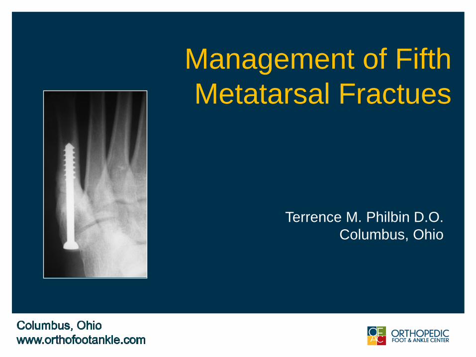
Management of Fifth Metatarsal Fractues
Terrence M. Philbin D.O.Columbus, Ohio
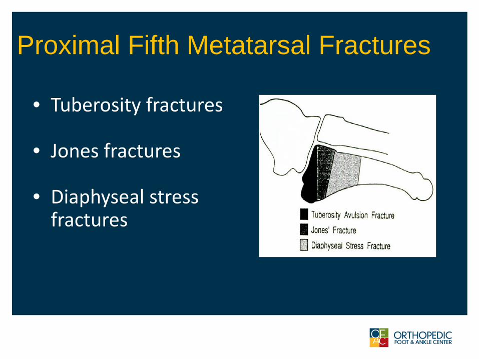
Proximal Fifth Metatarsal Fractures
• Tuberosity fractures
• Jones fractures
• Diaphyseal stress fractures
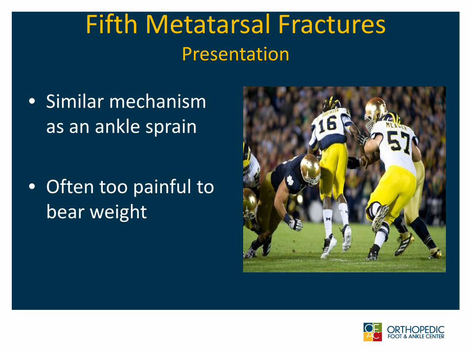
Fifth Metatarsal FracturesPresentation
• Similar mechanism as an ankle sprain
• Often too painful to bear weight
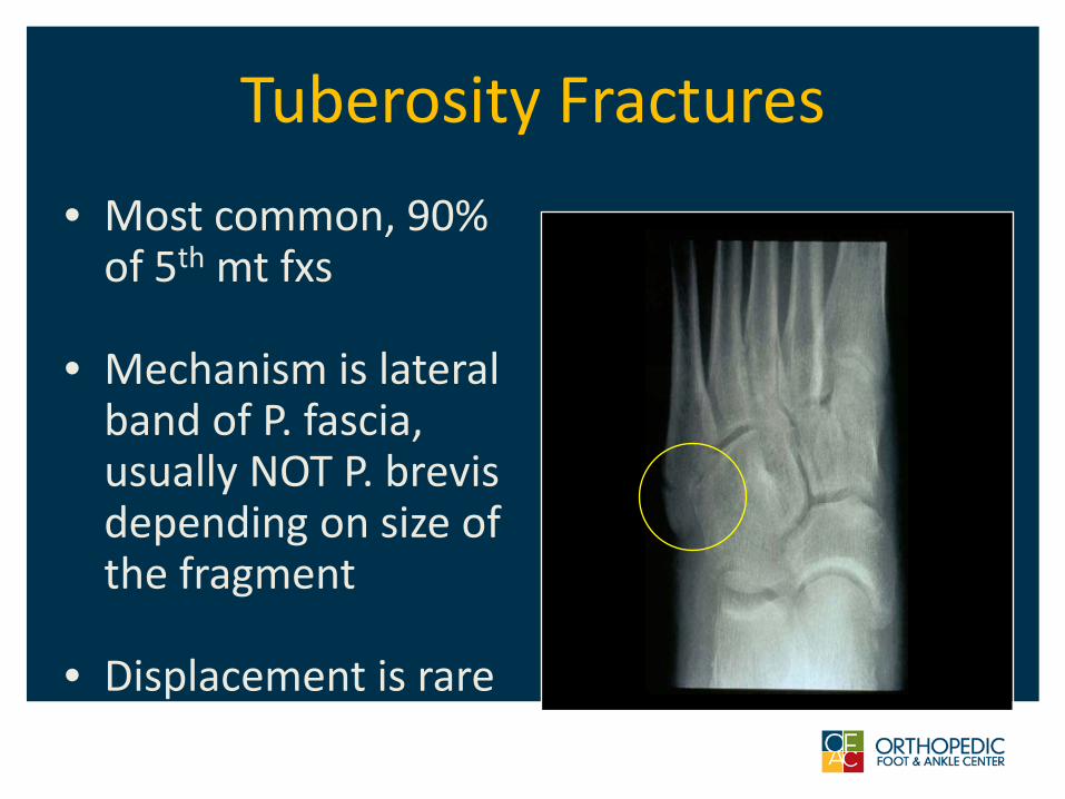
Tuberosity Fractures• Most common, 90%
of 5th mt fxs
• Mechanism is lateral band of P. fascia, usually NOT P. brevis depending on size of the fragment
• Displacement is rare
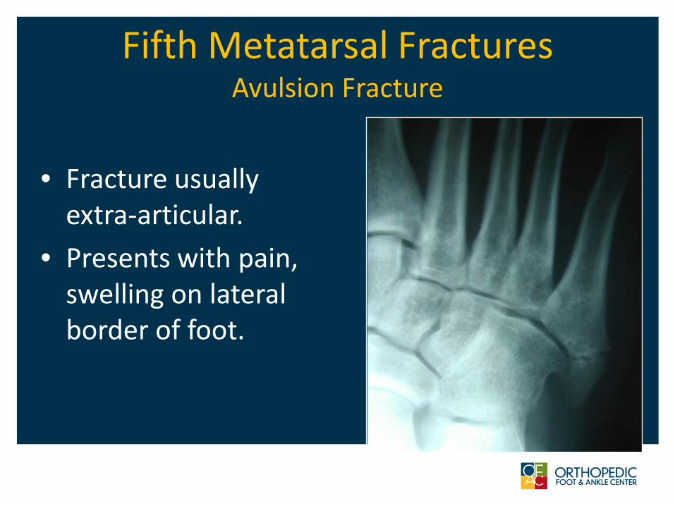
Fifth Metatarsal FracturesAvulsion Fracture
• Fracture usually extra-articular.
• Presents with pain, swelling on lateral border of foot.
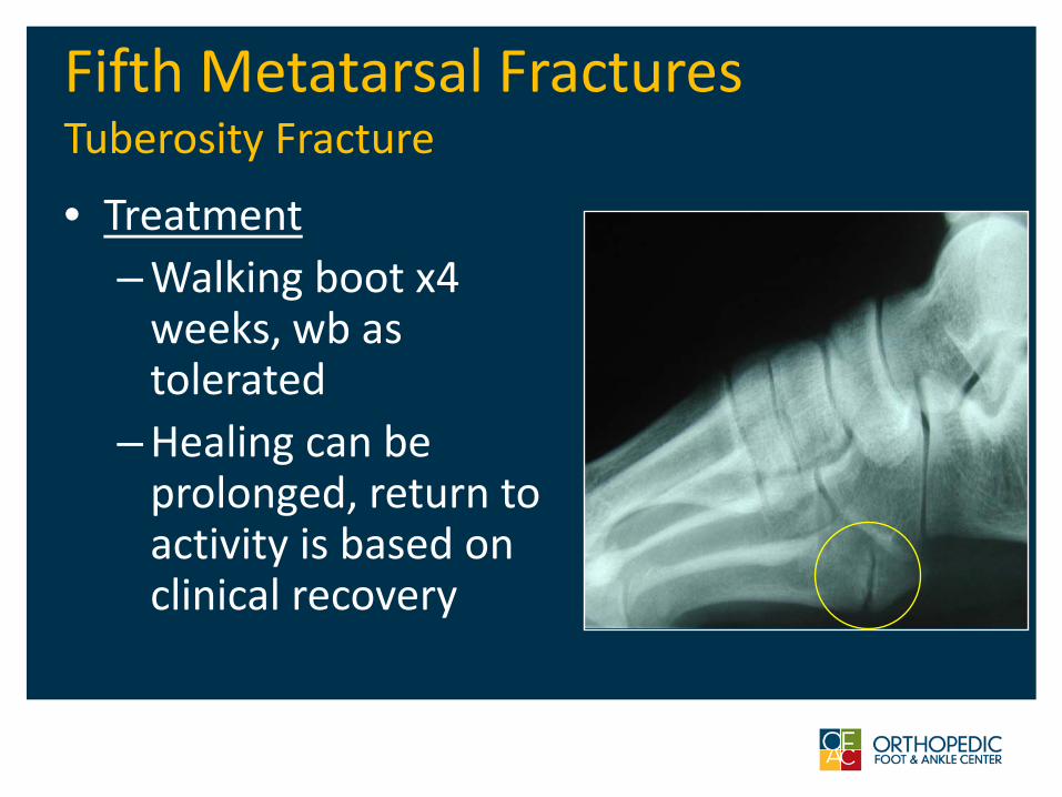
Fifth Metatarsal FracturesTuberosity Fracture
• Treatment– Walking boot x4
weeks, wb as tolerated
– Healing can be prolonged, return to activity is based on clinical recovery
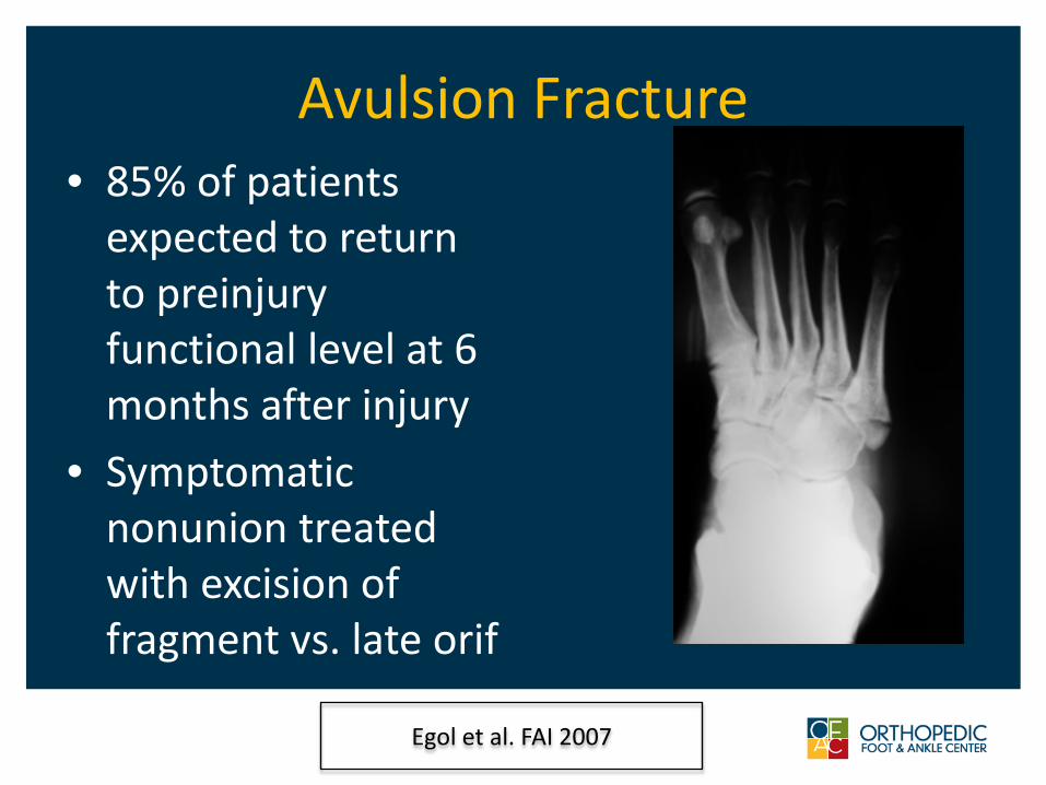
Avulsion Fracture• 85% of patients
expected to return to preinjury functional level at 6 months after injury
• Symptomatic nonunion treated with excision of fragment vs. late orif
Egol et al. FAI 2007
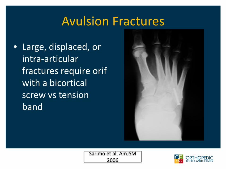
Avulsion Fractures
• Large, displaced, or intra-articular fractures require orif with a bicortical screw vs tension band
Sarimo et al. AmJSM2006
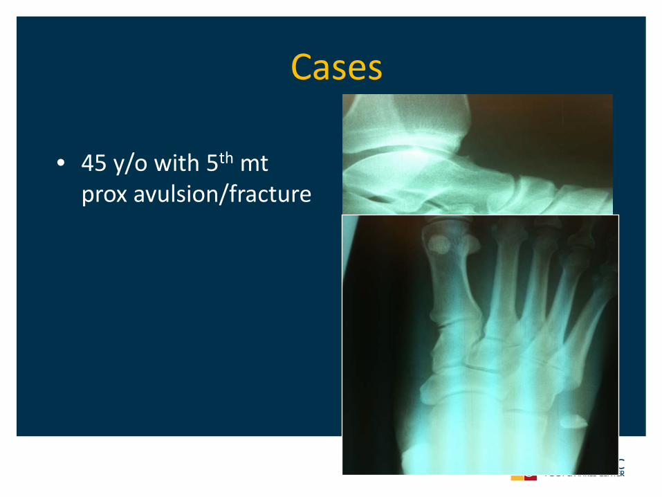
Cases
• 45 y/o with 5th mt prox avulsion/fracture
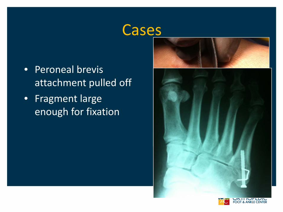
Cases
• Peroneal brevis attachment pulled off
• Fragment large enough for fixation
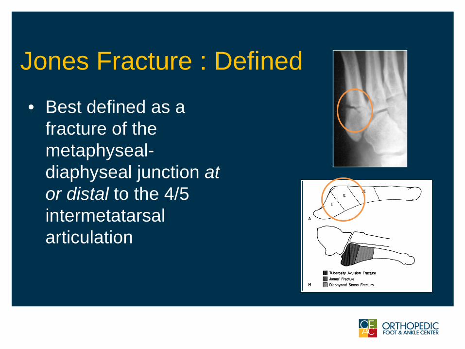
Jones Fracture : Defined
• Best defined as a fracture of the metaphyseal-diaphyseal junction at or distal to the 4/5 intermetatarsal articulation
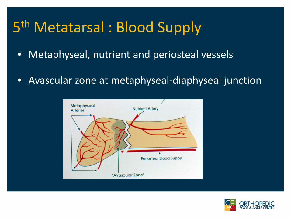
5th Metatarsal : Blood Supply
• Metaphyseal, nutrient and periosteal vessels
• Avascular zone at metaphyseal-diaphyseal junction
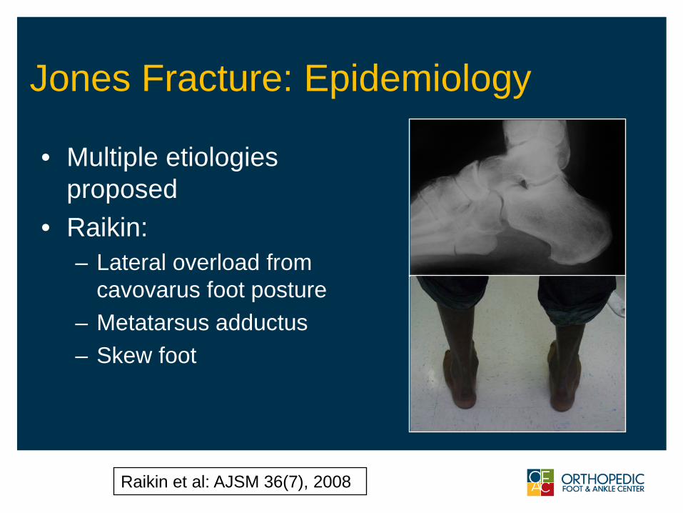
Jones Fracture: Epidemiology
• Multiple etiologies proposed
• Raikin:– Lateral overload from
cavovarus foot posture– Metatarsus adductus– Skew foot
Raikin et al: AJSM 36(7), 2008
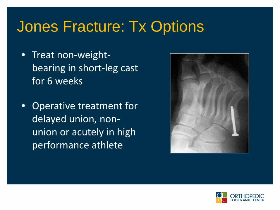
Jones Fracture: Tx Options
• Treat non-weight-bearing in short-leg cast for 6 weeks
• Operative treatment for delayed union, non-union or acutely in high performance athlete
ITEM 1
ITEM 2
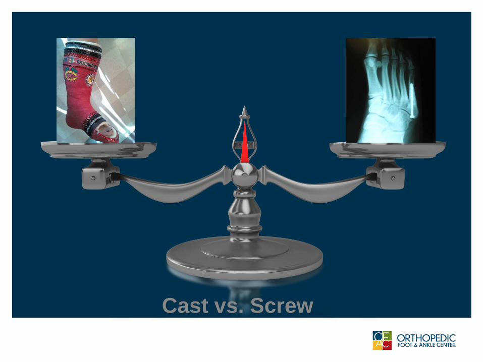
Cast vs. Screw
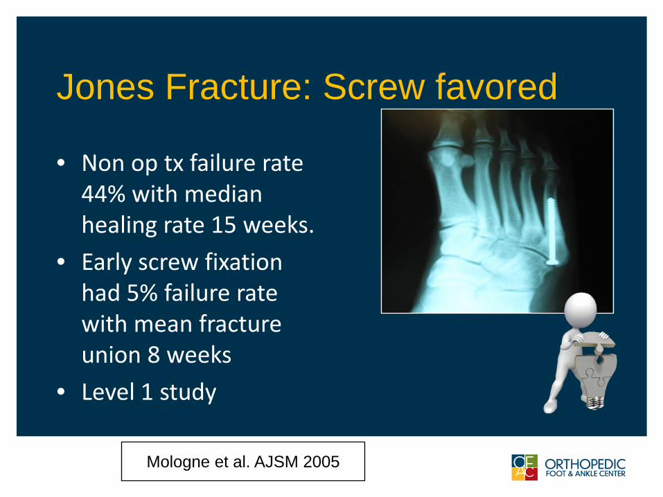
Jones Fracture: Screw favored
• Non op tx failure rate 44% with median healing rate 15 weeks.
• Early screw fixation had 5% failure rate with mean fracture union 8 weeks
• Level 1 study
Mologne et al. AJSM 2005
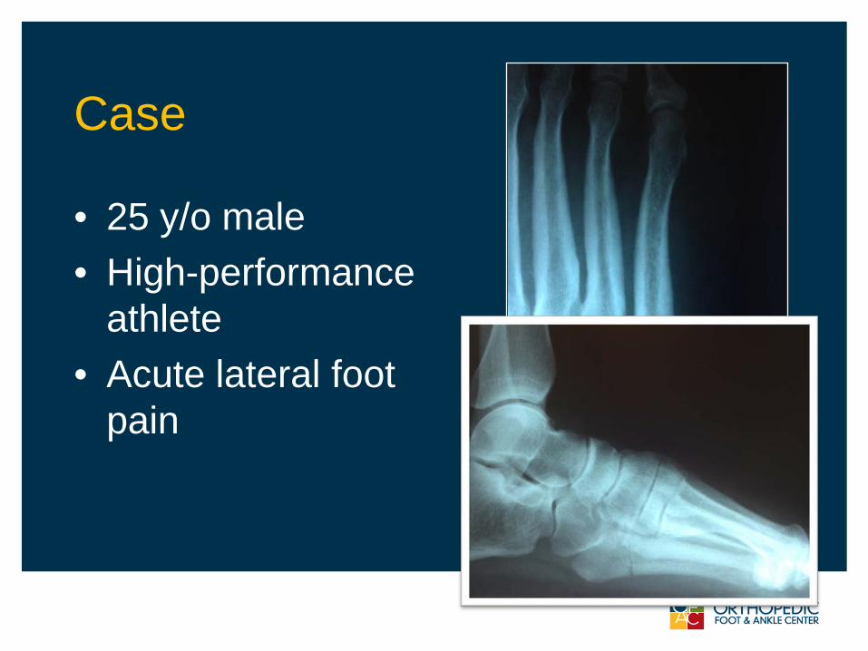
Case
• 25 y/o male• High-performance
athlete• Acute lateral foot
pain
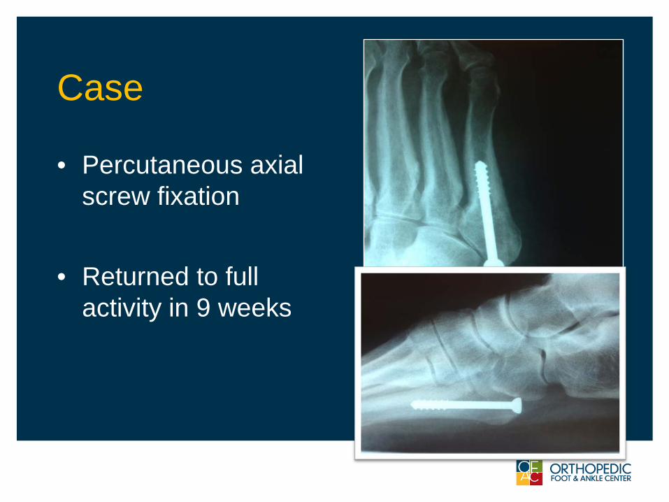
Case
• Percutaneous axial screw fixation
• Returned to full activity in 9 weeks
Jones Fractures- things to consider• Technique• Biomechanical Issues• Fixation choices:
– Type of screw ( solid or cannulated )– Metallurgy ( titanium or stainless steel)
• Return to sport timelines
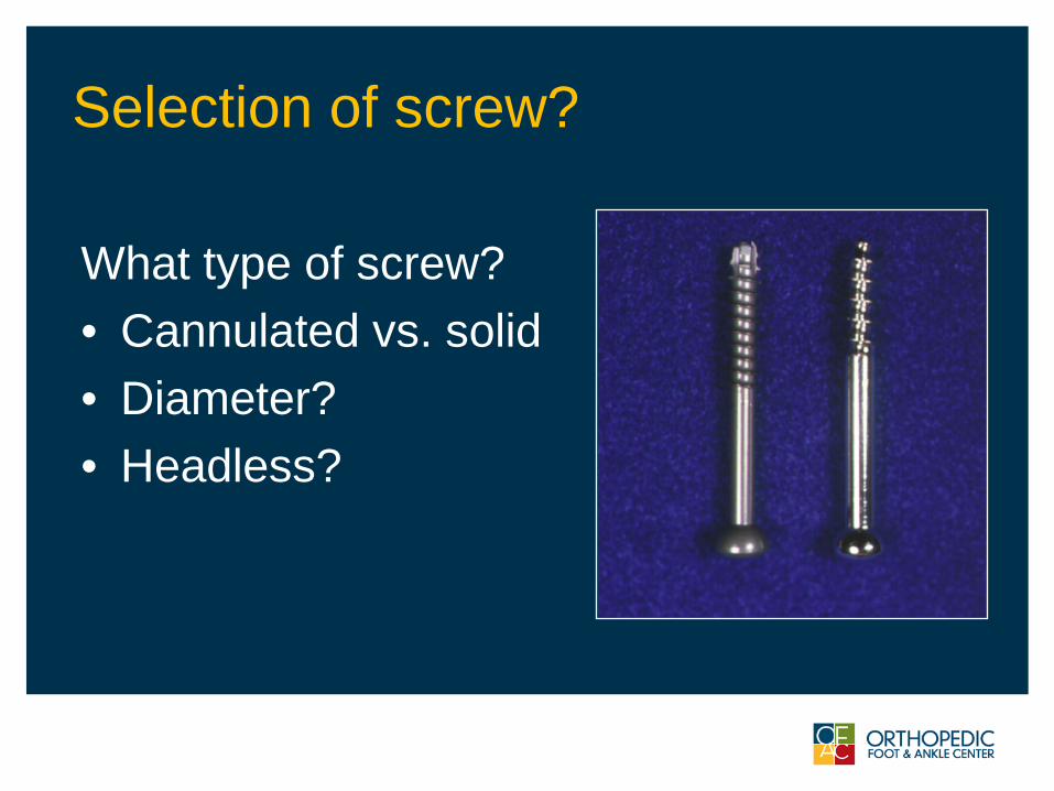
Selection of screw?
What type of screw?• Cannulated vs. solid• Diameter?• Headless?
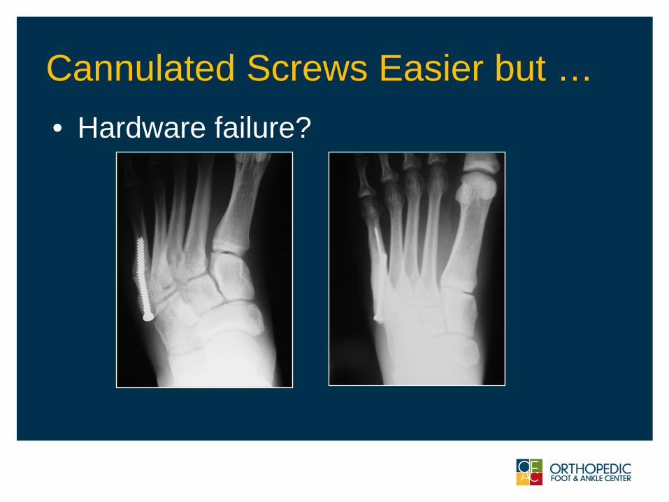
Cannulated Screws Easier but …• Hardware failure?
Screw Selection- literature• Pietropoali, FAI ’99
– No difference in cannulated vs. non-cannulated 4.5 mm
• Reese, AJSM ’04– Larger solid better (cycles to failure)– Avoid < 4 mm
• Devries JFAS 2011– No difference between cannulated 4.5 mm
titanium or stainless steel
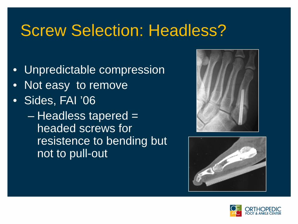
Screw Selection: Headless?
• Unpredictable compression• Not easy to remove• Sides, FAI ’06
– Headless tapered = headed screws for resistence to bending but not to pull-out
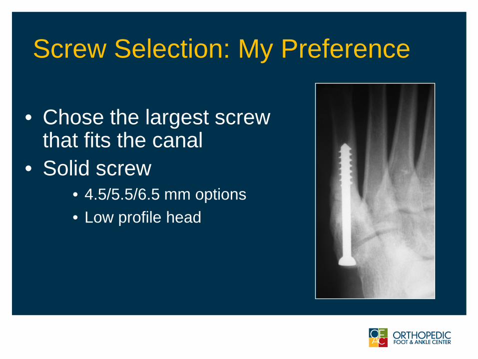
Screw Selection: My Preference
• Chose the largest screw that fits the canal
• Solid screw • 4.5/5.5/6.5 mm options• Low profile head
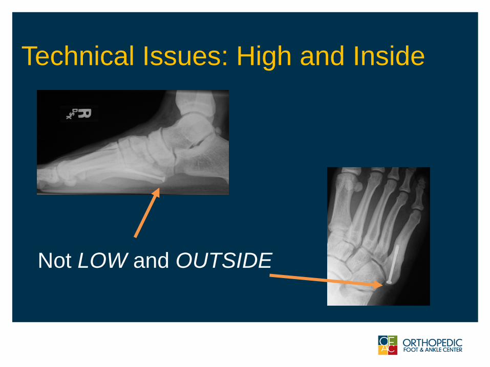
Technical Issues: High and Inside
Not LOW and OUTSIDE
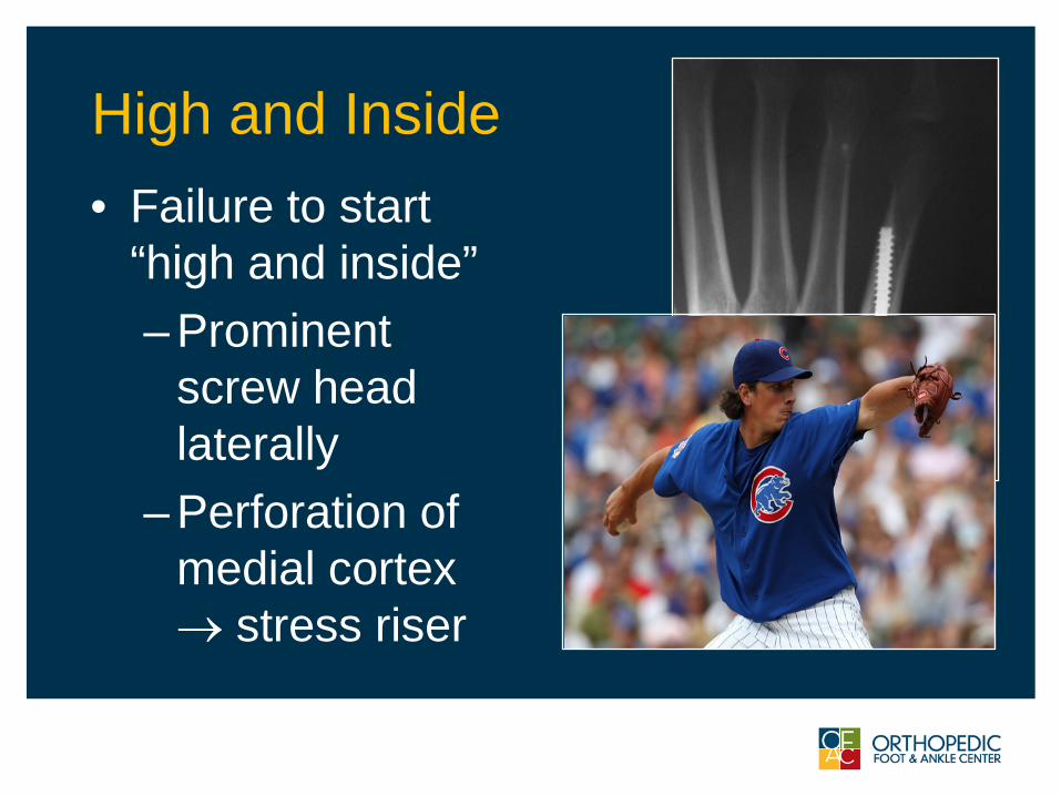
High and Inside • Failure to start
“high and inside”– Prominent
screw head laterally
– Perforation of medial cortex → stress riser
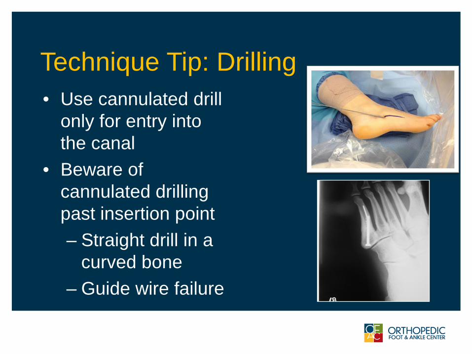
Technique Tip: Drilling• Use cannulated drill
only for entry into the canal
• Beware of cannulated drilling past insertion point– Straight drill in a
curved bone– Guide wire failure
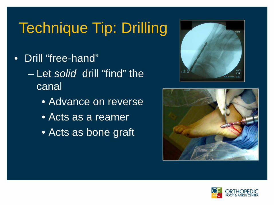
Technique Tip: Drilling
• Drill “free-hand”– Let solid drill “find” the
canal• Advance on reverse• Acts as a reamer• Acts as bone graft
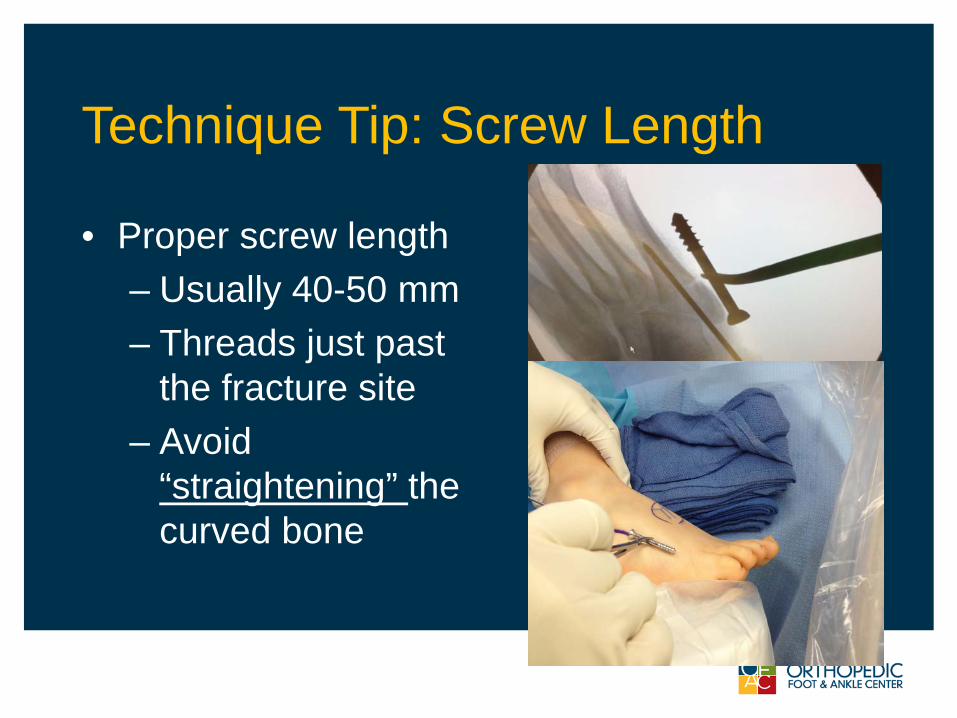
Technique Tip: Screw Length
• Proper screw length– Usually 40-50 mm– Threads just past
the fracture site– Avoid
“straightening” the curved bone
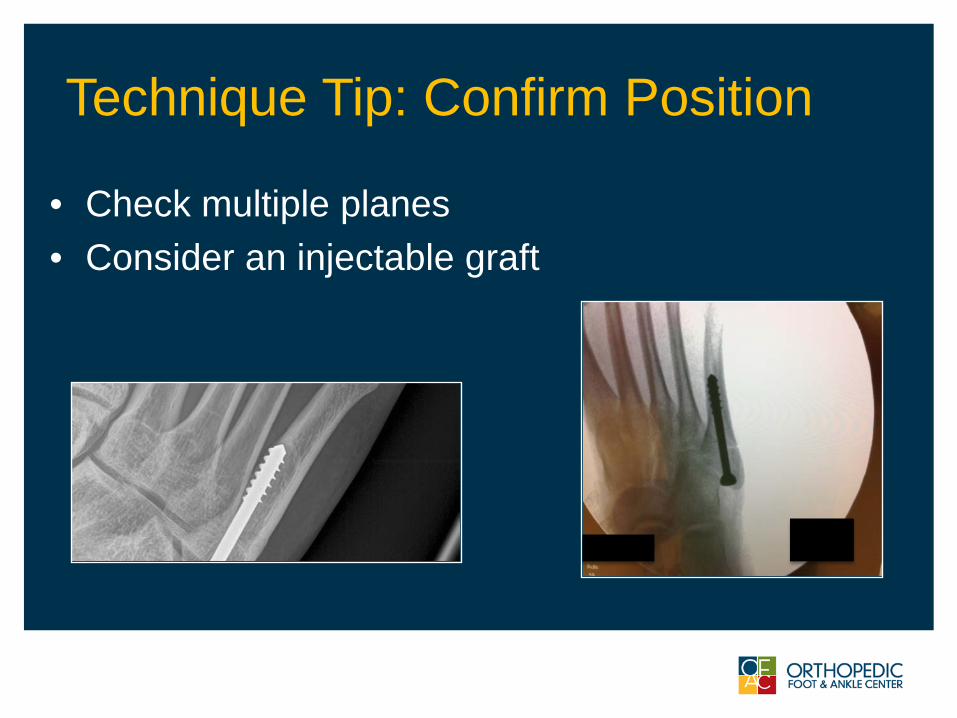
Technique Tip: Confirm Position
• Check multiple planes• Consider an injectable graft
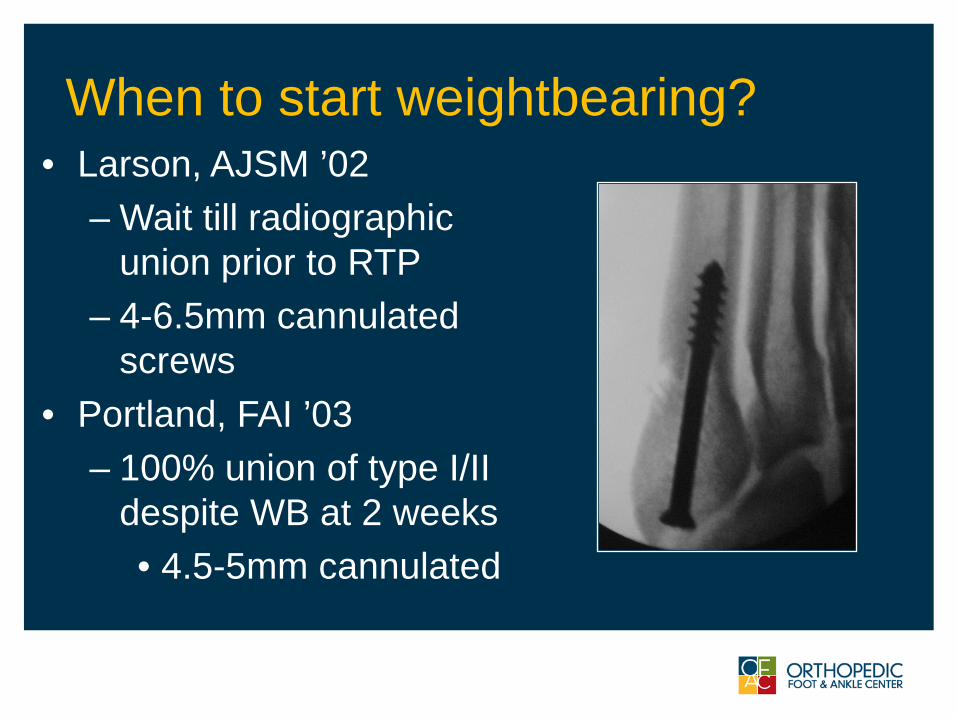
When to start weightbearing?• Larson, AJSM ’02
– Wait till radiographic union prior to RTP
– 4-6.5mm cannulated screws
• Portland, FAI ’03– 100% union of type I/II
despite WB at 2 weeks• 4.5-5mm cannulated

My Rehab Protocols : Athletes
More aggressive in athletes…• NWB x 2 weeks• WBTT in boot x 2-4 weeks• Pool therapy/bike• Bone stimulator
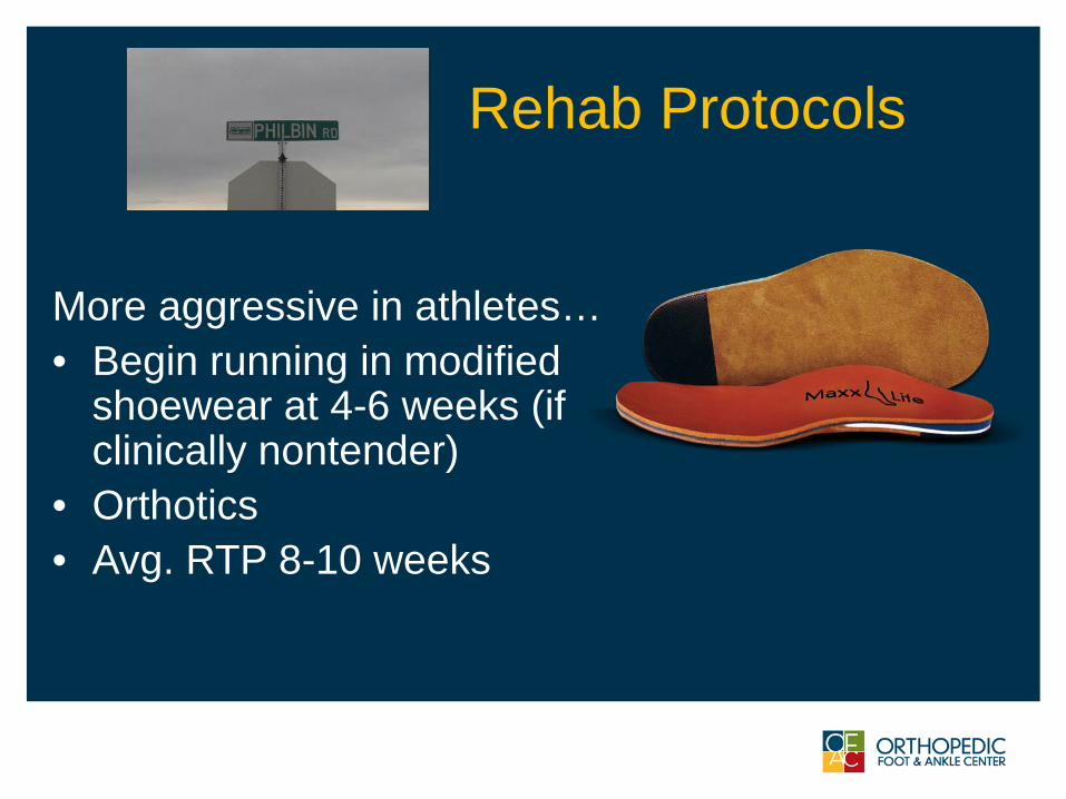
Rehab Protocols
More aggressive in athletes…• Begin running in modified
shoewear at 4-6 weeks (if clinically nontender)
• Orthotics• Avg. RTP 8-10 weeks
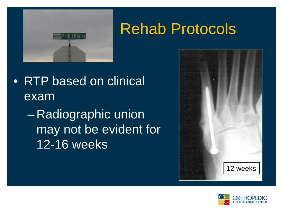
Rehab Protocols
• RTP based on clinical exam– Radiographic union
may not be evident for 12-16 weeks
12 weeks
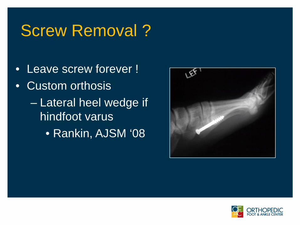
Screw Removal ?
• Leave screw forever !• Custom orthosis
– Lateral heel wedge if hindfoot varus• Rankin, AJSM ‘08
Outcomes: Good even if delayed treatment Habbu et al:• 14 patients• Mean duration from symptoms to surgery
= 28 weeks• Single intramedullary screw • 100 % union at an average of 13.3 weeks
Habbu et al: FAI 32(6), June 2011
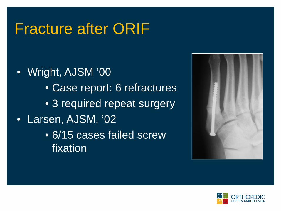
Fracture after ORIF
• Wright, AJSM ’00• Case report: 6 refractures• 3 required repeat surgery
• Larsen, AJSM, ’02• 6/15 cases failed screw
fixation
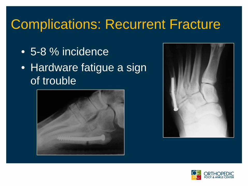
Complications: Recurrent Fracture
• 5-8 % incidence• Hardware fatigue a sign
of trouble
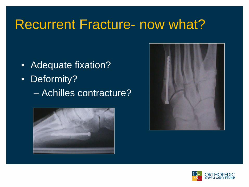
Recurrent Fracture- now what?
• Adequate fixation?• Deformity?
– Achilles contracture?
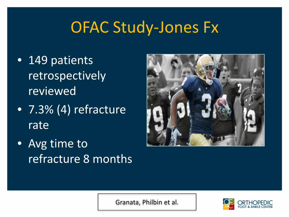
OFAC Study-Jones Fx
• 149 patients retrospectively reviewed
• 7.3% (4) refracture rate
• Avg time to refracture 8 months
Granata, Philbin et al.
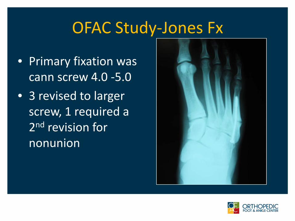
OFAC Study-Jones Fx
• Primary fixation was cann screw 4.0 -5.0
• 3 revised to larger screw, 1 required a 2nd revision for nonunion
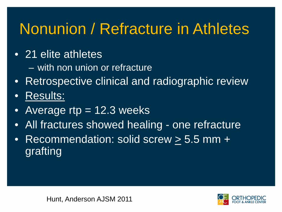
Nonunion / Refracture in Athletes• 21 elite athletes
– with non union or refracture• Retrospective clinical and radiographic review• Results:• Average rtp = 12.3 weeks• All fractures showed healing - one refracture• Recommendation: solid screw > 5.5 mm +
grafting
Hunt, Anderson AJSM 2011
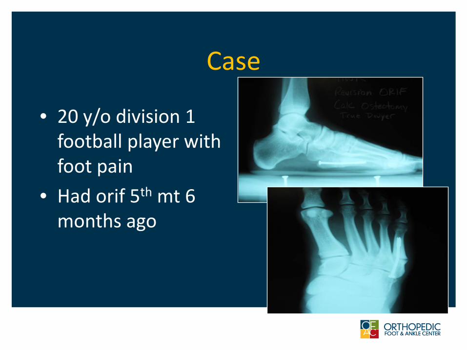
Case
• 20 y/o division 1 football player with foot pain
• Had orif 5th mt 6 months ago
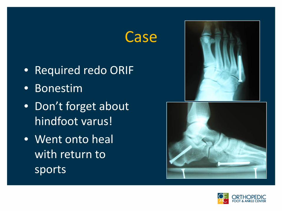
Case
• Required redo ORIF• Bonestim• Don’t forget about
hindfoot varus!• Went onto heal
with return to sports
Growth Factor Expression and Healing Time After Pulsed Electromagnetic Field Stimulation
of 5th Metatarsal Nonunions: A Prospective, Randomized, Double-Blind Trial
Terrence M. Philbin, DO1
Jaymes D. Granata, MD1
Hsuan-Ni Lin, PhD2
J. Patrick O’Connor, PhD2
Sheldon Lin, MD2
1. Advanced Orthopedic Foot & Ankle FellowshipOrthopedic Foot & Ankle Center, Columbus
2. New Jersey Medical SchoolDepartment of Orthopaedics
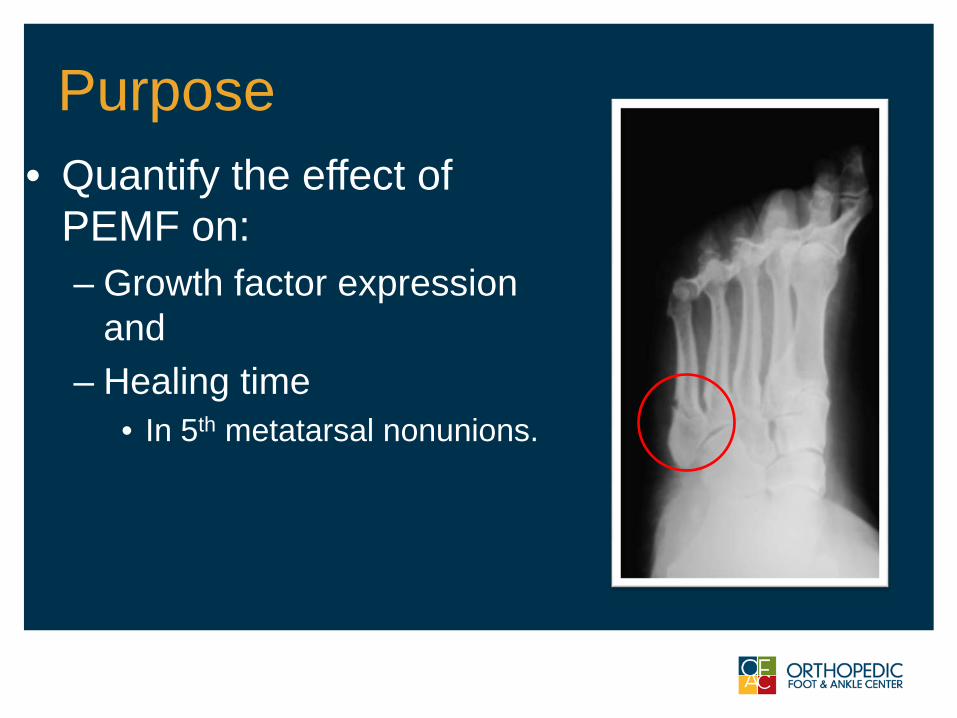
Purpose• Quantify the effect of
PEMF on:– Growth factor expression
and – Healing time
• In 5th metatarsal nonunions.
Methods• IRB approved prospective, randomized study
– Inclusion criteria • Between ages of 18 and 75 • Incompletely healed 5th metatarsal fracture after 3
months of conservative treatment – Exclusion criteria
• fracture gap greater than 5mm on CT scan• History of autoimmune or connective tissue
disease, history of cancer, current or previous infection of the 5th metatarsal, and pregnancy
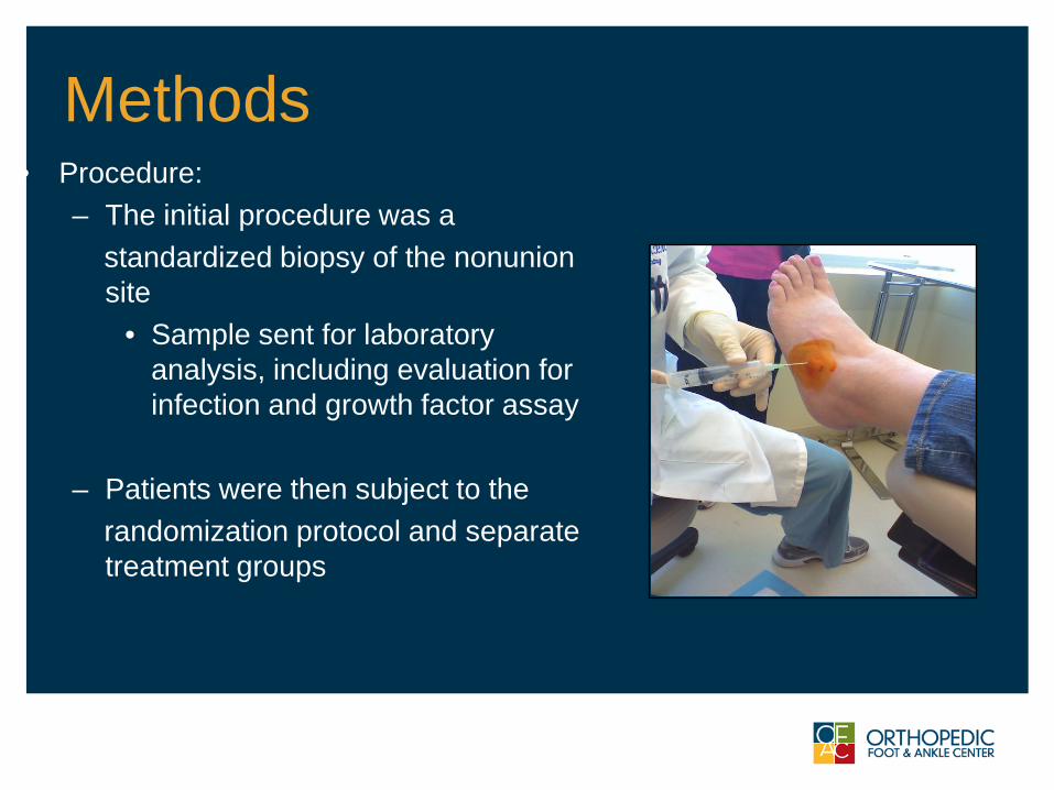
Methods• Procedure:
– The initial procedure was a standardized biopsy of the nonunion site
• Sample sent for laboratory analysis, including evaluation for infection and growth factor assay
– Patients were then subject to the randomization protocol and separate treatment groups
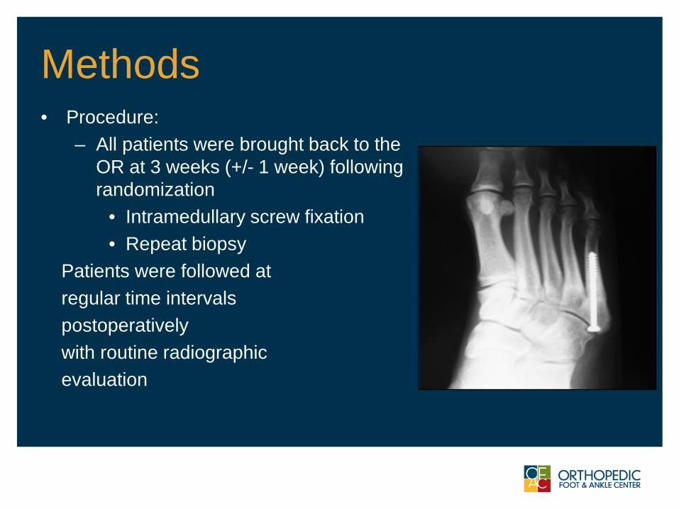
Methods• Procedure:
– All patients were brought back to the OR at 3 weeks (+/- 1 week) following randomization
• Intramedullary screw fixation • Repeat biopsy
Patients were followed at regular time intervals postoperatively with routine radiographic evaluation
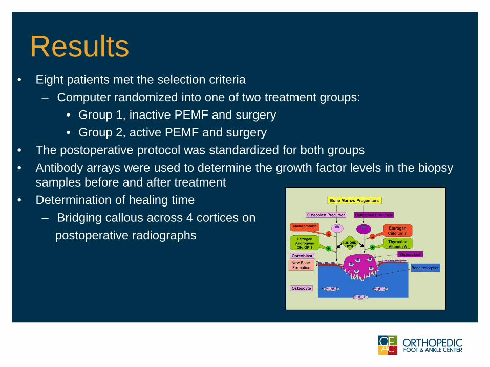
Results• Eight patients met the selection criteria
– Computer randomized into one of two treatment groups: • Group 1, inactive PEMF and surgery• Group 2, active PEMF and surgery
• The postoperative protocol was standardized for both groups • Antibody arrays were used to determine the growth factor levels in the biopsy
samples before and after treatment • Determination of healing time
– Bridging callous across 4 cortices on postoperative radiographs
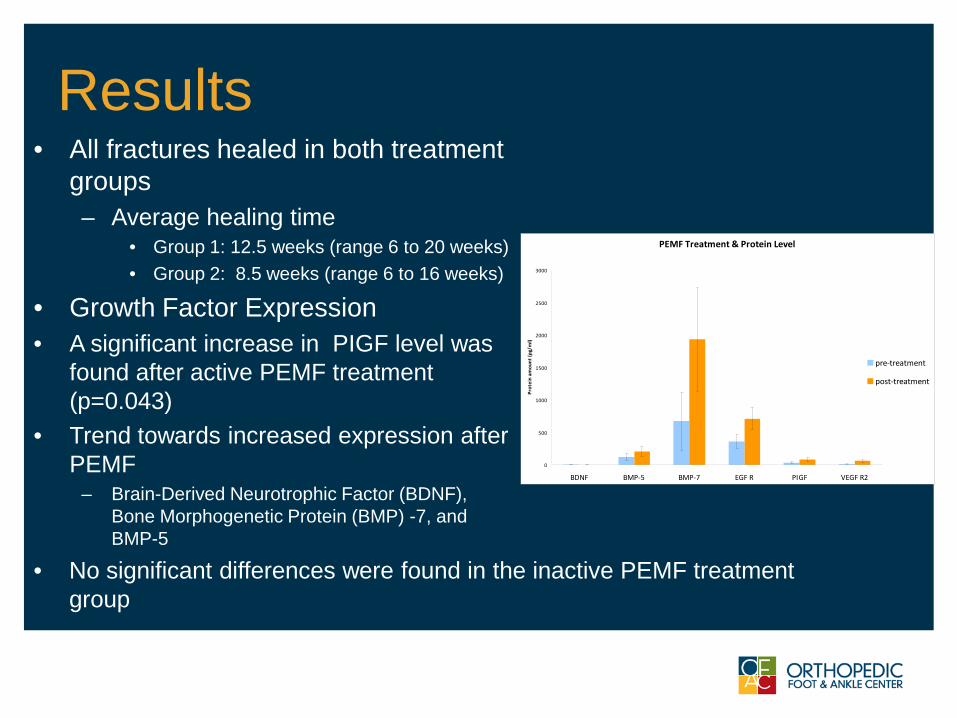
Results• All fractures healed in both treatment
groups– Average healing time
• Group 1: 12.5 weeks (range 6 to 20 weeks) • Group 2: 8.5 weeks (range 6 to 16 weeks)
• Growth Factor Expression• A significant increase in PIGF level was
found after active PEMF treatment (p=0.043)
• Trend towards increased expression after PEMF
– Brain-Derived Neurotrophic Factor (BDNF), Bone Morphogenetic Protein (BMP) -7, and BMP-5
• No significant differences were found in the inactive PEMF treatment group
PEMF Treatment & Protein Level
0
500
1000
1500
2000
2500
3000
BDNF BMP-5 BMP-7 EGF R PIGF VEGF R2
Prot
ein
amou
nt (p
g/m
l)
pre-treatment
post-treatment
Conclusions• The results of this study are consistent with
previous reports of increased growth factor expression after the use of PEMF in fracture healing
• A trend towards faster healing time was also noted in the active PEMF group
• Additional studies with larger treatment groups are needed to clarify the role of PEMF in delayed fracture healing and nonunions
Summary: Jones Fracture
• Jones fracture can still be a challenge• Always consider biomechanical issues
in recurrent situations and with revision surgery
• Be critical of your technique, fixation selection and rehab protocol

THANK YOU
Go Irish!!
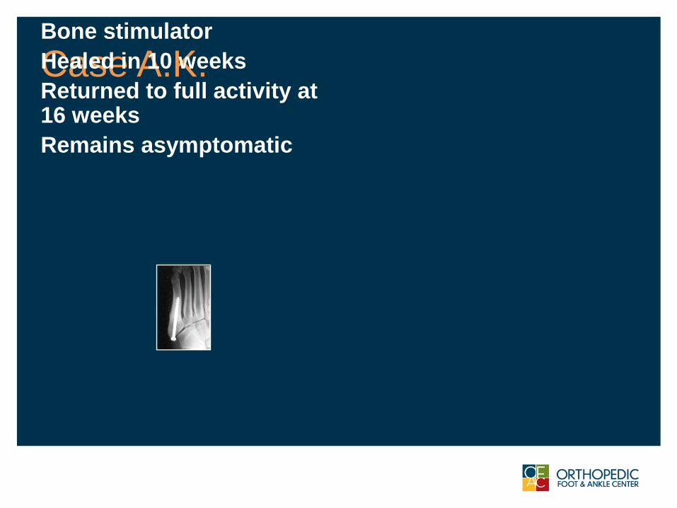
Case A.K.Bone stimulatorHealed in 10 weeksReturned to full activity at 16 weeksRemains asymptomatic
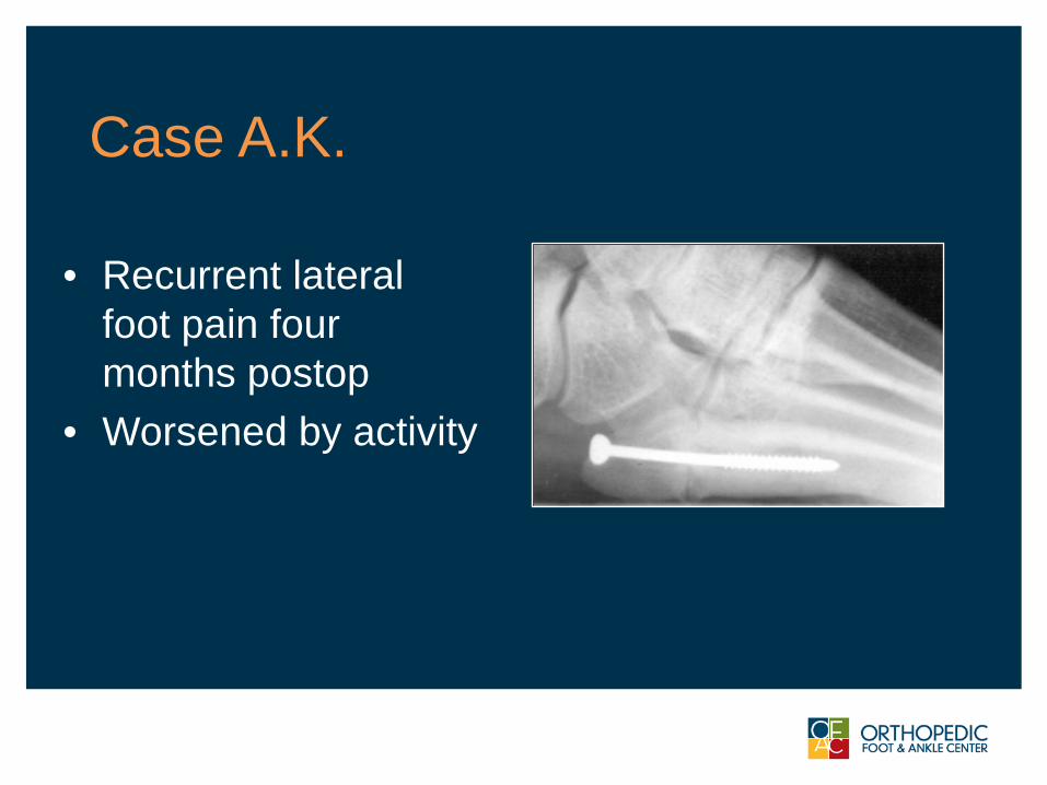
Case A.K.
• Recurrent lateral foot pain four months postop
• Worsened by activity
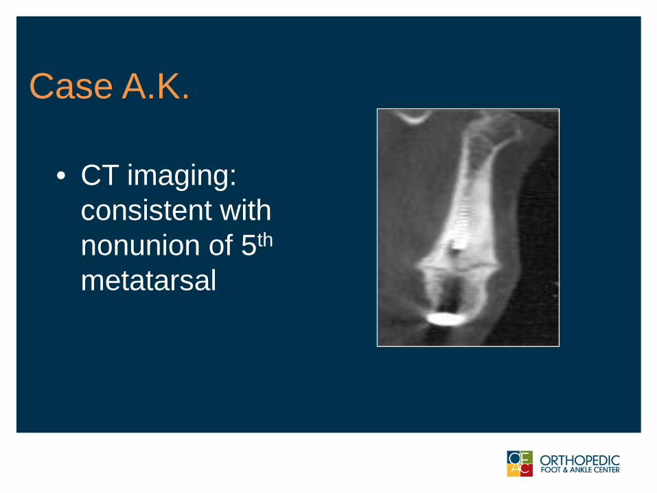
Case A.K.
• CT imaging: consistent with nonunion of 5th
metatarsal
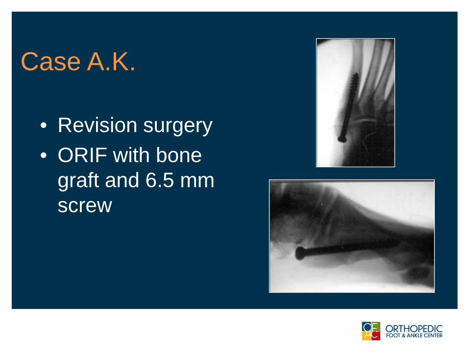
Case A.K.
• Revision surgery• ORIF with bone
graft and 6.5 mm screw
Complications: Recurrent Fracture
• Evaluation:• If asymptomatic • “good” screw
• Shoewear:• Rigid sole/turf toe plate• Pressure relieving orthosis
consider observation with shoewear protection
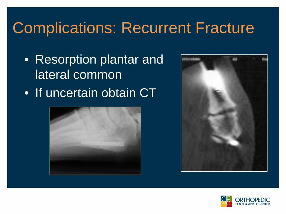
Complications: Recurrent Fracture
• Resorption plantar and lateral common
• If uncertain obtain CT