management of cardiac arrhythmias, 2 e (2011)
TRANSCRIPT


Management of Cardiac Arrhythmias

Contemporary CardiologyChristopher P. Cannon, MD
SERIES EDITOR
Annemarie M. Armani, MDEXECUTIVE EDITOR
For other titles published in this series, go tohttp://www.springer.com/series/7677

Management of CardiacArrhythmias
Edited by
Gan-Xin Yan, MD, PhDMain Line Health Heart CenterWynnewood, PA, USA
Peter R. Kowey, MDJefferson Medical CollegePhiladelphia, PA, USA

EditorsGan-Xin Yan, MD, PhDMain Line Health Heart CenterandLankenau Institute for Medical ResearchWynnewood, PA, [email protected]
Peter R. Kowey, MDJefferson Medical CollegePhiladelphia, PA, USAandMain Line Health Heart CenterWynnewood, PA, [email protected]
ISBN 978-1-60761-160-8 e-ISBN 978-1-60761-161-5DOI 10.1007/978-1-60761-161-5Springer New York Dordrecht Heidelberg London
Library of Congress Control Number: 2010934347
C© Springer Science+Business Media, LLC 2002, 2011All rights reserved. This work may not be translated or copied in whole or in part without the written permission of the publisher (HumanaPress, c/o Springer Science+Business Media, LLC, 233 Spring Street, New York, NY 10013, USA), except for brief excerpts in connectionwith reviews or scholarly analysis. Use in connection with any form of information storage and retrieval, electronic adaptation, computersoftware, or by similar or dissimilar methodology now known or hereafter developed is forbidden.The use in this publication of trade names, trademarks, service marks, and similar terms, even if they are not identified as such, is not to betaken as an expression of opinion as to whether or not they are subject to proprietary rights.While the advice and information in this book are believed to be true and accurate at the date of going to press, neither the authors nor theeditors nor the publisher can accept any legal responsibility for any errors or omissions that may be made. The publisher makes no warranty,express or implied, with respect to the material contained herein.
Printed on acid-free paper
Humana Press is part of Springer Science+Business Media (www.springer.com)

PrefaceIt is safe to say that few areas of medicine have moved faster than cardiac electrophysiology. In
three short decades, our field has grown from its infancy to a highly sophisticated subspecialty ofcardiology, complete with its own societies, scientific meetings, and board examination. Key to oursuccesses has been a progressively more in-depth understanding of pathophysiology from our basicscience laboratories. Burgeoning knowledge has been accompanied by a blitzkrieg of technology thathas allowed us to treat what used to be lethal rhythm disturbances and to improve the quality of lifeof millions of people the world over. In 2010, we stand on the threshold of an even more impressiveleap forward as we wrestle with defining how the genetic code predisposes to, or even causes, cardiacarrhythmias.
The price to pay for such rapid expansion of information is an ever-widening knowledge gap. It isobvious that practitioners who spend their time caring for patients find it difficult to keep up with thelatest developments in our field. The number of articles and journals that come across our desks everymonth is mind numbing. And few have the sophistication to understand the myriad of discoveriesthat are unwrapped at each of our congresses. Clearly there is a need to have complex informationpresented in an efficient and user-friendly way.
We believe that condensed texts represent one of the best ways for colleagues to stay current. Wealso think that there are individuals in our field, as in any endeavor, who are particularly skilled intaking a complex mass of information, condensing and formulating it, and producing a state-of-the-artmanuscript that makes clinical sense. Consequently, we agreed to recruit a stellar group of authorsand edit the text you are about to read. Its organization is standard, proceeding from basic scienceto diagnostic and therapeutic techniques, before ending in a discussion of specific patient types andsyndromes. We added an historical perspective that should be particularly gratifying to our youngerreaders. Since the time frame of development was short, the information is as current as possible andshould bring the interested reader up to speed rather quickly. We have tried to feature issues that willbe of continuing interest in our field over the next few years in order to provide a frame of reference forjournal reading. Finally, we have kept the level of science high to appeal to physicians and health-careprofessionals, or those in training, who have a deep interest in cardiac arrhythmias.
There are several we would like to acknowledge and thank, including our colleagues who helpedus with their knowledge and experience, our families who allowed us the time to write and edit, ourstaff who provided technical support, and the research foundations and granting agencies that keep usafloat. But most of all, we thank our patients who, by their courage and perseverance, inspire us to digdeeper so we can ultimately conquer the diseases that disrupt and end their lives.
Gan-Xin Yan, MD, PhDPeter R. Kowey, MD
v


AcknowledgmentsWe wish to acknowledge Ms. Rose Well and Drs. Ying Wu and Xiaoqing Quan for their assistance
in editing this book. We also wish to acknowledge American Heart Association, the W.W. SmithCharitable Trust, the Sharpe-Strumia Research Foundation, and the Albert M. Greenfield Foundationfor their generous support of our research and education in cardiac electrophysiology.
vii


ContentsPreface . . . . . . . . . . . . . . . . . . . . . . . . . . . . . . . . . . . . . . . . . . . . . . . v
Acknowledgments . . . . . . . . . . . . . . . . . . . . . . . . . . . . . . . . . . . . . . . . . vii
Contributors . . . . . . . . . . . . . . . . . . . . . . . . . . . . . . . . . . . . . . . . . . . . xi
Part I Introduction
1 Management of Ventricular Arrhythmias: An Historical Perspective . . . . . . . . . . . . 3David J. Callans and Mark E. Josephson
2 History of Supraventricular Tachycardia . . . . . . . . . . . . . . . . . . . . . . . . . . 17Yanfei Yang, Edmund C. Keung, and Melvin M. Scheinman
Part II Cardiac Electrophysiology
3 Ionic and Cellular Basis for Arrhythmogenesis . . . . . . . . . . . . . . . . . . . . . . . 41Charles Antzelevitch and Gan-Xin Yan
4 Genetic and Molecular Basis of Arrhythmias . . . . . . . . . . . . . . . . . . . . . . . . 65Shane B. Rowan and Dawood Darbar
Part III Diagnostic Testing
5 Diagnosis of Arrhythmias with Non-invasive Tools . . . . . . . . . . . . . . . . . . . . . 89Renee M. Sullivan, Wei Wei Li, Arthur C. Kendig, and Brian Olshansky
6 Electrophysiology Study: Indications and Interpretations . . . . . . . . . . . . . . . . . . 123Karen E. Thomas and Peter J. Zimetbaum
Part IV Specific Arrhythmias
7 Supraventricular Arrhythmias . . . . . . . . . . . . . . . . . . . . . . . . . . . . . . . . 141Khalid Almuti, Babak Bozorgnia, and Steven A. Rothman
8 Pharmacologic Management of Atrial Fibrillation and Flutter . . . . . . . . . . . . . . . 165Deepak Saluja, Kathleen Hickey, and James A. Reiffel
9 Catheter Ablation of Atrial Flutter and Atrial Fibrillation . . . . . . . . . . . . . . . . . . 195Joseph E. Marine and Hugh Calkins
10 Nonsustained Ventricular Tachycardia . . . . . . . . . . . . . . . . . . . . . . . . . . . . 225Peem Lorvidhaya and Alfred E. Buxton
11 Ventricular Tachycardia and Fibrillation: Pharmacologic Therapy . . . . . . . . . . . . . 243Gerald V. Naccarelli and John Field
12 Ablation for Ventricular Tachycardia . . . . . . . . . . . . . . . . . . . . . . . . . . . . 257Dusan Kocovic
13 Indications for Implantable Cardioverter-Defibrillators . . . . . . . . . . . . . . . . . . . 283Gustavo Lopera and Robert J. Myerburg
ix

x Contents
14 Bradyarrhythmias . . . . . . . . . . . . . . . . . . . . . . . . . . . . . . . . . . . . . . 305Ernest Matthew Quin, J. Marcus Wharton, and Michael R. Gold
Part V Arrhythmias in Specific Populations
15 Arrhythmias in the Athlete . . . . . . . . . . . . . . . . . . . . . . . . . . . . . . . . . . 323John A. Kalin, Mark S. Link, and N.A. Mark Estes III
16 Arrhythmias in Pregnancy and Postpartum . . . . . . . . . . . . . . . . . . . . . . . . . 339Kristen K. Patton and Richard L. Page
17 Arrhythmias in Children . . . . . . . . . . . . . . . . . . . . . . . . . . . . . . . . . . . 355Bhavya Trivedi and Ronald Kanter
Part VI Specific Syndromes
18 Syncope . . . . . . . . . . . . . . . . . . . . . . . . . . . . . . . . . . . . . . . . . . . 395Ilknur Can and David G. Benditt
19 Long QT Syndrome . . . . . . . . . . . . . . . . . . . . . . . . . . . . . . . . . . . . . 419Jonathan N. Johnson and Michael J. Ackerman
20 Short QT Syndrome: Clinical Presentation, Molecular, Genetic, Cellular,and Ionic Basis . . . . . . . . . . . . . . . . . . . . . . . . . . . . . . . . . . . . . . . . 441
Chinmay Patel, Gan-Xin Yan, and Charles Antzelevitch
21 J Wave Syndromes . . . . . . . . . . . . . . . . . . . . . . . . . . . . . . . . . . . . . . 453Jianfang Lian, Peter R. Kowey, and Gan-Xin Yan
Subject Index . . . . . . . . . . . . . . . . . . . . . . . . . . . . . . . . . . . . . . . . . . . 471

ContributorsMICHAEL J. ACKERMAN, MD, PHD, Departments of Medicine, Pediatrics, and Molecular Phar-
macology & Experimental Therapeutics/Divisions of Cardiovascular Diseases and PediatricCardiology/Windland Smith Rice Sudden Death Genomics Laboratory, Mayo Clinic, Rochester,MN, USA
KHALID ALMUTI, MD, Division of Cardiology, Lankenau Hospital, Wynnewood, PA, USACHARLES ANTZELEVITCH, PHD, Masonic Medical Research Laboratory, Utica, NY, USADAVID G. BENDITT, MD, Cardiac Arrhythmia Center, Cardiovascular Division, Department of
Medicine, University of Minnesota Medical School, Minneapolis, MN, USABABAK BOZORGNIA, MD, Naples Heart Rhythm Specialists, Naples, FL, USAALFRED E. BUXTON, MD, Department of Medicine, Rhode Island and Miriam Hospitals, The
Warren Alpert Medical School of Brown University, Providence, RI, USAHUGH CALKINS, MD, Department of Medicine and Cardiology, The Johns Hopkins Hospital,
Baltimore, MD, USADAVID J. CALLANS, MD, Section of Cardiology, Department of Medicine, The Hospital of the
University of Pennsylvania, Philadelphia PA, USAILKNUR CAN, MD, Cardiac Arrhythmia Center, Cardiovascular Division, Department of Medicine,
University of Minnesota Medical School, Minneapolis, MN, USADAWOOD DARBAR, MD, Department of Medicine, Vanderbilt University School of Medicine,
Nashville, TN, USAN.A. MARK ESTES III, MD, New England Cardiac Arrhythmia Center, Division of Cardiology,
Department of Medicine, Tufts Medical Center, Boston, MA, USAJOHN FIELD, MD, Penn State Hershey Heart & Vascular Institute, Penn State Milton S. Hershey
Medical Center, Hershey, PA, USAMICHAEL R. GOLD, MD, PHD, Division of Cardiology, Medical University of South Carolina,
Charleston, SC, USAKATHLEEN HICKEY, RN, DOCTOR OF NURSING, Columbia University Medical Center, New York,
NY, USAJONATHAN N. JOHNSON, MD, Department of Pediatrics, Division of Pediatric Cardiology, Mayo
Clinic, Rochester, MN, USAMARK E. JOSEPHSON, MD, Division of Cardiology, The Beth Israel – Deaconess Hospital, Boston,
MA, USAJOHN A. KALIN, MD, New England Cardiac Arrhythmia Center, Division of Cardiology, Depart-
ment of Medicine, Tufts Medical Center, Boston, MA, USARONALD KANTER, MD, Division of Cardiology, Department of Padiatrics and Medicine, Duke
University, Durham, NC, USAARTHUR C. KENDIG, MD, Department of Medicine, University of Iowa Hospitals, Iowa City, IA,
USAEDMUND C. KEUNG, MD, Cardiology Section, San Francisco Veterans Affairs Medical Center,
University of California, VA Medical Center, San Francisco, CA, USADUSAN KOCOVIC, MD, Division of Cardiology, Lankenau Hospital, Main Line Health Heart Center,
Wynnewood, PA, USAPETER R. KOWEY, MD, Main Line Health Heart Center, Wynnewood, PA, USA; Division of
Cardiovascular Diseases, Jefferson Medical College, Philadelphia, PA, USAWEI WEI LI, MD, Department of Medicine, University of Iowa Hospitals, Iowa City, IA, USA
xi

xii Contributors
JIANFANG LIAN, MD, PHD, Main Line Health Heart Center, Ning Bo Medical Center Li Hui LiHospital, Medical School, Ning Bo University, Ning Bo, P.R. China
MARK S. LINK, MD, New England Cardiac Arrhythmia Center, Division of Cardiology, Departmentof Medicine, Tufts Medical Center, Boston, MA, USA
GUSTAVO LOPERA, MD, Division of Cardiology, University of Miami/Miller School of Medicine andthe Veterans Affairs Medical Center, Miami, FL, USA
PEEM LORVIDHAYA, MD, Department of Medicine, The Warren Alpert Medical School of BrownUniversity, Providence, RI, USA
JOSEPH E. MARINE, MD, Department of Cardiology, The Johns Hopkins Hospital, Baltimore, MD,USA,
ROBERT J. MYERBURG, MD, Division of Cardiology, University of Miami/Miller School ofMedicine and the Veterans Affairs Medical Center, Miami, FL, USA
GERALD V. NACCARELLI, MD, Division of Cardiology, Penn State Hershey Heart and VascularInstitute, Penn State Milton S. Hershey Medical Center, Hershey, PA, USA
BRIAN OLSHANSKY, MD, Department of Medicine, University of Iowa Hospitals, Iowa City, IA,USA
RICHARD L. PAGE, MD, Department of Medicine, University of Wisconsin School of Medicine &Public Health, Madison, WI, USA
CHINMAY PATEL, MD, Lankenau Institute for Medical Research, Main Line Health Heart Center,Wynnewood, PA, USA
KRISTEN K. PATTON, MD, Division of Cardiology, Department of Medicine, University of Washing-ton School of Medicine, Seattle, WA, USA
ERNEST MATTHEW QUIN, MD, Division of Cardiology, Medical University of South Carolina,Charleston, SC, USA
JAMES A. REIFFEL, MD, Department of Medicine, Columbia University Medical Center, NY, USASTEVEN A. ROTHMAN, MD, Division of Cardiology, Lankenau Hospital, Main Line Health Heart
Center, Wynnewood, PA, USASHANE B. ROWAN, MD, Department of Medicine, Vanderbilt University School of Medicine,
Nashville, TN, USADEEPAK SALUJA, MD, Cardiac Care Unit, UMDNJ-Robert Wood Johnson Medical School, New
Brunswick, NJ, USAMELVIN M. SCHEINMAN, MD, Department of Medicine, Division of Cardiology, University of
California, San Francisco, CA, USARENEE M. SULLIVAN, MD, Department of Medicine, University of Iowa Hospitals, Iowa City, IA,
USAKAREN E. THOMAS, MD, Department of Medicine, Division of Cardiovascular Medicine, Beth
Israel Deaconess Medical Center, Harvard Medical School, Boston, MA, USABHAVYA TRIVEDI, MD, PHD, Pediatric Cardiology and Electrophysiology, Pediatrix Medical
Group/Pediatric Cardiology Associates, Tampa, FL, USAJ. MARCUS WHARTON, MD, Department of Medicine, Medical University of South Carolina,
Charleston, SC, USAGAN-XIN YAN, MD, PHD, Main Line Health Heart Center and Lankenau Institute for Medical
Research, Wynnewood, PA, USA; Division of Cardiovascular Diseases, Jefferson Medical College,Philadelphia, PA, USA; Xi’an Jiaotong University, Xi’an, China
YANFEI YANG, MD, Department of Medicine, Division of Cardiology, University of California, SanFrancisco, San Francisco, CA, USA
PETER J. ZIMETBAUM, MD, Department of Medicine, Division of Cardiology, Beth Israel Dea-coness Medical Center, Harvard Medical School, Boston, MA, USA

I INTRODUCTION

1 Management of Ventricular Arrhythmias:An Historical Perspective
David J. Callans and Mark E. JosephsonCONTENTS
NONPHARMACOLOGIC MANAGEMENT OF VT/VFCONCLUSIONS
REFERENCES
Abstract
The treatment of ventricular arrhythmias has undergone vibrant change in the last 40 years, evolving froma largely intellectual exercise to an evidence-based, guideline-supported set of patient care strategies. Thisprogress have been fueled by basic and clinical science and by the initial application of randomized clinicaltrials to the study of electrophysiology. Along the way, several strongly held beliefs were reconsidered, mostnotably the use of programmed stimulation or serial Holter montoring to guide pharmacologic therapy forventricular tachycardia. Although antiarrhythmic drugs are still considered useful in reducing the frequencyof recurrent VT episodes, our loss of confidence in guided drug therapy led to the development of device,surgical ablation, and catheter ablation therapies, which form the mainstay of treatment today.
Key Words: Ventricular tachycardia; ventricular fibrillation; premature ventricular complexes;electrophysiologic study; implantable cardioverter defibrillators (ICD); antitachycardia pacing; catheterablation; SCD-HeFT; Multicenter Automatic Defibrillator Trial; pace mapping.
A few hours before his death he told me . . . he did not feel any bodily ailments, and . . . without anysign of anything amiss, he passed away from this life.
. . . through failure of the artery that feeds the heart . . . which I found to be very parched and shrunkand withered.
– Leonardo DaVinci
Interest in the management of ventricular arrhythmias developed with the understanding that ven-tricular tachyarrhythmias were responsible for sudden cardiac arrest (Table 1). Although sudden deathhas been recognized for many centuries, true sudden cardiac arrest was probably initially describedby Leonardo DaVinci (see quote above). It was not until the second half of the twentieth centuryand the development of electrocardiographic monitoring that physicians recognized the initiation ofventricular fibrillation by premature ventricular complexes (PVCs), particularly in the early post-infarction period (Fig. 1) (1). With the advent of Holter monitoring, several studies demonstrated thatthe risk of sudden death and cardiac mortality increased as PVC frequency increased (particularly at a
From: Contemporary Cardiology: Management of Cardiac ArrhythmiasEdited by: Gan-Xin Yan, Peter R. Kowey, DOI 10.1007/978-1-60761-161-5_1
C© Springer Science+Business Media, LLC 2011
3

4 Part I / Introduction
Table 1Strategies for Treatment of VT/VF
PharmacologicEmpiricHolter-guidedEPS-guidedCombination
Non-phamacologicAntitachycardia pacingImplantable cardioverter defibrillator (ICD)Surgical ablationCatheter ablation
Fig. 1. Holter monitoring study demonstrating the rhythm recorded at the time of cardiac arrest in ambulatorypatients. The vast majority of sudden death in this study was caused by ventricular tachyarrhythmias, with ven-tricular tachycardia (at least as this initial arrhythmia) being the most common. Adapted from Ref. (1).
“threshold” of greater than 10 PVCs per hour) and/or the complexity of the PVCs increased (2, 3).In fact, PVC grading systems were developed by Lown that attempted to signify an increasing riskof sudden death with more malignant ventricular ectopic beats (4). This led to the PVC hypothesisthat treating spontaneous ventricular arrhythmias would prevent the induction of sustained ventriculararrhythmias, resulting in a reduction of sudden death risk. Initially, therapy was empiric use of antiar-rhythmic agents, particularly sodium channel blocking agents since these agents stabilized membranesand reduced the frequency of PVCs (Table 2). Unfortunately, as later trials would eventually demon-strate, none of these agents prevented sudden cardiac death, particularly in the post-infarction setting.Empiric use of beta-blockers, however, seemed to decrease mortality, both total and sudden in theBeta Blocker Heart Attack Study (BHAT) (5). Because of the failure of empiric use of antiarrhythmicagents, Holter guided therapy was attempted. It was clearly realized, however, that Holter monitor-ing itself had many limitations. First of all, the frequency and complexity of arrhythmias could varyfrom hour to hour and day to day. The longer one was monitored, the more frequent arrhythmias werenoted. This became even more evident when Holter monitoring was performed during the adminis-tration of antiarrhythmic drugs. Many studies showed that the frequency of spontaneous arrhythmiasbore no relationship to the spontaneous episodes of sustained ventricular arrhythmias (Fig. 2). Thus,the following basic assumptions of Holter guided therapy were shown to be in error.

Chapter 1 / Management of Ventricular Arrhythmias 5
Table 2Pharmacologic Therapy for Treatment of VT/VF
Advantages:• Noninvasive• No surgical morbidity or mortality• Inexpensive in short run• May be appropriate for certain subgroups:
– Refused ICD– Multisystem disease– Poor overall prognosis
Disadvantages:• Often empiric, even if EP-guided, since not all drugs can be
serially tested due to expense• Often associated with intolerable side effects, organ toxicity, and
non-compliance• Even if EP-guided, many patients remain non-suppressible and
have a poor prognosis
Fig. 2. The lack of ventricular ectopy and spontaneous episodes of sustained VT is one of the limitations ofmanaging antiarrhythmic therapy guided by Holter monitoring. In this example, multiple antiarrhythmic drugswere used and treatment efficacy was assessed with monitoring. Despite a marked reduction of ventricular ectopyduring quinidine and subsequently diisopyramide therapy, frequent episodes of sustained VT were observed.(From Ref. (38)).
1. Frequent and complex ectopy are specifically and causally related to VT/VF.2. Holter monitoring reliably identifies these arrhythmias.3. Elimination of PVCs prevents sudden death
This led to the demise of the use of Holter monitoring as a mode of prevention of lethal arrhythmias.Moreover, the Cardiac Arrhythmia Suppression Trial (CAST) demonstrated that in patients with coro-nary artery disease, moderately reduced injection fractions and chronic stable angina, use of 1C agentswas associated with an increase in mortality (6). The conclusions from CAST were that treatment ofasymptomatic or mildly symptomatic arrhythmias with Class IC agents in such patients was associatedwith excess mortality and no benefit. Proarrhythmia caused by these sodium channel blocking agents

6 Part I / Introduction
may occur late and elimination of ventricular ectopy did not provide protection against sudden cardiacdeath. It was the CAST Study that put an end to pharmacologic-directed therapy for the treatment ofspontaneous ectopy.
In the early 1970s, clinical electrophysiology began to develop as a tool to investigate the mecha-nisms of arrhythmias. Wellens in 1972 first demonstrated that sustained ventricular tachycardia couldbe initiated by programmed electrical stimulation (Fig. 3) (7). Shortly thereafter, Josephson and his col-leagues from the University of Pennsylvania demonstrated that using aggressive stimulation protocols,sustained ventricular tachycardia and coronary artery disease could be reproducibly initiated in the vastmajority of patients with healed myocardial infarction who presented with VT. Furthermore, VT couldbe reproducibly induced in patients who had more rapid arrhythmias, clinical arrhythmias associatedwith cardiac arrest and even those nonsustained ventricular arrhythmias, albeit in a much lower per-centage of cases (Fig. 4) (8). The sensitivity and specificity of programmed stimulation were validatedin a number of centers. As a result, the concept of electrophysiologic testing of VT induction as a wayto evaluate therapy was advanced (9). In this philosophy, one considered that there needed to be a sub-strate in which spontaneous or stimulated extra beats could initiate lethal arrhythmias. Studies fromHorowitz, Fisher, Mason and others demonstrated that one could use the response to programmed elec-trical stimulation to evaluate whether or not drugs could prevent spontaneous events (9–11). In Fig. 5,a series of drugs was administered and the response to programmed stimulation evaluated. A varietyof antiarrhythmic drugs were tested and it was established that the class 1A agents more frequentlyprevented induction than other agents. Moreover, noninducibility of arrhythmia was associated with
Fig. 3. Programmed ventricular stimulation for the induction of VT in one of Dr. Wellens’ original patients.Following a drive train at 600 msec, an extrastimulus at 510 msec does not induce VT (upper left). When theextrastimulus coupling interval is decreased to 500, VT is induced and the interval to the first tachycardia beatis 500 msec (lower left). When the coupling interval is decreased to 420 msec (upper right), VT is induced andthe interval to the first VT beat increases to 510 msec. Finally, when the coupling interval is decreased to 410msec, VT is no longer induced. The observations of an extrastimulus coupling interval “window” which resultsin VT induction, and a reciprocal relationship between the extrastimulus coupling interval and the timing of thefirst tachycardia beat provide evidence for a reentrant mechanism for VT in the setting of healed infarction.

Chapter 1 / Management of Ventricular Arrhythmias 7
Fig. 4. The ability to reproducibly induce VT in patients with healed infarction varies according to their clinicalpresentation. In patients who present with tolerated sustained VT, VT is induced in over 95%. The frequency ofVT induction is less in patients who present with cardiac arrest or nonsustained VT; in addition, the frequencyis less in patients with non-infarct-related forms of structural heart disease. Reproduced from Ref. (8) withpermission.
Fig. 5. The use of programmed stimulation to predict the efficacy of antiarrhythmic agents. A collection ofseparate electrophysiologic study data from the same patient being treated with different drug regimens is shown.Ventricular stimulation resulted in VT induction in the baseline state, which was not prevented by treatment withlidocaine, phenytoin or disopyramide, but was prevented by procainamide and quinidine. (From Ref. (39)).

8 Part I / Introduction
freedom of events with a predictive accuracy of approximately 80% in a 2-year follow-up. Unfortu-nately, VT inducibility on a given antiarrhythmic regimen did not always predict recurrence. This wasparticularly true with amiodarone. Despite this limitation, it was also noted that these sodium channelblocking drugs could frequently slow VT, resulting in well-tolerated recurrent events as opposed tosyncope or cardiac arrest (12). Thus, such agents not only could prevent arrhythmia but could makeit tolerated so that elective cardioversion or other stimulation techniques could be used to terminatethe arrhythmia. While these findings applied to patients with VT in the setting of prior myocardialinfarction, the ability to use programmed stimulation to predict successful antiarrhythmic therapy fornonischemic cardiomyopathy was unsuccessful. In such patients, reproducible initiation was less oftennoted, and response to drug therapy was not predictive of freedom from sudden cardiac death (13).
The use of electrophysiologic guided therapy had limitations aside from the inability to use it incardiomyopathy, however. First, none of the studies which demonstrated favorable outcomes wererandomized. Secondly, most results were actually a combination of prospective and retrospective data(i.e., prior clinical drug failure associated with inducible VT and EP studies). Finally, the follow-upof all of these studies was short, somewhere between 1 and 2 years. Nonetheless, all studies showed ahigher occurrence rate and/or mortality in those patients with persistently inducible VT.
The use of electrophysiologically guided antiarrhythmic drug therapy came to an abrupt halt withthe Electrophysiologic Study versus Electrocardiographic Monitoring (ESVEM) Study. This was thefirst prospective randomized trial to evaluate the role of antiarrhythmic therapy guided by the resultsof programmed stimulation versus Holter monitoring. This was a highly selected patient population ofpatients with sustained VT (15 beats or more), cardiac arrest (less than 15% of patients) and syncope(15%). In addition, the inclusion criteria required patients to have both >10 PVCs per hour on Holtermonitoring and inducible VT with programmed stimulation. The results of ESVEM Study showedthat both methodologies, as applied, were not useful to predict drug efficacy; recurrence was frequentindependent of which strategy was used (14). Sotalol was a well-tolerated, moderately successful drugin patients with reasonable ventricular function who had not failed prior antiarrhythmic therapy. How-ever, these results did not permit relative drug efficacy comparisons in untreated patients, since thoseeffectively treated were excluded from the ESVEM trial. These results are also not applicable to car-diac arrest patients with VT in clinical settings besides healed infarction, as these patients were not wellrepresented in this study. There were many limitations to the ESVEM Study. There was no placeboarmed to access mortality of untreated patients. Because of the known risks of many of the antiarrhyth-mic agents that were used, it is not inconceivable that a placebo group may even have faired better.The data may apply to only 15% of patients who present with frequent ectopy and/or nonsustainedVT on Holter who have inducible VT on EPS. There is a bias against EPS and a bias against type 1agents, because patients with VT who were successfully treated with class 1 agents were excluded andpatients with VT failing drug therapy (particularly class 1 agents) were included. This latter limitationis extremely important, since prior study suggested that failure of one or more class 1 agents (typicallyprocainamide) predicted failure of any antiarrhythmic agent as assessed by programmed stimulation.In total, 2/3 of the patients had failed standard drugs. In our opinion, there were other limitations thatprohibit generalization of the ESVEM trial. First, the stimulation protocol was inadequate and not uni-form. The number of extrastimuli was never more aggressive than during stimulation in the baselinestate; if three extrastimuli were not delivered at baseline, three extrastimuli would never be deliveredin interpreting the efficacy of a drug regimen. Thus, what was considered successful may not havebeen. Second, the ability for a patient to tolerate sotalol, particularly with regard to absence of severeLV dysfunction, may have biased the apparent response to sotalol. Many patients had already failedclass 1 agents, but none were included who had been successfully treated, a significant bias againstclass 1 agents. There is a very high recurrence rate despite “best” therapy, probably due to the priorfailure of class I agents in many subjects (which predicts failure of other drugs, as discussed above).Finally, amiodarone was not included in this trial. Regardless of these limitations which we believe

Chapter 1 / Management of Ventricular Arrhythmias 9
Table 3History of the Development of Nonpharmacologic Therapy for VT
• 1977–1985 – anti-tachycardia pacing for VT• 1978–1985 – development of surgery for VT• 1980–1999 – development and refinement of the implantable defibrillator• 1984–1993 – refinement of mapping techniques and development of
catheter ablation for monomorphic VT; change from direct currentablation (fulguration) to radiofrequency energy.
• 1998–2002 – application of catheter-based identification of the arrhythmiasubstrate to treat untolerated VTs by RF ablation
significant and biased against EP studies, the use of EP-guided pharmacologic therapy virtually endedfollowing completion of the ESVEM study. The recognition of significant pro-arrhythmia also shiftedthe approach towards nonpharmacologic therapy (Table 3).
NONPHARMACOLOGIC MANAGEMENT OF VT/VF
The nonpharmacologic modalities that have been developed to treat VT/VF include:
1. antitachycardia pacing2. antiarrhythmic surgery3. implantable cardioverter defibrillators (ICDs)4. catheter ablation
The concept of antitachycardia pacing was obvious at the time of the initial studies of programmedstimulation for ventricular tachycardia by Wellens and Josephson (15, 16). Both labs demonstrated thatreproducible termination of arrhythmias was possible in patients whose tachycardia could be repro-ducibly initiated. The group from The University of Pennsylvania demonstrated that the rate of tachy-cardia influenced the ability to terminate by programmed stimulation. The success of programmedstimulation (in the form of burst pacing, programmed stimuli or autodecrimental pacing) was high atrates <200 beats per minute or less; for more rapid VT, antitachycardia pacing was still successful inat least 50% VTs, but acceleration to ventricular fibrillation was more common. Furthermore, antiar-rhythmic drug therapy, which consistently slows VT rate, also has a favorable influence on terminationwith overdrive pacing. Although there was a period of time in which specific antitachycardia pacingdevices were used, the possibility of accelerating the tachycardia or producing ventricular fibrillationled to abandonment of this modality of therapy as a stand alone therapy.
Three forms of therapy eventually evolved. The earliest was antiarrhythmic surgery. Surgical pro-cedures evolved initially in the mid-1970s and continued to the early 1990s. Catheter endocardialmapping was developed in the mid-1970s and demonstrated for the first time that the majority ofarrhythmias in coronary artery disease originated on or near the endocardium (17). Intra-operativemapping of the endocardium and epicardium confirmed these findings in coronary artery disease (18).In addition, Cassidy et al. demonstrated that the substrate in which the tachycardia arose could bedefined by abnormal electograms, those of low amplitude, broad width, fractionation and those whichwere late (i.e., recorded beyond the termination of the QRS complex) (19). Studies by Kienzle inthe operating room confirmed these findings (20). Mapping in the catheterization lab could identifyexit sites of early activation of the ventricular myocardium giving rise to specific QRS morphologies.These data were confirmed by mapping in the operating room. As a result of these mapping studies,the group at the University of Pennsylvania developed the subendocardial resection to remove thearrhythmogenic substrate responsible for ventricular tachycardia and ventricular fibrillation (Fig. 6)

10 Part I / Introduction
Fig. 6. The technique of subendocardial resection for surgical ablation of VT. The aneursymal segment of theleft ventricle was opened, and after endocardial mapping, the subendocardial VT substrate was “peeled” fromthe surviving epicardial tissue. Ventricular stimulation was repeated, and if residual VT morphologies existedoutside of the dense infarct, they were typically treated with focal cryoablation.
(21). These investigations demonstrated that map-guided endocardial resections could successfullyprevent sudden cardiac death (4% in 5 years) or recurrent VT (8% in 5 years) (8). The surgical pro-cedure was successful in patients with recent or remote myocardial infarction. Other non-map-guidedprocedures such as encircling subendocardial resection or encircling cryoablation were developed byGuiraudon as a method to facilitate surgical procedures without the complexity of mapping equipment(22). When map-guided and non-map-guided therapies were compared at the University of Pennsyl-vania, map-guided therapy appeared to have significantly better outcomes. While surgical therapy wassuccessful for patients with coronary artery disease, there was a lesser experience and lower successrate in patients with cardiomyopathies. These patients appeared to have a lesser amount of endo-cardial electrical abnormalities and origins of VT and a greater amount of epicardial abnormalities.Not enough data were available to surgically address patients with cardiomyopathies and VT, andthey continued to be considered nonsurgical candidates. Because of relatively high operative mortality(10–15%) and requirement for “a surgical electrophysiological team” the procedure was used mini-mally with the development of the ICD.
The implantable defibrillator was developed initially by Mirowski and colleagues despite initialderidement by many in the field (23). Mirowski’s persistence, however, led to the development of animplantable cardioverter defibrillator, which was initially improved in 1985 (Fig. 7). The initial devicesrequired thoracotomy for the placement of epicardial patches. Since that time, the ICD has been minia-turized (20–25 cc) and has added an increasingly greater complexity in terms of number of leads (dualchamber or biventricular) or pacing (for rate support and antitachycardia pacing) as well as additionalleads to assure adequate defibrillation thresholds (subcutaneous or dual coil leads in the SVC). Theability to implant these devices as simply as a pacemaker with transvenous leads led to the widespreadapplication of this form of therapy throughout the world. The growth of ICD implantations has becomeexponential, such that more than 200,000 ICDs are implanted yearly in the United States alone. TheAntiarrhythmic Versus Implantable Defibrillator (AVID) Study was the first to demonstrate that theICD was superior to antiarrhythmic drugs (primarily amiodarone) in patients who had experienced a

Chapter 1 / Management of Ventricular Arrhythmias 11
Fig. 7. Demonstration of efficacy in defibrillation during ICD testing. Surface ECG lead II, the electrogramrecorded from the ICD lead and ICD sensing markers are recorded via the device. A low energy shock deliveredat the crest of the T wave results in induction of ventricular fibrillation. This is promptly detected by the device,and sinus rhythm is restored with the delivery of a 12 joule shock.
cardiac arrest or untolerated VT (24). The increase in survival was not impressive, but was dilutedby cross over from initial treatment assignment. It was not very cost-effective ($125,000 per year lifesaved). Nevertheless, guidelines were established that suggested ICDs should be implanted in patientssuffering from a hemodynamically untolerated VT or a cardiac arrest who had ejection fractions ofless than 40%. Although limited data are available, ICD therapy has not been demonstrated to beeffective (in terms of saving lives) in patients with ejection fractions of greater than 40% (except inspecific clinical situations, such as long QT syndrome or hypertrophic cardiomyopathy). This demon-strates the inherent limitation of basing guideline recommendations on even well-constructed ran-domized controlled trials. Clearly, young patients without structural heart disease and high-risk chan-nelopathies, whose only possible cause for death would be arrhythmic, require an ICD; however, thesepatients were underrepresented in the trials to make that point. In addition, the guidelines suggest thatpatients with well-tolerated VT also should receive devices because they had the same mortality as theuntreated patients with poorly tolerated VT/VF. This conclusion is invalid since there has never been atrial assessing other strategies (pharmacologic, ablation) versus ICD therapy for tolerated VT. This isfurther attested to by the fact that surgical therapy for VT/VF was more effective than any ICD, yet themortality was 50% at 5 years. As such, using total mortality as the primary endpoint, as was done inAVID as well as most contemporary randomized trials, carries with it significant limitations, particu-larly in patients with advanced structural heart disease. It is not reasonable to expect that ICD therapywould have any effect on mortality aside from sudden death mortality, and competing causes of deathremain high, even with contemporary pharmacologic therapy for heart failure. Finally, an additionallimitation of the AVID trial was that the control group received far less beta blockade than the ICDgroup. In fact, given the known benefit of beta-blockers in preventing both sudden and total mortality,it is conceivable that this difference could have been responsible for a majority of the difference insurvival between groups.
More recently, the use of ICDs for primary prevention has been championed. Several studies eval-uating ICDs alone versus paired with pharmacologic therapy or ICDs versus EP-guided therapy havedemonstrated in patients with low injection fractions (less than 30%) and coronary artery disease(Multicenter Automatic Defibrillator Trial – MADIT II) (25), less than 40% in coronary artery diseasewith nonsustained VT and inducible VTs (Multicenter Unsustained Tachycardia Trial – MUSTT) (26),

12 Part I / Introduction
less than 35% in coronary artery disease with nonsustained VT and failure to respond to intravenousprocainamide (Multicenter Automatic Defibrillator Trial MADIT I) (27) and those patients with injec-tion fractions of less than 35% who have class 2 or 3 heart failure (Sudden Cardiac Death in HeartFailure Trial -SCD-HeFT) (28) all demonstrated some benefit from ICD therapy. MUSTT and MADITI, trials that by design enriched the arrhythmic risk in the studied population prior to enrollment byprior EP studies and the presence of spontaneous nonsustained VT, had a significant mortality reduc-tion and good cost-effectiveness with number of needed to treat from three to fourpatients per lifesaved. However, MADIT II and SCD-HeFT had poorer number needed to treat parameters and lowerabsolute benefit. In fact in SCD-HeFT, the mortality benefit was 1.4% per year over 5 years. Whenone compares the noncoronary artery patients as a subgroup of SCD-HeFT as well as the noncoronaryartery patients seen in the Defibrillators in Nonischemic Cardiomyopathy Treatment Evaluation (DEF-INITE) (29) trial and the Amiodarone Versus Implantable Cardioverter-defibrillator (AMIOVIRT) (30)trial, there has been no consistent benefit in survival from primary prevention ICD therapy. This wasrecently re-evaluated by Tung et al. and raises the prospect of potential overuse of the device in suchpatients. Of note is the fact that in Europe, the use of ICDs for primary prevention of cardiomyopathypatients is a class 2 indication, whereas it is a class 1 in the United States. Viewing the lack of strikingbenefit from the device as well as potential complications (which have been recently summarized byJosephson and coworkers) (31) many in the United States have reassessed device usage.
With the reduction of the use of surgery, and the simultaneous development of newer mapping tools,the possibility of catheter ablation to cure VT or abolish the substrate of arrhythmias has become apossibility. This is particularly true in the case of scar-related VT due to healed infarction but with thedevelopment of epicardial approaches, ablation for control of VT for patients with cardiomyopathyand in whom the substrate appears to be primarily epicardial or subepicardial. In addition, many otherforms of ventricular arrhythmias which are highly symptomatic (RVOT and idiopathic LV tachycar-dia) and which can lead to cardiomyopathy can be cured by catheter ablation. Many of the mappingtechniques established to localize critical areas of re-entrance circuits of scar-related VT were estab-lished in the mid-1970s and early 1980s by a group at the University of Pennsylvania. Resetting andentrainment were further designed by Almendal, Josephson, Morady, and Stevenson et al. in the 1980sto allow precise localization of critical components of re-entry circuits of scar-related VT that couldbe destroyed, eliminating the arrhythmia (32–35). The findings during entrainment or resetting of VTwhich identify a critical isthmus through which an impulse must travel and is bordered by anatomicand/or functional barriers and is ideal for ablation and termination of VT include the following:
1. A paced QRS morphology which is identical to VT (concealed entrainment) which identifies that thepaced site is in, attached to or just proximal to a protected isthmus
2. The stimulus to QRS is approximately equal to the electogram to the QRS during VT, which means thatthe paced LV site is not a dead end pathway attached to the circuit.
3. The return cycle measured at the pacing site is equal to the VT cycle length, which means that the siteof pacing is within the tachycardia circuit.
These observations require pacing at rates not significantly faster than the VT cycle, to preventslowing of conduction or using very premature stimuli to reset the tachycardia. In addition, it requiresthat the current used may not be too high to capture more distant tissues. An example of a perfectentrainment map with successful ablation is shown (Fig. 8). Failure to terminate a tachycardia, evenwhen the entrainment map is apparently good may be related to a sub-epicardial location of the circuit,a wide isthmus or endocardial thrombus. Although dealing with insultating thrombus may be extremelydifficult, the sub-epicardial location can be dealt with an epicardial approach and a wide isthmus canbe dealt with by defining those sites which meet characteristics for an isthmus and ablating over alarger area.

Chapter 1 / Management of Ventricular Arrhythmias 13
Fig. 8. Entrainment mapping for well-tolerated uniform VT. Surface leads and intracardiac recordings from theablation catheter (ABL distal and proximal) and the right ventricle (RVA) are shown. Pacing is performed duringan episode of VT. The following characteristics suggest that the ablation catheter is within a protected isthmus ofthe VT circuit: (1) pacing from the catheter results in a perfect match in all surface ECG leads, (2) the stimulusto QRS onset during pacing equals the electrogram to QRS onset during VT (100 msec) and (3) the return cycle(the first spontaneous VT beat after pacing) measured at the pacing site is equal to the VT cycle length (480msec).
Although use of entrainment or reset mapping is useful for tolerated ventricular tachycardia, themajority of tachycardias occurring today are untolerated and cannot be mapped in detail by usingthese techniques. A different approach to those tachycardias is needed. Such an approach requiresan understanding of the pathophysiological substrate of the arrhythmia. While the basic principlesof substrate mapping were established in the mid-1980s by Cassidy et al., it was a development ofelectroanatomic mapping that allowed one to localize these electrograms in three-dimensional spaceand record them automatically, which allowed for the potential of ablating components of the sub-strate that were arrhythmogenic (36). The approach to mapping and ablating the substrate involvedfinding potential channels of activation, which could form critical isthmuses responsible for arrhyth-mias within the scar (37). The methods which are used include:
1. Pace mapping at a border zone to identify exit sites and isthmuses (long stimulus to QRS with the samemorphology as the pacing is moved deeper into the scar).
2. Redefining voltage windows to find potential channels of viability within scar initially in scar definedby a voltage of 0.5 millivolts.
3. Pacing at high voltage to identify inexcitable tissue that could form barriers through which viable tissueis identified.
4. Identification of split potentials to define potential barriers of an isthmus

14 Part I / Introduction
5. Define late potentials in order to identify critical isthmus sites leading to isolated mid-diastolicpotentials.
Examples of pace mapping to define an exit site and an isthmus are shown in Fig. 9. Once these exitsites or channels have been identified, ablation perpendicular to the channel and into the channel canbe used to prevent that channel from being used as an arrhythmia. Identifying channels of viable tissueeither by changing the voltage definitions or by looking for an excitable tissue surrounding excitablepathways can identify channels that can also be ablated. Finally, ablating all late potentials is anotherpotential methodology, but is much more difficult given the lack of ability for precisely identifyingand ablating all existing late potentials.
Fig. 9. Voltage mapping during sinus rhythm using electroanatomic mapping. A “shell” of the LV is made duringsinus rhythm – each mapped point is assigned a location in three dimensional space, and information about theelectrophysiologic characteristics of that point, in this case bipolar electrogram voltage, is presented in colorcoding: purple corresponds to normal, red to dense infarct (electrograms ≤ 0.5 mV) and the intervening colorsto the intervening voltages. A large apical infarction is demonstrated, and two VT morphologies are mapped andablated with substrate-based techniques. A right bundle right inferior axis VT (top panel) is mapped to the septalaspect of the infarct border, and linear ablation is performed perpendicularly to the presumed exit site (each redicon corresponds to a single ablation lesion), which was established as the site with the closest pacemap. A rightbundle right superior axis VT is similarly mapped and ablated to the lateral aspect of the infarct (bottom panel).
There are limitations to all of these techniques that involve both false positive and false negativeresults. Many of these are related to using high current outputs at the pacing site, which leads tocapture across circuit barriers, and effects more tissue than can be ablated with a single lesion. Despitethese limitations, a recent randomized trial using a substrate-based ablation strategy in patients withprior ICD implantations for cardiac arrest or documented syncope with inducible VT demonstratedthat this ablation strategy could reduce ICD therapies by nearly 70% in a 2-year follow-up period (40).

Chapter 1 / Management of Ventricular Arrhythmias 15
Further work is necessary to demonstrate whether this is a valid approach with the implantation ofdevices for both secondary and primary prevention. However, since this ablation carries a risk, thisstrategy needs to be compared to standard antiarrhythmic therapies, beta-blockers and ace inhibitors,before widespread use is accepted.
CONCLUSIONS
The history of EP therapy has been one of continuous evolution for understanding of the mechanismand underlying physiological substrate of arrhythmias. The development of new technology to allowprecise identification of arrhythmogenic sites has aided measurably to our ability to use catheter-basedablative procedures to treat these arrhythmias. It is our hope that with greater understanding of allthe processes involved in development of the substrate may lead to improved antiarrhythmic agentswhich are less toxic and more targeted as well as better based techniques to treat these arrhythmias.Moreover, the role of surgery, which deals with the arrhythmogenic substrate of VT/VF, coronaryartery disease, and adverse ventricular remodeling resulting in heart failure, needs to be reevaluated.Clearly, prevention of developing the physiological substrate by preventing infarction is the primeconsideration. Regardless of the therapeutic modality used, it is important to treat every patient as anindividual; since all trials have inherent limitations and biases, and one must recognize that one bulletdoes not fit all guns.
REFERENCES
1. Bayes de Luna A, Coumel P, Leclerq JF (1989) Ambulatory sudden cardiac death: mechanisms of production of fatalarrhythmia on the basis of data from 157 cases. Am Heart J 117:151–159
2. Bigger JT Jr, Fleiss JL, Kleiger R et al (1984) The relationships among ventricular arrhythmias, left ventricular dysfunc-tion, and mortality in the 2 years after myocardial infarction. Circulation 69:250–258
3. Ruberman W, Weinblatt E, Goldberg JD et al (1981) Ventricular premature complexes and sudden death after myocardialinfarction. Circulation 64:297–305
4. Lown B (1979) Sudden cardiac death – 1978. Circulation 60:1593–15995. The Beta Blocker Heart Attack Study Group (1981) The beta-blocker heart attack trial. JAMA 246:2073–20746. Echt DS, Liebson PR, Mitchell LB et al (1991) Mortality and morbidity in patients receiving encainide, flecainide, or
placebo. The cardiac arrhythmia suppression trial. N Engl J Med 324:781–7887. Wellens HJ, Schuilenburg RM, Durrer D (1972) Electrical stimulation of the heart in patients with ventricular tachycar-
dia. Circulation 46:216–2268. Josephson M (2008) Clinical cardiac electrophysiology: techniques and interpretations, 4th edn. Lippincott Williams &
Wilkins, Philadelphia9. Horowitz LN, Josephson ME, Farshidi A et al (1978) Recurrent sustained ventricular tachycardia 3. Role of the electro-
physiologic study in selection of antiarrhythmic regimens. Circulation 58:986–99710. Mason JW, Winkle RA (1978) Electrode-catheter arrhythmia induction in the selection and assessment of antiarrhythmic
drug therapy for recurrent ventricular tachycardia. Circulation 58:971–98511. Waspe LE, Seinfeld D, Ferrick A et al (1985) Prediction of sudden death and spontaneous ventricular tachycardia in
survivors of complicated myocardial infarction: value of the response to programmed stimulation using a maximum ofthree ventricular extrastimuli. J Am Coll Cardiol 5:1292–1301
12. Waller TJ, Kay HR, Spielman SR et al (1987) Reduction in sudden death and total mortality by antiarrhythmic therapyevaluated by electrophysiologic drug testing: criteria of efficacy in patients with sustained ventricular tachycardia. J AmColl Cardiol 10:83–89
13. Poll DS, Marchlinski FE, Buxton AE et al (1984) Sustained ventricular tachycardia in patients with idiopathic dilatedcardiomyopathy: electrophysiologic testing and lack of response to antiarrhythmic drug therapy. Circulation 70:451–456
14. Investigators E (1989) The ESVEM trial. Electrophysiologic study versus electrocardiographic monitoring for selectionof antiarrhythmic therapy of ventricular tachyarrhythmias. Circulation 79:1354–1360
15. Wellens HJ, Bar FW, Farre J et al (1980) Initiation and termination of ventricular tachycardia by supraventricular stimuli.Incidence and electrophysiologic determinants as observed during programmed stimulation of the heart. Am J Cardiol46:576–582

16 Part I / Introduction
16. Roy D, Waxman HL, Buxton AE et al (1982) Termination of ventricular tachycardia: role of tachycardia cycle length.Am J Cardiol 50:1346–1350
17. Josephson ME, Horowitz LN, Farshidi A et al (1978) Recurrent sustained ventricular tachycardia. 2. Endocardial map-ping. Circulation 57:440–447
18. Josephson ME, Horowitz LN, Spielman SR et al (1980) Comparison of endocardial catheter mapping with intraoperativemapping of ventricular tachycardia. Circulation 61:395–404
19. Cassidy DM, Vassallo JA, Miller JM et al (1986) Endocardial catheter mapping in patients in sinus rhythm: relationshipto underlying heart disease and ventricular arrhythmias. Circulation 73:645–652
20. Kienzle MG, Miller J, Falcone RA et al (1984) Intraoperative endocardial mapping during sinus rhythm: relationship tosite of origin of ventricular tachycardia. Circulation 70:957–965
21. Guiraudon G, Fontaine G, Frank R et al (1978) Encircling endocardial ventriculotomy: a new surgical treatment forlife-threatening ventricular tachycardias resistant to medical treatment following myocardial infarction. Ann ThoracicSurg 26:438–444
22. Mirowski M, Mower MM, Reid PR et al (1982) The automatic implantable defibrillator. New modality for treatment oflife-threatening ventricular arrhythmias. Pacing Clin Electrophysiol 5:384–401
23. AVID (1997) A comparison of antiarrhythmic-drug therapy with implantable defibrillators in patients resuscitated fromnear-fatal ventricular arrhythmias. The antiarrhythmics versus implantable defibrillators (AVID) investigators. N Engl JMed 337:1576–1583
24. Moss AJ, Zareba W, Hall WJ (2002) Prophylactic implantation of a defibrillator in patients with myocardial infarctionand reduced ejection fraction. N Engl J Med 348:1882–1890
25. Buxton AE, Lee KL, DiCarlo L et al (2000) Electrophysiologic testing to identify patients with coronary artery diseasewho are at risk for sudden death. Multicenter unsustained tachycardia trial investigators. N Engl J Med 342:1937–1945
26. Moss AJ, Zareba W, Hall WJ et al (2002) Prophylactic implantation of a defibrillator in patients with myocardialinfarction and reduced ejection fraction. N Engl J Med 346:877–883
27. Bardy GH, Lee KL, Mark DB et al (2005) Amiodarone or an implantable cardioverter-defibrillator for congestive heartfailure. 352:225–237
28. Kadish A, Quigg R, Schaechter A et al (2000) Defibrillators in nonischemic cardiomyopathy treatment evaluation.Pacing Clin Electrophysiol 23:338–343
29. Strickberger SA, Hummel JD, Bartlett TG et al (2003) Amiodarone versus implantable cardioverter-defibrillator: ran-domized trial in patients with nonischemic dilated cardiomyopathy and asymptomatic nonsustained ventricular tachy-cardia AMIOVIRT. J Am Coll Cardiol 41:1707–1712
30. Tung R, Zimetbaum P, Josephson ME (2008) A critical appraisal of implantable cardioverter-defibrillator therapy forprevention of sudden cardiac death. J Am Coll Cardiol 52:1111–1121
31. Almendral JM, Stamato NJ, Rosenthal ME et al (1986) Resetting response patterns during sustained ventricular tachy-cardia: relationship to the excitable gap. Circulation 74:722–730
32. Almendral JM, Gottlieb CD, Rosenthal ME et al (1988) Entrainment of ventricular tachycardia: explanation for sur-face electrocardiographic phenomena by analysis of electrograms recorded within the tachycardia circuit. Circulation77:569–580
33. Morady F, Kadish AH, Rosenheck S et al (1991) Concealed entrainment as a guide for catheter ablation of ventriculartachycardia in patients with prior myocardial infarction. J Am Coll Cardiol 17:678–689
34. Stevenson W, Khan H, Sager P et al (1993) Identification of reentry circuit sites during catheter mapping and radiofre-quency ablation of ventricular tachycardia late after myocardial infarction. Circulation 88:1647–1670
35. Marchlinski FE, Callans DJ, Gottlieb CD et al (2000) Linear ablation lesions for control of unmappable ventriculartachycardia in patients with ischemic and nonischemic cardiomyopathy. Circulation 101:1288–1296
36. Arenal A, del Castillo S, Gonzalez-Torrecilla E et al (2004) Tachycardia related channel in the scar tissue in patients withsustained monomorphic ventricular tachycardias. Influence of the voltage scar definition. Circulation 110:2568–2574
37. Reddy VY, Reynolds MR, Neuzil P et al (2007) Prophylactic catheter ablation for the prevention of defibrillator therapy.N Engl J Med 357:2657–2665
38. Horowitz LN, Josephson ME, Kastor JA (1980) Intracardiac electrophysiologic studies as a method for the optimizationof drug therapy in chronic ventricular arrhythmia. Prog Cardiovasc Dis 23:81–98
39. Kastor JA, Horowitz LN, Harken AH et al (1981) Clinical electrophysiology of ventricular tachycardia. N Engl J Med304:1004
40. Stevenson WG, Wilber DJ, Natale A, et al (2008) Irrigated radiofrequency catheter ablation guided by electroanatomicmapping for recurrent ventricular tachycardia after myocardial infarction: the multicenter thermocool ventricular tachy-cardia ablation trial. Circulation 118:2773–2782

2 History of Supraventricular Tachycardia
Yanfei Yang, Edmund C. Keung,and Melvin M. ScheinmanCONTENTS
INTRODUCTION
ATRIOVENTRICULAR NODAL REENTRY
WOLFF–PARKINSON–WHITE SYNDROME
ATRIAL FLUTTER
ATRIAL FIBRILLATION
REFERENCES
Abstract
In this chapter, we will discuss the history of supraventricular tachycardia (SVT) that includes foursections: atrioventricular (AV) nodal reentry, AV reentry, atrial flutter (AFL), and atrial fibrillation (AF).We will focus on the historical evolution of the electrophysiologic study of the mechanism and the devel-opment of surgical and catheter ablation of these SVTs. We will also discuss potentially newer therapeuticapproaches for these arrhythmias.
Key Words: Supraventricular tachycardia; atrioventricular (AV) nodal reentry; AV node; pre-excitation;accessory pathways; Wolf–Parkinson–White syndrome; atrial flutter; cavotricuspid isthmus-dependentatrial flutter; atypical atrial flutter; atrial fibrillation; catheter ablation; cardiac electrosurgery.
INTRODUCTION
We have divided the history of supraventricular tachycardia (SVT) into four sections: atrioventricu-lar (AV) nodal reentry, AV reentry, atrial flutter (AFL), and atrial fibrillation (AF).
ATRIOVENTRICULAR NODAL REENTRY
The debate about precise anatomic boundaries of AV nodal reentry has been lasting for more than 60years, since the first proposal that various mechanisms of SVT involve the region of the AV node (1).This debate continues even though the vast majority of these patients are cured by standard ablativemaneuvers.
From: Contemporary Cardiology: Management of Cardiac ArrhythmiasEdited by: Gan-Xin Yan, Peter R. Kowey, DOI 10.1007/978-1-60761-161-5_2
C© Springer Science+Business Media, LLC 2011
17
18 Part I / Introduction
Anatomy of AV Nodal and AV JunctionThe works by His (2) and Tawara (3) firmly established the electrical connection between the atrium
and ventricle. The AV node consists of closely packed nodal cells in open contact with atrial muscleat its proximal end (4). Its distal end is linked to the AV bundle which is normally completely insu-lated by the central fibrous body (5). The AV bundle links with the specialized ventricular conductionsystem (Purkinje) which is likewise insulated from ventricular myocardium. The proximal portion ofthe compact node is coated with layers of transitional cells. These morphologically distinct cells havehistologic features of both nodal and ordinary atrial myocardial cells. Of potentially great importanceis the recent rediscovery of posterior extensions of the AV node (6). These extensions may play a vitalrole in nodal reentrant circuits. One set of posterior extensions is covered by transitional cells over theleft margin of the node in contact with the left atrial (LA) myocardium. More elaborate and extensiveposterior extensions extend postero-inferiorly toward the area between the coronary sinus (CS) andtricuspid annulus (TA).
Strong evidence has been marshaled to place doubt on the existence of specialized atrial tracts(7). Instead, input into the AV node is thought to consist of an anterior input from the septal atrialmusculature and a posterior input emanating from the crista terminalis (CT) and skirting the inferiorvena cava (IVC) to proceed into the region between the CS and TA (slow pathway area) (8). The actualslow pathway may, in fact, be the nodal extension described above.
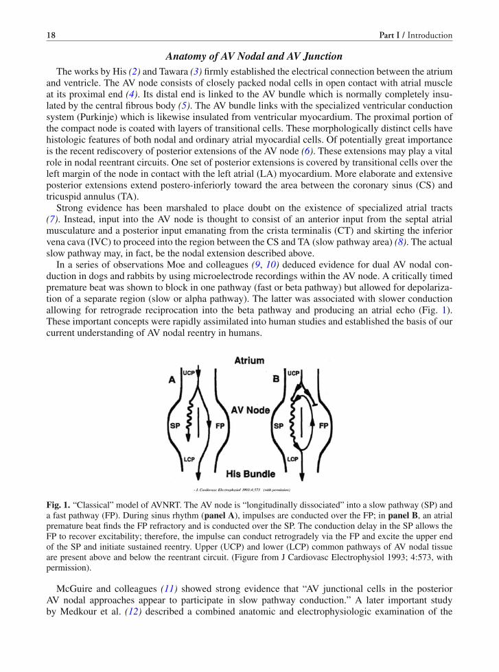
In a series of observations Moe and colleagues (9, 10) deduced evidence for dual AV nodal con-duction in dogs and rabbits by using microelectrode recordings within the AV node. A critically timedpremature beat was shown to block in one pathway (fast or beta pathway) but allowed for depolariza-tion of a separate region (slow or alpha pathway). The latter was associated with slower conductionallowing for retrograde reciprocation into the beta pathway and producing an atrial echo (Fig. 1).These important concepts were rapidly assimilated into human studies and established the basis of ourcurrent understanding of AV nodal reentry in humans.
Fig. 1. “Classical” model of AVNRT. The AV node is “longitudinally dissociated” into a slow pathway (SP) anda fast pathway (FP). During sinus rhythm (panel A), impulses are conducted over the FP; in panel B, an atrialpremature beat finds the FP refractory and is conducted over the SP. The conduction delay in the SP allows theFP to recover excitability; therefore, the impulse can conduct retrogradely via the FP and excite the upper endof the SP and initiate sustained reentry. Upper (UCP) and lower (LCP) common pathways of AV nodal tissueare present above and below the reentrant circuit. (Figure from J Cardiovasc Electrophysiol 1993; 4:573, withpermission).
McGuire and colleagues (11) showed strong evidence that “AV junctional cells in the posteriorAV nodal approaches appear to participate in slow pathway conduction.” A later important studyby Medkour et al. (12) described a combined anatomic and electrophysiologic examination of the

Chapter 2 / History of Supraventricular Tachycardia 19
Fig. 2. A schema showing anterior (septal – 2 upper arrows) inputs into the AV node (N) and posterior inputs(lower arrow) into the node. SVC = superior vena cava; IVC = inferior vena cava; CT = crista terminalis;ER = eustachian ridge; TT = tendon of Todaro: FO = foramen ovale; IAS = interatrial septum; CS os = ostiumof coronary sinus; PNE = posterior nodal extension; CFB = central fibrous body; His = His bundle; TA =tricuspid annulus.
posterior nodal extension (PNE) in the rabbit heart. As shown in Fig. 2, anatomically the extensionappeared as a bundle of specialized tissues between the CS and compact node. They found no distinctseparation between the compact node, lower nodal cell bundle, and the PNE. However, they founddistinct differences in electrophysiologic properties between the PNE and compact node. The PNEshowed cycle length-dependent slow conduction with its refractory period shorter than that of the node.Critically timed premature atrial depolorizations that blocked in the transitional cells could propagatein the PNE and thus explain the discontinuities in nodal conduction as well as in atrial echo beats(Fig. 3a and b). This study accumulated convincing evidence that the PNE provides substrate for slowpathway conduction.
Fig. 3. (a) A schema showing a premature atrial complex that is blocked in the transitional cells surroundingthe septal inputs to the node and the PNE as well as the node are engaged over the inferior inputs. (b) Thepathogenesis of an echo beat. The impulse blocked in the septal inputs proceeds over the inferior input andactivates the node via the PNE and is able to turn around in the node and reactivate the atrium.

20 Part I / Introduction
Human Electrophysiologic StudiesAs mentioned above, early observations by Moe and Menedez (9, 10) on reciprocal beats in rabbits
were rapidly applied to humans. These seminal findings were introduced just as the field of clin-ical invasive electrophysiology began to emerge. Early invasive electrophysiologic studies (13–16)attributed AV nodal reentry as cause of paroxysmal SVT. Of particular note was the work of Dr. KenRosen and colleagues (15) who demonstrated evidence for dual AV nodal physiology manifest by anabruptly increase in AV nodal conduction time in response to critically timed atrial premature depolar-izations. These data served as an excellent supportive compliment to the original observations of Moeand Menendez.
By the end of the 1970s, the concept of dual AV nodal conduction in humans had been wellestablished.
However, the precise anatomic components of the AV nodal reentrant circuit remained controver-sial. Josephson and colleagues (17) showed impressive evidence that the circuit was intranodal andthis concept was contested by Jackman et al. (18) and McGuire et al. (19, 20). The newer anatomicunderstanding of the node has made this debate largely moot. If one accepts the concept that the pos-terior nodal extensions as well as the transitional cells are part of the node (12) then the debate islargely resolved. Current understanding suggests that most subjects with AV nodal reentry have a finalcommon pathway within the AV node and an upper pathway involving the fast and slow pathwayssurrounding the compact node.
In 1993 McGuire et al. (19, 20) nicely summarized the available information and proposed variousmodels for tachycardia mechanisms which involve right-sided atrial inputs. Lately Jackman and col-leagues have expanded on various subforms of AV nodal reentrant tachycardia (AVNRT) (21). Theseinclude slow–fast form (antegrade conduction over the slow pathway and retrograde conduction overthe fast pathway) (81.4%), slow–slow form (both antegrade and retrograde conduction over the slowpathway) (13.7%), and fast–slow forms (antegrade conduction over the fast and retrograde conduc-tion over the slow pathway) (4.9%). The differentiation among these subforms is made based on thelocation of earliest atrial activation. The slow pathway retrograde conduction is manifest over the CSostium region while fast retro conduction occurs over the antero-septal area just superior to the Hisbundle-recording site. In addition, Jackman et al. (22) have suggested left-sided inputs as part of theAV nodal reentrant circuit. Recently Gonzalez et al. (23) proved the existence of LA input to the AVnode in humans with structurally normal hearts.
In addition, there are several case reports that documented the need to ablate AVNRT from theleft annulus or left posteroseptal area (24–26). One source of LA input is via the left-sided posteriornodal extension. The hypothetical left-sided inputs to the AV node and possible tachycardia circuitsare illustrated in Fig. 4.
Surgical Ablation of AVNRTRoss et al. (27) first introduced a non-pharmacologic therapy of AVNRT that involved surgical
dissection in Koch’s triangle, of which the results were confirmed by a number of surgical groups(28–30). This technique also led to a better understanding of this tachycardia. For example, high-resolution mapping of Koch’s triangle showed two distinct types of atrionodal connections in patientswith “typical” slow–fast AVNRT. In most patients the retrograde fast pathway (either during tachycar-dia or ventricular pacing) showed earliest atrial activation over the apex of Koch’s triangle while inthe minority earliest atrial activation occurred near the CS. This would nicely compliment the currentdesignation of AVNRT subforms (21).

Chapter 2 / History of Supraventricular Tachycardia 21
Fig. 4. Hypothesis of left-sided inputs to the AV node. In one iteration the coronary sinus musculature is involvedwith input into the region of the PNE. Ablation either within the coronary sinus or over the traditional slowpathway region (R) would be expected to ablate the circuit. Alternatively, the circuit may involve activation ofthe left atrium (LA) (shown by the broken arrow) either via the septum or Bachmann’s bundle. Activation towardthe AV node is through the tracts (L) along the mitral annulus. In the latter instance ablation over the putativeleft-sided inputs (L) will be required for arrhythmia cure.
Catheter Ablation of AVNRTCatheter ablation of the AV junction using high-energy direct current (DC) shocks for control of
drug-refractory SVT was first introduced in 1981 (31). In 1989, two groups (32, 33) almost simultane-ously reported success using high-energy discharge in the region of slow pathway. The subsequent useof radiofrequency (RF) energy completely revolutionized catheter cure of AVNRT. The initial attemptstargeted the fast pathway by applying RF energy superior and posterior to the His bundle region untilthe prolongation of AV nodal conduction occurred. Initial studies (32–36) showed a success rate of80–90%, but the risk of AV block was up to 21%. Jackman et al. (37) first introduced the techniqueof ablation of the slow pathway for AVNRT. Among experienced centers the current acute successrate for this procedure is 99% with a recurrence rate of 1.3% and a 0.4% incidence of AV block (38)requiring a pacemaker.
Ablation of the slow pathway is achieved by applying RF energy at the posterior–inferior septumin the region of the CS. This technique can be guided by either via discrete potentials (37, 39) orvia an anatomic approach (40); both have equal success rate. Radiofrequency energy is applied untiljunctional ectopics appear but at times successful slow pathway ablation may result without elicitingthe junctional ectopic complexes. Final testing involves proof that either the slow pathway has beeneliminated or no more than one AV nodal echo is present (37, 41).
More recently cryoenergy has been used for the slow pathway ablation (42, 43). The potential advan-tage of cryoenergy is the fact that the catheter sticks to adjacent endocardium during application ofenergy; hence, inadvertent catheter displacement and damage to the node are not possible. In addition,regions closer to the node may be explored since injury during the test procedure is reversible.
WOLFF–PARKINSON–WHITE SYNDROME
The Story of Wolff–Parkinson–White (WPW) SyndromeThe WPW syndrome holds particular interest not only for clinical cardiologists but also for
anatomists, surgeons as well as clinical and experimental electrophysiologists. The definition of thissyndrome was dependant upon a clear knowledge of both the normal conducting system and mecha-nism of reentrant arrhythmias.

22 Part I / Introduction
The first complete description of the syndrome by Drs. Wolff, Parkinson, and White was publishedin the American Heart Journal in August 1930 (44). They described 11 patients without structural car-diac disease who had a short P–R interval and “bundle branch block (BBB)” (Fig. 5) and paroxysmalSVT and/or AF. They made particular note of the fact that use of atropine or exercise would tend tonormalize the ECG in sinus rhythm, while increases in vagal tone had the opposite effect. They felt thatthe arrhythmias were due to “associated nervous control of the heart.” In their report they also creditedDr. F.N. Wilson (1915) and Dr. Wedd (1921) who described the pattern in case reports (45, 46).
Fig. 5. Surface 12-lead ECG in a patient with WPW syndrome. The ECG showed ventricular pre-excitation insinus rhythm with the presentation of short P–R interval and delta waves in 12 leads. The polarity of delta wavein 12 leads indicated that the atrioventricular accessory pathway was located at left posterior free wall.
Anatomic Studies of Atrioventricular (AV) ConnectionsAt the time of the initial observations it was appreciated that the atrium and ventricles were
electrically linked via the AV node and His bundle. Also, the bundle branches and Purkinje systemhad already been described and the electrocardiographic pattern of BBB had been identified (47–49).It is, therefore, clear why the early clinicians categorized ventricular pre-excitation as BBB.
We now need to digress a bit and discuss the work of the anatomists in the late nineteenth and earlytwentieth century. It was appreciated that electrical connections bridged the atrium and ventricles inmammalian hearts (50, 51) and the nature of these connections were of great interest. Stanley Kent (52)in 1893 described lateral AV connections and thought that these constituted the normal AV conductionsystem in man. This work proved controversial and was, in fact, rejected by Sir Thomas Lewis as wellas by Drs. Keith and Flack. In contrast the work of His (53) and Tawara (54) clearly defined the normalAV conducting system. Of interest, there was a later study by Kent, describing a lateral AV connectionand a node-like structure within the connection (55). While some have interpreted this finding as thefirst description of a right atriofascicular tract, but it should be appreciated that Kent felt that thisstructure was part of the normal AV conduction system. It is indeed odd that Kent is given credit forfirst describing accessory extranodal AV pathways since that credit clearly belongs to others, neithershould he be properly credited with the first description of atriofascicular pathways.
In contrast, the persons who deserve credit are Wood et al. (56) for first describing a right-sidedextranodal accessory pathway (1943) and Öhnell (57) for first reporting a left lateral pathway (1944).Other important contributions included the work of Mahaim (58) who described connections betweenthe AV node or His bundle, to the fascicles or ventricular muscle. It was Lev who found that Mahaim

Chapter 2 / History of Supraventricular Tachycardia 23
(59) tracts could produce a pattern of pre-excitation and nicely consolidated our modern understandingof the normal conduction system (60). In a landmark study Lev and Lerner presented detailed anatomicstudies of 33 fetal and neonatal hearts (60). They concluded that no accessory pathways existed out-side the AV conduction system; and that in fetal or neonatal hearts there were sparse development ofcollagen and hence there was close proximity but no communication between the atrium and ventricle.
Historical Evolution of Ventricular Pre-excitation and Circus-Movement TachycardiaThe early clinicians were focused on the “vagal” effects on the pre-excitation pattern and invoked
vague neuro-cardiac mechanisms to explain associated arrhythmias. The concept of reciprocal rhythmswas well established and Mines, who in fact, postulated a reciprocal rhythm involving the AV nodeand accessory pathway (61). According to TN James (62), Holzmann and Scherf (63) in 1932 were thefirst to describe pre-excitation as being due to an extranodal accessory pathway. Similar descriptionswere made by Wolferth and Wood (64) who labeled this pathway as “bundle of Kent.”
However, there were controversies regarding these findings at the time, and it leads to a profusion ofalternative ideas. For example, Hunter et al. (65) suggested that the syndrome was due to a fusion ofpacemakers (sinus conducted complexes and a pacemaker from the bundle branches). Printzmetal (66)attributed the findings to accelerated AV conduction with pathways around the node. Sodi-Pallares(1952) invoked “hyperexcitability of the right side of the septum” (67).
The work by Butterworth and Poindexter (68) in 1942 clearly demonstrated that an artificial con-nection between the atrium and ventricle could mimic classic pre-excitation and led to the acceptanceof an extranodal pathway as the cause for pre-excitation. The understanding of this syndrome wasenhanced by the observation of Pick, Langendorf, and Katz (69–71). They noted some 60 theoriesused to explain pre-excitation but felt that only the presence of extranodal accessory pathway couldexplain all their findings. By detailed and painstaking deductive analyses of literally thousands ofECGs, they amazingly described variations in the nodal vs. pathway refractoriness as a mechanism forinitiation and sustaining paroxysmal SVT. They studied the relationship between tachycardia and AFand distinguished extranodal from AV nodal pathways. Their incredible insights heavily influencedsubsequent human cardiac electrophysiologic studies.
Clinical Electrophysiologic StudyDrs. Durrer and Wellens (72, 73) were the first to use programmed electrical stimulation of the heart
in order to better define the mechanism(s) of arrhythmias. It should be emphasized that their obser-vation antedated the recording of His bundle activity in humans (74). They showed that reciprocatingtachycardia could be induced by premature atrial or ventricular stimulation and could be either ortho-dromic or antidromic; they also defined the relationship of the accessory pathway refractory period tothe ventricular response during AF. These workers provided the framework for use of intracardiac elec-trophysiological studies to define the location (Fig. 6) and electrophysiology of these atrioventricularaccessory pathways (75, 76).
Cardiac-Surgical ContributionPrior to the era of catheter ablation, patients with SVT that were refractory to medical therapy under-
went direct surgical ablation of the AV junction (77, 78). This approach, however, is not appropriatefor the management of the patient with AF with rapid conduction over a bypass tract. Durrer and Roos(79) were the first to perform intraoperative mapping and cooling to locate a right free wall accessorypathway. Burchell et al. (80) used intraoperative mapping to locate a right-sided pathway and showed

24 Part I / Introduction
Fig. 6. Anatomic locations of atrioventricular accessory pathway in a superior view of the cross-sectional planeat the level of atrioventricular annulus. PV = pulmonary valve; AV = aortic valve; MV = mitral valve; TV =tricuspid valve.
that pre-excitation could be abolished by injection of procainamide (1967). A limited surgical incisionover this area resulted in only transient loss of pre-excitation. Sealy (81) and the Duke team were thefirst to successfully ablate a right free wall pathway (1968). They initially used an epicardial approach.Their subsequent amazing results conclusively showed that a vast majority of WPW patients could becured by either direct surgical or cryoablation (82) of these pathways. Iwa from Japan concurrentlydemonstrated the effectiveness of cardiac electrosurgery for these patients (83). He is credited withbeing the first to use an endocardial approach for pathway ablation. The endocardial approach wassubsequently independently used by the Duke team of Sealy and Cox. Only later was the “closed”epicardial approach reintroduced by Guiraudon.
Catheter AblationThe technique of catheter ablation of the AV junction was introduced by Scheinman et al. in 1981
(31). The initial attempts used high-energy DC countershocks to destroy cardiac tissue, but expan-sion of its use to other arrhythmias was limited due to its high risk of causing diffuse damage frombarotrauma. Fisher et al. (84) also attempted to ablate left-sided accessory pathways through the CSby using the DC energy. This technique was eventually abandoned due to its limited efficacy and ahigh incidence of cardiac tamponade. Morady and Scheinman (85) introduced a catheter techniquefor disruption of posteroseptal accessory pathways. This was associated with a 65% efficacy and car-diac tamponade could be avoided by shock delivery just outside the CS (86). Warin et al. describedsuccessful ablation of non-septal pathways (87).
The introduction of RF energy in the late 1980s (88, 89) completely altered catheter ablative pro-cedures. The salient advances in addition to RF energy included much better catheter design, togetherwith the demonstration that pathway localization could be facilitated by direct recording of the path-way potential (Fig. 7). The remarkable work of Jackman et al. (89), Kuck et al. (90), and Calkins (91)ushered in the modern era of ablative therapy for patients with accessory pathways in all locations.
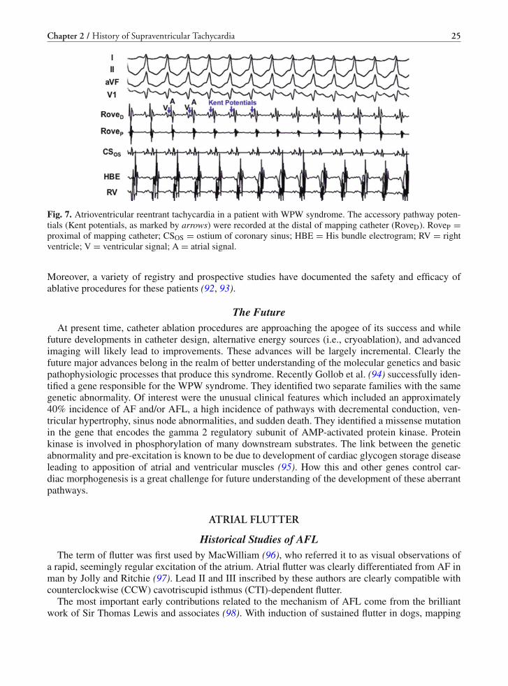
Chapter 2 / History of Supraventricular Tachycardia 25
Fig. 7. Atrioventricular reentrant tachycardia in a patient with WPW syndrome. The accessory pathway poten-tials (Kent potentials, as marked by arrows) were recorded at the distal of mapping catheter (RoveD). RoveP =proximal of mapping catheter; CSOS = ostium of coronary sinus; HBE = His bundle electrogram; RV = rightventricle; V = ventricular signal; A = atrial signal.
Moreover, a variety of registry and prospective studies have documented the safety and efficacy ofablative procedures for these patients (92, 93).
The FutureAt present time, catheter ablation procedures are approaching the apogee of its success and while
future developments in catheter design, alternative energy sources (i.e., cryoablation), and advancedimaging will likely lead to improvements. These advances will be largely incremental. Clearly thefuture major advances belong in the realm of better understanding of the molecular genetics and basicpathophysiologic processes that produce this syndrome. Recently Gollob et al. (94) successfully iden-tified a gene responsible for the WPW syndrome. They identified two separate families with the samegenetic abnormality. Of interest were the unusual clinical features which included an approximately40% incidence of AF and/or AFL, a high incidence of pathways with decremental conduction, ven-tricular hypertrophy, sinus node abnormalities, and sudden death. They identified a missense mutationin the gene that encodes the gamma 2 regulatory subunit of AMP-activated protein kinase. Proteinkinase is involved in phosphorylation of many downstream substrates. The link between the geneticabnormality and pre-excitation is known to be due to development of cardiac glycogen storage diseaseleading to apposition of atrial and ventricular muscles (95). How this and other genes control car-diac morphogenesis is a great challenge for future understanding of the development of these aberrantpathways.
ATRIAL FLUTTER
Historical Studies of AFLThe term of flutter was first used by MacWilliam (96), who referred it to as visual observations of
a rapid, seemingly regular excitation of the atrium. Atrial flutter was clearly differentiated from AF inman by Jolly and Ritchie (97). Lead II and III inscribed by these authors are clearly compatible withcounterclockwise (CCW) cavotriscupid isthmus (CTI)-dependent flutter.
The most important early contributions related to the mechanism of AFL come from the brilliantwork of Sir Thomas Lewis and associates (98). With induction of sustained flutter in dogs, mapping

26 Part I / Introduction
was obtained by direct recordings from the atrial epicardial surface together with surface ECG record-ings. They showed that there was an orderly sequence of atrial activation, and the absence of a truediastole which explained the surface pattern. They concluded that in most experiments the excitationwave was reentrant and appeared to involve both vena cava. The circulating wave front was inscribedeither in a caudo-cranial or in a cranial–caudal fashion in the right atrium (RA). It was felt that the cir-culating wave front could also involve the left atrium (LA). Incredibly, they suspected rotation aroundthe AV valves in several experiments. It should be emphasized that their observations were not univer-sally accepted. Others (e.g., Scherf and Schott) (99) favored enhanced automaticity as a mechanism.
Induction of AFL was uncommon in normal canines and stable mappable flutter was even lessfrequent. Rosenblueth and Garcia Ramos created a canine model of sustained flutter by introductionof a crush lesion between the cavae (100) and found that the flutter wave front rotated around the crushlesion using epicardial mapping. Of interest, they also noted that in some animals, an additional lesionthat “was made starting at the lower edge of the orifice of the IVC and extending the limit . . . towardthe AV groove” could make the flutter disappear without re-inducibility. The latter is the identicallesion currently used to ablate CTI-dependent flutter.
We are indebted to Waldo and his colleagues for their seminal observations explaining the phys-iology of AFL. He and his colleagues studied patients with postoperative flutter by means of fixedatrial electrodes (101, 102). They confirmed that the AFL cycle length was remarkably consistent withmean variations of less than 4 ms. Moreover, Waldo and colleagues taught us the importance of usingentrainment both for detection of reentrant circuits and for discerning whether a given region is criticalfor tachycardia maintenance (102). They also found an interesting relationship between AF and AFLin those patients in whom the onset of AFL was recorded that the transition to AFL was preceded byshort episodes of rapid transitional rhythm (or AF) (103).
Another major advance relates to the studies by Frame et al. (104, 105). They introduced a caninemodel with the intercaval lesion extended into the RA free wall (104). In this model, instead of the“expected” circuit around the “Y” lesion, mapping revealed evidence of electrical activation aroundthe tricuspid orifice. In a further study (105), they proved the reentrant circuit around the annulusby entrainment pacing and showed the presence of an excitable gap. This study established the needfor barriers for tachycardia initiation and perpetuation. Further observations supporting the reentrantnature of AFL were provided by the studies of Boineau et al. (colony of dogs with spontaneous flutter)(106), Hayden and Rytand (107), Kimura et al. (108), and Kato et al. (109).
Another notable contribution came from Klein and Guiraudon (110) who mapped two patients withAFL in the operating room. They found evidence of a large RA reentrant circuit and the narrowestpart of the circuit lay between the TA and the IVC. They successfully treated the flutter by usingcryoablation around the CS and surrounding atrium.
Following the report of Klein et al. (110), there appeared several studies using high-energy shocksin an attempt to cure AFL (111, 112)).
Subsequently both Drs. Feld and Cosio almost simultaneously described using RF energy for dis-ruption of CTI conduction in order to cure patients with AFL. Feld et al. (113) contributed an elegantstudy using endocardial mapping techniques and entrainment pacing to prove that the area posterioror inferior to the CS was a critical part of the flutter circuit and application of RF energy to this siteterminated AFL. Cosio et al. used similar techniques but placed the ablative lesion at the area betweenthe TA and IVC (114). The latter technique forms the basis for current ablation of CTI-dependentflutter.
Terminology of AFLRapid regular single macroreentrant atrial arrhythmias with little or no isoelectric interval are termed
AFL. Historically, AFL was defined as being typical, reverse typical, or atypical. With increased

Chapter 2 / History of Supraventricular Tachycardia 27
Fig. 8. Nomenclature of atrial flutter (AFL). CTI = cavotricuspid isthmus; CCW = counterclockwise AFLaround the tricuspid annulus (TA); CW = clockwise AFL around the TA; LLR = lower loop reentry aroundinferior vena cava; IIR = intra-isthmus reentry; DWR = double-wave reentry around the TA; LA = left atrium;RA = right atrium; PV = reentrant circuit around the pulmonary vein (s) with or without scar(s) in the LA;MA = reentrant circuit around mitral annulus; FO = reentrant circuit around the fossa ovalis; ULR = upperloop reentry in the RA.
knowledge a new terminology appears to be appropriate. As shown in Fig. 8, we have recently pro-posed a revised nomenclature of AFL.
CAVOTRICUSPID ISTHMUS-DEPENDENT AFLIn the majority of the RA flutter, the CTI is a critical part of the reentrant circuit, which is also the
target for AFL ablation.Counterclockwise or clockwise (CW) AFL: These are macroreentrant circuits around the TA
(115–120). The CW or CCW designation is made on the basis of wave front activation in the leftanterior oblique (LAO) projection. Counterclockwise AFL is the most common and the surface ECGshows continuous sawtooth-like flutter wave dominantly negative in the inferior leads (II, III, and aVF)and positive in V1; whereas CW flutter shows positive waves in the inferior leads and negative wavesin V1 (Fig. 9). These types of AFL may be seen both in patients with or without congenital heartdisease or cardiac surgery.
Double-wave reentry (DWR): Flutter circuits have an excitable gap. A critical timed premature beatis able to invade the circuit and if that impulse experiences antidromic block but yet is able to propagateorthodromically, then two wave fronts may occupy the circuit simultaneously (121). This arrhythmia ismanifest by acceleration of the tachycardia rate but with identical surface and intracardiac electrogrammorphology. Double-wave reentry is usually produced by extrastimuli and is usually short lived, butmay serve as a trigger for AFib (122).
Lower loop reentry (LLR): Lower loop reentry is a form of CTI-dependent flutter with a reentrantcircuit around the IVC, therefore, it is confined to the lower part of the RA (123–125). It often co-existswith typical CCW or CW flutter and involves posterior breakthrough(s) across the CT. Since the CTIis still a necessary part of the circuit, LLR is amenable to CTI ablation as is true of patients with CCWor CW flutter. The surface morphology of LLR usually is very similar to that of CCW or CW flutter(126).
Intra-isthmus reentry (IIR): We have recently reported a novel reentrant circuit within the region ofthe septal (medial) CTI and CS ostium (127, 128). In the initial report of this flutter form, entrainmentpacing from the antero-inferior (lateral) CTI shows post-pacing interval (PPI) > tachycardia cyclelength (TCL), indicating the lateral CTI is out of the reentrant circuit; whereas, pacing from the regionof septal isthmus or CS ostium shows concealed entrainment with PPI = TCL. Fractionated or double

28 Part I / Introduction
Fig. 9. Surface ECG of CTI-dependent AFL. Left panel shows the simultaneous 12-lead ECG in a patient withcounterclockwise (CCW) AFL; Right panel shows the surface ECG in a patient with clockwise (CW) AFL.
potentials usually can be recorded in this area and can be entrained. Applying RF energy at the site ofmaximal fractionated potentials can always abolish the tachycardia.
NON-CTI-DEPENDENT AFL
Right Atrial Flutter Circuits. Scar-related RA macroreentrant tachycardia: Previous studieshave shown that macroreentrant tachycardia can occur in patients with or without atriotomy or congen-ital heart disease (124, 129, 130). In these patients, electroanatomic voltage maps from the RA oftenshow “scar” or low-voltage area(s) which acts as the central obstacle or channels for the reentrantcircuit. The morphology of surface ECG may vary depending on where the scar(s) and low-voltagearea(s) are and how the wave fronts exit the circuits.
Upper loop reentry (ULR): This is a type of atypical AFL involving the upper portion of RA withtransverse conduction over the CT and wave front collision occurring at lower part of RA or withinthe CTI (124, 131). Therefore, this tachycardia is a non-CTI-dependent flutter and the CTI is foundto be outside of the circuit by entrainment pacing. Upper loop reentry was initially felt to involve areentrant circuit using the channel between the superior vena cava (SVC), fossa ovalis (FO), and CT(124). A study by Tai et al. using non-contact mapping technique showed that this form of AFL wasa macroreentrant tachycardia in the RA free wall with the CT as its functional central obstacle (131).They successfully abolished ULR by linear ablation of the gap in the CT. Like LLR, ULR also canoccur in conjunction with typical CW and/or CCW flutter, as well as LLR.
Left Atrial Flutter Circuits. Left AFL circuits are often related to AF. In recent years, thesecircuits have been better defined by use of electroanatomic or non-contact mapping techniques (132).Cardiac surgery involving the LA or atrial septum can produce different left flutter circuits. But, leftAFL circuits also can be found in patients without a history of atriotomy. Electroanatomic maps in

Chapter 2 / History of Supraventricular Tachycardia 29
these patients often show low-voltage or scar areas in the LA, which act as either a central obstacle ora barrier in the circuit. The surface ECG of left AFLs are variable due to different reentrant circuits.However, several sub-groups have been identified (Fig. 8).
Mitral annular (MA) AFL: This flutter circuit involves reentry around the MA either in a CCW or ina CW fashion. This arrhythmia is more common in patients with structural heart disease. However, ithas been described in patients without obvious structural heart disease (126, 132), but in these patients,electroanatomic voltage map often shows scar or low-voltage area(s) on the posterior wall of LA as aposterior boundary of this circuit. The surface ECG of MA flutter can mimic CTI-dependent CCW orCW flutter, but usually shows low flutter wave amplitude in most of the 12 leads (126).
Pulmonary vein(s) (PVs) with or without LA scar circuits: Various left AFL circuits involve the PVs,especially in those patients with AF or mitral valve disease. Reentry can circle around one or morePVs and/or posterior scar or low-voltage area(s) (126, 132). In order to cure these complex circuits,electroanatomic mapping is required to reveal the circuit and guide ablation (Fig. 10). Since thesecircuits are related to low-voltage or scar area(s), the surface ECG usually shows low-amplitude or flatflutter waves.
Fig. 10. Electroanatomic activation map in a patient with left atrial (LA) flutter with posterior–anterior (PA)project view. The activation map demonstrates a reentrant circuit (figure of eight) around the right upper pul-monary vein (RUPV) and scar on the LA posterior wall, as shown by the arrows.
ATRIAL FIBRILLATION
The history of AF has been described in a number of excellent articles (133–135). It is not clearwho first described AF. The legendary Chinese Yellow Emperor (Huangdi), who ruled China from2497 to 2398 BC, reportedly described irregular and weak pulses in the Yellow Emperor’s Classic ofInternal Medicine (133). Circa 1187, Moses Maimondides described irregular pulses suggesting AF(134). Hanon, Shapiro, and Schweitzer published a very detailed account of the original investigations
30 Part I / Introduction
and evolving theories of this important arrhythmia in more modern times (135). Irregular pulses inthe presence of mitral stenosis were first described by de Senac in the eighteenth century and laterby Robert Adams in 1827 (133, 136). Stokes described irregular pulses in 1854 and Wenckebach in1904 (137, 138). Rothberger and Winterberg described AF in two patients with arrhythmia perpetuain 1909 (139). Irregular pulse tracings showing discrepancies between simultaneously recorded apicaland radial pulses were published by Etienne Marey (1863), Heinrich Hering (1903), James Mackenzie(1907), and Thomas Lewis (1909) (135). Willem Einthoven and Thomas Lewis first recorded AF withelectrocardiograhy in 1909. In 1910, Lewis published a landmark treatise on AF and established theterm auricular fibrillation, which he defined as “a condition in which normal impulse formation in theauricle is replaced by stimulus production at multiple auricular foci. Co-ordinate contraction is lost;the normal and regular impulses transmitted to the ventricle are absent, while rapid and haphazardimpulses produced in the auricle take their place and produces gross irregularity of the ventricularaction” (140).
Mechanisms of AFCompeting models for mechanism underlying AF were reviewed by Garrey in 1924 (141).
Spontaneous rapidly discharging atrial foci and a single reentry circuit with fibrillatory conductionwere the first two mechanisms proposed for AF. Scherf developed the first animal model of AF byapplying aconitine on an atrial site causing rapid firing (142). Moe and his colleagues in 1964 pro-posed that AF was due to random reentry of multiple, simultaneously circulating reentrant wavelets(143). It was further developed by Allessie and his colleagues in the 1970s (144). It hypothesizedthat AF was due to multiple randomly propagating reentrant waves in the atrium and AF required atleast six to eight circular reentrant wave fronts and a critical atrial mass (145). It had been accepted asthe mechanism until the recent discovery of spontaneous electrical activity in pulmonary vein, first inisolated guinea pig pulmonary vein and later in humans during electrophysiologic study, clearly indi-cating a spontaneous rapidly discharging atrial focus can be the underlying mechanism for AF (146,147). Nattel reviewed the mechanisms of AF at the tissue and cellular levels and how our treatmentapproaches are modified and guided by our understanding of the underlying mechanisms (148, 149).
Clinical Implications of AFEpidemiology of AF: In 1965, a study reported that the incidence of AF was 4.3 per 1000 when
routine ECGs were recorded in one community with 5179 adults (150). Katz and Pick reported thatin 50,000 consecutive patients with suspected heart disease 3.1% had paroxysmal AF and 8.6% hadchronic AF (151). Most of our knowledge of the epidemiology of AF came from studies published inthe late 1970s–2000. And the Framingham Heart Study provided a majority of the data. The increasedprevalence of AF with age was clearly demonstrated in a number of epidemiology studies (152, 153).The Framingham Heart Study first established that AF increased mortality in older men and women(55–94 years of age) even after adjustment for the pre-existing cardiovascular conditions (154). Theeffect appears more prominent in women – 1.5-fold in men and 1.9-fold in women. Both heart failureand stroke contributed to the excessive mortality.
Thromboembolism and Anticoagulation: The role of nonrheumatic AF as a precursor of stroke andhigher stroke events at the onset of AF was first identified in the Framingham Heart Study in 1978(155, 156). The risks factors for stroke and the efficacy of warfarin in decreasing stroke were estab-lished in five randomized trials published between 1989 and 1992 (AF, Aspirin, Anticoagulation Studyfrom Copenhagen, Denmark (AFASAK), the Stroke Prevention in Atrial Fibrillation (SPAF) study, theBoston Area Anticoagulation Trial in Atrial Fibrillation (BAATAF), the Canadian Atrial FibrillationAnticoagulation (CAFA) study, and the Veterans Affairs Stroke Prevention in Nonrheumatic Atrial

Chapter 2 / History of Supraventricular Tachycardia 31
Fibrillation (SPINAF) study) (157–162). In patients with AF for whom warfarin was unsuitable, theaddition of clopidogrel to aspirin reduced the stroke risk (163). In the 1980s, transesophageal echocar-diography (TEE) was introduced as a sensitive and specific diagnostic tool to identify high-risk patientsby detecting thrombus in the LA appendage (164). In 1993, Manning and his coworkers introducedTEE-guided strategy for elective cardioversion without prolonged anticoagulation (165).
TherapyPharmacological Therapy: The treatment of AF had been limited to restoring normal sinus rhythm
with either DC shock or pharmacologic agents and maintenance of normal sinus rhythm with long-termdrug treatment. In the case of chronic AF, rate control was the alternative. For many years, quinidineand procaineamide were the only drugs available for chemical cardioversion and rhythm maintenance.Flecainide, propafenone, amiodarone, ibutilide, and dofetilide were introduced in the past 30 years.Their efficacy in maintaining normal sinus rhythm has not been satisfactory and their pro-rhythmicrisk well documented. In two randomized studies, the rate of recurrence ranged from about 35% foramiodarone to about 60% for sotalol and propafenone (166, 167). For controlling of rate only, theoldest drug is digitalis, which was first used by Jean Baptiste Bouilland in 1835 (133). Because ofits ineffectiveness, it has been replaced with beta-blockers and calcium channel blockers. For years,conversion to and maintenance of normal sinus rhythm have always been the main goal of AF therapy;rate control was attempted only if repeated attempts in rhythm control failed. Two large randomizedprospective clinical trials compared rate vs. rhythm control in AF therapy in elderly patients (168,169). In the AFFIRM trial, rate control lowered 5-year overall mortality rate (21.3% in rate vs. 23.8%in rhythm control, but without significant statistic difference). In the RACE trial, rate control increased3-year event-free survival (17.2 in rate vs. 22.6% in rhythm control). Events included cardiovasculardeath, heart failure, thromboembolism, bleeding, pacemaker implantation, and anti-arrhythmic drugside effects. Embolization occurs with equal frequency in both trials. More importantly, despite suc-cessful cardioversion, recurrence rate of AF was high and many AF episodes were asymptomatic.Accordingly, anticoagulation consideration is dependent on stroke risks and not on rate or rhythmcontrol management strategy.
Non-pharmacological Therapy: DC trans-thoracic defibrillation with synchronization was firstintroduced by Lown and his coworkers in 1962 (170). This method remains the most effective methodfor acute conversion of AF to normal sinus rhythm. Surgical interventions were designed to preventrandom reentry. These include reduction and fragmentation of atrial surface area, exclusion of the fib-rillating atrium, and creation of one-dimensional conduction with multiple surgical incisions (171).The best known surgical ablation of AF was the MAZE procedure first performed by Cox in 1987(172). Because of the complexity and morbidity surgical ablation quickly gave way to catheter abla-tion. Catheter ablation of the AV node with implantation of a permanent pacemaker was first usedto achieve rate control in AF with rapid ventricular response (31, 173). Since John F. Swartz intro-duced the procedure, curative catheter ablation therapy has been the major focus of electrophysiolo-gists since the early 1990s. Spragg and Caulkins recently published an overview of the developmentcatheter-based AF therapy and the relative merits of the different techniques (174). Major contribu-tors to the innovation and advancement of AF ablation include groups led by Michel Haissaguerre,Andrea Natale, Carlo Pappone, Morady, and Shih-Ann Chen. The first attempt entailed creation of lin-ear lesions in the RA only or in combination with the LA (including the PVs) (175–177). This was metwith little success. But the experience allowed Haissaguerre to discover that focal atrial triggers (pre-dominantly located in the PVs) are frequently responsible for initiation of AF (178). However, ablationof focal PV trigger sites carries risk of PV stenosis and high recurrence rate because the focus cannotbe consistently triggered during the procedure and precisely localized. Segmental ostial and circum-ferential PV isolation techniques were introduced to electrically isolate these triggering foci from the

32 Part I / Introduction
body of the LA by eliminate conduction between the pulmonary triggers and the atrial myocardium,thereby preventing initiation of AF (179, 180).
FutureThe last 30 years have seen enormous and rapid growth in our knowledge of AF. We learned the
epidemiology of AF, the stroke risks, the importance of anticoagulation, the use of echocardiographictechniques, especially TEE in managing AF, the merits of rate and rhythm management, and finally,catheter-based curative ablation. On the other hand, we may have not reached the end of the ascendingportion of our knowledge curve. It has been 10 years since catheter ablation of AF was first usedclinically, but we still do not have a standard ablation approach (181). The successful cure rates forparoxysmal, persistent, and chronic AF have not been significantly improved in the past few years.The success rates may be limited by our one-size-fit-all approach to a disease with diverse underlyingmechanisms. Our curative success in other tachyarrhythmias has largely been driven by our knowledgeof their underlying mechanisms. Similarly, higher success rate in AF ablation will be achieved largelyby improving our knowledge of its mechanisms and pathophysiology. Meanwhile, improvement inmapping techniques and equipment such as three-dimensional electroanatomical mapping system, CT,MRI and robotic navigation, catheter designs, and ablation energy sources will help to further reduceoperation time and possible the need for repeat procedures. The importance of anticoagulation in AFeven after apparent successful cardioversion cannot be overemphasized. There is an obvious need fornewer anticoagulation agents with a better safety profile and less monitoring. Left atrial appendageplug in prevention of clot formation has shown very promising results in clinical trial (PROTECT-AF). Lastly, insights into genetic determinants will help us to further understand the mechanismsof AF.
REFERENCES
1. Barker PS, Wilson FN, Johnston FD (1943) The mechanism of auricular paroxysmal tachycardia. Am Heart J 26:435–445
2. His W Jr (1893) Die Tatigkeit des embryonalen Herzens und deren bedeutung fur de Lehre von de Herzbewegung beimErwachsenen. Arbeiten aus der Medizinischen Klinik zu Leipzig 1:14–49
3. Tawara S (1906) Das Reizleitungssystem des Saugetirherzens: Eine anatomisch-histologische Studie uber das Atri-oventrikularbundel und die Purkinjeschen Faden. Jena, gustav Fischer, Germany, pp 9–70, 114–156
4. Ho SY, Kilpatrick L, Kanai T et al (1995) The architecture of the atrioventricular conduction axis in dog compared toman: its significance to ablation of the atrioventricular nodal approaches. J Cardiovasc Electrophysiol 6:26–39
5. Anderson RH, Ho SY, Becker AE (2000) Anatomy of the human atrioventricular junctions revisited. Anat Rec 260:81–91
6. Waki K, Kim JS, Becker AE (2000) Morphology of the human atrioventricular node is age-dependent: a feature ofpotential clinical significance. J Cardiovasc Electrophysiol 11:1144–1151
7. Mazgalev TN, Ho SY, Anderson RH (2001) Anatomic-electrophysiological correlations concerning the pathways foratrioventricular conduction. Circulation 103:2660–2667
8. Anderson RH, Becker AE, Brechenmacher C et al (1975) The human antrioventricular junctional area. A morphologi-cal study of the AV node and bundle. Eur J Cardiol 3:11–25
9. Moe GK, Preston JB, Burlington H (1956) Physiologic evidence for a dual A-V transmission system. Circ Res 4:357–375
10. Mendez C, Moe GK (1966) Demonstration of a dual A-V nodal conduction system in the isolated rabbit heart. CircRes 19:378–393
11. McGuire MA, Robotin M, Yip AS et al (1994) Electrophysiologic and histologic effects of dissection of the connec-tions between the atrium and posterior part of the atrioventricular node. J Am Coll Cardiol 23:693–701
12. Medkour D, Becker AE, Khalife K, Billette J (1998) Anatomic and functional characteristics of a slow posterior AVnodal pathway: role in dual-pathway physiology and reentry. Circulation 98:164–174
13. Goldreyer BN, Bigger JT (1971) The site of reentry in paroxysmal supraventricular tachycardia in man. Circulation43:15–26

Chapter 2 / History of Supraventricular Tachycardia 33
14. Goldreyer BN, Damato AN (1971) The essential role of atrioventricular conduction delay in the initiation of paroxys-mal supraventricular tachycardia. Circulation 43:679–687
15. Denes P, Wu D, Dhingra RC et al (1973) Demonstration of dual A-V nodal pathways in patients with paroxysmalsupraventricular tachycardia. Circulation 48:549–555
16. Schuilenburg RM, Durrer D (1968) Atrial echo beats in the human heart elicited by induced atrial premature beats.Circulation 37:680–693
17. Josephson ME, Kastor JA (1976) Paroxysmal supraventircular tachycardia. Is the atrium a necessary link? Circulation54:430–435
18. Jackman WM (1991) Participation of atrial myocardium (posterior septum) in AV nodal reentrant tachycardia: evidencefrom resetting by atrial extrastimuli. Pacing Clin Electrophysiol 14:646
19. Janse MJ, Anderson RH, McGuire MA et al (1993) “AV nodal” reentry: part I: “AV nodal” reentry revisited. J Cardio-vasc Electrophysiol 4:561–572
20. McGuire MA, Janse MJ, Ross DL (1993) “AV Nodal” reentry: part II: AV nodal, AV junctional, or atrionodal reentry?J Cardiovasc Electrophysiol 4:573–586
21. Heidbüchel H, Jackman WM (2004) Characterization of subforms of AV nodal reentrant tachycardia. Europace 6:316–29
22. Otomo K, Wang Z, Lazarra R, Jackman WM (1999) Atrioventricular nodal reentrant tachycardia: electrophysiologicalcharacteristics of four forms and implications for the reentrant circuit. In: Zipes DP, Jalife J (eds) Cardiac electrophys-iology: from cell to bedside, 3rd edn. W.B. Saunders Company, Philadelphia, PA, pp 504–521
23. Gonzalez MD, Contreras LJ, Cardona F et al (2002) Demonstration of a left atrial input to the atrioventricular node inhumans. Circulation 106:2930–2934
24. Jais P, Haissaguerre M, Shah DC et al (1999) Successful radiofrequency ablation of a slow atrioventricular nodalpathway on the left posterior atrial septum. Pacing Clin Electrophysiol 22:525–527
25. Tondo C, Otomo K, McClelland J et al (1996) Atrioventricular nodal reentrant tachycardia: is the reentrant circuitalways confined in the right atrium? (abstract) J Am Coll Cardiol 27:159A
26. Sousa J, El-Atassi R, Rosenheck S et al (1991) Radiofrequency catheter ablation of the atrioventricular junction fromthe left ventricle. Circulation 84:567–571
27. Ross D, Johnson D, Denniss A et al (1985) Curative surgery for atrioventricular junctional (“AV nodal”) reentranttachycardia. J Am Coll Cardiol 6:1383–1392
28. Cox J, Holman W, Cain M (1987) Cryosurgical treatment of atrioventricular node reentrant tachycardia. Circulation76:1329–1336
29. Guiraudon GM, Klein GJ, van Hemel N et al (1990) Anatomically guided surgery to the AV node. AV nodal skele-tonization: experience in 26 patients with AV nodal reentrant tachycardia. Eur J Cardiothorac Surg 4:464–465
30. Ruder MA, Mead RH, Smith NA et al (1990) Comparison of pre- and postoperative conduction patterns in patientssurgically cured of atrioventricular node reentrant tachycardia. J Am Coll Cardiol 17:397–402
31. Scheinman MM, Morady F, Hess DS, Gonzalez R (1982) Catheter-induced ablation of the atrioventricular junction tocontrol refractory supraventricular arrhythmias. JAMA 248:851–855
32. Haissaguerre M, Warin J, Lemetayer P et al (1989) Closed-chest ablation of retrograde conduction in patients withatrioventricular nodal reentrant tachycardia. N Engl J Med 320:426–433
33. Epstein LM, Scheinman MM, Langberg JJ et al (1989) Percutaneous catheter modification of the atrioventricular node:a potential cure for atrioventricular nodal tachycardia. Circulation 80:757–768
34. Lee MA, Morady F, Kadish A et al (1991) Catheter modification of the atrioventricular junction with radiofrequencyenergy for control of atrioventricular nodal reentry tachycardia. Circulation 83:827–835
35. Jazayeri MR, Hempe SL, Sra JS et al (1992) Selective transcatheter ablation of the fast and slow pathways usingradiofrequency energy in patients with atrioventricular nodal reentrant tachycardia. Circulation 85:1318–1328
36. Langberg JJ, Leon A, Borganelli M et al (1993) A randomized, prospective comparison of anterior and pos-terior approaches to radiofrequency catheter ablation of atrioventricular nodal reentry tachycardia. Circulation87:1551–1556
37. Jackman WM, Beckman KJ, McClelland JH et al (1992) Treatment of supraventricular tachycardia due to atri-oventricular nodal reentry, by radiofrequency catheter ablation of slow-pathway conduction. N Engl J Med 327:313–318
38. Morady F (2004) Catheter ablation of supraventricular tachycardia: state of the art. J Cardiovasc Electrophysiol15:124–139
39. Haissaguerre M, Gaita F, Fischer B et al (1992) Elimination of atrioventricular nodal reentrant tachycardia usingdiscrete slow potentials to guide application of radiofrequency energy. Circulation 85:2162–2175
40. Kalbfleisch SJ, Strickberger SA, Williamson B et al (1994) Randomized comparison of anatomic and electrogrammapping approaches to ablation of the slow pathway of atrioventricular node reentrant tachycardia. J Am Coll Cardiol23:716–723
34 Part I / Introduction
41. Hummel JD, Strickberger SA, Williamson BD et al (1995) Effect of residual slow pathway function on the time courseof recurrences of atrioventricular nodal reentrant tachycardia after radiofrequency ablation of the slow pathway. Am JCardiol 75:628–630
42. Skanes AC, Dubuc M, Klein GJ et al (2000) Cryothermal ablation of the slow pathway for the elimination of atrioven-tricular nodal reentrant tachycardia. Circulation 102:2856–2860
43. Rodriguez LM, Geller JC, Tse HF et al (2002) Acute results of transvenous cryoablation of supraventricular tachy-cardia (atrial fibrillation, atrial flutter, Wolff-Parkinson-White syndrome, atrioventricular nodal reentry tachycardia).J Cardiovasc Electrophysiol 13:1082–1089
44. Wolff L, Parkinson J, White PD (1930) Bundle-branch block with short P-R interval in healthy young people prone toparoxysmal tachycardia. Am Heart J 5:685–704
45. Wilson FN (1915) A case in which the vagus influenced the form of the ventricular complex of the electrocardiogram.Arch Intern Med 16:1008–1027
46. Wedd AM (1921) Paroxysmal tachycardia, with reference to nomotropic tachycardia and the role of the extrinsiccardiac nerves. Arch Intern Med 27:571–590
47. Eppinger H, Rothberger J (1909) Zur analyse des elektrokardiogramms. Wien Klin Wehnsehr 22:109148. Eppinger H, Rothberger J (1910) Ueber die folgen der durchschneidung der Tawaraschen schenkel des reizleitungssys-
tems. Zeitschr Klin Med 70:1–2049. Eppinger H, Stoerk O (1910) Zur klinik des elektrokardiogramms. Zeitschr Klin Med 71:15750. Paladino G (1876) Contribuzione all anatomia, istologia e fisiologia del cuore. Movemento Napoli 8:42851. Gaskell WH (1883–1884) On the innervation of the heart. with especial reference to the heart of the tortoise. J Physiol
4:4352. Kent AFS (1893) Researches on the structure and function of the mammalian heart. J Physiol 14:23353. His W (1893) die thatigkeit des embryonalen herzens unde deren bedeutung fur die lehre von de herzbewegung beim
erwachsenen. Med Klinik in Leipzig I:1454. Tawara S (1906) Des reizleitungsystem des Saugetierherzens. Eine anatomischhistologische studie uder das atrio-
ventrikularbundle under die Purkinjeschen faden. Jena, Germany, Verlag von Gustav Fischer, p 20055. Kent AFS (1914) A conducting path between the right auricle and the external wall of the right ventricle in the heart
of the mammal. J Physiol 48:5756. Wood FC, Wolferth CC, Geckeler GD (1943) Histologic demonstration of accessory muscular connections between
auricle and ventricle in a case of short P-R interval and prolonged QRS complex. Am Heart J 25:454–46257. Öhnell RF (1944) Pre-excitation, cardiac abnormality, pathophysiological, patho-anatomical and clinical studies of
excitatory spread phenomenon bearing upon the problem of the WPW (Wolff, Parkinson, and White) electrocardiogramand paroxysmal tachycardia. Acta Med Scand 152:1–167
58. Mahaim I, Benatt A (1937) Nouvelles recherches sur les connexions superieures de la branche gauche du faisceau deHis-Tawara avec la cloison interventriculaire, Cardiologia 1:61–73
59. Lev M, Leffler WB, Langendorf R, Pick A (1966) Anatomic findings in a case of ventricular pre-excitation (WPW)terminating in complete atrioventricular block. Circulation 34:718–733
60. Lev M, Lerner R (1955) The theory of Kent. A histologic study of the normal atrioventricular communications of thehuman heart. Circulation 12:176–184
61. Mines GR (1914) On circulating excitations in heart muscles and their possible relationship to tachycardia and fibril-lation. Proc Trans R Soc Can 8:43–52
62. James TN (1970) The Wolff-Parkinson-white syndrome: evolving concepts of its parthogenesis. Progr Cardiovasc DisXIII(2): 159–189
63. Holzmann M, Scherf D (1932) Uber elektrokardiogramme mit verkurzter Vorhof-Kammer Distanz und positiven P.Zacken Z Klin Med 121:404–410
64. Wolferth CC, Wood FC (1933) This mechanism of production of short P-R intervals and prolonged QRS complexesin patients with presumably undamaged hearts: hypothesis of an accessory pathway of auriculo-ventricular conduction(bundle of Kent). Am Heart J 8:297–311
65. Hunter A, Papp C, Parkinson J (1940) The syndrome of short P-R interval, apparent bundle branch block, and associ-ated paroxysmal tachycardia. Brit Heart J 2:107
66. Prinzmetal M et al (1952) Accelerated conduction: the Wolff-Parkinson-White syndrome and related conditions.Grune & Stratton, New York
67. Sodi-Pallares D, Cisneros F, Medrano GA et al (1963) Electrocardiographic diagnosis of myocardial infarction in thepresence of bundle branch block (right and left), ventricular premature beats and Wolff-Parkinson-white syndrome.Progr Cardiovasc Dis 6:107–136
68. Butterworth JS, Poindexter CA (1942) Short PR interval associated with a prolonged QRS complex. Arch Intern Med69:437–445
69. Pick A, Katz LN (1955) Disturbances of impulse formation and conduction in the pre-excitation (WPW) syndrome –their bearing on its mechanism. Am J Med 19:759–772

Chapter 2 / History of Supraventricular Tachycardia 35
70. Pick A, Langendorf R (1968) Recent advances in the differential diagnosis of A-V junctional arrhythmias. Am Heart J76:553–575
71. Katz LN, Pick A (1956) Clinical electrocardiography: part I. The arrhythmias: with an atlas of electrocardiograms, vol43. Lea & Febiger, Philadelphia, pp 679–708
72. Durrer D, Schoo L, Schuilenburg RM, Wellens HJ (1967) The role of premature beats in the initiation and the termi-nation of supraventricular tachycardia in the Wolff-Parkinson-White syndrome. Circulation 36:644–662
73. Wellens HJ, Schuilenburg RM, Durrer D (1971) Electrical stimulation of the heart in patients with Wolff-Parkinson-White syndrome, type A. Circulation 43:99–114
74. Scherlag BJ, Lau SH, Helfant RH et al (1969) Catheter technique for recording His bundle activity in man. Circulation39:13–18
75. Gallagher JJ, Pritchett ELC, Sealy WC et al (1976) The preexcitation syndrome. Circulation 54:571–59176. Jackman WM, Friday KJ, Scherlag BJ et al (1983) Direct endocardial recording from an accessory atrioventricular
pathway, localization of the site of block effect of antiarrhythmic drugs and attempt at nonsurgical ablation. Circulation68:906–916
77. Dreifus LS, Nichols H, Morse D et al (1968) Control of recurrent tachycardia of Wolff-Parkinson-White syndrome bysurgical ligature of the A-V bundle. Circulation 38:1030–1036
78. Edmunds JH, Ellison RG, Crews TL (1969) Surgically induced atrio-ventricular block as treatment for recurrent atrialtachycardia in Wolff-Parkinson-White syndrome. Circulation 39(Suppl):105–111
79. Durrer D, Roos JP (1967) Epicardial excitation of ventricles in patient with Wolff-Parkinson-White syndrome (typeB). Circulation 35:15–21
80. Burchell HB, Frye RL, Anderson MW, McGoon DC (1967) Atrioventricular and ventriculoatrial excitation in Wolff-Parkinson-White syndrome (type B). Circulation 36:663–669
81. Cobb FR, Blumenschein SD, Sealy WC, et al (1968) Successful surgical interruption of the bundle of Kent in a patientwith Wolff-Parkinson-White syndrome. Circulation 38:1018–1029
82. Cox JL (2004) NASPE history: Cardiac surgery for arrhythmias. Pacing Clin Electrophysiol 27:266–28283. Iwa T, Kazui T, Sugii S, Wada J (1970) Surgical treatment of Wolff-Parkinson-White syndrom. Kyobu Geka 23:513–884. Fisher JD, Brodman R, Kim SG et al (1984) Attempted nonsurgical electrical ablation of accessory pathways via the
coronary sinus in the Wolff-Parkinson-White syndrome. J Am Coll Cardiol 4:685–69485. Morady F, Scheinman MM (1984) Transvenous catheter ablation of a posteroseptal accessory pathway in a patient
with the Wolff-Parkinson-White syndrome. N Engl J Med 310:705–70786. Morady F, Scheinman MM, Kou WH et al (1989) Long-term results of catheter ablation of a posteroseptal accessory
atrioventricular connection in 48 patients. Circulation 79:1160–117087. Warin JF, Haissaguerre M, Lemetayer P et al (1988) Catheter ablation of accessory pathways with a direct approach.
Results in 35 patients. Circulation 78:800–81588. Borggrefe M, Budde T, Podczeck A, Breithardt G (1987) High frequency alternating current ablation of an accessory
pathway in humans. J Am Coll Cardiol 10:576–58289. Jackman WM, Wang XZ, Friday KJ et al (1991) Catheter ablation of accessory atrioventricular pathways (Wolff-
Parkinson-White syndrome) by radiofrequency current. N Engl J Med 334:1605–161190. Kuck KH, Schlüter M, Geiger M et al (1991) Radiofrequency current catheter ablation of accessory atrioventricular
pathways. Lancet 337:1557–156191. Calkins H, Sousa J el Atassi R et al (1991) Diagnosis and cure of the Wolff-Parkinson-white syndrome or paroxysmal
supraventricular tachycardias during a single electrophysiologic test. N Engl J Med 324:1612–161892. Scheinman MM (1995) NASPE survey on catheter ablation. Pacing Clin Electrophysiol 18:1474–147893. Hindricks G (1993) The multicentre European radiofrequency survey (MERFS) investigators of the work group on
arrhythmias of the European society of cardiology. The multicentre European radiofrequency survey (MERFS): com-plications of radiofrequency catheter ablation of arrhythmias. Eur Heart J 14:1644–1653
94. Gollob MH, Green MS, Tang AS et al (2001) Identification of a gene responsible for familial Wolff-Parkinson-Whitesyndrome. N Engl J Med 344:1823–1831
95. Wolf CM, Arad M, Ahmad F et al (2008) Reversibility of PRKAG2 glycogen-storage cardiomyopathy and electro-physiological manifestations. Circulation 117:144–154
96. McWilliam JA (1886) Fibrillar contraction of the heart. J Physiol 8:29697. Jolly WA, Ritchie TW (1911) Auricular flutter and fibrillation. Heart 3:177–22198. Lewis T, Feil HS, Stroud WD (1920) Observations upon flutter and fibrillation: part II: the nature of auricular flutter
and Part III: some effects of rhythmic stimulation of the auricle. Heart 7:191–29299. Scherf D, Schott A (1973) Extrasystoles and allied arrhythmias. Year Book Medical, Chicago, pp 402–420
100. Rosenblueth A, Garcia Ramos J (1947) Studies on flutter and fibrillation II. The Influence of artificial obstacles onexperimental auricular flutter. Am Heart J 33:677–684
101. Wells JL, MacLean WA, James TN et al (1979) Characterization of atrial flutter. Studies in man after open heart surgeryusing fixed atrial electrodes. Circulation 60:665–673

36 Part I / Introduction
102. Waldo AL, MacLean WAH, Karp RB et al (1977) Entrainment and interruption of atrial flutter with atrial pacing.Studies in man following open heart surgery. Circulation 56:737–745
103. Waldo AL, Cooper TB (1996) Spontaneous onset of type I atrial flutter in patients. J Am Coll Cardiol 28:707–712104. Frame LH, Page RL, Hoffman BF (1986) Atrial reentry around an anatomic barrier with a partially refractory excitable
gap. A canine model for atrial flutter. Circ Res 58:495–511105. Frame LH, Page RL, Boyden PA et al (1987) Circus movement in the canine atrium around the tricuspid ring during
experimental atrial flutter and during reentry in vitro. Circulation 76:1155–1175106. Boineau JP, Mooney CR, Hudson RD et al (1977) Observations on re-entrant excitation pathways and refractory period
distributions in spontaneous and experimental atrial flutter in the dog. In: Kulbertus HE (ed) Re-entrant arrhythmias –mechanisms and treatment. University Park, Baltimore, p 72
107. Hayden WG, Hurley EJ, Rytand DA (1967) The mechanism of canine atrial flutter. Circ Res 20:496–505108. Kimura E, Kato K, Murao S et al (1954) Experimental studies on the mechanism of the auricular flutter. Tohoku J
Exptl Med 60:197109. Kato K, Sato M, Harumi K et al (1957) Studies on auricular flutter. Observations upon F wave. Resp Circ (Tokyo)
5:837110. Klein GJ, Guiraudon GM, Sharma AD et al (1986) Demonstration of macroreentry and feasibility of operative therapy
in the common type of atrial flutter. Am J Cardiol 57:587–591111. Saoudi N, Atallah G, Kirkorian G et al (1990) Catheter ablation of the atrial myocardium in human type I atrial flutter.
Circulation 81:762–771112. Chauvin M, Brechenmacher C (1989) A clinical study of the application of endocardial fulguration in the treatment of
recurrent atrial flutter. Pacing Clin Electrophysiol 12:219–224113. Feld GK, Fleck RP, Chen PS et al (1992) Radiofrequency catheter ablation for the treatment of human type 1 atrial
flutter: identification of a critical zone in the reentrant circuit by endocardial mapping techniques. Circulation 86:1233–1240
114. Cosio FG, Lopez-Gil M, Goicolea A et al (1993) Radiofrequency ablation of the inferior vena cava-tricuspid valveisthmus in common atrial flutter. Am J Cardiol 71:705–709
115. Cosio FG, Arribas F, Barbero JM et al (1988) Validation of double spike electrograms as markers of conduction delayor block in atrial flutter. Am J Cardiol 61:775–780
116. Olshansky B, Okumura K, Hess PG et al (1990) Demonstration of an area of slow conduction in human atrial flutter.J Am Coll Cardiol 15:833–841
117. Olgin JE, Kalman JM, Fitzpatrick AP et al (1995) Role of right atrial structures as barriers to conduction duringhuman type I atrial flutter. Activation and entrainment mapping guided by intracardiac echocardiography. Circulation92:1839–1848
118. Nakagawa H, Lazzara R, Khastgir T et al (1996) Role of the tricuspid annulus and the eustacian valve/ridge on atrialflutter: relevance to catheter ablation of the septal isthmus and a new technique for rapid identification of ablationsuccess. Circulation 94:407–424
119. Kalman J, Olgin J, Saxon L et al (1996) Activation and entrainment mapping defines the tricuspid annulus as theanterior barrier in typical atrial flutter. Circulation 94:398–406
120. Friedman PA, Luria D, Fenton AM et al (2000) Global right atrial mapping of human atrial flutter: the presenceof posteromedial (sinus venosa region) functional block and double potentials: a study in biplane fluoroscopy andintracardiac echocardiography. Circulation 101:1568–1577
121. Cheng J, Scheinman MM (1998) Acceleration of typical atrial flutter due to double-wave reentry induced by pro-grammed electrical stimulation. Circulation 97:1589–1596
122. Yang Y, Mangat I, Glatter KA et al (2003) Mechanism of conversion of atypical right atrial flutter to atrial fibrillation.Am J Cardiol 91:46–52
123. Cheng J, Cabeen WR, Scheinman MM (1999) Right atrial flutter due to lower loop reentry; mechanism and anatomicsubstrates. Circulation 99:1700–1705
124. Yang Y, Cheng J, Bochoeyer A et al (2001) Atypical right atrial flutter patterns. Circulation 103:3092–3098125. Zhang S, Younis G, Hariharan R et al (2004) Lower loop reentry as a mechanism of clockwise right atrial flutter.
Circulation 109:1630–1635126. Bochoeyer A, Yang Y, Cheng J et al (2003) Surface electrocardiographic characteristics of right and left atrial flutter.
Circulation 108:60–66127. Yang Y, Varma N, Scheinman MM (2003) Reentry within the cavotricuspid isthmus: a novel isthmus dependent circuit.
(abstract) J Am Coll Cardiol 41:119A128. Yang Y, Varma N, Keung EC et al (2005) Reentry within the cavotricuspid isthmus: an isthmus dependent circuit.
PACE 28:808–818129. Kall JG, Rubenstein DS, Kopp DE et al (2000) Atypical atrial flutter originating in the right atrial free wall. Circulation
101:270–279

Chapter 2 / History of Supraventricular Tachycardia 37
130. Nakagawa H, Shah N, Matsudaira K et al (2001) Characterization of reentrant circuit in macroreentrant right atrialtachycardia after surgical repair of congenital heart disease: isolated channels between scars allow “focal” ablation.Circulation 103:699–709
131. Tai CT, Huang JL, Lin YK et al (2002) Noncontact three-dimensional mapping and ablation of upper loop re-entryoriginating in the right atrium. J Am Coll Cardiol 40:746–753
132. Jais P, Shah DC, Haissaguerre M et al (2000) Mapping and ablation of left atrial flutters. Circulation 101:2928–2934133. Lip GYH, Beevers DG (1995) ABC of atrial fibrillation: history, epidemiology, and importance of atrial fibrillation.
BMJ 311:1361134. Prystowsky EN (2008) The history of atrial fibrillation: the last 100 years. J Cardiovsasc Electrophysiol 19:575–582135. Hanon S, Shapiro M, Schweitzer P (2005) A troubled beginning: evolving concepts of an old arrhythmia. J Electrocar-
diol 38:213–217136. McMichael J (1982) History of atrial fibrillation 1628–1819. Harvey-de Senac-Laennec. Br Heart J 48:1973–197137. Stokes W (1854) The disease of the heart and the aorta. Hodges and Smith, Dublin138. Wenckebach KF (1904) Arrhythmia of the heart. A physiological and clinical study. William Green and Sons,
Edinburgh139. Rothberger CJ, Winterberg H (1909) Vorhofflimmenern und arrhythmia perpetua. Wein Klin Woschenschr 22:839140. Lewis T (1921) Auricular fibrillation. In: Clinical disorder of the heartbeat. Shaw & Son, London, pp 71–91141. Garrey WE (1924) Auricular fibrillation. Physiol Rev 4:215–250142. Sherf D (1947) Studies on auricular tachycardia caused by aconitine administration. Proc Exp Biol Med 64:
233–239143. Moe GK, Rheinboldt WC, Abildskov JA (1964) A computer model of atrial fibrillation. Am Heart J 67:200–220144. Allessie MA, Bonke FI, Schopman FJ (1977) Circus movement in rabbit atrial muscle as a mechanism of
tachycardia. III. The “leading circle” concept: a new model of circus movement in cardiac tissue without the involve-ment of an anatomical obstacle. Circ Res 41:9–18
145. Allessie MA, Lammers WJEP, Smeets J et al (1982) Tital mapping of atrial excitation during acetylcholine-inducedatrial flutter and fibrillation in the isolated canine heart. In: Kulbertus HE, Olsson SB, Schlepper M (eds) Atrial fibril-lation. A.B. Hassell, Molndal, Sweden, pp 44–59
146. Cheung DW (1981) Electrical activity of the pulmonary vein and its interaction with the right atrium in the guinea-pig.J Physiol 314:445–456
147. Haissaguerre M, Jais P, Shah DC et al (1998) Spontaneous initiation of atrial fibrillation by ectopic beats originatingfrom the pulmonary veins. N Engl J Med 339:659–666
148. Nattel S (2002) New ideas about atrial fibrillation 50 years on. Nature 415:219–226149. Nattel S (2003) Atrial electrophysiology and mechanisms of atrial fibrillation. J Cardiovasc Pharmacol Therapeut
8(Suppl):S5–S11150. Ostrander LD, Brandt RL, Kjelsberg MO et al (1965) Electrocardiographic findings among the adult population of a
total natural community, Tecumsch, Michigan. Circulation 31:888–898151. Katz LN, Pick A (1956) Clinical electrocardiography. Part I. The arrhythmias. Lea & Febiger, Philadelphia, PA152. Wolf PA, Abbott RD, Kannel WB (1987) Atrial fibrillation: a major contributor to stroke in the elderly. The Framing-
ham study. Arch Intern Med 147:1561–1564153. Go AS, Hylek EM, Phillips KA et al (2001) Prevalence of diagnosed atrial fibrillation in adults: national implications
for rhythm management and stroke prevention: the AnTicoagulation and Risk Factors in Atrial Fibrillation (ATRIA)Study. JAMA 285:2370–2375
154. Benjamin EJ, Wolf PA, D’Agostino RB et al (1998) Impact of atrial fibrillation on the risk of death: the Framinghamheart study. Circulation 98:946–952
155. Wolf PA, Dawber TR, Thomas HE et al (1978) Epidemiologic assessment of chronic atrial fibrillation and risk ofstroke: The Framingham study. Neurology 28:973–977
156. Wolf PA, Kannel WB, McGee DL et al (1983) Duration of atrial fibrillation and imminence of stroke: the Framinghamstudy. Stroke 14:664–667
157. Petersen P, Boysen G, Godtfredsen J et al (1989) Placebo-controlled, randomized trial of warfarin and aspirin forprevention of thromboembolic complications in chronic atrial fibrillation: the Copenhagen AFASAK study. Lancet1:175–178
158. Stroke Prevention in Atrial Fibrillation Investigators (1991) Stroke prevention in atrial Fibrillation study: final results.Circulation 84:527–539
159. The Boston Area Anticoagulation Trial for Atrial Fibrillation Investigators (1990) The effect of low-dose warfarin onthe risk of stroke in patients with nonrheumatic atrial fibrillation. N Engl J Med 323:1505–1511
160. Connolly SJ, Laupacis A, Gent M et al (1991) Canadian atrial fibrillation anticoagulation (CAFA) study. J Am CollCardiol 18:349–355

38 Part I / Introduction
161. Ezekowitz MD, Bridgers SL, James KE et al (1992) Warfarin in the prevention of stroke associated with nonrheumaticatrial fibrillation. N Engl J Med 327:1406–1412
162. Risk factors for stroke and efficacy of antithrombotic therapy in atrial fibrillation (1994) Analysis of pooled data fromfive randomized controlled trials. Arch Intern Med 154:1449–1457
163. The ACTIVE Investigators (2009) Effect of clopedogrel added to aspirin in patients with atrial fibrillation. N Engl JMed 360:2066–2078
164. Aschenberg W, Schluter M, Kremer P et al (1986) Transesophageal two-dimensional echocardiography for the detec-tion of left atrial appendage thrombus. J Am Coll Cardiol 7:163– 166
165. Manning WJ, Silverman DI, Gordon SP et al (1993) Cardioversion from atrial fibrillation without prolonged anti-coagulation with use of transesophageal echocardiography to exclude the presence of atrial thrombi. N Engl J Med328:750–755
166. Roy D, Talajic M, Dorian P et al (2000) Amiodarone to prevent recurrence of atrial fibrillation. Canadian trial of atrialfibrillation investigators. N Engl J Med 342:913–920
167. Singh BN, Singh SN, Reda DJ et al (2005) Amiodarone versus sotalol for atrial fibrillation. N Engl J Med 352:1861–1872
168. The Atrial Fibrillation Follow-up Investigation for Rhythm Management (AFFIRM) Investigators (2002) A comparisonof rate control and rhythm control in patients with atrial fibrillation. N Engl J Med 347:1825–1833
169. Van Gelder IC, Hagens VE, Bosker HA et al (2002) A comparison of rate control and rhythm control in patients withrecurrent persistent atrial fibrillation. N Engl J Med 347:1834–1840
170. Lown B, Amarasingham R, Neuman J, Berkovits BV (1962) New method for terminating cardiac arrhythmias. JAMA182:548–555
171. Guiraudon GM, Guiraudon CM, Klein GJ et al (1998) Atrial fibrillation: functional anatomy and surgical rationales.In: Farre J, Moro C (eds) Ten years of radiofrequency catheter ablation. Futura, Armonk, NY, pp 289–310
172. Cox JL, Boineau JP, Schuessler RB et al (1995) Electrophysiologic basis, surgical development and clinical results ofthe Maze procedure for atrial flutter and fibrillation. In: Karp RB, Wechsler AS (eds) Advances in cardiac surgery, vol6. Mosby-Year Book, St. Louis, MO, pp 1–67
173. Gallagher JJ, Swenson RH, Kasell JH et al (1982) Catheter technique for closed-chest ablation of the atrioventricularconduction system. N Engl J Med 266:1976–1980
174. Spragg DD, Calkins H (2008) Nonpharmacological therapy for atrial fibrillation. In: Calkins H, Jais P, Steinberg JS(eds) A practical approach to catheter ablation of atrial fibrillation. Wolters Kluwer, Lippincott Williams & Wilkins,Philadelphia, PA, pp 11–24
175. Haissaguerre M, Gencel L, Fischer B et al (1994) Successful catheter ablation of atrial fibrillation. J Cardiovasc Elec-trophysiol 5:1045–1052
176. Haissaguerre M, Jais P, Shah DC et al (1996) Right and left atrial radiofrequency catheter therapy of paroxysmal atrialfibrillation. J Cardiovasc Electrophysiol 7:1132–1144
177. Jais P, Shah DC, Haissaguerre M et al (1999) Efficacy and safety of septal and left-atrial linear ablation for atrialfibrillation. Am J Cardiol 84:139–146
178. Haisssaguerre M, Jais P, Shah DC et al (1998) Spontaneous initiation of atrial fibrillation by ectopic beats originatedfrom the pulmonary veins. N Engl J Med 339:659–666
179. Haissaguerre M, Shah DC, Jais P et al (2000) Electrophysiological breakthroughs from the left atrium to the pulmonaryveins. Circulation 102:2463–2465
180. Pappone C, Oreto G, Lamberti F et al (1999) Catheter ablation of paroxysmal atrial fibrillation using a 3D mappingsystem. Circulation 100:1203–1208
181. Calkins H, Brugada J, Packer DL et al (2007) HRS/EHRA/ECAS expert consensus statement on catheter and surgicalablation of atrial fibrillation: recommendations for personnel, policy, procedures, and follow-up. Heart Rhythm 4:816–861

II CARDIAC ELECTROPHYSIOLOGY

3 Ionic and Cellular Basisfor Arrhythmogenesis
Charles Antzelevitch and Gan-Xin YanCONTENTS
ABNORMAL IMPULSE FORMATION
REENTRANT ARRHYTHMIAS
REFERENCES
Abstract
Recent years have witnessed important advances in our appreciation of the mechanisms underlying thedevelopment of cardiac arrhythmias. Our understanding of these phenomena has been fueled by innovativeadvances in the genetic, ionic, and cellular basis for electrical disturbances of the heart. This chapterfocuses on our present understanding of ionic and cellular mechanisms responsible for common cardiacarrhythmias. The mechanisms responsible for cardiac arrhythmias are divided into two major categories:(1) enhanced or abnormal impulse formation and (2) reentry, which occurs when a propagating impulsefails to die out after normal activation of the heart and persists to re-excite the heart after expiration of therefractory period.
Key Words: Arrhythmias; automaticity; triggered activity; reentry; early afterdepolarization (EAD);delayed afterdepolarization (DAD); late-phase 3 EAD; M cells; transient outward current (Ito);hyperpolarization-activated inward current (If); IKs; IKr; late sodium current (INa,L); L-type calcium cur-rent (ICa,L); figure of eight model; reflection; spiral wave and rotor; leading circle model; monomorphicventricular tachycardia; ventricular fibrillation; phase 2 reentry; Brugada syndrome; J wave; J wave syn-dromes; transmural dispersion of repolarization; Tp–e interval; Tp–e/QT ratio; Torsade de Pointes.
Cardiac arrhythmias occur in approximately 5.3% of the population and contribute substantiallyto morbidity and mortality. Recent years have witnessed important advances in our understanding ofthe molecular and electrophysiologic mechanisms underlying the development of a variety of car-diac arrhythmias (Table 1). Our understanding of these phenomena has been fueled by innovativeadvances in our understanding of the genetic, ionic, and cellular basis for electrical disturbances of theheart. This chapter focuses our present understanding on ionic and cellular mechanisms responsiblefor common cardiac arrhythmias. The molecular mechanisms for arrhythmogenesis are discussed inChapter 4.
The mechanisms responsible for cardiac arrhythmias are generally divided into two major cate-gories: (1) enhanced or abnormal impulse formation and (2) reentry, i.e., circus movement (Fig. 1).Reentry occurs when a propagating impulse fails to die out after normal activation of the heart andpersists to re-excite the heart after expiration of the refractory period. Evidence implicating reen-try as a mechanism of cardiac arrhythmias stems back to the turn of century (1–4). Phase 2 reentry
From: Contemporary Cardiology: Management of Cardiac ArrhythmiasEdited by: Gan-Xin Yan, Peter R. Kowey, DOI 10.1007/978-1-60761-161-5_3
C© Springer Science+Business Media, LLC 2011
41

42 Part II / Cardiac Electrophysiology
Table 1Characteristics and Presumed Mechanisms of Cardiac Arrhythmias
Tachycardia Mechanism Origin Rate range, bpmAV or VA
conduction
Sinus node reentry Reentry Sinus node andright atrium
? 110–180 1:1 or variable
Atrial fibrillation Reentry, fibrillatoryconduction,triggered activity
Atria, pulmonaryveins, SVC
>350 Variable
Atrial flutter Reentry Right atrium, leftatrium(infrequent)
240–350, usually300 ± 20
2:1 or variable
Atrial tachycardia Reentry, automatic,triggered activity
Atria 150–240 1:1, 2:1, orvariable
AV nodal reentrytachycardia
Reentry AV node with dualpathways
120–250, usually150–220
1:1, very rarely2:1
AV reentry (WPWor concealedaccessory AVconnection)
Reentry Circuit includesaccessory AVconnection,atria, AV node,His–Purkinjesystem,ventricles
140–250, usually150–220
1:1
Accelerated AVjunctionaltachycardia
Automatic AV junction (AVnode and Hisbundle)
61–200, usually80–130
1:1 or variable
Acceleratedidioventricularrhythm
Abnormalautomaticity
Purkinje fibers >60–? Variable, 1:1, orAV dissociation
Ventriculartachycardia
Reentry, automatic,triggered
Ventricles 120–300, usually140–240
AV dissociation,variable
Bundle branchreentranttachycardia
Reentry Bundle branchesand ventricularseptum
160–250, usually195–240
AV dissociation,variable, or 1:1
Right ventricularoutflow tract
Automatic, triggeredactivity
Right ventricularoutflow tract
120–200 AV dissociation,variable, or 1:1
Torsade de Pointestachycardia
Triggered, butmaintaining viareentry
Ventricles (butmore likely inleft ventricle)
>200 AV dissociation
AV atrioventricular, DAD delayed afterdepolarization, WPW Wolff–Parkinson–White syndrome, EAD early afterdepolar-ization, bpm beats per minute, SVC superior vena cava (modified from Waldo and Wit-Hurst 11th edition, with permission)
(5–8) and fibrillatory conduction (9, 10) are interesting new concepts of reentrant activity advanced toexplain the development of extrasystolic activity and atrial as well as ventricular fibrillation, respec-tively. Mechanisms responsible for abnormal impulse formation include enhanced automaticity andtriggered activity. The triggered activity is consisting of (1) early afterdepolarizations (EADs) and (2)delayed afterdepolarizations (DADs). Recent studies have identified a novel mechanism, termed late-phase 3 EAD, representing a hybrid between those responsible for EAD and DAD activity (11–13).The underlying mechanisms for common cardiac arrhythmias are summarized in Table 1.

Chapter 3 / Ionic and Cellular Basis for Arrhythmogenesis 43
Fig. 1. Classification of active cardiac arrhythmias.
ABNORMAL IMPULSE FORMATION
Normal AutomaticityAutomaticity is the property of cardiac cells to generate spontaneous action potentials. Spontaneous
activity is the result of diastolic depolarization caused by a net inward current flowing during phase4 of the action potential, which progressively brings the membrane potential to threshold for excita-tion. The sino-atrial (SA) node normally displays the highest intrinsic rate and is the primary pace-maker in the normal heart. All other pacemakers are referred to as subsidiary or latent pacemakersincluding atrioventricular (AV) node and Purkinje system, since they take over the function of ini-tiating excitation of the heart only when the SA node is unable to generate impulses or when theseimpulses fail to exit SA. The automaticity of all subsidiary pacemakers within the heart is inhibitedwhen they are overdrive paced (14). This inhibition is called overdrive suppression. Under normal con-dition all subsidiary pacemakers are overdrive-suppressed by SA nodal activity. Overdrive suppressionis largely mediated by intracellular accumulation of Na+ leading to enhanced activity of the sodiumpump (Na+–K+ ATPase), which generates a hyperpolarizing electrogenic current that opposes phase 4depolarization (15, 16). The faster the overdrive rate or the longer the duration of overdrive, the greaterthe enhancement of sodium pump activity, so that the period of quiescence after cessation of overdriveis directly related to the rate and duration of overdrive.
The ionic mechanism underlying normal SA and AV nodes and Purkinje system automaticityincludes (1) a hyperpolarization-activated inward current (If) (17, 18) and/or (2) decay of outwardpotassium current (IK) (19, 20). The rate at which pacemaking cells initiate impulses is determinedby the interplay of the following three factors: (1) maximum diastolic potential, (2) threshold poten-tial, and (3) slope of phase 4 depolarization. Parasympathetic and sympathetic influences can alterone or more of these three parameters and thus modulate the intrinsic rate of discharge of biologi-cal pacemakers. In general, β-adrenergic receptor stimulation increases, whereas muscarinic receptorstimulation reduces the rate of phase 4 depolarization. Parasympathetic agonists such as acetylcholinealso hyperpolarize the cell leading to an increase in maximum diastolic potential. Acetylcholine exertsthese actions by activating a K current, IK–ACh (21), reducing inward Ca2+ current (ICa) as well asreducing the pacemaker current (If) (22). Vagal-induced hyperpolarization and slowing of phase 4depolarization act in concert to reduce sinus rate and are the principal causes of sinus bradycardia.

44 Part II / Cardiac Electrophysiology
Abnormal AutomaticityAbnormal automaticity can be enhanced activities in normal and/or subsidiary pacemakers or
acquired automaticity in those cells like atrial and ventricular myocardial cells that do not displayspontaneous diastolic depolarization under normal conditions (Fig. 1).
Enhanced automaticity like inappropriate sinus tachycardia and accelerated junctional rhythm maybe due to an enhanced sympathetic nerve activity or the flow of injury current between partially depo-larized myocardium due to ischemia or acute stretch and normally polarized latent pacemaker cells(23). This mechanism is also thought to be responsible for ectopic beats that arise at the borders ofischemic zones (24). Other causes of enhanced pacemaker activity include a decease in the extra-cellular potassium and enhanced hyperpolarization-activated If current. Recent data have shown thatblockade of If current with ivabradine is a promising treatment option for patients with inappropriatesinus tachycardia (25). It is noteworthy that myocytes isolated from failing and hypertrophied ani-mal and human hearts have been shown to manifest diastolic depolarization (26, 27) and to possessenhanced If pacemaker current (28, 29) suggesting that these mechanisms contribute to extrasystolicand tachyarrhythmias arising with these pathologies.
Atrial and ventricular myocardial cells do not display spontaneous diastolic depolarization undernormal conditions, but may develop repetitive impulse initiation under conditions of reduced restingmembrane potential, such as ischemia, stretch, or injury during heart surgery. This phenomenon istermed depolarization-induced automaticity. The membrane potential at which abnormal automaticitydevelops ranges between –70 and –30 mV (30).
The rate of abnormal automaticity is substantially higher than that of normal automaticity and is asensitive function of resting membrane potential (i.e., the more depolarized resting potential the fasterthe rate). Similar to normal automaticity, abnormal automaticity is enhanced by β-adrenergic agonistsand by reduction of external potassium (31). Compared to normal automaticity, abnormal automaticityin Purkinje fibers or ventricular and atrial myocardium is more readily suppressed by calcium channelblockers and shows little to no overdrive suppression (31, 32).
The ionic basis for diastolic depolarization in abnormal automaticity may be similar to that of nor-mal automaticity, consisting of a time-dependent activation of sodium current (33) and pacemakercurrent If, as well as decay of IK (34, 35). Action potential upstrokes of associated with abnormalautomaticity may be mediated either by sodium channel current (INa) or by ICa, depending on thetakeoff potential. In the range of takeoff potentials between approximately –70 and –50 mV, repeti-tive activity is dependent on INa and may be depressed or abolished by sodium channel blockers. In atakeoff potential range of –50 to –30 mV, repetitive activity depends on ICa and may be abolished bycalcium channel blockers.
Although abnormal automaticity is not responsible for most rapid tachyarrhythmias, it can precip-itate or trigger reentrant arrhythmias. Haissaguerre and co-workers have shown that atrial fibrillationcan be triggered by rapid automaticity arising in the pulmonary veins (36). It is noteworthy that atrialtissues isolated from patients with atrial fibrillation exhibit increased If mRNA levels (37).
Triggered ActivitiesOscillatory depolarizations that attend or follow the cardiac action potential and depend on preced-
ing transmembrane activity for their manifestation are referred to as afterdepolarizations. Two sub-classes traditionally recognized: (1) early and (2) delayed. Early afterdepolarizations (EAD) interruptor retard repolarization during phase 2 and/or phase 3 of the cardiac action potential, whereas delayedafterdepolarizations (DAD) arise after full repolarization. When EAD in cells with longer action poten-tial duration (APD) is able to initiate a new action potential in cells with relatively shorter APD (38)or DAD amplitude suffices to bring the membrane to its threshold potential, a spontaneous actionpotential referred to as a triggered response is the result. These triggered events may be responsible

Chapter 3 / Ionic and Cellular Basis for Arrhythmogenesis 45
for extrasystoles and tachyarrhythmias that develop under conditions predisposing to the developmentof afterdepolarizations.
EARLY AFTERDEPOLARIZATION-INDUCED TRIGGERED ACTIVITIES
EADs are observed in isolated cardiac tissues exposed to altered electrolytes, hypoxia, acidosis(39), catecholamines (40, 41), and QT-prolonging agents (42) (Fig. 2). Ventricular hypertrophy andheart failure also predispose to the development of EADs (43, 44). EAD-induced triggered activity isa sensitive function of stimulation rate. Agents with Class III action generally induce EAD activityat slow stimulation rates and totally suppress EADs at rapid rates (45, 46). In contrast, β-adrenergicagonist-induced EADs are fast rate dependent (40, 41).
Fig. 2. Early afterdepolarizations (EAD) and triggered activity. Each panel shows intracellular activity simul-taneously recorded from the two ends of a Purkinje preparation pretreated with quinidine. Left panels depictresponses displaying only EADs; right panels show responses manifesting triggered activity. (a) EAD and trig-gered activity occurring at the plateau level (phase 2). (b) EAD and triggered activity occurring during phase 3.Modified from Ref. (46) with permission. (c) IKr blocker prolonged action potential and caused phase 2 EAD inthe endocardium (ENDO) of the rabbit left ventricle (LV). Phase 2 EAD in the endocardium was able to producea new action potential in the epicardium (EPI), manifesting an R-on-T extrasystole capable of initiating a shortrun of polymorphic VT.
Our early understanding of the basis for the EAD was largely based studies involving Purkinjefiber preparations (Fig. 2). Recent studies have demonstrated that among three myocardial cell types,i.e., epicardium, midmyocardium (M cells), and endocardium, M cells readily develop EAD activitywhen exposed to QT-prolonging agents (47). Failure of epicardial tissues to develop EADs has beenascribed to the presence of a strong IKs in these cells (48). M cells have a weak IKs (48), predisposingthem to the development of EADs in the presence of IKr block. In the presence of chromanol 293B toblock IKs, IKr blockers such as E-4031 or sotalol induce EAD activity in canine-isolated epicardial andendocardial tissues as well as in M cells (49). The predisposition of cardiac cells to the development ofEADs depends principally on the reduced availability of IKr and IKs or increased late sodium current(INa,L) as occurs in many forms of cardiomyopathy (50–52). Under these conditions, EADs can appearin any part of the ventricular myocardium. While EAD-induced extrasystoles are capable of triggeringTdP, the arrhythmia is considered by many, but not all, to be maintained by a reentrant mechanism (38,53, 54).

46 Part II / Cardiac Electrophysiology
An EAD occurs when the balance of current active during phase 2 or 3 of the action potential shiftsin the inward direction due to either a reduction in outward currents or an increase in inward currentsor a combination of both. The upstroke of the EAD is generally carried by calcium current from reac-tivation of ICa,L during action potential phases 2 and 3 (10, 55). Since ICa,L recovery is time as well asvoltage dependent, pure IKr blocker, which may not only delay action potential repolarization but alsoproduce action potential triangulation in phases 2 and 3, facilitates ICa,L recovery, leading to develop-ment of EADs (56). It is noteworthy that drug-induced action potential triangulation itself should notbe treated as an arrhythmogenic parameter because its risk of arrhythmogenesis is dependent on howthe triangulation is produced. Although drugs that inhibit both outward currents and inward currentslike amiodarone may also produce action potential triangulation (57), their potential to produce EADsare low (42).
EAD-induced triggered activity plays a major role in the development of Torsade de Pointes undercondition of congenital and acquired long QT syndromes (38, 54) (Fig. 2). EAD activity may alsobe involved in the genesis of cardiac arrhythmias in cases of hypertrophy and heart failure which arecommonly associated with prolongation of the ventricular action potential (27, 43, 44).
It is noteworthy that EADs developing in select transmural subtypes (such as M cells) can exaggeratetransmural dispersion of repolarization (TDR), thus setting the stage for reentry. As will be discussedin more detail below, transmural dispersion of repolarization is thought to be an reentrant substrate formaintenance of TdP (38, 58, 59).
DELAYED AFTERDEPOLARIZATION-INDUCED TRIGGERED ACTIVITY
Oscillations of transmembrane activity that occur after full repolarization of the action potential anddepend on previous activation of the cell for their manifestation are referred to as delayed afterdepo-larizations (DAD) (Fig. 3). When DADs reach the threshold potential, they give rise to spontaneousaction potentials generally referred to as triggered activity. Delayed afterdepolarizations and accompa-
Fig. 3. Digitalis-induced delayed afterdepolarizations in M cells but not epicardium or endocardium. Effects ofacetylstrophanthidin (AcS) on transmembrane activity of an epicardial (Epi), endocardial (Endo), and M cellpreparation. [K+]o=4 mM. (a) Control. (b) Recorded after 90 min of exposure to 10–7 g/ml AcS. Each panelshows the last 3 beats of a train of 10 basic beats elicited at a basic cycle length (BCL) of 250 msec. Eachtrain is followed by a 3 s pause. AcS induced prominent delayed afterdepolarizations (DADs) in the M cellpreparation but not in epicardium or endocardium. (c) Rate dependence of coupling interval and amplitude ofthe AcS-induced DADs. Measured is the first DAD recorded from the M cell. From Ref. (47) with permission.

Chapter 3 / Ionic and Cellular Basis for Arrhythmogenesis 47
nying aftercontractions are caused by spontaneous release of calcium from the sarcoplasmic reticulum(SR) under calcium overload conditions. The afterdepolarization is believed to be induced by a tran-sient inward current (Iti) generated either by: (1) a non-selective cationic current (Ins) (60, 61), (2) theactivation of an electrogenic Na/Ca exchanger (60, 62–64), or (3) calcium-activated Cl– current (63,64). All are secondary to the release of Ca from the overloaded SR.
The overloaded SR and associated DAD-induced triggered activity are observed under conditions ofexposure to toxic levels of cardiac glycosides (digitalis) (Fig. 3) or catecholamines, in hypertrophiedand failing hearts (26, 65) and in Purkinje fibers surviving myocardial infarction (66). Interventioncapable of altering intracellular calcium, either by modifying transsarcolemmal calcium current orby inhibiting sarcoplasmic reticulum storage or release of calcium, can affect the manifestation ofthe DAD. DADs can also be modified by interventions capable of directly inhibiting or enhancingthe transient inward current, Iti. DADs are modified by extracellular K+, Ca2+, lysophosphoglycerides,and the metabolic factors such as ATP, hypoxia, and pH. Lowering extracellular K+ (<4 mM) promotesDADs, while increasing K+ attenuates or totally suppresses DADs. Lysophosphatidylcholine, in con-centrations similar to those that accumulate in ischemic myocardium, has been shown to induce DADactivity (67). Elevating extracellular Ca2+ promotes DADs and an increase of extracellular ATP poten-tiates isoproterenol-induced DAD (68). In contrast to EADs, DADs are always induced at relativelyrapid rates.
Pharmacological agents that affect the release and re-uptake of calcium by the SR, including caf-feine and ryanodine, can also influence the manifestation of DADs and triggered activity. Low con-centrations of caffeine facilitate Ca release from the SR and thus contribute to augmentation of DADand triggered activity. High concentration of caffeine prevents Ca uptake by the SR and thus abolishIti, DADs, aftercontractions, and triggered activity. Doxorubicin, an anthracycline antibiotic, has beenshown to be effective in suppressing digitalis-induced DADs, possibly through inhibition of the Na–Caexchange mechanism (69). Potassium channel activators, like pinacidil, can also suppress DAD andtriggered activity by activating ATP-regulated potassium current (IK–ATP) (70). Flunarizine is anotheragent shown to suppress DAD and triggered activity, in part through inhibition of both L-type andT-type calcium current (71, 72). Ranolazine, an antianginal agent with potent late sodium channelblocking action, has also been shown to reduce the amplitude of delayed afterdepolarizations (DADs)induced by isoproterenol, forskolin, or ouabain (73–75).
Although a wide variety of studies performed in isolated tissues and cells suggest an importantrole for DAD-induced triggered activity in the genesis of cardiac arrhythmias, especially bigemi-nal rhythms and tachyarrhythmias observed in the setting of digitalis toxicity, little direct evidenceof DAD-induced triggered activity is available in vivo. Consequently, even when triggered activityappears a likely mechanism, it is often impossible to completely rule out other mechanisms (e.g.,reentry, enhanced automaticity).
Clinical arrhythmias suggested to be caused by DAD-induced triggered activity include (1) idio-pathic ventricular tachyarrhythmias (55, 76–78), (2) idioventricular rhythms – accelerated A–V junc-tional escape rhythms that occur as a result of digitalis toxicity or in a setting of myocardial infarction.Other possible “DAD-mediated” arrhythmias include exercise-induced adenosine-sensitive ventricu-lar tachycardia as described by Lerman et al. (79), repetitive monomorphic ventricular tachycardiacaused presumably cAMP-mediated triggered activity (80), supraventricular tachycardias, includingarrhythmias originating in the coronary sinus, and some heart failure-related arrhythmias (81).
DADs are thought to play a prominent role in catecholaminergic or familial polymorphic ventric-ular tachycardia, a rare, autosomal dominant inherited disorder, predominantly affecting children oradolescents with structurally normal hearts. It is characterized by bidirectional ventricular tachycar-dia (BVT), polymorphic VT (PVT), and a high risk of sudden cardiac death (30–50% by the age of20–30 years) (82, 83). Recent molecular genetic studies have identified mutations in genes encod-ing for the cardiac ryanodine receptor 2 (RyR2) or calsequestrin 2 (CASQ2) in patients with this

48 Part II / Cardiac Electrophysiology
phenotype (84–86). Several lines of evidence point to delayed afterdepolarization (DAD)-induced trig-gered activity (TA) as the mechanism underlying monomorphic or bidirectional VT in these patients.These include the identification of genetic mutations involving Ca2+ regulatory proteins, a similarityof the ECG features to those associated with digitalis toxicity, and the precipitation by adrenergicstimulation. A recent model of catecholaminergic polymorphic ventricular tachycardia (CPVT) devel-oped using the left ventricular coronary-perfused wedge preparation was shown to recapitulate theelectrocardiographic and arrhythmic manifestations of the disease, most of which were secondary toDAD-induced triggered activity (87). DAD-induced extrasystolic activity arising from epicardium wasalso shown to provide the substrate for the development of reentrant tachyarrhythmias due to reversalof the direction of activation of the ventricular wall (87).
LATE-PHASE 3 EARLY AFTERDEPOLARIZATIONS (EAD) AND THEIR ROLE IN INITIATIONOF ATRIAL FIBRILLATION (AF)
Recent studies have uncovered a novel mechanism giving rise to triggered activity, termed “late-phase 3 EAD,” which combines properties of both EAD and DAD, but has its own unique character.Late-phase 3 EAD-induced triggered extrasystoles represent a new concept of arrhythmogenesis inwhich abbreviated repolarization permits “normal SR calcium release” to induce an EAD-mediatedclosely coupled triggered response, particularly under conditions permitting intracellular calcium load-ing (11, 12). These EADs are distinguished by the fact that they interrupt the final phase of repolariza-tion of the action potential (late-phase 3). In contrast to previously described DAD or Cai-dependedEAD, it is normal, not spontaneous SR calcium release that is responsible for the generation of theDAD. Late-phase 3 EADs are observed only when APD is markedly abbreviated as with acetylcholine(Fig. 4) (11). Based on the time-course of contraction, levels of intracellular calcium (Cai) wouldbe expected to peak during the plateau of the action potential (membrane potential of approximately–5 mV) under control conditions control, but during the late phase of repolarization (membrane poten-
Fig. 4. Proposed mechanism for the development of late-phase 3 EADs. Shown are superimposed action poten-tial (AP) and phasic tension recordings obtained under steady-state conditions and during the first regular post-rapid pacing beat in control and in the presence of acetylcholine. See text for further discussion (reproduced withpermission from Ref. (11)).
Chapter 3 / Ionic and Cellular Basis for Arrhythmogenesis 49
tial of approximately – 70 mV) in the presence of acetylcholine. As a consequence, the two principalcalcium-mediated currents, INa–Ca and ICl(Ca), would be expected to be weakly inward or even outward(ICl(Ca)) when APD is normal (control), but strongly inward when APD is very short (acetylcholine).Thus, abbreviation of the atrial APD allows for a much stronger recruitment of both INa–Ca and ICl(Ca)in the generation of a late-phase 3 EADs. It is noteworthy that the proposed mechanism is similar tothat thought to underlie the development of DADs and conventional Cai-dependent EAD (64, 88). Theprincipal difference is that in the case of these DADs/EADs, INa–Ca and ICl(Ca) are recruited secondaryto a spontaneous release of calcium from the SR, whereas in the case of late-phase 3 EADs, thesecurrents are accentuated as a consequence of the normal SR release mechanisms.
In the isolated canine atria, late-phase 3 EAD-induced extrasystoles have been shown to initiate AF,particularly following spontaneous termination of the arrhythmia (11). The appearance of late-phase3 EAD immediately following termination of AF or rapid pacing has been recently reported by in thecanine atria in vivo (12). Patterson et al. (89) recently described “tachycardia-pause”-induced EADin isolated superfused canine pulmonary vein muscular sleeve preparations in the presence of bothsimultaneous parasympathetic (to abbreviate APD) and sympathetic (to augment Cai) nerve stimula-tion. This Patterson also appears during late-phase 3 of the action potential and a similar mechanismhas been proposed (89). A similar mechanism has recently been invoked to explain catecholamine-induced afterdepolarizations and ventricular tachycardia in mice (90).
REENTRANT ARRHYTHMIAS
Circus Movement ReentryThe circuitous propagation of an impulse around an anatomical or functional obstacle leading to
re-excitation of the heart describes a circus movement reentry. Four distinct models of this form ofreentry have been described: (1) the ring model; (2) the leading circle model; (3) the figure of eightmodel; and (4) the spiral wave model. The ring model of reentry differs from the other three in that ananatomical obstacle is required. The leading circle, figure of eight, and spiral wave models of reentryrequire only a functional obstacle.
RING MODEL (ANATOMICAL REENTRY)The ring model is the simplest form of reentry in which an anatomical obstacle with a relative
fixed pathway is present. Mines was the first to develop the concept of circus movement reentry as amechanism responsible for cardiac arrhythmias (1). The criteria developed by Mines for identificationof circus movement reentry remains in use today: (1) an area of unidirectional block must exist; (2) theexcitatory wave progresses along a distinct pathway, returning to its point of origin and then followingthe same path again; and (3) interruption of the reentrant circuit at any point along its path shouldterminate the circus movement.
Schmitt and Erlanger in 1928 suggested that coupled ventricular extrasystoles in mammalian heartscould arise as a consequence of circus movement reentry within loops composed of terminal Purkinjefibers and ventricular muscle. Using a theoretical model consisting of a Purkinje bundle that dividesinto two branches which insert distally into ventricular muscle (Fig. 5), they suggested that a regionof depression within one of the terminal Purkinje branches could provide for unidirectional block andconduction slow enough to permit successful re-excitation within a loop of limited size (91).
It was recognized that successful reentry could occur only when the impulse was sufficiently delayedin an alternate pathway to allow for expiration of the refractory period in the tissue proximal to the siteof unidirectional block. Both conduction velocity and refractoriness determine the success or failureof reentry and the general rule is that the length of the circuit (pathlength) must exceed that of the

50 Part II / Cardiac Electrophysiology
Fig. 5. Ring models of reentry. (a) Schematic of a ring model of reentry. (b) Mechanism of reentry in theWolff–Parkinson–White syndrome involving the AV node and an atrioventricular accessory pathway (AP).(c) A mechanism for reentry in a Purkinje-muscle loop proposed by Schmitt and Erlanger. The diagram showsa Purkinje bundle (D) that divides into two branches, both connected distally to ventricular muscle. Circusmovement was considered possible if the stippled segment, A → B, showed unidirectional block. An impulseadvancing from D would be blocked at A, but would reach and stimulate the ventricular muscle at C by way ofthe other terminal branch. The wavefront would then reenter the Purkinje system at B traversing the depressedregion slowly so as to arrive at A following expiration of refractoriness. (d) Schematic representation of circusmovement reentry in a linear bundle of tissue as proposed by Schmitt and Erlanger. The upper pathway containsa depressed zone (shaded) serves as a site of unidirectional block and slow conduction. Anterograde conductionof the impulse is blocked in the upper pathway but succeeds along the lower pathway. Once beyond the zone ofdepression, the impulse crosses over through lateral connections and reenters through the upper pathway (panelsc and d are from Schmitt and Erlanger (91)).
wavelength that is defined as the product of the conduction velocity and the refractory period. Theopenings of the vena cava in the right atrium, an aneurysm or a scar secondary to myocardial infarctionin the ventricles, the presence of bypass tracts between atria and ventricles (Kent bundle), or dual AVnode pathways can form a ring-like path for the development of reentrant arrhythmias like atrial flutter,some forms of monomorphic ventricular tachycardia and supraventricular tachycardia via use of an AVaccessory pathway or dual AV node pathways.
Slowed or delayed conduction of the impulse can facilitate the development of reentrant arrhythmiasby reducing the wavelength (conduction velocity × action potential refractory period) of the reentrywavefront so that it can be accommodated by the available path length. There are several causes forslow conduction capable of leading to reentry: (1) slow or discontinuous conduction that may be a nor-mal property of some regions of the heart, such as the sinus and AV node, (2) depressed fast responsesor discontinuities of conduction caused by pathophysiological conditions like ischemia, heart failure,and use of sodium channel blockers like class Ic antiarrhythmic drugs, and (3) anisotropy, i.e., intra-cellular or extracellular resistance to the flow of current within the heart can result in differences inlongitudinal vs. transverse conduction velocities (92).
On the other hand, any agent that prolongs the wavelength of the reentry wavefront interrupts thecircus movement. Class III antiarrhythmic drugs like sotalol, amiodarone, and dofetilide that prolongaction potential duration and refractory period suppress not only anatomically reentrant arrhythmias

Chapter 3 / Ionic and Cellular Basis for Arrhythmogenesis 51
like atrial flutter and monomorphic VT using an anatomical obstacle, but also some forms of functionalreentry like atrial fibrillation.
FUNCTIONAL REENTRY
Leading circle model. In 1970s, Allessie and co-workers (93, 94) performed experiments in whichthey induced a tachycardia in isolated preparations of rabbit left atria by applying properly timed pre-mature extrastimuli. Using multiple intracellular electrodes, they showed that although the basic beatselicited by stimuli applied near the center of the tissue spread normally throughout the preparation,premature impulses propagate only in the direction of shorter refractory periods. An arc of block thusdevelops around which the impulse is able to circulate and re-excite the tissue. Recordings near thecenter of the circus movement showed only subthreshold responses. Thus arose the concept of theleading circle (94), a form of circus movement reentry occurring in structurally uniform myocardium,requiring no anatomic obstacle (Fig. 6). The functionally refractory region that develops at the vortexof the circulating wavefront prevents the centripetal waves from short circuiting the circus movementand thus serves to maintain the reentry. Since the head of the circulating wavefront usually travelson relatively refractory tissue, a fully excitable gap of tissue may not be present; unlike other formsof reentry the leading circle model may not be readily influenced by extraneous impulses initiated inareas outside the reentrant circuit and thus may not be easily entrained.
Fig. 6. Leading circle model of reentry. Activation maps during steady-state tachycardia induced by a prematurestimulus in an isolated rabbit atrium (upper right). On the left are transmembrane potentials recorded from sevenfibers located on a straight line through the center of the circus movement. Note that the central area is activatedby centripetal wavelets and that the fibers in the central area show double responses of subnormal amplitude.Both responses are unable to propagate beyond the center, thus preventing the impulse from short-cutting thecircuit. Lower right: the activation pattern is schematically represented, showing the leading circuit and theconverging centripetal wavelets. Block is indicated by double bars (from Allessie et al. (94) with permission).
Figure of eight model. The figure of eight model of reentry was first described by El-Sherif andco-workers in the surviving epicardial layer overlying infarction produced by occlusion of the leftanterior descending artery in canine hearts in the late 1980s (95). In the figure of eight model, thereentrant beat produces a wavefront that circulates in both directions around a long line of functional

52 Part II / Cardiac Electrophysiology
Fig. 7. Figure of eight model of reentry. Isochronal activation map during monomorphic reentrant ventriculartachycardia occurring in the surviving epicardial layer overlying an infarction. Recordings were obtained fromthe epicardial surface of a canine heart 4 days after ligation of the left anterior descending coronary artery. Acti-vation isochrones are drawn at 20 msec intervals. The reentrant circuit has a characteristic figure eight activationpattern. Two circulating wavefronts advance in clockwise and counterclockwise directions, respectively, aroundtwo zones (arcs) of conduction block (represented by heavy solid lines). The epicardial surface is depicted asif the ventricles were unfolded following a cut from the crux to the apex. A three-dimensional diagrammaticillustration of the ventricular activation pattern during the reentrant tachycardia is shown in the lower panel. RV,right ventricle; LV, left ventricle; EPI, epicardium; END, endocardium (from Ref. (95), with permission).
conduction block (Fig. 7) rejoining on the distal side of the block. The wavefront then breaks throughthe arc of block to re-excite the tissue proximal to the block. The single arc of block is thus dividedinto two and the reentrant activation continues as two circulating wavefronts that travel in clockwiseand counterclockwise directions around the two arcs in a pretzel-like configuration. The diameter ofthe reentrant circuit in the ventricle may be as small as a few millimeters or as large as a severalcentimeters. In 1999, Lin and co-workers (96) described a novel quatrefoil-shaped reentry induced bydelivering long stimuli during the vulnerable phase in rabbit ventricular myocardium. This pattern,a variant of figure of eight reentry, consists of two pairs of opposing rotors with all four circuitsconverging in the center.
Spiral waves and rotors. Originally used to describe reentry around an anatomical obstacle, the termspiral wave reentry was later adopted to describe circulating waves in the absence of an anatomicalobstacle (97, 98), similar to the circulating waves of the leading circle mechanism. Spiral wave theoryhas advanced our understanding of the mechanisms responsible for the functional form of reentry.Although leading circle and spiral wave reentry are considered by some to be similar, a number of

Chapter 3 / Ionic and Cellular Basis for Arrhythmogenesis 53
distinctions have been suggested (99–101). The curvature of the spiral wave is the key to the formationof the core (101). The curvature of the wave forms a region of high impedance mismatch (sink–sourcemismatch), where the current provided by the reentering wavefront (source) is insufficient to chargethe capacity and thus excite larger volume of tissue ahead (sink). A prominent curvature of the spiral
Fig. 8. Schematic representation of basic scroll-type reentry in three-dimensional and spiral wave phenotypeswith their possible clinical manifestations. Upper panel: Basic configurations of vortex-like reentry in threedimensions: a and a′, L-shaped scroll wave and filament, respectively. The scroll rotates in a clockwise direction(on the top) about the L-shaped filament (f, f′) shown in (a′, b, and b′), U-shaped scroll wave and filament,respectively. c and c′, O-shaped wave and filament, respectively (from Ref (128) with permission). Bottom panel:Four types of spiral wave phenotypes and associated clinical manifestations. A stable spiral wave mechanismgives rise to monomorphic ventricular tachycardia (VT) on the ECG. A quasi-periodic meandering spiral waveis responsible for Torsade de Pointes, whereas a chaotically meandering spiral wave is revealed as polymorphicVT. A ventricular fibrillation (VF) pattern is caused by spiral wave breakup. Second column, spiral waves areshown in gray; the path of their tip is shown as solid lines (from Ref. (129) with permission).

54 Part II / Cardiac Electrophysiology
wave is generally encountered following a wave break, a situation in which a planar wave encountersan obstacle and breaks up into two or more daughter waves. Because it has the greatest curvature,the broken end of the wave moves most slowly. As curvature decreases along the more distal parts ofthe spiral, propagation speed increases. Another difference between the leading circle and spiral waveis the state of the core; in former it is kept permanently refractory, whereas in the latter the core isexcitable but not excited. The term spiral wave is usually used to describe reentrant activity in twodimensions. The center of the spiral wave is called the core and the distribution of the core in threedimensions is referred to as the filament (Fig. 8).
The functional reentrant mechanisms can underlie many forms of tachyarrhythmias in ischemic or/infarcted hearts (102) and in structurally normal ventricles, as in the short QT (Chapter 20), long QT(Chapter 19), and J wave syndromes (Chapter 21). While the leading circle and spiral wave modelshave some conceptual differences (discussed above), it is difficult to determine which of these is morelikely to underlie a given functional reentrant arrhythmias in the heart. Spiral wave activity has beenused to explain the electrocardiographic patterns observed during monomorphic and polymorphic car-diac arrhythmias as well as during fibrillation (98, 103, 104). Monomorphic ventricular tachycardia(VT) results when the spiral wave is anchored and not able to drift within the ventricular myocardium.In contrast, a polymorphic VT and ventricular fibrillation (VF) such as that encountered with shortQT, LQTS, and the J wave syndromes is due to a meandering or drifting spiral wave. Similarly, spiralwaves are responsible for the development of atrial fibrillation (AF).
ReflectionDirect evidence in support of reflection as a mechanism of arrhythmogenesis was first provided by
Antzelevitch and co-workers in the early 1980s (105, 106). In a model with use of “ion-free” isotonicsucrose solution to create a narrow (1.5–2 mm) central inexcitable zone (gap) in unbranched Purkinjefibers (Fig. 9), stimulation of the proximal (P) segment elicits an action potential that propagates to theproximal border of the sucrose gap (105). Active propagation across the sucrose gap is not possiblebecause of the ion-depleted extracellular milieu, but local circuit current continues to flow throughthe intercellular low-resistance pathways (a Ag/AgCl extracellular shunt pathway is provided). Thislocal circuit or electrotonic current, very much reduced upon emerging from the gap, gradually dis-charges the capacity of the distal (D) tissue thus giving rise to a depolarization that manifests as eithera subthreshold response (last distal response) or a foot potential that brings the distal excitable tissueto its threshold potential.(Fig. 9b). Active impulse propagation stops and then resumes after a delaythat can be as long as several hundred milliseconds. When anterograde (P to D) transmission time issufficiently delayed to permit recovery of refractoriness at the proximal end, electrotonic transmis-sion of the impulse in the retrograde direction is able to re-excite the proximal tissue, thus generating aclosely coupled reflected reentry. Reflection therefore results from the to and fro electrotonically medi-ated transmission of the impulse across the same inexcitable segment; neither longitudinal dissociationnor circus movement need be invoked to explain the phenomenon.
Success or failure of reflection depends on the degree to which conduction is delayed in both direc-tions across the functionally inexcitable zone. These transmission delays in turn depend on the widthof the inexcitable segment, the intracellular and extracellular resistance to the flow of local circuitcurrent across the inexcitable zone, and the excitability of the distal active site (sink). Because theexcitability of cardiac tissues continues to recover for hundreds of milliseconds after an action poten-tial, impulse transmission across the inexcitable zone is a sensitive function of frequency (105, 107,108). Because reflection can occur within areas of tissue of limited size (as small as 1–2 mm2), it islikely to appear as focal in origin. Its identification as a mechanism of arrhythmia may be difficult evenwith very high spatial resolution mapping of the electrical activity of discrete sites. The delineationof delayed impulse conduction mechanisms at discrete sites generally requires the use of intracellu-lar microelectrode techniques in conjunction with high-resolution extracellular mapping techniques.

Chapter 3 / Ionic and Cellular Basis for Arrhythmogenesis 55
Fig. 9. (a) Delayed transmission and reflection across an inexcitable gap created by superfusion of the centralsegment of a Purkinje fiber with an “ion-free” isotonic sucrose solution. The two traces were recorded fromproximal (P) and distal (D) active segments. P–D conduction time (indicated in the upper portion of the figure,in msec) increased progressively with a 4:3 Wenckebach periodicity. The third stimulated proximal responsewas followed by a reflection (from Antzelevitch (130), with permission). (b) Discontinuous conduction (B) andconduction block (A) in a Purkinje strand with a central inexcitable zone (C). The schematic illustration is basedon transmembrane recordings obtained from canine Purkinje fiber-sucrose gap preparations. An action potentialelicited by stimulation of the proximal (P) side of the preparation conducts normally up to the border of theinexcitable zone. Active propagation of the impulse stops at this point, but local circuit current generated by theproximal segment continues to flow through the preparation encountering a cumulative resistance (successivegap junctions). Transmembrane recordings from the first few inexcitable cells show a response not very differentfrom the action potentials recorded in the neighboring excitable cells, in spite of the fact that no ions may bemoving across the membrane of these cells. The responses recorded in the inexcitable region are the electrotonicimages of activity generated in the proximal excitable segment. The resistive–capacitive properties of the tissuelead to an exponential decline in the amplitude of the transmembrane potential recorded along the length of theinexcitable segment and to a slowing of the rate of change of voltage as a function of time. If, as in panel b, theelectrotonic current is sufficient to bring the distal excitable tissue to its threshold potential, an action potentialis generated after a step delay imposed by the slow discharge of the capacity of the distal (D) membrane by theelectrotonic current (foot-potential). Active conduction of the impulse therefore stops at the proximal border ofthe inexcitable zone and resumes at the distal border after a step delay that may range from a few to tens orhundreds of milliseconds. Modified from Ref. (131) with permission.
These limitations considered, reflection has been suggested as the mechanism underlying reentrantextrasystolic activity in ventricular tissues excised from a one-day-old infarcted canine heart (109) andin a clinical case of incessant ventricular bigeminy in a young patient with no evidence of organic heartdisease (110).
56 Part II / Cardiac Electrophysiology
Phase 2 ReentryAnother reentrant mechanism that can appear to be of focal origin is phase 2 reentry. Phase 2 reentry
occurs when the Ito-mediated dome of the action potential, most commonly in the epicardium, propa-gates from sites at which it is maintained to sites at which it is abolished, causing local re-excitation ofthe epicardium and the generation of a closely coupled extrasystole. The closely coupled extrasystolevia Phase 2 reentry can serve as a trigger to initiate polymorphic VT and VF (Fig. 10). Phase 2 reentryis a key mechanism responsible for arrhythmogenesis in J wave syndromes (111) and is discussed inmore detail in Chapter 21.
Fig. 10. Spontaneous and programmed electrical stimulation-induced polymorphic VT in RV wedge prepara-tions pretreated with terfenadine (5–10 μM). (a) Phase 2 reentry in epicardium gives rise to a closely coupledextrasystole. (b) Phase 2 reentrant extrasystole triggers a brief episode of polymorphic VT. (c) Phase 2 reentryfollowed by a single circus movement reentry in epicardium gives rise to a couplet. (d) Extrastimulus (S1–S2 =250 msec) applied to epicardium triggers a polymorphic VT (modified from Ref. (132), with permission).
Reentry due to Spatial Dispersion of RepolarizationStudies conducted overt the past 15 years have established that ventricular myocardium is not
homogeneous, as previously thought, but is comprised of at least three electrophysiologically andfunctionally distinct cell types: epicardial, M, and endocardial cells (112, 113). These three principalventricular myocardial cell types differ with respect to phase 1 and phase 3 repolarization character-istics (Fig. 11). Ventricular epicardial and M, but not endocardial, cells generally display a prominentphase 1, due to a large 4-aminopyridine (4-AP)-sensitive transient outward current (Ito), giving theaction potential a spike and dome or notched configuration. Differences in the magnitude of the actionpotential notch and corresponding differences in Ito have also been described between right and left

Chapter 3 / Ionic and Cellular Basis for Arrhythmogenesis 57
Fig. 11. Ionic distinctions among epicardial, M, and endocardial cells. (a) Action potentials recorded frommyocytes isolated from the epicardial, endocardial, and M regions of the canine left ventricle. (b) I–V rela-tions for IK1 in epicardial, endocardial, and M region myocytes. Values are mean ± S.D. (c) Transient outwardcurrent (Ito) recorded from the three cell types (current traces recorded during depolarizing steps from a holdingpotential of –80 mV to test potentials ranging between –20 and +70 mV (d) The average peak current–voltagerelationship for Ito for each of the three cell types. Values are mean ± S.D. (e) Voltage-dependent activation of theslowly activating component of the delayed rectifier K+ current (IKs) (currents were elicited by the voltage pulseprotocol shown in the inset; Na+-, K+-, and Ca2+-free solution). (f) Voltage dependence of IKs (current remain-ing after exposure to E-4031) and IKr (E-4031-sensitive current). Values are mean ± S.E. ∗p<0.05 comparedwith Epi or Endo (from Refs. (48, 117, 133) with permission). (g) Reverse-mode sodium–calcium exchangecurrents recorded in potassium- and chloride-free solutions at a voltage of –80 mV. INa–Ca was maximally acti-vated by switching to sodium-free external solution at the time indicated by the arrow. (h) Midmyocardialsodium–calcium exchanger density is 30% greater than endocardial density, calculated as the peak outwardINa–Ca normalized by cell capacitance. Endocardial and epicardial densities were not significantly different. (i)TTX-sensitive late sodium current. Cells were held at –80 mV and briefly pulsed to –45 mV to inactivate fastsodium current before stepping to –10 mV. (j) Normalized late sodium current measured 300 msec into the testpulse was plotted as a function of test pulse potential (modified from Ref. (117) with permission).

58 Part II / Cardiac Electrophysiology
ventricular epicardium (114). Similar interventricular differences in Ito have also been described forcanine ventricular M cells (115).
Between the surface epicardial and endocardial layers are transitional and M cells. M cells are dis-tinguished by the ability of their action potential to prolong disproportionately relative to the actionpotential of other ventricular myocardial cells in response to a slowing of rate and/or in responseto action potential duration (APD)-prolonging agents (47, 112). In the dog, the ionic basis for thesefeatures of the M cell includes the presence of a smaller slowly activating delayed rectifier current(IKs) (48), a larger late sodium current (late INa) (116), and a larger Na–Ca exchange current (INa–Ca)(117). In the canine heart, the rapidly activating delayed rectifier (IKr) and inward rectifier (IK1)currents are similar in the three transmural cell types. Histologically M cells are similar to epicar-dial and endocardial cells. Electrophysiologically and pharmacologically, they appear to be a hybridbetween Purkinje and ventricular cells. Like Purkinje fibers, M cells show a prominent APD prolon-gation and develop early afterdepolarizations (EAD) in response to IKr blockers, whereas epicardiumand endocardium do not. Like Purkinje fibers, M cells develop delayed afterdepolarizations (DAD)in response to agents that calcium load or overload the cardiac cell; epicardium and endocardiumdo not.
Distribution of M cells within the ventricular wall has been investigated in greatest detail in the leftventricle of the canine heart. Although transitional cells are found throughout the wall in the canine leftventricle, M cells displaying the longest action potentials (at BCLs ≥ 2000 msec) are often localized inthe deep subendocardium to midmyocardium in the anterior wall (118) and throughout the wall in theregion of the right ventricular (RV) outflow tracts (113). M cells are also present in the deep cell lay-ers of endocardial structures, including papillary muscles, trabeculae, and the interventricular septum(119). Unlike Purkinje fibers, M cells are not found in discrete bundles or islets (119, 120), althoughthere is evidence that they may be localized in discrete muscle layers. Cells with the characteristics ofM cells have been described in the canine, guinea pig, pig, and human ventricles (113). Amplificationof pre-existed transmural dispersion of repolarization may serve as a reentrant substrate for functionalreentry. In the temporal window when some cells have completed their repolarization while others arestill in their repolarization phases 2 and 3, any trigger that occurs within this window can potentiallyinitiate functional reentry, leading to polymorphic VT or VF.
Repolarization of M cells and its interaction with that of the epicardial and endocardial layers con-tribute important to the genesis of T wave and determine QT interval under physiological conditions(10, 118). On the ECG, the time interval from the T wave peak to the end (Tp–e) has been shown toprovide an index of transmural dispersion of repolarization (10, 42, 113, 121). The available data sug-gest that Tp–e measurements are generally limited to precordial leads (V1–V6) since these leads moreaccurately reflect transmural dispersion of repolarization. While the clinical applicability of these con-cepts remains to be carefully validated, significant progress toward validation of the Tp–e interval as anindex of transmural dispersion has been achieved. Lubinski et al. (122) demonstrated that this inter-val is increased in patients with congenital long QT syndrome. Recent studies suggest that the Tp–einterval may be a useful index of transmural dispersion and thus may be prognostic of arrhythmic riskunder a variety of conditions (123–126).
It also appears that Tp–e interval is proportional to QT interval under physiological conditions acrossa variety of species (10). In other words, the ratio of Tp–e/QT remains relatively constant among differ-ent species despite the fact that their QTc intervals are significantly different. In addition, the Tp–e/QTalso remains relatively constant in physiological heart rates in normal human individuals (121). Indrug-induced QT prolongation, the Tp–e/QT increases markedly prior to the onset of Torsade de Pointes(42, 127). Amplification of transmural heterogeneities is observed in a variety of arrhythmias associ-ated with channelopathies, including J wave syndromes (Chapter 21), long QT syndrome (Chapter 19)and short QT syndrome (Chapter 20) as well as catecholaminergic VT. Interestingly, the Tp–e/QT isincreased in these arrhythmias (Fig. 12) (121).

Chapter 3 / Ionic and Cellular Basis for Arrhythmogenesis 59
Fig. 12. Recordings from experiments involving canine left ventricular wedge preparations demonstrating cellu-lar basis of ECG phenotype in long QT syndrome (LQTS), short QT syndrome (SQTS), and Brugada syndrome.In LQTS, preferential prolongation of action potential of M cells leads to amplification of Tp–e interval (TDR atcellular level). In SQTS, preferential abbreviation of epicardium leads to amplification of Tp–e interval. In caseof Brugada syndrome, all or none repolarization in epicardium at the end of phase 1 leads to increase in Tp–einterval. (b) Body surface ECG of the patients with corresponding disease closely resembles the pseudo-ECG.Epi, Epicardium; M, M cell; Endo, Endocardium; LQTS, long QT syndrome; SQTS, short QT syndrome. Notethat the time scale of the figure is not absolutely correct due to reproduction from multiple references and use ofdifferent experiments (modified and reproduced with permission from references (134–136).
REFERENCES
1. Mines GR (1914) Further experiments on the action of the vagus on the electrogram of the frog’s heart. J Physiol47(6):419–430
2. Moe GK (1975) Evidence for reentry as a mechanism of cardiac arrhythmias. Rev Physiol Biochem Pharmacol 72:55–81
3. Hoffman BF, Dangman KH (1987) Mechanisms for cardiac arrhythmias. Experientia 43(10):1049–10564. Lazzara R (1988) Electrophysiological mechanisms for ventricular arrhythmias. Clin Cardiol 11(3 Suppl 2):II1–II45. Lukas A, Antzelevitch C (1996) Phase 2 reentry as a mechanism of initiation of circus movement reentry in canine
epicardium exposed to simulated ischemia. Cardiovasc Res 32(3):593–6036. Yan GX, Antzelevitch C (1999) Cellular basis for the Brugada syndrome and other mechanisms of arrhythmogenesis
associated with ST-segment elevation. Circulation 100(15):1660–16667. Antzelevitch C (2005) In vivo human demonstration of phase 2 reentry. Heart Rhythm 2(8):804–8068. Yan GX, Joshi A, Guo D, Hlaing T, Martin J, Xu X et al (2004) Phase 2 reentry as a trigger to initiate ventricular
fibrillation during early acute myocardial ischemia. Circulation 110(9):1036–10419. Jalife J (2003) Rotors and spiral waves in atrial fibrillation. J Cardiovasc Electrophysiol 14(7):776–780
10. Guo D, Zhou J, Zhao X, Gupta P, Kowey PR, Martin J et al (2008) L-type calcium current recovery versus ventricularrepolarization: preserved membrane-stabilizing mechanism for different QT intervals across species. Heart Rhythm5(2):271–279
11. Burashnikov A, Antzelevitch C (2003) Reinduction of atrial fibrillation immediately after termination of the arrhythmiais mediated by late phase 3 early afterdepolarization-induced triggered activity. Circulation 107(18):2355–2360
12. Burashnikov A, Antzelevitch C (2006) Late-phase 3 EAD. A unique mechanism contributing to initiation of atrialfibrillation. Pacing Clin Electrophysiol 29(3):290–295
13. Patterson E, Lazzara R, Szabo B, Liu H, Tang D, Li YH et al (2006) Sodium-calcium exchange initiated by the Ca2+
transient: an arrhythmia trigger within pulmonary veins. J Am Coll Cardiol 47(6):1196–120614. Vassalle M (1977) Cardiac automaticity and its control. Am J Physiol 233(6):H625–H63415. Glitsch HG (1979) Characteristics of active Na transport in intact cardiac cells. Am J Physiol 236(2):H189–H199

60 Part II / Cardiac Electrophysiology
16. Gadsby DC, Cranefield PF (1979) Direct measurement of changes in sodium pump current in canine cardiac Purkinjefibers. Proc Natl Acad Sci USA 76(4):1783–1787
17. DiFrancesco D (1985) The cardiac hyperpolarizing-activated current, if. Origins and developments. Prog Biophys MolBiol 46(3):163–183
18. DiFrancesco D (1995) The pacemaker current (I(f)) plays an important role in regulating SA node pacemaker activity.Cardiovasc Res 30(2):307–308
19. Satoh H, Hashimoto K (1984) Effect of 3, 4-dihydro-6-[4-(3,4-dimethoxybenzoyl)-1-piperazinyl]-2(1H)-qu inolinone(OPC-8212) on the membrane currents of rabbit sino-atrial node cells. Arzneimittelforschung 34(3A):376–380
20. Vassalle M, Yu H, Cohen IS (1995) The pacemaker current in cardiac Purkinje myocytes. J Gen Physiol 106(3):559–578
21. Soejima M, Noma A (1984) Mode of regulation of the ACh-sensitive K-channel by the muscarinic receptor in rabbitatrial cells. Pflugers Arch 400(4):424–431
22. DiFrancesco D, Tromba C (1988) Inhibition of the hyperpolarization-activated current (if) induced by acetylcholine inrabbit sino-atrial node myocytes. J Physiol 405:477–491
23. Katzung BG, Hondeghem LM, Grant AO (1975) Letter: cardiac ventricular automaticity induced by current of injury.Pflugers Arch 360(2):193–197
24. Janse MJ, Wilms-Schopman F (1982) Effect of changes in perfusion pressure on the position of the electrophysiologicborder zone in acute regional ischemia in isolated perfused dog and pig hearts. Am J Cardiol 50(1):74–82
25. Winum PF, Cayla G, Rubini M, Beck L, Messner-Pellenc P (2009) A case of cardiomyopathy induced by inappropriatesinus tachycardia and cured by ivabradine. Pacing Clin Electrophysiol 32(7):942–944
26. Vermeulen JT, McGuire MA, Opthof T, Coronel R, de Bakker JM, Klopping C et al (1994) Triggered activity andautomaticity in ventricular trabeculae of failing human and rabbit hearts. Cardiovasc Res 28(10):1547–1554
27. Nuss HB, Kaab S, Kass DA, Tomaselli GF, Marban E (1999) Cellular basis of ventricular arrhythmias and abnormalautomaticity in heart failure. Am J Physiol 277(1 Pt 2):H80–H91
28. Hoppe UC, Jansen E, Sudkamp M, Beuckelmann DJ (1998) Hyperpolarization-activated inward current in ventricularmyocytes from normal and failing human hearts. Circulation 97(1):55–65
29. Cerbai E, Barbieri M, Mugelli A (1996) Occurrence and properties of the hyperpolarization-activated current If inventricular myocytes from normotensive and hypertensive rats during aging. Circulation 94(7):1674–1681
30. Hauswirth O, Noble D, Tsien RW (1969) The mechanism of oscillatory activity at low membrane potentials in cardiacPurkinje fibres. J Physiol 200(1):255–265
31. Imanishi S, Surawicz B (1976) Automatic activity in depolarized guinea pig ventricular myocardium. Characteristicsand mechanisms. Circ Res 39(6):751–759
32. Dangman KH, Hoffman BF (1983) Antiarrhythmic effects of ethmozin in cardiac Purkinje fibers: suppression of auto-maticity and abolition of triggering. J Pharmacol Exp Ther 227(3):578–586
33. Rota M, Vassalle M (2003) Patch-clamp analysis in canine cardiac Purkinje cells of a novel sodium component in thepacemaker range. J Physiol 548(Pt 1):147–165
34. Katzung BG, Morgenstern JA (1977) Effects of extracellular potassium on ventricular automaticity and evidence for apacemaker current in mammalian ventricular myocardium. Circ Res 40(1):105–111
35. Pappano AJ, Carmeliet EE (1979) Epinephrine and the pacemaking mechanism at plateau potentials in sheep cardiacPurkinje fibers. Pflugers Arch 382(1):17–26
36. Haissaguerre M, Jais P, Shah DC, Takahashi A, Hocini M, Quiniou G et al (1998) Spontaneous initiation of atrialfibrillation by ectopic beats originating in the pulmonary veins. N Engl J Med 339(10):659–666
37. Lai LP, Su MJ, Lin JL, Tsai CH, Lin FY, Chen YS et al (1999) Measurement of funny current (I(f)) channel mRNAin human atrial tissue: correlation with left atrial filling pressure and atrial fibrillation. J Cardiovasc Electrophysiol10(7):947–953
38. Yan GX, Wu Y, Liu T, Wang J, Marinchak RA, Kowey PR (2001) Phase 2 early afterdepolarization as a trigger ofpolymorphic ventricular tachycardia in acquired long-QT syndrome : direct evidence from intracellular recordings inthe intact left ventricular wall. Circulation 103(23):2851–2856
39. Adamantidis MM, Caron JF, Dupuis BA (1986) Triggered activity induced by combined mild hypoxia and acidosis inguinea-pig Purkinje fibers. J Mol Cell Cardiol 18(12):1287–1299
40. Priori SG, Corr PB (1990) Mechanisms underlying early and delayed afterdepolarizations induced by catecholamines.Am J Physiol 258(6 Pt 2):H1796–H1805
41. Volders PG, Kulcsar A, Vos MA, Sipido KR, Wellens HJ, Lazzara R et al (1997) Similarities between earlyand delayed afterdepolarizations induced by isoproterenol in canine ventricular myocytes. Cardiovasc Res 34(2):348–359
42. Liu T, Brown BS, Wu Y, Antzelevitch C, Kowey PR, Yan GX (2006) Blinded validation of the isolated arteriallyperfused rabbit ventricular wedge in preclinical assessment of drug-induced proarrhythmias. Heart Rhythm 3(8):948–956

Chapter 3 / Ionic and Cellular Basis for Arrhythmogenesis 61
43. Volders PG, Sipido KR, Vos MA, Kulcsar A, Verduyn SC, Wellens HJ (1998) Cellular basis of biventricular hyper-trophy and arrhythmogenesis in dogs with chronic complete atrioventricular block and acquired Torsade de Pointes.Circulation 98(11):1136–1147
44. Yan GX, Rials SJ, Wu Y, Liu T, Xu X, Marinchak RA et al (2001) Ventricular hypertrophy amplifies transmuralrepolarization dispersion and induces early afterdepolarization. Am J Physiol Heart Circ Physiol 281(5):H1968–H1975
45. Roden DM (1986) Pharmacologic information required for design of programmed electrical stimulation protocols.Circulation 73(2 Pt 2):II39–II44
46. Davidenko JM, Cohen L, Goodrow R, Antzelevitch C (1989) Quinidine-induced action potential prolongation, earlyafterdepolarizations, and triggered activity in canine Purkinje fibers. Effects of stimulation rate, potassium, and mag-nesium. Circulation 79(3):674–686
47. Sicouri S, Antzelevitch C (1991) Afterdepolarizations and triggered activity develop in a select population of cells(M cells) in canine ventricular myocardium: the effects of acetylstrophanthidin and Bay K 8644. Pacing Clin Electro-physiol 14(11 Pt 2):1714–1720
48. Liu DW, Antzelevitch C (1995) Characteristics of the delayed rectifier current (IKr and IKs) in canine ventricularepicardial, midmyocardial, and endocardial myocytes. A weaker IKs contributes to the longer action potential of theM cell. Circ Res 76(3):351–365
49. Burashnikov A, Antzelevitch C (2002) Prominent I(Ks) in epicardium and endocardium contributes to developmentof transmural dispersion of repolarization but protects against development of early afterdepolarizations. J CardiovascElectrophysiol 13(2):172–177
50. Yan GX, Rials SJ, Wu Y, Liu T, Xu X, Marinchak RA et al (2001) Ventricular hypertrophy amplifies transmuralrepolarization dispersion and induces early afterdepolarization. Am J Physiol Heart Circ Physiol 281(5):H1968–H1975
51. Xu X, Rials SJ, Wu Y, Salata JJ, Liu T, Bharucha DB et al (2001) Left ventricular hypertrophy decreases slowly but notrapidly activating delayed rectifier potassium currents of epicardial and endocardial myocytes in rabbits. Circulation103(11):1585–1590
52. Valdivia CR, Chu WW, Pu J, Foell JD, Haworth RA, Wolff MR et al (2005) Increased late sodium current in myocytesfrom a canine heart failure model and from failing human heart. J Mol Cell Cardiol 38(3):475–483
53. Belardinelli L, Antzelevitch C, Vos MA (2003) Assessing predictors of drug-induced Torsade de Pointes. TrendsPharmacol Sci 24(12):619–625
54. Lankipalli RS, Zhu T, Guo D, Yan GX (2005) Mechanisms underlying arrhythmogenesis in long QT syndrome.J Electrocardiol 38(4 Suppl):69–73
55. Viswanathan PC, Rudy Y (1999) Pause induced early afterdepolarizations in the long QT syndrome: a simulationstudy. Cardiovasc Res 42(2):530–542
56. Guo D, Zhao X, Wu Y, Liu T, Kowey PR, Yan GX (2007) L-type calcium current reactivation contributes to arrhyth-mogenesis associated with action potential triangulation. J Cardiovasc Electrophysiol 18(2):196–203
57. Hondeghem LM, Lu HR, van Rossem K, De Clerck F (2003) Detection of proarrhythmia in the female rabbit heart:blinded validation. J Cardiovasc Electrophysiol 14(3):287–294
58. Antzelevitch C, Sun ZQ, Zhang ZQ, Yan GX (1996) Cellular and ionic mechanisms underlying erythromycin-inducedlong QT intervals and Torsade de Pointes. J Am Coll Cardiol 28(7):1836–1848
59. Antzelevitch C, Belardinelli L (2006) The role of sodium channel current in modulating transmural dispersion ofrepolarization and arrhythmogenesis. J Cardiovasc Electrophysiol 17(Suppl 1):S79–S85
60. Kass RS, Tsien RW, Weingart R (1978) Ionic basis of transient inward current induced by strophanthidin in cardiacPurkinje fibres. J Physiol 281:209–226
61. Cannell MB, Lederer WJ (1986) The arrhythmogenic current ITI in the absence of electrogenic sodium-calciumexchange in sheep cardiac Purkinje fibres. J Physiol 374:201–219
62. Fedida D, Noble D, Shimoni Y, Spindler AJ (1987) Inward current related to contraction in guinea-pig ventricularmyocytes. J Physiol 385:565–589
63. Laflamme MA, Becker PL (1996) Ca2+-induced current oscillations in rabbit ventricular myocytes. Circ Res78(4):707–716
64. Zygmunt AC, Goodrow RJ, Weigel CM (1998) INaCa and ICl(Ca) contribute to isoproterenol-induced delayed afterdepolarizations in midmyocardial cells. Am J Physiol 275(6 Pt 2):H1979–H1992
65. Aronson RS (1981) Afterpotentials and triggered activity in hypertrophied myocardium from rats with renal hyperten-sion. Circ Res 48(5):720–727
66. Lazzara R, el Sherif N, Scherlag BJ (1973) Electrophysiological properties of canine Purkinje cells in one-day-oldmyocardial infarction. Circ Res 33(6):722–734
67. Pogwizd SM, Onufer JR, Kramer JB, Sobel BE, Corr PB (1986) Induction of delayed afterdepolarizations and triggeredactivity in canine Purkinje fibers by lysophosphoglycerides. Circ Res 59(4):416–426
68. Song Y, Belardinelli L (1994) ATP promotes development of afterdepolarizations and triggered activity in cardiacmyocytes. Am J Physiol 267(5 Pt 2):H2005–H2011

62 Part II / Cardiac Electrophysiology
69. Caroni P, Villani F, Carafoli E (1981) The cardiotoxic antibiotic doxorubicin inhibits the Na+/Ca2+ exchange of dogheart sarcolemmal vesicles. FEBS Lett 130(2):184–186
70. Spinelli W, Sorota S, Siegal M, Hoffman BF (1991) Antiarrhythmic actions of the ATP-regulated K+ current activatedby pinacidil. Circ Res 68(4):1127–1137
71. So HS, Park C, Kim HJ, Lee JH, Park SY, Lee JH et al (2005) Protective effect of T-type calcium channel blockerflunarizine on cisplatin-induced death of auditory cells. Hear Res 204(1–2):127–139
72. Chattipakorn N, Ideker RE (2003) Delayed afterdepolarization inhibitor: a potential pharmacologic intervention toimprove defibrillation efficacy. J Cardiovasc Electrophysiol 14(1):72–75
73. Letienne R, Vie B, Puech A, Vieu S, Le Grand B, John GW (2001) Evidence that ranolazine behaves as a weakbeta1- and beta2-adrenoceptor antagonist in the cat cardiovascular system. Naunyn Schmiedebergs Arch Pharmacol363(4):464–471
74. Antzelevitch C, Belardinelli L, Zygmunt AC, Burashnikov A, Di Diego JM, Fish JM et al (2004) Electrophysiologicaleffects of ranolazine, a novel antianginal agent with antiarrhythmic properties. Circulation 110(8):904–910
75. Song Y, Shryock JC, Wu L, Belardinelli L (2004) Antagonism by ranolazine of the pro-arrhythmic effects of increasinglate INa in guinea pig ventricular myocytes. J Cardiovasc Pharmacol 44(2):192–199
76. Ritchie AH, Kerr CR, Qi A, Yeung-Lai-Wah JA (1989) Nonsustained ventricular tachycardia arising from the rightventricular outflow tract. Am J Cardiol 64(10):594–598
77. Wilber DJ, Davis MJ, Rosenbaum M, Ruskin JN, Garan H (1987) Incidence and determinants of multiple morpholog-ically distinct sustained ventricular tachycardias. J Am Coll Cardiol 10(3):583–591
78. Cardinal R, Scherlag BJ, Vermeulen M, Armour JA (1992) Distinct activation patterns of idioventricular rhythmsand sympathetically-induced ventricular tachycardias in dogs with atrioventricular block. Pacing Clin Electrophysiol15(9):1300–1316
79. Lerman BB, Belardinelli L, West GA, Berne RM, DiMarco JP (1986) Adenosine-sensitive ventricular tachycardia:evidence suggesting cyclic AMP-mediated triggered activity. Circulation 74(2):270–280
80. Lerman BB, Stein K, Engelstein ED, Battleman DS, Lippman N, Bei D et al (1995) Mechanism of repetitive monomor-phic ventricular tachycardia. Circulation 92(3):421–429
81. Pogwizd SM, McKenzie JP, Cain ME (1998) Mechanisms underlying spontaneous and induced ventricular arrhythmiasin patients with idiopathic dilated cardiomyopathy. Circulation 98(22):2404–2414
82. Leenhardt A, Thomas O, Cauchemez B, Maison-Blanche P, Denjoy I, de Jode P et al (1995) Value of the exercise testin the study of arrhythmia. Arch Mal Coeur Vaiss 88(Spec No 1):59–66
83. Swan H, Piippo K, Viitasalo M, Heikkila P, Paavonen T, Kainulainen K et al (1999) Arrhythmic disorder mapped tochromosome 1q42–q43 causes malignant polymorphic ventricular tachycardia in structurally normal hearts. J Am CollCardiol 34(7):2035–2042
84. Priori SG, Napolitano C (2002) Genetic defects of cardiac ion channels. The hidden substrate for Torsades de Pointes.Cardiovasc Drugs Ther 16(2):89–92
85. Priori SG, Napolitano C, Tiso N, Memmi M, Vignati G, Bloise R et al (2001) Mutations in the cardiac ryan-odine receptor gene (hRyR2) underlie catecholaminergic polymorphic ventricular tachycardia. Circulation 103(2):196–200
86. Laitinen PJ, Brown KM, Piippo K, Swan H, Devaney JM, Brahmbhatt B et al (2001) Mutations of the cardiac ryanodinereceptor (RyR2) gene in familial polymorphic ventricular tachycardia. Circulation 103(4):485–490
87. Nam GB, Burashnikov A, Antzelevitch C (2005) Cellular mechanisms underlying the development of catecholamin-ergic ventricular tachycardia. Circulation 111(21):2727–2733
88. Volders PG, Vos MA, Szabo B, Sipido KR, de Groot SH, Gorgels AP et al (2000) Progress in the understandingof cardiac early afterdepolarizations and Torsades de Pointes: time to revise current concepts. Cardiovasc Res 46(3):376–392
89. Patterson E, Po SS, Scherlag BJ, Lazzara R (2005) Triggered firing in pulmonary veins initiated by in vitro autonomicnerve stimulation. Heart Rhythm 2(6):624–631
90. Kirchhof P, Klimas J, Fabritz L, Zwiener M, Jones LR, Schafers M et al (2007) Stress and high heart rate provokeventricular tachycardia in mice expressing triadin. J Mol Cell Cardiol 42(5):962–971
91. Schmitt FO, Erlanger J (1928) directional differences in the conduction of the impulse through heart muscle and theirpossible relation to extrasystolic and fibrillary contractions. Am J Physiol 87:326–341
92. Spach MS (1999) Anisotropy of cardiac tissue: a major determinant of conduction? J Cardiovasc Electrophysiol10(6):887–890
93. Allessie MA, Bonke FI, Schopman FJ (1976) Circus movement in rabbit atrial muscle as a mechanism of tachycardia.II. The role of nonuniform recovery of excitability in the occurrence of unidirectional block, as studied with multiplemicroelectrodes. Circ Res 39(2):168–177
94. Allessie MA, Bonke FI, Schopman FJ (1977) Circus movement in rabbit atrial muscle as a mechanism of tachycardia.III. The “leading circle” concept: a new model of circus movement in cardiac tissue without the involvement of ananatomical obstacle. Circ Res 41(1):9–18

Chapter 3 / Ionic and Cellular Basis for Arrhythmogenesis 63
95. el Sherif N (1988) Reentry revisited. Pacing Clin Electrophysiol 11(9):1358–136896. Lin SF, Roth BJ, Wikswo JP Jr (1999) Quatrefoil reentry in myocardium: an optical imaging study of the induction
mechanism. J Cardiovasc Electrophysiol 10(4):574–58697. Davidenko JM, Kent PF, Chialvo DR, Michaels DC, Jalife J (1990) Sustained vortex-like waves in normal isolated
ventricular muscle. Proc Natl Acad Sci USA 87(22):8785–878998. Pertsov AM, Davidenko JM, Salomonsz R, Baxter WT, Jalife J (1993) Spiral waves of excitation underlie reentrant
activity in isolated cardiac muscle. Circ Res 72(3):631–65099. Anumonwo JM, Delmar M, Vinet A, Michaels DC, Jalife J (1991) Phase resetting and entrainment of pacemaker
activity in single sinus nodal cells. Circ Res 68(4):1138–1153100. Athill CA, Ikeda T, Kim YH, Wu TJ, Fishbein MC, Karagueuzian HS et al (1998) Transmembrane potential properties
at the core of functional reentrant wave fronts in isolated canine right atria. Circulation 98(15):1556–1567101. Vaidya D, Morley GE, Samie FH, Jalife J (1999) Reentry and fibrillation in the mouse heart. A challenge to the critical
mass hypothesis. Circ Res 85(2):174–181102. Waldecker B, Coromilas J, Saltman AE, Dillon SM, Wit AL (1993) Overdrive stimulation of functional reentrant
circuits causing ventricular tachycardia in the infarcted canine heart. Resetting and entrainment. Circulation 87(4):1286–1305
103. Davidenko JM (1993) Spiral wave activity: a possible common mechanism for polymorphic and monomorphic ven-tricular tachycardias. J Cardiovasc Electrophysiol 4(6):730–746
104. Qu Z, Weiss JN, Garfinkel A (1999) Cardiac electrical restitution properties and stability of reentrant spiral waves: asimulation study. Am J Physiol 276(1 Pt 2):H269–H283
105. Antzelevitch C, Jalife J, Moe GK (1980) Characteristics of reflection as a mechanism of reentrant arrhythmias and itsrelationship to parasystole. Circulation 61(1):182–191
106. Antzelevitch C, Moe GK (1981) Electrotonically mediated delayed conduction and reentry in relation to “slowresponses” in mammalian ventricular conducting tissue. Circ Res 49(5):1129–1139
107. Antzelevitch C, Bernstein MJ, Feldman HN, Moe GK (1983) Parasystole, reentry, and tachycardia: a canine preparationof cardiac arrhythmias occurring across inexcitable segments of tissue. Circulation 68(5):1101–1115
108. Delmar M, Michaels DC, Jalife J (1989) Slow recovery of excitability and the Wenckebach phenomenon in the singleguinea pig ventricular myocyte. Circ Res 65(3):761–774
109. Rosenthal JE (1988) Reflected reentry in depolarized foci with variable conduction impairment in 1 day old infarctedcanine cardiac tissue. J Am Coll Cardiol 12(2):404–411
110. Van Hemel NM, Swenne CA, de Bakker JM, Defauw JJ, Guiraudon GM (1988) Epicardial reflection as a cause ofincessant ventricular bigeminy. Pacing Clin Electrophysiol 11(7):1036–1044
111. Antzelevitch C, Yan GX (2010) J Wave Syndromes. Heart Rhythm 7(4):549–558112. Antzelevitch C, Sicouri S, Litovsky SH, Lukas A, Krishnan SC, Di Diego JM et al (1991) Heterogeneity within
the ventricular wall. Electrophysiology and pharmacology of epicardial, endocardial, and M cells. Circ Res 69(6):1427–1449
113. Antzelevitch C, Shimizu W, Yan GX, Sicouri S, Weissenburger J, Nesterenko VV et al (1999) The M cell: its con-tribution to the ECG and to normal and abnormal electrical function of the heart. J Cardiovasc Electrophysiol 10(8):1124–1152
114. Di Diego JM, Sun ZQ, Antzelevitch C (1996) I(to) and action potential notch are smaller in left vs. right canineventricular epicardium. Am J Physiol 271(2 Pt 2):H548–H561
115. Volders PG, Sipido KR, Vos MA, Spatjens RL, Leunissen JD, Carmeliet E et al (1999) Downregulation of delayed rec-tifier K(+) currents in dogs with chronic complete atrioventricular block and acquired Torsades de Pointes. Circulation100(24):2455–2461
116. Zygmunt AC, Eddlestone GT, Thomas GP, Nesterenko VV, Antzelevitch C (2001) Larger late sodium conductancein M cells contributes to electrical heterogeneity in canine ventricle. Am J Physiol Heart Circ Physiol 281(2):H689–H697
117. Zygmunt AC, Goodrow RJ, Antzelevitch C (2000) I(NaCa) contributes to electrical heterogeneity within the canineventricle. Am J Physiol Heart Circ Physiol 278(5):H1671–H1678
118. Yan GX, Shimizu W, Antzelevitch C (1998) Characteristics and distribution of M cells in arterially perfused canineleft ventricular wedge preparations. Circulation 98(18):1921–1927
119. Sicouri S, Antzelevitch C (1993) Drug-induced afterdepolarizations and triggered activity occur in a discrete subpop-ulation of ventricular muscle cells (M cells) in the canine heart: quinidine and digitalis. J Cardiovasc Electrophysiol4(1):48–58
120. Sicouri S, Fish J, Antzelevitch C (1994) Distribution of M cells in the canine ventricle. J Cardiovasc Electrophysiol5(10):824–837
121. Gupta P, Patel C, Patel H, Narayanaswamy S, Malhotra B, Green JT et al (2008) T(p-e)/QT ratio as an index ofarrhythmogenesis. J Electrocardiol 41(6):567–574

64 Part II / Cardiac Electrophysiology
122. Lubinski A, Lewicka-Nowak E, Kempa M, Baczynska AM, Romanowska I, Swiatecka G (1998) New insight intorepolarization abnormalities in patients with congenital long QT syndrome: the increased transmural dispersion ofrepolarization. Pacing Clin Electrophysiol 21(1 Pt 2):172–175
123. Wolk R, Mazurek T, Lusawa T, Wasek W, Rezler J (2001) Left ventricular hypertrophy increases transepicardial dis-persion of repolarisation in hypertensive patients: a differential effect on QTpeak and QTend dispersion. Eur J ClinInvest 31(7):563–569
124. Tanabe Y, Inagaki M, Kurita T, Nagaya N, Taguchi A, Suyama K et al (2001) Sympathetic stimulation produces agreater increase in both transmural and spatial dispersion of repolarization in LQT1 than LQT2 forms of congenitallong QT syndrome. J Am Coll Cardiol 37(3):911–919
125. Yamaguchi M, Shimizu M, Ino H, Terai H, Uchiyama K, Oe K et al (2003) T wave peak-to-end interval and QTdispersion in acquired long QT syndrome: a new index for arrhythmogenicity. Clin Sci (Lond) 105(6):671–676
126. Takenaka K, Ai T, Shimizu W, Kobori A, Ninomiya T, Otani H et al (2003) Exercise stress test amplifies genotype-phenotype correlation in the LQT1 and LQT2 forms of the long-QT syndrome. Circulation 107(6):838–844
127. Wang D, Patel C, Cui C, Yan GX (2008) Preclinical assessment of drug-induced proarrhythmias: role of the arteriallyperfused rabbit left ventricular wedge preparation. Pharmacol Ther 119(2):141–151
128. Pertsov AM, Jalife J (1995) Three-dimensional vortex-like reentry. In: Zipes DP, Jalife J (eds) Cardiac electrophysiol-ogy: from cell to bedside. W.B. Saunders, Philadelphia, pp 403–410
129. Qu Z, Garfinkel A (2004) Nonlinear dynamics of excitation and propagation in cardiac muscle. In: Zipes DP, JalifeJ (eds) Cardiac electrophysiology: from cell to bedside. W.B. Saunders, Philadelphia, pp 327–335
130. Antzelevitch C (1983) Clinical application of new concepts of parasystole, reflection, and tachycardia. Cardiol Clin1(1):39–50
131. Antzelevitch C (1990) Electrotonus and reflection. In: Rosen MR, Janse MJ, Wit AL (eds) Cardiac electrophysiology:a textbook. Futura, Mount Kisco, NY, pp 491–516
132. Fish JM, Antzelevitch C (2004) Role of sodium and calcium channel block in unmasking the Brugada syndrome. HeartRhythm 1(2):210–217
133. Liu DW, Gintant GA, Antzelevitch C (1993) Ionic bases for electrophysiological distinctions among epicardial, mid-myocardial, and endocardial myocytes from the free wall of the canine left ventricle. Circ Res 72(3):671–687
134. Yan GX, Antzelevitch C (1998) Cellular basis for the normal T wave and the electrocardiographic manifestations ofthe long-QT syndrome. Circulation 98(18):1928–1936
135. Antzelevitch C (2006) Brugada syndrome. Pacing Clin Electrophysiol 29(10):1130–1159136. Patel C, Antzelevitch C (2008) Cellular basis for arrhythmogenesis in an experimental model of the SQT1 form of the
short QT syndrome. Heart Rhythm 5(4):585–590

4 Genetic and Molecular Basisof Arrhythmias
Shane B. Rowan and Dawood DarbarCONTENTS
INTRODUCTION
BASIC GENETIC PRINCIPLES
THE MOLECULAR BASIS OF ARRHYTHMIAS
THE CONGENITAL LONG QT SYNDROME – A CASE STUDY OF
MONOGENIC ARRHYTHMIA SYNDROMES
FAMILIAL ATRIAL FIBRILLATION (AF)BRUGADA SYNDROME
CATECHOLAMINERGIC POLYMORPHIC VENTRICULAR
TACHYCARDIA (CPVT)ARRHYTHMOGENIC RIGHT VENTRICULAR CARDIOMYOPATHY
(ARVC)CONCLUSIONS
GENETICS GLOSSARY
ACKNOWLEDGEMENTS
REFERENCES
Abstract
In recent years, the identification of gene defects in a vast array of monogenic disorders has revolution-ized our understanding of the basic mechanisms underlying many disease processes. Mutations in cardiacion channels have been identified as the basis of a wide range of inherited arrhythmia syndromes, includingthe long QT syndrome (LQTS), catecholaminergic polymorphic ventricular tachycardia (CPVT), Brugadasyndrome, short QT syndrome (SQTS), Andersen–Tawil syndrome (ATS), progressive cardiac conduc-tion disease, familial atrial fibrillation (AF), and idiopathic ventricular fibrillation (VF). More recently, ithas been observed that not only transmembrane cardiac ion channels cause cardiac arrhythmias, but alsointracellular channel and non-ion conduction proteins that may be pathophysiologically linked to inher-ited arrhythmias. The identification of genes underlying the inherited arrhythmia syndromes has greatlycontributed to our understanding of the substrates for arrhythmia development. Although the fundamentalpathogenic mechanisms responsible for many inherited arrhythmia syndromes have been partially eluci-dated, it is apparent that there is marked clinical and genetic heterogeneity, i.e., several genes and allelicvariants can be responsible for disease pathogenesis. Hundreds of disease-causing or contributing muta-tions at the single nucleotide level have been identified in nearly 20 channelopathy-susceptibility genes:12 LQTS genes; 3 SQTS genes, 3 CPVT genes, and 6 susceptibility genes associated with the Brugada
From: Contemporary Cardiology: Management of Cardiac ArrhythmiasEdited by: Gan-Xin Yan, Peter R. Kowey, DOI 10.1007/978-1-60761-161-5_4
C© Springer Science+Business Media, LLC 2011
65

66 Part II / Cardiac Electrophysiology
syndrome. Most represent primary pathogenic disease-causing mutations only identified in disease cohorts,but others represent common or rare polymorphisms identified not only in disease but also in health thatmight confer an increased risk for arrhythmias under certain settings.
Key Words: Arrhythmias; genetics; molecular; mechanisms; long QT syndrome; Brugada syndrome;atrial fibrillation; catecholaminergic polymorphic ventricular tachycardia.
INTRODUCTION
Although we have witnessed significant strides in our understanding of the genetic basis of inher-ited arrhythmia syndromes in the past decade, many challenging issues still need to be addressed.Developments are likely to come from studies using new model systems that assess the function ofmutant proteins in preparations that are more closely allied to the physiological environment in whichthese proteins are distributed. These more physiological expression systems will be useful not only tocharacterize individual mutations or polymorphisms, but also to elucidate the effects of mutations onthe complex physiology of cardiac cells. In this chapter we summarize our present understanding ofthe molecular basis for cardiac arrhythmias using the congenital LQTS as a case study in monogenicdiseases.
BASIC GENETIC PRINCIPLES
Hereditary information is encoded as a series of bases – adenine, guanine, cytosine, and thymine –on a backbone of sugar and phosphate groups, referred to as deoxyribonucleic acid (DNA). A matchedpair of strands of DNA is called a chromosome. The normal human complement of chromosomescomprises 23 pairs (22 autosomal pairs, 1 sex chromosome pair). One member of each pair is inher-ited from each parent. Sequences of base pairs along a chromosome that encode proteins are referredto as genes. There are an estimated 30,000–35,000 genes in the human genome, each of which existsat a specific location, or locus. The sequence of a particular gene can vary in a population (or betweenchromosomes in an individual); when such variation exists, there are said to be multiple possibleforms, or alleles, of a gene. Classically, genes are thought to serve as a code for protein manufac-ture within the cell. In this model, the sequence of base pairs is transcribed via a specialized enzymeapparatus into messenger ribonucleic acid (mRNA). These mRNA molecules are then translated inthe cytoplasm into strings of amino acids, or proteins. After translation of mRNA into a protein,additional modifications to the protein can be made via post-translational processing. Proteins des-tined for the cell surface must then be shuttled via intracellular transport systems to their ultimatedestination.
The transmission of genetic information between generations and the translation of the geneticcode into proteins are both highly regulated processes with error-reducing safeguards; nonetheless,mistakes can occur. Mistakes in the transmission of DNA cause mutations and polymorphisms, orrare and common alterations in the sequence of base pairs on a chromosome, respectively. It isthe study of these genetic changes that has increased our understanding of the molecular basis ofarrhythmias.
Classically, genetics revolved around the study of monogenic disorders caused by chromosomalrearrangement or point mutation. Chromosomal rearrangements include duplication, insertion, andtranslocation. Point mutations are changes, additions, or deletions of single base pairs. While some ofthese changes may be silent (i.e., they do not change the amino acid sequence of a protein), others have

Chapter 4 / Genetic and Molecular Basis of Arrhythmias 67
profound consequences for protein structure. Such changes may be broadly categorized as missense,nonsense, and frameshift mutations. Missense mutations result in substitution of one amino acid foranother. Nonsense mutations result in premature truncation of a protein. Frameshift mutations alter allof the succeeding information downstream from the site of mutation.
Because each person inherits two copies of the genetic code for autosomal proteins, they may beheterozygous (having only one copy) or homozygous (having two copies) for a particular mutation.This allows further classification of mutations into dominant (a single mutant copy is sufficient forexpression of the clinical phenotype), recessive (a single copy does not change the phenotype fromwild type; two mutant copies are required for clinical expression), incompletely dominant (a singlecopy causes a phenotype between homozygous wild-type and mutant phenotypes), and co-dominant(both the mutant copy and the wild-type copy are expressed).
Although it was initially assumed that all carriers of pathogenic mutations would manifest the corre-sponding phenotype, it soon became apparent that the clinical consequences of genetic defects are farmore variable than expected. Some of this variability is related to incomplete penetrance and variableexpressivity. The penetrance of a monogenic disease is defined as the percentage of individuals with amutant allele who develop the phenotype of the related disease and it can vary from 10 to 100%. Theexpressivity of a disease is defined as the different phenotypical manifestations that can be observedamong carriers of the same genetic defect. Consequently, the combination of variable penetrance andexpressivity dictates that carriers of a DNA mutation may manifest either no clinical phenotype orphenotypes that are not the classical “textbook” type. Thus genotyping patients affected by “simple”monogenic diseases is more complicated than initially envisioned.
In contrast to the classical study of monogenic disorders, attention in the last several years hasfocused on genome-wide association studies (GWAS). In these studies, a phenotype of interest is cor-related with the incidence of common genetic variants. For example, recent papers have identifiedloci on chromosome 4q25 and 16q22 that confer a predisposition to developing AF (1, 2). GWASdiffer significantly from classical genetic association studies, and a discussion of these details isbeyond the scope of this chapter. However, it is important to recognize that such association stud-ies do not, by themselves, inform us about the mechanism of an arrhythmia. The genetic loci thatare described are markers for areas of the chromosome, and it is presumably a gene or gene modifierin that area that causes the studied effect. Thus, while genetic association studies can provide valu-able guidance when searching for mechanisms, they do not, by themselves, describe a mechanism orcause.
THE MOLECULAR BASIS OF ARRHYTHMIAS
The cardiac action potential is mediated by a well-orchestrated activity of a diversity of ion channels(Fig. 1). The cardiac ion channels are proteins located in the sarcolemma of cardiomyocytes which,via highly regulated opening and closing (gating), conduct a selective and rapid flow of ions througha central pore. Spatial heterogeneity of ion channel expression underlies the different action potentialmorphology of the different parts of the heart which in turn ensure coordinated contraction. Abnor-malities in ion channels function can have dramatic consequences that manifest themselves not onlyas electrocardiographic (ECG) abnormalities but also as arrhythmias. These disorders of ion channels,commonly referred to as “cardiac channelopathies,” have been brought into focus in recent years asmutation in genes coding for specific ion channels was shown to underlie specific forms of herita-ble arrhythmogenic disorders occurring in the structurally normal heart. There include the long QTsyndrome (LQTS), short QT syndrome (SQTS), Brugada syndrome, catecholaminergic polymorphicventricular tachycardia (CPVT), familial atrial fibrillation (AF), and idiopathic ventricular fibrilla-tion (VF).

68 Part II / Cardiac Electrophysiology
Fig. 1. The relationship between ionic currents and the duration of cardiac repolarization recorded from theECG and the myocardial action potential. The upper panels show an idealized ECG recording aligned with aschematized ventricular myocyte action potential. Repolarization is controlled by a balance between inward (red)and outward (black) currents and repolarization is driven to completion by a relative increase of outward overinward currents. The action potential is initiated by inward sodium current (phase 0) and proceeds through early(phases 1 and 2) and late (phase 3) stages of repolarization. Increases in net inward current prolong repolarization.The “plateau” phases (2 and 3) are vulnerable to minor increases in net inward current, which can initiate earlyafter-depolarizations that are one likely cause of Torsades de Pointes in LQTS.
When considering the molecular causes of arrhythmia, it is vital to recall that each gene and theprotein or proteins that it encodes are but one step in a biological pathway. Therefore, multiple differentmutations can result in similar phenotypes if they occur at distinct steps in the pathway or if they affectone step in different ways. For example, an inherited arrhythmia syndrome may be caused by oneof several mutations in an ion channel protein, a mutation in another ion channel opposed to first, aprotein involved in calcium handling within the cell, or a protein involved in cell-to-cell connections.This concept, that mutations in various genes can give rise to a common phenotype, is referred to as“genetic heterogeneity.”
Cardiac arrhythmias generally result from abnormalities in four classes of protein: (1) ion chan-nels, exchangers, and their modulators (primary electrical disease); (2) cell-to-cell junction proteins,such as those responsible for arrhythmogenic right ventricular cardiomyopathy (ARVC); (3) con-tractile sarcomeric proteins, such as those responsible for hypertrophic cardiomyopathy (HCM); and(4) cytoskeletal proteins, which are responsible for dilated cardiomyopathy (DCM). Tables 1 and 2present a list of known atrial and ventricular inherited arrhythmia syndromes based on phenotypiccharacteristics.

Chapter 4 / Genetic and Molecular Basis of Arrhythmias 69
Table 1Atrial Arrhythmias Due to Monogenic Disorders
Phenotype Rhythm Inheritance LocusIon
channel/protein Gene
AF AF AD 10q22–24 – –11p15 IKs KCNQ16q14–16 – –5p15 – –21q22 IKs KCNE217q23 IK1 KCNJ212p13 IKur KCNA521q22 IKr KCNH21q21.1 Connexin 40 GJA51p35–36 ANP NPPA
AR 5p13 Nucleoporin NUP155SND Sinus brady-
cardia, AFAD HCN4 HCN4
Atrial standstill SND, AF AD 3q21 INa SCN5AWPW syndrome AVRT AD PRKAG2Progressive
conductiondisease
AVB AD 19q13 3q21 INa SCN5A
AD autosomal dominant, AF atrial fibrillation, ANP atrial natriuretic peptide, AR autosomal recessive, AVRT atrio-ventricular reentrant tachycardia, AVB atrio-ventricular block, SND sinus node dysfunction, WPW Wolff–Parkinson–White
Table 2Genetic Arrhythmia Syndromes Associated with Ventricular Arrhythmias
Syndrome Inheritance LocusIon
channel/protein Gene
LQT syndrome (RW)LQT1LQT2LQT3LQT4LQT5LQT6LQT7LQT8LQT9LQT10LQT11LQT12
AD 11p15.57q35–q363p214q25–q2721q22.1q22.221q22.117q23.1–q24.212p13.33p2511q23.37q21–q2220q11.2
IKsIKrINaAnkyrin-BIKsIKrIK1ICa–LCaveolin-3NaV1.5 β4YotiaoA1-syntrophin
KCNQ1KCNH2SCN5AANK2KCNE1KCNE2KCNJ2CACNA1CCAV3SCN4BAKAP9SNTA1
JLN1JLN2
ARAR
11p1521q22
IKsIKr
KCNQ1KCNE1
Catecholaminergicpolymorphic VT
ADAR
1q421p13-p117p14-p22
RYR2CASQ2
(Continued)

70 Part II / Cardiac Electrophysiology
Table 2(Continued)
Syndrome Inheritance LocusIon
channel/protein Gene
Brugada syndrome AD 3p213p22–2512p13.310p12.3319q13.111q13–q1411q23.3
INaINaCavβ1.2Cavβ2NavβMiRP2Navβ3
SCN5AGPDL-1CACNA1CCACNB2SCN1BKCNE3SCN3B
Short QT syndrome AD 21q2221q2217q23
IKrIKsIK1
KCNH2KCNQ1KCNJ2
ARVCARVC1ARVC2
ARVC3ARVC4ARVC5
ARVC6ARVC7ARVC8ARVC9ARVC10ARVC11Naxos disease
AD
AR
14q23–241q42
14q11–q122q323p23
10p12–p1410q226p2812p1118q1218q12.117q21
TGFβ-3Ryanodine
Receptor 2
Transmembraneprotein 43
DesmoplakinPlakophilin-2Desmoglein-2Desmocollin-2Plakoglobin
RYR2
DSPPKP2DSG2DSC2JUP
ARVC arrhythmogenic right ventricular cardiomyopathy, JLN Jervell and Lange-Nielsen, LQT long QT, RV right ven-tricular, RW Romano-Ward, TGF-β transforming growth factor – beta, VT ventricular tachycardia. See Table 1 for remainingabbreviations
THE CONGENITAL LONG QT SYNDROME – A CASE STUDY OF MONOGENICARRHYTHMIA SYNDROMES
Since the initial description of congenital deafness, prolongation of the QT interval, and suddencardiac death (SCD) by Jervell and Lange-Nielsen in 1957 (3), our understanding of the molecular andgenetic basis of the congenital LQTS has progressed significantly. Shortly after the autosomal recessiveJervell–Lange-Nielsen syndrome was described, Romano and Ward each independently described an“autosomal dominant” form without congenital deafness (4, 5). The findings that the QT interval couldbe prolonged by right stellectomy and the successful treatment of a medically refractory young patientwith LQTS by left stellectomy led to the hypothesis that the disease was primarily a disorder of cardiacsympathetic innervation (6). Although we now know that this is not the primary cause of LQTS, theimportance of these early observations is evident in that autonomic modulation remains an importanttherapeutic approach in LQTS patients (7).
The subsequent theory that the underlying cause of LQTS was an alteration of one of the repolariz-ing potassium currents was proposed nearly 10 years prior to the identification of the first LQTS genesin 1995 (8–11). Indeed, four forms of LQTS, and the two most common forms (LQT1 and LQT2) are

Chapter 4 / Genetic and Molecular Basis of Arrhythmias 71
caused by mutations in genes that encode proteins that form repolarizing potassium channels. In thelate 1990s the first five LQTS genes were identified (Table 2), all of which encoded proteins that formion channels that underlie the cardiac action potential (Fig. 1). The most commonly identified genes,KCNQ1 and KCNH2, encode proteins that form the α-subunits of two major repolarizing potassiumcurrents, IKs and IKr. Two other LQTS genes encode for the corresponding β-subunits (KCNE1 andKCNE2). The other major LQTS gene, SCN5A, encodes the α-subunit of the cardiac sodium channel.Either “loss-of-function” mutations in potassium channel genes, or “gain-of-function” mutations in thecardiac sodium or calcium channels lead to prolongation of ventricular repolarization, and therefore, aprolonged QT interval. Furthermore, we now know that the Jervell–Lange-Nielsen syndrome is sim-ply a more severe form of LQTS, as patients with “Romano-Ward syndrome” carry a single mutation,while it is now accepted that homozygous mutations of KCNQ1 or KCNE1 cause Jervell–Lange-Nielsen syndrome (12, 13). The extracardiac finding of congenital deafness requires the presenceof two mutant alleles and results from lack of functioning IKs in the inner ear (14). Jervell–Lange-Nielsen syndrome patients are also thought to be highly susceptible to arrhythmias; thus, arrhythmiarisk seems partly dependent on “gene dosage.” These first five forms of LQTS represent classic LQTS,in that single mutations in ion channel genes resulted in action potential prolongation and prolongedQT intervals with an increased risk for Torsades de Pointes and SCD with no significant extracardiacmanifestations.
With the finding of ANK2 mutations underlying LQT4 (15), we find that the spectrum of LQTSgenes are not limited to genes that encode ion channel proteins. ANK2 encodes ankyrin-B, one isoformof a ubiquitously expressed family of proteins originally identified in the erythrocyte as a link betweenmembrane proteins. Cardiomyocytes heterozygous for a null mutation in ankyrin-B display reducedexpression and abnormal localization of the Na/Ca exchanger, Na/K ATPase, and InsP3 receptor, butnormal expression and localization of other cardiac proteins, including the L-type Ca2+ channel andthe cardiac Na+ channel (16). Action potential duration is normal, but myocytes display abnormalitiesin calcium homeostasis leading to early and delayed after-depolarizations. Further clinical character-ization of patients with ANK2 mutations reveals that these patients, while at risk for sudden death,do not uniformly display prolonged QT intervals (16), suggesting that ankyrin-B diseases are distinctfrom LQTS (17). Andersen–Tawil syndrome (ATS), termed by some as LQT7 (18) is due to mutationsin KCNJ2 and is associated with significant extracardiac findings, including periodic paralysis, hyper-telorism, and clinodactyly (19). As many patients with ATS have mild or no prolongation of the QTinterval, and as the clinical manifestations, ECG characteristics, and outcomes are quite different fromLQTS, it seems that ATS is not a subtype of long QT syndrome, and it has been recommended thatthe annotation of KCNJ2-positive ATS individuals should be ATS1 rather than LQT7 (20). Recently,mutations in the gene encoding the L-type calcium channel have been found to underlie Timothy Syn-drome, a rare multisystem disorder characterized by QT prolongation as well as syndactyly, autism,and immune deficiencies (21). This has been termed LQT8 and does cause a “gain of function” of ICadue to slowed inactivation, which directly prolongs the QT interval (Fig. 1), similar to the other formsof LQTS. However, the first described case of Timothy syndrome differs from classic forms of LQTSin that it is associated with significant extracardiac manifestations. More recently, other Ca2+ channelgene (CACNA1C) mutations have been identified that increased QT interval but result in less severeextracardiac manifestations (22).
Even when limited to “classic” forms of LQTS, important clinical differences among affectedpatients depending on the specific gene (and in some cases the specific mutation) have been observed –so called genotype–phenotype correlation. Much of the clinical information in large numbers of geno-typed patients is possible primarily because of the international LQTS registry (23). As the majority(>90%) of genotyped LQTS patients have LQT1, LQT2, or LQT3 (24), most of the differences areobserved among these genotypes. Important differences among LQTS types 1–3 include differentECG T-wave patterns (25), clinical course (26), triggers of cardiac events (27), response to sympa-thetic stimulation (28, 29), and effectiveness and limitations of β-blocker therapy (30). These clinical

72 Part II / Cardiac Electrophysiology
observations, combined with an improved understanding of the molecular mechanisms underlying thevarious forms of LQTS, have allowed a framework for developing some genotype-specific therapies.Since beta-adrenergically mediated increases in IKs are the predominant mechanism of QT shorteningwith adrenergic stimulation (31), patients with defective IKs (LQT1 and LQT5) are most sensitive toautonomic influences, and beta-blockers and left cardiac sympathetic denervation seem most effectivefor these LQTS types. Increased extracellular potassium paradoxically increases IKr (32); therefore,supplemental, potassium seems most appropriate for patients with defective IKr (LQT2 and LQT6) andhas been shown to shorten QT interval in patients with LQT2, both acutely and chronically (33, 34).Patients with augmented INa (LQT3) would be expected to improve with sodium channel blockers andhave been shown to shorten QT intervals with mexilitene (35). LQT3 patients also display bradycardiaand shorten their QT interval with increases in heart rate, leading to the recommendation for treatmentwith pacemakers.
Despite genotype-specific mechanisms, many therapies have beneficial effects on patients with anyform of LQTS. Beta-blockers are recommended for all patients with LQTS, and mexiletine may havebeneficial effects in LQT1 and LQT2 (36, 37). This is consistent with the concept of “reduced repolar-ization reserve” (38); multiple redundant mechanisms contribute to normal repolarization, and whenone or more of these cause a net increase in inward current during repolarization it manifests as QTprolongation with increased risk for Torsades de Pointes. A specific therapy may target the under-lying defect as when a sodium channel blocker normalizes the QT interval in a LQT3 patient withabnormally augmented sodium current. Alternatively, a therapy may enhance a normal mechanismand thereby offset the diseased one. For example, a patient with LQT1 with reduced IKs may benefitfrom supplemental potassium, which augments normal IKr and thereby helps restore the balance ofrepolarization.
Important clinical distinctions can be made amongst the different forms of long QT syndrome. Insome cases, it also appears that disease “hot spots” may confer enhanced proarrhythmic risk. For exam-ple, among patients with LQT2, increased risk has been associated with mutations located in the poreregion of the protein, compared to mutations in other locations (39). In patients with LQT1, transmem-brane domain mutations were associated with more frequent events, as well as longer QTc and longerpeak to end of QT interval than those with C-terminal mutations (40). Understanding the proteinstructure–function underpinnings of disease severity will require improved cellular models. Despitethese findings and the hope for genotype-guided therapy, there is tremendous clinical variability evenwithin families with a single specific mutation, with variable penetrance and expression, likely due tomodifier genes which alter the phenotype (41). Multiple genes may influence cardiac repolarizationand alter the phenotype in patients with the same LQTS mutation.
The identification of LQTS disease genes represents a crucial first step in developing an understand-ing of the molecular basis for normal cardiac repolarization. This information will be important notonly for identifying new therapies in LQTS, but also for further understanding arrhythmias, and theirpotential therapies, in situations such as heart failure, cardiac hypertrophy, myocardial infarction, orsudden infant death syndrome, where abnormal repolarization has been linked to sudden death (42).The congenital LQTS thus represents a paradigm for studying monogenic arrhythmia syndromes, butthe revealed complexities have highlighted the need for more integrated systems-based approaches tounderstanding, modeling, and predicting arrhythmia risk in patients. The congenital LQTS also sharessimilarities with proarrhythmic “electrical remodeling” that occurs in common forms of structuralheart disease. The action potential duration is prolonged in human heart failure due to a variety ofcauses, including myocardial infarction, hypertension, and genetic mutations. It now seems likely thatarrhythmia mechanisms identified in models of LQTS, such as after-depolarizations and transmuraldispersion of repolarization, are also operative and contribute to SCD in patients with common formsof structural heart disease. Thus, genetically defined arrhythmias may reveal important insights intomechanisms of arrhythmias that impact large numbers of people and represent a significant publichealth challenge.

Chapter 4 / Genetic and Molecular Basis of Arrhythmias 73
FAMILIAL ATRIAL FIBRILLATION (AF)
AF has not previously been considered one of the classical inherited arrhythmia syndromes,although inherited forms of the disease have been recognized for decades. Recently, new geneticapproaches have become powerful enough to elucidate the mutations that cause some forms of familialAF. These discoveries have, in turn, informed our understanding of the molecular and electrical eventsthat give rise to AF.
Monogenic familial AF was first reported in 1943 (43), and while it may be uncommon, there hasbeen no attempt to determine the overall prevalence of familial AF. Recent analysis of the Framinghamdata, however, has shown genetic susceptibility to AF; in this cohort, parental AF increased the risk ofthe arrhythmia in the offspring (44). Furthermore, studies indicate that 5% of the patients with typicalAF and up to 15% of the individuals with lone AF may have a familial form of the disease (45). Thissuggests that familial AF may have a higher prevalence than previously suspected. A gene locus for AFwas first reported in 1997, based on genetic mapping studies in three families from Spain who appearedto share common ancestry (46). However, the gene responsible for AF in this kindred has not yet beenidentified, but resides within a relatively large chromosomal region spanning 14 centi-Morgans. Inaddition, three other loci on chromosomes 6q14–16 (47), 5p13 (48), and 10p11–q21 (49) have beenreported, with no defective genes yet identified. Recently, a novel AF locus on chromosome 5p15was identified by genome-wide linkage analysis of a four-generation kindred (50). Importantly, thisstudy established an abnormally prolonged P wave (>155 ms) determined by signal-averaged ECGanalysis as an “endophenotype” that improved statistical power of the linkage study in this family.Taken together, these reports support the idea that familial AF is a genetically heterogeneous diseasemuch like many other inherited arrhythmia syndromes.
Since these initial chromosomal studies, the first genes for AF have been identified, providing alink between ion channelopathies and the disease. In a single four-generation Chinese family in whichthe LQT syndrome and early-onset AF co-segregate, a mutation (S140G) in KCNQ1 gene on chro-mosome 11p15.5 has been reported (51). The KCNQ1 gene encodes the pore-forming α subunit ofthe cardiac IKs (KCNQ1/KCNE1), the KCNQ1/KCNE2, and the KCNQ1/KCNE3 potassium channels.Functional analysis of the S140G mutant revealed a gain-of-function effect in the KCNQ1/KCNE1and KCNQ1/KCNE2 currents, which contrasts with the dominant negative or loss-of-function effectsof the KCNQ1 mutations previously identified with LQTS. The gain-of-function mutation is a logicalexplanation for the shortening of the action potential duration and effective refractory period that islinked to the mechanism of AF but is at odds with the loss-of-function mechanism thought to underliethe LQTS phenotype. More recently, also from the same group in China, a link between KCNE2 andAF has been provided with the identification of the same mutation in two families with AF (52). Themutation R27C caused a gain of function when co-expressed with KCNQ1 but had no effect whenexpressed with HERG.
Another study linking potassium channel mutations to AF identified mutations in KCNA5, a genethat encodes Kv1.5, a potassium channel in human atria. The mutation studied caused loss of functionof the encoded protein and a decrease in IKur, the ultrarapid delayed rectifier current that participates inatrial repolarization. In contrast to the potassium channel mutations previously discussed, this mutationprolonged action potential duration; however, it resulted in an increase in triggered activity, particularlywith adrenergic stimulation (53). Triggered atrial activity such as this is the target of many currentablative procedures for AF. We recently identified a novel KCNA5 variant in a kindred with early-onset lone familial AF (Fig. 2) (54). This variant disrupts a proline-rich motif involved in tyrosinekinase modulation of IKur, reduces wild-type current but renders the channel kinase-resistant (Fig. 3).These data implicate abnormal atrial potential control due to abnormal tyrosine kinase signaling as amechanism in this familial form of AF, and thereby suggest a role for modulation of this pathway inAF and its treatment. These studies clearly establish several potential mechanisms by which cardiacpotassium ion channel abnormalities may cause AF.

74 Part II / Cardiac Electrophysiology
Fig. 2. (a) A KCNA5 mutation identified in family with familial AF. Solid symbols denote AF, and open symbolsindividuals without a documented history of AF. Male subjects are shown as squares and female subjects ascircles. The proband (arrow) and the presence (+) or absence (–) of the KCNA5 71–81deletion (del) are indicated.(b) The location of the del in the KCNA5 protein and a portion of the KCNA5 N-terminus amino acid showingthe indel mutant sequence in red.
In addition to the evidence linking cardiac potassium channels to AF, there is also research implicat-ing the cardiac sodium channel. Olson et al. (55) identified mutations in SCN5A, the gene encoding thecardiac sodium channel, in a group of patients with dilated cardiomyopathy. Mutations in this groupalso predisposed to AF. AF in the incident kindred often preceded the development of cardiomyopathy.Since this publication, multiple other mutations in SCN5A and related proteins have been associatedwith AF (56–58). Both gain-of-function (58) and loss-of-function (57) mutations have been described.Paralleling developments in potassium channel research, mechanisms invoking decreased refractoryperiods and increased excitability have also been proposed.
Non-ion channel mutations associated with AF have also been identified. As might be expected inan electrical syncytium such as the human atrium, abnormalities in cell-to-cell connections have alsobeen associated with AF. Gollob et al. (59) showed that a mutation in connexin-40 affected intracel-lular transport of the mutant protein and affected cell surface gap junction formation. A recent studydemonstrated a unique association between a frameshift mutation in NPPA, the gene that encodes atrialnatriuretic peptide (ANP), and AF (60). The frameshift disrupts the protein’s usual stop codon and cre-ates an elongated version of ANP. In a whole-heart model, the mutant protein caused shortening of theaction potential and effective refractory period. A particularly novel aspect of this study was that it isthe first to identify a mutation in a circulating protein that predisposes to AF.

Chapter 4 / Genetic and Molecular Basis of Arrhythmias 75
Fig. 3. The 71–81del in the N-terminus of KCNA5 expressed a reduced current. Panels A, B, and C show wild-type (WT), 71–81del and the WT+71–81del currents, respectively, with a dominant negative effect. Panels Dand E summarize both steady-state and tail currents in the three groups.
Although mutations in multiple genes have been implicated in familial AF, the underlying mecha-nisms, and thus implications for therapy, remain ill-defined. Recently, we identified a novel mutationin KCNQ1 and separately a mutation in the NPPA gene, both segregating with early-onset lone AFin different kindreds (Figs. 4 and 5) (61). The functional effects of these mutations yielded strik-ingly similar IKs “gain of function.” In Chinese Hamster Ovary (CHO) cells, coexpression of mutantKCNQ1 with its ancillary subunit KCNE1 generated ∼3-fold larger currents that activated much fasterthan wild-type (WT)-IKs. Application of the WT NPPA peptide fragment produced similar changes inWT-IKs, and these were exaggerated with the mutant NPPA S64R peptide fragment. Anantin, a com-petitive ANP receptor antagonist, completely inhibited the changes in IKs gating observed with NPPA-S64R. Computational simulations identified accelerated transitions into open states as the mechanismfor variant IKs gating. Incorporating these IKs changes into computed human atrial action potentialsresulted in 37% shortening (120 vs. 192 ms at 300 ms cycle length), reflecting loss of the phase IIdome which is dependent on L-type calcium channel current. This study found striking functionalsimilarities due to mutations in KCNQ1 and NPPA genes which led to IKs “gain of function,” atrialaction potential shortening, and consequent altered calcium current as a common mechanism betweendiverse familial AF syndromes.
The classical paradigm for AF was that of multiple sites of functional reentry causing rapid, collid-ing, and chaotic wavefronts of atrial conduction that gave rise to the characteristic electrocardiographicappearance of AF. More recently, this concept has been challenged by the clinical observations thatthe pulmonary veins are sites of active foci that appear to give rise to AF and that electrical isolationof the pulmonary veins from the atrium may cure AF. Knowledge of the mutations that have beenassociated with AF to date fits well with this model; increased excitability and triggered activity inthe pulmonary veins combined with shortened atrial refractory periods would provide the substrate toinitiate and sustain AF.
In 2007, a genome-wide association study (GWAS) in Icelanders identified a locus on chromosome4q25 associated with AF in subjects of all ages (1). Within this locus, two non-coding single nucleotide

76 Part II / Cardiac Electrophysiology
Fig. 4. A KCNQ1 mutation identified in a kindred with familial AF. (a) The family pedigree. The proband(arrow) and the presence (+) or absence (–) of the KCNQ1-IAP54–56 indel are indicated. (b) The location of thevariant in the KCNQ1 protein. (c) The portion of the KCNQ1 N-terminus amino acid WT and mutant sequence.Symbols as in Fig. 2.
Fig. 5. NPPA mutation identified in a kindred with familial AF. Shown is the family pedigree (AF 1111). Symbolsas in Fig. 2.

Chapter 4 / Genetic and Molecular Basis of Arrhythmias 77
polymorphisms (SNPs) were independently associated with AF, and these findings were replicated intwo populations of European descent and one of Asian descent. The SNP most strongly associatedwith AF, rs2200733, conferred a 1.71-fold increased odds of AF while the other SNP, rs10033464,conferred a 1.42-fold increased odds of AF. Kaab et al. (62) replicated this association in a studyof four large populations with ambulatory AF, and most recently a study has also shown that thisassociation holds in post-cardiac surgery AF, a setting thought to be related to inflammation (63).
Although the potential mechanism of action of the chromosome 4q25 locus identified by the twonon-coding SNPs is unknown and may be mediated through effects of distant genes, it is interesting tonote that the closest gene, located approximately 90 kb centromeric, is the paired-like homeodomaintranscription factor 2 (PITX2). Mouse PITX2 knockouts have demonstrated a critical role for oneisoform, PITX2c, in left–right asymmetry, specifically the development of the left atrium (64). Lossof PITX2c leads to right atrial isomerization and a failure to suppress a default pathway for sinusnode development of the pulmonary myocardium, or the sleeve of cardiomyocytes extending fromthe left atrium into the initial portion of the pulmonary vein (65). Importantly, this area has beenimplicated as a source for the ectopic atrial activity necessary for the initiation and propagation ofAF (66). Furthermore, recent data also strongly implicate PITX2 as the causative gene. In a recentGWAS, conducted by Chung et al. (67), a single SNP (rs4611994) in the same LD block on 4q25met genome-wide significance and was again genotyped in 46 left atrial appendage tissue samples,with PITX2 expression levels also obtained. This analysis demonstrated a significant association ofrs4611994 genotype with the C isoform of PITX2 (67).
BRUGADA SYNDROME
The Brugada syndrome – a characteristic pattern of “right bundle branch block”, ST segment ele-vation, and SCD – was initially described as a clinical entity in 1992 and is an archetypical exampleof an arrhythmia syndrome arising from ion channel dysfunction (Fig. 6) (68). Consideration of thissyndrome illustrates several important points touched upon in the previous section. First, multiplemutations in a protein can yield similar or highly variable phenotypes depending on how they affectthe function of the protein. Second, knowledge of the genetic cause of a disease can be helpful indetermining the molecular mechanism. Third, mutations in multiple proteins can cause similar clinicalphenotypes.
Fig. 6. Twelve-lead ECG of patient with the Brugada syndrome showing the characteristic coved-type ST-segment elevation in leads V1 and V2. This is a 12-lead ECG showing sinus rhythm with marked ST-segmentelevation that is characteristic of some patients with Brugada syndrome.

78 Part II / Cardiac Electrophysiology
The genetic basis for the syndrome was first identified several years after the description of thesyndrome itself (69). Researchers identified three genetic mutations leading to alterations in the α-subunit of the cardiac sodium channel (SCN5A). The first was a missense mutation, the second was asplice-donor mutation (a mutation that occurs at the site of joining of two exons after elimination ofan intron), and the third was a frameshift mutation caused by the deletion of a single codon.
As mentioned in the previous section, multiple mutations within a single protein can yield a similarphenotype. In this fashion, the three distinct alterations in the SCN5A gene described in this chapterlead to alterations in the function of the sodium channel protein. These alterations lead, in turn, to theclinical phenotype of Brugada syndrome. Since this original description of SCN5A mutations, investi-gators have identified over 90 other mutations in this gene in patients with the Brugada syndrome. Themutations described to date have in common that they cause loss of function of the sodium channel.The mechanisms by which the mutations cause this loss of function vary. For example, mutations havebeen described that produce a non-functioning protein product, change time constants of inactivationor recovery of inactivation of the sodium channel, abolish sodium current in the cells, and affect intra-cellular trafficking of the sodium channel (70–72). Notably, mutations in SCN5A do not always causethe Brugada syndrome. Mutations at residues or near residues associated with the Brugada syndromehave been associated with the LQTS3. In the case of LQT3, rather than causing loss of function,mutations have been associated with gain of function (70).
The identification of SCN5A mutations associated with the Brugada syndrome led to further explo-ration of the mechanism of this syndrome. Yan and Antzelevitch were able to reproduce the electro-cardiographic findings of Brugada syndrome in a canine model (73). Pharmacologic manipulation ledto the loss of the normal action potential dome in regions of the epicardium with preservation of thedome in the endocardium. This variation led to a transmural voltage gradient, giving rise to the char-acteristic ST elevation of the Brugada syndrome. Additionally, they noted dispersion of repolarizationon the epicardial surface and phase 2 reentry. Thus, they recapitulated the electrocardiographic find-ings of Brugada syndrome and described a potential mechanism for the induction of the characteristicventricular arrhythmias. As they noted in their discussion, the dependence of their findings on the lossof the action potential dome points to the loss of currents that balance Ito. One such current that theyspecifically cite is INa, the current carried by the sodium channel (74).
While this elegant experiment used genetic information to generate a model that was consistentwith the putative molecular mechanism, other research invokes selective heterogeneous slowing ofconduction in the right ventricle to explain the electrical manifestations of Brugada syndrome (75).
Not only can several mutations within one gene cause the same clinical phenotype, but mutations indistinct genes can also do so. For example, mutations in two other genes that encode proteins that affectsodium channel function have recently been implicated in Brugada syndrome. In one case, a mutationin GPD1-L is hypothesized to alter sodium channel trafficking within the cell, leading to an effectiveloss of function at the cell membrane (76). In the second, mutations in SCN1B, the gene encodingthe β-subunit of the cardiac sodium channel, were identified in patients with the Brugada syndrome(77). The β-subunit modifies the function of the sodium channel; these mutations again caused loss offunction of the channel.
There have also been mutations identified that do not appear to affect sodium channel function thatcause the Brugada syndrome phenotype. Antzelevitch et al. (78) described mutations in the L-typecalcium channel genes CACNA1C and CACNB2b. Despite the large number of identified causativemutations in SCN5A and other genes, in the majority of patients with Brugada syndrome a geneticmutation has not been identified. This fact emphasizes the concept that the initiation and propaga-tion of the action potential are events that involve cellular and signaling pathways; therefore, similarphenotypes may arise as a consequence of defects at multiple steps along these pathways. It is likelythat further research will uncover additional mutations in these and other pathways and allow furtherexplanation of the cause of the Brugada syndrome.

Chapter 4 / Genetic and Molecular Basis of Arrhythmias 79
As might be predicted with so many mutations, the degree of loss of function of the sodium channelcan vary as can the expression of the clinical phenotype. A genetic mutation may be necessary to allowabnormal function of the sodium channel but may be, by itself, insufficient to cause overt Brugadasyndrome. Physiologic and pharmacologic stressors can alter sodium channel function sufficiently toexpose subclinical abnormalities in the sodium channel. For example, fever has been associated withventricular fibrillation and worsened sodium channel function (79, 80). Likewise, procainamide andother sodium channel blockers are used clinically to provoke electrocardiographic changes in patientsin whom Brugada syndrome is suspected but who do not have diagnostic findings at baseline (81).
In summary, the Brugada syndrome is an arrhythmic disease characterized primarily by mutationsthat result in loss of normal cardiac sodium channels. The mutations that cause this disorder maybe in the sodium channel itself, or they may arise in genes that modify the function or intracellularhandling of the sodium channel. The mutations vary in degree of effect on sodium channel function;this variance, in combination with environmental factors, affects phenotypic expression of a geneticdisorder.
CATECHOLAMINERGIC POLYMORPHIC VENTRICULAR TACHYCARDIA (CPVT)
CPVT also is a classic example of an inherited arrhythmia syndrome. Unlike Brugada syndrome, adisease of sodium channel dysfunction, CPVT is primarily a disorder of cytoplasmic proteins involvedwith intracellular calcium handling. First described in 1975 and clinically characterized as a dis-tinct entity in 1999, CPVT is a malignant arrhythmia syndrome characterized by stress (exercise oremotional)-induced bidirectional ventricular tachycardia, polymorphic ventricular tachycardia, andventricular fibrillation in the absence of apparent structural heart disease (82, 83). More than 25 yearsafter the initial description of CPVT in a pediatric patient, Priori et al. identified missense mutations inthe ryanodine receptor gene (hRyR2) in several patients with CPVT (84). In a follow-up paper a yearlater, Priori et al. showed that hRyR2 mutations were present in ∼50% of patients with CPVT (84).The ryanodine receptor is the largest protein of the calcium release channel, a protein complex thatconnects the cytosolic and luminal portions of the sarcoplasmic reticulum. During a normal cardiaccycle, small amounts of calcium that enter the cell through L-type calcium channels stimulate therelease of large amounts of calcium from the sarcoplasmic reticulum (SR) through the calcium releasechannel. As the concentration of calcium in the SR decreases, calcium movement through the calciumrelease channel is inhibited. Knowledge of the location of the mutations in the ryanodine receptor thatcause CPVT led to the hypothesis that these mutations alter the sensitivity of the protein to intralumi-nal calcium and cause additional calcium release. This, in turn, leads to delayed after-depolarizationsand arrhythmia (85). Importantly, in addition to providing valuable insights about the mechanism ofthe disease, the work by Priori et al. also showed another aspect of genetic research into arrhythmias.In their paper, patients with a mutation in hRyR2 had a distinctly worsened prognosis when comparedwith those patients without a described mutation. Knowledge of the genetic basis of arrhythmia maysometimes be helpful for risk prediction and therapeutic recommendations.
Recent research has shown that the ryanodine receptor is not the lone culprit in CPVT. As was thecase with Brugada syndrome, other proteins involved in the same pathway, in this case, calcium han-dling, have also been implicated. Mutations in CASQ2, the gene that encodes the cardiac form of calse-questrin, have also been demonstrated in patients with CPVT. These have included both missense and,in more severe forms, nonsense mutations (86, 87). The mechanism by which CASQ2 mutations causeCPVT appears to be tied intimately to the ryanodine receptor. CASQ2 is located in the SR and servesas a calcium sensor that helps inhibit the ryanodine receptor when intra-SR calcium levels decrease.Mutations in CASQ2 affect its ability to sense calcium levels or to inhibit the ryanodine receptor. In

80 Part II / Cardiac Electrophysiology
either case, this leads again to increased cytosolic calcium influx and delayed after-depolarizations(85).
Knowledge of the mechanism of increased intracellular calcium release and spontaneous delayedafter-depolarizations paved the way for recent work showing that a sodium channel blocker, flecainide,can achieve a remarkable reduction in ventricular tachycardia burden in patients with medically refrac-tory CPVT (88). The story of CPVT demonstrates how a clinical description of a syndrome can leadto genetic insight, which in turn can lead to information on a molecular mechanism. Therapy directedat the molecular mechanism can then close the circle and lead to effective treatments in humans. Leen-hardt et al.’s (83) insight in their 1999 description of the entity that “This ECG pattern is most com-monly described in digitalis toxicity. . .” led researchers to focus on characterization of genes involvedin calcium handling. Knowledge of the genes involved in calcium homeostasis permitted the creationof murine models which then allowed elucidation of the underlying electrophysiologic mechanismsof the disorder. Armed with this knowledge, researchers were then able to pursue a mechanism-basedapproach to treat this life-threatening syndrome.
ARRHYTHMOGENIC RIGHT VENTRICULAR CARDIOMYOPATHY (ARVC)
In contrast to Brugada syndrome and CPVT, most cases of ARVC do not arise from ion channelor cytoplasmic protein dysfunction; instead, ARVC commonly arises from problems with the desmo-some, an integral component of cell-to-cell connections. ARVC is a genetic cardiomyopathy charac-terized by ventricular arrhythmias and structural abnormalities of the right ventricle, resulting fromprogressive fibrofatty infiltration of right ventricular myocardium (89). It may be a relatively com-mon cause of ventricular tachycardia in young patients with previously unrecognized heart disease,and accounts for up to 10% of unexpected SCD (90), though its prevalence may vary by geographiclocation (91). The diagnosis of ARVC is based on clinical criteria including structural abnormalities,tissue characterization, abnormalities in depolarization or repolarization, arrhythmias, and family his-tory (92). These criteria are often subjective, and may be too stringent, especially for family membersof patients with known ARVC (93). Familial occurrence of ARVC is well recognized, most commonlywith autosomal dominant inheritance.
To date, five genes have been identified in autosomal dominant ARVC: the cardiac ryanodine recep-tor (RYR2) in ARVC2 (94), desmoplakin (DSP) in ARVC8 (95), plakophilin-2 (PKP2) in ARVC9(96), desmoglein-2 (DSG2) (97), and desmocollin-2 (DSC2) (98). ARVC2 is a rare and atypical form,with only mild structural abnormalities and a characteristic bidirectional ventricular tachycardia verysimilar to CPVT. It is still unclear whether such patients fulfill the diagnostic criteria for ARVC. Muta-tions in the non-coding region of the transforming growth factor β3 have been identified in one kindredwith ARVC1 (99). One form of ARVC, inherited in an autosomal recessive fashion, is associated withwooly hair and palmoplantar keratoderma (Naxos disease) and caused by mutations in plakoglobin(JUP) (100). Five of the seven identified genes encode key components of the desmosome–proteincomplexes which provide structural and functional integrity to adjacent cells, suggesting that ARVCis a disease of the desmosome.
Although mutations in desmosomal proteins have been clearly implicated in ARVC, the mecha-nisms by which disruption in the normal function of the desmosome translates into the clinicopatho-logic disease are uncertain. Hypotheses about simple structural changes, disruption of WNT-β (beta)-catenin signaling, increased apoptosis, and remodeling of the gap junction are currently active areasof research (101). For example, a 2007 study identified a mutation in the gene encoding plakoglobinwith in vitro studies of this mutation demonstrating increased ubiquitinization of the mutant protein,leading to increased turnover in the cell (102). A biopsy specimen from a patient with this muta-tion showed decreased localization of plakoglobin and other desmosomal components to intercalated

Chapter 4 / Genetic and Molecular Basis of Arrhythmias 81
disks, suggesting that the mutant protein interfered with these cell-to-cell junctions. Similarly, a sep-arate study identified missense mutations in PKP2, the gene that encodes plakophilin-2, that wereassociated with abnormalities of cell-to-cell junctions. Garcia-gras and colleagues showed in cellularand murine models that inhibition of expression of desmoplakin (encoded by Dsp) leads to inhibitionof WNT-β(beta)-catenin signaling (involved in the regulation of adipogenesis) and caused increasedmyocardial adipocytes, fibrosis, and arrhythmias. Finally, cellular work in rat myocytes shows thatsilencing of PKP2 that encodes plakophilin leads to effects on Cx43 gap junctions. Gap junctions arean integral component of electrical communication between cells; an abnormality in these structuresprovides a plausible structural mechanism for the arrhythmias characteristic of ARVC. Whatever themechanism, the presence of structural heart disease differentiates this disease from primary electri-cal diseases such as Brugada syndrome and CPVT. The zones of fibrofatty tissue that replace normalmyocardium in the right ventricle serve as zones of scar that allow reentry and ventricular arrhythmias.
While the mechanism by which desmosomal abnormalities cause the disease remains uncertain,knowledge of desmosomal involvement underpins a recent study that looked at a novel diagnostic testfor ARVC. In this study, immunohistochemical labeling of plakoglobin proved to be a sensitive andspecific method of differentiating ARVC from other cardiomyopathies (103). If this result is confirmedin further study, this will be an invaluable tool for the notoriously difficult diagnosis of this disease.
CONCLUSIONS
Over the last decade, we have witnessed remarkable advances in our understanding of cardiacarrhythmia syndromes. These advances have stemmed from the discovery of mutations, primarilyin ion channel genes, underlying inherited arrhythmia syndromes such as LQTS, Brugada syndrome,and familial AF. The recognition of the genetic substrate underlying these syndromes has providedremarkable insight into the molecular basis of arrhythmias.
Based on current knowledge, it is clear that many arrhythmia syndromes arise from abnormalitiesin the protein systems used by the heart to initiate and propagate the cardiac action potential. Untilrecently, much research has focused on abnormalities of ion channels that are intimately involved indepolarization and repolarization of the cell; however, proteins that affect intracellular ion storageand intercellular communication have now also been implicated in arrhythmogenesis. Importantly,even in the well-described inherited arrhythmia syndromes reviewed in this chapter, many patientswith the disease phenotype do not have an identified mutation. This suggests that other basic geneticcauses and cellular mechanisms remain obscure, and that, as demonstrated in the case of ANP andAF, further research may well identify novel pathways essential for the normal electrical function ofthe heart. While traditional candidate gene association studies have provided much of the knowledgeof the genetic causes of arrhythmias to date, the recent availability of high-throughput resequencingand GWASs will likely identify novel genes and pathways. GWAS, by virtue of being inherently“agnostic,” or free of any preconceived notions about mechanism, offer this possibility.
With greater understanding of the genetic and molecular mechanisms of arrhythmias comes thehope that we will then be better able to diagnose, counsel, and treat patients with these arrhythmiasyndromes.
GENETICS GLOSSARY
Allele: Alternate form of a gene.Alternative splicing: A regulatory mechanism by which variations in the incorporation of a gene’s exons,
or coding regions, into mRNA lead to the production of more than one related protein, or isoform.Codon: A three-base sequence of DNA or RNA that specifies a single amino acid.

82 Part II / Cardiac Electrophysiology
Conservative mutation: A change in a DNA or RNA sequence that leads to the replacement of one aminoacid with a biochemically similar one.
Complex disease: Combining the effects of several gene(s) and environment; also referred to as multifacto-rial or polygenic.
Compound heterozygote: Carrying two different mutations for one gene, each provided by a different parent.Exon: A region of a gene that codes for a protein.Frameshift mutation: The addition or deletion of a number of DNA bases that is not a multiple of three,
thus causing a shift in the reading frame of the gene. This shift leads to a change in the reading frame ofall parts of the gene that are downstream from the mutation, often leading to a premature stop codon andultimately, to a truncated protein.
Gain-of-function mutation: A mutation that produces a protein that takes on a new or enhanced function.Gene dose effect: A relationship between the number of diseased alleles and phenotype severity.Genomics: The study of functions and interactions of all the genes in the genome, including their interac-
tions with environmental factors.Genotype: The genetic makeup of an individual; it also refers to the alleles at a given locus.Genetic heterogeneity: Mutations in several genes causing similar phenotype.Haplotype: A group of nearby alleles that are inherited together.Heterozygous: Carrying two different alleles of a given gene.Homozygous: Carrying two identical alleles of a given gene.Intron: A region of a gene that does not code for a protein.Loss-of-function mutation: A mutation that decreases the production or function of a protein (or does both).Locus: Location of a gene on a chromosome.Missense mutation: Substitution of a single DNA base that results in a codon that specifies an alternative
amino acid.Monogenic: Caused by a mutation in a single gene.Modifier gene: Genes that are not primarily responsible for a trait but alters a trait’s expression or severity.Nonsense mutation: Substitution of a single DNA base that results in a stop codon, thus leading to truncation
of a protein.Penetrance: The likelihood that a person carrying a particular mutant gene will have an altered phenotype.
ACKNOWLEDGEMENTS
Supported in part by grants from the United States Public Health Service (HL085690, HL092217,and U01 HL65962) and the AHA 0940116 N.
REFERENCES
1. Gudbjartsson DF, Arnar DO, Helgadottir A et al (2007) Variants conferring risk of atrial fibrillation on chromosome4q25. Nature 488:353–357
2. Gudbjartsson DF, Holm H, Gretarsdottir S et al (2009) A sequence variant in ZFHX3 on 16q22 associates with atrialfibrillation and ischemic stroke. Nat Genet published online 13 July 2009
3. Jervell A, Lange-Nielsen F (1957) Congenital deaf-mutism, functional heart disease with prolongation of the Q-Tinterval and sudden death. Am Heart J 54:59–68
4. Romano C, Gemme G, Pongiglione R (1963) Rare cardiac arrhythmias of the pediatric age. Ii. Syncopal attacks dueto paroxysmal ventricular fibrillation. Presentation of 1st case in Italian pediatric literature. Clin Pediatr (Bologna)45:656–683
5. Ward OC (1964) A new familial cardiac syndrome in children. J Ir Med Assoc 54:103–1066. Schwartz PJ, Periti M, Malliani A (1975) The long Q-T syndrome. Am Heart J 89:378–3907. Schwartz PJ (2009) Cutting nerves and saving lives. Heart Rhythm 6:760–7638. Moss AJ (1986) Prolonged QT-interval syndromes. JAMA 256:2985–29879. Wang Q, Shen J, Splawski I et al (1995) SCN5A mutations associated with an inherited cardiac arrhythmia, long QT
syndrome. Cell 80:805–811

Chapter 4 / Genetic and Molecular Basis of Arrhythmias 83
10. Curran ME, Splawski I, Timothy KW et al (1995) A molecular basis for cardiac arrhythmia: HERG mutations causelong QT syndrome. Cell 80:795–803
11. Wang Q, Curran ME, Splawski I et al (1996) Positional cloning of a novel potassium channel gene: KVLQT1 mutationscause cardiac arrhythmias. Nat Genet 12:17–23
12. Schulze-Bahr E, Wang Q, Wedekind H et al (1997) KCNE1 mutations cause Jervell and Lange-Nielsen syndrome. NatGenet 17:267–268
13. Tyson J, Tranebjaerg L, Bellman S et al (1997) IsK and KvLQT1: mutation in either of the two subunits of the slowcomponent of the delayed rectifier potassium channel can cause Jervell and Lange-Nielsen syndrome. Hum Mol Genet6:2179–2185
14. Neyroud N, Tesson F, Denjoy I et al (1997) A novel mutation in the potassium channel gene KVLQT1 causes theJervell and Lange-Nielsen cardioauditory syndrome. Nat Genet 15:186–189
15. Mohler PJ, Schott JJ, Gramolini AO et al (2003) Ankyrin-B mutation causes type 4 long-QT cardiac arrhythmia andsudden cardiac death. Nature 421:634–639
16. Mohler PJ, Splawski I, Napolitano C et al (2004) A cardiac arrhythmia syndrome caused by loss of ankyrin-B function.Proc Natl Acad Sci USA 101:9137–9142
17. Mohler PJ, Bennett V (2005) Ankyrin-based cardiac arrhythmias: a new class of channelopathies due to loss of cellulartargeting. Curr Opin Cardiol 20:189–193
18. Tristani-Firouzi M, Jensen JL, Donaldson MR et al (2002) Functional and clinical characterization of KCNJ2 mutationsassociated with LQT7 (Andersen syndrome). J Clin Invest 110:381–388
19. Plaster NM, Tawil R, Tristani-Firouzi M et al (2001) Mutations in Kir2.1 cause the developmental and episodic elec-trical phenotypes of Andersen’s syndrome. Cell 105:511–519
20. Zhang L, Benson DW, Tristani-Firouzi M et al (2005) Electrocardiographic features in Andersen-Tawil syndromepatients with KCNJ2 mutations: characteristic T-U-wave patterns predict the KCNJ2 genotype. Circulation 111:2720–2726
21. Splawski I, Timothy KW, Sharpe LM et al (2004) Ca(V)1.2 calcium channel dysfunction causes a multisystem disorderincluding arrhythmia and autism. Cell 119:19–31
22. Splawski I, Timothy KW, Decher N et al (2005) Severe arrhythmia disorder caused by cardiac L-type calcium channelmutations. Proc Natl Acad Sci USA 102:8089–8968
23. Moss AJ, Schwartz PJ (2005) 25th anniversary of the international long-QT syndrome registry: an ongoing quest touncover the secrets of long-QT syndrome. Circulation 111:1199–1201
24. Splawski I, Shen J, Timothy KW et al (2000) Spectrum of mutations in long-QT syndrome genes. KVLQT1, HERG,SCN5A, KCNE1, and KCNE2. Circulation 102:1178–1185
25. Moss AJ, Zareba W, Benhorin J et al (1995) ECG T-wave patterns in genetically distinct forms of the hereditary longQT syndrome. Circulation 92:2929–2934
26. Zareba W, Moss AJ, Schwartz PJ et al (1998) Influence of genotype on the clinical course of the long-QT syndrome.International long-QT syndrome registry research group. N Engl J Med 339:960–965
27. Schwartz PJ, Priori SG, Spazzolini C et al (2001) Genotype-phenotype correlation in the long-QT syndrome: gene-specific triggers for life-threatening arrhythmias. Circulation 103:89–95
28. Shimizu W, Noda T, Takaki H et al (2004) Diagnostic value of epinephrine test for genotyping LQT1, LQT2, andLQT3 forms of congenital long QT syndrome. Heart Rhythm 276–283
29. Noda T, Takaki H, Kurita T et al (2002) Gene-specific response of dynamic ventricular repolarization to sympatheticstimulation in LQT1, LQT2 and LQT3 forms of congenital long QT syndrome. Eur Heart J 23:975–983
30. Moss AJ, Zareba W, Hall WJ et al (2000) Effectiveness and limitations of beta-blocker therapy in congenital long-QTsyndrome. Circulation 101:616–623
31. Marx SO, Kurokawa J, Reiken S et al (2002) Requirement of a macromolecular signaling complex for beta adrenergicreceptor modulation of the KCNQ1-KCNE1 potassium channel. Science 295:496–499
32. Sanguinetti MC, Jurkiewicz NK (1992) Role of external Ca2+ and K+ in gating of cardiac delayed rectifier K+ currents.Pflugers Arch 420:180–186
33. Compton SJ, Lux RL, Ramsey MR et al (1996) Genetically defined therapy of inherited long-QT syndrome. Correctionof abnormal repolarization by potassium. Circulation 94:1018–1022
34. Etheridge SP, Compton SJ, Tristani-Firouzi M et al (2003) A new oral therapy for long QT syndrome: long-term oralpotassium improves repolarization in patients with HERG mutations. J Am Coll Cardiol 42:1777–1782
35. Schwartz PJ, Priori SG, Locati EH et al (1995) Long QT syndrome patients with mutations of the SCN5A and HERGgenes have differential responses to Na+ channel blockade and to increases in heart rate. Implications for gene-specifictherapy. Circulation 92:3381–3386
36. Shimizu W, Antzelevitch C (1997) Sodium channel block with mexiletine is effective in reducing dispersion of repo-larization and preventing torsade des pointes in LQT2 and LQT3 models of the long-QT syndrome. Circulation 96:2038–2047

84 Part II / Cardiac Electrophysiology
37. Shimizu W, Antzelevitch C (1998) Cellular basis for the ECG features of the LQT1 form of the long-QT syndrome:effects of beta-adrenergic agonists and antagonists and sodium channel blockers on transmural dispersion of repolar-ization and torsade de pointes. Circulation 98:2314–2322
38. Roden DM (1998) Taking the “idio” out of “idiosyncratic”: predicting torsades de pointes. Pacing Clin Electrophysiol21:1029–1034
39. Moss AJ, Zareba W, Kaufman ES et al (2002) Increased risk of arrhythmic events in long-QT syndrome with mutationsin the pore region of the human ether-a-go-go-related gene potassium channel. Circulation 105:794–799
40. Shimizu W, Antzelevitch C (2000) Differential effects of beta-adrenergic agonists and antagonists in LQT1, LQT2 andLQT3 models of the long QT syndrome. J Am Coll Cardiol 35:778–786
41. Priori SG, Napolitano C, Schwartz PJ (1999) Low penetrance in the long-QT syndrome: clinical impact. Circulation99:529–533
42. Schwartz PJ, Priori SG, Dumaine R et al (2000) A molecular link between the sudden infant death syndrome and thelong-QT syndrome. N Engl J Med 343:262–267
43. Wolff L (1943) Familial auricular fibrillation. N England J Med 396–39744. Fox CS, Parise H, D’Agostino RB Sr et al (2004) Parental atrial fibrillation as a risk factor for atrial fibrillation in
offspring. JAMA 291:2851–285545. Darbar D, Herron KJ, Ballew JD et al (2003) Familial atrial fibrillation is a genetically heterogeneous disorder. J Am
Coll Cardiol 41:2185–219246. Brugada R, Tapscott T, Czernuszewicz GZ et al (1997) Identification of a genetic locus for familial atrial fibrillation.
N Engl J Med 336:905–91147. Ellinor PT, Shin JT, Moore RK et al (2003) Locus for atrial fibrillation maps to chromosome 6q14–16. Circulation
107:2880–288348. Oberti C, Wang L, Li L et al (2004) Genome-wide linkage scan identifies a novel genetic locus on chromosome
5p13 for neonatal atrial fibrillation associated with sudden death and variable cardiomyopathy. Circulation 110:3753–3759
49. Volders PG, Zhu Q, Timmermans C et al (2007) Mapping a novel locus for familial atrial fibrillation on chromosome10p11-q21. Heart Rhythm 4:469–475
50. Darbar D, Hardy A, Haines JL et al (2008) Prolonged signal-averaged P-wave duration as an intermediate phenotypefor familial atrial fibrillation. J Am Coll Cardiol 51:1083–1089
51. Chen YH, Xu SJ, Bendahhou S et al (2003) KCNQ1 gain-of-function mutation in familial atrial fibrillation. Science299:251–254
52. Yang Y, Xia M, Jin Q et al (2004) Identification of a KCNE2 gain-of-function mutation in patients with familial atrialfibrillation. Am J Hum Genet 75:899–905
53. Olson TM, Alekseev AE, Liu XK et al (2006) Kv1.5 channelopathy due to KCNA5 loss-of-function mutation causeshuman atrial fibrillation. Hum Mol Genet 15:2185–2191
54. Darbar D, Yang T, Yang P et al (2006) KCNA5 loss-of-function implicates tyrosine kinase signaling in human atrialfibrillation. Circulation Res 99:3 (abstract)
55. Olson TM, Michels VV, Ballew JD et al (2005) Sodium channel mutations and susceptibility to heart failure and atrialfibrillation. JAMA 293:447–454
56. Darbar D, Kannankeril PJ, Donahue BS et al (2008) Cardiac sodium channel (SCN5A) variants associated with atrialfibrillation. Circulation 117:1927–1935
57. Ellinor PT, Nam EG, Shea MA et al (2008) Cardiac sodium channel mutation in atrial fibrillation. Heart Rhythm5:99–105
58. Benito B, Brugada R, Perich RM et al (2008) A mutation in the sodium channel is responsible for the association oflong QT syndrome and familial atrial fibrillation. Heart Rhythm 5:1434–1440
59. Gollob MH, Jones DL, Krahn AD et al (2006) Somatic mutations in the connexin 40 gene (GJA5) in atrial fibrillation.N Engl J Med 354:2677–2688
60. Hodgson-Zingman DM, Karst ML, Zingman LV et al (2008) Atrial natriuretic peptide frameshift mutation in familialatrial fibrillation. N Engl J Med 359:158–165
61. Abraham R, Yang T, Kucera G et al (2010) Augmented potassium current is associated with a shared phenotype acrossmultiple genetic defects causing familial atrial fibrillation. J Mol Cell Cardiol 48:181–190
62. Kaab S, Darbar D, van Noord C et al (2009) Large scale replication and meta-analysis of variants on chromosome4q25 associated with atrial fibrillation. Eur Heart J 30:813–819
63. Body S, Collard CD, Shernan KS et al (2009) Variation in the 4q25 Chromosomal Locus Predicts Atrial Fibrillationafter Coronary Artery Bypass Graft Surgery. Circ Cardiovasc Genet 2:499–506
64. Tessari A, Pietrobon M, Notte A et al (2008) Myocardial Pitx2 differentially regulates the left atrial identity andventricular asymmetric remodeling programs. Circ Res 102:813–822
65. Mommersteeg MT, Brown NA, Prall OW et al (2007) Pitx2c and Nkx2–5 are required for the formation and identityof the pulmonary myocardium. Circ Res 101:902–909

Chapter 4 / Genetic and Molecular Basis of Arrhythmias 85
66. Haissaguerre M, Jais P, Shah DC et al (1998) Spontaneous initiation of atrial fibrillation by ectopic beats originatingin the pulmonary veins. N Engl J Med 339:659–666
67. Chung MK, Van Wagoner DR, Smith JD et al (2008) Significant single nucleotide polymorphism associated with atrialfibrillation located on chromosome 4q25 in a whole genome association study and association with left atrial geneexpression [abstract]. Circulation 118:S882
68. Brugada J, Brugada P, Brugada R (2002) The syndrome of right bundle branch block, ST segment elevation in V1 toV3 and sudden death. Cardiovasc Drugs Ther 16:25–27
69. Chen Q, Kirsch GE, Zhang D et al (1998) Genetic basis and molecular mechanism for idiopathic ventricular fibrillation.Nature 392:293–296
70. Deschenes I, Baroudi G, Berthet M et al (2000) Electrophysiological characterization of SCN5A mutations causinglong QT (E1784K) and Brugada (R1512W and R1432G) syndromes. Cardiovasc Res 46:55–65
71. Valdivia CR, Tester DJ, Rok BA et al (2004) A trafficking defective, Brugada syndrome-causing SCN5A mutationrescued by drugs. Cardiovasc Res 62:53–62
72. Wang DW, Makita N, Kitabatake A et al (2000) Enhanced Na(+) channel intermediate inactivation in Brugada syn-drome. Circ Res 87:E37–E43
73. Yan GX, Antzelevitch C (1999) Cellular basis for the Brugada syndrome and other mechanisms of arrhythmogenesisassociated with ST-segment elevation. Circulation 100:1660–1666
74. Ruan Y, Liu N, Priori SG (2009) Sodium channel mutations and arrhythmias. Nat Rev Cardiol 6:337–34875. Coronel R, Casini S, Koopmann TT et al (2005) Right ventricular fibrosis and conduction delay in a patient with
clinical signs of Brugada syndrome: a combined electrophysiological, genetic, histopathologic, and computationalstudy. Circulation 112:2769–2777
76. London B, Michalec M, Mehdi H et al (2007) Mutation in glycerol-3-phosphate dehydrogenase 1 like gene (GPD1-L)decreases cardiac Na+ current and causes inherited arrhythmias. Circulation 116:2260–2268
77. Watanabe H, Koopmann TT, Le Scouarnec S et al (2008) Sodium channel beta1 subunit mutations associated withBrugada syndrome and cardiac conduction disease in humans. J Clin Invest 118:2260–2268
78. Antzelevitch C, Pollevick GD, Cordeiro JM et al (2007) Loss-of-function mutations in the cardiac calcium channelunderlie a new clinical entity characterized by ST-segment elevation, short QT intervals, and sudden cardiac death.Circulation 115:442–449
79. Dumaine R, Towbin JA, Brugada P et al (1999) Ionic mechanisms responsible for the electrocardiographic phenotypeof the Brugada syndrome are temperature dependent. Circ Res 85:803–809
80. Keller DI, Huang H, Zhao J et al (2006) A novel SCN5A mutation, F1344S, identified in a patient with Brugadasyndrome and fever-induced ventricular fibrillation. Cardiovasc Res 70:521–529
81. Brugada R, Brugada J, Antzelevitch C et al (2000) Sodium channel blockers identify risk for sudden death in patientswith ST-segment elevation and right bundle branch block but structurally normal hearts. Circulation 101:510–515
82. Reid DS, Tynan M, Braidwood L et al (1975) Bidirectional tachycardia in a child. A study using His bundle electrog-raphy. Br Heart J 37:339–344
83. Leenhardt A, Lucet V, Denjoy I et al (1995) Catecholaminergic polymorphic ventricular tachycardia in children. A7-year follow-up of 21 patients. Circulation 91:1512–1519
84. Priori SG, Napolitano C, Tiso N, Memmi M, Vignati G, Bloise R, Sorrentino V, Danieli GA (2001) Mutations in thecardiac ryanodine receptor gene (hRyR2) underlie catecholaminergic polymorphic ventricular tachycardia. Circulation103:196–200
85. Gyorke S (2009) Molecular basis of catecholaminergic polymorphic ventricular tachycardia. Heart Rhythm 6:123–12986. Eldar M, Pras E, Lahat H (2002) A missense mutation in a highly conserved region of CASQ2 is associated with
autosomal recessive catecholamine-induced polymorphic ventricular tachycardia in Bedouin families from Israel. ColdSpring Harb Symp Quant Biol 67:333–337
87. Postma AV, Denjoy I, Hoorntje TM et al (2002) Absence of calsequestrin 2 causes severe forms of catecholaminergicpolymorphic ventricular tachycardia. Circ Res 91:e21–26
88. Watanabe H, Chopra N, Laver D et al (2009) Flecainide prevents catecholaminergic polymorphic ventricular tachycar-dia in mice and humans. Nat Med 15:380–383
89. Calkins H (2006) Arrhythmogenic right-ventricular dysplasia/cardiomyopathy. Curr Opin Cardiol 21:55–6390. Tabib A, Loire R, Chalabreysse L et al (2003) Circumstances of death and gross and microscopic observations in a
series of 200 cases of sudden death associated with arrhythmogenic right ventricular cardiomyopathy and/or dysplasia.Circulation 108:3000–3005
91. Corrado D, Basso C, Thiene G (2001) Sudden cardiac death in young people with apparently normal heart. CardiovascRes 50:399–408
92. McKenna WJ, Thiene G, Nava A et al (1994) Diagnosis of arrhythmogenic right ventricular dysplasia/cardiomyopathy.Task force of the working group myocardial and pericardial disease of the european society of cardiology and of thescientific council on cardiomyopathies of the international society and federation of cardiology. Br Heart J 71:215–218

86 Part II / Cardiac Electrophysiology
93. Hamid MS, Norman M, Quraishi A et al (2002) Prospective evaluation of relatives for familial arrhythmogenic rightventricular cardiomyopathy/dysplasia reveals a need to broaden diagnostic criteria. J Am Coll Cardiol 40:1445–1450
94. Tiso N, Stephan DA, Nava A et al (2001) Identification of mutations in the cardiac ryanodine receptor gene in familiesaffected with arrhythmogenic right ventricular cardiomyopathy type 2 (ARVD2). Hum Mol Genet 10:189–194
95. Rampazzo A, Nava A, Malacrida S et al (2002) Mutation in human desmoplakin domain binding to plakoglobin causesa dominant form of arrhythmogenic right ventricular cardiomyopathy. Am J Hum Genet 71:1200–1206
96. Gerull B, Heuser A, Wichter T et al (2004) Mutations in the desmosomal protein plakophilin-2 are common in arrhyth-mogenic right ventricular cardiomyopathy. Nat Genet 36:1162–1164
97. Pilichou K, Nava A, Basso C et al (2006) Mutations in desmoglein-2 gene are associated with arrhythmogenic rightventricular cardiomyopathy. Circulation 113:1171–1179
98. Heuser A, Plovie ER, Ellinor PT et al (2006) Mutant desmocollin-2 causes arrhythmogenic right ventricular cardiomy-opathy. Am J Hum Genet 79:1081–1088
99. Beffagna G, Occhi G, Nava A et al (2005) Regulatory mutations in transforming growth factor-beta3 gene causearrhythmogenic right ventricular cardiomyopathy type 1. Cardiovasc Res 65:366–373
100. McKoy G, Protonotarios N, Crosby A et al (2000) Identification of a deletion in plakoglobin in arrhythmogenic rightventricular cardiomyopathy with palmoplantar keratoderma and woolly hair (Naxos disease). Lancet 355:2119–2124
101. Awad MM, Calkins H, Judge DP (2008) Mechanisms of disease: molecular genetics of arrhythmogenic right ventric-ular dysplasia/cardiomyopathy. Nat Clin Pract Cardiovasc Med 5:258–267
102. Lahtinen AM, Lehtonen A, Kaartinen M et al (2008) Plakophilin-2 missense mutations in arrhythmogenic right ven-tricular cardiomyopathy. Int J Cardiol 126:92–100
103. Asimaki A, Tandri H, Huang H et al (2009) A new diagnostic test for arrhythmogenic right ventricular cardiomyopathy.N Engl J Med 360:1075–1084

III DIAGNOSTIC TESTING

5 Diagnosis of Arrhythmiaswith Non-invasive Tools
Renee M. Sullivan, Wei Wei Li, Arthur C. Kendig,and Brian OlshanskyCONTENTS
INTRODUCTION
WHAT ARE NON-INVASIVE DIAGNOSTIC ARRHYTHMIA TOOLS?INITIAL ARRHYTHMIA ASSESSMENT – HISTORY AND PHYSICAL
EXAMINATION
ELECTROCARDIOGRAPHY
IN-HOSPITAL TELEMETRY
HOLTER MONITORING
EVENT RECORDER
MINIMALLY INVASIVE IMPLANTABLE LOOP RECORDER
TRANSTELEPHONIC RECORDING OF PACEMAKER AND
DEFIBRILLATOR ACTIVITY
EXERCISE TESTING
PROGNOSIS
SIGNAL-AVERAGED ELECTROCARDIOGRAM
T-WAVE ALTERNANS
HEART RATE VARIABILITY
BAROREFLEX SENSITIVITY
HEART RATE TURBULENCE
OTHER PREDICTORS
A GLIMPSE AT THE FUTURE OF MONITORING
CONCLUSION
REFERENCES
Abstract
Non-invasive tools play a crucial role in the diagnosis, evaluation, and management of virtually allpatients suspected to have or who have had cardiac arrhythmias. These non-invasive tools can help deter-mine the clinical significance and prognostic importance of the arrhythmia and can help assess risk ofserious arrhythmic consequences including death. Based on a non-invasive assessment, the need for pro-phylactic and/or potentially interventional therapy can be better ascertained. We provide a comprehensive,yet, concise overview of non-invasive tools to address practical, important, and contemporaneous issues
From: Contemporary Cardiology: Management of Cardiac ArrhythmiasEdited by: Gan-Xin Yan, Peter R. Kowey, DOI 10.1007/978-1-60761-161-5_5
C© Springer Science+Business Media, LLC 2011
89

90 Part III / Diagnostic Testing
with a focus on patient management. We emphasize strategies to approach challenging clinical scenariosand address how monitoring adds value in specific clinical situations.
Key Words: Non-invasive tools; non-invasive evaluation; history and physical; electrocardiography;in-hospital telemetry; Holter monitor; event recorder; minimally invasive implantable loop recorder;transtelephonic monitoring; exercise testing; signal averaged electrocardiogram; T-wave alternans; heartrate variability; baroreflex sensitivity; heart rate turbulence; arrhythmia; use of non-invasive tools in diag-nosis; assessment; prognosis; surveillance; treatment/response to therapy; risk stratification.
INTRODUCTION
A number of early ideas have led to the concept of breaking away from the limitations of orthodoxelectrocardiography to solve the scientific problem of adequate sampling and the medical problem ofobtaining electrocardiograms in situations other than the highly artificial and unrealistic situation ofresting quietly on a comfortable pad after a good sleep, with no breakfast, and with calm confidencein one’s physician.
− Norman J. Holter (1961) (1)
Written decades ago, the words of Norman J. Holter revolutionized medicine by bringing forth theidea of ambulatory non-invasive cardiac monitoring. What originally began as a method to monitorheart rhythm “giving the subject the greatest possible freedom of activity” (1) has lead to the devel-opment of various technologies to capture cardiac data for diagnostic and prognostic purposes. Stilltoday, when it comes to arrhythmia management, establishing a diagnosis remains the critical first stepbefore considering any therapy. Non-invasive recording tools continue to be the principal diagnosticagents, but little focus is given to their proper use. This curious disconnect reflects a basic assumptionthat the use of non-invasive techniques is well established and self-evident. Nothing could be furtherfrom the truth. Some of these tools are used with reckless and purposeless abandon, while others arealmost never considered, even for appropriate purposes.
Guidelines (2–8) regarding arrhythmia management include recommendations for the use of non-invasive tools. Recent technological enhancements have emerged to expand and modify the armamen-tarium of available tools. The role of non-invasive tools is being redefined as evidence from clinicaltrials has emerged. Ironically, non-invasive assessment is often dictated by individual clinical condi-tions rather than consensus or controlled clinical trials. This chapter addresses available non-invasivetools for arrhythmia diagnosis and management and provides a framework for their use in the practicalcare of patients.
WHAT ARE NON-INVASIVE DIAGNOSTIC ARRHYTHMIA TOOLS?
Non-invasive tools record cardiac electrical activity from the body surface (Table 1). Data areobtained while patients are engaged in their usual activities, making cardiovascular evaluation possibleunder physical, psychological, and neurohumoral conditions typical for each patient. They remain the“gold standard” by which a definitive arrhythmia diagnosis is possible, as linking an arrhythmia to asymptom can be diagnostic.
The use of the tools does not end there. They can diagnose dangerous arrhythmias during which apatient may have no symptoms (Fig. 1). Also, they can provide prognostic information to help pinpointan individual’s risk of ventricular tachyarrhythmias, sudden cardiac death, arrhythmic death, and totalmortality. Moreover, non-invasive monitoring can be used as a surveillance tool to identify problematic– but asymptomatic – arrhythmias (e.g., atrial fibrillation) that may be otherwise difficult to detect.

Chapter 5 / Diagnosis of Arrhythmias with Non-invasive Tools 91
Non-invasive tools include the electrocardiogram and monitoring systems such as Holter monitors,event recorders, in-hospital telemetry, and the minimally invasive implantable loop recorder. Othernovel approaches have been developed to assess and process subtle signals present on the electro-cardiogram, namely the signal averaged electrocardiogram (SAECG) and T wave alternans (TWA).Provocative maneuvers, such as exercise testing, can be used to secure a diagnosis or predict prog-nosis. Further, recorded information can be processed and, based on specific algorithms, be used tobetter understand autonomic and other effects on the risk of arrhythmic or sudden cardiac death. Theseapproaches include assessment of heart rate turbulence, heart rate variability (HRV), and baroreflexsensitivity (BRS).
Table 1Advantages, Disadvantages, and Uses of Non-Invasive Tools for Diagnosis and Management of Arrhythmias
Tools Advantages Disadvantages Uses
Electrocardiography – Diagnosis of arrhythmia orevaluation of baseline EKGabnormalities
– May determine focus ofarrhythmia from 12 leadrecording if captured duringrhythm disturbance
– Easily accessible, inexpensive,safe
– Short recording periodmaking it possible tomiss arrhythmia
– Obscuration ofelectrical data frombody surface (poorlead placement, bodyhabitus)
– Artifact is possible
– All cases ofarrhythmia or potentialarrhythmia
Telemetry – Multi-channel, real-time, fulldisclosure monitoring
– Data sent to central work station– Correlation of symptoms with
arrhythmia– Technology allows patients to be
ambulatory– Alarm systems alert to presence
of arrhythmia– Heart rate, rhythm, and other
trends may be reviewed
– Recordings mayrepresent artifact
– Recordings may notcorrelate withsymptoms
– It is possible to miss atrue arrhythmia ifalarm does not sound
– Recordings requirecareful scrutiny bytrained personnel
– Patients admitted forevaluation ofarrhythmia or thosewith underlyingconditions which maypredispose toarrhythmia
– Determination of heartrate and rhythm
– Monitors pacemakersand ICDs
Holter monitor – 24 or 48 h evaluation withtimestamps and event markers
– Records without patientactivation
– Multiple channels are recordedto decrease possibility of artifact
– Recordings are analyzedautomatically but require shortperiod of over reading bypersonnel
– Heart rate, PR interval, QTinterval, QRS width, pacemakerfunction, and heart ratevariability may be determined
– Spontaneousvariability in rhythmmay allow forarrhythmia to bemissed duringmonitoring period
– Recordings mayrepresent artifact
– Patient recordeddiaries are essential incorrelating symptomswith rhythmdisturbance; hencepatient must becooperative
– Patients withsymptoms that mightrepresent arrhythmiawhich occur almostdaily
– Patients withpacemakers or ICDs todetermine ifprogrammed settingsare adequate
– Surveillance ofasymptomaticarrhythmia aftertherapy initiated
(Continued)

92 Part III / Diagnostic Testing
Table 1(Continued)
Tools Advantages Disadvantages Uses
Event monitor – Evaluation over a longer periodthan Holter monitor, usually 30days
– Looping monitors record bypatient activation orauto-triggering bypre-determined parameters
– Nonlooping recorders arebeneficial in patients withinfrequent symptoms who canactivate the monitor quickly
– Data is sent to central locationand physician alerted if seriousarrhythmia or symptoms
– Could potentiallydelay diagnosis givenlength of monitoringperiod
– Patient triggering ofevents requirescooperative patientwho is not abruptlyincapacitated
– Landline phonerequired to transmitdata accurately
– Patients withsymptoms that occuron at least a monthlybasis
– Surveillance ofarrhythmia therapy
Minimally invasivemonitor
– Evaluation for longer periods oftime, often 1–2 years
– Data may be senttranstelephonically
– Single lead recordingmay be fraught withartifact
– Requires surgicalimplantation
– Patients withunexplained syncopethought due toarrhythmia
Transtelephonicmonitoring ofpacemakers andICDs
– Takes advantage of alreadyimplanted devices to giveinformation about arrhythmiasor the device itself
– Information may be obtainedover the phone
– None – Patients withpacemakers or ICDs,to assess symptoms,function of devices, orcause for defibrillation
Exercise testing – Attempts to reproduce activityso that arrhythmia may beprovoked in patients withsymptoms upon exercise
– Not all arrhythmiasmay be reproduced inthis manner; some areinduced only by otherforms of activity
– Patients withsymptoms withactivity
Fig. 1. Event monitor with ventricular tachycardia in a 58-year-old man with dilated cardiomyopathy and a leftventricular ejection fraction of 30% for the past 6 months. He reported intermittent dizziness prior to the monitorbeing placed but reported no symptoms with this arrhythmia.

Chapter 5 / Diagnosis of Arrhythmias with Non-invasive Tools 93
Non-invasive approaches are moving in the direction of seamless integration with the electronicmedical record. Transtelephonic transmission and internet data transfer can provide extensive anddetailed rhythm information. This improves convenience for both the clinician and the patient, andwith prompt data transfer in real time, can further enhance efficient delivery of therapy and safety forthe patient.
Although their potential benefits are legion, non-invasive tools have drawbacks. For one, sponta-neous variability may result in a non-diagnostic test even with prolonged recordings. Second, it ispossible that an arrhythmia is either present but not linked to symptoms (and ultimately is of littleclinical consequence) (Fig. 2), or is present during a symptom but is not its cause. The attempt tolink an asymptomatic finding to a symptom is common and remains a concern because a presump-tive diagnostic maneuver can be fraught with error. Even if a rhythm diagnosis is established, themechanism of the symptom, assumed to be related to the arrhythmia, may not be clear (e.g., neu-rocardiogenic syncope and relative bradycardia). Third, in some instances, prolonged non-invasiveoutpatient monitoring may ultimately not be beneficial if the suspected arrhythmia is immediatelylife-threatening. Fourth, patient compliance is a factor that is difficult to control, even with implantableloop recorders (e.g., not recording symptoms). Finally, artifacts due to non-cardiac muscle potentialsor motion may complicate diagnostic assessment. Risk assessment using non-invasive tools is far fromfoolproof.
Fig. 2. Event monitor from a 48-year-old female with palpitations. Recording shows sinus rhythm with develop-ment of narrow complex tachycardia. The patient had no symptoms at the time of the recording.
Invasive evaluation and/or treatment may be necessary based on, or in lieu of, non-invasive results.Unlike an extended non-invasive approach, a primary invasive strategy can provide answers quickly,but the results may be “non-clinical” and of no clear significance. Provocative invasive arrhythmiainduction by electrophysiological testing, for example, may not explain symptoms or even providedata of prognostic importance. Nonetheless, non-invasive and invasive strategies can have a synergisticrole but should be used strategically, based upon an individual’s clinical presentation.
Another important use of non-invasive tools is to evaluate the response to therapy and aid in bettercharacterizing symptoms that occur after an apparent definitive therapy is delivered (e.g., palpitationsafter curative ablation). Non-invasive tools can also be used to determine the efficacy of a therapyeven if the goals are not to reduce symptoms. For instance, if rate control is the treatment strategyfor a patient with atrial fibrillation, an ambulatory monitor can evaluate the heart rate during routineactivity and assess the need for further therapy to prevent the development of tachycardia-mediatedcardiomyopathy or symptoms of heart failure (Fig. 3).
Likewise, non-invasive techniques may serve as surveillance tools in patients with asymptomaticarrhythmias (9). For example, after atrial fibrillation ablation, a monitor can assess the subsequent

94 Part III / Diagnostic Testing
Fig. 3. Holter monitor recording from a 46-year-old female with atrial fibrillation, here with a rapid ventricularrate. The heart rate was 72 in the clinic, but a Holter monitor was placed to assess the quality of rate control. Asa result of the Holter monitor, medications were adjusted.
Fig. 4. Holter monitor recording in a 68-year-old male with asymptomatic atrial fibrillation after atrial fibrillationablation. The monitor shows that 83.8% of total beats are atrial fibrillation, with a maximum rate of 150 beats perminute. Ventricular ectopy with ventricular tachycardia is also noted. The patient reported no symptoms duringthe monitoring period.
burden of atrial fibrillation as morbidity and mortality may be dependent on the continued presenceof the arrhythmia despite lack of subsequent symptoms (10) (Fig. 4). Monitoring may also help planstrategies for concomitant therapy, such as anticoagulation (11–13). Clinicians can also utilize theresults of non-invasive tests for risk stratification and establishment of prognosis in patients with symp-toms or with specific underlying conditions that predispose to arrhythmias.

Chapter 5 / Diagnosis of Arrhythmias with Non-invasive Tools 95
INITIAL ARRHYTHMIA ASSESSMENT – HISTORY AND PHYSICAL EXAMINATION
A thorough history and physical examination are the pillars on which to build a solid evaluation. Theinformation gathered may establish a diagnosis, determine the impact of symptoms on a patient’s lifeand the need for further assessment, as well as target selection of the proper non-invasive diagnosticmodality. The “H&P” also provides clues to a patient’s compliance with diagnostic interventions andtreatment.
The history should include a description of symptoms, including their frequency, severity, dura-tion, and context (14). If symptoms occur daily, for instance, short-term but continuous non-invasive monitoring such as a 24 or 48 h Holter monitor would be the best choice. Alternatively,if symptoms occur once a month, a 30-day event monitor would be more likely to document anepisode.
Some modern monitoring devices can combine the capabilities of a Holter monitor with that of along-term recorder. If the symptoms are long-lasting, a patient-activated short-term event recorder thatrequires only intermittent connection to the patient is the best option. For those who have an occasionaldifficult to record symptom, an automatically triggered or patient-activated endless loop recorder canbe utilized. This is especially true for a patient with syncope who is unable to attach the recorderfor any meaningful recording after the fact. Technology is evolving with mobile cardiac telemetry(15–16).
If a patient reports incapacitating, dangerous, or potentially life-threatening symptoms, in-hospitalevaluation is most appropriate. For such a patient, long-term non-invasive testing would not beinitially recommended and an invasive strategy, if predictive and potentially diagnostic, would bepreferable.
Careful characterization of symptoms, including a description of their onset and recovery, may pro-vide important diagnostic clues that can help shape the subsequent evaluation. An irregular rhythm ofsudden onset may represent atrial fibrillation or frequent ventricular ectopy. Episodic “skipped” beatsare likely atrial or ventricular ectopy. A fast regular rhythm that includes a sensation of neck poundingis likely AV node reentry tachycardia (17–18). A regular rapid rhythm without such pounding in theneck is more likely AV reentry, atrial, sinoatrial reentry, or ventricular tachycardia (18).
An exhaustive past medical history may also provide clues, and patients should be queried toascertain if co-morbid conditions that predispose to arrhythmia, such as a history of arrhythmias,structural heart disease (including coronary disease), cardiomyopathy, or cardiac transplant, arepresent, which can further help to focus the subsequent non-invasive assessment strategy. Medi-cations used at the time of symptoms may predispose to an arrhythmia or affect its clinical pre-sentation (14). Furthermore, the family history can be crucial if there is a history of suddencardiac death, Brugada syndrome, long QT interval syndrome, atrial fibrillation, or other suchconditions that may explain a patient’s symptoms and define risk. Data from prior assessmentsincluding careful analysis of old records (including those from other institutions and physicians)can assist the clinician in better understanding the complaints a patient may have in relation tosymptoms.
Physical examination including properly determined vital signs (including an orthostatic assess-ment), presence or absence of a murmur and/or gallop and evidence for congestive heart failure basedon neck veins, lower extremity edema, and lung sounds may better define the problem and furtherfocus subsequent non-invasive assessment.
In summary, with a careful initial assessment similar to what is outlined here, non-invasive toolsmay then be used to correlate and better understand the link between a patient’s symptoms and anarrhythmia.

96 Part III / Diagnostic Testing
ELECTROCARDIOGRAPHY
When an arrhythmia is suspected, a 12 lead surface electrocardiographic recording can facilitatediagnosis. Some arrhythmias are more evident during a 12 lead recording. A disadvantage of the 12lead electrocardiogram (ECG) recording is that it represents only a small-time aliquot that may not beadequate to make a diagnosis. In addition, patient body habitus, artifact, and other factors may provethis evaluation difficult. An esophageal recording, although rarely used, may assist in determiningatrial activity to better clarify an arrhythmia mechanism (19, 20).
If the arrhythmia has ceased, additional clues may be evident (e.g., Wolff-Parkinson-White pattern,a long QT interval, a myocardial infarction, Brugada pattern, ventricular hypertrophy, or evidencefor arrhythmogenic right ventricular cardiomyopathy). The presence of a myocardial infarction or abundle branch block in a patient with syncope may indicate the presence of asystole, AV block, orventricular tachycardia. Arrhythmias may persist in patients with prior symptoms. Reflex hemody-namic adjustments can transform syncopal ventricular tachycardia into a well-tolerated rhythm withresolution of symptoms.
Advanced electrocardiographic methods using body surface potential mapping provide a detailedmethod to analyze electrical activation. Utilizing up to 180 chest wall points, the location of anaccessory pathway or the site of origin of a focal tachycardia can be mapped (21). Perhaps such amethod can ultimately facilitate curative and even non-invasive ablative procedures. Although suchtechnology has been explored, no practical application for body surface potential mapping has yetemerged.
Twelve lead electrocardiography is an easily accessible, useful, safe and inexpensive initial studywhen an arrhythmia is suspected.
IN-HOSPITAL TELEMETRY
The advent of continuous electrocardiographic recording by hard-wired telemetry was the impetusthat led to development of modern intensive care units (22). It became evident that recognition andurgent treatment of life-threatening arrhythmias can improve outcomes, especially if monitors arewatched (23).
Since then, modern telemetry monitoring has transformed medical care by providing multi-channel,real-time, full-disclosure arrhythmia monitoring. Telemetry monitoring is possible from a centralworkstation where all signals are transmitted. Patients need not be hard-wired to a bedside moni-tor, but can be ambulatory. Modern equipment has noise reduction software and requires less policingas alarm systems triggered by pre-programmed and manually selected arrhythmia specifications areeffective and generally reliable. Recordings can be stored for off-line playback and analysis. Effortshave been made to integrate this information into the electronic medical record.
Various telemetry monitoring systems exist. While a standard 12 lead electrocardiogram can providedetailed information about QRS morphology and AV relationships to help diagnose an arrhythmia,such continuous inpatient monitoring is simply not feasible. Inpatient monitoring systems instead relyon the use of a limited number of electrodes placed strategically on the chest. Many modern systemscan derive a 12 lead ECG from the recorded data.
The simplest portable inpatient monitoring is a 3-electrode bipolar lead system that utilizes leads I,II, and III, or a modified chest lead (MCL1). While this allows for detection of heart rate and rhythmanalysis, without a true V1 lead, this approach is less sensitive in diagnosing bundle branch block,confirming appropriate right ventricular pacing, and distinguishing supraventricular from ventriculartachycardia (24).

Chapter 5 / Diagnosis of Arrhythmias with Non-invasive Tools 97
The 5-electrode approach is the most common in telemetry monitoring. In this method, four limbleads and one precordial lead are used. This system has the advantage of easy accessibility to the chestfor physical exam, x-rays, and cardiac resuscitation. The limb leads, moved to the torso, create fewerartifacts. While the exact position of the electrodes may differ, one validated method, the EASITM
system, is used commonly (25). In this configuration, electrodes are placed at the upper sternum,lower sternum at the fifth intercostal space, the right and left midaxillary lines at the fifth intercostalspace, and a “ground” lead anywhere on the chest. A derived 12 lead ECG can be generated from thisarrangement.
In-hospital telemetry is an invaluable tool. It can be used to determine heart rate, diagnose arrhyth-mias, and record pacemaker or ICD activity or malfunction. Most importantly, it can record life-threatening arrhythmias and alert clinicians who then must respond in a timely manner to adminis-ter Advanced Cardiac Life Support (ACLS) when appropriate. Telemetry helps manage patients witharrhythmias by recording and highlighting arrhythmia events and by following trends in arrhythmiaburden and heart rate. Careful review is possible during off-line assessment.
In many hospitals, telemetry monitoring is expected, and even mandated, in virtually every patienton a cardiovascular service (despite lack of indication in many cases). Monitoring may benefit thosewith recent onset heart failure, unstable cardiac conditions (hypotension, critical aortic stenosis, leftmain lesion), resuscitation from cardiac arrest, syncope of uncertain etiology, post-myocardial infarc-tion, post-operative from cardiac surgery, diagnosed but uncontrolled arrhythmia (rapid rates in atrialfibrillation), recent arrhythmia with or without a pacemaker or ICD device, and post-catheter abla-tion. Patients admitted to other medical and surgical services may derive benefit if their primaryissue is noncardiac but have cardiovascular complications or a significant history of life-threateningarrhythmias.
Overall, patients admitted with suspected arrhythmias or those who have risk for life-threateningproblems are those most likely to benefit from inpatient monitoring. Recent recommendations havebeen published (26). In terms of monitoring patients with an implanted pacemaker or ICD, indica-tions include significant symptoms, arrhythmias, shocks, suspected malfunction, or other instabilitythat requires continuous telemetry assessment. Ultimately, the decision to use monitoring is a clinicalone as little hard data support outcomes benefits for many who are suspected of requiring telemetrymonitoring.
All telemetry monitors were not created equal (27), and monitoring itself can lead to potentialharm. Arrhythmias recorded but not related to clinical events may be non-specific, confusing, or evendangerous as they can lead to inappropriate and unnecessary care. Artifacts can be mistaken for life-threatening ventricular arrhythmias, leading to inappropriate ICD implantation (28) (Fig. 5).
Although not commonly harmful, telemetry monitoring may not always be helpful. For instance,a given recording may be non-diagnostic, and if poorly tolerated arrhythmias are not detected, actedupon, and treated promptly, even the best system will have no impact. While technology has advanced,
Fig. 5. A 52-year-old man was admitted to the hospital for evaluation of chest pain. In-hospital telemetry showedatrial fibrillation with the development of a wide complex tachycardia thought to be ventricular tachycardia. Anelectrophysiology consult was called for evaluation of ventricular tachycardia, but review of the strip showedatrial fibrillation with artifact. No further treatment for the heart rhythm was required.

98 Part III / Diagnostic Testing
it alone cannot manage the recorded arrhythmias; advanced technology is not necessarily coupled withimproved implementation of advanced therapy.
In addition to potential harm or uselessness, telemetry monitoring can be a liability, can be ignored,and can be an unnecessary expense. Proper monitoring may not occur even if continuous recordingsare made throughout the hospital stay. Problems with artifact (29), signal loss, and issues of copi-ous false alarms simply blending into the background noise of a hospital unit may lead to a lack ofattention to specific alarms and increase the liability of missing true alarms. Often, telemetry is notcarefully scrutinized by nurses or physicians. While immediate availability of resuscitation equipmentis necessary on wards with telemetry monitoring, just being monitored and prepared does not nec-essarily change outcomes. A recent study of hospital variation in defibrillation times for in-hospitalcardiac arrests showed there was often a delay in defibrillation (>2 min from onset of cardiac arrest).With the exception of the ICU setting, there was no significant difference in defibrillation time amongpatients on telemetry or non-telemetry units (30). While not completely explained, this finding demon-strates the need for prompt arrhythmia recognition and subsequent treatment to improve outcomes(Fig. 6).
Fig. 6. In-hospital telemetry includes full disclosure recordings. This 75-year-old man was admitted for acutecoronary syndrome. His heart rhythm changed from sinus rhythm with Mobitz I to ventricular tachycardia thatgoes unnoticed by staff for several minutes. The patient was asymptomatic, hemodynamically stable, and con-verted to sinus rhythm with lidocaine.

Chapter 5 / Diagnosis of Arrhythmias with Non-invasive Tools 99
Table 2Indications for In-Hospital Telemetry Monitoring for Arrhythmia Assessment (24, 155)
Class I indications: Patients at significant risk of immediate, life-threatening arrhythmia• Post-resuscitation from cardiac arrest while cause is being determined and treatment is being initiated• Minimum of 24 h after uncomplicated acute MI or for at least 24 h after complications have resolved• Unstable angina or “rule out” myocardial infarction, until infarction is ruled out and patient is symptom
free for 24 h• Newly diagnosed left main or equivalent disease prior to revascularization• After uncomplicated cardiac surgery for 48–72 h• After coronary angioplasty or stenting for at least 24 h, especially if procedural complications• Patients with intra-aortic balloon pump• Until definitive therapy is initiated in patients requiring temporary transvenous or transcutaneous
pacemakers• After implantation of pacemaker or defibrillator for 12–24 h• Mobitz II, advanced second degree AV block, complete heart block, or new onset bundle branch block• Complete heart block or long sinus pauses with sick sinus syndrome as patients are predisposed to
torsades de pointes• Until definitive management in patients with arrhythmias exhibiting rapid antegrade conduction over
accessory pathway• Patients with long QT interval• Patients receiving drugs which may prolong the QT interval (48–72 h for quinidine, procainamide,
disopyramide, sotalol, dofetilide and 4–5 h for ibutilide)• Patients admitted for drug overdose with proarrhythmic agent, until drug levels decrease and there is no
prolonged QT or arrhythmia• Patients with acute heart failure or pulmonary edema• Indications for ICU management• Patients undergoing conscious sedation• Metabolic derangements which may predispose to arrhythmia
Class II indications: May be beneficial but not essential for all patients• Patients with recent MI (monitoring > 48 h may help predict mortality if patient has arrhythmia at that
time)• Chest pain without ECG changes or elevated biomarkers• Uncomplicated non-urgent coronary intervention (at least 6–8 h after stent and 12–24 h after
angioplasty)• Not pacemaker dependent with spontaneous rhythm in absence of pacing that does not cause
hemodynamic instability (should monitor 12–24 h post procedure to assure pacing function andprogramming are appropriate)
• Uncomplicated arrhythmia ablation (12–24 h after prolonged rapid heart rates fromincessant tachycardia and patients with organic heart disease who underwent VT ablation)
• Immediately after coronary angiography as vasovagal reaction possible• Subacute heart failure while medications, device therapy, or both are being altered• Patients with syncope when there is suspicion for arrhythmic cause or an EP disorder (monitor
24–48 h or until arrhythmic cause has been ruled out by invasive EP testing)• Symptomatic palliative care patients, when adjustments in medications will increase comfort• Patients with history of prolonged QT who require medications which may increase risk of torsades de
pointes• Patients with subarachnoid hemorrhage (predisposition to QT prolongation but rarely torsades)
Class III indications• Post-operative patients at low risk of arrhythmia• Obstetric patients without heart disease• Permanent, rate controlled atrial fibrillation• Hemodialysis• Stable patients with chronic PVCs

100 Part III / Diagnostic Testing
In a time of austerity, the critical question of which patients require, and truly benefit from, teleme-try monitoring has not yet been answered definitively. Despite this, consensus recommendations haveemerged (Table 2). It is likely that important advances in telemetry monitoring may help provideenhanced continuous online recordings for more efficient and rapid diagnostic assessment followed byeffective therapeutic intervention. For this to translate into better patient outcomes, clinicians adept atarrhythmia management must take responsibility for effective and rapid patient care. Perhaps, central-ized telemetry control will be required.
HOLTER MONITORING
The first advance in diagnostic outpatient ambulatory electrocardiographic monitoring was theHolter monitor created by Norman “Jeff” Holter in 1961 (1). The original Holter monitor was bulky(85 pounds) and could only record a single electrocardiographic channel over 10 h on a reel-to-reelcassette tape. The evaluation and assessment was complex and time-consuming. Ambulatory moni-tors have since advanced to become more diminutive battery-operated digital devices that can recordmultiple electrocardiographic channels continuously over 24–48 h coupled with timestamps and eventmarkers. Symptom diaries have been an integral part of the process of Holter monitor recordings, inthat patients can press a button at the time they have symptoms and document what they experience toallow for arrhythmia/symptom correlation.
Holter monitors have also become less arduous to analyze. Digital data acquisition and storagehas advanced with enhanced quality, length of recordings, accuracy, and reliability of interpretation.Automatic analysis of the full data set is performed, but a manual over read is generally completed fromcompressed data. A 24-h monitor can generally be reviewed manually in 20 min by an experiencedclinician.
The present standard monitor analysis reports average, minimum, and maximum heart rate, in addi-tion to the presence and morphology of atrial and ventricular ectopy, nonsustained atrial and ventric-ular arrhythmias (including atrial flutter and fibrillation), AV block, QT interval, and asystole/pauses.Holter monitors generally record three leads (lead II, V1, and V5, for example), but a 12 lead ECGcan be derived from the recorded multichannel information (31).
Heart rate, PR and QT intervals, as well as QRS width and pacing can be assessed accurately.Thus, even individual beats of recorded arrhythmias can be classified by morphology, which then canbe used to evaluate the approach to treatment. For instance, if ventricular premature complexes havea single morphology, their focus can potentially be targeted and ablated, if necessary (32) (Fig. 7).The information can also be used to derive heart rate variability and can signal average multiple beats.Ultimately, a Holter monitor may be used to assess T wave alternans or QT dispersion, but this requireshigh-resolution data acquisition (33).
There are several limitations to Holter monitoring. Recording of artifact, mostly related to motionand lead attachment, is a concern. Another limitation is missing an arrhythmia due to spontaneousvariability within the limits of the Holter recording. As such, arrhythmias that occur frequently andreproducibly are most likely to be recorded.
The ambulatory monitor provides continuous recordings. It is best suited to evaluate short-livedarrhythmias frequent enough to be detected in the recorded time with the goal of linking the rhythm tothe symptom and/or a specific diagnosis (Fig. 8). Recent developments in technology enhance record-ing time by coupling continuous Holter recording to subsequent event recordings if detection is notpossible in a short-time window.
Findings from Holter monitoring may provide prognostic data, but there are limitations. For one,determination of risks from ventricular ectopy and nonsustained ventricular tachycardia are diseasedependent. Frequent PVCs (>10/h) are a concern regarding risk for sudden death in post-myocardial
Chapter 5 / Diagnosis of Arrhythmias with Non-invasive Tools 101
Fig. 7. Holter monitor recordings show two distinct morphologies of premature ventricular contractions (PVCs).The patient was referred to the electrophysiology clinic for evaluation of PVCs. He was asymptomatic at thetime of these tracings, had a normal left ventricular ejection fraction and no structural heart disease by echocar-diogram, and was treated with beta-blockers.
a
b
c
Fig. 8. Holter monitor recordings from a 19-year-old female reporting symptoms with tachycardia. Figure (a)shows narrow complex tachycardia consistent with AVNRT. Figure (b) depicts atrial tachycardia with AV block.Figure (c) demonstrates sinus tachycardia. She underwent ablation of AVNRT and atrial tachycardia and iscurrently symptom-free.

102 Part III / Diagnostic Testing
infarction patients, but may not be in other clinical scenarios (34–36). Mortality is associated withfrequency and “complexity” (couplets, triplets, non-sustained ventricular tachycardia) of PVCs espe-cially in patients with ischemic heart disease and impaired left ventricular function (37, 38). Thereproducibility of abnormalities from one Holter monitor to another is low; and for a drug to showventricular ectopy suppression, the reduction in frequency must be at least 90%. The assumption thatsuppression of ectopic beats would improve prognosis proved incorrect.
Ectopy serves as a marker of risk, but suppression by an antiarrhythmic drug did not improve, and,in fact, could substantially worsen survival (39–42). As identification of ventricular ectopy lacks sen-sitivity and specificity to predict outcome, and as suppression of ectopy does not improve survival,interest in the prognostic utility of Holter monitoring has waned and remains low. Other prognos-tic information can be detected by ambulatory monitor recordings, such as heart rate variability andturbulence, but currently Holter recordings are rarely used for this purpose (43).
Holter monitoring is generally utilized to detect the cause for symptoms and diagnose rhythm dis-turbances. Unfortunately, a Holter monitor is often either prescribed for the wrong condition (whenyield is likely low to detect an arrhythmia) or when the chance to correlate a symptom to an arrhythmiais unlikely. An example is using Holter monitoring to diagnose the cause for syncope. The chance ofmaking a diagnosis by Holter monitor is low (less than 3%, in some studies) depending on patient
Table 3Indications for Ambulatory Electrocardiography in Assessment of Arrhythmia or Determination of
Therapy Efficacy (2)
Class I• Evaluation of pre-syncope, syncope, episodic dizziness in patients without obvious cause• Evaluation of palpitations that are unexplained and recurrent• Assessment of response to antiarrhythmic drug in patients with reproducible and frequent
arrhythmia• Evaluation of palpitations or other symptoms in patients with pacemakers to assess device
function and to assist in programming rate responsive features and mode switching• Assessment of potential device failure when interrogation of device is not sufficient• Assessment of medication therapy in patients with devices
Class IIa• Detection of proarrhythmic response to antiarrhythmic drugs in high-risk patients
Class IIb• Unexplained episodic shortness of breath, chest pain, or fatigue• Assessment of neurological events in patients with possible atrial fibrillation or flutter• Patients with pre-syncope, syncope, or dizziness which continues despite treatment of another
cause which is more likely than arrhythmia• Assessment of rate control in atrial fibrillation• Documentation of arrhythmia (symptomatic or asymptomatic) during outpatient treatment• Post implantation evaluation in patients with pacemaker or ICD as alternative or adjunct
to in-hospital telemetry• Evaluation of supraventricular tachycardia rate in patients with ICDs
Class III• Evaluation in patients with pre-syncope, syncope, dizziness, or palpitations with other
identified causes• Assessment of cerebrovascular events without other evidence for arrhythmia• Assessment of device malfunction when underlying cause can be made by other means• Follow up in asymptomatic patients

Chapter 5 / Diagnosis of Arrhythmias with Non-invasive Tools 103
selection and the length of monitoring (44–48). Nevertheless, non-invasive monitoring with Holterrecordings can provide important clues about the cause of syncope (49, 50). Loop recorders have amuch higher diagnostic yield (48).
Similarly, patients who have dizziness, lightheadedness, palpitations, weakness, fatigue, and othernonspecific symptoms potentially related to an arrhythmia rarely have a diagnosis secured by Holterunless the symptoms are frequent and occur during the recording. Further, an asymptomatic arrhyth-mia may have little clinical meaning. It is crucial to know that an arrhythmia is cause for a symp-tom; otherwise, therapy may fail to provide any benefit, may be unnecessary, and may even bedangerous.
Thus, Holter monitors are most useful for individuals who have frequent symptoms that are expectedto occur within a 24 or 48-h monitoring period. Table 3 describes some potential reasons to use Holtermonitoring to diagnose arrhythmias.
EVENT RECORDER
Similar to Holter monitors, event recorders can correlate symptoms to the presence or absence ofan arrhythmia. They differ as they are utilized for a longer duration. The indications for these devicesinclude diagnosis and management of palpitations, supraventricular tachycardias, and symptoms sus-pected to be directly caused by an arrhythmia (such as syncope, collapse, lightheadedness, dizziness,fatigue, and weakness) (50). Event recorders are especially useful when symptoms occur less thanevery 24–48 h, the usual period for Holter monitoring, but are likely to happen within a 21–30 dayperiod (51). For infrequent episodes, they are also not very helpful (52).
A small crossover study of patients with palpitations comparing diagnostic yield and cost-effectiveness of Holter monitoring versus event recorders showed that 67% of patients sent at leastone interpretable recording while using an event recorder compared with only one third of the samepatients wearing the Holter monitor (53). The same study also showed that the diagnosis of a clin-ically significant arrhythmia was more likely with an event recorder (20%) versus a Holter monitor(0%). Other studies show similar utility of event recorders (48, 54, 55).
There are several types of event recorders that are differentiated mainly by the presence or absenceof “looping” during which the device can record information continuously but allow manual triggeringto record an event after it occurs for subsequent playback and analysis. Such a device may also havethe ability to record automatically depending on specific predetermined and preprogrammed parame-ters. Data from event recorders are usually transmitted from a patient transtelephonically to a centrallocation for interpretation and dispersal. The data may then be sent via internet transmission or fax tothe clinician (who may also be alerted if there is an urgent event), thereby improving the time fromsymptoms to diagnosis and treatment. An important caveat is that in rural practice areas where patientsmay not have a telephone, or landline (which anecdotally seems to transmit data more smoothly thanmobile phones), rapid transmission of data may not be feasible.
Looping monitors record ECG data (possibly several electrocardiographic leads continuously) andstore data lasting several minutes to an hour (depending upon the programming of the device) priorto a patient-triggered event recording. When the patient develops symptoms and activates the device,a recording of this time interval is saved. Looping monitors may also have an auto-trigger capabilitythat allows for the detection of arrhythmias with predetermined parameters regardless of whether ornot the patient is symptomatic or conscious during and/or after the event. This is especially useful forthe diagnosis of profound bradycardias (including pauses) and tachycardias (Fig. 9). Auto-detectionand triggering of atrial fibrillation, which may commonly be asymptomatic, are possible (LifeWatch,Inc., personal communication).

104 Part III / Diagnostic Testing
Fig. 9. Event monitor from an 80-year-old man with syncope shows sinus rhythm with sinus pause of 5.4 sec.Patient reported dizziness at the time of the pause. A pacemaker was placed and he is now doing well withoutsymptoms.
Another indication for this type of device is surveillance in patients undergoing treatment or evalua-tion for an arrhythmia, especially in the asymptomatic patient. Patients with sudden incapacitation dueto syncope or collapse may benefit from such algorithms as they do not have to trigger the device torecord. Moreover, monitoring may also be useful to determine the presence of both asymptomatic andsymptomatic atrial fibrillation (11, 56). Newer devices can detect atrial fibrillation automatically andhave demonstrated higher diagnostic yield and earlier diagnosis compared to memory loop recordingalone (57). The stepwise analysis involves measurement of RR variability. If irregular, an attempt ismade to detect P waves. The sensitivity and specificity of detection algorithms have been touted asexcellent by some and they may be improving; this is disputed. There are little hard data to support theaccuracy of detecting atrial fibrillation at this time.
Non-looping recorders rely on patient-triggered recording and save data only during and after asymptomatic event in “real time” when the device is attached to the body. The duration of recordingsis seconds to minutes and data can be stored for subsequent downloading. These recorders may not useattached electrodes, and are therefore ideal for use in patients with sensitivity or allergy to adhesives.This type of device is suitable for patients who have symptoms that last for at least several seconds sothe recorder may be placed over the chest (or on the wrist) and information can be recorded. Given thetechnical aspects of this device, it is not practical for patients who have abrupt or severe symptoms thatare transient as there may not be time to attach and trigger the device to record the event. Likewise,patients with physical or cognitive impairments may not be able to activate the device.
While event recorders have inherent advantages, there are drawbacks. For instance, most eventrecorders rely upon the patient to activate the device at the time of or directly after a given episode.As noted previously, patients with cognitive or physical impairments, including incapacitation at thetime of symptoms, may not be good candidates for event recording. Similarly, patients without reliableaccess to a landline phone may not be able to transmit the recorded data for interpretation.
Newer capabilities include extended-term full disclosure ECG recording and remote monitoring inwhich devices are able to transmit heart rate trends, arrhythmia burden, and alert/high risk events on adaily basis or as needed. This type of data collection may improve arrhythmia management as well asmorbidity and mortality. Ambulatory ECG monitoring may be enhanced by the features and capabili-ties of modern devices. According to the 1999 ACC/AHA guidelines for ambulatory ECG monitoring(33), Class I indications include unexplained syncope, dizziness, or palpitations. Class IIb indicationsinclude unexplained chest pain or dyspnea, “neurological events” suspected to be arrhythmia-related,or syncope likely due to a non-arrhythmic cause that persists despite treatment. Class III indications

Chapter 5 / Diagnosis of Arrhythmias with Non-invasive Tools 105
(i.e., not indicated) are evaluation for occult arrhythmias as causes of stroke, or for syncope or dizzi-ness due to a non-arrhythmic cause. Other uses include evaluation of antiarrhythmic drug efficacy,monitoring of pacemaker function, extended-term QT interval monitoring, diagnosis of long-QT syn-drome, assessment of heart rate variability, surveillance for occult atrial flutter or fibrillation as a causefor stroke, home monitoring for sleep apnea, and even diagnosis of Brugada syndrome.
MINIMALLY INVASIVE IMPLANTABLE LOOP RECORDER
Implantable loop recorders (ILRs) have extended diagnostic capabilities beyond those of externalloop recorders (58–64). They are generally indicated to diagnose the causes for syncope and associatedsymptoms and can be used to rule in or rule out an arrhythmic cause for symptoms (65). These smallleadless devices are implanted subcutaneously and record local (single lead) electrocardiographicactivity with P waves and QRS complexes generally being visible. The device can be programmedto record for a specific length of time and is dependent on the memory of the device. Typically, ILRshave a battery life of 11/2–2 years. Some monitors require interrogation by placing a programming headover the device while others allow transtelephonic transmission of data and events. All devices can beprogrammed to allow for manual and/or automatic recordings based on prespecified parameters.
The advantage of these devices is they can record longer episodes and the device is available forlonger periods than an external loop recorder, allowing for better correlation of events with arrhythmiasin patients with only occasional symptoms (66, 67). ILRs can record for and save data from longperiods (up to 45 min). The average time to diagnosis for patients with syncope who have ILRs isgenerally 3–4 months, indicating a potential advantage over external loop recorders.
There are a few disadvantages of ILRs. Only one local chest lead is recorded. There can be noiseand artifactual signals, sometimes related to patient position alone (68). Finally, the device requiressurgical implantation with inherent associated risks.
For any arrhythmia monitor, including ILRs, the mechanism of events remains a critical issue. Itcan be difficult to know the role of arrhythmia as the cause for symptoms in some cases. For example,recording sinus tachycardia, bradycardia, or asystole, in a patient with syncope, does not necessarilyindicate that the arrhythmia caused the problem. Sinus arrest due to sick sinus syndrome may bedifficult to differentiate from asystole due to a neurocardiogenic cause. ILRs show that asystole occursmore often during a spontaneous episode of neurocardiogenic syncope compared to a tilt table test(69, 70).
Newer devices are designed for specific clinical conditions such as syncope or atrial fibrillation.They not only can be helpful in diagnosing an arrhythmic cause of symptoms but can be just as usefulin determining that symptoms are not related to an arrhythmia. It is possible that newer devices will bedeveloped that will be highly sensitive and specific for atrial fibrillation, whereas others may be betterdesigned to assess syncope. Present indications for these devices have been published (50, 71).
TRANSTELEPHONIC RECORDING OF PACEMAKER AND DEFIBRILLATORACTIVITY
Pacemakers and implantable defibrillators can be monitored transtelephonically to help determinethe cause of symptoms and aid in surveillance of arrhythmias (72). Although transtelephonic pace-maker monitoring technology has been evaluated and available since the 1970s (73), recent advancesnow allow detailed information to be downloaded and transmitted for physician interpretation (74, 75).It is now possible to understand better why a patient has syncope or ICD shocks via home monitoring(Fig. 10). This information can also be used to determine if there are problems with the leads or thedevice itself.

106 Part III / Diagnostic Testing
Fig. 10. Transtelephonic monitoring from a 72 year-old female who had an ICD shock while at home. Therecording shows ventricular fibrillation, ICD shock, and then junctional escape with eventual return to sinusrhythm.
Typically, a patient has an interface device at home that communicates with the pacemaker or defib-rillator. The external device can automatically download information from the implanted device andtransmit it by telephone line where it is delivered via an internet site where electrograms and recordedevents can be downloaded at a moment’s notice. This new technology provides a methodology todiagnose rhythm disturbances rapidly.
All pacemaker and defibrillator companies have developed advanced means to interrogate thesedevices, determine the programming characteristics, and evaluate stored electrogram data from adistance (72, 74). This can be extraordinarily helpful for a patient who has syncope, ICD shocks,or other symptoms that may be related to pacemaker or defibrillator failure. It can help assess theburden of atrial fibrillation. These devices can monitor changes in lead characteristics. This may be ofcrucial importance in patients who have recalled leads or recalled devices that have a low failure ratebut are associated with the potential for catastrophic outcomes.
EXERCISE TESTING
Exercise testing can diagnose arrhythmias, particularly if symptoms due to a suspected arrhythmiaoccur with activity (76–82). It can be useful for exercise-induced AV block, chronotropic incompe-tence, supraventricular tachycardia, and ventricular tachycardia, and has even been used to assessprognostic significance of the presence of an accessory pathway (83). Exercise testing increases sym-pathetic activity and decreases parasympathetic activity. By increasing heart rate, cardiac output, andoxygen consumption, clinically important arrhythmias may be initiated.
While some forms of ventricular tachycardia are inducible with exercise (such as right ventricularoutflow tract ventricular tachycardia in patients with normal hearts), some other forms are not. Theutilization of treadmill testing in lieu of other forms of monitoring depends specifically on the type of

Chapter 5 / Diagnosis of Arrhythmias with Non-invasive Tools 107
Table 4Indications for Exercise Testing in Assessment of Heart Rhythm Disorders (84)
Class I• Determination of appropriate settings for rate-adaptive pacemakers• Evaluation of congenital complete heart block prior to increased activity or competitive sports
Class IIa• Evaluation of patients with known or suspected exercise-induced arrhythmia• Evaluation of medical, surgical, or ablative management in patients with exercise-induced arrhythmia
Class IIb• Evaluation of ventricular ectopic beats in middle aged patients without evidence of coronary artery
disease• Evaluation of patients with first degree atrioventricular block or Mobitz I block, left or right bundle
branch blocks, or isolated ectopic beats in young patients prior to participating in competitive sports
Class III• Evaluation of ectopic beats in young patients
arrhythmia, the clinical circumstances under which the patient had the episodes, and the type of heartdisease suspected. The ACC/AHA has established indications for exercise testing in the assessment ofpatients with possible cardiac arrhythmias (84) (Table 4).
Exercise testing can also be useful to determine prognosis with respect to arrhythmias. One studyfound that the 12-month mortality is three times greater in patients with exercise-induced ectopy com-pared to those with ectopy at rest only (85). The mortality rate is higher for patients with complexectopy compared to simple ectopy (86). Class IC antiarrhythmic drugs can cause wide complex tachy-cardia due to 1:1 conduction of atrial tachycardia or slowed atrial flutter (Fig. 11). Treadmill testingmay have a role to determine the presence of proarrhythmic effects from these drugs used to treat atrialflutter and atrial fibrillation, but the sensitivity and specificity of treadmill testing for this purpose is
a
b
Fig. 11. A 50-year-old woman was started on propafenone for control of atrial fibrillation. While wearing aHolter monitor she developed symptomatic atrial flutter with variable conduction (a) and then a wide complextachycardia consistent with atrial flutter with 1:1 conduction and aberrancy (b). She was asymptomatic duringthe episode.

108 Part III / Diagnostic Testing
low (87). It has also been used in the past to assess antegrade conduction capabilities of the Kent bun-dle in patients with the Wolff-Parkinson-White syndrome (88). Although exercise testing is a powerfultool to assess prognosis, it is rarely used in patients with arrhythmias for this purpose.
The exact role of treadmill testing in the assessment and management of arrhythmias remains some-what uncertain as arrhythmias triggered by exercise are not necessarily reproducibly inducible. Theymay not occur with the type of exercise protocol that is typically performed in a monitored circum-stance on a treadmill. For example, athletes may have specific and even life-threatening arrhythmias,but only during competition or playing a specific type of sport. In this instance, monitoring may pro-vide additional data that could not be gleaned from routine treadmill testing (Fig. 12).
Fig. 12. A 24 year-old man reported dizziness and near syncope while playing hockey. He underwent a treadmillstress test during which he attained 100% maximum age predicted heart rate, had no ectopy, and remained freeof symptoms. His symptoms were considered benign but he was given a monitor. Ventricular tachycardia wasrecorded while he was playing hockey.
PROGNOSIS
Numerous attempts have been made to characterize the individual at greatest risk for malignantventricular arrhythmias and ultimately sudden and/or arrhythmic cardiac death. This issue is compli-cated by the fact that what appears to be sudden cardiac death due to an arrhythmia may in fact besudden cardiac death, instead, with (or even without) an arrhythmia. Treating an arrhythmia may notprevent sudden death, and arrhythmic death is not necessarily equal to sudden cardiac death. Preven-tion of sudden cardiac death may not necessarily reduce total mortality, which is the ultimate goal.Any tool used to prognosticate has to be able to reduce the risk of total mortality and catastrophicevents related to arrhythmia. To make this issue even more complex, tools used for prognostic pur-poses years ago may not necessarily apply to today’s patients who are better treated with statins,beta-blockers, and ACE inhibitors, to name a few drugs. The mortality of patients with ischemicand non-ischemic cardiomyopathy and New York Heart Association (NYHA) Functional Class II–III congestive heart failure is lower now than it once was. Using any prognostic tool in the generalpopulation now would have to grapple with sensitivity and specificity issues and may not even beapplicable.
The purpose of using a non-invasive prognostic tool must consider the potential benefits of knowinga patient’s outcome. Presumably, there would be some therapeutic benefit since simply knowing whichpatient will die over time has little appeal. ICDs may offset the risk of an arrhythmic death and therebyreduce total mortality in select patients. A prognostic tool that can enhance the ability to determinewhich patients will benefit from an ICD, then, would have general appeal.
Prediction of life-threatening arrhythmias and total mortality often is based on historical featuresand determination of the severity of the underlying heart disease. The most commonly used clinicalparameters of risk include the assessment of NYHA Functional Class and left ventricular ejectionfraction (LVEF) in patients with ischemic and/or structural heart disease. The list of available clinical

Chapter 5 / Diagnosis of Arrhythmias with Non-invasive Tools 109
predictors involves parameters that cannot or will not be changed including gender, religious beliefs,alcohol use, and even geographic location.
Non-invasive tools have been evaluated regarding their ability to assess risk for arrhythmic death,sudden cardiac death, and total mortality. These techniques may help the clinician better understandwhich patient will derive the greatest benefit from a therapeutic intervention such as an ICD implant.These techniques may also help to better clarify the relationship between arrhythmias and some oftheir triggers (consider the relationship between autonomic tone as determined by heart rate variabil-ity measurements and arrhythmic death). Some of these tools use standalone technologies specifi-cally designed to determine risk. Others attempt to glean information from routine monitoring sys-tems using specific mathematical programs or algorithms. Risk stratification using these non-invasivetools involves the measurement of several unrelated parameters that represent different physiologicalparameters.
Important information can be obtained by careful study of not only the ECG and Holter monitoringbut also by evaluating signal-averaged electrocardiography, T wave alternans, baroreceptor testing,heart rate variability, QT dispersion methodologies, spectral turbulence assessment, as well as a vari-ety of clinical and historical features. These tools and techniques can provide insight into alterationsin autonomic tone, repolarization and conduction that can be arrhythmogenic. Despite the potentialpredictive value of non-invasive modalities that measure these and other important parameters, theyremain underutilized or not utilized at all. They have generally been relegated to obscurity in the dust-bin of clinical management.
In 1991, Farrell (89) assessed the value of several non-invasive predictors to assess outcome inpost-myocardial infarction patients. Heart rate variability, signal-averaged ECG, and frequent ventric-ular ectopy were highly predictive and significantly associated with outcomes (p value for each was0.0000). In contrast, a LVEF <40% and Killip class, while predictive, were not as significant as predic-tors (P<0.02 for each). The combination of positive heart rate variability and positive signal-averagedelectrocardiogram to predict arrhythmic events had a relative risk of 18.5. Despite these astoundingresults, the non-invasive predictors continue to be underutilized in comparison to other less powerfulclinical predictors. The focus on LVEF and NYHA Functional Class continues.
Until this disconnect between practice and data is better understood, even the best non-invasive toolsto predict outcomes may not be used. There are various potential reasons for this obvious dichotomybetween evidence and practice. Part of the problem is that it is not entirely clear what risk of suddendeath is too high and what proportion of deaths are due to arrhythmias and are sudden. When it comesto ICDs, Stevenson and Ridker (90) noted that the number needed to treat to prevent one death of apost-myocardial infarction patient with a LVEF <0.40 was 1 in 14. They asserted that the risk of suddendeath in these patients was so low that “routine referral to arrhythmia specialist is not warranted”.Using the concept of number needed to treat, preventing one death in 14 with an ICD is now consideredan expected appropriate benefit.
The idea of “high risk” of sudden cardiac death and total mortality is a moving target. The actualdefinition of risk and benefit from therapy when it comes to ICDs still remains uncertain. When consid-ering the use of a non-invasive risk predictor, the following issues need to be well defined: the absoluterisk of mortality of a given patient, the relative risk of a positive test, the influence of confound-ing variables and comorbidities, the sensitivity and specificity of the test and the value of knowing theinformation regarding risk of arrhythmic death, sudden cardiac death, and total mortality. For example,even if a patient has a very high risk of arrhythmic death based on a variety of non-invasive predictors,other less specific predictors may be just as good at predicting time to total mortality, which is the realreason to assess risk. Even if non-invasive predictors could provide some evidence for increased risk,if that risk remains low despite evidence for benefit, it likely will not be used under any circumstance.
Recently, several novel predictors have been carefully evaluated. These include T wave alternansand spectral turbulence. Some non-linear algorithmic predictors may also be powerful predictors.

110 Part III / Diagnostic Testing
While these methodologies may have value in predicting outcomes, we suspect that these, and othernon-invasive predictors, will suffer the same fate of underutilization despite potential compelling andcogent clinical information based on good trials. We suspect that non-invasive predictors, unless 100%reliable, will never see the light of day for routine screening use. Even if they are entirely reliable, alter-ing therapy based on the test results would have to be shown to be beneficial in terms of long-termsurvival and not just mode of death.
Evaluation of non-invasive tools to attempt to predict risk of sudden arrhythmic and total mortal-ity has generally fallen into disfavor despite the fact that some of the information is very provoca-tive and persuasive. In fact, non-invasive tools can predict outcomes of patients fairly accurately.It remains puzzling why some of these tools have not been developed further and are not usedclinically.
This section will address some of these tools in broad strokes but not deal with the details of mea-surements and analysis as these technologies have not yet been developed for widespread practicalclinical purposes. While potentially highly valuable as predictors, the diverse group of tests, measur-ing a multiplicity of parameters, further drives home the point that there are many avenues to sudden,arrhythmic, and total mortality in patients with heart disease, with a genetic predisposition of arrhyth-mic death and even the population at large.
SIGNAL-AVERAGED ELECTROCARDIOGRAM
The signal-averaged electrocardiogram (SAECG) has been correlated with, and purportedly detects,slow ventricular and atrial conduction. Delayed ventricular activation, due to slowed ventricular con-duction, caused by fibrosis or scar, is thought to be responsible for potentially life-threatening reen-trant ventricular tachycardia. On the SAECG, high-frequency low-amplitude delayed signals (“latepotentials”) not seen on the standard electrocardiogram can be detected. Noise reduction, high-gainamplification, and proper filtering allow detection of these small amplitude signals at the end of thesquare root of the summation of the voltage of the squares of each of the X, Y, and Z leads (91, 92).Interpretation is based upon the total QRS width and the voltage and length of the low-amplitude signal(Fig. 13).
SAECG abnormalities are associated with increased risk of ventricular arrhythmias in multiple clini-cal settings, including myocardial infarction, non-ischemic cardiomyopathy, and arrhythmogenic rightventricular cardiomyopathy (93–96). The sensitivity and specificity are dependent on the underlyingclinical condition. The test cannot be interpreted when the 12 lead QRS width exceeds 120 msec. Thesensitivity may also depend on location of infarction (inferior vs. anterior).
In patients with ischemic heart disease, the SAECG is abnormal in more patients with history ofventricular tachycardia compared to those without ventricular tachycardia (prevalence of 93% and18–33%, respectively) (97–99). The SAECG has high negative predictive value for sudden cardiacdeath, ranging from 95% to 99% (89, 100). However, the positive predictive value is low, rangingfrom 7% to 40% (89, 100).
The relationship of the SAECG to arrhythmic events and total mortality is clear. It was assessedas a substudy of the Multicenter Unsustained Tachycardia Trial (MUSTT) where prolonged filteredSAECG QRS > 114 msec predicted significant increase in arrhythmic events (28% vs. 17%, P =0.0001) and total mortality (43% vs. 35%, P = 0.0001) during 5 years of follow-up (101). As a preludeto the CABG-Patch trial, patients with coronary artery disease and an abnormal SAECG were foundto have a 28% 3-year mortality rate vs. 14% mortality in a similar group with a normal SAECG (102).
SAECG abnormalities are lower in patients treated with thrombolytic therapy or angioplasty(103, 104). The Late Assessment of Thrombolytic Efficiency (LATE) study investigators assessedthe SAECG in patients who were administered thrombolytic therapy 6–24 h after myocardial

Chapter 5 / Diagnosis of Arrhythmias with Non-invasive Tools 111
Fig. 13. Signal-averaged electrocardiogram showing late potential was obtained using a 40 Hz high-pass bidi-rectional filter. SAECG shows a filtered QRS duration of 137 msec, a signal of 18.27 μV in the terminal QRS,and voltage < 40 μV for 53 msec.
infarction (105). SAECG abnormalities were 37% lower in those who received thrombolytic ther-apy vs. placebo. In another report, 9.3% who received primary reperfusion therapy had an abnormalSAECG, but these abnormalities did not correlate with cardiac death and arrhythmic events over a 34-month follow-up (106). In separate studies, however, the SAECG is strongly associated with increasedrisk of arrhythmic events with a positive predictive accuracy of 11–25% (103–104).
In one study, the predictive value of the SAECG, the LVEF, Holter monitor, and heart rate variabilitywere tested in a cohort of patients after myocardial infarction (107). In an univariate analysis, allfour variables predicted arrhythmic and cardiac death, but in a multivariate analysis, only heart ratevariability and nonsustained ventricular tachycardia were independent predictors of arrhythmic death.It appears that many of these non-invasive methods to assess arrhythmic outcomes lack sensitivity andspecificity. The predictive value of any one non-invasive test may be low, but multiple non-invasivepredictors can increase sensitivity to determine the risk of arrhythmic and total mortality (108, 109).
Likewise, the value of SAECG in patients with non-ischemic cardiomyopathy remains speculative(109–111). Among 114 patients with non-ischemic cardiomyopathy, those with abnormal SAECGhad 39% 1-year event-free survival vs. 95% in patients with normal SAECG (112). In a separate studyof 131 patients with non-ischemic cardiomyopathy followed at 54 months, those with an abnormalSAECG had an increased incidence of all-cause mortality (relative risk 3.3) and arrhythmic events(relative risk 7.2) (113). The negative and positive predictive accuracies for VT were 94–100% and25–45%, respectively (110, 111). In an advanced heart failure population, abnormal SAECG was notpredictive of worse outcomes (114).
Present use of the SAECG is limited. It is rarely used to define risk of arrhythmias or sudden death.It may have some diagnostic capabilities to detect ventricular tachycardia in select patients. Some sce-narios in which the SAECG may be used include patients with an undiagnosed cause for syncope andevidence for mild to moderately impaired ventricular function. Patients with severe ventricular dys-function, when severe symptoms are present, are likely to have an implantable defibrillator regardless

112 Part III / Diagnostic Testing
of SAECG results. The SAECG may have some use to identify high-risk patients with arrhythmo-genic right ventricular cardiomyopathy (115, 116), but the majority of such patients have an abnormalSAECG (115).
The 2008 AHA/ACC/Heart Rhythm Society does not support the routine use of the SAECG asnon-invasive risk stratification to identify patients at high risk for Sudden Cardier Death (117).The 2006 AHA/ACC syncope guidelines also do not recommend using SAECG in the workup ofsyncope (4).
T-WAVE ALTERNANS
T wave alternans (TWA) is defined as beat-to-beat alternation of the morphology and amplitudeof the T wave on the ECG (118, 119). Visible TWA has been linked to ventricular arrhythmias andsudden cardiac death. Contemporary TWA testing uses ECG signal processing and can detect TWAon the order of a few microvolts, termed MTWA (120).
Evidence shows that alternans of intracardiac repolarization may be linked with MTWA (121, 122).This could be due to spatial repolarization dispersion, in which regions of myocardium could havelonger action potential duration and depolarize every other cycle. Alternatively, action potential dura-tion may alternate at a single site (temporal dispersion) (123, 124).
TWA is not generally present at rest and is rate related. Because of this, exercise testing or atrial pac-ing is used to increase the heart rate. Measurement at less than 109 beats per minute minimizes falsepositives (125). Sequential ECG cycles are recorded and aligned to their QRS complexes. T waveamplitudes at a predefined point are measured. Computer algorithms are used to detect and quantifybeat-to-beat amplitude variations. The most widely applied technique is the spectral analysis that con-siders TWA as a periodic signal superimposed on T waves at a frequency of half the heart rate (126).The computer itself does the interpretation and there is little information to review. Therefore, thisappears to be almost a black box or “divining rod” often based on interpretation using proprietarysoftware. It appears that approximately 30% of individuals who have this test will have an indetermi-nate result. Indeterminate results appear to be associated with higher risk so that the lowest risk groupincludes only those who have a negative result.
TWA is linked to ventricular arrhythmias (127, 128) and it may be an important tool to predict therisk of ventricular arrhythmia in diverse populations, including those with ischemic and non-ischemicheart disease. In a large, prospective, multicenter study, Bloomfield (128) tested if TWA was associatedwith increased risk of death and arrhythmia events. Patients with LVEF <40% were enrolled, with halfhaving ischemic cardiomyopathy. The all-cause mortality and sustained ventricular arrhythmia ratewas 15% in the TWA-positive group vs. 2.5% in the TWA-negative group. TWA appeared to identifyhigh-risk patients who may benefit from an ICD and low-risk patients unlikely to benefit from an ICD.
In patients with ischemic cardiomyopathy (LVEF ≤35%) and no prior history of ventricular arrhyth-mia, an abnormal TWA was associated with a higher risk of all-cause and arrhythmic mortality (129).ICD therapy was associated with significantly lower all-cause mortality in TWA-positive patients butnot in TWA-negative patients (130).
The Alternans before Cardioverter Defibrillator (ABCD) Trial was the first trial to use TWA to guideprophylactic ICD insertion (131). TWA and electrophysiology study were performed in 566 patientswith ischemic cardiomyopathy and LVEF <40%. ICDs were implanted if either test was positive.During 1.9 years of follow-up, TWA had 1-year positive (9%) and negative (95%) predictive values thatwere comparable to electrophysiology study (11 and 95%, respectively). Positive TWA and positiveelectrophysiology study gave a hazard ratio of 2.1 and 2.4, respectively. A risk stratification strategy

Chapter 5 / Diagnosis of Arrhythmias with Non-invasive Tools 113
using non-invasive TWA was as effective as electrophysiology study in predicting VT events at 1 yearin enrolled patients.
This trial confirmed findings from an earlier study, in which TWA had negative predictive value of97% for 1-year arrhythmic mortality in patients with ischemic cardiomyopathy (LVEF 39% ± 18%)(132). In patients with severely decreased LVEF (<30%), the effect of TWA was less clear. In a studyof 144 patients with ischemic cardiomyopathy, TWA was a potent predictor of events in patients withLVEF 30–40%, but did not predict events in those with LVEF <30% (HR = 1.1, P > 0.5) (133).
The predictive value of TWA is less in patients with prolonged QRS duration. Shalaby tested TWAto predict outcomes in patients with ischemic cardiomyopathy (LVEF <40%) who had bundle branchblock or interventricular conduction delay or did not (134). TWA predicted the combined endpoint ofventricular arrhythmia or death in patients with narrow QRS (HR = 1.64, P = 0.04) but not in thosewith prolonged QRS (HR = 1.04, P = 0.91). In multivariate analysis, QRS width and TWA, as wellas QRS width and electrophysiology study, were independent predictors of events. Similarly, Rashbashowed that TWA was a significant predictor of arrhythmic events and death in patients with normalQRS, but not among patients with prolonged QRS (133).
The Microvolt T wave Alternans testing for Risk Stratification of Post-Myocardial InfarctionPatients (MASTER) I trial included 654 patients who met the Multicenter Automatic DefibrillatorImplantation Trial II (MADIT-II) indication for ICD implantation. After TWA testing, patients under-went ICD implant with programming to minimize risk of a non-life-threatening arrhythmia shock.TWA was negative in 214 patients (37%), positive in 293 patients (51%), and indeterminate in 68patients (12%). The primary endpoint of life-threatening ventricular tachyarrhythmia events (assessedby ICD shocks) was not different between patients with negative and non-negative TWA tests, but theevent rates in this population were much less than suspected (135).
The T-wave Alternans in Patients with Heart Failure (ALPHA) study investigated TWA predictionsin 446 patients with non-ischemic cardiomyopathy (LVEF <40%, NYHA FC II/III) who were followed18–24 months. Primary endpoint (combination of cardiac death and life-threatening arrhythmias) eventrates in patients with abnormal and normal MTWA tests were 6.5 and 1.6%, respectively (136). Thenegative predictive value for TWA was >97%. Similar results were obtained from a previous trial of547 patients with LVEF <40% and NYHA Functional Class II/III heart failure enrolled and followedfor 2 years. In this non-ischemic cohort, patients with abnormal TWA had event rates (death andventricular arrhythmia) of 13.3% vs. 0% in those with normal TWA (128).
A substudy of 490 patients (average LVEF 25% and NYHA functional class II/III heart failure) fromthe Sudden Cardiac Death in Heart Failure Trial (SCD-HeFT) showed different results. The primaryend point, sudden cardiac death, sustained VT, or appropriate ICD discharge, during 30-month follow-up was the same for TWA-positive and negative patients (HR = 1.24 for TWA, P = 0.56). TWA didnot predict arrhythmic events or mortality (137). Similarly, TWA did not predict ventricular arrhythmiaor sudden cardiac death in the MArburg CArdiomyopathy Study (MACAS). Left ventricular ejectionfraction was the only significant arrhythmia risk predictor (138).
Such heterogeneous results may be due to the populations studied, the endpoints, and/or the method-ology used. The underlying pathology of left ventricular dysfunction and the presence or absence ofadditional clinical risk factors may affect the pretest probability of ventricular arrhythmic events. Nev-ertheless, TWA has high negative predictive value in broad populations, and may provide importantinformation in identifying a low-risk group with left ventricular dysfunction who is unlikely to benefitfrom prophylactic ICD. Thus, patients with negative TWA tests may not benefit from ICD implanta-tion. TWA has a class IIa indication in the ACC/AHA/ESC 2006 guidelines (4), but further studies areneeded to better explore the technical features of the technologies used before it is incorporated intoICD implantation guidelines.

114 Part III / Diagnostic Testing
HEART RATE VARIABILITY
Heart rate variability is a marker of autonomic control. While the resting heartbeat in the normalheart is influenced mainly by parasympathetic innervation from the vagus, there is also sympatheticinput as well. Heart rate variability evaluates variations in RR intervals and may be reported by timedomain or frequency domain measures. Data have shown that patients with decreased heart rate vari-ability are more susceptible to arrhythmias (139).
Time domain measures assess the heart rate by recognizing and recording the intervals betweenQRS complexes. The NN interval (the interval between adjacent QRS complexes initiated by the sinusnode) is determined. Other measures include mean heart rate, difference between NN intervals, anddifference in day and night heart rate.
Frequency domain measures use power spectral analysis to determine the density of frequencybands. Recordings considered short term (2–5 min) can decipher very low-, low-, and high-frequencysignals while long-term (24 h) recordings demonstrate ultra low-frequency components. While low-and high-frequency components comprise only 5% of the total analysis, they appear to be the mostmeaningful (140). High-frequency bands represent vagal stimulation; low-frequency bands representsympathetic activation (140).
Heart rate variability may be useful in assessing patients after myocardial infarction, in patientswith heart failure, or in those with diabetes. In post-myocardial infarction patients, decreased vagalactivity, and hence decreased heart rate variability, represents an increase in sympathetic stimulationand possible electrical lability, independent of other risk factors (140). In conjunction with other riskfactors such as ejection fraction and presence of ectopic beats on 24 h monitoring, sensitivity is 25–75% (140).
Another group reported that heart rate variability was more valuable in predicting arrhythmia thanwas a depressed ejection fraction after myocardial infarction (141). It has also been reported that heartrate variability may be decreased after recent myocardial infarction, but may recover over time (142).As such, it is recommended that HRV be evaluated 1 week after myocardial infarction (140). Similar topatients after myocardial infarction, patients with left ventricular dysfunction demonstrate decreasedheart rate variability, usually based upon sympathetic activation.
Heart rate variability measures that indicate increased sympathetic activation at rest and or decreasedparasympathetic activation appear to be associated with increased risk for sudden cardiac death andtotal mortality. It appears that alterations in autonomic innervation (143) are associated with increasedrisk, but the mechanism by which this may occur is uncertain. It is unclear if sympathetic activa-tion or lack of parasympathetic activation is the primary cause or if these just reflect the severity ofunderlying disease. Heart rate variability does not appear to be an independent predictor in all clinicalcircumstances. It seems to be a dynamic issue that may vary with severity of heart failure or heart dis-ease. Various methodologies have incorporated the assessment of heart rate variability as a predictorof outcomes or worsening heart failure in patients, but it remains a rather non-specific entity and istherefore not yet used commonly to assess arrhythmia risk.
BAROREFLEX SENSITIVITY
The baroreflex affects autonomic input based on sensed pressure from the baroreptors. There arevarious methodologies by which to measure baroreflex sensitivity. In some instances, phenylephrineis given and alterations in blood pressure and heart rate are compared. This can give an indicationof the sensitivity of the baroreflex (144). During this reflex, the presence of bradycardia that occurswith transient increases in blood pressure is a result of sensitivity of the arterial baroreceptors. Theinterrelationships of blood pressure and heart rate are complex and may vary over time. Baroreflex

Chapter 5 / Diagnosis of Arrhythmias with Non-invasive Tools 115
sensitivity can be a potent marker of arrhythmic events and total mortality in a patient populationwith ischemic heart disease and/or cardiomyopathy (145, 146). Alterations in the baroreflex are notnecessarily fixed and abnormalities in baroreflex sensitivity may be due to many factors. Nevertheless,it is a reasonable predictor.
HEART RATE TURBULENCE
Heart rate turbulence (HRT) is the evaluation of heart rate following a premature ventricular contrac-tion. Under normal circumstances, there is a short period of acceleration followed by deceleration ofthe pulse. This change in rate is thought to reflect baroreflex sensitivity, at least in part. Diminished tur-bulence slope has been shown to predict mortality and cardiac arrest in patients with myocardial infarc-tion (147, 148). The technology has also been used in patients to predict heart failure decompensation(149). It has shown promise in evaluating patients with diabetes as many have autonomic dysfunc-tion (150). In addition, a future use may be to diagnose sleep apnea as HRT correlates with theapnea–hypopnea index (151). HRT is a promising approach to predict risk in patients and it maybe one of the best, if not the best, non-invasive predictor of outcomes for patients with structural heartdisease.
OTHER PREDICTORS
The complexity of the relationship between non-invasive tools and arrhythmic outcomes is clear.This relates to the complexity of physiologic perturbations and outcomes in patients with variousunderlying cardiovascular conditions. Winfree has attempted to describe the dynamic and complexinterplay mathematically (152). He has considered that sudden cardiac death can be considered topo-logically and that arrhythmias are initiated by multiple interrelated factors. Based on the complexity ofthe variables involved and their interaction, it may be somewhat of a surprise that the heart has enoughdynamic checks and balances to prevent sudden death with each heartbeat.
There are many algorithms used to assess the risk of sudden, arrhythmic, and total mortality. QTdispersion is one non-specific marker derived from the 12 lead ECG. A variety of nonlinear arrhyth-mia predictors including symbolic dynamics (every heart beat compared to a preceding beat), fractaldimension of the RR and QT intervals, QT variability, non-linear compression entropy, and joint sym-bolic dynamics (assessing systolic blood pressure respiration and heart rate in a moving window) havebeen used to associate the risk of death in cardiovascular events (153, 154). The predictive values ofthese methodologies have not been tested rigorously.
A GLIMPSE AT THE FUTURE OF MONITORING
Modern ambulatory monitoring offers the possibility of multichannel real-time recording and instan-taneous full disclosure transmission. It also offers the possibility of automatically, or by patient trigger,detecting, storing, and transmitting arrhythmia abnormalities for evaluation. Advancements in arrhyth-mia monitoring will provide improved diagnostic capabilities over longer periods to help secure diag-noses more accurately and for a broader range of conditions.
Determining a correlation between the heart rate, the rhythm, and symptoms is crucial. It is possiblethat a patient reports symptoms at the time of a tachycardia or bradycardia, but the symptoms are not aresult of the arrhythmia itself. If monitors had an accelerometer, it would be possible to correlate heartrate with symptoms and specific activities to determine, for example, if sinus tachycardia is appropriateor not. Inappropriate increases in heart rate could be diagnosed while the patient is at rest or relativelyinactive. If position and activity could be linked and recorded, the presence or absence of postural

116 Part III / Diagnostic Testing
orthostatic tachycardia syndrome could also be documented. Chronotropic incompetence, difficult todiagnose but potentially ameliorated by a pacemaker, could be diagnosed by linking normal activitieswith heart rate rather than testing rate response artificially on a treadmill or by other means.
Monitoring may provide measurements of respiratory rate and minute ventilation which can then belinked to heart rate. The temporal relationship between these and a given activity may make it possibleto determine the primary cause of a patient complaint or symptom. For example, it could be determinedif the patient had worsening heart failure or if symptoms were due instead to an underlying lungcondition that caused a change in heart rate. The diagnosis of functional disorders, such as anxiety orpanic attacks, could also be made by linking respiratory rate with heart rate. Measurements includingoxygen saturation, blood pressure, and pH may help determine relationships between respiratory rateand other clinical and physiological variables.
Blood pressure is an important variable to measure in addition to heart rate when it comes to symp-toms such as syncope. Heart rate measurements alone do not necessarily indicate why a patient maylose consciousness, but knowing the heart rate in relation to blood pressure can help to identify themechanism responsible for the episode. An asystolic or tachycardia episode associated with syncopedoes not mean that either is responsible for the problem. The temporal relationship between the bloodpressure drop and/or change in heart rate may provide greater insight into the mechanism. For exam-ple, hypotension may precede bradycardia in patients with neurocardiogenic syncope and a pacemakermay not be useful. Alternatively, if asystole is the primary cause for the event, a pacemaker may betherapeutic.
Markers of autonomic pertubations such as skin conductance and heart rate variability may helpbetter characterize the cause for symptoms. Transthoracic impedance can help diagnose congestiveheart failure as it correlates with lung water. Sophisticated monitoring may also diagnose and aid inthe treatment of sleep apnea.
Non-invasive monitors of the future will provide enhanced diagnostic and prognostic information.In order to do so, monitors must be capable of being worn for periods long enough to provide pertinentand definitive information concerning the relationship of symptoms to arrhythmias and arrhythmias tooutcomes. Rapid advances are likely.
CONCLUSION
Non-invasive arrhythmia assessment is the critical first step to arrive at a definitive diagnosis. Tech-nology has advanced rapidly, is highly refined, and can now provide important prognostic informa-tion. Despite evidence supporting the use of non-invasive tools to predict patient outcomes, these toolsremain underutilized for this purpose.
Norman Holter dreamed of the day that, a then, “unorthodox” monitor would give patients greaterfreedom while providing clinicians with reliable information to secure an arrhythmia diagnosis. Useof these monitors now seems commonplace. Yet just as Holter saw beyond the realities of his day,visions of the future inspire today’s pioneers to advance non-invasive technology.
There will always be room for improvement of the equipment, but, basically, “full freedom” meansfreedom to make long, continuous records of physiological phenomena as close as possible to thegeographic site of occurrence. Thus the future – and this development may not be remote, in viewof the present increasing interest in medical electronics – will see human beings and other animals“wired for research”.
– Norman J. Holter (1961) (1)

Chapter 5 / Diagnosis of Arrhythmias with Non-invasive Tools 117
REFERENCES
1. Holter NJ (1961) New method for heart studies. Science 134:1214–202. Crawford MH et al (1999) ACC/AHA guidelines for ambulatory electrocardiography. A report of the American College
of Cardiology/American Heart Association Task Force on Practice Guidelines (committee to revise the guidelinesfor ambulatory electrocardiography). Developed in collaboration with the North American Society for Pacing andElectrophysiology. J Am Coll Cardiol 34(3):912–948
3. Lohman JE (1989) ACC/AHA task force report: guidelines for ambulatory electrocardiography. Circulation80(4):1098–1100
4. Zipes DP et al (2006) ACC/AHA/ESC 2006 guidelines for management of patients with ventricular arrhythmias andthe prevention of sudden cardiac death: a report of the American college of cardiology/American Heart AssociationTask Force and the European Society of Cardiology Committee for Practice Guidelines (writing committee to developguidelines for management of patients with ventricular arrhythmias and the prevention of sudden cardiac death). J AmColl Cardiol 48(5):e247–e346
5. Kadish AH et al (2001) ACC/AHA clinical competence statement on electrocardiography and ambulatory electrocar-diography: a report of the ACC/AHA/ACP-ASIM task force on clinical competence (ACC/AHA committee to developa clinical competence statement on electrocardiography and ambulatory electrocardiography) endorsed by the Interna-tional Society for Holter and Noninvasive Electrocardiology. Circulation 104(25):3169–3178
6. Epstein AE et al (2008) ACC/AHA/HRS 2008 guidelines for device-based therapy of cardiac rhythm abnormalities:executive summary. Heart Rhythm 5(6):934–955
7. Fuster V et al (2006) ACC/AHA/ESC 2006 guidelines for the management of patients with atrial fibrillation: full text:a report of the American College of Cardiology/American Heart Association Task Force on Practice Guidelines and theEuropean Society of Cardiology Committee for Practice Guidelines (writing committee to revise the 2001 guidelinesfor the management of patients with atrial fibrillation) developed in collaboration with the European heart rhythmassociation and the heart rhythm society. Europace 8(9):651–745
8. Blomstrom-Lundqvist C et al (2003) ACC/AHA/ESC guidelines for the management of patients with supraventric-ular arrhythmias – executive summary. a report of the American College of Cardiology/American Heart AssociationTask Force on Practice Guidelines and the European Society of Cardiology Committee for Practice Guidelines (writ-ing committee to develop guidelines for the management of patients with supraventricular arrhythmias) developed incollaboration with NASPE-Heart Rhythm Society. J Am Coll Cardiol 42(8):1493–1531
9. Benjamin EJ et al (2009) Prevention of atrial fibrillation: report from a National Heart, Lung, and Blood Instituteworkshop. Circulation 119(4):606–618
10. Vasamreddy CR et al (2006) Symptomatic and asymptomatic atrial fibrillation in patients undergoing radiofrequencycatheter ablation. J Cardiovasc Electrophysiol 17(2):134–139
11. Lickfett L et al (2008) Outcome of atrial fibrillation ablation: assessment of success. Minerva Cardioangiol 56(6):635–641
12. Wiesel J, Wiesel DJ, Messineo FC (2007) Home monitoring with a modified automatic sphygmomanometer to detectrecurrent atrial fibrillation. J Stroke Cerebrovasc Dis 16(1):8–13
13. Elijovich L et al (2009) Intermittent atrial fibrillation may account for a large proportion of otherwise cryptogenicstroke: a study of 30-day cardiac event monitors. J Stroke Cerebrovasc Dis 18(3):185–189
14. Buxton AE et al (2006) ACC/AHA/HRS 2006 key data elements and definitions for electrophysiological studies andprocedures: a report of the American College of Cardiology/American Heart Association Task Force on Clinical DataStandards (ACC/AHA/HRS writing committee to develop data standards on electrophysiology). Circulation 114(23):2534–2570
15. Rothman SA et al (2007) The diagnosis of cardiac arrhythmias: a prospective multi-center randomized study com-paring mobile cardiac outpatient telemetry versus standard loop event monitoring. J Cardiovasc Electrophysiol 18(3):241–247
16. Olson JA et al (2007) Utility of mobile cardiac outpatient telemetry for the diagnosis of palpitations, presyncope,syncope, and the assessment of therapy efficacy. J Cardiovasc Electrophysiol 18(5):473–477
17. Laurent G et al (2009) Influence of ventriculoatrial timing on hemodynamics and symptoms during supraventriculartachycardia. J Cardiovasc Electrophysiol 20(2):176–181
18. Gonzalez-Torrecilla E et al (2009) Combined evaluation of bedside clinical variables and the electrocardiogram forthe differential diagnosis of paroxysmal atrioventricular reciprocating tachycardias in patients without pre-excitation.J Am Coll Cardiol 53(25):2353–2358
19. Li Y et al (2006) The combined use of esophageal electrocardiogram and multiple right parasternal chest leads inthe diagnosis of PSVT and determination of accessory pathways involved: a new simple noninvasive approach. Int JCardiol 113(3):311–319
20. Bagliani G et al (2003) Atrial activation analysis by surface P wave and multipolar esophageal recording after car-dioversion of persistent atrial fibrillation. Pacing Clin Electrophysiol 26(5):1178–1188

118 Part III / Diagnostic Testing
21. Liebman J et al (1991) Electrocardiographic body surface potential mapping in the Wolff-Parkinson-White syndrome.Noninvasive determination of the ventricular insertion sites of accessory atrioventricular connections. Circulation83(3):886–901
22. Julian DG (1987) The history of coronary care units. Br Heart J 57(6):497–50223. Funk M et al (1997) Effect of dedicated monitor watchers on patients’ outcomes. Am J Crit Care 6(4):318–32324. Drew BJ et al (2005) AHA scientific statement: practice standards for electrocardiographic monitoring in hospital
settings: an American Heart Association Scientific Statement from the Councils on Cardiovascular Nursing, ClinicalCardiology, and Cardiovascular Disease in the Young: endorsed by the International Society of Computerized Electro-cardiology and the American Association of Critical-Care Nurses. J Cardiovasc Nurs 20(2):76–106
25. Drew BJ et al (1999) Accuracy of the EASI 12-lead electrocardiogram compared to the standard 12-lead electrocar-diogram for diagnosing multiple cardiac abnormalities. J Electrocardiol 32(Suppl):38–47
26. Dhillon SK et al (2009) Telemetry monitoring guidelines for efficient and safe delivery of cardiac rhythm monitoringto noncritical hospital inpatients. Crit Pathw Cardiol 8(3):125–126
27. Brugada J, Brugada P, Brugada R (2000) Sudden death (VI). The Brugada syndrome and right myocardiopathies as acause of sudden death. The differences and similarities. Rev Esp Cardiol 53(2):275–285
28. Knight BP et al (1999) Clinical consequences of electrocardiographic artifact mimicking ventricular tachycardia.N Engl J Med 341(17):1270–1274
29. Kumar SP, Yans J, Kwatra M (1979) Unusual artifacts in electrocardiographic monitoring. J Electrocardiol 12(3):295–298
30. Chan PS et al (2009) Hospital variation in time to defibrillation after in-hospital cardiac arrest. Arch Intern Med169(14):1265–1273
31. Nanke T et al (2004) New olter monitoring analysis system: a calculation of the lead vectors. Circ J 68(8):751–756
32. Seidl K et al (1999) Radiofrequency catheter ablation of frequent monomorphic ventricular ectopic activity. J Cardio-vasc Electrophysiol 10(7):924–934
33. Crawford MH et al (1999) ACC/AHA guidelines for ambulatory electrocardiography: executive summary and rec-ommendations. A report of the American College of Cardiology/American Heart Association Task Force on PracticeGuidelines (committee to revise the guidelines for ambulatory electrocardiography). Circulation 100(8):886–893
34. Hallstrom AP et al (1992) Prognostic significance of ventricular premature depolarizations measured 1 year aftermyocardial infarction in patients with early postinfarction asymptomatic ventricular arrhythmia. J Am Coll Cardiol20(2):259–264
35. Bigger JT Jr (1983) Definition of benign versus malignant ventricular arrhythmias: targets for treatment. Am J Cardiol52(6):47C–54C
36. Bigger JT Jr et al (1981) Prevalence, characteristics and significance of ventricular tachycardia (three or more com-plexes) detected with ambulatory electrocardiographic recording in the late hospital phase of acute myocardial infarc-tion. Am J Cardiol 48(5):815–823
37. Odemuyiwa O et al (1992) Influence of age on the relation between heart rate variability, left ventricular ejec-tion fraction, frequency of ventricular extrasystoles, and sudden death after myocardial infarction. Br Heart J 67(5):387–391
38. Lown B (1982) Management of patients at high risk of sudden death. Am Heart J 103(4 Pt 2):689–69739. Preliminary report: effect of encainide and flecainide on mortality in a randomized trial of arrhythmia suppression
after myocardial infarction (1989) The Cardiac Arrhythmia Suppression Trial (CAST) investigators. N Engl J Med321(6):406–412
40. Waldo AL et al (1996) Effect of d-sotalol on mortality in patients with left ventricular dysfunction after recent andremote myocardial infarction. The SWORD investigators. Survival with oral d-sotalol. Lancet 348(9019):7–12
41. International Mexiletine and Placebo Antiarrhythmic Coronary Trial: I. Report on arrhythmia and other findings (1984)Impact research group. J Am Coll Cardiol 4(6):1148–1163
42. Echt DS et al (1991) Mortality and morbidity in patients receiving encainide, flecainide, or placebo. The CardiacArrhythmia Suppression Trial. N Engl J Med 324(12):781–788
43. Koyama J et al (2002) Evaluation of heart-rate turbulence as a new prognostic marker in patients with chronic heartfailure. Circ J 66(10):902–907
44. Gibson TC, Heitzman MR (1984) Diagnostic efficacy of 24-hour electrocardiographic monitoring for syncope. Am JCardiol 53(8):1013–1017
45. Bass EB et al (1990) The duration of Holter monitoring in patients with syncope. Is 24 hours enough? Arch Intern Med150(5):1073–1078
46. Lipski J et al (1976) Value of Holter monitoring in assessing cardiac arrhythmias in symptomatic patients. Am J Cardiol37(1):102–107
47. Ayabakan C et al (2000) Analysis of 2017 Holter records in pediatric patients. Turk J Pediatr 42(4):286–293

Chapter 5 / Diagnosis of Arrhythmias with Non-invasive Tools 119
48. Sivakumaran S et al (2003) A prospective randomized comparison of loop recorders versus Holter monitors in patientswith syncope or presyncope. Am J Med 115(1):1–5
49. Gatzoulis KA et al (2009) Correlation of noninvasive electrocardiography with invasive electrophysiology in syncopeof unknown origin: implications from a large syncope database. Ann Noninvasive Electrocardiol 14(2):119–127
50. Moya A et al (2009) Guidelines for the diagnosis and management of syncope (version 2009): the task force for thediagnosis and management of syncope of the European Society of cardiology (ESC). Eur Heart J 30(21):2631–2671
51. Gula LJ et al (2004) External loop recorders: determinants of diagnostic yield in patients with syncope. Am Heart J147(4):644–648
52. Schuchert A et al (2003) Diagnostic yield of external electrocardiographic loop recorders in patients with recurrentsyncope and negative tilt table test. Pacing Clin Electrophysiol 26(9):1837–1840
53. Kinlay S et al (1996) Cardiac event recorders yield more diagnoses and are more cost-effective than 48-hour Holtermonitoring in patients with palpitations. A controlled clinical trial. Ann Intern Med 124(1 Pt 1):16–20
54. Grupi CJ et al (1998) The contribution of event monitor recorder to the diagnosis of symptoms. Arq Bras Cardiol70(5):309–314
55. Linzer M et al (1990) Incremental diagnostic yield of loop electrocardiographic recorders in unexplained syncope. AmJ Cardiol 66(2):214–219
56. Joshi S et al (2009) Prevalence, predictors, and prognosis of atrial fibrillation early after pulmonary vein isolation:findings from 3 months of continuous automatic ECG loop recordings. J Cardiovasc Electrophysiol 20(10):1089–1094
57. Reiffel JA et al (2005) Comparison of autotriggered memory loop recorders versus standard loop recorders versus24-hour Holter monitors for arrhythmia detection. Am J Cardiol 95(9):1055–1059
58. Krahn AD et al (2004) The use of monitoring strategies in patients with unexplained syncope–role of the external andimplantable loop recorder. Clin Auton Res 14(Suppl 1):55–61
59. Mason PK et al (2003) Usefulness of implantable loop recorders in office-based practice for evaluation of syncope inpatients with and without structural heart disease. Am J Cardiol 92(9):1127–1129
60. Brignole M et al (2005) Proposed electrocardiographic classification of spontaneous syncope documented by animplantable loop recorder. Europace 7(1):14–18
61. Entem FR et al (2009) Utility of implantable loop recorders for diagnosing unexplained syncope in clinical practice.Clin Cardiol 32(1):28–31
62. Giada F et al (2007) Recurrent Unexplained Palpitations (RUP) study comparison of implantable loop recorder versusconventional diagnostic strategy. J Am Coll Cardiol 49(19):1951–1956
63. Simantirakis EN et al (2004) Severe bradyarrhythmias in patients with sleep apnoea: the effect of continuous positiveairway pressure treatment: a long-term evaluation using an insertable loop recorder. Eur Heart J 25(12):1070–1076
64. Huikuri HV et al (2003) Cardiac Arrhythmias and Risk Stratification after MyocArdial infarction: results of theCARISMA pilot study. Pacing Clin Electrophysiol 26(1 Pt 2):416–419
65. Kabra R et al (2009) The dual role of implantable loop recorder in patients with potentially arrhythmic symptoms: aretrospective single-center study. Pacing Clin Electrophysiol 32(7):908–912
66. Nierop PR et al (2000) Heart rhythm during syncope and presyncope: results of implantable loop recorders. PacingClin Electrophysiol 23(10 Pt 1):1532–1538
67. Rossano J et al (2003) Efficacy of implantable loop recorders in establishing symptom-rhythm correlation in youngpatients with syncope and palpitations. Pediatrics 112(3 Pt 1):e228–e233
68. van Dam P et al (2009) Improving sensing and detection performance in subcutaneous monitors. J Electrocardiol42(6):580–583
69. Brignole M (2009) Different electrocardiographic manifestations of the cardioinhibitory vasovagal reflex. Europace11(2):144–146
70. Brignole M (2007) International Study on Syncope of Uncertain aEtiology 3 (ISSUE 3): pacemaker therapy for patientswith asystolic neurally-mediated syncope: rationale and study design. Europace 9(1):25–30
71. Brignole M et al (2009) Indications for the use of diagnostic implantable and external ECG loop recorders. Europace11(5):671–87
72. Chen J et al (2008) Design of the Pacemaker Remote Follow-up Evaluation and Review (PREFER) trial to assess theclinical value of the remote pacemaker interrogation in the management of pacemaker patients. Trials 9:18
73. Parsonnet V, Crawford CC, Bernstein AD (1984) The 1981 United States survey of cardiac pacing practices. J Am CollCardiol 3(5):1321–1332
74. Joseph GK et al (2004) Remote interrogation and monitoring of implantable cardioverter defibrillators. J Interv CardElectrophysiol 11(2):161–166
75. Gessman LJ et al (1995) Accuracy and clinical utility of transtelephonic pacemaker follow-up. Pacing Clin Electro-physiol 18(5 Pt 1):1032–1036
76. Hayashi M et al (2009) Incidence and risk factors of arrhythmic events in catecholaminergic polymorphic ventriculartachycardia. Circulation 119(18):2426–2434
120 Part III / Diagnostic Testing
77. Tan JH, Scheinman MM (2008) Exercise-induced polymorphic ventricular tachycardia in adults without structuralheart disease. Am J Cardiol 101(8):1142–1146
78. Pappas LK et al (2006) Exercise-induced second-degree atrioventricular block. Int J Cardiol 111(3):461–46379. Beckerman J et al (2005) Exercise-induced ventricular arrhythmias and cardiovascular death. Ann Noninvasive Elec-
trocardiol 10(1):47–5280. Morshedi-Meibodi A et al (2004) Clinical correlates and prognostic significance of exercise-induced ventricular pre-
mature beats in the community: the Framingham Heart Study. Circulation 109(20):2417–242281. Bunch TJ et al (2004) The prognostic significance of exercise-induced atrial arrhythmias. J Am Coll Cardiol
43(7):1236–124082. Partington S et al (2003) Prevalence and prognostic value of exercise-induced ventricular arrhythmias. Am Heart J
145(1):139–14683. Sharma AD et al (1987) Sensitivity and specificity of invasive and noninvasive testing for risk of sudden death in
Wolff-Parkinson-White syndrome. J Am Coll Cardiol 10(2):373–38184. Gibbons RJ et al (1997) ACC/AHA guidelines for exercise testing: executive summary. A report of the American
College of Cardiology/American Heart Association Task Force on Practice Guidelines (committee on exercise testing).Circulation 96(1):345–354
85. Podrid PJ, Graboys TB (1984) Exercise stress testing in the management of cardiac rhythm disorders. Med Clin NorthAm 68(5):1139–1152
86. Graboys TB et al (1982) Long-term survival of patients with malignant ventricular arrhythmia treated with antiarrhyth-mic drugs. Am J Cardiol 50(3):437–443
87. Falk RH (1989) Flecainide-induced ventricular tachycardia and fibrillation in patients treated for atrial fibrillation. AnnIntern Med 111(2):107–111
88. Bricker JT et al (1985) Exercise testing in children with Wolff-Parkinson-White syndrome. Am J Cardiol 55(8):1001–1004
89. Farrell TG et al (1991) Risk stratification for arrhythmic events in postinfarction patients based on heart rate variabil-ity, ambulatory electrocardiographic variables and the signal-averaged electrocardiogram. J Am Coll Cardiol 18(3):687–697
90. Stevenson WG, Ridker PM (1996) Should survivors of myocardial infarction with low ejection fraction be routinelyreferred to arrhythmia specialists? JAMA 276(6):481–485
91. Gardner PI et al (1985) Electrophysiologic and anatomic basis for fractionated electrograms recorded from healedmyocardial infarcts. Circulation 72(3):596–611
92. Richards DA et al (1984) Electrophysiologic substrate for ventricular tachycardia: correlation of properties in vivo andin vitro. Circulation 69(2):369–381
93. Steinberg JS, Berbari EJ (1996) The signal-averaged electrocardiogram: update on clinical applications. J CardiovascElectrophysiol 7(10):972–988
94. Korhonen P et al (2006) QRS duration in high-resolution methods and standard ECG in risk assessment after first andrecurrent myocardial infarctions. Pacing Clin Electrophysiol 29(8):830–836
95. Folino AF et al (2006) Long-term follow-up of the signal-averaged ECG in arrhythmogenic right ventricular cardiomy-opathy: correlation with arrhythmic events and echocardiographic findings. Europace 8(6):423–429
96. Bauce B et al (2010) Differences and similarities between arrhythmogenic right ventricular cardiomyopathy and ath-lete’s heart adaptions. Br J Sports Med 44(2):148–154
97. Denes P et al (1983) Quantitative analysis of the high-frequency components of the terminal portion of the body surfaceQRS in normal subjects and in patients with ventricular tachycardia. Circulation 67(5):1129–1138
98. Simson MB (1981) Use of signals in the terminal QRS complex to identify patients with ventricular tachycardia aftermyocardial infarction. Circulation 64(2):235–242
99. Breithardt G et al (1983) Prognostic significance of late ventricular potentials after acute myocardial infarction. EurHeart J 4(7):487–495
100. Goldberger JJ et al (1994) Assessment of effects of autonomic stimulation and blockade on the signal-averaged elec-trocardiogram. Circulation 89(4):1656–1664
101. Gomes JA et al (2001) Prediction of long-term outcomes by signal-averaged electrocardiography in patients withunsustained ventricular tachycardia, coronary artery disease, and left ventricular dysfunction. Circulation 104(4):436–441
102. Gottlieb S et al (1995) Improvement in the prognosis of patients with acute myocardial infarction in the 1990s com-pared with the prethrombolytic era: an analysis by age subgroups. Am J Geriatr Cardiol 4(6):17–31
103. Pedretti R et al (1992) Influence of thrombolysis on signal-averaged electrocardiogram and late arrhythmic events afteracute myocardial infarction. Am J Cardiol 69(9):866–872
104. Denes P et al (1994) Prognostic significance of signal-averaged electrocardiogram after thrombolytic therapy and/orangioplasty during acute myocardial infarction (CAST substudy). Cardiac Arrhythmia Suppression Trial (CAST)SAECG substudy investigators. Am J Cardiol 74(3):216–220

Chapter 5 / Diagnosis of Arrhythmias with Non-invasive Tools 121
105. Steinberg JS et al (1994) Effects of thrombolytic therapy administered 6 to 24 hours after myocardial infarction on thesignal-averaged ECG. Results of a multicenter randomized trial. LATE ancillary study investigators. Late Assessmentof Thrombolytic Efficacy. Circulation 90(2):746–752
106. Bauer A et al (2005) Reduced prognostic power of ventricular late potentials in post-infarction patients of the reperfu-sion era. Eur Heart J 26(8):755–761
107. Hartikainen JE et al (1996) Distinction between arrhythmic and nonarrhythmic death after acute myocardial infarctionbased on heart rate variability, signal-averaged electrocardiogram, ventricular arrhythmias and left ventricular ejectionfraction. J Am Coll Cardiol 28(2):296–304
108. Kuchar DL, Rosenbaum DS (1989) Noninvasive recording of late potentials: current state of the art. Pacing ClinElectrophysiol 12(9):1538–1551
109. Middlekauff HR et al (1990) Comparison of frequency of late potentials in idiopathic dilated cardiomyopathy andischemic cardiomyopathy with advanced congestive heart failure and their usefulness in predicting sudden death. AmJ Cardiol 66(15):1113–1117
110. Poll DS et al (1985) Abnormal signal-averaged electrocardiograms in patients with nonischemic congestive cardiomy-opathy: relationship to sustained ventricular tachyarrhythmias. Circulation 72(6):1308–1313
111. Keeling PJ et al (1993) Usefulness of signal-averaged electrocardiogram in idiopathic dilated cardiomyopathy foridentifying patients with ventricular arrhythmias. Am J Cardiol 72(1):78–84
112. Mancini DM, Wong KL, Simson MB (1993) Prognostic value of an abnormal signal-averaged electrocardiogram inpatients with nonischemic congestive cardiomyopathy. Circulation 87(4):1083–1092
113. Fauchier L et al (2000) Long-term prognostic value of time domain analysis of signal-averaged electrocardiography inidiopathic dilated cardiomyopathy. Am J Cardiol 85(5):618–623
114. Silverman ME et al (1995) Prognostic value of the signal-averaged electrocardiogram and a prolonged QRS in ischemicand nonischemic cardiomyopathy. Am J Cardiol 75(7):460–464
115. Turrini P et al (1999) Late potentials and ventricular arrhythmias in arrhythmogenic right ventricular cardiomyopathy.Am J Cardiol 83(8):1214–1219
116. Blomstrom-Lundqvist C et al (1988) Quantitative analysis of the signal-averaged QRS in patients with arrhythmogenicright ventricular dysplasia. Eur Heart J 9(3):301–312
117. Epstein AE et al (2008) ACC/AHA/HRS 2008 guidelines for device-based therapy of cardiac rhythm abnormalities: areport of the American College of Cardiology/American Heart Association Task Force on Practice Guidelines (writ-ing committee to revise the ACC/AHA/NASPE 2002 guideline update for implantation of cardiac pacemakers andantiarrhythmia devices) developed in collaboration with the American Association for Thoracic Surgery and Societyof Thoracic Surgeons. J Am Coll Cardiol 51(21):e1–e62
118. Haghjoo M, Arya A, Sadr-Ameli MA (2006) Microvolt T-wave alternans: a review of techniques, interpretation, utility,clinical studies, and future perspectives. Int J Cardiol 109(3):293–306
119. Narayan SM (2008) T-wave alternans testing for ventricular arrhythmias. Prog Cardiovasc Dis 51(2):118–127120. Adam DR et al (1984) Fluctuations in T-wave morphology and susceptibility to ventricular fibrillation. J Electrocardiol
17(3):209–218121. Selvaraj RJ et al (2007) Endocardial and epicardial repolarization alternans in human cardiomyopathy: evidence
for spatiotemporal heterogeneity and correlation with body surface T-wave alternans. J Am Coll Cardiol 49(3):338–346
122. Narayan SM (2007) T-wave alternans and human ventricular arrhythmias: what is the link? J Am Coll Cardiol49(3):347–349
123. Chinushi M et al (1998) Electrophysiological basis of arrhythmogenicity of QT/T alternans in the long-QT syndrome:tridimensional analysis of the kinetics of cardiac repolarization. Circ Res 83(6):614–628
124. Koller ML et al (2005) Altered dynamics of action potential restitution and alternans in humans with structural heartdisease. Circulation 112(11):1542–1548
125. Bloomfield DM, Hohnloser SH, Cohen RJ (2002) Interpretation and classification of microvolt T wave alternans tests.J Cardiovasc Electrophysiol 13(5):502–512
126. Smith JM et al (1988) Electrical alternans and cardiac electrical instability. Circulation 77(1):110–121127. Richter S, Duray G, Hohnloser SH (2005) How to analyze T-wave alternans. Heart Rhythm 2(11):1268–1271128. Bloomfield DM et al (2006) Microvolt T-wave alternans and the risk of death or sustained ventricular arrhythmias in
patients with left ventricular dysfunction. J Am Coll Cardiol 47(2):456–463129. Chow T et al (2006) Prognostic utility of microvolt T-wave alternans in risk stratification of patients with ischemic
cardiomyopathy. J Am Coll Cardiol 47(9):1820–1827130. Chow T et al (2007) Microvolt T-wave alternans identifies patients with ischemic cardiomyopathy who benefit from
implantable cardioverter-defibrillator therapy. J Am Coll Cardiol 49(1):50–58131. Costantini O et al (2009) The ABCD (Alternans Before Cardioverter Defibrillator) trial: strategies using T-wave alter-
nans to improve efficiency of sudden cardiac death prevention. J Am Coll Cardiol 53(6):471–479

122 Part III / Diagnostic Testing
132. Gold MR et al (2000) A comparison of T-wave alternans, signal averaged electrocardiography and programmed ven-tricular stimulation for arrhythmia risk stratification. J Am Coll Cardiol 36(7):2247–2253
133. Rashba EJ et al (2004) Enhanced detection of arrhythmia vulnerability using T wave alternans, left ventricular ejectionfraction, and programmed ventricular stimulation: a prospective study in subjects with chronic ischemic heart disease.J Cardiovasc Electrophysiol 15(2):170–176
134. Shalaby AA et al (2007) Microvolt T-wave alternans during atrial and ventricular pacing. Pacing Clin Electrophysiol30(Suppl 1):S178–S182
135. Chow T et al (2008) Does microvolt T-wave alternans testing predict ventricular tachyarrhythmias in patients withischemic cardiomyopathy and prophylactic defibrillators? The MASTER (Microvolt T wave alternans testing for riskstratification of post-myocardial infarction patients) trial. J Am Coll Cardiol 52(20):1607–1615
136. Salerno-Uriarte JA et al (2007) Prognostic value of T-wave alternans in patients with heart failure due to nonischemiccardiomyopathy: results of the ALPHA study. J Am Coll Cardiol 50(19):1896–1904
137. Gold MR et al (2008) Role of microvolt T-wave alternans in assessment of arrhythmia vulnerability among patientswith heart failure and systolic dysfunction: primary results from the T-wave alternans Sudden Cardiac Death in HeartFailure Trial substudy. Circulation 118(20):2022–2028
138. Grimm W et al (2003) Noninvasive arrhythmia risk stratification in idiopathic dilated cardiomyopathy: results of theMarburg cardiomyopathy study. Circulation 108(23):2883–2891
139. Stein PK et al (1994) Heart rate variability: a measure of cardiac autonomic tone. Am Heart J 127(5):1376–1381140. Heart rate variability (1996) Standards of measurement, physiological interpretation, and clinical use. Task force of
the European society of cardiology and the North American Society of Pacing and Electrophysiology. Eur Heart J17(3):354–381
141. Odemuyiwa O et al (1991) Comparison of the predictive characteristics of heart rate variability index and left ventricu-lar ejection fraction for all-cause mortality, arrhythmic events and sudden death after acute myocardial infarction. AmJ Cardiol 68(5):434–439
142. Ortak J et al (2005) Changes in heart rate, heart rate variability, and heart rate turbulence during evolving reperfusedmyocardial infarction. Pacing Clin Electrophysiol 28(Suppl 1):S227–S232
143. Chiou CW, Zipes DP (1998) Selective vagal denervation of the atria eliminates heart rate variability and baroreflexsensitivity while preserving ventricular innervation. Circulation 98(4):360–368
144. La Rovere MT, Pinna GD, Raczak G (2008) Baroreflex sensitivity: measurement and clinical implications. Ann Non-invasive Electrocardiol 13(2):191–207
145. Adamson PB, Vanoli E (2001) Early autonomic and repolarization abnormalities contribute to lethal arrhythmias inchronic ischemic heart failure: characteristics of a novel heart failure model in dogs with postmyocardial infarction leftventricular dysfunction. J Am Coll Cardiol 37(6):1741–1748
146. Schwartz PJ, La Rovere MT, Vanoli E (1992) Autonomic nervous system and sudden cardiac death. Experimental basisand clinical observations for post-myocardial infarction risk stratification. Circulation 85(1 Suppl):I77–I91
147. Stein PK, Deedwania P (2009) Usefulness of abnormal heart rate turbulence to predict cardiovascular mortality inhigh-risk patients with acute myocardial infarction and left ventricular dysfunction (from the EPHESUS study). Am JCardiol 103(11):1495–1499
148. Exner DV et al (2007) Noninvasive risk assessment early after a myocardial infarctions, the REFINE study. J Am CollCardiol 50(24):2275–2284
149. Moore RK et al (2006) Heart rate turbulence and death due to cardiac decompensation in patients with chronic heartfailure. Eur J Heart Fail 8(6):585–590
150. Balcioglu S et al (2007) Heart rate variability and heart rate turbulence in patients with type 2 diabetes mellitus withversus without cardiac autonomic neuropathy. Am J Cardiol 100(5):890–893
151. Yang A et al (2005) Influence of obstructive sleep apnea on heart rate turbulence. Basic Res Cardiol 100(5):439–445152. Winfree AT (1983) Sudden cardia death: a problem in topology. Sci Am 248(5):144–149, 152–157, 160–161153. Baumert M et al (2004) Forecasting of life threatening arrhythmias using the compression entropy of heart rate. Meth-
ods Inf Med 43(2):202–206154. Baumert M et al (2002) Heart rate and blood pressure interaction in normotensive and chronic hypertensive pregnancy.
Biomed Tech (Berl) 47(Suppl 1 Pt 2):554–556155. Recommended guidelines for in-hospital cardiac monitoring of adults for detection of arrhythmia (1991) Emergency
Cardiac Care Committee members. J Am Coll Cardiol 18(6):1431–143

6 Electrophysiology Study: Indicationsand Interpretations
Karen E. Thomas and Peter J. ZimetbaumCONTENTS
INTRODUCTION
THE BASIC ELECTROPHYSIOLOGY STUDY
BASIC INTRACARDIAC INTERVALS
INDICATIONS FOR ELECTROPHYSIOLOGY STUDY
INDICATIONS FOR AN ELECTROPHYSIOLOGY STUDY
FOR THE EVALUATION OF SYNCOPE
INTERPRETATIONS OF AN ELECTROPHYSIOLOGY STUDY FOR
SYNCOPE
BRADYARRHYTHMIAS
TACHYARRHYTHMIAS
SUPRAVENTRICULAR ARRHYTHMIAS
VENTRICULAR ARRHYTHMIAS
INDICATIONS FOR AN ELECTROPHYSIOLOGY STUDY
TO DIAGNOSE AN ARRHYTHMIA
THE ROLE OF THE ELECTROPHYSIOLOGY STUDY IN PATIENTS
WITH WOLFF-PARKINSON-WHITE
THE ROLE OF THE EP STUDY IN RISK STRATIFICATION
FOR SUDDEN CARDIAC DEATH
THE ROLE OF THE ELECTROPHYSIOLOGY STUDY IN
NON-ISCHEMIC CARDIOMYOPATHIES
PERFORMANCE AND INTERPRETATION OF EP STUDY
PERFORMED FOR RISK STRATIFICATION
CONCLUSION
REFERENCES
Abstract
The electrophysiology study is central to the field of cardiac electrophysiology. This chapter begins bydescribing maneuvers performed in a basic electrophysiology study. Indications for an electrophysiologystudy are then discussed. They include (1) evaluation of syncope, (2) diagnosis of an arrhythmia, (3) evalu-ation of patients with Wolff–Parkinson–White, and (4) risk stratification of sudden cardiac death. The roleof EP studies in non-ischemic cardiomyopathies is also addressed.
From: Contemporary Cardiology: Management of Cardiac ArrhythmiasEdited by: Gan-Xin Yan, Peter R. Kowey, DOI 10.1007/978-1-60761-161-5_6
C© Springer Science+Business Media, LLC 2011
123

124 Part III / Diagnostic Testing
Key Words: Arrhythmogenic right ventricular cardiomyopathy; AV node function; AV Wenckebach;bradyarrhythmias; corrected sinus node recovery time; electrophysiology study; His bundle; His Purkinjefunction; HV interval; hypertrophic cardiomyopathy; inducible ventricular tachycardia; intracardiac inter-vals; Multicenter Automatic Defibrillator Implantation Trial (MADIT); Multicenter Unsustained Tachy-cardia Trial (MUSTT); non-ischemic cardiomyopathy; sinus node function; sinus node recovery time;sinoatrial conduction time; supraventricular arrhythmias; syncope; ventricular arrhythmias; ventricularstimulation; Wolff-Parkinson-White ECG pattern; Wolff-Parkinson-White syndrome.
INTRODUCTION
Central to the field of cardiac electrophysiology and cardiac arrhythmia management is the electro-physiology study (EPS). In broad terms, a diagnostic electrophysiology study is an invasive, catheter-based procedure which makes intracardiac recordings of baseline cardiac intervals in sinus rhythm, aswell as intracardiac recordings during supraventricular and ventricular arrhythmias. Additionally, fixedcycle length pacing and extrastimulus testing can be performed to evaluate a patient’s native conduc-tion system and to help induce and determine the nature of an arrhythmia. Finally, an electrophysiologystudy can be therapeutic and potentially curative when radiofrequency ablation is performed.
Indications and interpretations of an electrophysiology study is the subject of the current chap-ter. Major indications for an EPS include (1) the evaluation of syncope, (2) definitive diagnosis ofsubjective palpitations or of a documented cardiac arrhythmia, (3) evaluation of patients with Wolff-Parkinson-White, and (4) risk stratification of sudden cardiac death. Interpretations of the electrophys-iology study will be included in the relevant indication for the EPS. Specific details regarding therole of the electrophysiology study in the evaluation and treatment of supraventricular and ventriculararrhythmias will not be discussed in this chapter, but will be the topic of subsequent chapters.
THE BASIC ELECTROPHYSIOLOGY STUDY
A basic electrophysiology study is performed in a dedicated electrophysiology lab where, withfluoroscopic guidance, catheters are placed in the cardiac chambers. In our lab, the right and leftfemoral veins are the most common access sites. From the femoral veins, catheters are passed viathe inferior vena cava into the right atrium and right ventricle. If required, a transseptal punctureperformed from the right atrium can be used to access the left atrium. In our lab, we prefer to use thefemoral artery to access the left ventricle via the retrograde aortic approach. Alternatively, a transseptalapproach can be employed. It is also possible to use the antecubital, brachial, or subclavian veins forcatheter placement in the right heart, particularly the coronary sinus.
Four catheters are generally used for a complete diagnostic electrophysiology study. One catheter isplaced in the high posterolateral wall of the right atrium, designated high right atrium (HRA). Catheterplacement is near the junction of the right atrium and superior vena cava in the region of the sinus node.A second catheter is placed in the coronary sinus (CS). The CS catheter allows for indirect left atrialrecording and stimulation. Direct catheter placement in the left atrium, via transseptal puncture, is notusually required for a diagnostic EPS. A third catheter is placed in the right ventricle (RV) and is mostoften positioned in the right ventricular apex, but can be positioned anywhere along the intraventricularseptum as well as the right ventricular outflow tract. A fourth catheter is placed across the tricuspidannulus in the region of the His bundle electrogram. Ideally the most proximal His bundle potentialis recorded. Often, the proximal His bundle electrogram is present when the atrial and ventricularpotentials are of approximately equal size (1). Notably, a His bundle electrogram can be recordedfrom the left side of the heart when the catheter is placed in the noncoronary cusp of the aortic valve.See Figs. 1 and 2 for RAO and LAO views of diagnostic catheter position.

Chapter 6 / Electrophysiology Study: Indications and Interpretations 125
Fig. 1. Right anterior oblique (RAO) projection of diagnostic catheter position.
Fig. 2. Left anterior oblique (LAO) projection of diagnostic catheter position.
BASIC INTRACARDIAC INTERVALS
An electrophysiology study begins with measurements of the basic intracardiac intervals. The atrio-His (AH) interval represents conduction time from the low right atrium through the (atrio-ventricular)AV node to the His bundle. Conduction through the AV node constitutes the majority of the AH intervaland as such the AH interval largely represents AV nodal function. The AH interval is measured fromthe atrial electrogram seen on the His bundle recording to the onset of the His deflection. It is importantto note that a patient’s autonomic state can dramatically affect conduction through the AV node andtherefore the AH interval. Thus, the absolute value of an AH interval should not represent a definiteassessment of AV nodal function. Instead the response of the AH interval to pacing maneuvers or drugsprovides the most meaningful information about the functional state of the AV node (1).
A normal AH interval is 60–125 msec. Causes of AH prolongation include heightened vagal tone,ischemia (during inferior myocardial infarction), infectious processes such as Lyme disease, and fibro-

126 Part III / Diagnostic Testing
calcific degeneration. Causes of AH shortening include heightened sympathetic tone or rarely an atrio-His bypass tract (2).
The His-ventricular (HV) interval is the time from the earliest recorded His potential to the earliestrecorded ventricular deflection measured on either the His tracing or on the surface QRS. It is importantto emphasize that earliest ventricular deflection may not be on the His-bundle catheter, but rather onthe surface ECG.
A normal HV interval is 35–55 msec. Unlike the AH interval, the HV interval is not significantlyaffected by autonomic tone. Causes of HV prolongation include aortic and mitral annular calcification,infiltrative processes, surgery, or anti-arrhythmic medications. Conduction over an accessory pathwaycan cause shortening of the HV interval. Occasionally, HV measurements are falsely shortened. Thiscan occur when premature ventricular contractions cause retrograde activation of the His or when aright bundle potential is mistaken for a His bundle potential. See Fig. 3 for basic intracardiac recordingsand AH and HV interval measurements.
Fig. 3. Basic intracardiac intervals are shown on this intracardiac tracing in sinus rhythm. AH interval measures96 msec. HV interval measures 55 msec.
In addition to recording baseline cardiac intervals, the basic electrophysiology study includes per-forming several pacing maneuvers. Fixed cycle length pacing and pacing with the introduction of pre-mature stimuli are typically performed to assess a patient’s conduction system and to induce arrhyth-mias. Fixed cycle length pacing is performed by introducing electrical stimuli (S1) at a fixed cyclelength (i.e., 600, 500 msec). This pacing technique is used to assess the functional properties of thesinus node, the AV node, and His-Purkinje system and is discussed below. Pacing with the introductionof premature stimuli is performed by pacing at a fixed cycle length for a predefined number of beatsfollowed by the introduction of premature stimuli. Typically, eight beats (S1) at a cycle length of 600msec are followed by a premature stimuli (S2) at 400 msec. The interval of the premature stimuli (S2)is then shortened by 10–20 msec increments until the extrastimuli fails to capture the myocardium.

Chapter 6 / Electrophysiology Study: Indications and Interpretations 127
The introduction of multiple stimuli (S2, S3, S4) may be used to induce reentrant arrhythmias. In gen-eral, premature stimuli are not introduced at a coupling interval shorter than 180 msec to prevent theinduction of atrial or ventricular fibrillation.
Complications of the EP study include pain and minor bleeding at the access sites, usually thefemoral veins. Rarely, significant hemorrhage, groin or retroperitoneal hematoma formation, pseu-doanerysm or fistula formation can occur. Phlebitis or thromboembolism may occur although theseare relatively rare in a diagnostic EPS. Catheter placement and manipulation can cause vascular dam-age and extremely rarely lead to perforation of the atrium or ventricle causing tamponade. More often,the regrettable complications of an electrophysiology study including tamponade, heart attack, andstroke occur during radiofrequency ablation, particularly in the left atrium or ventricle. Despite thesepotential complications, the electrophysiology study, particularly a diagnostic electrophysiology study,is considered a relatively safe procedure which is performed worldwide on a daily basis.
INDICATIONS FOR ELECTROPHYSIOLOGY STUDY
As mentioned, major indications for a diagnostic electrophysiology study include the evaluation ofsyncope, diagnosis of subjective palpitations or a documented arrhythmia, evaluation of patients withWolff-Parkinson-White, and risk stratification of sudden cardiac death.
INDICATIONS FOR AN ELECTROPHYSIOLOGY STUDY FOR THE EVALUATIONOF SYNCOPE
Syncope is the sudden and transient loss of consciousness accompanied by a loss of postural tone.There are many etiologies of true syncope including bradyarrhythmias and tachyarrhythmias. Formany patients, the etiology of syncope can be determined by history and physical examination alone.This is often the case with vasovagal syncope. Additional evaluation, including ECG, telemetry mon-itoring, and ECHO, can definitively determine the etiology of syncope in some patients (i.e., patientswith conversion pauses caught on telemetry).
For some patients who continue to have unexplained syncope after initial evaluation, an electro-physiology study is warranted. The current ACC/AHA recommendation regarding patients with unex-plained syncope in whom an electrophysiology study is warranted is: “EP testing can be useful inpatients with syncope when bradyarrhythmias or tachyarrhythmias are suspected and in whom nonin-vasive diagnostic are not conclusive” (3). Findings on ECG such as sinus bradycardia, prolonged AVconduction, and bundle branch block have been shown to predict bradyarrhythmic findings on EPSwith 79% sensitivity. Findings of organic heart disease on ECHO or non-sustained ventricular tachy-cardia (NSVT) on Holter monitor have been demonstrated to predict ventricular tachyarrhythmias onEPS with 100% sensitivity (4). Thus, patients with electrocardiographic evidence of conduction dis-ease or ventricular ectopy and those patients with structural heart disease deserve an electrophysiologystudy for unexplained syncope. The ACC/AHA makes a specific recommendation to perform an elec-trophysiology study in patients with unexplained syncope and impaired left ventricular function (3).This recommendation is likely due to the concern for the higher rate of ventricular arrhythmias in thisgroup of patients.
Our general practice is consistent with the published ACC/AHA guidelines that patients with clinicalcharacteristics suggestive of possible bradyarrhythmic or tachyarrhythmic causes of syncope undergoEPS. This includes patients with ECG and ECHO findings as mentioned above, as well as patients whohave syncope and associated palpitations. Additionally, we believe that in patients with “concerning”syncope (i.e., syncope that was unheralded, syncope that resulted in significant trauma, or syncopein patients with high-risk jobs (e.g., pilot, firefighter)), an electrophysiology study may be warranted.

128 Part III / Diagnostic Testing
That said, we realize that published studies have shown that an electrophysiology study is relativelylow yield in patients with normal ECG and normal ECHO (4).
INTERPRETATIONS OF AN ELECTROPHYSIOLOGY STUDY FOR SYNCOPE
The goal of an electrophysiology study in patients with syncope should be to determine sinus nodefunction, AV node function, and His-Purkinje function to evaluate for potential bradyarrhythmias. Sub-sequently, atrial and then ventricular stimulation should be performed to evaluate for supraventricularor ventricular arrhythmias that lead to hypotension and could potentially cause syncope.
BRADYARRHYTHMIAS
Sinus Node FunctionIn the electrophysiology lab, the study of bradyarrhythmias begins with the evaluation of sinus node
function. Direct measurement of the sinus node electrogram is possible in a research setting, but is notroutinely performed in the clinical EP lab. Instead, the response of the sinus node to sustained fixedcycle length atrial pacing and atrial premature depolarizations is used as an indirect measure of sinusnode function.
Fixed cycle length atrial pacing causes overdrive suppression of the sinus node. The degree ofoverdrive suppression is a measure of sinus node function and is called the sinus node recoverytime (SNRT). In the electrophysiology lab, sinus node recovery times are obtained by measuring theresponse to fixed cycle length pacing from the HRA catheter. Stimulation is begun at a cycle lengthjust below the sinus node cycle length, is continued at a stable rate for 60 sec, and is then terminated.The interval from the last paced beat to the first sinus beat is the sinus node recovery time. The sinusnode recovery time should be calculated at multiple fixed pacing cycle lengths. Generally, SNRT isperformed at 100 msec increments from pacing rates just under the sinus rate to 200 msec. A patient’ssinus node recovery time is considered the longest pause between the last paced atrial depolarization tothe first sinus return cycle. Although there is some variation, in general, SNRT <1.5 sec is considerednormal.
The corrected sinus node recovery time (CSRT) is a second way to evaluate the automaticity of thesinus node. The corrected sinus node recovery time was developed because evaluation of SNRT inpatients with resting bradycardia can be inaccurate. The CSRT = SNRT – sinus cycle length. NormalCSRT are considered <550 msec.
Findings of abnormal SNRT or CSRT are clinically meaningful. Two studies performed by Gangand Engel nearly 30 ago revealed that prolonged pauses after the cessation of atrial pacing correlatewith conversion pauses after atrial arrhythmias in patients with tachy-brady syndromes (5, 6). There-fore, particularly in patients with atrial fibrillation or other supraventricular arrhythmias, findings ofmarkedly abnormal SNRT or CSRT implicate a conversion pause as the cause of syncope.
Sinoatrial conduction time (SACT) is a third measure of sinus node function. Specifically, the SACTmeasures the time it takes for a sinus impulse to exit the sinus node and conduct to the surroundingatrial tissue. SACT is assessed by introducing premature atrial stimulation into sinus rhythm. ElevatedSACT suggest a problem with sinus node conduction, whereas prolonged SNRT and CSRT suggest aproblem with sinus node automaticity. In general, evaluations of sinus node automaticity are consid-ered the more useful evaluations of sinus node conduction (1).
Finally, the response to carotid sinus pressure can be evaluated in the clinic setting, but should beincluded in an EP study for the evaluation of syncope as well. A sinus pause >3 sec is thought besignificant, particularly if it reproduces the patient’s symptoms.

Chapter 6 / Electrophysiology Study: Indications and Interpretations 129
AV Node FunctionAV nodal function is evaluated by determining the AV nodal Wenckebach cycle length. AV
Wenckebach (AVWB) is determined by pacing at progressively rapid fixed cycle lengths. AVWBis reached when there is atrial capture without capture of the His bundle or ventricle. AVWenckebach cycle length >500 msec is considered abnormal. AV nodal conduction is markedlyaffected by a patient’s autonomic state as well as medications; interpretations of AV nodal Wenckebachmeasurements should be considered in this light.
Although an electrophysiology study can detect abnormalities in sinus node and AV nodal function,the EP study is relatively poor at detecting transient conduction disturbances that cause syncope. In astudy by Fujimura, 21 patients with documented sinus pauses (n = 8) or AV block (n = 13) causingsyncope underwent an electrophysiology study prior to pacer implantation. Of the eight patients withsinus pauses, three had abnormalities on EP study – one had a prolonged sinus node recovery time andtwo had carotid hypersensitivity. Of the 13 patients with documented AV or infranodal block, only twohad findings on EP study consistent with AV conduction disease. In this study the sensitivity of theEP study to detect transient sinus node dysfunction was 37.5% and the sensitivity to detect transientAV nodal dysfunction was 15.4% (7). This study, along with others, highlights the fact that transientconduction disturbances which cause syncope may not be detected on EP study. In other words, thereis a relatively high false negative rate for EPS when considering transient bradyarrhythmias causingsyncope. Thus, in patients with syncope, normal conduction on an electrophysiology study does notexclude a transient bradyarrhythmia as the cause of syncope.
When patients with syncope do have markedly abnormal findings of sinus or AV nodal function onelectrophysiology study, a permanent pacer is recommended.
His-Purkinje FunctionThe final evaluation for potential bradyarrhythmias in patients with unexplained syncope involves
evaluation of the infranodal conduction system or His-Purkinje system. The HV interval, as describedabove, normally measures 35–55 msec. Prolonged HV intervals are almost always associated with anabnormal QRS complex because impairment of infra-Hisian conduction is not homogenous (1).
In patients with bundle branch block, measurement of the HV interval predicts progression to com-plete heart block. In a study by Scheinman, 313 patients with chronic bundle branch block underwentEP study and measurement of the HV interval. Patients were divided into groups according to mea-sured HV intervals. Group I had normal HV intervals <55 msec. Group II had HV intervals 55–69msec. Group III had HV intervals >70 msec. In 3-year follow-up, Groups I, II, and III had progres-sion to high-degree heart block 4, 2, and 12% of the time. Of patients with markedly prolonged HVintervals (>100 msec), 24% developed high-degree block during follow-up (8). This study demon-strated that patients with bundle branch block and prolonged HV intervals are at relatively high riskfor developing high-grade block. Thus, transient high-grade block must be considered as the etiologyof syncope in patients with bundle branch block and prolonged HV intervals on EPS.
Currently, the ACC/AHA guidelines state that permanent pacemaker implantation is reasonable forpatients with a markedly prolonged HV interval (greater than or equal to 100 msec) (3). This recom-mendation is based largely on the study by Scheinman. The ACC/AHA recommendation for pacerimplantation applies to patients with syncope as well as asymptomatic patients who are incidentallyfound to have prolonged HV intervals on EPS.
In addition to HV interval measurement, fixed cycle length pacing can be performed to evaluate His-Purkinje function. Block in or below the His-Purkinje system at cycle lengths >400 msec is abnormaland an indication for implantation of a permanent pacemaker. At times block in the AV node impairsthe ability to evaluate the His-Purkinje system. In these instances, atropine can be administered toimprove conduction through the AV node to allow assessment of infranodal conduction.

130 Part III / Diagnostic Testing
Numerous studies have been performed to evaluate the ability of the electrophysiology study toidentify the etiology of syncope. Although these studies vary somewhat in their inclusion criteria andmethod of EPS, in general, the results are similar. In patients with unexplained syncope, findingsat EP study suggestive of bradyarrhythmias as the cause of syncope occur approximately 15% ofthe time (9–11). Thus, the electrophysiology study is relatively low yield in identifying undiagnosedbradyarrhythmias as the cause of syncope although abnormalities on ECG may increase the sensitivityof an EPS slightly.
TACHYARRHYTHMIAS
Tachyarrhythmias, both supraventricular and ventricular, can be diagnosed during electrophysiologystudy and may be the etiology of syncope. As previously mentioned, patients with organic heart dis-ease or non-sustained ventricular tachycardia and syncope have a higher rate of ventricular tachycardiaon EPS (4). Numerous studies have shown that, in general, in patients with undiagnosed syncope andstructurally normal hearts who undergo EP study, approximately 10% are diagnosed with supraven-tricular arrhythmias and 15% are diagnosed with ventricular tachycardia (9, 10, 12, 13).
SUPRAVENTRICULAR ARRHYTHMIAS
For patients with supraventricular arrhythmias, these arrhythmias can usually be induced on EPS.Whether or not a supraventricular arrhythmia identified at EP study is the cause of a patient’s syncopecan be difficult to determine. Including invasive arterial monitoring as part of the electrophysiologystudy can sometime be of help as it allows rapid demonstration of the hemodynamic consequencesof an arrhythmia. In our experience, syncope with supraventricular tachycardia is relatively rare.When syncope does occur, it tends to occur at the onset of the SVT and is likely due to baroreceptorreflexes (2).
Atrial fibrillation may be induced during an electrophysiology study. Easily inducible atrial fibril-lation at electrophysiology study may suggest undiagnosed atrial fibrillation in a patient. Syncope inpatients with paroxysmal atrial fibrillation is not usually due to the arrhythmia itself, but rather due topauses at the time of conversion from atrial fibrillation to sinus rhythm.
VENTRICULAR ARRHYTHMIAS
The final portion of an electrophysiology study conducted for the evaluation of syncope is ventricu-lar stimulation. Ventricular stimulation with single, double, or triple extrastimuli should be performedat two locations in the right ventricle, usually RV apex and RV outflow tract. (Please see the section onthe role of the EPS in risk stratification for sudden cardiac death for detailed description of ventricularstimulation.)
The ability to induce ventricular tachycardia by ventricular stimulation is highly dependent on apatient’s underlying myocardial substrate. In patients with ischemic heart disease and structurallyabnormal hearts, stable monomorphic tachycardia can be induced in approximately 30% of patients(14). These patients should be identified as high risk for ventricular tachycardia prior to EP study basedon abnormal findings on ECG and echocardiography. Identifying patients by electrophysiology studywith ischemic heart disease and abnormal left ventricular systolic function who are at risk for suddencardiac death will be discussed in detail later in this chapter.
In patients without structural heart disease, induction of ventricular tachycardia at electrophysiol-ogy study occurs at a rate of 10–15% (13, 15). Aggressive stimulation protocols may be more likelyto induce ventricular tachycardia, but these may be false-positive results. In general, ventricular stim-ulation in patients with unexplained syncope is highest when restricted to up to stimulation from twosights (2).

Chapter 6 / Electrophysiology Study: Indications and Interpretations 131
INDICATIONS FOR AN ELECTROPHYSIOLOGY STUDY TO DIAGNOSEAN ARRHYTHMIA
Evaluation of subjective palpitations or documented arrhythmia is a second indication for an elec-trophysiology study. Supraventricular and ventricular arrhythmias are often experienced by the patientas palpitations or heart racing, potentially with associated with shortness of breath, chest pressure,dizziness, or syncope.
All patients with a history of palpitations should undergo history, physical examination, and ECG.Occasionally, a baseline sinus rhythm ECG can suggest the cause of palpitations, as for example inpatients with Wolff-Parkinson-White syndrome. If there is no documentation of the patient’s arrhyth-mia, a 2-week event monitor should be ordered to document a patient’s palpitations. In many instances,sinus tachycardia will be documented. At other times, a symptomatic arrhythmia will be documented.At times these arrhythmias can be diagnosed on the event monitor, as in cases of atrial fibrillation oratrial flutter. Other times, a narrow complex arrhythmia may be documented on the monitor, but thenature of the arrhythmia cannot be determined. In fact, in many instances, even when a 12 lead ECGis obtained of a supraventricular tachycardia, a definitive diagnosis cannot be made. In these instancesof undifferentiated SVT, an electrophysiology study is warranted to definitively diagnose (and poten-tial cure via radiofrequency ablation) the patient’s arrhythmia. While an EP study can be performedfor diagnosis and treatment purposes of a supraventricular arrhythmia, it is not required. For non-lifethreatening supraventricular arrhythmias, empiric medical management with a beta-blocker or calciumchannel blocker is a reasonable initial approach. In our practice, decisions regarding performing an EPstudy are largely influenced by patient preference.
In some instances, patients who present with palpitations have a documented wide complex tachy-cardia (WCT). Like for narrow complex arrhythmias, at times it is difficult to determine the trueetiology of the WCT. For patients with undifferentiated WCT, an electrophysiology study is indicated.
At times patients describe subjective palpitations, but no arrhythmia can be documented. In someinstances, particularly if patients have concerning associated symptoms, like syncope, an EP study iswarranted to determine if there is any arrhythmia present and the nature of the arrhythmia.
ACC/AHA have not published widely applicable recommendations regarding indications for EPstudy in patients with symptomatic palpitations or in patients with documented undifferentiated nar-row or wide complex arrhythmias. In patients with coronary heart disease and wide-QRS complextachycardias, the ACC/AHA states that “EP testing is useful for diagnostic evaluation” (3). The NorthAmerican Society of Pacing and Electrophysiology (now Heart Rhythm Society) published guidelinesin 2002 regarding recommendations for catheter ablation of specific tachycardias, but no mentionis made regarding indications for a diagnostic EP study to diagnose these arrhythmias. Thus, in theabsence of published guidelines, our general approach is outlined above.
THE ROLE OF THE ELECTROPHYSIOLOGY STUDY IN PATIENTSWITH WOLFF-PARKINSON-WHITE
Patients with manifest AV accessory pathways, characterized by short PR interval and delta waveon the surface ECG, have Wolff-Parkinson-White (WPW) ECG. The accessory AV bypass tract inthese patients predisposes them to AV reentrant tachycardias. Patients with manifest preexcitation onsurface ECG and documented supraventricular tachycardia have WPW syndrome. In general, patientswith WPW syndrome should undergo electrophysiology study for curative ablation of the AV acces-sory pathway. It is important to note that atrial fibrillation occurs in 10–30% of patients with WPWsyndrome and can be life-threatening when atrial fibrillation conducts rapidly to the ventricle over thebypass tract causing ventricular fibrillation (16, 17). This concern contributes, in part, to the rationaleto study and ablation patients with symptomatic WPW.

132 Part III / Diagnostic Testing
For asymptomatic patients with WPW ECG, the role of the electrophysiology study is controversial.Indeed, a recent “Controversies in Cardiovascular Medicine” was published in Circulation to addressthe question: “Should catheter ablation be performed in asymptomatic patients with Wolff-Parkinson-White Syndrome?” (18, 19).
Proponents of performing an EPS in asymptomatic patients with WPW argue that some patientshave ventricular fibrillation as their first “symptomatic” episode of WPW and that an EPS can identifysome features of asymptomatic patients who are considered “high risk” (20–22). Timmermans et al.,and others, have demonstrated that short anterograde refractory period of the accessory pathway andRR intervals <200 msec during atrial fibrillation on an EP study correlate with increased risk of suddendeath. Moreover, in the past several years Pappone et al. have published data which demonstrated that:(1) asymptomatic patients with WPW with inducible arrhythmias on EPS were more likely to developsymptomatic SVT in the future, (2) patients with inducible arrhythmias at EPS who undergo ablationwere less likely to develop arrhythmias, and (3) patients who do not undergo ablation continued tohave arrhythmias, including asymptomatic atrial fibrillation, which in some cases lead to ventricularfibrillation and death (19, 23, 24).
Opponents of performing an EPS in asymptomatic patients with WPW argue that Pappone’s argu-ments are flawed. In his portion of “Controversies in Cardiovascular Medicine” Wellens submits that:(1) the electrophysiological properties of the AV node and accessory pathway change over time, (2)inducibility on EP study is not stable over time, and (3) Pappone reported a much higher incidenceof serious arrhythmias in his studies than previously published reports. Moreover, Wellens points outthat EP studies are not without risks and that non-invasive methods can help risk stratify asymptomaticpatients with WPW (18).
The ACC/AHA/ESC take a neutral stand on the EP study in asymptomatic patients with WPW.The 2003 guidelines for the management of patients with supraventricular arrhythmias states, “Therole of electrophysiological testing and catheter ablation in asymptomatic patients with pre-excitationis controversial.” “Patients with asymptomatic pre-excitation should be encouraged to seek medicalexpertise whenever arrhythmia related symptoms occur. The potential value of electrophysiologicaltesting in identifying high-risk patients who may benefit from catheter ablation must be balancedagainst the approximately 2% risk of a major complication associated with catheter ablation” (25). Ourpractice is similar to that recommended by the ACC/AHA. Most patients with asymptomatic WPWundergo non-invasive testing and are found to be “low risk”. There is a rare asymptomatic patient withWPW for whom we believe EP study and ablation is warranted.
THE ROLE OF THE EP STUDY IN RISK STRATIFICATIONFOR SUDDEN CARDIAC DEATH
A final indication for an electrophysiology study is risk stratification for sudden cardiac death. It iswell known that patients with coronary artery disease and depressed left ventricular systolic functionare at increased risk for ventricular tachycardia and sudden cardiac death. However, only a minorityof patients with ischemic cardiomyopathies will develop ventricular tachycardia. Thus, tools to strat-ify patients with structural heart disease according to their risk of sudden cardiac death have beendeveloped.
The electrophysiology study and ventricular stimulation in particular have long been thought topredict future risk of sudden cardiac death. Decades ago numerous small studies demonstrated thatinducible ventricular tachycardia in patients with ischemic heart disease predicted future arrhythmicevents (26–28). More recently, data from the Multicenter Unsustained Tachycardia Trial (MUSTT)and Multicenter Automatic Defibrillator Implantation Trial II (MADIT II) have demonstrated that aninvasive EP study has a modest ability to predict future arrhythmic events. Reviewing data from these
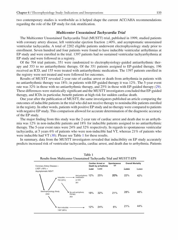
Chapter 6 / Electrophysiology Study: Indications and Interpretations 133
two contemporary studies is worthwhile as it helped shape the current ACC/AHA recommendationsregarding the role of the EP study for risk stratification.
Multicenter Unsustained Tachycardia TrialThe Multicenter Unsustained Tachycardia Trial (MUSTT) trial, published in 1999, studied patients
with coronary artery disease, left ventricular ejection fraction ≤40%, and asymptomatic unsustainedventricular tachycardia. A total of 2202 eligible patients underwent electrophysiology study prior toenrollment. Seven hundred and four patients were found to have inducible ventricular arrhythmias atEP study and were enrolled in the trial. 1397 patients had no sustained ventricular tachyarrhythmia atEP study and were followed in a registry.
Of the 704 trial patients, 351 were randomized to electrophysiology-guided antiarrhythmic ther-apy and 353 to no antiarrhythmic therapy. Of the 351 patients assigned to EP-guided therapy, 196received an ICD, and 155 were treated with antiarrhythmic medication. The 1397 patients enrolled inthe registry were not treated and were followed for outcomes.
Results of MUSTT revealed 2-year rate of cardiac arrest or death from arrhythmia in patients withno antiarrhythmic therapy was 18%; in patients with EP-guided therapy it was 12%. The 5-year eventrate was 32% in those with no antiarrhythmic therapy, and 25% in those with EP-guided therapy (29).These differences were statistically significant and the MUSTT investigators concluded that EP-guidedtherapy, and ICDs in particular, benefit patients at high risk for sudden cardiac death.
One year after the publication of MUSTT, the same investigators published an article comparing theoutcomes of inducible patients in the trial who did not receive therapy to noninducible patients enrolledin the registry. In other words, patients with positive EP study and no therapy were compared to patientswith negative EP study. This comparison allowed for accurate determination of the diagnostic accuracyof the EP study.
The major finding from this study was the 2-year rate of cardiac arrest and death due to an arrhyth-mia was 12% in non-inducible patients and 18% for inducible patients assigned to no antiarrhythmictherapy. The 5-year event rates were 24% and 32% respectively. In regards to spontaneous ventriculartachycardia, at 5 years 6% of patients who were non-inducible had VT, whereas 21% of patients whowere inducible had VT (30). Please see Table 1 for these results.
In summary, data from the MUSTT investigators revealed that inducibility on EP study accuratelypredicts increased risk of ventricular tachycardia, cardiac arrest, and death due to arrhythmia. Patients
Table 1Results from Multicenter Unsustained Tachycardia Trial and MUSTT-EPS
Cardiac Arrest or Spontaneous VT
Overall MortalityCoronary Artery Disease +LVEF ≤ 40% +Asymptomatic NSVT
Anti-arrhythmic
Death by arrhythmia2 year 5 year
12% 25%
2 years 5 years
22% 42%20%Inducible
704 (34%)
medication
or ICD
351
20%
Electrophysiology Study
2202No anti-arrhythmic therapy
353
18% 32% 28% 48%21%
Non-inducible 12% 24% 21% 44%6%1397 (66%)

134 Part III / Diagnostic Testing
with a positive EP study had higher event rates than those patients who were non-inducible. However,these studies also revealed that the ability of the EP study to predict future events is limited. Differencesin event rates between the two groups were statistically significant, but small. At 5 years the differencein rates of cardiac death and death due to an arrhythmia was only 12%. Thus, although an EP study candistinguish between patients who are at high and low risk, the differences between these two groups issmall.
Multicenter Automatic Defibrillator Implantation TrialThe MADIT II trial is the second large contemporary trial which gives data regarding the predictive
value of the electrophysiology study. The Multicenter Automatic Defibrillator Implantation Trial II(MADIT II) published in 2002 enrolled 1232 patients with prior myocardial infarction and left ven-tricular ejection fraction ≤30%. Patients were randomized in a 3:2 fashion to ICD (742 patients) orconventional medical therapy (490 patients). The primary outcome was death from any cause.
MADIT II did not require inducibility on EP study for enrollment in the trial, but in patients assignedto the ICD arm, an EP study was strongly encouraged at the time of ICD implantation. After publica-tion of MADIT II, II investigators published a substudy of patients who underwent elective EP studyat the time of ICD implantation. Of the 720 patients in the ICD arm, 593 underwent an EP study. Ofthose, 207(36%) met a “standard” definition of inducibility. Patients who were found to be inducible onEP study were subsequently compared to patients who were non-inducible on EP study. The primaryendpoint of this substudy was the incidence of spontaneous VT or VF on ICD interrogation.
Results from the MADIT II substudy revealed the rates of ventricular tachycardia and ventricularfibrillation (VF) in patients who were inducible (by the standard definition) were similar to rates ofpatients who were non-inducible. At 2 years, the incidence of ICD therapy for VT or VF was 29.4%in inducible patients and 25.5% in non-inducible patients (14). This suggests that the EP study cannotdifferentiate among patients who will receive appropriate shocks.
MADIT II investigators then employed a “narrow” definition of inducibility, namely inductionof sustained monomorphic ventricular tachycardia, and evaluated arrhythmic outcomes. In this set-ting, the prognostic ability of the EP study was dramatically improved. Patients in whom sustainedmonomorphic VT was induced on EP study were significantly more likely to develop both VT (hazardratio 1.89) and VT or VF (hazard ratio 1.56) than patients who were not inducible (14). Thus, whensustained monomorphic ventricular tachycardia is induced, it accurately predicts future ventriculartachycardia.
In conclusion, among patients with chronic coronary artery disease and reduced left ventricularsystolic function electrophysiology studies can identify patients who are at high risk for sudden cardiacdeath. Data collected as part of MUSTT and MADIT II trials revealed that patients with coronary arterydisease and reduced left ventricular ejection fraction who are inducible on EP testing are at increasedrisk of spontaneous stable ventricular tachycardia, cardiac arrest, and death by arrhythmia. Although,this increased risk may be relatively small when compared to patients who are non-inducible. With thisavailable data, the ACC/AHA/ESC guidelines state: “EP testing is reasonable for risk stratificationin patients with remote MI, NSVT, and LVEF equal to or less than 40%” (3). This is a Class IIarecommendation.
THE ROLE OF THE ELECTROPHYSIOLOGY STUDY IN NON-ISCHEMICCARDIOMYOPATHIES
In addition to patients with ischemic cardiomyopathies, there are other subgroups of patients whoare known to be at increased risk of sudden cardiac death due to ventricular arrhythmias. Thesesubgroups include patients with non-ischemic dilated cardiomyopathy, hypertrophic cardiomyopathy,

Chapter 6 / Electrophysiology Study: Indications and Interpretations 135
arrhythmogenic right ventricular cardiomyopathy, Brugada syndrome, and long QT syndrome. In gen-eral, the role of the EP study for risk stratification in these patients is less well characterized, althoughin some cases it may be of minor benefit.
In patients with non-ischemic dilated cardiomyopathy, studies have shown mixed results in regardsto the ability of EP study to predict future arrhythmic events (31–33). Thus, the ACC/AHA do notrecommend to undertake extrastimulus testing in this group of patients for the purposes of risk strat-ification. For patients with non-ischemic cardiomyopathies who have documented ventricular tachy-cardia, an EP study can be useful as bundle-branch reentry is relatively common in these patients. TheACC/AHA make a Class I recommendation in this regarding stating, “EP testing is useful to diag-nose bundle-branch reentrant tachycardia and to guide ablation in patients with nonischemic dilatedcardiomyopathy” (3). In patients with hypertrophic cardiomyopathy, the role of the electrophysiologystudy is controversial. In general, inducibility on EP study is not thought to have predictive power inthis patient population. As such, the ACC/AHA make a Class IIb recommendation that “EP testingmay be considered for risk assessment for SCD in patients with HCM”(3). As a general rule, we donot undertake extrastimulus testing in patients with hypertrophic cardiomyopathy.
In patients with arrhythmogenic right ventricular cardiomyopathy (ARVC), the ACC/AHA state:“EP testing might be useful for risk assessment of SCD in patients with ARVC.” Historically, EP test-ing had been used not so much for risk stratification, but to guide ablation in patients with ventriculartachycardia. Recent studies have shown that voltage mapping of the right ventricle shows promise inhelping to diagnose this type of cardiomyopathy (34).
In patients with Brugada syndrome and long QT syndrome, the ACC/AHA does not make a recom-mendation regarding EP testing, as its role is debated at this time.
PERFORMANCE AND INTERPRETATION OF EP STUDY PERFORMEDFOR RISK STRATIFICATION
The electrophysiology study performed to stratify patients according to their risk for sudden deathfollows a relatively standard protocol. In general, programmed stimulation is performed with a fixedcycle length pacing (S1) of eight beats immediately followed by single, double, and triple ventricularextrastimuli (S2, S3, S4) (35, 36). The standard protocol includes at least two different cycle lengths(typically 600 and 400 msec) as well as stimulation in sinus rhythm.
The optimal number of extrastimuli that should be used during ventricular stimulation is debatable.In general, the greater the number of extrastimuli used, the higher the sensitivity, but lower the speci-ficity of the resulting arrhythmia. We believe the use of up to three extrastimuli is optimal. Stimulationshould be performed in two different sites, typically the right ventricular apex and right ventricularoutflow tract. The protocol described by Josephson by which single ventricular extra stimuli (VES)are delivered at all sites and all cycle lengths before double VES, and double VES before triple VES isrecommended (1). This format is less likely to result in ventricular fibrillation at one site which wouldrequire ending stimulation for the safety of the patient, when another site could produce monomorphicventricular tachycardia.
Additional maneuvers can be employed to induce ventricular tachycardia. These include: (1) pacingfrom alternative sites (left ventricle or atrium), (2) infusing medication (isoproterenol or a type I agentlike procainamide), (3) increasing the current used, and (4) using long-short sequences. However, theseare not standard when performing ventricular stimulation for the purposes of risk stratification.
When a standard electrophysiology study is performed and no ventricular arrhythmias are induced,it is considered a negative EPS and the patient non-inducible. However, an electrophysiology studyin which a ventricular arrhythmia is induced is not necessarily considered positive. The nature andduration of the ventricular arrhythmia need to be considered, as does the stimuli which induced it.

136 Part III / Diagnostic Testing
While the induction of some arrhythmias suggests a patient is at high risk for future spontaneousventricular arrhythmias, the induction of other arrhythmias is non-specific and may be of no clinicalimport.
The strictest definition of a positive EPS is one in which monomorphic ventricular tachycardia isreproducibly induced and is sustained (>15 sec) or is hemodynamically unstable requiring cardiover-sion. This is the definition we employ in our lab. A broader definition of a positive EP study includespatients who develop sustained monomorphic VT, or polymorphic VT or ventricular fibrillation withone or two extrastimuli. Responses that are not interpreted as a positive EPS include nonsustainedmonomorphic or polymorphic ventricular tachycardia, bundle branch reentry ventricular tachycar-dia, repetitive polymorphic response, or polymorphic ventricular tachycardia or ventricular fibrillationinduced with three extrastimuli.
It is important to note that sustained monomorphic ventricular tachycardia, especially in patientswith coronary artery disease, can be reproducibly initiated on electrophysiology studies over time(37, 38). If VT can be induced today, it will be induced one year from now. This finding supports theidea that in patients with chronic ischemic heart disease the myocardial scar that forms the substrateof ventricular arrhythmias is fixed and stable over time.
In accordance with the evidence and the ACC/AHA/ESC guidelines, it is our general practiceto perform programmed ventricular stimulation on patients with coronary artery disease and LVEF31–40% to evaluate for inducibility. Patients in whom sustained monomorphic or polymorphic ven-tricular tachycardia, or ventricular fibrillation, are induced subsequently undergo ICD implantation.Although we realized that non-inducible patients are still at moderately high risk for ventriculararrhythmias, we do not implant ICDs in these patients.
CONCLUSION
In conclusion, there are numerous indications for a diagnostic electrophysiology study. Asdescribed, these include determining the etiology of unexplained syncope, making a definitive diag-nosis of subjective palpitations or documented narrow or wide complex tachycardia, evaluate patientswith Wolff-Parkinson-White syndrome, and stratifying a patient’s risk of sudden cardiac death, partic-ularly patients with coronary artery disease and depressed left ventricular function.
As mentioned, the EP study is limited in its ability to determine a bradyarrhythmic or tachyarrhyth-mic etiology of syncope. Moreover, the EP study is limited in its ability to broadly distinguish betweenpatients who are at high and low risk of sudden cardiac death. However, the EP study is excellent,indeed the gold standard, for determining the nature of a narrow complex or wide complex tachycar-dia. Much of electrophysiology is based on the ability of the EP study to make these diagnoses. Thesedays, once a diagnostic EP study has made a definitive diagnosis of a particular arrhythmia, the EPstudy can be therapeutic with the application of radiofrequency ablation. It is for this reason that theEP study will remain at the heart of the field of electrophysiology.
REFERENCES
1. Josephson ME (2002) Clinical cardiac electrophysiology: techniques and interpretations. Lippincott Williams &Wilkins, Philadelphia
2. Zimetbaum PJ, Josephson ME (2009) Practical clinical electrophysiology. Lippincott, Williams & Wilkins, Philadelphia3. Zipes DP, Camm AJ, Borggrefe M, Buxton AE, Chaitman B, Fromer M et al (2006) ACC/AHA/ESC 2006 guidelines
for management of patients with ventricular arrhythmias and the prevention of sudden cardiac death: a report of theAmerican college of cardiology/American heart association task force and the European society of cardiology com-mittee for practice guidelines (writing committee to develop guidelines for management of patients with ventriculararrhythmias and the prevention of sudden cardiac death). J Am Coll Cardiol 48(5):e247–e346

Chapter 6 / Electrophysiology Study: Indications and Interpretations 137
4. Bachinsky WB, Linzer M, Weld L, Estes NA III (1992) Usefulness of clinical characteristics in predicting the outcomeof electrophysiologic studies in unexplained syncope. Am J Cardiol 69(12):1044–1049
5. Engel TR, Luck JC, Leddy CL, Gonzalez AD (1979) Diagnostic implications of atrial vulnerability. Pacing ClinElectrophysiol 2(2):208–214
6. Gang ES, Reiffel JA, Livelli FD Jr, Bigger JT Jr (1983) Sinus node recovery times following the spontaneous terminationof supraventricular tachycardia and following atrial overdrive pacing: a comparison. Am Heart J 105(2):210–215
7. Fujimura O, Yee R, Klein GJ, Sharma AD, Boahene KA (1989) The diagnostic sensitivity of electrophysiologic testingin patients with syncope caused by transient bradycardia. N Engl J Med 321(25):1703–1707
8. Scheinman MM, Peters RW, Suave MJ, Desai J, Abbott JA, Cogan J et al (1982) Value of the H-Q interval in patientswith bundle branch block and the role of prophylactic permanent pacing. Am J Cardiol 50(6):1316–1322
9. Brembilla-Perrot B, Beurrier D, de la Chaise AT, Suty-Selton C, Jacquemin L, Thiel B et al (1996) Significanceand prevalence of inducible atrial tachyarrhythmias in patients undergoing electrophysiologic study for presyncopeor syncope. Int J Cardiol 53(1):61–69
10. Doherty JU, Pembrook-Rogers D, Grogan EW, Falcone RA, Buxton AE, Marchlinski FE et al (1985) Electrophysiologicevaluation and follow-up characteristics of patients with recurrent unexplained syncope and presyncope. Am J Cardiol55(6):703–708
11. Hess DS, Morady F, Scheinman MM (1982) Electrophysiologic testing in the evaluation of patients with syncope ofundetermined origin. Am J Cardiol 50(6):1309–1315
12. Denes P, Uretz E, Ezri MD, Borbola J (1988) Clinical predictors of electrophysiologic findings in patients with syncopeof unknown origin. Arch Intern Med 148(9):1922–1928
13. Morady F, Scheinman MM (1983) The role and limitations of electrophysiologic testing in patients with unexplainedsyncope. Int J Cardiol 4(2):229–234
14. Daubert JP, Zareba W, Hall WJ, Schuger C, Corsello A, Leon AR et al (2006) Predictive value of ventricular arrhyth-mia inducibility for subsequent ventricular tachycardia or ventricular fibrillation in multicenter automatic defibrillatorimplantation trial (MADIT) II patients. J Am Coll Cardiol 47(1):98–107
15. Denniss AR, Ross DL, Richards DA, Uther JB (1992) Electrophysiologic studies in patients with unexplained syncope.Int J Cardiol 35(2):211–217
16. Campbell RW, Smith RA, Gallagher JJ, Pritchett EL, Wallace AG (1977) Atrial fibrillation in the preexcitation syn-drome. Am J Cardiol 40(4):514–520
17. Wellens HJ, Durrer D (1974) Wolff-Parkinson-White syndrome and atrial fibrillation. Relation between refractory periodof accessory pathway and ventricular rate during atrial fibrillation. Am J Cardiol 34(7):777–782
18. Wellens HJ (2005) Should catheter ablation be performed in asymptomatic patients with Wolff-Parkinson-White syn-drome? When to perform catheter ablation in asymptomatic patients with a Wolff-Parkinson-White electrocardiogram.Circulation 112(14):2201–2207
19. Pappone C, Manguso F, Santinelli R, Vicedomini G, Sala S, Paglino G et al (2004) Radiofrequency ablation in childrenwith asymptomatic Wolff-Parkinson-White syndrome. N Engl J Med 351(12):1197–1205
20. Klein GJ, Bashore TM, Sellers TD, Pritchett EL, Smith WM, Gallagher JJ (1979) Ventricular fibrillation in the Wolff-Parkinson-White syndrome. N Engl J Med 301(20):1080–1085
21. Montoya PT, Brugada P, Smeets J, Talajic M, Della BP, Lezaun R et al (1991) Ventricular fibrillation in the Wolff-Parkinson-White syndrome. Eur Heart J 12(2):144–150
22. Timmermans C, Smeets JL, Rodriguez LM, Vrouchos G, van den DA, Wellens HJ (1995) Aborted sudden death in theWolff-Parkinson-White syndrome. Am J Cardiol 76(7):492–494
23. Pappone C, Santinelli V, Rosanio S, Vicedomini G, Nardi S, Pappone A et al (2003) Usefulness of invasive electrophys-iologic testing to stratify the risk of arrhythmic events in asymptomatic patients with Wolff-Parkinson-White pattern:results from a large prospective long-term follow-up study. J Am Coll Cardiol 41(2):239–244
24. Pappone C, Santinelli V, Manguso F, Augello G, Santinelli O, Vicedomini G et al (2003) A randomized study ofprophylactic catheter ablation in asymptomatic patients with the Wolff-Parkinson-White syndrome. N Engl J Med349(19):1803–1811
25. Blomstrom-Lundqvist C, Scheinman MM, Aliot EM, Alpert JS, Calkins H, Camm AJ et al (2003) ACC/AHA/ESCguidelines for the management of patients with supraventricular arrhythmias–executive summary: a report of theAmerican college of cardiology/American heart association task force on practice guidelines and the European soci-ety of cardiology committee for practice guidelines (writing committee to develop guidelines for the management ofpatients with supraventricular arrhythmias). Circulation 108(15):1871–1909
26. Richards DA, Byth K, Ross DL, Uther JB (1991) What is the best predictor of spontaneous ventricular tachycardia andsudden death after myocardial infarction? Circulation 83(3):756–763
27. Wilber DJ, Olshansky B, Moran JF, Scanlon PJ (1990) Electrophysiological testing and nonsustained ventricular tachy-cardia. Use and limitations in patients with coronary artery disease and impaired ventricular function. Circulation82(2):350–358

138 Part III / Diagnostic Testing
28. Schmitt C, Barthel P, Ndrepepa G, Schreieck J, Plewan A, Schomig A et al (2001) Value of programmed ventricu-lar stimulation for prophylactic internal cardioverter-defibrillator implantation in postinfarction patients preselected bynoninvasive risk stratifiers. J Am Coll Cardiol 37(7):1901–1907
29. Buxton AE, Hafley GE, Lehmann MH, Gold M, O’Toole M, Tang A et al (1999) Prediction of sustained ventriculartachycardia inducible by programmed stimulation in patients with coronary artery disease. Utility of clinical variables.Circulation 99(14):1843–1850
30. Buxton AE, Lee KL, DiCarlo L, Gold MR, Greer GS, Prystowsky EN et al (2000) Electrophysiologic testing to iden-tify patients with coronary artery disease who are at risk for sudden death. Multicenter unsustained tachycardia trialinvestigators. N Engl J Med 342(26):1937–1945
31. Turitto G, Ahuja RK, Caref EB, el Sherif N (1994) Risk stratification for arrhythmic events in patients with nonischemicdilated cardiomyopathy and nonsustained ventricular tachycardia: role of programmed ventricular stimulation and thesignal-averaged electrocardiogram. J Am Coll Cardiol 24(6):1523–1528
32. Milner PG, Dimarco JP, Lerman BB (1988) Electrophysiological evaluation of sustained ventricular tachyarrhythmiasin idiopathic dilated cardiomyopathy. Pacing Clin Electrophysiol 11(5):562–568
33. Brembilla-Perrot B, Donetti J, de la Chaise AT, Sadoul N, Aliot E, Juilliere Y (1991) Diagnostic value of ventricularstimulation in patients with idiopathic dilated cardiomyopathy. Am Heart J 121(4 Pt 1):1124–1131
34. Avella A, d’Amati G, Pappalardo A, Re F, Silenzi PF, Laurenzi F et al (2008) Diagnostic value of endomyocar-dial biopsy guided by electroanatomic voltage mapping in arrhythmogenic right ventricular cardiomyopathy/dysplasia.J Cardiovasc Electrophysiol 19(11):1127–1134
35. Waldo AL, Akhtar M, Brugada P, Henthorn RW, Scheinman MM, Ward DE et al (1985) The minimally appropriateelectrophysiologic study for the initial assessment of patients with documented sustained monomorphic ventriculartachycardia. J Am Coll Cardiol 6(5):1174–1177
36. Vandepol CJ, Farshidi A, Spielman SR, Greenspan AM, Horowitz LN, Josephson ME (1980) Incidence and clinicalsignificance of induced ventricular tachycardia. Am J Cardiol 45(4):725–731
37. Josephson ME, Almendral JM, Buxton AE, Marchlinski FE (1987) Mechanisms of ventricular tachycardia. Circulation75(4 Pt 2):III41–III47
38. Roy D, Marchand E, Theroux P, Waters DD, Pelletier GB, Cartier R et al (1986) Long-term reproducibility and signifi-cance of provokable ventricular arrhythmias after myocardial infarction. J Am Coll Cardiol 8(1):32–39

IV SPECIFIC ARRHYTHMIAS
7 Supraventricular Arrhythmias
Khalid Almuti, Babak Bozorgnia,and Steven A. RothmanCONTENTS
INTRODUCTION
NONINVASIVE DIAGNOSIS OF SVTMECHANISMS OF SVTNON-INVASIVE AND PHARMACOLOGIC THERAPIES FOR SVTPHARMACOTHERAPY
ELECTROPHYSIOLOGIC TESTING AND TACHYCARDIA ABLATION
CATHETER ABLATION OF AVNRTATRIAL TACHYCARDIA
SUMMARY
REFERENCES
Abstract
Paroxysmal supraventricular tachycardia is a common arrhythmia with multiple etiologies, includingatrio-ventricular nodal reentrant tachycardia, atrio-ventricular reentrant tachycardia, and atrial tachycardia.Treatment of these arrhythmias depends greatly upon the proper diagnosis as well as an understanding ofthe arrhythmia’s mechanism. A preliminary diagnosis can be often be inferred from the patient’s historyalong with noninvasive testing and can help guide initial management strategies. Pharmacologic therapy,however, is often limited by side effects, compliance, and marginal efficacy. More definitive treatment of thearrhythmia requires an invasive electrophysiology study to confirm the diagnosis followed by catheter abla-tion of the arrhythmogenic substrate. The success rate for catheter ablation can approach 95% dependingon the mechanism of the arrhythmia and is the treatment of choice for patients with severe symptoms.
Key Words: Activation mapping; adenosine; afterdepolarizations; amiodarone; antidromic atrio-ventricular reentrant tachycardia; atrial extrastimuli; atrial tachycardia; atrio-ventricular nodal reentranttachycardia; atrio-ventricular reentrant tachycardia; automaticity; beta-blockers; digoxin, diltiazem;dofetilide; entrainment; flecainide; ibutilide; isoproterenol; macroreentry; metoprolol; microreentry; ortho-dromic atrio-ventricular reentrant tachycardia; pace mapping; para-Hisian pacing; pharmacotherapy; proar-rhythmia; procainamide; propafeone; propranolol; radiofrequency catheter ablation; sotalol; supraven-tricular tachycardia; triggered activity; ventricular extrastimuli; verapamil; Wolff–Parkinson–Whitesyndrome.
From: Contemporary Cardiology: Management of Cardiac ArrhythmiasEdited by: Gan-Xin Yan, Peter R. Kowey, DOI 10.1007/978-1-60761-161-5_7
C© Springer Science+Business Media, LLC 2011
141

142 Part IV / Specific Arrhythmias
INTRODUCTION
The term “supraventricular tachycardia” (SVT) technically refers to arrhythmias originating abovethe AV node. This includes rhythms as disparate as sinus tachycardia and atrial fibrillation (AF),but in practice, the term “supraventricular tachycardia” is mostly used to refer to a finite numberof abnormal rhythms that are paroxysmal in nature and include atrio-ventricular nodal reentrant tachy-cardia (AVNRT), atrio-ventricular reentrant tachycardia (AVRT), atrial tachycardia (AT), and, lesscommonly, junctional ectopic tachycardia and sino-atrial reentrant tachycardia. The prevalence ofthese paroxysmal SVT’s is 2.25 per 1000 persons with a female preponderance especially before age65 years (1). In this chapter, the most common paroxysmal supraventricular arrhythmias (AVNRT,AVRT, and AT) will be discussed. AF and atrial flutter will be covered in more detail in separatechapters.
NONINVASIVE DIAGNOSIS OF SVT
HistoryIn the absence of an electrocardiographic documentation of an SVT, history can be extremely help-
ful in differentiating SVT from other cardiac arrhythmias. If an SVT is documented on an ECG(or a cardiac monitor) then a detailed history can predict the mechanism of the SVT in a high per-centage of patients (2). Useful information includes descriptions of the onset and termination of theepisode, instigating and terminating factors, symptoms during the episode, and age at the onset ofsymptoms (3).
Reentrant SVTs such as AVNRT and AVRT are usually abrupt in onset and offset while automaticatrial arrhythmias, including sinus tachycardia, will usually initiate and subside gradually. Symptomsmay include palpitations, dizziness, shortness of breath, and chest tightness. Some patients may expe-rience diaphoresis, numbness in the extremities, and flushing. If asked, the patient will usually be ableto tap out a rapid but regular demonstration of the episode. Many patients may also feel pulsationsin the neck representing contraction of the atria against a closed AV valve. This phenomenon is morecommon in AVNRT (2). More severe symptoms, such as syncope, are less frequent, but can occur inup to 20% of patients (4).
Aside from the description of SVT episodes, history should also include any underlying cardiac dis-eases such as congenital heart disease or prior heart surgery. A history of heart surgery with resultingscar tissue may represent an arrhythmogenic substrate and makes a diagnosis of AT or atrial fluttermore likely (5). A history of prior catheter-based ablation therapy is also important to obtain for thesame reason. The age and gender of the patient may, in some cases, help narrow the differential diag-nosis of the SVT. For example, AVNRT tends to have a female preponderance with a bimodal agedistribution (2).
ECG FeaturesSeveral features on the cardiac electrocardiogram can be useful in determining the mechanism of
SVT. Most important of these is the P wave location (Fig. 1). If discernable P waves are visible, thendetermining the length of the RP interval can be used to categorize the tachycardia as either a short-or a long-RP tachycardia. If the interval from the start of the P wave to the preceding QRS complex isshorter than the interval from the same P wave to the subsequent QRS complex, then the tachycardiais described as a short-RP tachycardia. The converse is true for a long-RP tachycardia (6).
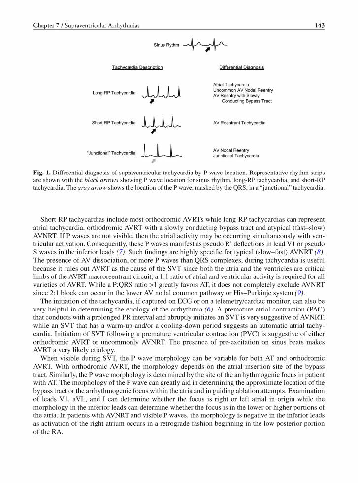
Chapter 7 / Supraventricular Arrhythmias 143
Fig. 1. Differential diagnosis of supraventricular tachycardia by P wave location. Representative rhythm stripsare shown with the black arrows showing P wave location for sinus rhythm, long-RP tachycardia, and short-RPtachycardia. The gray arrow shows the location of the P wave, masked by the QRS, in a “junctional” tachycardia.
Short-RP tachycardias include most orthodromic AVRTs while long-RP tachycardias can representatrial tachycardia, orthodromic AVRT with a slowly conducting bypass tract and atypical (fast–slow)AVNRT. If P waves are not visible, then the atrial activity may be occurring simultaneously with ven-tricular activation. Consequently, these P waves manifest as pseudo R’ deflections in lead V1 or pseudoS waves in the inferior leads (7). Such findings are highly specific for typical (slow–fast) AVNRT (8).The presence of AV dissociation, or more P waves than QRS complexes, during tachycardia is usefulbecause it rules out AVRT as the cause of the SVT since both the atria and the ventricles are criticallimbs of the AVRT macroreentrant circuit; a 1:1 ratio of atrial and ventricular activity is required for allvarieties of AVRT. While a P:QRS ratio >1 greatly favors AT, it does not completely exclude AVNRTsince 2:1 block can occur in the lower AV nodal common pathway or His–Purkinje system (9).
The initiation of the tachycardia, if captured on ECG or on a telemetry/cardiac monitor, can also bevery helpful in determining the etiology of the arrhythmia (6). A premature atrial contraction (PAC)that conducts with a prolonged PR interval and abruptly initiates an SVT is very suggestive of AVNRT,while an SVT that has a warm-up and/or a cooling-down period suggests an automatic atrial tachy-cardia. Initiation of SVT following a premature ventricular contraction (PVC) is suggestive of eitherorthodromic AVRT or uncommonly AVNRT. The presence of pre-excitation on sinus beats makesAVRT a very likely etiology.
When visible during SVT, the P wave morphology can be variable for both AT and orthodromicAVRT. With orthodromic AVRT, the morphology depends on the atrial insertion site of the bypasstract. Similarly, the P wave morphology is determined by the site of the arrhythmogenic focus in patientwith AT. The morphology of the P wave can greatly aid in determining the approximate location of thebypass tract or the arrhythmogenic focus within the atria and in guiding ablation attempts. Examinationof leads V1, aVL, and I can determine whether the focus is right or left atrial in origin while themorphology in the inferior leads can determine whether the focus is in the lower or higher portions ofthe atria. In patients with AVNRT and visible P waves, the morphology is negative in the inferior leadsas activation of the right atrium occurs in a retrograde fashion beginning in the low posterior portionof the RA.
144 Part IV / Specific Arrhythmias
MECHANISMS OF SVT
ReentryReentry is the most common mechanism of narrow QRS complex tachycardia (10). It requires two
distinct pathways with different electrophysiologic properties that are linked proximally and distally,forming an anatomic or functional circuit (5, 11). Reentry occurs when an impulse initially excitesand conducts through the first pathway (or area of cardiac tissue), while failing to conduct through thesecond part of the circuit because it is refractory and therefore not excitable. Via the distal connectionof the circuit, the impulse then enters the previously refractory tissue of the second pathway exciting itin a retrograde direction. The impulse must conduct sufficiently slowly within one limb of the circuit toallow the previously refractory tissue to recover excitability. If the impulse conducted in a retrogrademanner in the second pathway reaches the proximal portion of the circuit when the first pathway isagain excitable, then the impulse is able to reenter the first pathway resulting in a “circus movement”or reentrant arrhythmia.
The reentrant circuit may become repetitively activated, producing a sustained reentrant tachy-cardia. The type of arrhythmia that ensues is determined by the characteristics and location of thereentrant circuit. Reentry may use a large macroreentrant circuit (as in atrial flutter and AVRT) orsmall microreentrant circuits (as in some atrial tachycardias and AVNRT). Anatomic structures (e.g.,the crista terminalis and eustachian ridge in the case of typical atrial flutter) or areas of fibrosis andscar may form the boundaries of the reentrant circuit (12). Alternatively, the circuit may result fromfunctional electrophysiologic properties of normal or diseased tissue that creates the milieu for reentry(13).
Automaticity and Triggered ActivityA less common mechanism of narrow QRS complex tachycardia is automaticity. Automaticity is
caused by enhanced diastolic phase 4 depolarization and when the firing rate exceeds the sinus rate,the abnormal rhythm will occur. Tissues capable of causing a narrow complex tachycardia due toautomaticity may be found in the atria, AV junction, vena cava, and pulmonary veins. These rhythmscan be either incessant or episodic.
Triggered activity is another arrhythmogenic mechanism due to abnormal impulse initiation (14).This type of tachycardia results from interruptions of the repolarization process called an afterdepo-larizations. When an afterdepolarization reaches a threshold, an action potential is triggered. Afterde-polarizations are characterized as either “early,” occurring during repolarization, or “delayed” whichoccur at the end of repolarization or immediately after completion of repolarization (15). Atrial tachy-cardias associated with digoxin toxicity or theophylline are examples of a triggered arrhythmia (16).
Management of SVTThe management of SVT is based on the clinical presentation of the arrhythmia and the patient’s
preferences. While electrophysiologic testing may be used to assess the risk of life-threatening arrhyth-mias in patients with asymptomatic WPW (17), treatment is typically not indicated for patients whohave pre-excitation on their ECG without a clinical syndrome. Individuals with high-risk occupations(e.g., airplane pilots) and asymptomatic WPW, however, may require more aggressive managementincluding “prophylactic” catheter-based ablation. Patients with mild, infrequent symptoms may bene-fit from intermittent pharmacologic therapy (e.g., “pill-in-pocket” approach), while patients with fre-quent symptomatic episodes are candidates for chronic therapy or catheter-based ablation. Patientswith infrequent, but poorly tolerated arrhythmias also require a more definitive approach. An indi-vidual’s lifestyle and personal preferences along with overall health and the presence of significantcomorbidities should be considered when making long-term management decisions (10).

Chapter 7 / Supraventricular Arrhythmias 145
NON-INVASIVE AND PHARMACOLOGIC THERAPIES FOR SVT
The development of catheter-based ablation technology for the treatment of SVT, providing higharrhythmia cure rates, has greatly diminished the role of pharmacologic therapy for SVT. Currently,the main role of pharmacotherapy is in the acute termination of an arrhythmia or for control of theventricular response rate during SVT episodes. The chronic use of pharmacologic agents to suppressSVT is usually reserved for patients who are not candidates for catheter-based ablation procedures orpatients who prefer a pharmacologic option.
PHARMACOTHERAPY
Acute TerminationIn general, SVT is considered to be a non life-threatening condition with a good long-term prog-
nosis. Nevertheless, certain episodes of SVT can present with hemodynamic compromise and/or sig-nificant symptoms. An acute intervention may be necessary to restore hemodynamic stability or topalliate severe symptoms. Pharmacotherapy, vagal maneuvers, and electrical cardioversion are optionsthat can be used to achieve these goals.
Maneuvers that increase vagal tone, such as carotid sinus massage and the Valsalva maneuver, alterthe refractoriness and conduction properties of the AV node and can terminate the SVT if the AVnode is an integral part of the SVT circuit (e.g., AVNRT or AVRT) (18). Alternatively, they can slowdown the rate of the ventricular response to the SVT (i.e., in AT) and help differentiate the mechanismof the tachycardia (6). If these measures are ineffective, then pharmacological intervention should beconsidered. Intravenous verapamil and adenosine are the drugs of choice for reentrant arrhythmias(10, 19, 20). They exert their activity principally at the level of the AV node. Similar to vagal maneu-vers, these agents may either terminate or slow down the tachycardia.
Adenosine’s ultra-short duration of action makes it a preferred agent before resorting to emergentDC cardioversion in patients with a tenuous hemodynamic state. Caution has to be exercised whenusing adenosine due to a potential proarrhythmic effect stemming from a transient increase in atrialvulnerability to AF (21–23). In patients with an AT, adenosine may result in transient AV block, help-ing determine the diagnosis. Occasionally, adenosine may terminate an AT, especially if the arrhythmiais due to a triggered or automatic mechanism (24).
Intravenous verapamil is also effective for the acute termination of AVRT, but has a later onset ofaction and longer effect. It should not be used in patients with profound hypotension or those withseverely depressed ventricular systolic function (5). It should also be avoided in patients with pre-excited atrial fibrillation due to its potential to accelerate the ventricular response rate (25, 26). Likeadenosine, calcium channel blockers can occasionally terminate AT but the most common outcomeis slowing down the ventricular response rate, making the tachycardia more hemodynamically stablewithout terminating it (19, 27). Intravenous diltiazem and beta-blockers (propranolol and metoprolol)are also effective in the acute treatment of SVT.
Intravenous procainamide is a class IA agent that depresses conduction and prolongs refractorinessin atrial and ventricular myocardium, in accessory pathways, and in the His–Purkinje system (28, 29).It may also cause slight shortening of the AV nodal refractory period but often has no discernableeffect on AV nodal refractoriness (13). Procainamide is most effective in terminating reentrant atrialtachycardia and AVRT; it is less effective in terminating AVNRT. In patients presenting with a wideQRS complex tachycardia of unknown etiology, procainamide is considered one of the safest and mosteffective drugs to administer (30). Its electrophysiologic effects may result in the termination of bothventricular tachycardia and antidromic AVRT. Ibutilide can also be used in the acute management ofpatients with pre-excited atrial fibrillation (10, 31).
146 Part IV / Specific Arrhythmias
Maintenance PharmacotherapyThe goals of long-term maintenance therapy for SVT are to suppress future episodes and to control
the rate of the ventricular response if episodes do recur. The selection of a pharmacologic agent isbased on certain patient characteristics and on the unique electrophysiologic properties of the arrhyth-mia. Patient characteristics include existing comorbidities, baseline cardiac function, severity of symp-toms during SVT, and drug sensitivities. Pharmacologic agents that are well tolerated with low organtoxicity are preferred.
Agents with AV nodal-specific activity (beta-blockers, calcium channel blockers, and to a lesserextent digoxin) are often used as first-line therapy and are most useful in suppressing reentrant arrhyth-mias that use the AV node for at least one limb of the tachycardia, especially AVNRT. Overall, theseagents may improve symptoms in up to 60–80% of patients (5), but are sometimes inadequate asmonotherapy because of their inability to directly slow conduction and alter the refractoriness of anaccessory pathway or to significantly reduce the frequency of arrhythmia-triggering ectopy (32–34).
Class IC antiarrhythmic agents (i.e., flecainide and propafenone) prolong both antegrade and ret-rograde refractoriness in the accessory pathway (35) making them useful in the chronic treatment ofAVRT and other paroxysmal SVTs (36–40). An important contraindication to the use of these agents isthe presence of known coronary disease or structural heart disease as the risk of proarrhythmic effectsin those settings is considerable (41). Other antiarrhythmic agents that are effective in the treatmentof paroxysmal SVT include sotalol (42, 43), dofetilide (44, 45), and amiodarone (46–48). These arebest considered as second-line agents, however, due to their side effect profiles and increased risk ofproarrhythmia.
Chronic drug therapy usually requires continuous dosing at regular intervals for an indefinite periodof time. However, there are patients with infrequent and well-tolerated episodes of SVT that cause mildsymptoms. Such patients may benefit from regimens of intermittent oral drugs or “pill-in-the-pocket”therapy (49) that terminate SVT episodes. Drugs that can be used in this manner include shorter actingbeta-blockers, calcium channel blocker, and class IC AAD such as propafenone and flecainide (50–53).
ELECTROPHYSIOLOGIC TESTING AND TACHYCARDIA ABLATION
The invasive electrophysiology procedure in patients with SVT has two purposes: determination ofthe mechanism of the arrhythmia and catheter ablation of the anatomic substrate causing the tachy-cardia. To evaluate the patient’s clinical arrhythmia, the tachycardia must first be initiated in the elec-trophysiology laboratory. Reentrant arrhythmias can be initiated with a variety of pacing maneuvers,although intravenous isoproterenol, a beta agonist, may be needed to enhance conduction of the AVnode (54). Triggered arrhythmias usually require the addition of isoproterenol along with programmedstimulation for initiation while automatic arrhythmias are generally not inducible with programmedstimulation, but can be facilitated with isoproterenol (55). In addition to its utility in initiating theclinical tachycardia, programmed stimulation can also be used to define the arrhythmogenic substrate.
Atrial extrastimuli (AES) are atrial premature depolarizations delivered at sequentially shorter cou-pling intervals (usually 10 msec decrements) after the last beat of a fixed cycle length drivetrain or dur-ing the spontaneous rhythm. Atrial extrastimuli are used to assess the refractory periods of supraven-tricular tissues and also to facilitate the induction of SVT. Measurement of the AH interval associatedwith each decremental AES will usually demonstrate a slight increase in the AH interval due to thedecremental conduction properties of the AV node. Plotting of the AH interval as a function of theAES coupling interval results in an AV nodal conduction curve. Dual AV nodal physiology is demon-strated by a discontinuity in this curve (56) as well as by an abrupt increase in the AH interval (usually>50 msec) in response to a 10 msec decrement in the coupling interval of the AES (Fig. 2). AES can

Chapter 7 / Supraventricular Arrhythmias 147
Fig. 2. Dual AV nodal pathways. AV nodal conduction is measured (AH interval) in response to decrementalatrial depolarizations delivered after an eight-beat pacing drive. The left hand panel shows an AH interval of168 msec in response to a coupling interval of 310 msec. The right hand panel shows an abrupt increase in theAH interval to 254 msec in response to a 10 msec decrease in the coupling interval (300 msec). This abruptincrease is consistent with dual AV nodal pathways as the fast pathway is now refractory and conduction occursover the slow pathway. An AV nodal echo beat also occurs as retrograde conduction is now present through thefast pathway. (Surface leads I, II, III, V1, and V6 are shown with intracardiac electrograms: HRA = high rightatrium, HB = His bundle, CS = coronary sinus, RVA = right ventricular apex; p = proximal, d = distal, s =stimuli, T = time).
also be used to determine the refractory period of an accessory pathway’s antegrade conduction, whichcould have prognostic implications should the patient develop AF with rapid conduction (57).
Ventricular extrastimuli (VES) are ventricular premature beats that are also delivered at sequentiallyshorter coupling intervals after a fixed cycle length drivetrain or other spontaneous rhythm. The atrialactivation sequence with normal retrograde AV nodal activation typically shows earliest atrial activityin the septal region near the His bundle recording site, although occasionally may be earliest in theposterior septum and proximal coronary sinus recordings. Accessory pathways located on the left freewall of the mitral annulus will have early atrial activity in the distal CS recordings while right free wallpathways will have early atrial activation in the lateral RA catheter. Measurement of the VA intervalwill allow assessment of the retrograde refractory periods of the AV node or accessory pathways.Retrograde dual AV nodal pathways may be manifested by an abrupt increase in the VA conductiontime through the AV node ( >50 msec) in response to a 10 msec decrement in the coupling interval.
Careful assessment of the atrial activation sequence during VES is very important. When morethan one retrograde pathway is present (i.e., AV node and accessory pathway), fusion of atrial activa-tion may result in early atrial activation at multiple sites. As the refractory period of one pathway isapproached with decremental VES, a change in the atrial activation sequence may signifying a shiftin retrograde conduction through only one of the pathways, confirming the presence of an accessory

148 Part IV / Specific Arrhythmias
pathway. Multiple shifts in the retrograde atrial activation sequence can be seen in cases where morethan one accessory pathway is present. Retrograde dual AV nodal pathways, however, may also causea shift in atrial activation. Earliest activation may shift more posteriorly and inferiorly as AV nodalconduction changes from the fast to slow pathway (58).
Para-Hisian pacing can also be performed to evaluate retrograde atrial activation and is used todifferentiate anteroseptal accessory pathways from normal retrograde AV nodal conduction (59). Inthe presence of an accessory pathway, pacing the His bundle without capturing local ventricular tissuewill require atrial activation to occur via an impulse that must first conduct over the His–Purkinjesystem to the ventricle and then through the ventricular myocardium back to the accessory pathway.If local ventricular tissue is captured, however, then conduction occurs over a small area of ventriculartissue and directly then to the AP. This results in a shortening of the His (or pacing stimulus) to atrialinterval (Fig. 3). Capture of local ventricular tissue without His bundle capture would also result inthe shorter HA interval. Since AV nodal conduction requires conduction from the His bundle to theatrium via the AV node only, there would be no change with or without local ventricular capture. But iflocal ventricular capture occurs without His bundle stimulation, then the HA interval would lengthen(Fig. 4).
Fig. 3. Para-Hisian pacing in the presence of an accessory pathway. Pacing is performed from the anteroseptumwith the first 2 complexes resulting in capture of both the His bundle and local ventricular tissue. Subsequentpacing stimuli show capture of only the His bundle with a narrowing of the QRS (i.e., pure His bundle capture).Local ventricular capture allows conduction back to the atrium to occur directly over the accessory pathway,resulting in a shorter stimulus to atrial electrogram (S–A) interval of 150 msec (surface leads I, III, aVF, and V1are shown with intracardiac electrograms: HRA = high right atrium, His = His bundle, CS = coronary sinus,RVA = right ventricular apex; p = proximal, m = mid, d = distal, s = stimuli, T = time).
Induction of SVTThe induction of reentrant SVT with extrastimuli requires block in one pathway while the second
pathway conducts with sufficient delay to allow recovery and retrograde conduction in the first (15).In AV nodal reentry, the antegrade effective refractory period (ERP) of the fast AV nodal pathway isusually longer than the ERP of the slow pathway such that common type AVNRT can be induced withAES. The retrograde ERP of the fast AV nodal pathway, however, tends to be shorter than the slow

Chapter 7 / Supraventricular Arrhythmias 149
Fig. 4. Para-Hisian pacing in the absence of an accessory pathway. Pacing is performed from the anteroseptumwith the second complex showing capture of both the His bundle and local ventricular tissue and the third com-plex showing capture of only local ventricular tissue (wider QRS duration). The stimulus to atrial electrogram(SA) interval is lengthened when His bundle capture is lost since ventriculoatrial conduction is AV nodal depen-dent (surface leads I, II, III, and V1 are shown with intracardiac electrograms: HRA = high right atrium, His =His bundle, CS = coronary sinus, RVA = right ventricular apex; p = proximal, m = mid, d = distal, s = stimuli,T = time).
pathway and typical AVNRT is usually not induced with VES. When the retrograde slow pathwayERP is shorter than the fast pathway ERP, however, uncommon AVNRT can be induced with VES(60). Rapid atrial pacing near the AV nodal Wenckebach cycle length (CL) can also be used to inducecommon AVNRT.
In patients with Wolff–Parkinson–White syndrome, if the antegrade accessory pathway ERP islonger than the AV nodal pathway ERP, and there is sufficient prolongation of AV nodal conduction,then AVRT can be induced with AES. For patients with concealed accessory pathways, only suffi-cient prolongation in AV nodal conduction is usually necessary to induce AVRT as antegrade blockis already present in the accessory pathway. More commonly, AVRT can be induced with ventricularextrastimuli as the retrograde refractory period of the accessory pathway is usually shorter than that ofthe AV node. Delivering VES at shorter drive cycle lengths can be helpful as AV nodal refractorinesswill increase while most bypass tract refractory periods will decrease.
Atrial tachycardias can be either reentrant, triggered or automatic and each mechanism typicallyrequires a different mode of induction (55). For microreentrant atrial tachycardia, multiple extrastim-uli are commonly needed to achieve block in one limb of the circuit and cause significant prolongationof conduction in the other to allow reentry. Rapid (burst) atrial pacing is commonly used to inducetriggered arrhythmias, especially during the infusion of an intravenous catecholamine, such as isopro-terenol. Automatic AT is usually not initiated with either AES or burst pacing, but may be enhancedby isoproterenol.
Electrophysiologic Diagnostic TechniquesOnce SVT is initiated, careful assessment of the ventricular and atrial timing, along with pro-
grammed stimulation and rapid pacing, can be used to differentiate the mechanism of the SVT. If

150 Part IV / Specific Arrhythmias
spontaneous AV block is observed, then AVRT is definitively ruled out and atrial tachycardia is themost likely diagnosis. Rarely, AVNRT can have a 2:1 AV ratio due to block in the lower common AVnodal pathway or His bundle (9). For tachycardias with a VA time of <60 msec, measured from theonset of ventricular activation to the earliest atrial activation, a diagnosis of AVNRT is most likely(61). In AVRT, conduction from the ventricle to the atrium, via the bypass tract, would be expected totake longer than 60 msec. Atrial tachycardia with a prolonged PR interval, such that the P wave fallson the preceding QRS, would be an exception to this.
Other observations can also be helpful in diagnosing the SVT mechanism. Bundle branch blockthat results in an increase in the tachycardia CL or a >20 msec increase in the VA interval is consis-tent with AVRT utilizing an ipsilateral accessory pathway (62) (Fig. 5). This is due to the extra timerequired to traverse a circuit with conduction proceeding down the opposite bundle branch and thenacross the septum. If spontaneous termination is observed, then it should be noted if the last beat endswith ventricular activation (VA block) or atrial activation (AV block). If the tachycardia reproduciblyterminates with atrial activation, then an atrial tachycardia would be very unlikely since both block inthe atrial circuit and AV nodal block would have to occur simultaneously.
The effect caused by ventricular stimulation during His bundle refractoriness can also be very usefulin differentiating the tachycardia mechanism (63). Ventricular extrastimuli are delivered either simul-
Fig. 5. Orthodromic SVT with bundle branch block. The panel on the left shows surface and intracardiac record-ings of orthodromic SVT utilizing a left lateral accessory pathway. The VA interval from earliest ventricularactivation to earliest atrial activation is 86 msec. The panel on the right shows the same orthodromic SVT withleft bundle aberration. Because conduction must now proceed via the right bundle branch and then across theseptum to the left ventricle, there is an increase in the VA interval to 118 msec and an increase in the orthodromicSVT cycle length to 420 msec (surface leads I, II, V1, and V6 are shown with intracardiac electrograms: HRA= high right atrium, His = His bundle, CS = coronary sinus, RVA = right ventricular apex; p = proximal, m =mid, d = distal).

Chapter 7 / Supraventricular Arrhythmias 151
Fig. 6. His bundle refractory ventricular extrastimuli. A ventricular extrastimulus is delivered during orthodromicSVT when the His bundle is refractory due to antegrade activation, prohibiting retrograde conduction over theHis bundle and AV node. The subsequent atrial activation is advanced by 30 msec demonstrating the presence ofan accessory pathway over which retrograde conduction can occur (surface leads I, III, aVF, and V1 are shownwith intracardiac electrograms: HRA = high right atrium, His = His bundle, CS = coronary sinus, RVA = rightventricular apex; p = proximal, m = mid, d = distal, s = stimulus, T = time).
taneously or up to 55 msec before the expected His bundle activation, such that retrograde conductionthrough the AV node is prevented. Any effect on the subsequent atrial activation or cycle length wouldtherefore require a separate retrograde pathway. Several responses can be observed as follows:
(1) Atrial activation is advanced (Fig. 6):
• AP is present if the atrial activation sequence remains unchanged and the tachycardia resets• A bystander AP is present if the atrial activation sequence is changed
(2) Atrial activation is prolonged (Fig. 7):
• An AP with decremental conduction is present and participating in the circuit
(3) Tachycardia breaks without atrial activation:
• AP is present and participating in the circuit
(4) Atria activation is not advanced while ventricular activation advances 30 msec without modification oftachycardia CL:
• Excludes the presence of a bypass tract
Overdrive ventricular pacing is another diagnostic maneuver and is performed by pacing from theventricle at a cycle length faster than the tachycardia CL by 10–20 msec (64). The SVT is entrainedif 1:1 VA conduction is maintained. If the SVT resumes at the end of ventricular pacing, then thepattern of continuation can be helpful in differentiating AVNRT and AVRT (VAVA pattern) from AT(VAAV pattern). The post-pacing interval (or return cycle length) can also be measured. A PPI minus

152 Part IV / Specific Arrhythmias
Fig. 7. His bundle refractory ventricular extrastimuli in the presence of slowly conducting accessory pathway.A ventricular extrastimulus is delivered during His bundle refractoriness, resulting in a delay in the subsequentatrial activation (A2) by 20 msec due to conduction over a decrementally conducting accessory pathway (surfaceleads I, II, III, and V1 are shown with intracardiac electrograms: HB = His bundle, CS = coronary sinus, RVA =right ventricular apex; p = proximal, d = distal, s = stimulus, T = time).
the tachycardia CL of >115 msec supports a diagnosis of AVNRT (65). If the tachycardia terminates,then a termination pattern of VAVA would support AVNRT or AVRT. In contrast, a termination patternof VAAV would support a diagnosis of AT.
Differentiation of AVNRT from AVRT can often be done by measuring the HA interval during SVTand comparing it to the HA interval with ventricular pacing at the tachycardia CL. In typical AVNRT,the SVT circuit involves reentry between antegrade conduction down the slow AV nodal pathwayand retrograde through the fast pathway. Usually, the conducted impulse enters the fast pathway ina retrograde manner while continuing antegrade conduction through a lower “common pathway” oftissue before activating the His bundle. Measuring the HA interval may therefore result in a falseshortening of the HA interval when compared with ventricular pacing, which must conduct throughboth the “common lower pathway” and fast pathway in series (Fig. 8).
For AVRT, the HA interval measured during SVT requires conduction through the His–Purkinjesystem, ventricular tissue, and finally the AP. In contrast, ventricular pacing will result in conductionfrom the passing point through ventricular tissue to the AP, while simultaneously conducting throughthe His–Purkinje system to the His bundle. Therefore, the HA interval measured during ventricularpacing will be shorter than that during SVT, opposite to that seen during AVNRT (Fig. 9).

Chapter 7 / Supraventricular Arrhythmias 153
Fig. 8. Differential HA interval with ventricular pacing during AV nodal reentrant SVT. During AV nodal reen-try, early retrograde conduction over the fast pathway occurs simultaneously with conduction over a commonlower pathway. This may result in a false shortening of the His bundle to atrial electrogram (HA) interval whencompared to ventricular pacing, which requires conduction over both the lower common AV nodal tissue and theretrograde fast pathway (surface leads I, III, and V1 are shown with intracardiac electrograms: HB = His bundle,CS = coronary sinus, RVA = right ventricular apex; p = proximal, m= mid, d = distal).
Fig. 9. Differential HA interval with ventricular pacing during AV reentrant SVT. During AV reentry, the reen-trant circuit from the His bundle to atria involves conduction through the lower His–Purkinje system and thenover ventricular myocardium to the accessory pathway. During ventricular pacing, conduction will occur simul-taneously retrograde through the His–Purkinje system to the His bundle and over the ventricular myocardiumto the accessory pathway. This results in a shorter HA interval during ventricular pacing when compared to theHA interval during SVT (surface leads I, III, aVF, and V1 are shown with intracardiac electrograms: HRA =right atrium, His = His bundle, CS = coronary sinus, RVA = right ventricular apex; p = proximal, m= mid,d = distal, s= stimuli, T = time).

154 Part IV / Specific Arrhythmias
CATHETER ABLATION OF AVNRT
The approach to the catheter ablation of AVNRT is based upon the concept of dual, or multiple,AV nodal pathways. These pathways are thought of as being anatomically continuous and possessingdifferent electrophysiologic properties making them functionally separate and distinct (66–68). In thetypical and most common form of AVNRT, the dual pathways have the following characteristics: (1)A “fast” pathway with rapid conduction and relatively long refractory period and (2) a “slow” pathwaywith relatively slower conduction, but possessing a shorter refractory period.
During normal sinus rhythm, a sinus beat conducts down both the fast and slow pathways, but therapid conduction of the fast pathway allows the impulse to reach the His bundle region first. Theimpulse traveling down the slow pathway will usually be unable to activate the His bundle regionsince it is still refractory, nor can it conduct retrograde up the fast pathway since that pathway is alsostill refractory. This scenario results in a single impulse reaching the ventricle and the PR interval isusually normal in length.
Atrial premature beats, however, may encounter the fast AV nodal pathway while it is still refractoryand preferentially conduct down the slow pathway (now excitable due to its shorter refractory period).This is manifested on the surface ECG by a long PR interval. In addition, the long conduction timedown the “slow” pathway will allow recovery of the fast pathway and the impulse can then conductretrograde to the atrium and initiate a reentrant tachycardia that conducts back down the slow pathway.The resulting rhythm is typical AVNRT and accounts for approximately 90% of all cases of AVNRT.Atypical forms of AVNRT account for the other 10% of cases and involve either the reverse circuit,with antegrade conduction down the fast pathway and retrograde conduction up a slow pathway (fast-slow tachycardia) or a circuit in which both the antegrade and retrograde limbs are relatively “slow”pathways with distinct electrophysiologic properties (slow–slow AVNRT).
AV Node Modification Using Radiofrequency EnergyThe target in catheter ablation of AVNRT is to modify or eliminate the SP of the AV node while
carefully preserving FP conduction. The SP is usually found in the mid to posterior low septal region(Koch′s triangle) (68). The exact target site is usually determined by the anatomic position on flu-oroscopic views and by the morphology of the intracardiac electrogram (69). Ablation of the slowpathway preserves fast pathway function with a normal PR interval after the ablation and has a lowerrisk of complete heart block than fast pathway modification (70).
Using fluoroscopic guidance, the ablation electrode is typically positioned near the tricuspid valveannulus at the level of the coronary sinus ostium and along its anterior lip. A good ablation site recordsa small fractionated or multicomponent atrial potential with an atrial amplitude that is 10–15% of thelocal ventricular amplitude (71, 72) (Fig. 10). Approximately 90% of successful slow pathway ablationsites are found between the coronary sinus ostium and the tricuspid valve. The occurrence of transientjunctional rhythm during RF energy application is indicative of a potentially effective site for ablation(73). Fast junctional rhythms with CLs <350 msec, however, may predict a higher risk of conductionblock and energy application should be terminated during such lesions (74). Successful ablation isconfirmed by the inability to reinduce the tachycardia and either elimination of the slow pathway ormodification of the slow pathway with prolongation of the refractory period (75). In patients withatypical forms of AVNRT, ablation can be performed in a similar manner or by targeting the site ofearliest retrograde atrial activity during the atypical AVNRT (76).
ABLATION SUCCESS RATE
In experienced hands, the posterior approach described above successfully eliminates arrhythmiarecurrence in over 95% of patients (71, 77–80). Evidence of dual pathway physiology can persist inone-third to one-half of cases since it is not necessary to eliminate all slow pathway conduction to

Chapter 7 / Supraventricular Arrhythmias 155
Fig. 10. Catheter position for radiofrequency modification of the AV nodal slow pathway. Fluoroscopic imag-ing in an RAO projection is shown of the ablation catheter position on the posterior septum. The intracardiacelectrogram recording at this position is shown on the left-hand side (surface leads I, II, and V1 are shown withintracardiac electrograms: His = His bundle, Abl = ablation catheter; p = proximal, d = distal, T = time. Posi-tion of the high right atrial (HRA), coronary sinus (CS), His bundle (His), right ventricular apical (RVA), andablation (Abl) catheters are shown on the fluoroscopic image).
achieve clinical success (i.e., elimination of arrhythmia recurrence). If the slow pathway is damagedbut not completely abolished, a “jump” and single atrial echoes may still be present (77, 75, 81).Persistence of double echo beats is not acceptable as an endpoint since the substrate for AVNRT is stillintact.
ATRIAL TACHYCARDIA
Focal atrial tachycardia represents a rapid, usually narrow QRS rhythm emanating from an atrialsource other than the sinus node and then spreading centrifugally to activate the rest of the atria (30).The arrhythmogenic focus may originate in either the right or the left atrium, with the region of thecrista terminalis and the pulmonary vein ostias being frequent locations. Up to 80% of focal AT arisesfrom the right atrium (82, 83). Overall, AT is less common than AVNRT and AVRT, accounting foronly 5–15% of all adult SVT’s seen in clinical practice, and is frequently associated with structurallyabnormal hearts (55, 84).
Atrial tachycardias can be caused by one of three mechanisms: (1) enhanced or abnormal automatic-ity, (2) triggered activity, or (3) reentry (55). Focal AT is usually associated with a tachycardia cyclelength (CL) of ≥250 ms (heart rate <240 bpm) (30). While the surface ECG is not helpful in determin-ing the exact tachycardia mechanism, the P wave morphology can be used to determine the approx-imate site of the arrhythmogenic focus. In contrast to macroreentrant atrial flutter, the surface ECGin a focal AT usually demonstrates isoelectric baselines between P waves. When due to an automaticmechanism, the focal AT may be associated with an onset characterized by a progressively faster rate(warm-up) and termination with progressive slowing of the rate (cool-down). The tachycardia cyclelength can vary over time. In contrast, microreentrant and triggered focal ATs are characterized byacute onset and termination.
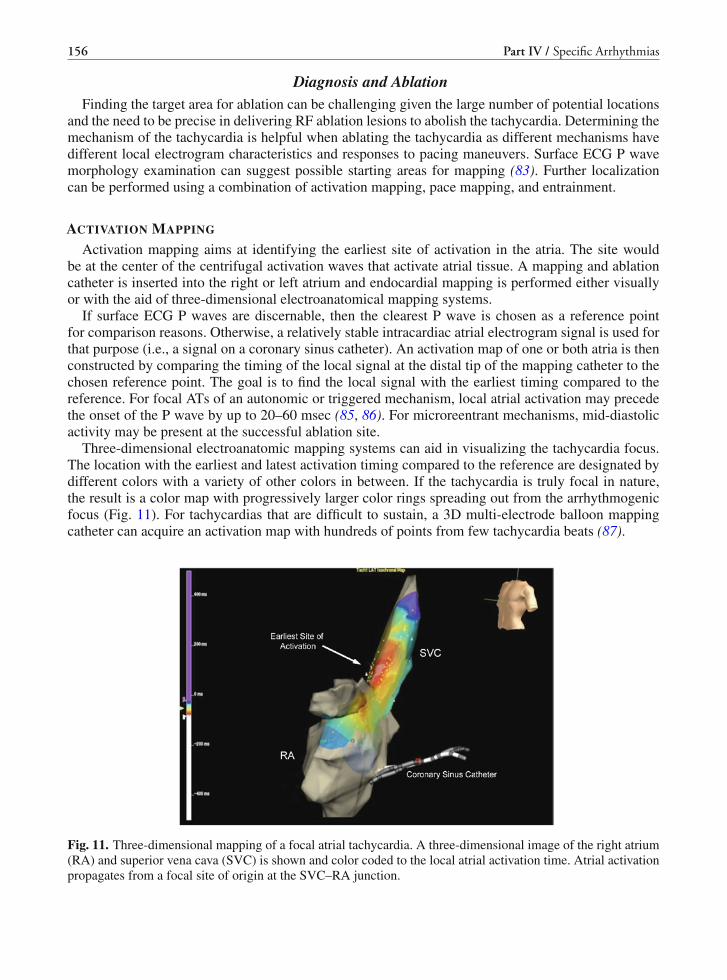
156 Part IV / Specific Arrhythmias
Diagnosis and AblationFinding the target area for ablation can be challenging given the large number of potential locations
and the need to be precise in delivering RF ablation lesions to abolish the tachycardia. Determining themechanism of the tachycardia is helpful when ablating the tachycardia as different mechanisms havedifferent local electrogram characteristics and responses to pacing maneuvers. Surface ECG P wavemorphology examination can suggest possible starting areas for mapping (83). Further localizationcan be performed using a combination of activation mapping, pace mapping, and entrainment.
ACTIVATION MAPPING
Activation mapping aims at identifying the earliest site of activation in the atria. The site wouldbe at the center of the centrifugal activation waves that activate atrial tissue. A mapping and ablationcatheter is inserted into the right or left atrium and endocardial mapping is performed either visuallyor with the aid of three-dimensional electroanatomical mapping systems.
If surface ECG P waves are discernable, then the clearest P wave is chosen as a reference pointfor comparison reasons. Otherwise, a relatively stable intracardiac atrial electrogram signal is used forthat purpose (i.e., a signal on a coronary sinus catheter). An activation map of one or both atria is thenconstructed by comparing the timing of the local signal at the distal tip of the mapping catheter to thechosen reference point. The goal is to find the local signal with the earliest timing compared to thereference. For focal ATs of an autonomic or triggered mechanism, local atrial activation may precedethe onset of the P wave by up to 20–60 msec (85, 86). For microreentrant mechanisms, mid-diastolicactivity may be present at the successful ablation site.
Three-dimensional electroanatomic mapping systems can aid in visualizing the tachycardia focus.The location with the earliest and latest activation timing compared to the reference are designated bydifferent colors with a variety of other colors in between. If the tachycardia is truly focal in nature,the result is a color map with progressively larger color rings spreading out from the arrhythmogenicfocus (Fig. 11). For tachycardias that are difficult to sustain, a 3D multi-electrode balloon mappingcatheter can acquire an activation map with hundreds of points from few tachycardia beats (87).
Fig. 11. Three-dimensional mapping of a focal atrial tachycardia. A three-dimensional image of the right atrium(RA) and superior vena cava (SVC) is shown and color coded to the local atrial activation time. Atrial activationpropagates from a focal site of origin at the SVC–RA junction.

Chapter 7 / Supraventricular Arrhythmias 157
PACE MAPPING
For atrial tachycardias that are difficult to induce during the electrophysiology study, pace mappingis a technique that may aid in locating an arrhythmogenic focus in a small area of potential targetsfor ablation. Pace mapping requires ECG documentation of the tachycardia P wave morphology alongwith the pattern of intracardiac chamber activation. The mapping catheter is moved to various posi-tions within the suspected chamber and pacing is initiated at the lowest output needed for capture ofthe atria. The paced P wave morphology and pattern of chamber activation is then compared to theclinical tachycardia (88). An area with a high level of concordance in surface and intracardiac electro-gram signals is suggestive of proximity to the arrhythmogenic focus. Unlike ventricular arrhythmiaswhere pace mapping utilizes the usually clear QRS signals, P wave morphology is much more diffi-cult to discern. Often, the P wave is superimposed on the T wave preventing attempts at morphologiccomparisons. Rapid pacing to create AV block and separate the P waves from adjoining T waves maybe necessary at times.
ENTRAINMENT MAPPING
Entrainment mapping is used in cases of microreentrant tachycardia to assess whether areas ofmid-diastolic activity are necessary in the reentrant circuit and therefore potentially successful sitesfor ablation. Using the mapping catheter during SVT, sites of early atrial activation and mid dias-tolic activity are located (Fig. 12). Pacing is then performed for a brief period at cycle lengths of20–30 msec shorter than that of the tachycardia itself. If the tachycardia continues at the terminationof pacing then the return cycle length (defined as the time from the last pacing impulse to the first
Fig. 12. Activation mapping of a focal atrial tachycardia. Local mid-diastolic, atrial activity is present on thedistal ablation electrogram (Abl-d). The fractionated signal precedes the onset of the P wave by 95 msec (arrow)(surface leads I, aVF, and V1 are shown with intracardiac electrograms: HRA = right atrium, His = His bundle,CS = coronary sinus, RA = right atrial, Abl = ablation/mapping catheter; p = proximal, m= mid, d = distal,s= stimuli, T = time).

158 Part IV / Specific Arrhythmias
Fig. 13. Entrainment mapping of a focal atrial tachycardia with concealed fusion. Pacing is performed from theablation catheter at the site of mid-diastolic atrial activity (see Fig. 12). Surface P wave morphology and theintracardiac atrial activation sequence are identical to that of the clinical atrial tachycardia (surface leads I, aVF,and V1 are shown with intracardiac electrograms: HRA = right atrium, His = His bundle, s= stimuli, T = time).
recorded local electrical impulse on the ablation catheter) is documented. If the return cycle lengthis equal or close to the tachycardia cycle length, the finding would be suggestive that the tip of themapping catheter is within the reentant circuit. Concealed fusion occurs with the paced P wave mor-phology and intracardiac activation pattern is identical to the clinical tachycardia and signifies a sitewith high success for termination of the tachycardia (Fig. 13) (86).
Catheter AblationOnce a focus for the AT is identified, then ablation can be targeted at that location. Depending on
the location, radiofrequency (RF) energy (with or without cooling) or cryoablation can be used. Inarrhythmias of an automatic or triggered mechanism, the initiation of RF energy causes heating ofthe local tissue, often leading to transient acceleration of the tachycardia with subsequent termination.In cases of a reentrant arrhythmia, slowing of the tachycardia may precede termination. In eithercase, if termination is not achieved within approximately 15 sec, despite adequate energy delivery,then ablation should be halted and re-evaluation of the site with further mapping performed. If ATterminates, then additional lesions may be delivered to the small area of tissue surrounding the target toassure destruction of the arrhythmogenic focus. Thereafter, attempts at re-induction of the tachycardiaare necessary to confirm abolition of the tachycardia.
In recent years, ablation of focal AT has been associated with an overall success rate of greaterthan 80%. Tachycardias with right and left atrial origins have higher ablation success rates comparedto AT’s originating from septal foci (80, 86, 89). Complications related to ablation of tachycardiaare relatively uncommon, occurring in 1–2% of cases (80, 89). These include vascular injury, cardiacperforation, and injury to surrounding intra- and extra-cardiac structures. Atrial tachycardias arisingfrom the posterolateral aspect of the right atrium may result in damage to the phrenic nerve (90). Inthese regions, high output pacing can be performed to assess for diaphragmatic capture, signifying the

Chapter 7 / Supraventricular Arrhythmias 159
location of the phrenic nerve, and varying techniques or different energy sources for ablation can beused to avoid diaphragmatic paralysis (91).
Catheter Ablation of AVRTThe approach for catheter ablation of AVRT is more complicated than AVNRT. While the ablation
site for AVNRT is fairly well defined and limited to the posterior septum, potential ablation sites forAVRT are as variable as the locations of the accessory pathways. Most pathways, however, are leftlateral in location followed by paraseptal and right lateral tracts. In addition, up to 10% of patientswith AVRT may have more than one bypass tract (92–94). Successful ablation in up to 94% of patientswith accessory pathways has been reported in large series of patients with AVRT (92, 95).
Mapping of the accessory pathway location can be performed by determining the earliest antegradeventricular activation during sinus rhythm if pre-excitation is present or by the earliest retrogradeatrial activation during either orthodromic SVT or ventricular pacing. In the case of left-sided path-ways, placement of the coronary sinus catheter should be performed such that the earliest retrogradeatrial activation is “bracketed” by more proximal and distal electrode pairs. For right-sided pathways,placement of a circular catheter around the tricspid annulus can be helpful in localizing the bypasstract. The retrograde atrial activation of septal accessory pathways is best mapped during orthodromicAVRT or with fast enough ventricular pacing to avoid fusion with retrograde atrial activation via theAV node.
Fig. 14. Catheter ablation of an accessory pathway. Right and left anterior oblique fluoroscopic images of thecatheter positions for ablation of a left lateral accessory pathway are demonstrated on the right-hand panel.The intracardiac electrogram recordings at these sites are shown on the left-hand panel (surface leads I, II, III,V1, and V6 are shown with intracardiac electrograms: His = His bundle, CS = coronary sinus, RVA = rightventricular apex, Abl = ablation/mapping catheter; p = proximal, m= mid, d = distal, uni = unipolar. Positionof the coronary sinus (CS), His bundle (His), right ventricular apical (RVA), and ablation catheters are shown onthe fluoroscopic images).

160 Part IV / Specific Arrhythmias
Local electrogram characteristics will vary depending on the method of mapping. Mapping ante-grade conduction of the acessory pathway will locate the ventricular insertion of the pathway and thelocal ventricular electrogram should precede the onset of the surface ECG delta wave by up to 20 msec(Fig. 14) (96). Unipolar recordings are particularly helpful and will show a QS deflection, demonstrat-ing that all ventricular activation is propagating from that point (97). The local bipolar electrogram willshow a continuous signal with a local atrio-ventricular interval of less than or equal to 40 msec (98). Adiscrete accessory pathway potential, when present, also predicts a higher probability of success (99).
In mapping the atrial insertion of an accessory pathway, the onset of earliest atrial activation islocated during either orthodromic SVT or ventricular pacing. The bipolar electrogram will typicallydemonstrate continuous electrical activity (100) with a local ventriculoatrial interval of less than 60msec and a surface QRS onset to local atrial electrogram time of 80 msec (98, 101). Pacing at ventric-ular CLs that result in 2:1 block, or with ventricular premature depolarizations that block in the bypasstract, is sometimes needed to determine which part of the signal is atrial or ventricular in origin. Thepresence of possible accessory pathway potentials is seen in only 30% of successful ablation sites(101).
SUMMARY
The paroxysmal supraventricular tachycardias are a diverse group of arrhythmias with the major-ity being due to either AV nodal reentry or atrio-ventricular reentry. While pharmacologic therapy isstill used for suppression and treatment, especially for the atrial arrhythmias, efficacy and side effectshave limited their application. Radiofrequency catheter ablation has become the treatment of choicefor most symptomatic patients due to the procedure’s high rate of success and infrequent complica-tions. Catheter ablation, however, still requires a diligent approach in determining the diagnosis andmechanism of the arrhythmia during the invasive electrophysiology procedure.
REFERENCES
1. Orejarena LA, Vidaillet HJ, DeStefano F, Nordstrom DL, Vierkant RA, Smith PN, Hayes JJ (1998) Paroxysmalsupraventricular tachycardia in the general population. J Am Coll Cardiol 31:150–157
2. Gonzalez-Torrecilla E, Almendral J, Arenal A, Atienza F, Atea LF, del Castillo S, Fernandez-Aviles F (2009) Com-bined evaluation of bedside clinical variables and the electrocardiogram for the differential diagnosis of paroxysmalatrioventricular reciprocating tachycardias in patients without pre-excitation. J Am Coll Cardiol 53:2353–2358
3. Zimetbaum P, Josephson ME (1998) Evaluation of patients with palpitations. N Engl J Med 338:1369–13734. Wood KA, Drew BJ, Scheinman MM (1997) Frequency of disabling symptoms in supraventricular tachycardia. Am J
Cardiol 79:145–1495. Ferguson JD, DiMarco JP (2003) Contemporary management of paroxysmal supraventricular tachycardia. Circulation
107:1096–10996. Wellens HJ (1996) The value of the ECG in the diagnosis of supraventricular tachycardias. Eur Heart J 17(Suppl
C):10–207. Kumar UN, Rao RK, Scheinman MM (2006) The 12-lead electrocardiogram in supraventricular tachycardia. Cardiol
Clin 24:427–37, ix8. Kalbfleisch SJ, el-Atassi R, Calkins H, Langberg JJ, Morady F (1993) Differentiation of paroxysmal narrow QRS
complex tachycardias using the 12-lead electrocardiogram. J Am Coll Cardiol 21:85–899. Man KC, Brinkman K, Bogun F, Knight B, Bahu M, Weiss R, Goyal R, Harvey M, Daoud EG, Strickberger SA,
Morady F (1996) 2:1 atrioventricular block during atrioventricular node reentrant tachycardia. J Am Coll Cardiol28:1770–1774
10. Blomstrom-Lundqvist C, Scheinman MM, Aliot EM, Alpert JS, Calkins H, Camm AJ, Campbell WB, Haines DE,Kuck KH, Lerman BB, Miller DD, Shaeffer CWJ, Stevenson WG, Tomaselli GF, Antman EM, Smith SCJ, Alpert JS,Faxon DP, Fuster V, Gibbons RJ, Gregoratos G, Hiratzka LF, Hunt SA, Jacobs AK, Russell ROJ, Priori SG, BlancJJ, Budaj A, Burgos EF, Cowie M, Deckers JW, Garcia MA, Klein WW, Lekakis J, Lindahl B, Mazzotta G, MoraisJC, Oto A, Smiseth O, Trappe HJ (2003) ACC/AHA/ESC guidelines for the management of patients with supraven-

Chapter 7 / Supraventricular Arrhythmias 161
tricular arrhythmias–executive summary: a report of the American college of cardiology/American heart associationtask force on practice guidelines and the European society of cardiology committee for practice guidelines (writingcommittee to develop guidelines for the management of patients with supraventricular arrhythmias). Circulation 108:1871–1909
11. Ganz LI, Friedman PL (1995) Supraventricular tachycardia. N Engl J Med 332:162–17312. Shah D, Jais P, Haissaguerre M (2002) Electrophysiological evaluation and ablation of atypical right atrial flutter. Card
Electrophysiol Rev 6:365–37013. Akhtar M, Jazayeri MR, Sra J, Blanck Z, Deshpande S, Dhala A (1993) Atrioventricular nodal reentry. Clinical,
electrophysiological, and therapeutic considerations. Circulation 88:282–29514. Cranefield PF (1977) Action potentials, afterpotentials, and arrhythmias. Circ Res 41:415–42315. Wit AL, Rosen MR (1983) Pathophysiologic mechanisms of cardiac arrhythmias. Am Heart J 106:
798–81116. Akhtar M, Tchou PJ, Jazayeri M (1988) Mechanisms of clinical tachycardias. Am J Cardiol 61:9A–19A; Marchlin-
ski FE, Miller JM (1985) Atrial arrhythmias exacerbated by theophylline. Response to verapamil and evidence fortriggered activity in man. Chest 88:931–934
17. Pappone C, Santinelli V, Rosanio S, Vicedomini G, Nardi S, Pappone A, Tortoriello V, Manguso F, Mazzone P, GullettaS, Oreto G, Alfieri O (2003) Usefulness of invasive electrophysiologic testing to stratify the risk of arrhythmic eventsin asymptomatic patients with Wolff-Parkinson-White pattern: results from a large prospective long-term follow-upstudy. J Am Coll Cardiol 41:239–244
18. Waxman MB, Wald RW, Sharma AD, Huerta F, Cameron DA (1980) Vagal techniques for termination of paroxysmalsupraventricular tachycardia. Am J Cardiol 46:655–664
19. Gonzalez R, Scheinman MM (1981) Treatment of supraventricular arrhythmias with intravenous and oral verapamil.Chest 80:465–470
20. DiMarco JP, Sellers TD, Berne RM, West GA, Belardinelli L (1983) Adenosine: electrophysiologic effects and thera-peutic use for terminating paroxysmal supraventricular tachycardia. Circulation 68:1254–1263
21. Pelleg A, Pennock RS, Kutalek SP (2002) Proarrhythmic effects of adenosine: one decade of clinical data. Am J Ther9:141–147
22. Kaltman JR, Tanel RE, Shah MJ, Vetter VL, Rhodes LA (2006) Induction of atrial fibrillation after the routine use ofadenosine. Pediatr Emerg Care 22:113–115
23. Strickberger SA, Man KC, Daoud EG, Goyal R, Brinkman K, Knight BP, Weiss R, Bahu M, Morady F (1997)Adenosine-induced atrial arrhythmia: a prospective analysis. Ann Intern Med 127:417–422
24. Iwai S, Markowitz SM, Stein KM, Mittal S, Slotwiner DJ, Das MK, Cohen JD, Hao SC, Lerman BB (2002) Responseto adenosine differentiates focal from macroreentrant atrial tachycardia: validation using three-dimensional elec-troanatomic mapping. Circulation 106:2793–2799
25. Gulamhusein S, Ko P, Carruthers SG, Klein GJ (1982) Acceleration of the ventricular response during atrial fibrillationin the Wolff-Parkinson-White syndrome after verapamil. Circulation 65:348–354
26. Jacob AS, Nielsen DH, Gianelly RE (1985) Fatal ventricular fibrillation following verapamil in Wolff-Parkinson-Whitesyndrome with atrial fibrillation. Ann Emerg Med 14:159–160
27. Markowitz SM, Stein KM, Mittal S, Slotwiner DJ, Lerman BB (1999) Differential effects of adenosine on focal andmacroreentrant atrial tachycardia. J Cardiovasc Electrophysiol 10:489–502
28. Wellens HJ, Durrer D (1974) Effect of procaine amide, quinidine, and ajmaline in the Wolff-Parkinson-White syn-drome. Circulation 50:114–120
29. Windle J, Prystowsky EN, Miles WM, Heger JJ (1987) Pharmacokinetic and electrophysiologic interactions of amio-darone and procainamide. Clin Pharmacol Ther 41:603–610
30. Saoudi N, Cosio F, Waldo A, Chen SA, Iesaka Y, Lesh M, Saksena S, Salerno J, Schoels W (2001) Classification ofatrial flutter and regular atrial tachycardia according to electrophysiologic mechanism and anatomic bases: a statementfrom a joint expert group from the Working Group of Arrhythmias of the European society of cardiology and the NorthAmerican society of pacing and electrophysiology. J Cardiovasc Electrophysiol 12:852–866
31. Glatter KA, Dorostkar PC, Yang Y, Lee RJ, Van Hare GF, Keung E, Modin G, Scheinman MM (2001) Electrophysio-logical effects of ibutilide in patients with accessory pathways. Circulation 104:1933–1939
32. Akhtar M, Tchou P, Jazayeri M (1989) Use of calcium channel entry blockers in the treatment of cardiac arrhythmias.Circulation 80:IV31–IV39
33. Gmeiner R, Ng CK (1982) Metoprolol in the treatment and prophylaxis of paroxysmal reentrant supraventriculartachycardia. J Cardiovasc Pharmacol 4:5–13
34. Lindsay BD, Saksena S, Rothbart ST, Herman S, Barr MJ (1987) Long-term efficacy and safety of beta-adrenergicreceptor antagonists for supraventricular tachycardia. Am J Cardiol 60:63D–67D
35. Hellestrand KJ, Nathan AW, Bexton RS, Spurrell RA, Camm AJ (1983) Cardiac electrophysiologic effects of flecainideacetate for paroxysmal reentrant junctional tachycardias. Am J Cardiol 51:770–776

162 Part IV / Specific Arrhythmias
36. Dorian P, Naccarelli GV, Coumel P, Hohnloser SH, Maser MJ (1996) A randomized comparison of flecainide versusverapamil in paroxysmal supraventricular tachycardia. The Flecainide multicenter investigators group. Am J Cardiol77:89A–95A
37. Ward DE, Jones S, Shinebourne EA (1986) Use of flecainide acetate for refractory junctional tachycardias in childrenwith the Wolff-Parkinson-White syndrome. Am J Cardiol 57:787–790
38. Musto B, D’Onofrio A, Cavallaro C, Musto A (1988) Electrophysiological effects and clinical efficacy of propafenonein children with recurrent paroxysmal supraventricular tachycardia. Circulation 78:863–869
39. Musto B, D’Onofrio A, Cavallaro C, Musto A, Greco R (1988) Electrophysiologic effects and clinical efficacy offlecainide in children with recurrent paroxysmal supraventricular tachycardia. Am J Cardiol 62:229–233
40. Kim SS, Lal R, Ruffy R (1986) Treatment of paroxysmal reentrant supraventricular tachycardia with flecainide acetate.Am J Cardiol 58:80–85
41. Echt DS, Liebson PR, Mitchell LB, Peters RW, Obias-Manno D, Barker AH, Arensberg D, Baker A, Friedman L,Greene HL et al (1991) Mortality and morbidity in patients receiving encainide, flecainide, or placebo. The Cardiacarrhythmia suppression trial.N Engl J Med 324:781–788
42. Mitchell LB, Wyse DG, Duff HJ (1987) Electropharmacology of sotalol in patients with Wolff-Parkinson-White syn-drome. Circulation 76:810–818
43. Kunze KP, Schluter M, Kuck KH (1987) Sotalol in patients with Wolff-Parkinson-White syndrome. Circulation75:1050–1057
44. Tendera M, Wnuk-Wojnar AM, Kulakowski P, Malolepszy J, Kozlowski JW, Krzeminska-Pakula M, Szechinski J,Droszcz W, Kawecka-Jaszcz K, Swiatecka G, Ruzyllo W, Graff O (2001) Efficacy and safety of dofetilide in theprevention of symptomatic episodes of paroxysmal supraventricular tachycardia: a 6-month double-blind comparisonwith propafenone and placebo. Am Heart J 142:93–98
45. Kobayashi Y, Atarashi H, Ino T, Kuruma A, Nomura A, Saitoh H, Hayakawa H (1997) Clinical and electrophysiologiceffects of dofetilide in patients with supraventricular tachyarrhythmias. J Cardiovasc Pharmacol 30:367–373
46. Rosenbaum MB, Chiale PA, Ryba D, Elizari MV (1974) Control of tachyarrhythmias associated with Wolff-Parkinson-White syndrome by amiodarone hydrochloride. Am J Cardiol 34:215–223
47. Wellens HJ, Lie KI, Bar FW, Wesdorp JC, Dohmen HJ, Duren DR, Durrer D (1976) Effect of amiodarone in theWolff-Parkinson-White syndrome. Am J Cardiol 38:189–194
48. Feld GK, Nademanee K, Weiss J, Stevenson W, Singh BN (1984) Electrophysiologic basis for the suppression byamiodarone of orthodromic supraventricular tachycardias complicating pre-excitation syndromes. J Am Coll Cardiol3:1298–1307
49. Alboni P, Tomasi C, Menozzi C, Bottoni N, Paparella N, Fuca G, Brignole M, Cappato R (2001) Efficacy and safetyof out-of-hospital self-administered single-dose oral drug treatment in the management of infrequent, well-toleratedparoxysmal supraventricular tachycardia. J Am Coll Cardiol 37:548–553
50. Rae AP (1998) Placebo-controlled evaluations of propafenone for atrial tachyarrhythmias. Am J Cardiol 82:59 N–65 N51. Musto B, Cavallaro C, Musto A, D′Onofrio A, Belli A, De Vincentis L (1992) Flecainide single oral dose
for management of paroxysmal supraventricular tachycardia in children and young adults. Am Heart J 124:110–115
52. Rose JS, Bhandari A, Rahimtoola SH, Wu D (1986) Effective termination of reentrant supraventricular tachycardia bysingle dose oral combination therapy with pindolol and verapamil. Am Heart J 112:759–765
53. Yeh SJ, Lin FC, Chou YY, Hung JS, Wu D (1985) Termination of paroxysmal supraventricular tachycardia with asingle oral dose of diltiazem and propranolol. Circulation 71:104–109
54. Cossu SF, Rothman SA, Chmielewski IL, Hsia HH, Vogel RL, Miller JM, Buxton AE (1997) The effects ofisoproterenol on the cardiac conduction system: site-specific dose dependence. J Cardiovasc Electrophysiol 8:847–853
55. Chen SA, Chiang CE, Yang CJ, Cheng CC, Wu TJ, Wang SP, Chiang BN, Chang MS (1994) Sustained atrial tachycar-dia in adult patients. Electrophysiological characteristics, pharmacological response, possible mechanisms, and effectsof radiofrequency ablation. Circulation 90:1262–1278
56. Wu D, Denes P, Dhingra R, Wyndham C, Rosen KM (1975) Determinants of fast- and slow-pathway conduction inpatients with dual atrioventricular nodal pathways. Circ Res 36:782–790
57. Patruno N, Critelli G, Pulignano G, Urbani P, Villanti P, Reale A (1989) [Asymptomatic pre-excitation. Identificationof potential risk using transesophageal pacing]. Cardiologia 34:777–781
58. Sung RJ, Waxman HL, Saksena S, Juma Z (1981) Sequence of retrograde atrial activation in patients with dual atri-oventricular nodal pathways. Circulation 64:1059–1067
59. Hirao K, Otomo K, Wang X, Beckman KJ, McClelland JH, Widman L, Gonzalez MD, Arruda M, Nakagawa H,Lazzara R, Jackman WM (1996) Para-Hisian pacing. A new method for differentiating retrograde conduction over anaccessory AV pathway from conduction over the AV node. Circulation 94:1027–1035
60. Strasberg B, Swiryn S, Bauernfeind R, Palileo E, Scagliotti D, Duffy CE, Rosen KM (1981) Retrograde dual atrioven-tricular nodal pathways. Am J Cardiol 48:639–646

Chapter 7 / Supraventricular Arrhythmias 163
61. Benditt DG, Pritchett EL, Smith WM, Gallagher JJ (1979) Ventriculoatrial intervals: diagnostic use in paroxysmalsupraventricular tachycardia. Ann Intern Med 91:161–166
62. Coumel P, Attuel P (1974) Reciprocating tachycardia in overt and latent preexcitation. Influence of functional bundlebranch block on the rate of the tachycardia. Eur J Cardiol 1:423–436
63. Sellers TDJ, Gallagher JJ, Cope GD, Tonkin AM, Wallace AG (1976) Retrograde atrial preexcitation following pre-mature ventricular beats during reciprocating tachycardia in the Wolff-Parkinson-White syndrome. Eur J Cardiol 4:283–294
64. Knight BP, Ebinger M, Oral H, Kim MH, Sticherling C, Pelosi F, Michaud GF, Strickberger SA, Morady F (2000)Diagnostic value of tachycardia features and pacing maneuvers during paroxysmal supraventricular tachycardia. J AmColl Cardiol 36:574–582
65. Michaud GF, Tada H, Chough S, Baker R, Wasmer K, Sticherling C, Oral H, Pelosi FJ, Knight BP, Strickberger SA,Morady F (2001) Differentiation of atypical atrioventricular node re-entrant tachycardia from orthodromic recipro-cating tachycardia using a septal accessory pathway by the response to ventricular pacing. J Am Coll Cardiol 38:1163–1167
66. McGuire MA, Lau KC, Johnson DC, Richards DA, Uther JB, Ross DL (1991) Patients with two types of atrioventric-ular junctional (AV nodal) reentrant tachycardia. Evidence that a common pathway of nodal tissue is not present abovethe reentrant circuit. Circulation 83:1232–1246
67. Janse MJ, Anderson RH, McGuire MA, Ho SY (1993) “AV nodal” reentry: part I: “AV nodal” reentry revisited.J Cardiovasc Electrophysiol 4:561–572
68. McGuire MA, Bourke JP, Robotin MC, Johnson DC, Meldrum-Hanna W, Nunn GR, Uther JB, Ross DL (1993) Highresolution mapping of Koch’s triangle using sixty electrodes in humans with atrioventricular junctional (AV nodal)reentrant tachycardia. Circulation 88:2315–2328
69. Kalbfleisch SJ, Strickberger SA, Williamson B, Vorperian VR, Man C, Hummel JD, Langberg JJ, Morady F (1994)Randomized comparison of anatomic and electrogram mapping approaches to ablation of the slow pathway of atri-oventricular node reentrant tachycardia. J Am Coll Cardiol 23:716–723
70. Lee MA, Morady F, Kadish A, Schamp DJ, Chin MC, Scheinman MM, Griffin JC, Lesh MD, Pederson D, Gold-berger J et al (1991) Catheter modification of the atrioventricular junction with radiofrequency energy for control ofatrioventricular nodal reentry tachycardia. Circulation 83:827–835
71. Jackman WM, Beckman KJ, McClelland JH, Wang X, Friday KJ, Roman CA, Moulton KP, Twidale N, Hazlitt HA,Prior MI et al (1992) Treatment of supraventricular tachycardia due to atrioventricular nodal reentry, by radiofrequencycatheter ablation of slow-pathway conduction. N Engl J Med 327:313–318
72. Yamabe H, Okumura K, Tsuchiya T, Tabuchi T, Iwasa A, Yasue H (1998) Slow potential-guided radiofrequencycatheter ablation in atrioventricular nodal reentrant tachycardia: characteristics of the potential associated with suc-cessful ablation. Pacing Clin Electrophysiol 21:2631–2640
73. Jentzer JH, Goyal R, Williamson BD, Man KC, Niebauer M, Daoud E, Strickberger SA, Hummel JD, Morady F (1994)Analysis of junctional ectopy during radiofrequency ablation of the slow pathway in patients with atrioventricular nodalreentrant tachycardia. Circulation 90:2820–2826
74. Lipscomb KJ, Zaidi AM, Fitzpatrick AP, Lefroy D (2001) Slow pathway modification for atrioventricular nodere-entrant tachycardia: fast junctional tachycardia predicts adverse prognosis. Heart 85:44–47
75. Lindsay BD, Chung MK, Gamache MC, Luke RA, Schechtman KB, Osborn JL, Cain ME (1993) Therapeutic endpoints for the treatment of atrioventricular node reentrant tachycardia by catheter-guided radiofrequency current. J AmColl Cardiol 22:733–740
76. Strickberger SA, Kalbfleisch SJ, Williamson B, Man KC, Vorperian V, Hummel JD, Langberg JJ, Morady F (1993)Radiofrequency catheter ablation of atypical atrioventricular nodal reentrant tachycardia. J Cardiovasc Electrophysiol4:526–532
77. Clague JR, Dagres N, Kottkamp H, Breithardt G, Borggrefe M (2001) Targeting the slow pathway for atrioventric-ular nodal reentrant tachycardia: initial results and long-term follow-up in 379 consecutive patients. Eur Heart J 22:82–88
78. Haissaguerre M, Gaita F, Fischer B, Commenges D, Montserrat P, d′Ivernois C, Lemetayer P, Warin JF (1992) Elimina-tion of atrioventricular nodal reentrant tachycardia using discrete slow potentials to guide application of radiofrequencyenergy. Circulation 85:2162–2175
79. Calkins H, Yong P, Miller JM, Olshansky B, Carlson M, Saul JP, Huang SK, Liem LB, Klein LS, Moser SA, BlochDA, Gillette P, Prystowsky E (1999) Catheter ablation of accessory pathways, atrioventricular nodal reentrant tachy-cardia, and the atrioventricular junction: final results of a prospective, multicenter clinical trial. The Atakr multicenterinvestigators group. Circulation 99:262–270
80. Scheinman MM, Huang S (2000) The 1998 NASPE prospective catheter ablation registry. Pacing Clin Electrophysiol23:1020–1028

164 Part IV / Specific Arrhythmias
81. Hummel JD, Strickberger SA, Williamson BD, Man KC, Daoud E, Niebauer M, Bakr O, Morady F (1995) Effect ofresidual slow pathway function on the time course of recurrences of atrioventricular nodal reentrant tachycardia afterradiofrequency ablation of the slow pathway. Am J Cardiol 75:628–630
82. Kistler PM, Roberts-Thomson KC, Haqqani HM, Fynn SP, Singarayar S, Vohra JK, Morton JB, Sparks PB, KalmanJM (2006) P-wave morphology in focal atrial tachycardia: development of an algorithm to predict the anatomic site oforigin. J Am Coll Cardiol 48:1010–1017
83. Tang CW, Scheinman MM, Van Hare GF, Epstein LM, Fitzpatrick AP, Lee RJ, Lesh MD (1995) Use of P waveconfiguration during atrial tachycardia to predict site of origin. J Am Coll Cardiol 26:1315–1324
84. Porter MJ, Morton JB, Denman R, Lin AC, Tierney S, Santucci PA, Cai JJ, Madsen N, Wilber DJ (2004) Influence ofage and gender on the mechanism of supraventricular tachycardia. Heart Rhythm 1:393–396
85. Walsh EP, Saul JP, Hulse JE, Rhodes LA, Hordof AJ, Mayer JE, Lock JE (1992) Transcatheter ablation of ectopic atrialtachycardia in young patients using radiofrequency current. Circulation 86:1138–1146
86. Lesh MD, Van Hare GF, Epstein LM, Fitzpatrick AP, Scheinman MM, Lee RJ, Kwasman MA, Grogin HR, Grif-fin JC (1994) Radiofrequency catheter ablation of atrial arrhythmias. Results and mechanisms. Circulation 89:1074–1089
87. Schmitt C, Zrenner B, Schneider M, Karch M, Ndrepepa G, Deisenhofer I, Weyerbrock S, Schreieck J, Schomig A(1999) Clinical experience with a novel multielectrode basket catheter in right atrial tachycardias. Circulation 99:2414–2422
88. Tracy CM, Swartz JF, Fletcher RD, Hoops HG, Solomon AJ, Karasik PE, Mukherjee D (1993) Radiofrequency catheterablation of ectopic atrial tachycardia using paced activation sequence mapping. J Am Coll Cardiol 21:910–917
89. O’Hara GE, Philippon F, Champagne J, Blier L, Molin F, Cote JM, Nault I, Sarrazin JF, Gilbert M (2007) Catheterablation for cardiac arrhythmias: a 14-year experience with 5330 consecutive patients at the Quebec heart institute,Laval Hospital. Can J Cardiol 23(Suppl B):67B–70B
90. Swallow EB, Dayer MJ, Oldfield WL, Moxham J, Polkey MI (2006) Right hemi-diaphragm paralysis following cardiacradiofrequency ablation. Respir Med 100:1657–1659
91. Lee JC, Steven D, Roberts-Thomson KC, Raymond JM, Stevenson WG, Tedrow UB (2009) Atrial tachycardias adja-cent to the phrenic nerve: recognition, potential problems, and solutions. Heart Rhythm 6:1186–1191; Bastani H,Insulander P, Schwieler J, Tabrizi F, Braunschweig F, Kenneback G, Drca N, Sadigh B, Jensen-Urstad M (2009) Safetyand efficacy of cryoablation of atrial tachycardia with high risk of ablation-related injuries. Europace 11:625–629
92. Calkins H, Langberg J, Sousa J, el-Atassi R, Leon A, Kou W, Kalbfleisch S, Morady F (1992) Radiofrequencycatheter ablation of accessory atrioventricular connections in 250 patients. Abbreviated therapeutic approach to Wolff-Parkinson-White syndrome. Circulation 85:1337–1346
93. Lesh MD, Van Hare GF, Schamp DJ, Chien W, Lee MA, Griffin JC, Langberg JJ, Cohen TJ, Lurie KG, Scheinman MM(1992) Curative percutaneous catheter ablation using radiofrequency energy for accessory pathways in all locations:results in 100 consecutive patients. J Am Coll Cardiol 19:1303–1309
94. Weng KP, Wolff GS, Young ML (2003) Multiple accessory pathways in pediatric patients with Wolff-Parkinson-Whitesyndrome. Am J Cardiol 91:1178–1183
95. Chen YJ, Chen SA, Tai CT, Chiang CE, Lee SH, Chiou CW, Ueng KC, Wen ZC, Yu WC, Huang JL, Feng AN, ChangMS (1997) Long-term results of radiofrequency catheter ablation in patients with Wolff-Parkinson-White syndrome.Zhonghua Yi Xue Za Zhi (Taipei) 59:78–87
96. Lin JL, Schie JT, Tseng CD, Chen WJ, Cheng TF, Tsou SS, Chen JJ, Tseng YZ, Lien WP (1995) Value of localelectrogram characteristics predicting successful catheter ablation of left-versus right-sided accessory atrioventricularpathways by radiofrequency current. Cardiology 86:135–142
97. Grimm W, Miller J, Josephson ME (1994) Successful and unsuccessful sites of radiofrequency catheter ablation ofaccessory atrioventricular connections. Am Heart J 128:77–87
98. Silka MJ, Kron J, Halperin BD, Griffith K, Crandall B, Oliver RP, Walance CG, McAnulty JH (1992) Analysis of localelectrogram characteristics correlated with successful radiofrequency catheter ablation of accessory atrioventricularpathways. Pacing Clin Electrophysiol 15:1000–1007
99. Calkins H, Kim YN, Schmaltz S, Sousa J, el-Atassi R, Leon A, Kadish A, Langberg JJ, Morady F (1992) Electrogramcriteria for identification of appropriate target sites for radiofrequency catheter ablation of accessory atrioventricularconnections. Circulation 85:565–573
100. Haissaguerre M, Fischer B, Warin JF, Dartigues JF, Lemetayer P, Egloff P (1992) Electrogram patterns predictive ofsuccessful radiofrequency catheter ablation of accessory pathways. Pacing Clin Electrophysiol 15:2138–2145
101. Swartz JF, Tracy CM, Fletcher RD (1993) Radiofrequency endocardial catheter ablation of accessory atrioventricularpathway atrial insertion sites. Circulation 87:487–499

8 Pharmacologic Management of AtrialFibrillation and Flutter
Deepak Saluja, Kathleen Hickey,and James A. ReiffelCONTENTS
INTRODUCTION
PATTERNS OF AFGOALS OF TREATMENT
SPECIFIC DRUGS FOR MAINTENANCE OF OR CONVERSION
TO SINUS RHYTHM
SPECIFIC DRUGS FOR THE CONTROL OF VENTRICULAR RATE IN AFDRUG SELECTION IN SPECIFIC POPULATIONS
ANTICOAGULATION
ATRIAL FLUTTER
REFERENCES
Abstract
Atrial fibrillation is the most commonly encountered arrhythmia requiring therapy and is associatedwith significant morbidity, a reduced quality of life, an increased risk for mortality, a significant likelihoodof hospitalization, an increased risk for stroke and systemic embolism, and significant costs. Both ratecontrol and rhythm control have been shown to improve quality of life. Neither as a strategy has beenproven to reduce mortality versus the other; however, in some series the attainment and maintenance ofsinus rhythm has had superior outcomes if risks from therapy are not encountered (especially those ofamiodarone). In patients with markers of an increased risk for stroke, anticoagulation with warfarin isessential. In all patients, rate control is also necessary so as to reduce both symptoms and the risk for atachycardia-induced cardiomyopathy. The choice of rhythm control strategy must be individualized basedon patient characteristics. This chapter will review the considerations necessary for choosing appropriatepharmacologic therapies as well as the therapeutic agents themselves.
Key Words: Atrial fibrillation; Atrial flutter; Antiarrhythmic drugs; Rate control; Rhythm control;Anticoagulation.
INTRODUCTION
Atrial fibrillation (AF) is the most commonly encountered sustained arrhythmia in clinical practice,affecting an estimated 2.3−5.1 million people in the United States, with the prevalence expected totriple by 2050 as the population ages (1). AF is associated with physical symptoms, a shortened lifeexpectancy, and reduced quality of life (QoL).
From: Contemporary Cardiology: Management of Cardiac ArrhythmiasEdited by: Gan-Xin Yan, Peter R. Kowey, DOI 10.1007/978-1-60761-161-5_8
C© Springer Science+Business Media, LLC 2011
165

166 Part IV / Specific Arrhythmias
The impact AF can have on a patient’s health may take several forms. AF can cause palpita-tions, angina, dyspnea, lightheadedness, chronic fatigue, and/or impaired exercise tolerance. Even inthe absence of such symptoms, AF may lead to several potentially significant associated conditions,including tachycardia-induced cardiomyopathy and thromboembolism.
Quality of life is adversely affected by the presence of AF in most populations, either due to thepresence of symptoms, side effects of medications, or due to lifestyle disruptions associated withanticoagulation (2). All-cause mortality in patients with AF is 1.5−1.9 fold higher than those withoutAF, regardless of the presence of symptoms (3). This may be due to the effects of AF itself, toxicitiesassociated with treatment, or comorbidities such as hypertension, heart failure, and valvular diseasemore commonly seen in AF patients.
While antiarrhythmic drug (AAD) therapy can mitigate both symptoms and decreased QoL by main-taining sinus rhythm (SR), clinical trials have thus far failed to conclusively demonstrate that the pur-suit of SR with currently available AADs decreases mortality (4–7). Further analysis of these trialshas suggested the absence of AF may be associated with decreased mortality in those who can achieveit without significant proarrhythmia or toxicity (8). Although this may become achievable with thedevelopment of newer agents such as dronedarone, which was recently shown in the ATHENA trialto be associated with a reduction in a combined endpoint of cardiovascular hospitalization and deathin a high-risk AF population (9), given the currently available data, the main focus of drug treatmentof AF at present remains improving symptoms and QoL in a way that minimizes the side effects andrisks from therapy. While rate control and anticoagulation to reduce the risk of thromboembolismremain vital components in achieving this goal, it must be emphasized that many patients will nothave adequate relief of symptoms until a normal rhythm is established, even after controlling the ven-tricular rate. Rhythm control, therefore, remains an important and commonly employed (10) part ofthe physician’s armamentarium in treating AF in selected cases.
PATTERNS OF AF
Characterizing the pattern of AF is critical in selecting the appropriate rate or rhythm control treat-ment strategy for an individual patient (Table 1). Treatment algorithms in the 2006 ACC/AHA/ESCGuidelines for the Treatment of Patients with Atrial Fibrillation are based on this division (11). Whileseveral systems have been proposed to classify AF based on various characteristics, the system that ismost practical for clinical use divides AF into groups based on duration of AF episodes and therapeutichistory.
Table 1Classification of Patterns of AF
Patterns of AF
ParoxysmalEpisodes of AF last less than 1 weekTerminates spontaneously, without electrical or pharmacological cardioversion
PersistentEpisodes of AF last longer than 1 week orElectrical or pharmacologic cardioversion is used to terminate AF
PermanentA strategy of rhythm control has been abandoned or never tried andAF is continually present

Chapter 8 / Pharmacologic Management of Atrial Fibrillation and Flutter 167
AF is termed paroxysmal when it terminates without drug or electrical cardioversion therapy andrecurs within 1 week; persistent when self-terminating episodes last longer than 1 week or whentherapy is delivered to terminate the rhythm; and permanent when a strategy of rhythm control hasbeen either abandoned or never tried and the patient remains in AF indefinitely. The paroxysmal andpersistent categories are not mutually exclusive, as both may be seen in a particular individual atdifferent times. The classification of a patient who presents with AF for the first time presents a specialsituation, since the history and clinical decision making required for classification under this systemhas not yet been established. In this case, some time may be necessary to assess the patient’s likelihoodfor spontaneous termination, recurrence, and response to any treatments delivered. In all categories,it is assumed that the duration of any episode of AF is > 30 sec, and that AF is not due to reversiblecauses (hyperthyroidism, pneumonia, pericarditis, etc.) (11).
Paroxysmal AFBy definition, paroxysmal AF is self-terminating and limited in duration. The choice of therapy is
therefore dependent on the presence and severity of symptoms during episodes of AF. Asymptomaticpatients whose rates are controlled off of medications may not require AAD therapy. Patients withrapid ventricular responses require rate control, which may be sufficient to control symptoms in certaincases. Rhythm monitoring devices such as 24-h Holter monitors can be valuable tools to ensure thatrates are reasonable not only at rest but also during daily activities. Rhythm control strategies aremost often reserved for those patients who have intolerable symptoms during AF not alleviated by ratecontrolling medications.
Persistent AFPatients with persistent AF have either self-terminating episodes of AF that last longer than 1 week
or episodes in which therapy is delivered for rhythm conversion. Patients in this category occasionallyhave more significant degrees of abnormalities in left ventricular ejection fraction, left atrial size, andleft atrial contractile function than paroxysmal patients; abnormalities that may be reversible with therestoration of sinus rhythm (12). For those patients who have disabling symptoms that are not allevi-ated by rate control, a rhythm control strategy is indicated. Electrical or pharmalcologic cardioversionmay be used to terminate events. For patients with infrequent events, as-needed cardioversion withoutfurther antiarrhythmic therapy may suffice. Pharmacologic cardioversion can be achieved with eithera single dose of an AAD at the onset of episodes (the so-called “pill-in-the-pocket” approach) (13),or daily dosing of an AAD over a limited period of time. Alternatively, for patients in whom intermit-tent therapy is inadequate or inconvenient, longer term daily maintenance antiarrhythmic drug (AAD)therapy may be used. For those patients who fail AAD therapy or for those who are intolerant of orunwilling to take them, catheter ablation therapy is an efficacious method of rhythm control in theparoxysmal and persistent AF populations whose availability is increasing (14). As with PAF, anti-coagulation of patients at “high risk” for thromboembolism with warfarin is employed. It should becontinued even if sinus rhythm is restored.
Permanent AFBy definition, a patient with permanent AF is one in whom a rhythm control strategy has been aban-
doned. Pharmacologic therapy in these patients therefore consists of rate control and, in “high-risk”patients, anticoagulation. As recent advances in catheter ablation techniques expand the populationof patients in whom maintenance of sinus rhythm can be achieved to include certain patients withlong-term AF (15) as well as left ventricular dysfunction (16), it may become necessary to reconsider

168 Part IV / Specific Arrhythmias
the possibility of achieving sinus rhythm in the patient with permanent AF. In many of these patients,adequate rhythm control may require adjunctive AAD therapy in addition to ablation.
GOALS OF TREATMENT
Rate Control vs. Rhythm ControlThe goal in the treatment of pharmacological treatment of AF is, as in any other disease, to improve
the quality and, if possible, the quantity of life while minimizing any potential toxic effects of ther-apy. Other therapeutic goals specific to the treatment of AF include preventing or reversing structuralchanges caused by the persistence of AF and the prevention of thromboembolism.
In general, the approach to AF therapy has been divided into two possible strategies: (1) controllingthe ventricular rate during AF episodes (rate control) and (2) preventing AF from recurring (rhythmcontrol). Although this division is useful conceptually, it is somewhat artificial in clinical practice,since a strategy of rhythm control does not assure that achievement and maintenance of SR occur,and a strategy of rate control in PAF patients does not define the overall burden of AF. Nevertheless,treatment decisions are based on such factors as the severity and type of symptoms, the presence ofother comorbidities such as atherosclerotic disease and ventricular hypertrophy, the functionality ofthe AV node, the toxicities of the treatments under consideration, and the preferences of the patient.In recent years, several trials have been done in order to establish an evidence basis to guide decisionmaking (4–7, 17, 18). Below is a discussion of the relative efficacy of these two treatment strategies inachieving selected endpoints.
MortalityAs noted above, AF is associated with a significant increase in all-cause mortality compared to
patients with SR (3). It was reasonably expected, therefore, that if this association was causal, themaintenance of SR should lead to reduced mortality. Maintaining SR would then become an endpointin its own right, without consideration to the presence or severity of symptoms. Several trials havebeen completed in recent years addressing this hypothesis (4–7, 17, 18).
The largest such trial completed to date is the AFFIRM trial, a study of 4060 patients aged 65 orolder with AF and other risk factors for stroke or death randomized to either a rate control or rhythmcontrol strategy. In the rate control arm, patients were treated with a combination of β (beta)-blockers,calcium channel blockers, and digoxin. Patients in the rhythm control arm were treated generally withClass III AADs (largely amiodarone). Overall mortality (the primary endpoint) was not statisticallydifferent between the two groups. There was, in fact, a trend toward increased mortality in the rhythmcontrol arm (7).
Other, smaller, trials have found similar results. The RACE trial compared rate and rhythm controlstrategies in 522 patients with persistent AF and mild-to-moderate heart failure. The rates of achieve-ment of a combined endpoint that included mortality, heart failure hospitalization, pacemaker implan-tation, hemorrhage, thrombotic complications, and severe adverse events were no different betweenthe two groups (6). AF-CHF, the first trial to compare rhythm and rate control strategies in heartfailure patients, found no difference in total mortality, with a higher rate of hospitalizations in therhythm control group (17). Other smaller randomized trials of treatment strategies in AF includingSTAF (4), HOT-CAFÉ (5), and PIAF (18) similarly found no differences in mortality between groups(although none were adequately powered to do so). These findings are also in keeping with the resultsobtained from subanalyses of AAD therapy in other populations, including DIAMOND-AF (19) andCHF-STAT (20). A metaanalysis of available rate vs. rhythm control therapy trials came to the same

Chapter 8 / Pharmacologic Management of Atrial Fibrillation and Flutter 169
conclusion and further found that a rhythm control strategy was associated with higher health-careutilization and costs (21).
Several features of these trials bear noting. First, they were designed to compare different treatmentstrategies, as opposed to different rhythms. This distinction is necessary because many patients withAF in whom rate control is pursued may continue to have periods of SR, while those seeking rhythmcontrol may be in AF a significant proportion of time despite AAD or ablative therapy. Second, therewas a high overall degree of crossover between groups (14.9% from the rate control group and 37.5%in the rhythm control group at 5 years in the AFFIRM trial), such that in intention-to-treat analyses, afair number of patients were analyzed as belonging to a treatment assignment that they did not remainin during the study (7).
Although an orthodox analysis of the randomized data described consistently fails to demonstrate asurvival advantage for a strategy of rhythm control, there are data that suggest that when sinus rhythmcan be maintained and the toxicities of AAD therapy avoided, patients do significantly better than whenleft in AF. An on-treatment reanalysis of the AFFIRM data that classified patients into subgroups basedon which treatment they actually received, as opposed to which treatment they were randomized to,found that the presence of SR was associated with a lower risk of mortality, and AAD therapy wasassociated with increased mortality after adjustment for the presence of SR (8). This suggests thatbeneficial effects of SR may have been cancelled out by a harmful effect of the drugs used to achieveit (again, largely amiodarone, which was associated with an increase in non-cardiovascular mortality).Other data from the DIAMOND-AF (19) and the CHF-STAT (20) found a similar positive survivalbenefit for the presence of SR, although similar analyses in some other drug trials have not duplicatedthese results (4–6, 18).
An additional point to consider is that patients enrolled in the above-mentioned rate vs. rhythmcontrol trials had to be willing to be randomized to either strategy. Patients who were already ratecontrolled and still symptomatic were highly unlikely to enroll in such trials; hence, such trials shouldnot be taken to indicate that rate control is a reasonable strategy for all AF patients. Similarly, thesetrials enrolled patients at increased risk for thromboembolism or death. Hence, their results should notbe generalized to a lower risk population for whom AAD therapy may be less problematic.
Taken together, the above data suggest that an a priori strategy of rhythm control with currentlyavailable AADs should not be undertaken with the expectation of a survival advantage. It is importantto emphasize, however, that given the lack of clear increased mortality with rhythm control agents,these data do not suggest that a strategy of rhythm control be withheld from specific patients who haveintolerable symptoms in AF in the presence of a controlled rate. It is possible that newer AADs withmore favorable safety profiles will deliver the benefits of SR without the toxicities of current therapies,as was suggested by the ATHENA data (9).
Quality of Life and Exercise ToleranceIt is known that AF can adversely effect patients’ QoL, exercise tolerance, and functional status
in ways unrelated to objective measures of its severity (22), and treatment of AF can improve theseendpoints compared to leaving AF untreated (7, 18).
In randomized trials, treatment with a strategy of rhythm control has not conclusively been found todeliver QoL improvements superior to those attained by a strategy of rate control (23–25). Most studieshave found, however, that those patients that are able to achieve SR do appear to have improvementsin QoL that exceed those in who remain in AF (24, 26). An important exception is the AFFIRM trial,in which QoL was comparable whether patients were in sinus rhythm or in AF (25). One explanationfor this discrepancy may be the relatively low number of highly symptomatic patients enrolled in theolder AFFIRM population.

170 Part IV / Specific Arrhythmias
Rhythm control may be superior to rate control in improving exercise tolerance. In several trials,exercise capacity improved more in patients treated with rhythm control strategies than with a ratecontrol strategy (5, 18, 26, 27).
Ventricular and Atrial Structure and FunctionAF is associated with changes in the electrical, contractile, and structural functions of the heart. In
the atria, structural changes are thought to play a role in the persistence of AF (28) and may explainwhy SR is more difficult to maintain after cardioversion in patients with a longer duration of precedingAF (29).
Maintenance of SR has been associated with benefits in chamber remodeling in patients with andwithout clinical heart failure at baseline in some trials (12, 30, 31). In the RACE, SR was associatedwith improvements in LV function and reduction in atrial sizes in patients with mild-to-moderate heartfailure. Similar improvements were not seen in patients whom the ventricular rate was controlled,although it did prevent deterioration in LV function (30). In a small study of patients with AF andheart failure that underwent catheter ablation, patients who achieved SR had reductions in LA and LVsizes and increases in ejection fraction, while those that remained in AF showed none of these changes(31). Similar changes were noted in a larger study of patients undergoing catheter ablation who hadrelatively preserved baseline LV function (12). Changes in electrophysiological parameters are alsoseen with the achievement of SR. Patients who maintain SR 1 week after cardioversion have increasedatrial refractory periods and decreased sinus node recovery times and P-wave durations compared toimmediately after cardioversion (32).
Prevention of ThromboembolismAF can be associated with a substantially increased rate of thromboembolic events that is reduced
with the use of oral anticoagulants (33, 34). Warfarin therapy, however, can be inconvenient (2), carriesits own risks, and does not completely eliminate thromboembolism (35). In years past, a strategy ofrhythm control was often used with the anticipated goal of discontinuing oral anticoagulant therapy.Data from both the RACE and AFFIRM trials showed that the rates of thromboembolism were higherin rhythm control patients than in rate control patients, illustrating that SR by itself does not eliminateembolic risk in patients with risk markers associated with their prior AF (6, 7). Among the rhythmcontrol patients with thromboembolism, most had stopped taking warfarin. Important to note is thatmost of these patients were in SR at the time of their event.
Several observations may explain the above findings. First, restoring electrical normality to a fib-rillating atrium does not immediately restore mechanical normality (36), and the persistence of atrialdysfunction may result in a continued risk of stroke after SR is restored. Second, recurrences of AF canbe asymptomatic and may go unnoticed unless rhythm monitoring is continuous (37). Third, AF mayeither cause or be a marker of biochemical alterations at the endothelial and intraatrial level that mayincrease stroke risk independent of mechanical considerations and that may persist after SR has beenrestored (38). That is, AF and clot propensity may both be downstream effects of atrial dysfunctionand that AF itself may not be the (sole) causative factor of increased thromboembolic risk.
The above suggests that stroke risk persists in “high-risk” patients with AF even after SR has beenrestored. As a general rule, patients in AF with indications for anticoagulation (see below) should becontinued on it even after rhythm conversion. Pursuing rhythm control in an AF patient with strokerisk factors has not yet been shown to reduce their risk of stroke and should not be undertaken for thispurpose alone.

Chapter 8 / Pharmacologic Management of Atrial Fibrillation and Flutter 171
SPECIFIC DRUGS FOR MAINTENANCE OF OR CONVERSION TO SINUSRHYTHM
Antiarrhythmic drugs are commonly divided into classes based on the Vaughan-Williams classifi-cation system, which divides drugs based on their major mechanism of action (Table 2). This sectionwill review the role of these drugs in the conversion to and maintenance of sinus rhythm in AF.
Table 2The Vaughan-Williams Antiarrhythmic Drug Classification
Vaughan-Williams classification
Na+ channel β-receptor1 K+ channel Ca+ channel
Ia Ib Ic II III IVProcainamide Lidocaine Propafenone Metoprolol Amiodarone DiltiazemQuinidine Mexilitine Flecainide Propranolol Sotalol VerapamilDisopyrimide Esmolol
Cavedilol2
1Among other β-receptor antagonists2Carvedilol also exhibits α-receptor blocking properties
Class IaClass Ia agents include quinidine, disopyrimide, and procainamide. They are sodium channel block-
ing agents that prolong the action potential in a use dependent fashion, although drugs in this class alsohave other important ion channel and autonomic effects, such as Ikr inhibition in the case of all threeof these agents, Ito inhibition in the case of quinidine, and vagolytic effects in the case of disopyrimideand less so, quinidine. They have been used for the acute conversion of AF as well as maintenanceof SR after conversion. The potassium channel inhibitory effects may be associated with Torsades dePointes type (TdP) proarrhythmia. This risk may be highest with quinidine.
QUINIDINE
Quinidine is among the first used and best studied of the AADs in AF. Oral quinidine has found toincrease the odds of SR maintenance over placebo by about twofold in a recent systematic review of 44antiarrhythmic trials (39). It can also be used for the conversion of AF to SR. The PAFAC trial showedthat the combination of quinidine plus verapamil maintained sinus rhythm after electrical cardioversionin 65% of treated patients (40). This efficacy was equally to sotalol and better than placebo. In this trial,verapamil appeared to have substantially reduced the risk of TdP from quinidine. Overall, however,results have been more variable, success rates varying from 20 to 80% in different reports (41, 42). Itsuse has always been limited by its substantial side effect profile, which includes diarrhea and upper GIintolerance in up to a quarter of patients. More concerning, however, are several reports linking the useof quinidine with an increase in overall mortality (43, 44). If used for AF, quinidine must be initiatedin-hospital.
PROCAINAMIDE
Procainamide has an efficacy for the conversion of AF that is approximately equal to that ofquinidine (45). Negative inotropic effects, QT prolongation, and hypotension complicate intravenous

172 Part IV / Specific Arrhythmias
administration for this purpose. In one series, intravenous procainamide converted 52% of treatedpatients, although serious side effects occurred in 10%, including hypotension, bradycardia, and heartblock (45). While these concerns limit its use in most circumstances, it continues to be a drug optionin the pharmacological conversion of AF in patients with WPW, where administration of conventionalrate controlling agents is limited by concerns over AV nodal suppression and acceleration of ventricu-lar response through enhanced accessory conduction.
Its long-term use for the maintenance of SR is limited by a side effect profile that includes rash,GI intolerance, neutropenia, and the development of a lupus-like syndrome in up to 30% of long-termusers. Oral procainamide is now very difficult to obtain in the United States as a result of significantmarket decline over the past decade.
DISOPYRIMIDE
There are limited amount of data regarding the use of disopyrimide for the treatment of AF. One trialreported efficacy vs. placebo in the maintenance of SR after electrical cardioversion (46). Substantialnegative inotropic and vagolytic effects can limit its use in most patients. Its vagolytic actions may beof some benefit in patients with vagally mediated AF, such as those patients with lone AF that developsnocturnally.
Due to the side effects outlined above, concerns over mortality, and the availability of other classesof drugs, current guidelines no longer consider Class Ia drugs as playing a role drug treatment of AFin the majority of patients (11).
Class IbDrugs of this class include lidocaine, mexiletine, and phenytoin. They have little effect on atrial
myocardium and do not play a role in treating AF.
Class IcClass Ic agents include flecainide, propafenone, encainide, and moricizine. These class Ic agents
potently block sodium channels in a use-dependent manner, have slow association/dissociation char-acteristics, and have little effect on action potential duration. Flecainide and propafenone have beenused for the acute conversion of AF as well as maintenance of SR. Moricizine is no longer availablein the United States.
PROPAFENONE AND FLECAINIDE
The largest studies of propafenone for maintenance of SR were the RAFT and ERAFT studies.RAFT (done in the US and Canada) and ERAFT (done in Europe) were two trials with similar designsthat studied the efficacy of twice-daily sustained-release propafenone in patients with PAF. Togetherthese trials studied over 1100 patients with PAF and randomized them to 325 mg twice a day, 425 mgtwice a day, or placebo. In RAFT, a 225 mg twice-daily arm was studied as well. There was a dose-responsive increase in the time to recurrent symptomatic AF over placebo in all propafenone arms (47,48). This is consistent with other trials, which have reported similar efficacy (49). The absolute efficacyrates for the same doses were greater in the RAFT than in the ERAFT trial; a result of a population inERAFT that had a longer history of AF, a history of more frequent AF events, and more prior therapyresistance.
Flecainide has been found to prolong time to first AF relapse and time between symptomaticepisodes in patients with PAF. In one placebo-controlled study, patients treated with flecainide hadon average 27 days between symptomatic attacks vs. 6 days for placebo patients (50). The efficacyof flecainide is probably similar to that of propafenone; one study comparing the drugs head to head

Chapter 8 / Pharmacologic Management of Atrial Fibrillation and Flutter 173
in 200 patients found the chances of safe and effective treatment to be 77% for flecainide-treated and75% for propafenone-treated patients (51).
Immediate release propafenone, given as 600 mg orally, has been studied for the acute conversionof AF (initially in the observed setting), where it has been found to achieve SR in 62% of patients8 h after administration (about double that of placebo) (52). Other trials have conversion rates at 8 hof ∼70–80% with both single doses of immediate release propafenone (600 mg) or single doses offlecainide (300 mg). Intravenous administration is equally efficacious, but not commercially availablein the US (53).
Recently the safety and efficacy of single out-of-hospital doses of AADs for the conversion of AFhave been established. Alboni et al. studied this “pill-in-the-pocket” approach by giving either weight-based doses of flecainide (200 or 300 mg) or propafenone (450 or 600 mg) to patients with PAF andrecurrent AF in whom pharmacologic conversion was initially achieved in the hospital. Both drugswere effective in terminating palpitations within 6 h in 94% of episodes and markedly reduced thenumber of hospitalizations vs. before the trial began (13). When used, the first administration of theseagents is usually given under observation and in successful respondents, subsequent events may betreated at home. This is especially true of patients with unknown sinus and AV node function. Inpatients without concomitant sinus node disease, conduction disease, or associated structural heartdisease, however, initial administration as an outpatient may be employed. We have done so in over150 patients during the past 5 years without difficulty. In patients with known dysfunction, cautionshould be used when administering Ic drugs in any setting.
Propafenone and flecainide are generally considered safe drugs for the chronic suppression of AFin patients with no or minimal heart disease. Perhaps the most serious cardiac side effect of thesedrugs is the risk of conversion to atrial flutter, which can occasionally conduct 1:1. To prevent thispotentially dangerous situation, many physicians administer these drugs with nodal blocking agents.Non-cardiac side effects of flecainide include dizziness and visual changes. The most common sideeffects of propafenone are taste disturbance and GI intolerance. While generally well tolerated, sometrials have reported excessive rates of discontinuation due to these effects (54). In RAFT and ERAFT,discontinuation rates in excess of placebo were only seen with the highest dose, and serious adverseevents did not exceed placebo event rates with any dose (47, 55).
The use of Ic agents is limited to patients without coronary disease and with structurally normalventricles. This major limitation is in large part due to the results of the CAST trial. CAST studiedthe effects of flecainide, encainide, and moricizine on mortality in patients with premature ventriculardepolarizations after myocardial infarction. The trial was stopped early after it was shown that patientsin the encainide and flecainide arms had a higher rate of total mortality (3.0%) as well as fatal andnonfatal arrhythmic events (4.5%) and than patients in the placebo arm (1.6 and 1.7%, respectively)(56). Similar results were later reported in the moricizine arm (57).
As a result of the above findings, patients with risk factors for coronary disease must undergo stresstesting for inducible ischemia before the administration of a Class Ic agent, and these agents must bediscontinued in those patients with clinical ischemic events. Likewise, Class Ic agents should not beadministered to patients with other ventricular pathophysiological states in which cell-to-cell conduc-tion may be impaired, such as by fibrosis, infiltration, or inflammation, or in the presence of significantventricular hypertrophy.
Class IIClass II agents block β (beta)1 and β (beta)2-adrenergic receptors in the heart and vasculature with
varying proportional affinity, and are conventionally known as β (beta)-blockers. Some agents haveeffects on α (alpha)-adrenergic receptors as well.
While β (beta)-blockade is generally prescribed for control of the ventricular rate in AF, somebasic and clinical data suggest that it may have direct effects in preventing AF occurrence. Data from

174 Part IV / Specific Arrhythmias
experiments using isolated human right atrial cardiomyoctes show that chronic treatment with β (beta)-blockers decreases Ito current density and phase 1 of the action potential, which prolongs action poten-tial duration and the atrial refractory period (58). Clinical data support this association. Observationaldata have suggested a link between β (beta)-blockers use and a decreased incidence of AF (59), andrandomized data studying the role of β (beta)-blockers after cardioversion suggest an effect in main-taining sinus rhythm. One placebo-controlled trial of extended release metoprolol showed a decreasein the rate of relapse after cardioversion for persistent AF from 59.9% in the placebo arm to 48.7%(60). Atenolol and bisoprolol have also been studied in similar circumstances and have been foundto be equally effective as sotalol in decreasing AF recurrence after cardioversion without the risk oflife-threatening Torsades (61, 62).
Randomized and metaanalysis data (63) suggest that β (beta)-blockade appears to have particularefficacy in preventing AF after cardiac surgery. The administration of metoprolol after cardiac surgerydecreases the incidence of AF occurrence modestly (39% vs. 31% in the placebo arm in one trial)(64). This efficacy of metoprolol is increased substantially by the use of a strategy 48-h titrated-doseintravenous metoprolol infusion (65). Carvedilol may be particularly effective in post-cardiac surgeryAF prevention, having been shown to superior to both placebo (66) and metoprolol for this purpose.In one randomized active treatment comparison, the incidence of post-heart surgery AF was reducedfrom 36% in the metoprolol arm to 16% in the carvedilol arm (67).
Less evidence is available regarding the ability of β (beta)-blockers to achieve conversion toSR without electrical cardioversion. Although in one small trial, 50% of the patients given intra-venous esmolol converted to SR compared with 12% of patients given verapamil (68), no placebo-controlled data are available, and the administration of β (beta)-blockers for this purpose alone is notrecommended (11).
Class IIIThe most commonly used Class III agents in AF are amiodarone, sotalol, dofetilide, and ibutilide.
They all have in common their ability to block K+ current during repolarization, which has the effect ofprolonging the APD and the surface QT interval, although they differ in their route of administration,pharmacology, and full spectrum of antiarrhythmic effects. Ibutilide is only available intravenously forrapid cardioversion. Dofetilide is only available orally. Sotalol is only available orally in the UnitedStates, but is available both orally and for IV administration in parts of Europe, where it is used forboth cardioversion (IV) and sinus rhythm maintenance (orally).
The major limitation to the use of Class III antiarrhythmics is the risk of TdP, a specific type ofpolymorphic ventricular tachycardia characterized by a rotating axis and QRS amplitude. The basis ofthe increased risk of TdP with Class III agents is potassium channel inhibition, which prolongs repo-larization and with it, early afterdepolarizations, and phase 2 reentry. Although TdP most commonlyterminates spontaneously, sustained episodes yielding syncope as well as degeneration to ventricularfibrillation can occur. Although all Class III agents have this effect on the QT, some Class III agents(dofetilide, ibutilide, sotalol) appear to have a greater propensity to cause TdP than others (amiodarone,azimilide) (69). These differences may be a result of degree of ion channel specificity or of differencesin the degree of use dependence (70). Patient factors appear to be important as well. For example,women have a longer QT interval than men at baseline and are twice as likely to develop TdP withall Class III agents. Risk increases in the setting of hypokalemia, hypomagnesemia, and bradycardia(71). It also increases in the presence of ventricular hypertrophy and impaired metabolism/eliminationof the causative drugs. Vigilance in monitoring the QT interval, electrolyte status, renal function isrequired to mitigate proarrhythmic risk. The time of greatest risk in AF patients given these drugs is atthe time of conversion when post-conversion pauses or bradycardia may occur.

Chapter 8 / Pharmacologic Management of Atrial Fibrillation and Flutter 175
An important issue in the assessment of proarrhythmic risk is the difficulty in accurately measuringthe QT interval in AF. Commonly used corrections do not account for variability in the RR interval andare inaccurate at the extremes of heart rate often seen in AF. Recently, a new QT correction formulahas been derived for use in AF that appears to be more accurate than conventional corrections (72).
AMIODARONE
Amiodarone is a complicated antiarrhythmic that, while classified as Class III, displays character-istics of all antiarrhythmic drug classes. It has a very large volume of distribution due to extensiveaccumulation in various locations and therefore must be “loaded” before steady-state dosages canbe prescribed. It has an extensive side effect profile, including optic nerve, pulmonary, neurologic,skin, thyroid, and liver toxicity that necessitates long-term follow-up and screening of liver function,thyroid, eye, and pulmonary status on a scheduled basis, regardless of the absence of symptoms forthose chronically exposed. Additionally, amiodarone may increase defibrillation thresholds in patientswith implantable defibrillators. Amiodarone is burdened with innumerable drug interactions. No drugshould be co-administered with amiodarone without first checking on the interaction potential andrequired dosing adjustment. Despite this formidable description, data from several randomized trialsindicate that amiodarone is the most effective antiarrhythmic currently available for rhythm control intreatment of AF (73–77).
The CTAF trial studied 403 patients with persistent and paroxysmal AF with least one episodein the preceding 6 months to open-label amiodarone or another AAD. The other AAD was eitherpropafenone or sotalol (in randomized modest doses), given sequentially in an order that was deter-mined in a second randomization. After initial cardioversion, 65% of amiodarone-treated patients werefree from recurrent AF (defined as symptomatic AF lasting at least 10 min) compared with 37% of thepropafenone or sotalol treated patients. Among those with AF relapse, amiodarone was associated witha longer time to first recurrence than propafenone and sotalol (>498 compared with 98 days). Therewas a trend toward increased discontinuation of study medication due to side effects in the amiodaronearm (18% vs. 11%) (73). Some, but not all, additional comparisons of amiodarone to propafenone (76)and sotalol (77) in patients with paroxysmal AF are consistent with the above findings.
Amiodarone has also been studied in patients with persistent AF. In the SAFE-T trial, 665 patientswere randomized to amiodarone, sotalol, or placebo in a blinded fashion. At 1 year, SR was maintainedin 52% of amiodarone, 32% of sotalol, and 13% of placebo-treated patients. The median time of recur-rence of AF was 487, 74, and 6 days, respectively. In subgroup analysis, patients with ischemic disease,median time to recurrence was not significantly different between amiodarone and sotalol. Restorationand maintenance of SR were associated with improvements in QoL and exercise capacity (74).
The results described above are supported by data obtained from the AFFIRM trial, where amio-darone was associated with a greater frequency of cardioversion-free SR maintenance than eithersotalol or Class I agents (75), as well as by results of a systematic metaanalysis of 44 trials, whichfound that amiodarone was the most effective drug for the treatment of AF (odds ratios of 0.19, 0.31,and 0.43 for the maintenance of SR compared to placebo, Class I drugs, and sotalol, respectively) (39).Amiodarone’s efficacy may be lowered somewhat by a side effect profile that may be higher than withother AADs. One trial found that when taking both tolerability and efficacy into account, propafenonewas favored over amiodarone in maintaining SR (76).
Intravenous amiodarone is also effective in converting AF to SR, although its particular pharma-cokinetics require a substantial dosing period (usually >24 h) and total dose (usually 1–2 gm) beforeadequate tissue levels accumulate, reducing its immediate-term efficacy. Slowing of ventricular rate,however, may occur with as little as 300–400 mg of IV amiodarone. For the acute conversion ofrecent-onset AF, one metaanalysis concluded that oral amiodarone was minimally more effective thanplacebo for conversion at 6–8 but significantly so at 24 h, (78) while another found an odds ratio of

176 Part IV / Specific Arrhythmias
4.33 for conversion to SR after 48 h but 1.4 before (79). In AF of more chronic duration, amiodaronewas found to be equally as efficacious as sotalol in the SAFE-T trial after 28 days of treatment (27%vs. 24% respectively) (74) and equal to propafenone in another study (47% vs. 41% respectively) (80).Although efficacy was equal in this latter study, all conversions on amiodarone occurred after 7 daysof therapy.
Amiodarone is also effective at increasing the efficacy of electrical cardioversion of patients withchronic AF who do not achieve spontaneous cardioversion on drug. Amiodarone pretreatment renderselectrical cardioversion in chronic AF more effective than placebo or diltiazem pretreatment (68–88%efficacy) (81, 82). It also decreases the frequency (37% amiodarone vs. 80% placebo) and duration(8.8 months amiodarone vs. 2.7 months placebo) of relapses after cardioversion (81).
SOTALOL
Sotalol is a medication with both Class II (β (beta)-blocking) and Class III (K+-channel blocking)effects. It is administered as a racemic mixture of L- and D-sotalol, and has efficacy for both ventricularand atrial arrhythmias. Although both isomers contribute to sotalol’s Class III properties by blockingthe rapid component of the delayed rectifier current (Ikr) and thereby prolonging repolarization, theL-isomer is responsible for virtually all of sotalol’s β (beta)-blocking properties. As opposed to amio-darone, sotalol may decrease defibrillation thresholds in patients with implantable defibrillators (83).
Oral sotalol has generally been found to be ineffective for the conversion of AF to SR (84–86)and is not recommended for this purpose (11). It has, however, been found to be effective for themaintenance of SR (87, 88). In a double-blind placebo-controlled multicenter trial comparing 80, 120,and 160 mg twice-daily sotalol regimens with placebo in 253 patients with AF who were in SR at thetime of enrollment, time to recurrence of AF was significantly longer in the groups taking the highertwo dosages of sotalol (229 and 175 days) than placebo (27 days) (87). Similar results were found inanother trial that included patients with paroxysmal supraventricular tachycardias in addition to AF(88). No deaths or episodes of TdP were reported in either trial, which followed strict exclusion anddosing protocols.
Direct comparisons of sotalol with amiodarone have generally found sotalol to be the inferior agentfor the maintenance of SR (73–75). As mentioned above, both the CTAF and SAFE-T trials foundgreater efficacy for amiodarone than sotalol in freedom from recurrence of AF (73, 74). Analysisof the AFFIRM data as well as systematic metaanalysis reached the same conclusion (39, 75). Apossible exception may be in patients with ischemic disease. In the SAFE-T trial, the median timeto AF recurrence among ischemic patients was equal between the amiodarone and sotalol groups(74).
Although less efficacious than amiodarone, sotalol has generally been found to have an efficacyequal to Class Ic agents. A 100 patient randomized trial of propafenone and sotalol in patients whofailed previous treatment with Ia agents found no difference between the drugs, with ∼40% of patientsin each group maintaining SR (89). This is consistent with data from the CTAF trial, where an equalpercentage of patients (37%) in the propafenone and sotalol arms were free of AF at 1 year (73). Oneof sotalol’s biggest advantages over Class Ic agents is that its β (beta)-blocking properties make itmore effective in controlling the ventricular rate during periods of AF breakthrough. In fact, β (beta)-blockers are often administered with Ic agents to mitigate the risk of rapid conduction after conversionof AF to atrial flutter. The β (beta)-blocking effects of sotalol begin at doses as low as 80 mg/d andplateau at doses between 240 and 320 mg/d while, in the presence of normal renal function, its ClassIII effects only begin at doses of 160 mg/d and increase linearly with dose thereafter. Because of therisk of TdP, sotalol should not be used only as a rate control agent.
The most important concerns regarding sotalol, which is not organ toxic, are bradycardia, whichis dose dependent, and proarrhythmic TdP. The risk of TdP is 2–4% for all indications, although

Chapter 8 / Pharmacologic Management of Atrial Fibrillation and Flutter 177
it is highest in those treated for sustained VT and VF and lower in those treated for AF (90). TdPpointes is more common at higher doses of sotalol (>320 mg/day), in the presence of decreased renalfunction (the drug is renally excreted), and female gender (71, 91). Sotalol may be given to patientswith heart failure, but caution should be used in patients with congestive symptoms. In the heart failurepopulation, sotalol is associated with an increased risk of TdP (91) but a decrease in the incidence ofshocks in those with an implantable defibrillator (92). Due to concerns about increased risk of TdP inhypertrophic hearts, the current guidelines do not recommend the use of sotalol in patients with morethan minimal hypertrophy (11).
DOFETILIDE
Dofetilide is a highly specific blocker of the rapid component of the delayed rectifier potassiumcurrent (Ikr). Its effects on the action potential are similar to that of other Class III agents; namely,prolongation of phases 2 and 3 and therefore the surface QT interval. Its main drawback is the potentialfor proarrhythmic TdP (see below), which is influenced by dose, QT interval, and renal function.Dofetilide also has numerous drug interactions, including some with verapamil and diltiazem that mustbe noted. For these reasons, it is only available for prescription by individuals who have completedspecialized training administered by the manufacturer. In its clinical AF trials, TdP was often notself-terminating.
Dofetilide can be used for the conversion of AF to SR. Intravenous dofetilide, which is not availablein the United States, converted 31% of patients with either AF or atrial flutter into SR compared to 0%with placebo, although efficacy was significantly greater in patients with atrial flutter (54%) than thosewith AF (15%) (93). Other trials using oral dofetilide have found comparable conversion rates fromAF as well as atrial flutter (19, 94, 95), but the availability of ibutilide, an available and establishedintravenous Ikr blocker, coupled with the need for in-hospital initiation of dofetilide makes its use forthis purpose unusual.
Oral dofetilide has been studied in several randomized trials and shown to be efficacious in themaintenance of SR. The SAFIRE-D trial evaluated three doses of dofetilide in patients with AF andatrial flutter. Among 250 patients who converted to SR, all doses of dofetilide achieved higher ratesof SR maintenance at 1 year (up to 58% for 500 mcg BID dofetilide) than placebo. Adverse eventsincluded TdP in 0.8% (two patients) and one sudden cardiac death, thought to be arrhythmic (95).The SAFIRE data are consistent with the findings of the EMERALD trial, in which 671 patients withAF were randomized to dofetilide (in one of three dosages), sotalol, or placebo. Dofetilide was moreeffective than either sotalol or placebo. The findings of the EMERALD trial may be limited, however,in that the dosage of sotalol used (80 mg) was comparatively low for the treatment of AF, and that thetrial was only presented in abstract form.
The DIAMOND trial was a study of the effect of dofetilide compared to placebo on mortality inpatients with heart failure with or without ischemic heart disease. Overall survival was not differentbetween the two groups. Among those patients with AF or atrial flutter, cardioversion occurred in 59%compared with 34% with placebo and was maintained at 1 year in 79 and 42% of patients, respec-tively. Restoration of SR was associated with decreased mortality, and dofetilide treatment overall wasassociated with a decreased rate of hospitalizations. Torsades occurred in four patients (1.6%) with nofatalities (19).
Overall, dofetilide is an effective drug for the conversion of AF or maintenance of SR. Althoughdofetilide probably has a neutral effect on mortality in patients with heart failure (96), it does have ameasurable, if low, risk of TdP, which may not be self-terminating. It must therefore be initiated ina monitored setting, with careful dose adjustments for changes in creatinine clearance (it is renallyexcreted) and QTc. Since direct comparisons to other AADs with more established efficacy and safetyprofiles are lacking, it is not considered first line in the current guidelines (11).

178 Part IV / Specific Arrhythmias
IBUTILIDE
Ibutilide, like dofetilide, is a blocker of Ikr, although it also delays inactivation of the slow inwardsodium currents that occur during early repolarization to some degree (likely more so in ventricularthan in atrial tissue). In addition to prolonging the QT interval, ibutilide may cause some mild slowingof the sinus rate (97, 98). Ibutilide is only available as intravenous infusion and is therefore only usedfor acute arrhythmia termination.
Two separate trials of ibutilide in patients with AF or atrial flutter of relatively recent onset usingtwo 10-min 1 mg infusions separated by 10 min showed acute conversion rates of 35–47% (99, 100).Conversion was more common in patients with atrial flutter (63%) than in those with AF (31%), inthose with a shorter duration of arrhythmia, and in those with a normal left atrial size (99). Con-version rates of acute AF and atrial flutter after cardiac surgery have been even higher. Concomitantadministration of 4 g of intravenous magnesium with ibutilide enhanced the efficacy of conversionand attenuate increases in QT interval in one trial (101). Alternatively, MgSO4 may be administeredupon conversion to SR or upon the development of ventricular ectopy to reduce the subsequent riskof TdP.
The rate of TdP with ibutilide has been 1.7% (sustained) to 8.3% (overall) in large trials (99, 100,102) and is more common in woman (103). Like sotalol, ibutilide exhibits reverse use dependence.Prolongation of the QT is therefore exaggerated at slower heart rates, which may explain an increasedpropensity toward Torsades during bradycardia.
Class IVNon-dihydropyridine calcium channel antagonists block L-type calcium current in cardiomyocytes
and AV nodal tissue. Like β (beta)-blockers, they are generally used for the control of ventricular ratein AF. Several studies have investigated potential inherent antiarrhythmic effects of calcium channelblockade. Some studies have suggested that treatment with verapamil can abrogate atrial electricalremodeling seen in acute AF (104–106), suggesting it may have particular benefits when used forrate control in these patients. Some studies, however, have suggested possible proarrhythmic effectsof acute calcium blocker administration (107). Although some data have suggested a modest effectin preventing atrial arrhythmias after thoracic surgery compared to placebo (108), data for its use inmaintaining SR after cardioversion have been disappointing (109).
Overall, the data supporting a role for calcium channel blockers in AF outside of rate control areweak, and they are not recommended for this purpose (11).
Newer AgentsSeveral new agents are under development for the treatment of AF. Some have novel mechanisms
of action, and others have safety profiles that are improved over existing agents of the same class. Afull review of newer agents is beyond the scope of this chapter. Several agents that are in particularlyadvanced stages of development or well studied are described below.
DRONEDARONE
Dronedarone is a non-iodinated benzfuran derivative of amiodarone that shows promise in exhibit-ing many of amiodarone’s beneficial antiarrhythmic effects without thyroid or pulmonary toxicitiesseen in trials to date. At the time of writing, it was awaiting FDA approval for use. Approval came inJuly 2009.
In similarly designed phase III trials (ADONIS and EURIDIS) of dronedarone in patients withparoxysmal or persistent AF that had been cardioverted, dronedarone was associated with a significantincrease in time to recurrence compared with placebo (158 vs. 59 days in ADONIS and 96 vs. 41days in EURIDIS) and a decrease in the ventricular rate during recurrent episodes (110). Enthusiasm

Chapter 8 / Pharmacologic Management of Atrial Fibrillation and Flutter 179
for dronedarone waned after the results of the ANDROMEDA trial, which evaluated the effect ofdronedarone on mortality on patients with Class IV heart failure in which a trend toward increasedmortality with dronedarone therapy was noted (111). However, in a retrospective analysis, mortalitywas increased only in those patients in whom angiotensin converting enzyme (ACE) inhibitors andangiotensin receptor blockers (ARBs) had been discontinued.
Insufficiently appreciated at the time, dronedarone decreases renal tubular secretion of creatinine,increasing serum creatinine levels without actually effecting filtration rate, creating the false impres-sion of renal dysfunction. Inappropriate withdrawal of the ACE inhibitors and ARBs, stalwarts of heartfailure therapy, rather than a direct effect of dronedarone, may have been an important contributor tothe ANDROMEDA results. Subsequent to ANDROMEDA, the results of the ATHENA trial, whichevaluated the safety of dronedarone on mortality and rhythm control in 2628 high-risk patients withAF were reported. Without any excessive discontinuation of ACE inhibitors in this trial, which studiedpatients with similar characteristics to those in AFFIRM, there was a 24% decrease in the combinationof all-cause mortality and cardiovascular hospitalization (the trial’s primary endpoint) (9). Reductionsin arrhythmic death, acute coronary syndrome, and other clinically important endpoints, including AF,also occurred. In the U.S. dronedarone was approved to decrease cardiovascular hospitalization in nonpermanent AF patients with characteristics similar to ATHENA trial enrollees, who do not have ClassIV heart failure or recent decompensation.
VERNAKALANT
Vernakalant is an atrial-specific AAD that blocks the ultra-rapid K+ current (Ikur), the transientoutward current (Ito), and has a mild effect on Na+ channels. Trials of intravenous vernakalant inmedical patients have found it to have an efficacy for the acute conversion of relatively recent-onset AFof 38% compared with 3% of placebo-treated patients. Vernakalant was not efficacious in convertingatrial flutter, and there were no reported instances of TdP (112). Intravenous vernakalant was submittedto the FDA for an indication of AF conversion. An additional trial to increase the size of the overallpopulation studied has been requested prior to approval. Oral vernakalant is in an earlier stage ofdevelopment.
AZIMILIDE
Azimilide is a Class III medication that, similar to amiodarone, has a long half-life and blocks Ikr,Iks, sodium, and calcium currents without use dependence. Trials evaluating azimilide in AF have beenmixed. Initial trials showed increased arrhythmia-free survival in patients with a history of AF or atrialflutter (113) with a neutral effect in overall mortality and a low incidence of TdP in a large populationwith ischemic heart disease (114). More recently, however, larger multicenter trials have failed to showsignificant efficacy in AF rhythm control in the dose used and some increased risk of both TdP andneutropenia (55, 113, 115). Based on this data, the manufacturer of azimilide is no longer seekingFDA approval for use in AF.
Non-antiarrhythmic DrugsMany drugs that do not directly affect the action potential and are not conventionally classified as
antiarrhythmics have been studied for their ability to treat AF. These agents include statins, fish oil, andACE-I/ARBs. A full review of these medications is beyond the scope of this chapter, but the interestedreader is referred to recent reviews (116, 117).
180 Part IV / Specific Arrhythmias
SPECIFIC DRUGS FOR THE CONTROL OF VENTRICULAR RATE IN AF
The main determinant of the ventricular response to AF in patients without an accessory AV nodalconnection is the functional refractory period of the AV node, which is in turn determined by intrinsicfactors and the balance of sympathetic and parasympathetic tone. Drugs used to slow the ventric-ular response in AF act either by increasing directly altering AV nodal refractory properties or bychanging autonomic characteristics. Multiple drugs are available for this function, including β (beta)-blockers, non-dihydropyridine calcium channel antagonists, digoxin, and, much less commonly, cloni-dine. Other medications usually reserved for control of the atrial rhythm, including amiodarone,sotalol, and to a lesser extent, propafenone, also have rate-slowing effects in AF but are seldom usedfor this purpose alone, although dronedarone may find a role for this purpose in part of its clinicalprofile, and it has been approved for such in Europe.
Drugs for rate control can be combined, especially in patients with normal ventricular function.Negative inotropy is a concern in patients with systolic failure, while combination therapy mayincrease the risk of bradycardia, especially in those patients with paroxysmal AF. Permanent pac-ing may be indicated for paroxysmal patients in whom the ventricular rate in AF cannot be controlledwithout periods of symptomatic bradycardia in SR or for permanent AF patients who have large rateswings in the arrhythmia with rapid rates requiring AV nodal suppression but slow rates necessitatingpacer implantation.
Class IClass I agents are not used for the control of ventricular rate in AF. Propafenone has weak β (beta)-
blocking properties (∼1/40th the potency of propranolol on a milligram for milligram basis) that istoo small to provide any clinically meaningful ventricular rate control. Other Cass I agents, such asprocainamide and disopyrimide, have vagolytic effects and have the potential to increase AV nodalconduction.
Class IIBy inhibiting adrenergic input into the AV node, β (beta)-blockers act to increase AV nodal refrac-
toriness, thereby prolonging the functional refractory period of the AV node. They are especiallyeffective under circumstances of increased sympathetic tone, such as post-operatively. Although usedmainly for this purpose, they may also have antiarrhythmic effects in certain circumstances, as notedabove.
For the acute control of rapid AF, intravenous formulations of β (beta)-blockers such as metoprolol,atenolol, and propranolol can effectively control ventricular rate. Esmolol, a short-acting selectiveβ (beta)1-antagonist, can be particularly useful as a continuous infusion in difficult-to-control cases.
For long-term management, oral β (beta)-blockers such as metoprolol, atenolol, nadolol, bisoprolol,carvedilol, or propranolol (among others) may be given. A systematic review of the use of β (beta)-blockers for rate control found that compared to placebo, β (beta)-blockers are effective for the controlof ventricular rate in AF both at rest and during exercise, usually without deleterious effects on exer-cise tolerance. Among those β (beta)-blockers studied, nadolol and atenolol were the most efficacious(118). In the AFFIRM trial, β (beta)-blockers were the most effective class of drugs for rate control,achieving target heart rates in 70% of patients vs. 54% for calcium channel blockers (119).
Newer agents such as carvedilol, a non-selective agent with β (beta)- and α (alpha)-adrenergic block-ing activity, are also effective in controlling ventricular rate in AF. In the heart failure population, inwhom β (beta)-blocker administration is indicated for its beneficial effects on overall survival (120),carvedilol has been shown to decrease the ventricular rate both at rest and during exercise (121). Theefficacy of β (beta)-blockade in this population is of substantial clinical utility, as the administration

Chapter 8 / Pharmacologic Management of Atrial Fibrillation and Flutter 181
of diltiazem has been associated with increased mortality events in patients with baseline reductionsin ejection fraction (122). In patients with the brady–tachy syndrome, pindolol is the beta-blocker ofchoice as its intrinsic sympathomimetic actions prevent worsening of sinus bradyarrhythmias in about85% of such patients while its beta-blocking effects are useful in control of the ventricular rate in AF(123).
Class IIIBoth amiodarone and sotalol can slow the ventricular rate in AF, although their toxicities and avail-
ability of alternative rate control agents limit their use for this purpose alone. Sotalol has effectiveβ (beta)-blocking properties at higher doses and has an advantage over other antiarrhythmics in thatbreakthrough AF occurs at a slower rate. In one study, sotalol was more effective in controlling ven-tricular rate during exercise than metoprolol (83).
Amiodarone also has rate-slowing effects for in patients that do not achieve conversion. It mayhave a limited role in rate control in critically ill patients with rapid AF in whom intravenous admin-istration of conventional agents may precipitate or exacerbate hypotension, and in whom electricalcardioversion in contraindicated or not desirable. In one study, intravenous amiodarone decreased themean heart rate in critically ill patients with rapid AF by 37 beats per minute and increased meanblood pressure, while intravenous esmolol, diltiazem, and digoxin had no effect on the heart rate andreduced mean blood pressure overall (124). Dronedarone, in addition to its effects on preventing AF,is useful for slowing the ventricular rate, as was shown in the ERATO trial (125). Rates at rest onHolter monitoring and rates during exercise testing were both improved with this agent (averagingabout 10–15 bpm).
Class IVCalcium channel blockers slow the ventricular response in AF by decreasing L-type calcium current
in the AV node, which reduces the height of the action potential, prolongs its duration, and increasesthe AV nodal functional refractory period.
Both of the most common non-dihydropyridine calcium channel blockers currently in use, diltiazemand verapamil, are effective medications for rate control in AF. They are subject to the same cautionsnoted above as β (beta)-blockers with regards to bradycardia and decreased inotropy in specific popu-lations (122). Chronic treatment of patients with systolic function may be a particular issue, as notedabove, although calcium channel blockers may be particularly helpful in patients with bronchospasticdisease which β (beta)-blockers may aggravate.
Diltiazem and verapamil appear to be equally effective in decreasing ventricular response in AFboth at rest and with exercise, and may provide more benefits in QoL and exercise tolerance than β
(beta)-blockers (126, 127). A systematic review of calcium channel blockers for control of ventricularrate in AF found similar results (118). Intravenous formulations may be useful for the acute controlof ventricular rate, although infusions are generally required, and hypotension may be a limiting sideeffect (128).
DigoxinDigoxin is a cardiac glycoside derived from the foxglove plant. Its main effects are to increase
parasympathetic tone, decreasing the functional refractory period of the AV node. It also increasesinotropy by inhibiting the Na+/K+ ATPase pump, which indirectly increases myocyte Ca2+ availability.It is renally cleared, and doses must therefore be adjusted in renal failure.
Digoxin has no role in the conversion of AF, having been shown in randomized trials to be ineffec-tive for this purpose (129, 130). Digoxin is, however, extensively used for the control of the ventricularrate in AF. However, because of its mechanism of action, it is most efficacious under conditions in

182 Part IV / Specific Arrhythmias
which modulation of parasympathetic tone is most relevant. For example, in a systematic review, themajority of trials of rate controlling agents in AF that involved digoxin found that while it was effectivein slowing resting heart rate, it did not control the ventricular response under exercise conditions (118).Although some data have suggested that digoxin can be as effective as other agents at rate control inAF (119), most active patients will not achieve adequate control with digoxin monotherapy and mustbe managed with at least one additional agent. It is therefore most appropriate to use for rate controlin sedentary patients and in those with systolic heart failure. Digitalis may increase vagal-induced AF.
DRUG SELECTION IN SPECIFIC POPULATIONS
In all cases, a thorough history and physical exam including basic laboratory tests and evaluation ofcardiac function should be performed in every AF patient in order to identify possible reversible causesand precipitants of AF, such as valvular disease, metabolic abnormalities, and thyroid function. Theeffects of lesser recognized factors, such as caffeine, stress, alcohol, and obesity on the initiation of AFshould not be underestimated. One recent study demonstrated acute stress, high coffee consumption,and body mass index over 30 as independent risk factors for the development of AF, and acute ingestionof excessive amounts of alcohol is known to precipitate AF in otherwise healthy individuals. Theseissues should be addressed in all patients with AF.
When the decision to initiate antiarrhythmic therapy has been made, drug selection in a particu-lar patient is based on the balance of efficacy and safety of the various available agents in particularpatient populations. The relevant patient characteristics include left ventricular systolic function, ven-tricular mass, presence of coronary disease, previous infarction, and renal function (Table 3). Currentrecommendations are outlined in the 2006 ACC/AHA/ESC AF practice guidelines and summarized inTable 4 (11).
Table 3Important Clinical Characteristics in Choosing AADs for AF
Important clinical characteristics in choosing AADs
LV functionCongestive symptoms preclude sotalol useArrhythmic risk precludes Class Ic useAmiodarone considered first lineDofetilide may be considered as second line
LV massSignificant LVH precludes Class Ic, sotalol, or dofetilide useAmiodarone considered first line
Coronary artery diseaseClass Ic agents contraindicated in those with previous infarctionClass Ic generally withheld in those with CAD but without infarctionSotalol considered first lineAmiodarone considered second line due to toxicities
Impaired renal functionToxic metabolites may accumulate with procainamide useProarrhythmic risk precludes sotalol useDofetilide contraindicated with severe renal dysfunctionCaution advised with Class Ic use
Prolonged baseline QT intervalIncreased proarrhythmic risk with procainamide, sotalol, dofetilide, ibutilide

Chapter 8 / Pharmacologic Management of Atrial Fibrillation and Flutter 183
Table 4Trials of Anticoagulation for the Prevention of Stroke in Nonvalvular AF
Stroke prevention trials in AF
sTrial Summary References
EAFT Wafarin vs placebo (secondary prevention) (139)BAATAF Low-intensity warfarin vs. placebo (or ASA) (148)SPAF I Warfarin vs. ASA vs. placebo (149)AFASAK Warfarin vs. ASA (150)CAFA Warfarin vs. placebo (151)VA Study Low-intensity wafarin vs. placebo (152)
Minimal DiseaseFor patients with structurally normal ventricles and normal renal function, first-line drugs include
propafenone and flecainide as well as sotalol. These agents are both safe and efficacious in this pop-ulation and can spare patients the toxicities of long-term amiodarone use. All three are essentiallydevoid of organ toxicity. The Class Ic AADs are also essentially devoid of ventricular proarrhythmicrisk in this population and the TdP risk of sotalol in this circumstance should be less than 1–2% ifadministered properly. Dronedarone is also reasonable in this patient group.
Although the use of Ia agents for AF rhythm control is discouraged, and they no longer appear in thelatest practice guidelines, the vagolytic properties of disopyrimide may be of benefit for those cases inwhich vagal tone is known to be a substantial AF precipitant.
Coronary DiseaseFor patients with coronary disease who require drug therapy beyond β (beta)-blockade, amiodarone
(131, 132), sotalol (133), and azimilide (114) have neutral effects on mortality in the general post-infarct population. As noted earlier, however, azimilide is not available clinically. Sotalol is consideredfirst-line therapy given its β (beta)-blocking capacity, although dofetilide plus a rate control agent mayalso be used. Amiodarone, although effective, is reserved for second-line therapy given its toxicities.
For patients with coronary disease and previous infarction, Class Ic agents are contraindicated givenan increased mortality when used in this population (56). These agents are also generally withheld inpatients with coronary disease who have not yet had infarction given concern over the potential forproarrhythmia in even reversible ischemia. Their use in patients without coronary disease but withsubstantial risk factors is up to the discretion of the physician but requires vigilance in screening forthe development of coronary lesions. In CAD patients without severe heart failure dronedarone isanother alternative.
Heart FailurePatients with left ventricular systolic dysfunction and congestive failure are generally treated with
ACE inhibitors/ARB and β (beta)-blocker for their beneficial effects on overall survival. These medi-cations, by improving overall hemodynamics and possibly independently, may lower the AF burden inthis population. In two large trials of carvedilol in patients with congestive failure (134, 135) patientstreated with carvedilol had lower rates of AF and atrial flutter than those treated with placebo.
For patients who require additional drug therapy, the risk of proarrhythmia precludes the use of Icagents and congestive symptoms would preclude sotalol. Amiodarone is the preferred agent in thispopulation, given its established safety and efficacy (136). Dofetilide may also be considered as it has

184 Part IV / Specific Arrhythmias
been shown to have efficacy in the heart failure population with a neutral effect on overall mortality. Ifheart failure is severe, dronedarone is contraindicated.
LVHSubstantial left ventricular hypertrophy is a risk factor for thromboembolism in AF (33), is a risk
factor for coronary disease and demand ischemia, and is thought to increase the risk of TdP (137,138). What constitutes “substantial” is not well established, but in practice a cutoff wall thickness of14 mm is commonly used. Because of these concerns, current guidelines advise against the routineuse of Class Ic agents, sotalol, or dofetilide in the presence of substantial LVH. Class Ia agents shouldalso be avoided when LVH is present for the same reason. Amiodarone, which carries a lower risk ofTdP, is the first and only medication recommended by the guidelines when substantial LVH is present.Those patients with conditions that have a predisposition toward LVH, such as hypertension, but whohave minimal or no hypertrophy on imaging, are treated in the algorithm as if they have no cardiacdisease. Ongoing management of their underlying condition, however, is essential.
ANTICOAGULATION
Stroke is one of the most feared complications of AF, occurring in 5% of AF patients per yearoverall, and 14% of those who have already had a stroke (139). Clinical characteristics that increasethe risk of stroke include female sex, diabetes, hypertension, clinical heart failure, prior stroke, andpossibly coronary disease (140–142). In one study, the relative risk of stroke was 1.4 for patientswith congestive failure, 1.6 for hypertension, 1.7 for diabetes, and 2.5 for prior stroke. Age is also animportant risk factor, with a continuous relative risk of 1.4 per decade of life (143). In patients over75, AF accounts for half of all strokes, and AF is the most common cause of disabling strokes amongelderly women (144, 145). Patients under age 60 with none of the above risk factors (so-called loneAF) have an extremely low incidence of stroke over the long term (1.3% in 15 years) (146).
Although there is some evidence that time spent in AF is related to clot formation (147), it appearsthat overall, patients with paroxysmal AF have about the same overall stroke risk as those with per-sistent AF (148, 149). Similarly, as noted above, patients treated with a strategy of rhythm controlhave a continued risk of stroke that is at least equal to those treated with rate control. Current practice,therefore, is to continue preventative stroke therapy in indicated AF patients, even if they have periodsof SR whether with or without antiarrhythmic therapy or ablation.
The mainstay of stroke prevention in AF for those at highest risk is oral anticoagulation with war-farin. Several risk stratification tools have been created identify high-risk patients based on clinicalcharacteristics. The most commonly used and best validated is the CHADS2 score (Table 5), whichwas studied in a population of 1733 patients aged 65–95 with nonrheumatic AF. For each one-pointincrease in the CHADS2 score, the risk of stroke without treatment increases by 1.5-fold (33).
Six large clinical trials done published between 1989 and 1999 have largely provided the bodyof knowledge that has determined the current approach taken to reduce strokes in patients with AF(Table 6) (141, 150–154). Together, these trials showed a 62% decrease in stroke for adjusted-dosewarfarin monotherapy; significantly better than aspirin (ASA) or placebo. The benefit of warfarin ispresent in all age groups, risk profiles, and in those both with and without prior stroke (141, 143, 155).The generally recommended INR range is 2–3. Stroke rates rise steeply at values below 2, while higherlevels of anticoagulation increase rates of major bleeding without equal benefit in stroke prevention(156, 157). The rate of increase in bleeding with INRs above 3.0 is more gradual than the steep increasein risk for embolism at INRs below 2.0.

Chapter 8 / Pharmacologic Management of Atrial Fibrillation and Flutter 185
Table 5The CHADS2 Risk Score and Associated Stroke Risk (33)
CHADS2 score
Clinical risk Score
Congestive heart failure 1Hypertension 1Age ≥ 75 1Diabetes mellitus 1Stroke or TIA history 2
CHADS2 score
ScoreStroke rate per 100
patient-years 95% CI
0 1.9 1.2–3.01 2.8 2.0–3.82 4.0 3.1–5.13 5.9 4.6–7.34 8.5 6.3–11.15 12.5 8.2–17.56 18.2 10.5–27.4
Table 6The Selection of AADs in Specific Populations
Approach to AAD selection for AF rhythm control
Characteristic First line Second line
No or minimal disease FlecainidePropafenoneSotalol
AmiodaroneDofetilide
Hypertension with LVH Amiodarone –Coronary disease Sotalol
DofetilideAmiodarone
Heart failure AmiodaroneDofetilide
–
The effect of ASA on stroke risk is more modest than that of warfarin and has not been seen in everytrial. In one metaanalysis, ASA therapy was associated with a 20% reduction in stroke events overall,although it seems to be less effective in those at highest risk (139). Part of the utility of ASA may be inits superior prevention of non-cardioembolic stroke in patients with atherosclerotic risk factors (158).ASA appears to be most useful in patients judged to be at lower risk of stroke. One pooled analysisfound that in patients aged 65–75 without other stroke risk factors, the baseline stroke rate (4.3%/year)was decreased to nearly the same degree by ASA therapy (1.4%/year) as with warfarin (1.1%/year)and concluded that ASA treatment seemed adequate in low-risk populations (155). There are no dataregarding the effects of different ASA dosing regimens, although only 325 mg daily showed efficacyin the ASA–warfarin trials. Adding low-dose warfarin therapy to ASA is inferior to adjusted-dosewarfarin monotherapy in stroke prevention, as shown in the SPAF III trail, and is not recommended(159). ASA therapy in combination with clopidogrel is inferior to warfarin (160) but superior to ASAtherapy alone, although with a small increase in bleeding endpoints (161).

186 Part IV / Specific Arrhythmias
The greatest hazard of anticoagulant or antiplatelet therapy is the risk of bleeding, particularlyintracranial hemorrhage. Hemorrhage risk is higher with warfarin therapy than with ASA (162) andis highest in those given combined therapy (163). Particular care must be taken in elderly patients,in whom stroke prevention therapy is effective, although is associated with a higher risk of bleedingthan in younger patients (150, 164). Current guidelines have suggested that a lower INR range mightbe a reasonable compromise between toxicity and efficacy for patients above age 75 at high risk forbleeding (11).
In practice, warfarin therapy with an INR of 2–3 is given to patients with a CHADS2 score of 2or greater who have paroxysmal or persistent AF and do not have contraindications to anticoagulationtherapy. Patients with CHADS2 scores of 0 that are below age 60 are at extremely low risk of stroke andcan probably be followed without ASA or warfarin. Patients with a CHADS2 score of 0 that are aboveage 60 or those with a CHADS2 score of 1 are often treated with warfarin or ASA, the choice beingdependent on factors such as the assessment of bleeding risk, the availability of timely monitoring,and patient preference.
In the near future, easier to use and possibly more effective alternatives to warfarin are likely toappear. Many direct thrombin inhibots and factor Xa inhibitors are under study, with one, dabigatran,currently under consideration by the FDA.
ATRIAL FLUTTER
Atrial flutter can be defined generally by surface EKG as a non-sinus atrial arrhythmia with an atrialrate between 200 and 400 bpm (165). Defined mechanistically, “atrial flutter” as will be discussedhere refers to a reentrant circuit rotating around the tricuspid valve. Also known as “typical” flutter,this rhythm most commonly travels in a counterclockwise manner and produces characteristic negativesawtooth flutter waves in the inferior leads on EKG.
Both atrial flutter and AF may occur in the same patient. Most often, periods of AF initiate flutterby forming functional components of the flutter circuit (166), although atrial flutter may exist in theabsence of AF. A large population cohort study showed that among patients hospitalized with anepisode of atrial flutter, nearly 50% were hospitalized with AF in the ensuing 5 years (167).
Atrial flutter can cause a similar spectrum of symptoms as AF, including thromboembolism. In somepatients, because of the rapid organized manner of impulses reaching the AV node, the ventricularresponse to atrial flutter is more rapid and more difficult to control in AF than during AF. Conductionthat occurs in a 1:1 manner may be poorly tolerated and result in syncope. The goals of treatment inatrial flutter are therefore similar to those in AF. That is, the alleviation of symptoms, prevention ofthromboembolism, and the minimization of treatment toxicities.
One of the main differences in treatment of AF and atrial flutter is that since atrial flutter is causedby a defined circuit that can be easily accessed through the venous system, catheter ablation can beaccomplished in with little morbidity and with an approximately 90% cure rate (168). As a result, abla-tion is considered much earlier in the course of treatment than in AF. When ablation is not pursuedor desired, AAD treatment may be used for the pharmacologic conversion, as an adjunct to electricalcardioversion, and for the long-term maintenance of SR. Although Class Ia agents (45, 54) have beenshown to be effective for cardioverting atrial flutter, the best evidence is for Class III drugs, partic-ularly dofetilide and ibutilide. Intravenous administration of ibutilide has a 63% efficacy rate for theconversion of atrial flutter to SR compared to 31% for AF (99), while oral treatment with dofetilideis significantly more likely to maintain SR when given for atrial flutter than for AF (95). Studies ofAADs in atrial flutter have generally included AF patients, so efficacy in this population alone has notbeen well established for most drugs. As with AF, clinical characteristics must be taken into accountto choose an AAD that maximizes safety.

Chapter 8 / Pharmacologic Management of Atrial Fibrillation and Flutter 187
Anticoagulation for the prevention of systemic embolization is an important component of atrialflutter therapy, as it is with AF. The risk of stroke may be slightly lower for patients solely with atrialflutter than with AF, but it remains elevated compared to normal controls, and the frequent coexistenceof the two rhythms makes this distinction unnecessary in many patients (167, 169). Patients with atrialflutter are therefore treated with the same anticoagulation strategy as those with AF described above.That is, adjusted-dose warfarin therapy for those patients with clinical risk factors for stroke.
REFERENCES
1. Miyasaka Y, Barnes ME, Gersh BJ et al (2006) Secular trends in incidence of atrial fibrillation in Olmsted County,Minnesota, 1980 to 2000, and implications on the projections for future prevalence. Circulation 114(2):119–125
2. Luderitz B, Jung W (2000) Quality of life in patients with atrial fibrillation. Arch Intern Med 160(12):1749–17573. Benjamin EJ, Wolf PA, D’Agostino RB et al (1998) Impact of atrial fibrillation on the risk of death: the Framingham
heart study. Circulation 98(10):946–5924. Carlsson J, Miketic S, Windeler J et al (2003) Randomized trial of rate-control versus rhythm-control in persistent
atrial fibrillation: the strategies of treatment of atrial fibrillation (STAF) study. J Am Coll Cardiol 41(10):1690–16965. Opolski G, Torbicki A, Kosior DA et al (2004) Rate control vs rhythm control in patients with nonvalvular persistent
atrial fibrillation: the results of the polish how to treat chronic atrial fibrillation (HOT CAFE) Study. Chest 126(2):476–486
6. Van Gelder IC, Hagens VE, Bosker HA et al (2002) A comparison of rate control and rhythm control in patients withrecurrent persistent atrial fibrillation. N Engl J Med 347(23):1834–1840
7. Wyse DG, Waldo AL, DiMarco JP et al (2002) A comparison of rate control and rhythm control in patients with atrialfibrillation. N Engl J Med 347(23):1825–1833
8. Corley SD, Epstein AE, DiMarco JP et al (2004) Relationships between sinus rhythm, treatment, and survival in theatrial fibrillation follow-up investigation of rhythm management (AFFIRM) study. Circulation 109(12):1509–1513
9. Hohnloser SH, Crijns HJ, van Eickels M et al (2009) Effect of dronedarone on cardiovascular events in atrial fibrillation.N Engl J Med 360(7):668–678
10. Reiffel J (2009) Current assignment of rate vs rhythm control for atrial fibrillation by US cardiologists: the AFFECTSregistry. Heart Rhythm 6(5):S104–S105
11. Fuster V, Ryden LE, Cannom DS et al (2006) ACC/AHA/ESC 2006 Guidelines for the management of patients withatrial fibrillation: a report of the American college of cardiology/American heart association task force on practiceguidelines and the European society of cardiology committee for practice guidelines (writing committee to revise the2001 guidelines for the management of patients with atrial fibrillation): developed in collaboration with the Europeanheart rhythm association and the heart rhythm society. Circulation 114(7):e257–e354
12. Reant P, Lafitte S, Jais P et al (2005) Reverse remodeling of the left cardiac chambers after catheter ablation after 1year in a series of patients with isolated atrial fibrillation. Circulation 112(19):2896–2903
13. Alboni P, Botto GL, Baldi N et al (2004) Outpatient treatment of recent-onset atrial fibrillation with the “pill-in-the-pocket” approach. N Engl J Med 351(23):2384–2391
14. O’Neill MD, Jais P, Hocini M et al (2007) Catheter ablation for atrial fibrillation. Circulation 116(13):1515–152315. Oral H, Pappone C, Chugh A et al (2006) Circumferential pulmonary-vein ablation for chronic atrial fibrillation.
N Engl J Med 354(9):934–94116. Hsu LF, Jais P, Sanders P et al (2004) Catheter ablation for atrial fibrillation in congestive heart failure. N Engl J Med
351(23):2373–238317. Roy D, Talajic M, Nattel S et al (2008) Rhythm control versus rate control for atrial fibrillation and heart failure.
N Engl J Med 358(25):2667–267718. Hohnloser SH, Kuck KH, Lilienthal J (2000) Rhythm or rate control in atrial fibrillation – pharmacological intervention
in atrial fibrillation (PIAF): a randomised trial. Lancet 356(9244):1789–179419. Pedersen OD, Bagger H, Keller N et al (2001) Efficacy of dofetilide in the treatment of atrial fibrillation-flutter in
patients with reduced left ventricular function: a Danish investigations of arrhythmia and mortality on dofetilide (dia-mond) substudy. Circulation 104(3):292–296
20. Deedwania PC, Singh BN, Ellenbogen K et al (1998) Spontaneous conversion and maintenance of sinus rhythm byamiodarone in patients with heart failure and atrial fibrillation: observations from the veterans affairs congestive heartfailure survival trial of antiarrhythmic therapy (CHF-STAT). The department of veterans affairs CHF-STAT investiga-tors. Circulation 98(23):2574–2579
21. Vidaillet H, Greenlee RT (2005) Rate control versus rhythm control. Curr Opin Cardiol 20(1): 15–2022. Dorian P, Jung W, Newman D et al (2000) The impairment of health-related quality of life in patients with intermittent
atrial fibrillation: implications for the assessment of investigational therapy. J Am Coll Cardiol 36(4):1303–1309

188 Part IV / Specific Arrhythmias
23. Gronefeld GC, Lilienthal J, Kuck KH et al (2003) Impact of rate versus rhythm control on quality of lifein patients with persistent atrial fibrillation. Results from a prospective randomized study. Eur Heart J 24(15):1430–1436
24. Hagens VE, Ranchor AV, Van Sonderen E et al (2004) Effect of rate or rhythm control on quality of life in persis-tent atrial fibrillation. Results from the rate control versus electrical cardioversion (RACE) study. J Am Coll Cardiol43(2):241–247
25. Jenkins LS, Brodsky M, Schron E et al (2005) Quality of life in atrial fibrillation: the atrial fibrillation follow-upinvestigation of rhythm management (AFFIRM) study. Am Heart J 149(1):112–120
26. Singh SN, Tang XC, Singh BN et al (2006) Quality of life and exercise performance in patients in sinus rhythmversus persistent atrial fibrillation: a veterans affairs cooperative studies program substudy. J Am Coll Cardiol 48(4):721–730
27. Chung MK, Shemanski L, Sherman DG et al (2005) Functional status in rate- versus rhythm-control strategies foratrial fibrillation: results of the atrial fibrillation follow-up investigation of rhythm management (AFFIRM) functionalstatus substudy. J Am Coll Cardiol 46(10):1891–1899
28. Lin CS, Pan CH (2008) Regulatory mechanisms of atrial fibrotic remodeling in atrial fibrillation. Cell Mol Life Sci65(10):1489–1508
29. Dittrich HC, Erickson JS, Schneiderman T et al (1989) Echocardiographic and clinical predictors for outcome ofelective cardioversion of atrial fibrillation. Am J Cardiol 63(3):193–197
30. Hagens VE, Crijns HJ, Van Veldhuisen DJ et al (2005) Rate control versus rhythm control for patients with persistentatrial fibrillation with mild to moderate heart failure: results from the RAte control versus electrical cardioversion(RACE) study. Am Heart J 149(6):1106–1111
31. Efremidis M, Sideris A, Xydonas S et al (2008) Ablation of atrial fibrillation in patients with heart failure: reversal ofatrial and ventricular remodelling. Hellenic J Cardiol 49(1):19–25
32. Raitt MH, Kusumoto W, Giraud G et al (2004) Reversal of electrical remodeling after cardioversion of persistent atrialfibrillation. J Cardiovasc Electrophysiol 15(5):507–512
33. Gage BF, Waterman AD, Shannon W et al (2001) Validation of clinical classification schemes for predicting stroke:results from the national registry of atrial fibrillation. JAMA 285(22):2864–2870
34. Stroke Prevention in Atrial Fibrillation Investigators (1994) Warfarin versus aspirin for prevention of thromboembolismin atrial fibrillation: stroke prevention in atrial fibrillation II study. Lancet 343(8899):687–691
35. Hart RG, Halperin JL (1999) Atrial fibrillation and thromboembolism: a decade of progress in stroke prevention.Ann Intern Med 131(9):688–695
36. Manning WJ, Silverman DI, Katz SE et al (1994) Impaired left atrial mechanical function after cardioversion: relationto the duration of atrial fibrillation. J Am Coll Cardiol 23(7):1535–1540
37. Page RL, Wilkinson WE, Clair WK et al (1994) Asymptomatic arrhythmias in patients with symptomatic paroxysmalatrial fibrillation and paroxysmal supraventricular tachycardia. Circulation 89(1):224–227
38. Cai H, Li Z, Goette A et al (2002) Downregulation of endocardial nitric oxide synthase expression and nitricoxide production in atrial fibrillation: potential mechanisms for atrial thrombosis and stroke. Circulation 106(22):2854–2858
39. Lafuente-Lafuente C, Mouly S, Longas-Tejero MA et al (2006) Antiarrhythmic drugs for maintaining sinus rhythmafter cardioversion of atrial fibrillation: a systematic review of randomized controlled trials. Arch Intern Med166(7):719–728
40. Fetsch T, Bauer P, Engberding R et al (2004) Prevention of atrial fibrillation after cardioversion: results of the PAFACtrial. Eur Heart J 25(16):1385–1394
41. Volgman AS, Carberry PA, Stambler B et al (1998) Conversion efficacy and safety of intravenous ibutilide comparedwith intravenous procainamide in patients with atrial flutter or fibrillation. J Am Coll Cardiol 31(6):1414–1419
42. Kirpizidis C, Stavrati A, Geleris P et al (2001) Safety and effectiveness of oral quinidine in cardioversion of persistentatrial fibrillation. J Cardiol 38(6):351–354
43. Flaker GC, Blackshear JL, McBride R et al (1992) Antiarrhythmic drug therapy and cardiac mortality in atrial fibrilla-tion. The stroke prevention in atrial fibrillation investigators. J Am Coll Cardiol 20(3):527–532
44. Lafuente-Lafuente C, Mouly S, Longas-Tejero MA et al (2007) Antiarrhythmics for maintaining sinus rhythm aftercardioversion of atrial fibrillation. Cochrane Database Syst Rev (4):CD005049
45. Stiell IG, Clement CM, Symington C et al (2007) Emergency department use of intravenous procainamide for patientswith acute atrial fibrillation or flutter. Acad Emerg Med 14(12):1158–1164
46. Karlson BW, Torstensson I, Abjorn C et al (1988) Disopyramide in the maintenance of sinus rhythm after electrocon-version of atrial fibrillation. A placebo-controlled one-year follow-up study. Eur Heart J 9(3):284–290
47. Meinertz T, Lip GY, Lombardi F et al (2002) Efficacy and safety of propafenone sustained release in the prophylaxis ofsymptomatic paroxysmal atrial fibrillation (The European rythmol/rytmonorm atrial fibrillation trial (ERAFT) study).Am J Cardiol 90(12):1300–1306

Chapter 8 / Pharmacologic Management of Atrial Fibrillation and Flutter 189
48. Pritchett EL, Page RL, Carlson M et al (2003) Efficacy and safety of sustained-release propafenone (propafenone SR)for patients with atrial fibrillation. Am J Cardiol 92(8):941–946
49. Stroobandt R, Stiels B, Hoebrechts R (1997) Propafenone for conversion and prophylaxis of atrial fibrillation.Propafenone atrial fibrillation trial investigators. Am J Cardiol 79(4):418–423
50. Anderson JL, Gilbert EM, Alpert BL et al (1989) Prevention of symptomatic recurrences of paroxysmal atrial fibril-lation in patients initially tolerating antiarrhythmic therapy. A multicenter, double-blind, crossover study of flecainideand placebo with transtelephonic monitoring. Flecainide supraventricular tachycardia study group. Circulation 80(6):1557–1570
51. Chimienti M, Cullen MT Jr, Casadei G (1996) Safety of long-term flecainide and propafenone in the managementof patients with symptomatic paroxysmal atrial fibrillation: report from the flecainide and propafenone Italian studyinvestigators. Am J Cardiol 77(3):60A–75A
52. Capucci A, Boriani G, Rubino I et al (1994) A controlled study on oral propafenone versus digoxin plus quinidine inconverting recent onset atrial fibrillation to sinus rhythm. Int J Cardiol 43(3):305–313
53. Boriani G, Capucci A, Lenzi T et al (1995) Propafenone for conversion of recent-onset atrial fibrillation. A controlledcomparison between oral loading dose and intravenous administration. Chest 108(2):355–358
54. Aliot E, Denjoy I (1996) Comparison of the safety and efficacy of flecainide versus propafenone in hospital out-patients with symptomatic paroxysmal atrial fibrillation/flutter. The flecainide AF French study group. Am J Cardiol77(3):66A–71A
55. Pritchett EL, Kowey P, Connolly S et al (2006) Antiarrhythmic efficacy of azimilide in patients with atrial fibrillation.Maintenance of sinus rhythm after conversion to sinus rhythm. Am Heart J 151(5):1043–1049
56. Ruskin JN (1989) The cardiac arrhythmia suppression trial (CAST). N Engl J Med 321(6):386–38857. The Cardiac Arrhythmia Suppression Trial II Investigators (1992) Effect of the antiarrhythmic agent moricizine on
survival after myocardial infarction. N Engl J Med 327(4):227–23358. Workman AJ, Kane KA, Russell JA et al (2003) Chronic beta-adrenoceptor blockade and human atrial cell electro-
physiology: evidence of pharmacological remodelling. Cardiovasc Res 58(3):518–52559. Psaty BM, Manolio TA, Kuller LH et al (1997) Incidence of and risk factors for atrial fibrillation in older adults.
Circulation 96(7):2455–246160. Kuhlkamp V, Schirdewan A, Stangl K et al (2000) Use of metoprolol CR/XL to maintain sinus rhythm after conversion
from persistent atrial fibrillation: a randomized, double-blind, placebo-controlled study. J Am Coll Cardiol 36(1):139–146
61. Steeds RP, Birchall AS, Smith M et al (1999) An open label, randomised, crossover study comparing sotalol andatenolol in the treatment of symptomatic paroxysmal atrial fibrillation. Heart 82(2):170–175
62. Plewan A, Lehmann G, Ndrepepa G et al (2001) Maintenance of sinus rhythm after electrical cardioversion of persistentatrial fibrillation; sotalol vs bisoprolol. Eur Heart J 22(16):1504–1510
63. Crystal E, Connolly SJ, Sleik K et al (2002) Interventions on prevention of postoperative atrial fibrillation in patientsundergoing heart surgery: a meta-analysis. Circulation 106(1):75–80
64. Connolly SJ, Cybulsky I, Lamy A et al (2003) Double-blind, placebo-controlled, randomized trial of prophylacticmetoprolol for reduction of hospital length of stay after heart surgery: the beta-blocker length of stay (BLOS) study.Am Heart J145(2):226–232
65. Halonen J, Hakala T, Auvinen T et al (2006) Intravenous administration of metoprolol is more effective than oraladministration in the prevention of atrial fibrillation after cardiac surgery. Circulation 114(1 Suppl):I1–I4
66. Tsuboi J, Kawazoe K, Izumoto H et al (2008) Postoperative treatment with carvedilol, a beta-adrenergic blocker,prevents paroxysmal atrial fibrillation after coronary artery bypass grafting. Circ J 72(4):588–591
67. Acikel S, Bozbas H, Gultekin B et al (2008) Comparison of the efficacy of metoprolol and carvedilol for preventingatrial fibrillation after coronary bypass surgery. Int J Cardiol 126(1):108–113
68. Platia EV, Michelson EL, Porterfield JK et al (1989) Esmolol versus verapamil in the acute treatment of atrial fibrilla-tion or atrial flutter. Am J Cardiol 63(13):925–929
69. Wolbrette DL (2003) Risk of proarrhythmia with class III antiarrhythmic agents: sex-based differences and other issues.Am J Cardiol 91(6A):39D–44D
70. Shantsila E, Watson T, Lip GY (2007) Drug-induced QT-interval prolongation and proarrhythmic risk in the treatmentof atrial arrhythmias. Europace 9(Suppl 4):iv37–iv44
71. Gowda RM, Khan IA, Wilbur SL et al (2004) Torsade de pointes: the clinical considerations. Int J Cardiol 96(1):1–672. Saluja D, Guyotte J, Reiffel J (2008) An improved QT correction method for use in atrial fibrillation and a comparison
with the assessment of QT in sinus rhythm. J Atrial Fib 1(1):1–1173. Roy D, Talajic M, Dorian P et al (2000) Amiodarone to prevent recurrence of atrial fibrillation. Canadian trial of atrial
fibrillation investigators. N Engl J Med 342(13):913–92074. Singh BN, Singh SN, Reda DJ et al (2005) Amiodarone versus sotalol for atrial fibrillation. N Engl J Med
352(18):1861–1872

190 Part IV / Specific Arrhythmias
75. Maintenance of sinus rhythm in patients with atrial fibrillation: an AFFIRM substudy of the first antiarrhythmic drug(2003). J Am Coll Cardiol 42(1):20–29
76. Kochiadakis GE, Igoumenidis NE, Hamilos MI et al (2004) Long-term maintenance of normal sinus rhythm in patientswith current symptomatic atrial fibrillation: amiodarone vs propafenone, both in low doses. Chest 125(2):377–383
77. Kochiadakis GE, Igoumenidis NE, Marketou ME et al (2000) Low dose amiodarone and sotalol in the treatment ofrecurrent, symptomatic atrial fibrillation: a comparative, placebo controlled study. Heart 84(3):251–257
78. Chevalier P, Durand-Dubief A, Burri H et al (2003) Amiodarone versus placebo and class Ic drugs for cardioversionof recent-onset atrial fibrillation: a meta-analysis. J Am Coll Cardiol 41 (2):255–262
79. Letelier LM, Udol K, Ena J et al (2003) Effectiveness of amiodarone for conversion of atrial fibrillation to sinus rhythm:a meta-analysis. Arch Intern Med 4163(7):777–785
80. Kochiadakis GE, Igoumenidis NE, Parthenakis FI et al (1999) Amiodarone versus propafenone for conversion ofchronic atrial fibrillation: results of a randomized, controlled study. J Am Coll Cardiol 33(4):966–971
81. Galperin J, Elizari MV, Chiale PA et al (2001) Efficacy of amiodarone for the termination of chronic atrial fibrilla-tion and maintenance of normal sinus rhythm: a prospective, multicenter, randomized, controlled, double blind trial.J Cardiovasc Pharmacol Ther 6(4):341–350
82. Capucci A, Villani GQ, Aschieri D et al (2000) Oral amiodarone increases the efficacy of direct-current cardioversionin restoration of sinus rhythm in patients with chronic atrial fibrillation. Eur Heart J 21(1):66–73
83. Anderson JL, Prystowsky EN (1999) Sotalol: an important new antiarrhythmic. Am Heart J 137(3):388–40984. Hohnloser SH, van de Loo A, Baedeker F (1995) Efficacy and proarrhythmic hazards of pharmacologic cardioversion
of atrial fibrillation: prospective comparison of sotalol versus quinidine. J Am Coll Cardiol 26(4):852–85885. Halinen MO, Huttunen M, Paakkinen S et al (1995) Comparison of sotalol with digoxin-quinidine for conversion of
acute atrial fibrillation to sinus rhythm (the sotalol-digoxin-quinidine trial). Am J Cardiol 76(7):495–49886. Singh S, Saini RK, DiMarco J et al (1991) Efficacy and safety of sotalol in digitalized patients with chronic atrial
fibrillation. The sotalol study group. Am J Cardiol 68(11):1227–123087. Benditt DG, Williams JH, Jin J et al (1999) Maintenance of sinus rhythm with oral D,L-sotalol therapy in patients with
symptomatic atrial fibrillation and/or atrial flutter. D,L-sotalol atrial fibrillation/flutter study group. Am J Cardiol 84(3):270–277
88. Wanless RS, Anderson K, Joy M et al (1997) Multicenter comparative study of the efficacy and safety of sotalol in theprophylactic treatment of patients with paroxysmal supraventricular tachyarrhythmias. Am Heart J 133(4):441–446
89. Reimold SC, Cantillon CO, Friedman PL et al (1993) Propafenone versus sotalol for suppression of recurrent symp-tomatic atrial fibrillation. Am J Cardiol 71(7):558–563
90. MacNeil DJ, Davies RO, Deitchman D (1993) Clinical safety profile of sotalol in the treatment of arrhythmias. AmJ Cardiol 72(4):44A–50A
91. Lehmann MH, Hardy S, Archibald D et al (1996) Sex difference in risk of torsade de pointes with D,L-sotalol. Circu-lation 94(10):2535–2541
92. Pacifico A, Hohnloser SH, Williams JH et al (1999) Prevention of implantable-defibrillator shocks by treatment withsotalol. D,L-sotalol implantable cardioverter-defibrillator study group. N Engl J Med 340(24):1855–1862
93. Falk RH, Pollak A, Singh SN et al (1997) Intravenous dofetilide, a class III antiarrhythmic agent, for the terminationof sustained atrial fibrillation or flutter. Intravenous dofetilide investigators. J Am Coll Cardiol 29(2):385–390
94. Ferguson JJ (1999) Meeting highlights. Highlights of the 71st scientific sessions of the American heart association.Circulation 99(19):2486–2491
95. Singh S, Zoble RG, Yellen L et al (2000) Efficacy and safety of oral dofetilide in converting to and maintainingsinus rhythm in patients with chronic atrial fibrillation or atrial flutter: the symptomatic atrial fibrillation investigativeresearch on dofetilide (SAFIRE-D) study. Circulation 102(19):2385–2390
96. Pritchett EL, Wilkinson WE (1999) Effect of dofetilide on survival in patients with supraventricular arrhythmias. AmHeart J 138(5 Pt 1):994–997
97. Lee KS (1992) Ibutilide, a new compound with potent class III antiarrhythmic activity, activates a slow inward Na+current in guinea pig ventricular cells. J Pharmacol Exp Ther 262(1):99–108
98. Buchanan LV, Kabell G, Brunden MN et al (1993) Comparative assessment of ibutilide, D-sotalol, clofilium, E-4031,and UK-68,798 in a rabbit model of proarrhythmia. J Cardiovasc Pharmacol 22(4):540–549
99. Stambler BS, Wood MA, Ellenbogen KA et al (1996) Efficacy and safety of repeated intravenous doses of ibutilide forrapid conversion of atrial flutter or fibrillation. Ibutilide repeat dose study investigators. Circulation 94(7):1613–1621
100. Abi-Mansour P, Carberry PA, McCowan RJ et al (1998) Conversion efficacy and safety of repeated doses of ibutilidein patients with atrial flutter and atrial fibrillation. Study investigators. Am Heart J 136(4 Pt 1):632–642
101. Tercius AJ, Kluger J, Coleman CI et al (2007) Intravenous magnesium sulfate enhances the ability of intravenousibutilide to successfully convert atrial fibrillation or flutter. Pacing Clin Electrophysiol 30(11):1331–1335
102. Ellenbogen KA, Stambler BS, Wood MA et al (1996) Efficacy of intravenous ibutilide for rapid termination of atrialfibrillation and atrial flutter: a dose-response study. J Am Coll Cardiol 28(1):130–136

Chapter 8 / Pharmacologic Management of Atrial Fibrillation and Flutter 191
103. Gowda RM, Khan IA, Punukollu G et al (2004) Female preponderance in ibutilide-induced torsade de pointes. IntJ Cardiol 95(2–3):219–222
104. Tieleman RG, De Langen C, Van Gelder IC et al (1997) Verapamil reduces tachycardia-induced electrical remodelingof the atria. Circulation 95(7):1945–1953
105. Daoud EG, Knight BP, Weiss R et al (1997) Effect of verapamil and procainamide on atrial fibrillation-induced elec-trical remodeling in humans. Circulation 96(5):1542–1550
106. Moriguchi M, Niwano S, Yoshizawa N et al (2003) Verapamil suppresses the inhomogeneity of electrical remodelingin a canine long-term rapid atrial stimulation model. Pacing Clin Electrophysiol 26(11):2072–2082
107. Shenasa M, Kus T, Fromer M et al (1988) Effect of intravenous and oral calcium antagonists (diltiazem and verapamil)on sustenance of atrial fibrillation. Am J Cardiol 62(7):403–407
108. Amar D, Roistacher N, Rusch VW et al (2000) Effects of diltiazem prophylaxis on the incidence and clinical outcomeof atrial arrhythmias after thoracic surgery. J Thorac Cardiovasc Surg 120(4):790–798
109. Villani GQ, Piepoli MF, Terracciano C et al (2000) Effects of diltiazem pretreatment on direct-current cardioversion inpatients with persistent atrial fibrillation: a single-blind, randomized, controlled study. Am Heart J 140(3):e12
110. Singh BN, Connolly SJ, Crijns HJ et al (2007) Dronedarone for maintenance of sinus rhythm in atrial fibrillation orflutter. N Engl J Med 357(10):987–999
111. Kober L, Torp-Pedersen C, McMurray JJ et al (2008) Increased mortality after dronedarone therapy for severe heartfailure. N Engl J Med 358(25):2678–2687
112. Roy D, Pratt CM, Torp-Pedersen C et al (2008) Vernakalant hydrochloride for rapid conversion of atrial fibrillation: aphase 3, randomized, placebo-controlled trial. Circulation 117(12):1518–1525
113. Pritchett EL, Page RL, Connolly SJ et al (2000) Antiarrhythmic effects of azimilide in atrial fibrillation: efficacyand dose-response. Azimilide supraventricular arrhythmia program 3 (SVA-3) investigators. J Am Coll Cardiol 36(3):794–802
114. Camm AJ, Pratt CM, Schwartz PJ et al (2004) Mortality in patients after a recent myocardial infarction: a randomized,placebo-controlled trial of azimilide using heart rate variability for risk stratification. Circulation 109(8):990–996
115. Kerr CR, Connolly SJ, Kowey P et al (2006) Efficacy of azimilide for the maintenance of sinus rhythm in patients withparoxysmal atrial fibrillation in the presence and absence of structural heart disease. Am J Cardiol 98(2):215–218
116. Morrow JP, Reiffel JA (2009) Drug therapy for atrial fibrillation: what will its role be in the era of increasing use ofcatheter ablation? Pacing Clin Electrophysiol 32(1):108–118
117. Morrow JP, Cannon CP, Reiffel JA (2007) New antiarrhythmic drugs for establishing sinus rhythm in atrial fibrillation:what are our therapies likely to be by 2010 and beyond? Am Heart J 154(5):824–829
118. Segal JB, McNamara RL, Miller MR et al (2000) The evidence regarding the drugs used for ventricular rate control.J Fam Pract 49(1):47–59
119. Olshansky B, Rosenfeld LE, Warner AL et al (2004) The atrial fibrillation follow-up investigation of rhythm manage-ment (AFFIRM) study: approaches to control rate in atrial fibrillation. J Am Coll Cardiol 43(7):1201–1208
120. Packer M, Bristow MR, Cohn JN et al (1996) The effect of carvedilol on morbidity and mortality in patients withchronic heart failure. US carvedilol heart failure study group. N Engl J Med 334(21):1349–1355
121. Agarwal AK, Venugopalan P (2001) Beneficial effect of carvedilol on heart rate response to exercise in digitalisedpatients with heart failure in atrial fibrillation due to idiopathic dilated cardiomyopathy. Eur J Heart Fail 3(4):437–440
122. Goldstein RE, Boccuzzi SJ, Cruess D et al (1991) Diltiazem increases late-onset congestive heart failure in postinfarc-tion patients with early reduction in ejection fraction. The adverse experience committee; and the multicenter diltiazempostinfarction research group. Circulation 83(1):52–60
123. Reiffel JA (1992) Improved rate control in atrial fibrillation. Am Heart J 123(4 Pt 1):1094–1098124. Clemo HF, Wood MA, Gilligan DM et al (1998) Intravenous amiodarone for acute heart rate control in the critically
ill patient with atrial tachyarrhythmias. Am J Cardiol 81(5):594–598125. Davy JM, Herold M, Hoglund C et al (2008) Dronedarone for the control of ventricular rate in permanent atrial
fibrillation: the efficacy and safety of dRonedArone for the control of ventricular rate during atrial fibrillation (ERATO)study. Am Heart J 156(3):527 e1–e9
126. Tamariz LJ, Bass EB (2004) Pharmacological rate control of atrial fibrillation. Cardiol Clin 22(1):35–45127. Lundstrom T, Ryden L (1990) Ventricular rate control and exercise performance in chronic atrial fibrillation: effects of
diltiazem and verapamil. J Am Coll Cardiol 16(1):86–90128. Delle Karth G, Geppert A, Neunteufl T et al (2001) Amiodarone versus diltiazem for rate control in critically ill patients
with atrial tachyarrhythmias. Crit Care Med 29(6):1149–1153129. Jordaens L, Trouerbach J, Calle P et al (1997) Conversion of atrial fibrillation to sinus rhythm and rate control by
digoxin in comparison to placebo. Eur Heart J 18(4):643–648130. Falk RH, Knowlton AA, Bernard SA et al (1987) Digoxin for converting recent-onset atrial fibrillation to sinus rhythm.
A randomized, double-blinded trial. Ann Intern Med 106(4):503–506

192 Part IV / Specific Arrhythmias
131. Julian DG, Camm AJ, Frangin G et al (1997) Randomised trial of effect of amiodarone on mortality in patients withleft-ventricular dysfunction after recent myocardial infarction: EMIAT. European myocardial infarct amiodarone trialinvestigators. Lancet 349(9053):667–674
132. Cairns JA, Connolly SJ, Roberts R et al (1997) Randomised trial of outcome after myocardial infarction in patients withfrequent or repetitive ventricular premature depolarisations: CAMIAT. Canadian amiodarone myocardial infarctionarrhythmia trial investigators. Lancet 349(9053):675–682
133. Julian DG, Prescott RJ, Jackson FS et al (1982) Controlled trial of sotalol for one year after myocardial infarction.Lancet 1(8282):1142–1147
134. Packer M, Coats AJ, Fowler MB et al (2001) Effect of carvedilol on survival in severe chronic heart failure. N EnglJ Med 344(22):1651–1658
135. Dargie HJ (2001) Effect of carvedilol on outcome after myocardial infarction in patients with left-ventricular dysfunc-tion: the CAPRICORN randomised trial. Lancet 357(9266):1385–1390
136. Massie BM, Fisher SG, Radford M et al (1996) Effect of amiodarone on clinical status and left ventricular function inpatients with congestive heart failure. CHF-STAT investigators. Circulation 93(12):2128–2134
137. Ben-David J, Zipes DP, Ayers GM et al (1992) Canine left ventricular hypertrophy predisposes to ventricular tachy-cardia induction by phase 2 early afterdepolarizations after administration of BAY K 8644. J Am Coll Cardiol 20(7):1576–1584
138. Prystowsky EN (2000) Management of atrial fibrillation: therapeutic options and clinical decisions. Am J Cardiol85(10A):3D–11D
139. Hart RG, Benavente O, McBride R et al (1999) Antithrombotic therapy to prevent stroke in patients with atrial fibril-lation: a meta-analysis. Ann Intern Med 131(7):492–501
140. Hart RG, Pearce LA, McBride R et al (1999) Factors associated with ischemic stroke during aspirin therapy in atrialfibrillation: analysis of 2012 participants in the SPAF I-III clinical trials. The stroke prevention in atrial fibrillation(SPAF) investigators. Stroke 30(6):1223–1229
141. EAFT (1993) Secondary prevention in non-rheumatic atrial fibrillation after transient ischaemic attack or minor stroke.EAFT (European atrial fibrillation trial) study group. Lancet 342(8882):1255–1262
142. Fang MC, Singer DE, Chang Y et al (2005) Gender differences in the risk of ischemic stroke and peripheral embolismin atrial fibrillation: the Anticoagulation and Risk factors In Atrial fibrillation (ATRIA) study. Circulation 112(12):1687–1691
143. Risk factors for stroke and efficacy of antithrombotic therapy in atrial fibrillation (1994) Analysis of pooled data fromfive randomized controlled trials. Arch Intern Med 154(13):1449–1457
144. Moulton AW, Singer DE, Haas JS (1991) Risk factors for stroke in patients with nonrheumatic atrial fibrillation: acase-control study. Am J Med 91(2):156–161
145. Boysen G, Nyboe J, Appleyard M et al (1988) Stroke incidence and risk factors for stroke in Copenhagen, Denmark.Stroke 19(11):1345–1353
146. Kopecky SL, Gersh BJ, McGoon MD et al (1987) The natural history of lone atrial fibrillation. A population-basedstudy over three decades. N Engl J Med 317(11):669–674
147. Capucci A, Santini M, Padeletti L et al (2005) Monitored atrial fibrillation duration predicts arterial embolic eventsin patients suffering from bradycardia and atrial fibrillation implanted with antitachycardia pacemakers. J Am CollCardiol 46(10):1913–1920
148. Hart RG, Pearce LA, Rothbart RM et al (2000) Stroke with intermittent atrial fibrillation: incidence and predictorsduring aspirin therapy. Stroke prevention in atrial fibrillation investigators. J Am Coll Cardiol 35(1):183–187
149. Hohnloser SH, Pajitnev D, Pogue J et al (2007) Incidence of stroke in paroxysmal versus sustained atrial fibrillationin patients taking oral anticoagulation or combined antiplatelet therapy: an ACTIVE W Substudy. J Am Coll Cardiol50(22):2156–2161
150. The effect of low-dose warfarin on the risk of stroke in patients with nonrheumatic atrial fibrillation (1990) The Bostonarea anticoagulation trial for atrial fibrillation investigators. N Engl J Med 323(22):1505–1511
151. Stroke Prevention in Atrial Fibrillation Study (1991) Final results. Circulation 84(2):527–539152. Petersen P, Boysen G, Godtfredsen J et al (1989) Placebo-controlled, randomised trial of warfarin and aspirin for
prevention of thromboembolic complications in chronic atrial fibrillation. The Copenhagen AFASAK study. Lancet1(8631):175–179
153. Connolly SJ, Laupacis A, Gent M et al (1991) Canadian atrial fibrillation anticoagulation (CAFA) study. J Am CollCardiol 18(2):349–355
154. Ezekowitz MD, Bridgers SL, James KE et al (1992) Warfarin in the prevention of stroke associated with nonrheumaticatrial fibrillation. Veterans affairs stroke prevention in nonrheumatic atrial fibrillation investigators. N Engl J Med327(20):1406–1412
155. Ezekowitz MD, Levine JA (1999) Preventing stroke in patients with atrial fibrillation. JAMA 281(19):1830–1835

Chapter 8 / Pharmacologic Management of Atrial Fibrillation and Flutter 193
156. Hylek EM, Skates SJ, Sheehan MA et al (1996) An analysis of the lowest effective intensity of prophylactic anticoag-ulation for patients with nonrheumatic atrial fibrillation. N Engl J Med 335(8):540–546
157. Optimal oral anticoagulant therapy in patients with nonrheumatic atrial fibrillation and recent cerebral ischemia (1995)The European atrial fibrillation trial study group. N Engl J Med 333(1):5–10
158. The efficacy of aspirin in patients with atrial fibrillation (1997) Analysis of pooled data from 3 randomized trials. Theatrial fibrillation investigators. Arch Intern Med 157(11):1237–1240
159. Stroke Prevention in Atrial Fibrillation III randomised clinical trial (1996) Adjusted-dose warfarin versus low-intensity,fixed-dose warfarin plus aspirin for high-risk patients with atrial fibrillation. Lancet 348(9028):633–638
160. Connolly S, Pogue J, Hart R et al (2006) Clopidogrel plus aspirin versus oral anticoagulation for atrial fibrillation inthe Atrial fibrillation Clopidogrel Trial with Irbesartan for prevention of Vascular Events (ACTIVE W): a randomisedcontrolled trial. Lancet 367(9526):1903–1912
161. Connolly SJ, Pogue J, Hart RG et al (2009) Effect of clopidogrel added to aspirin in patients with atrial fibrillation.N Engl J Med 360(20):2066–2078
162. van Walraven C, Hart RG, Singer DE et al (2002) Oral anticoagulants vs aspirin in nonvalvular atrial fibrillation: anindividual patient meta-analysis. JAMA 288(19):2441–2448
163. Hart RG, Benavente O, Pearce LA (1999) Increased risk of intracranial hemorrhage when aspirin is combined withwarfarin: a meta-analysis and hypothesis. Cerebrovasc Dis 9(4):215–217
164. Shireman TI, Howard PA, Kresowik TF et al (2004) Combined anticoagulant-antiplatelet use and major bleeding eventsin elderly atrial fibrillation patients. Stroke 35(10):2362–2367
165. Surawicz B, Knilans T (2001) Atrial flutter and atrial fibrillation. In: Chou’s electrocardiography in clinical practice,5th edn. Saunders, Philadelphia, PA, p 345
166. Waldo AL (2003) Inter-relationships between atrial flutter and atrial fibrillation. Pacing Clin Electrophysiol26(7 Pt 2):1583–1596
167. Biblo LA, Yuan Z, Quan KJ et al (2001) Risk of stroke in patients with atrial flutter. Am J Cardiol 87(3):346–349, A9168. Andrew P, Montenero AS (2007) Atrial flutter: a focus on treatment options for a common supraventricular tach-
yarrhythmia. J Cardiovasc Med (Hagerstown) 8(8):558–567169. Ghali WA, Wasil BI, Brant R et al (2005) Atrial flutter and the risk of thromboembolism: a systematic review and
meta-analysis. Am J Med 118(2):101–107

9 Catheter Ablation of Atrial Flutterand Atrial Fibrillation
Joseph E. Marine and Hugh CalkinsCONTENTS
INTRODUCTION
ATRIAL FLUTTER
ATRIAL FIBRILLATION
REFERENCES
Abstract
Atrial flutter and atrial fibrillation are two of the most common arrhythmias seen in clinical practice andthe most common indications for referral for catheter ablation. While they are historically grouped together,research over the past 20 years has shown that atrial flutter and atrial fibrillation are distinct arrhythmiaswith different electrophysiological mechanisms. This review will consider the development of and currentapproach to catheter ablation of atrial flutter and atrial fibrillation.
Key Words: Atrial fibrillation; atrial flutter; atrioventricular (AV) conduction; entrainment;cavotricuspid isthmus; reentry; catheter ablation; anticoagulation; meta-analysis; complications; pericar-dial tamponade; stroke; fluoroscopy; cardioversion; guidelines; counterclockwise atrial flutter; clockwiseatrial flutter; programmed stimulation; electroanatomical mapping; perimitral flutter; phrenic nerve injury;transseptal puncture; antiarrhythmic drug; multiple wavelet hypothesis; atrial fibrosis; rate control; rhythmcontrol; AFFIRM trial; AV junction ablation; Maze-III procedure; linear ablation; pulmonary veins; focaltriggers; pulmonary vein stenosis; pulmonary vein isolation; circumferential ablation; mitral isthmus; proar-rhythmia; vagal denervation; ganglionated plexi; complex fractionated atrial electrogram (CFAE); stepwiseapproach (to AF ablation); amiodarone; paroxysmal atrial fibrillation; persistent atrial fibrillation; esoph-agus; atrial-esophageal fistula; randomized trial; quality of life; worldwide survey; HRS expert consensusstatement; cryoablation; remote navigation.
INTRODUCTION
Atrial flutter and atrial fibrillation are two of the most common arrhythmias seen in clinical prac-tice and the most common indications for referral for catheter ablation. While they are historicallygrouped together, research over the past 20 years has shown that atrial flutter and atrial fibrillation aredistinct arrhythmias with different electrophysiological mechanisms. While they share common riskfactors, cause similar symptoms, and often coexist in the same patients, ablation strategies for the twoarrhythmias differ and depend critically upon an understanding of their distinct natural histories and
From: Contemporary Cardiology: Management of Cardiac ArrhythmiasEdited by: Gan-Xin Yan, Peter R. Kowey, DOI 10.1007/978-1-60761-161-5_9
C© Springer Science+Business Media, LLC 2011
195

196 Part IV / Specific Arrhythmias
mechanisms. This review will therefore largely consider catheter ablation for atrial flutter and atrialfibrillation separately, although we will point out several important points of overlap between them.
ATRIAL FLUTTER
Historical BackgroundSince its first electrocardiographic description, atrial flutter has been recognized as a common atrial
tachyarrhythmia causing palpitations, fatigue, congestive heart failure, and thromboembolism. ByECG criteria, atrial flutter is distinguished from atrial fibrillation by the presence of regular, orga-nized atrial activity, usually at a rate of 240–320 beats per minute (bpm) with 2:1 conduction to theventricles. In patients with relatively slower atrial rates of 200–250 bpm and short atrioventricular(AV) nodal refractory periods, 1:1 AV conduction may be seen. Other patients may have 3:1, 4:1, orvariable AV conduction. In most cases, the atrial activation is seen on the ECG as a “sawtooth” wave-form in the inferior leads (II, III, and aVF) with positive P-waves in lead V1 and negative P-waves inlead V6.
Since its first description in 1913 through the 1960s, electrophysiologists debated whether the mech-anism of the arrhythmia is focal or reentrant. However, since the 1970s, evidence mounted favoringreentry as the fundamental mechanism. Waldo et al. demonstrated that most cases of atrial flutter aftercardiac surgery can be entrained and terminated with atrial pacing (1). Josephson and others demon-strated that the arrhythmia could be reproducibly and specifically induced with programmed atrialstimulation in patients who demonstrated the arrhythmia clinically (2).
By 1990, several investigators had demonstrated by multipolar catheter recordings in patients withatrial flutter that right atrial activation showed a stereotypical reentrant pattern with the wave of depo-larization spreading up the interatrial septum, over the top of the right atrium, down the lateral wall,through the floor of the right atrium, and then up the interatrial septum again in a counterclockwisepattern when the right atrium is viewed in a left anterior oblique position (3). The left atrium is acti-vated passively and is not required for the maintenance of the arrhythmia. Other investigators usingintracardiac ultrasound and pacing entrainment mapping demonstrated that the tricuspid valve annulusforms the anterior border of the reentrant circuit while the Eustachian ridge and crista terminalis formthe posterior border of the reentrant circuit (4, 5).
Typical Atrial Flutter Pathway: Contemporary ModelIn normal sinus rhythm, atrial depolarization begins in the sinus node, at the anterolateral junction of
the superior vena cava and the right atrium. From there, depolarization spreads simultaneously downthe anterolateral wall, posterior wall, and interatrial septum until reaching the AV node. The atriumis then electrically silent until another wave of depolarization is initiated from the pacemaker in thesinus node. In typical atrial flutter, there is no physiologic pacemaker regulating the atrial rate. Rather,a continuous reentrant wave of depolarization drives atrial activity, spreading down the anterolateralwall, through the relatively narrow cavotricuspid isthmus, up the interatrial septum, over the roof ofthe RA, and then down the anterolateral wall again. The left atrium is activated passively from right toleft. The rate of the flutter is determined by the length of the circuit (which is longer in dilated rightatria) and conduction velocity of the atrial myocardium (which may be affected by intrinsic disease,autonomic tone, electrolyte status, and antiarrhythmic drugs). Of central importance is the observationthat the continuity of the circuit depends on conduction through the relatively narrow cavotricuspidisthmus region. The work of numerous investigators in the late 1980s and 1990s showed that dis-rupting conduction in the isthmus region both terminates typical atrial flutter acutely and renders itnoninducible.
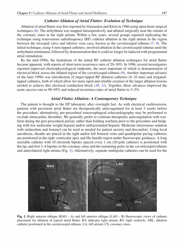
Chapter 9 / Catheter Ablation of Atrial Flutter and Atrial Fibrillation 197
Catheter Ablation of Atrial Flutter: Evolution of TechniqueAblation of atrial flutter was first reported by Guiraudon and Klein in 1986 using open-heart surgical
techniques (6). The arrhythmia was mapped intraoperatively and ablated surgically near the ostium ofthe coronary sinus in the right atrium. Within a few years, several groups reported replicating thetechnique using transvenous radiofrequency (RF) catheter ablation in the right atrium in the regionbetween the tricuspid valve and inferior vena cava, known as the cavotricuspid isthmus (7, 8). Theinitial technique, using 4-mm-tipped catheters, involved ablation in the cavotricuspid isthmus until thearrhythmia terminated, followed by demonstration that it could no longer be induced with programmedatrial stimulation.
By the mid-1990s, the limitations of the initial RF catheter ablation techniques for atrial flutterbecame apparent, with reports of short-term recurrence rates of 20–30%. In 1996, several investigatorsreported improved electrophysiological endpoints, the most important of which is demonstration ofelectrical block across the ablated region of the cavotricuspid isthmus (9). Another important advanceof the later 1990s was introduction of larger-tipped RF ablation catheters (8–10 mm) and irrigated-tipped catheters, both of which allow for more rapid and reliable creation of the larger ablation lesionsneeded to achieve this electrical conduction block (10, 11). Together, these advances improved theacute success rate to 90–95% and reduced recurrence rates of atrial flutter to 3–5%.
Atrial Flutter Ablation: A Contemporary TechniqueThe patient is brought to the EP laboratory after overnight fast. As with electrical cardioversion,
patients with persistent atrial flutter are therapeutically anticoagulated for at least 3 weeks beforethe procedure; alternatively, pre-procedural transesophageal echocardiography may be performed toexclude intracardiac thrombus. We generally prefer to continue therapeutic anticoagulation with war-farin during the peri-procedural period, rather than holding warfarin prior to the procedure and bridg-ing with low molecular weight heparin and/or unfractionated heparin. Moderate intravenous sedationwith midazolam and fentanyl can be used as needed for patient anxiety and discomfort. Using localanesthesia, sheaths are placed in the right and/or left femoral veins and quadripolar pacing cathetersare positioned in the right ventricular apex and His bundle region under fluoroscopic guidance. A longsteerable catheter with 10 electrode bipoles spaced every 1 cm (20-pole catheter) is positioned withthe tip, and first 3–4 bipoles in the coronary sinus and the remaining poles in the cavotricuspid isthmusand anterolateral right atrium (Fig. 1). Alternatively, separate multipolar catheters can be used for the
Fig. 1. Right anterior oblique (RAO – A) and left anterior oblique (LAO – B) fluoroscopic views of catheterplacement for ablation of typical atrial flutter. RA indicates right atrium; RV, right ventricle; ABL, ablationcatheter positioned in the cavotricuspid isthmus; LA, left atrium; CS, coronary sinus.

198 Part IV / Specific Arrhythmias
coronary sinus and anterolateral right atrium. A radiofrequency ablation catheter with 8–10 mm tip or4 mm irrigated tip is positioned in the cavotricuspid isthmus.
If the patient is in sinus rhythm, attempts are made to induce atrial flutter using programmed atrialstimulation. The arrhythmia is then mapped to determine the right atrial activation pattern and to provethat the cavotricuspid isthmus is an obligate part of the reentrant circuit. The ablation catheter is thenpositioned in the cavotricuspid isthmus and advanced so that the tip lies on the tricuspid valve annulus(Fig. 2). RF energy is then applied, which causes resistive heating and necrosis of the myocardiumlying in immediate contact with the tip. Every 30–60 sec, the tip is pulled back several millimeterstoward the inferior vena cava until a complete line has been created connecting the tricuspid valveannulus with the inferior vena cava. Ablation is continued until atrial flutter terminates to sinus rhythm.
Fig. 2. Anatomy of the right atrium relevant to atrial flutter ablation. The figure shows right anterior obliqueview of a heart model of the right atrium and right ventricle with the anterolateral walls removed. The ablationcatheter is positioned on the annulus of the tricuspid valve (TV) in the cavotricuspid isthmus (CTI), the startingpoint for ablation. Dashed line shows the path along which the catheter is withdrawn to create a line of block tothe inferior vena cava (IVC). CS indicates the ostium of the coronary sinus.
After termination, pacing on each side of the ablation line is performed to prove that no electricalconduction across the line is possible. If bidirectional conduction block is not present, the isthmusregion is probed with the ablation catheter to find gaps of viable myocardium, which allow conduction.Once bidirectional block is present and stable over a brief observation period, an atrial pacing protocolis repeated to confirm noninducibility of the clinical arrhythmia and to look for other arrhythmias.Catheters and sheaths are then removed. Typical total procedure times are 2–3 h from start of sheathinsertion, with fluoroscopy times of 10–20 min and RF ablation time of 10–20 min. Patients who do notrequire immediate intravenous anticoagulation can often be discharged after a 4-h observation period,with the remainder leaving after an overnight stay. Patients who arrive for the ablation procedurein atrial flutter are effectively being cardioverted to sinus rhythm, and therefore require continuoustherapeutic anticoagulation for at least 4 weeks afterward.
Atrial Flutter Ablation: ResultsIn a recently published meta-analysis, Perez et al. examined 158 studies reporting results of atrial
flutter ablation involving a total of 10,719 patients with a mean of 14.3 + 0.3 months of follow-up (12).

Chapter 9 / Catheter Ablation of Atrial Flutter and Atrial Fibrillation 199
Adjusting for potential reporting bias, the overall acute success rate was 91.1% with an adjusted recur-rence rate of atrial flutter of 10.9%. To reflect contemporary practice, the authors also examined the 49studies (3098 patients) in which 8–10 mm or irrigated-tipped RF ablation catheters were used with aprocedural endpoint of bidirectional cavotricuspid isthmus block. In this contemporary subgroup, theadjusted acute success rate was improved to 93.6% with a recurrence rate of atrial flutter of 6.7%.
Two randomized controlled trials of catheter ablation for atrial flutter have been reported, bothshowing marked superiority of the procedure compared with medical therapy. Natale et al. randomized61 patients with at least two episodes of atrial flutter to either undergo RF catheter ablation or toreceive sequential trials of antiarrhythmic drug therapy (13). During a mean follow-up period of 21 +11 months, atrial flutter recurred in 93% of the medically treated group vs. only 6% of the grouptreated with catheter ablation. Quality of life was shown to be significantly improved in the catheterablation group.
Da Costa et al. randomized 104 patients after a single episode of symptomatic atrial flutter to treat-ment with either RF catheter ablation or medical therapy with amiodarone (14). After a mean follow-upof 13 + 6 months, 30% of amiodarone-treated patients experienced recurrence of atrial flutter vs. only4% of the ablation-treated group. Moreover, there were no complications of catheter ablation duringthe trial while 10% of the medically treated group experienced complications of amiodarone treatment,including sinus node dysfunction (4%) and thyroid abnormalities (6%). The high recurrence rates ofatrial flutter with medical treatment in these trials is also supported by a study of Babaev et al., who fol-lowed 53 patients after initial hospital presentation with the first onset of atrial flutter (15). All patientswere cardioverted and medically treated, 36% with antiarrhythmic drugs. After a mean follow-up of22 + 12 months, 92% of patients experienced recurrence of atrial flutter, with a 42% recurrence rate inthe first month alone.
An important limitation to catheter ablation of atrial flutter that has been noted in numerous studies isa high incidence of atrial fibrillation in follow-up, likely due to common atrial substrate and risk factorsshared by the two arrhythmias. A prior history of atrial fibrillation is the most potent predictor ofatrial fibrillation after successful atrial flutter ablation. In the Perez meta-analysis, the overall adjustedoccurrence of atrial fibrillation after atrial flutter ablation was 33.6%. The adjusted incidence of post-ablation atrial fibrillation was 52.7% among patients with a prior history of atrial fibrillation, vs. 23.1%among those with no history of atrial fibrillation. The meta-analysis also showed that incidence ofatrial fibrillation after atrial flutter ablation increases with duration of follow-up. Other investigatorshave shown that risk of post-procedural atrial fibrillation is also increased by coexisting heart failureand left ventricular dysfunction. In an analysis of 110 patients after atrial flutter ablation, Paydak et al.found that 74% of patients with both pre-procedural atrial fibrillation and left ventricular dysfunctionexperienced atrial fibrillation in follow-up, vs. only 10% of patients with no history of atrial fibrillationand normal left ventricular function (16).
Atrial Flutter Ablation: RisksCatheter ablation of atrial flutter has a long track record of safety, and significant complications of
the procedure are uncommon. As with most cardiac endovascular procedures, femoral hematoma is themost common complication, but rarely requires intervention. Deep venous thrombosis and infectionof the vascular access occur with similarly low frequency as in standard cardiac catheterization. Atrialpacing protocols may induce atrial fibrillation, which may require electrical or chemical cardioversion;ventricular arrhythmias are rarely seen.
Pericardial tamponade due to perforation and hemopericardium has been reported in about 0.5%of cases in large series of catheter ablations for SVT (17). These series report procedure-relatedmortality of 0.1–0.3%, which also includes complications related to ablation on the left side of theheart. To our knowledge, no case of death caused by atrial flutter ablation has been reported to date.

200 Part IV / Specific Arrhythmias
RF ablation can potentially injure cardiac structures adjacent to the area being ablated, such as thetricuspid valve, AV node, and right coronary artery. The risk is minimized, however, with appro-priate training and experience, as well as with careful monitoring of electrograms and fluoroscopicimages during ablation. Rare cases of AV block requiring pacemaker placement have been reportedwhen the medial or septal cavotricuspid isthmus region is targeted. As with electrical or chemicalcardioversion, stroke is a potential complication of ablation in a patient with persistent atrial flut-ter, which can be minimized with appropriate anticoagulation and/or screening with transesophagealechocardiography.
In a recent meta-analysis of atrial flutter ablation, complication rates in 93 studies which included6293 patients were summarized (12). The overall complication rate was 2.6%. Of the 77 specificcomplications reported in these studies, significant vascular injury occurred in 26 patients (0.4%),complete AV block in 12 (0.2%), pericardial effusion in 8 (0.1%), stroke in 3 (0.05%), and myocardialinfarction due to right coronary injury in 1 (0.02%).
Atrial Flutter Ablation: Evidence-Based GuidelinesBecause of the high degree of efficacy and safety, catheter ablation for atrial flutter has become
the first-line therapy for treatment of symptomatic arrhythmia, and is clearly superior to antiarrhyth-mic drug therapy. In the 2003 American Heart Association (AHA) – American College of Cardiol-ogy (ACC) – European Society of Cardiology (ESC) Guidelines for Management of Patients withSupraventricular Arrhythmias, catheter ablation carries a class I recommendation for the treatment ofatrial flutter that is recurrent or poorly tolerated (18). For a first occurrence of well-tolerated atrialflutter, catheter ablation has a class IIa recommendation. In contrast, almost all antiarrhythmic drugsfor recurrent atrial flutter carry a class IIb recommendation, based on Level C (limited) evidence ofefficacy.
Atrial Flutter: Atypical FormsThe preceding discussion has generally referred to the common or typical form of atrial flutter which
has classical appearance on 12-lead ECG and requires conduction through the cavotricuspid isthmusfor its maintenance. This typical form comprises approximately 90% of atrial flutters seen in clinicalpractice. The remaining 10% form a spectrum of arrhythmias, which may involve the right or leftatrium and utilize a variety of reentrant pathways (Table 1).
Table 1Types of Atypical Atrial Flutter
Right atrial originCavotricuspid isthmus-dependent
1. Clockwise typical atrial flutter2. Type Ic atrial flutter3. Lower loop reentry
Non-isthmus dependent1. Incisional reentry (post-cardiac surgery)2. Upper loop reentry
Left atrial originPerimitral flutterIncisional reentry (post-cardiac surgery)Post-ablation for atrial fibrillation

Chapter 9 / Catheter Ablation of Atrial Flutter and Atrial Fibrillation 201
One of the more common variants is so-called “clockwise” typical atrial flutter. This arrhythmiautilizes the same reentrant pathway in the right atrium as typical counterclockwise atrial flutter, butin the opposite direction. This variant can be induced with programmed stimulation in most patientswith typical atrial flutter, but for unknown reasons is far less common clinically (19). The ablationtechnique, success rates, and risks of catheter ablation are same similar to typical atrial flutter.
Another variant of typical atrial flutter is that seen after antiarrhythmic drug treatment of atrialfibrillation, or so-called “type Ic” atrial flutter (20). This arrhythmia has been described mainly aftertreatment with type Ic sodium channel-blocking antiarrhythmic drugs (flecainide and propafenone) aswell as with amiodarone. Almost all cases utilize the same reentrant right atrial circuit as with typicalatrial flutter, and the ablation technique used is the same. Rates of acute success of catheter ablation aswell as recurrence rates of atrial flutter are felt to be similar to those for typical atrial flutter ablation.Importantly, successful ablation of atrial flutter in this group does not obviate the need for continuationof antiarrhythmic drug therapy to suppress atrial fibrillation. However, recurrences of atrial fibrillationare often seen in this group, even with the continuation of antiarrhythmic drug therapy.
Most other forms of atrial flutter do not utilize the cavotricuspid isthmus and therefore will be unaf-fected by the standard ablation technique for typical atrial flutter. In these so-called “atypical” forms ofatrial flutter, a large variety of electrocardiographic patterns have been described, and there is no ECGpattern that has been found to be characteristic of any type of reentrant circuit. In these atypical forms,the reentrant circuit travels around one or more fixed anatomical or functional barriers. In these cases,one or more barriers is often created as a result of atrial fibrosis from surgical incision, pericardiotomy,placement of surgical patches for treatment of congenital heart disease, or prior catheter or surgicalablation of atrial fibrillation (21). Atypical atrial flutter is occasionally seen in patients who have neverundergone cardiac procedures; such patients are usually found to have developed spontaneous atrialfibrosis as a component of a cardiomyopathy.
Atypical Atrial Flutter: Ablation TechniqueThe technique for mapping and ablation of atypical flutters is different from that for typical atrial
flutter. After placement of standard electrophysiological catheters, atrial pacing maneuvers are per-formed to identify whether the right atrium or left atrium is the site of origin of the arrhythmia. Oncethis is determined, that atrium undergoes electroanatomical mapping. In this procedure, the cardiacchamber of interest (in this case, the right or left atrium) is mapped in a point-by-point fashion usingone of several specialized computer systems that can integrate electrical and spatial information. Afterapproximately 50–100 points are gathered over a 15–30 min period, a three-dimensional shell of themapped atrium is created with activation and voltage encoded in color format. If created accuratelyand in sufficient detail, the activation pathway of the atypical flutter is usually apparent. An ablationstrategy is then devised, which usually involves ablation of a critical narrow portion of the circuit byconnecting two or more electrical barriers.
One of the most common forms of atypical flutter occurs after right atriotomy, and usually consistsof a reentrant circuit which revolves around a lateral right atrial scar. In such cases, the atrial flutteris usually easily ablated by connecting the lateral right atrial scar to the ostium of the inferior venacava with a 1–2 cm ablation line (Fig. 3). Less commonly, right atrial flutter circuits may form aroundposterior or septal scars or patches. Ablation of such atrial flutters usually involves connecting thescar to the ostium of the inferior vena cava or the tricuspid valve annulus. The most difficult formsof right atrial flutter usually result from complex congenital heart disease and associated correctiveoperations. Nakagawa et al. described a series of such cases, some of which had 6–8 distinct fluttercircuits (22). With lengthy procedures involving extremely detailed electroanatomical mapping andablation lines connecting a number of scars and other barriers, they were able to render approximately70% of patients free of atrial arrhythmias.

202 Part IV / Specific Arrhythmias
Fig. 3. Electroanatomical map of atypical atrial flutter due to incisional reentry late after surgical repair of atrialseptal defect. Figure shows a right lateral view of electroanatomical map of right atrium during atypical atrialflutter in a 44-year-old woman who underwent surgical repair of atrial septal defect 20 years previously. Area ofscar, probably representing a surgical incision, is shown in gray. The large red dots represent RF ablation lesions.Direction of depolarization during atrial flutter is indicated by arrows. SVC indicates superior vena cava; TVA,tricuspid valve annulus, IVC, inferior vena cava.
With the rise in utilization of ablation therapy for atrial fibrillation, this has become the most com-mon cause for the development of atypical atrial flutter arising in the left atrium (23). The most com-mon form of left atrial flutter, so-called “perimitral flutter,” utilizes a circuit which travels aroundthe mitral valve annulus. Ablation of this form of atypical atrial flutter generally involves creating aline of ablation lesions, which connects the mitral valve annulus to one of the left-sided pulmonaryveins (Fig. 4). Creating a full transmural ablation line in this region may be very challenging, andmay require ablation within the coronary sinus to achieve complete conduction block. Other formsof left atrial flutter may utilize one or more pulmonary veins as a central barrier. Ablation in thesecases may involve connecting the ostia of the right and left superior pulmonary veins and/or connect-ing the ostium of one or more pulmonary veins to the mitral valve annulus. As with atypical rightatrial flutters, many different pathways are possible, and successful ablation usually requires carefulelectroanatomical mapping of the reentrant circuit and its associated barriers in order to formulate asuccessful ablation strategy (24).
In addition to macro-reentrant forms of atypical atrial flutter following ablation of atrial fibrillation,it is also common to observe atypical atrial flutter with a presumed micro-reentrant mechanism dueto a gap in an ablation line. These micro-reentrant forms of atypical atrial flutter can be recognizedby their centrifugal spread of activation as well as a more discrete P-wave morphology with a clearisoelectric interval on the 12-lead ECG (25).

Chapter 9 / Catheter Ablation of Atrial Flutter and Atrial Fibrillation 203
Fig. 4. Two approaches to ablation of peri-mitral left atrial flutter occurring after AF ablation. Left anterioroblique view of an electroanatomical map of the left atrium is shown. The lower line of red dots indicate ablationlesions connecting the mitral valve annulus (MVA) to the ostium of the left inferior pulmonary vein (LIPV),which is the standard approach. The upper line indicates alternative approach connecting the MVA to the leftsuperior pulmonary vein (LSPV). RSPV indicates right superior pulmonary vein; RIPV, right inferior pulmonaryvein.
Atypical Atrial Flutter: Results, Risks, and GuidelinesCompared with ablation of typical right atrial flutter, there is much less published data on the results
of catheter ablation of atypical atrial flutter. Most data have been reported in the form of case series,usually with fewer than 25 patients and less than 1 year of follow-up. In general, success rates of70–80% have been reported, considerably less than what has been achieved with catheter ablationof typical atrial flutter. Recurrence rates have not been well described in the literature, but are likelyhigher than the 5–10% reported for typical atrial flutter owing to the more complex nature of the atrialsubstrate that gives rise to these arrhythmias.
Complication rates cannot be precisely determined from the limited literature on this subject, butare probably similar to those for ablation of typical atrial flutter described above when only rightatrial ablation is performed. One potential additional complication that may result from ablation ofatypical right atrial flutter is right phrenic nerve injury resulting in temporary or permanent right hemi-diaphragmatic paralysis. This complication usually arises from ablation along the posterolateral rightatrium and/or lateral ostium of the superior vena cava, and may be reduced testing with high-outputpacing looking for diaphragmatic stimulation at each high-risk site prior to delivering RF ablation. Inaddition, the risk of AV block may be higher when ablating atypical atrial flutter in the septal region,and AV conduction must be carefully monitored whenever delivering RF lesions near the interatrialseptum. Ablation of atypical left atrial flutter likely carries a higher complication rate than that of rightatrial flutters owing to the need for transseptal puncture and catheterization as well as delivery of RFlesions in the left atrium. Some of these potential risks will be covered in the later sections on ablationof atrial fibrillation.
Because of the more complex nature of atypical atrial flutters and the broad diversity of atrialsubstrates and cardiac and noncardiac comorbidities, including history of atrial fibrillation, patientselection for catheter ablation of atypical atrial flutter must be individualized, and uniform treatment

204 Part IV / Specific Arrhythmias
recommendations cannot be make. In contrast to typical atrial flutter, a trial of antiarrhythmic drugtherapy may be warranted, and a rate control strategy may be effective in the occasional patient withminimal or no symptoms. Reflecting this clinical heterogeneity, the 2003 ACC/AHA/ESC Guidelinesfor Treatment of Supraventricular Arrhythmias accord ablation of atypical atrial flutter a class IIarecommendation (18).
Summary: Catheter Ablation of Atrial FlutterRF catheter ablation of typical, cavotricuspid isthmus-dependent atrial flutter is a safe and highly
effective curative treatment for this troublesome atrial arrhythmia and is recommended as first-linetherapy for most symptomatic patients. Atypical atrial flutter is a heterogeneous clinical problem andrequires individualized clinical decision-making. Successful ablation of atypical atrial flutter usuallyrequires careful electroanatomical mapping with ablation of all induced sustained atrial flutter circuits.Development of atrial fibrillation after successful ablation of atrial flutter remains a significant clinicalproblem.
ATRIAL FIBRILLATION
Clinical BackgroundAtrial fibrillation (AF) is the most common sustained arrhythmia, with an estimated prevalence of at
least 2 million in the United States, a number which is likely to rise due to the aging of the population(26). Prevalence increases with age in a nearly exponential fashion, with estimated prevalence of 10%in people over the age of 80 years, and the estimated lifetime risk for developing AF for a 40-year-old is about 25% (27). Patients with AF may be asymptomatic or suffer from a variety of symptoms,including palpitations, dyspnea, chest discomfort, fatigue, malaise, and loss of exercise tolerance. Insome cases, tachycardia-induced cardiomyopathy may result from uncontrolled ventricular rate. It isan important cause of stroke, with estimated attributable risk of 20–25%. Quality of life is reduced inmost patients, and epidemiologic studies have linked AF to increased risk of mortality.
For most of the past 50 years, the dominant model of AF mechanism has been the multiple wavelethypothesis of Moe and Allessie (28). According to this model, six to eight randomly moving reentrantwavelets are required to maintain AF. The wavelets do not follow any fixed pathways, but do observefixed anatomical boundaries and may collide with one another, extinguish, and reform, resulting inan apparently chaotic pattern of atrial activation. In recent years, increasing attention has focused onelectrophysiologic triggers of AF, particularly focal arrhythmias arising from the pulmonary veins.Contemporary research has also established that, in contrast to other supraventricular arrhythmias, AFis often a progressive disease which becomes increasingly self-sustaining and resistant to treatmentover time due to electrical and anatomical remodeling, resulting in atrial fibrosis and enlargement.
Management of AF begins with medical control of ventricular rate and anticoagulation if strokerisk factors are present. A trial of cardioversion is appropriate for most patients who first presentwith sustained AF, although it is recognized that some older patients with minimal or no symptomsmay be managed successfully in permanent AF. Longer-term treatment is guided by the patient’s age,symptoms, comorbidities, and personal preferences. Disease management has been affected by theAFFIRM and RACE trials, which showed no reduction in stroke risk or overall mortality in patientswith AF who were treated with antiarrhythmic drugs to maintain sinus rhythm over a simpler rate con-trol and anticoagulation strategy (29, 30). Subgroup analysis of the AFFIRM trial, however, did showimproved outcomes in the subset of patients who successfully maintained sinus rhythm, suggestingthat there may be significant benefit to maintaining sinus rhythm, which is offset by adverse effectsand relative inefficacy of antiarrhythmic drugs (31).

Chapter 9 / Catheter Ablation of Atrial Flutter and Atrial Fibrillation 205
Patients enrolled in the AFFIRM and RACE trials had either asymptomatic or minimally symp-tomatic AF, thereby making them appropriate candidates for either rate or rhythm control strategy. Itis therefore inappropriate to conclude that rate and rhythm control are equivalent treatment strategiesfor patients with symptomatic AF. Since publication of the AFFIRM trial in 2002, the safety and effi-cacy of catheter ablation of AF has improved significantly, and it is continuing to improve further. Inaddition, committing a patient to a rate control strategy will likely to render the atrium more resistantto AF ablation several years later due to progressive atrial remodeling. We are therefore reluctant tocommit younger patients (<70 years) to a rate control strategy without attempting an initial rhythmcontrol approach. On the other hand, we quickly move to a rate control strategy for very elderly AFpatients who are either asymptomatic or minimally symptomatic.
For those patients with recurrent AF who do not have acceptable symptom control with rate controlmedications, rhythm control generally begins with a choice of antiarrhythmic drug therapy. Random-ized trials have shown that antiarrhythmic drugs maintain sinus rhythm in 30–60% of patients over a1–2 year follow-up period (32, 33). Success rates of antiarrhythmic drugs depend upon cardiac comor-bidities, left atrial size and fibrosis, and duration of AF. Selection of antiarrhythmic drug therapydepends upon patients’ comorbidities, other medications, renal function, and personal preferences. Auseful algorithm can be found in the 2006 ACC/AHA/ESC Guidelines for the management of patientswith atrial fibrillation (26).
For patients with symptomatic AF who cannot tolerate or achieve acceptable symptom control witheither a rate control strategy or with antiarrhythmic drug therapy, catheter ablation has evolved froman investigational procedure to one that is now performed on thousands of patients annually in manymedical centers throughout the world. The growing acceptance of this procedure has been broughtabout by a growing number of reports showing safety and efficacy of catheter ablation of AF targetingthe pulmonary veins and posterior left atrium. The remainder of this section will review the develop-ment of catheter ablation procedures, highlighting current techniques, outcomes, complications, andremaining questions.
Catheter Ablation of AF: Historical BackgroundThe era of catheter ablation therapy for cardiac arrhythmias began in 1982 with reports of direct
current ablation of the AV junction for control of ventricular rate in patients with intractable supraven-tricular tachyarrhythmias, including AF (34). While rarely performed now for other supraventriculararrhythmias, catheter ablation of the AV junction with permanent pacemaker implantation remains auseful treatment for selected AF patients with refractory symptoms due to uncontrolled ventricularresponse despite medical therapy, particularly in elderly patients (35). In recent years, biventricularpacemakers have been increasingly used in patients undergoing AV junction ablation who have preex-isting heart failure symptoms due to left ventricular dysfunction and/or mitral regurgitation, due to thewell-established risk for right ventricular apical pacing to exacerbate these problems (36). In the early1990s, several groups reported success in modifying AV node conduction with partial catheter abla-tion with acceptable ventricular rate control, but without complete AV block or need for pacemakerimplantation. These early results proved not to be durable, with a majority of patients either regressingto rapid ventricular response or progressing to high-degree AV block and pacemaker-dependency, andAV node modification is now rarely performed for AF.
Concurrently with the development of catheter ablation techniques described above, Cox and col-leagues developed a series of surgical techniques for the disruption of AF, first in a canine model andthen in series of patients (37). The most advanced version of the operation, the Maze-III procedure,was based upon the multiple wavelet hypothesis, in which maintenance of the arrhythmia requiresmaintenance of a critical number of circulating wavelets of reentry, each of which requires a criticalmass of atrial tissue to sustain it. In the Maze-III operation, after induction of cardiopulmonary bypass,

206 Part IV / Specific Arrhythmias
a series of transmural incisions are made in the left and right atria, with preservation of the sinus nodeand an uninterrupted corridor of atrial tissue to allow conduction to the AV node. In addition, the rightand left atrial appendages are removed, which may further reduce stroke risk. The end result of theprocedure is that the atria are divided into electrically isolated subsections such that maintenance ofAF is prevented regardless of the mode of initiation. In the course of their research, these investigatorsfound that the left atrial lesion set was largely sufficient to prevent AF, while right atrial lesions wererequired to prevent development of atrial flutter. Interestingly, isolation of the pulmonary veins andposterior left atrium was a feature common to all successful iterations of the procedure.
With the Maze-III approach, Cox and colleagues have reported maintenance of sinus rhythm in95% of patients at long-term follow-up with stroke rates of 0.1%/year (38). Fifteen percent of patientsrequired permanent pacemakers, and 7% lost left atrial transport function. Several other surgical seriesin other centers have reported success rates nearly as high, above 85%. Widespread application ofthe Maze-III operation as a stand-alone treatment for AF has been limited by the complexity of theprocedure and the morbidity and risk associated with sternotomy/thoracotomy and cardiopulmonarybypass, as well as by limited adoption among cardiothoracic surgeons. However, long-term resultsof the Maze-III operation still stand as the gold standard for what can be achieved with catheter andsurgical ablation techniques.
Catheter Ablation of AF – Linear ApproachThe success of the Maze-III procedure in the early 1990s led some interventional cardiac elec-
trophysiologists to attempt to reproduce the procedure with RF catheter lesions using a transvenousapproach. In 1994, Schwartz and colleagues reported reproducing the Maze-III lesion set in a smallseries of patients using specially designed sheaths and standard RF ablation catheters (39). Efficacywas modest, complication rates were high, and procedure and fluoroscopy times exceedingly long,and further attempts to reproduce the full Maze-III lesion set were abandoned. However, this reportdemonstrated a proof-of-concept that led others to try to improve the catheter-based approach.
In 1996, Haissaguerre and colleagues reported the outcomes of linear catheter ablation in 45 patientsundergoing right and/or left atrial ablation for paroxysmal AF (40). They found that right atrial abla-tion (with two or more procedures in 19 patients) led to rhythm control in 15 patients (33%), 9 ofwhom required antiarrhythmic drugs. Ten patients in whom right atrial ablation was unsuccessfulthen underwent left atrial ablation via transseptal approach, leading to rhythm control in six patients(60%). Further refinement of linear catheter ablation technique by this group was reported in 1999, ina series of 44 patients (91% with paroxysmal AF) who underwent RF catheter ablation to create spe-cific lesions sets in the right (2 lines) and left (3–4 lines) atria (41). A mean of 2.7 +/– 1.3 procedureswere required to complete the lesions and to ablate post-procedure atrial flutters, which occurred in70% patients and were generally observed to be caused by incomplete lines of block. Interestingly, 29patients (66%) were observed to have areas of focal electrical discharge initiating AF and underwentattempted ablation of these foci, 97% of which were located in the pulmonary veins.
Of the 44 patients in this study, maintenance of sinus rhythm was achieved without antiarrhythmicdrugs in 25 (57%) and 27% had improved rhythm control on antiarrhythmic drugs. The investigatorsnoted significant complications in nine patients (20%). In retrospect, it is likely that ablation of initi-ating pulmonary vein foci significantly improved their results compared with a pure linear approach.Multicenter trials of linear ablation of AF using novel multipolar ablation catheters were attempted,but were terminated prematurely due to high complication rates and poor efficacy. On the basis ofthese observations, and the rapid advances in ablation of AF targeting initiating focal triggers, thecatheter-based linear ablation approach for AF has been largely abandoned.

Chapter 9 / Catheter Ablation of Atrial Flutter and Atrial Fibrillation 207
Catheter Ablation of AF: Focal ApproachOne important observation which came from the early work of Haissaguerre and colleagues in linear
ablation of AF was the fact that some patients with paroxysmal AF appear to have a focal source ofelectrical activity triggering some or all paroxysms of AF. They published findings of a series of 45patients in a landmark publication in 1998 (42). Patients had a mean age of 54 +/– 11 years and hadfrequent (often daily) paroxysms of drug-refractory AF for a mean of 6 +/– 6 years. Most patients(69%) had structurally normal hearts. Patients also had at least 700 isolated premature atrial ectopicbeats on a 24-h Holter. All patients underwent intracardiac mapping to localize the source of thepremature atrial ectopic beats; 69 ectopic foci were found and targeted for ablation. Remarkably, 65foci (94%) originated within pulmonary veins, most within 2–4 cm of the ostium. AF was initiatedwith a burst of rapid firing from the foci and was abolished with RF ablation at the site of origin. Aftera follow-up period of 8 +/– 6 months, 28 patients (62%) were free of AF off antiarrhythmic drugs, asubstantial effect given the frequency and long duration of symptoms preceding the procedure. Thesefindings were soon replicated by other groups.
The electrophysiologic basis of these focal sources appeared to be sleeves of left atrial muscleinvesting the pulmonary veins, an anatomic observation that had been made decades ago. One ormore veins develop abnormal, paroxysmal, rapid automaticity, triggering AF as a result (43). Despiteextensive research, the pathophysiology of this process remains poorly understood. Further researchestablished that non-pulmonary vein foci were an important source of AF in some patients, althoughpercentages vary among different groups from 5 to 20% (44). Sources identified included severalthoracic veins: the vein/ligament of Marshall (a remnant of the fetal left superior vena cava on theposterior left atrial wall), the coronary sinus, and the superior vena cava. Other reported sources includethe left atrial posterior wall and various right atrial sites. Rare cases of AF triggered by ectopic foci inthe inferior vena cava and in a persistent left superior vena cava have been described.
With increasing use of focal ablation of pulmonary vein triggers, the problem of pulmonary veinstenosis quickly became recognized due to its serious sequelae, including hemoptysis, pulmonaryedema, and pulmonary hypertension. Another problem with the focal approach to pulmonary veinablation was the recognition that after successful ablation, recurrence rates were high and remappingusually showed new foci in the ablated vein or in other veins rather than recurrence of the originalfocus. Paroxysmal AF appears to be a field effect, rendering patients susceptible to recurrences frommultiple pulmonary vein foci. A final major limitation of this approach is that it requires direct obser-vation of atrial premature beats triggering AF during the ablation procedure, limiting it to patients withfrequent spontaneous or inducible atrial ectopy.
Catheter Ablation of AF: Segmental Ostial ApproachRecognition of these major limitations of focal ablation led to techniques to electrically isolate the
entire pulmonary vein. Because the pulmonary vein musculature conducts to left atrial musculatureby discrete connections, investigators targeted those connections using multipolar catheters shapedinto rings or baskets (45, 46). After placement of a diagnostic electrophysiology catheter in the coro-nary sinus, two long sheaths are placed across the interatrial septum with a double transseptal tech-nique. After defining pulmonary vein anatomy with contrast venography, a circular mapping catheteris placed at the ostium of the targeted pulmonary vein. Ablation is performed with a separate rovingcatheter through the second transseptal sheath at the site of earliest activation sequentially until pul-monary vein electrical activity disappears or becomes dissociated from left atrial activity. Using thisstrategy, between 20% and 60% of the pulmonary vein circumference is typically targeted for ablation.It was found that by ablating at or just outside the pulmonary vein ostium, the incidence of significantpulmonary vein stenosis could be reduced to less than 5%. Pulmonary vein isolation has the additional

208 Part IV / Specific Arrhythmias
advantage of simultaneously treating all triggering foci within the vein, thereby obviating the need toelicit and map those foci individually.
Marchlinski et al. compared their experience with focal vs. segmental ostial isolation for treatmentof AF in 107 patients and found freedom from AF at 1 year in 80% of patients treated with pulmonaryvein isolation vs. 45% of patients treated with focal ablation (47). For similar reasons, investigatorswere soon led to attempt to isolate as many pulmonary veins as possible at the initial ablation ses-sion. Comparative case series ultimately demonstrated that empiric four vein isolation led to superioroutcomes over isolating fewer veins.
Using this approach, electrophysiology centers began reporting success rates of 60–80% for patientswith paroxysmal AF with medium-term follow-up. Success rates for this approach are clearly betterfor patients with paroxysmal atrial fibrillation than for those with persistent atrial fibrillation. Recur-rences generally result from reconnection of previously isolated veins or ectopy in pulmonary veinsnot targeted at the initial procedure. A minority of recurrences have been shown to be due to electricaltriggers outside of the pulmonary veins.
Table 2 summarizes the outcomes of eight case series or trials that have reported on the safety andefficacy of segmental ostial ablation for the treatment of AF (46, 48–54). Medium-term success wasachieved in 196 of 280 patients with paroxysmal AF (70%) and 21 of 70 patients with persistent atrialfibrillation (30%). The overall major complication rate was 6.3%, including stroke (0.7%), pericardialtamponade (1.2%), and significant pulmonary vein stenosis (4.3%).
The technique of segmental ostial pulmonary vein isolation represented an important advance incatheter treatment of AF over the focal approach. Medium-term success for patients with paroxysmalAF is good, but results are generally worse for persistent AF. It remains the preferred approach toparoxysmal AF in some centers, although most have moved toward techniques to modify left atrialsubstrate.
Catheter Ablation of AF: Circumferential ApproachConcurrently with the development of the segmental approach to pulmonary vein isolation, Pap-
pone and colleagues working in Milan developed the circumferential approach using electroanatomicmapping (55, 56). After placement of one or more diagnostic electrophysiology catheters, a singletransseptal puncture is performed and a long transseptal sheath is placed in the left atrium. Using anelectroanatomic mapping system, an anatomic shell is constructed with the mapping/ablation catheter,including the pulmonary veins. RF ablation is then performed circumferentially around each vein, welloutside the ostium, with the endpoint of ablation being the elimination or marked reduction (80–90%)in the amplitude of electrical signals within the encircling lesions. In cases where the inferior and supe-rior veins were closely spaced or shared a common ostium, a single large circumferential RF lesionset would be performed. Despite lack of evidence showing pulmonary veins treated in this way werecompletely electrically isolated from the left atrium, this group began reporting results for paroxysmalAF, which were as good or better than those working with the ostial segmental approach. Furthermore,patients with persistent AF treated with the circumferential approach achieved results nearly as goodas those with paroxysmal AF, and far better than reports of patients treated with segmental pulmonaryvein isolation.
In 2001, Pappone and colleagues reported results of this approach in 251 consecutive patients withdrug-refractory AF (paroxysmal in 179 and persistent in 72) (57). Electroanatomic mapping immedi-ately after ablation showed an estimated 23 +/– 9% of the entire left atrial surface area was transformedto a low-voltage or electrically silent state. After a mean follow-up period of 10.4 +/– 4.5 months, 85%of patients with paroxysmal AF and 68% of patients with persistent AF were free of symptomatic AF.
Results from other centers using the circumferential approach have not been as favorable. Table 3summarizes the outcomes of six case series or trials which have reported on the safety and efficacy of

Chapter 9 / Catheter Ablation of Atrial Flutter and Atrial Fibrillation 209
Tab
le2
Sum
mar
yof
Clin
ical
Stud
ies
ofSe
gmen
talP
ulm
onar
yVe
in(P
V)
Abl
atio
n
Succ
ess
Com
plic
atio
ns
Stud
yYe
arFo
llow
-up
(mon
ths)
Ove
rall
Paro
xysm
alA
FPe
rsis
tent
orpe
rman
entA
F
PV
sten
osis
(>50
%)
Stro
keC
ardi
acta
mpo
nade
Mit
ralv
alve
inju
ry
Hai
ssag
uerr
e(4
6)20
004±
551
/70
(73)
51/7
0(7
3)–
00
00
Ora
l(48
)20
025±
344
/70
(63)
41/5
8(7
1)3/
12(2
5)0
1(1
.4)
00
Dei
senh
ofer
(49)
2003
8±4
38/7
5(5
1)N
/AN
/A6
(8)
04
(5.3
)0
Mar
rouc
he(5
0)20
0314
±527
1/31
5(8
6)N
/AN
/A22
(7)
2(0
.6)
00
Are
ntz
(51)
2003
1234
/55
(62)
26/3
7(7
0)8/
18(4
4)1
(1.8
)0
1(1
.8)
0O
ral(
52)
2003
627
/40
(67)
27/4
0(6
7)–
00
00
Man
sour
(53)
2004
21±5
22/4
0(5
5)19
/33
(58)
3/7
(43)
00
2(5
)0
Vas
amre
ddy
(54)
2004
11±8
39/7
5(5
2)32
/42
(76)
7/33
(21)
3(4
)2
(2.6
)2
(2.6
)1
(1.3
)O
vera
ll–
–52
6/74
0(7
1)19
6/28
0(7
0)21
/70
(30)
32(4
.3)
5(0
.7)
9(1
.2)
1(0
.1)
Val
ues
are
give
nas
n(%
);Su
cces
sw
asde
fined
asfr
eeof
atri
alfib
rilla
tion
(AF)
recu
rren
cew
ithou
tant
iarr
hyth
mic
drug
s.

210 Part IV / Specific Arrhythmias
circumferential ablation for treatment of AF (52, 53, 57–60). Medium-term success was achieved in290 of 393 patients with paroxysmal AF (74%) and 73 of 149 patients with persistent/permanent AF(49%). The overall major complication rate was 2.4%, including stroke (0.4%), pericardial tamponade(0.7%), and PV stenosis (0.4%).
Catheter Ablation of AF: Additional ApproachesThe reported success of the circumferential approach raised several important questions and has
led investigators to attempt modifications to the standard circumferential lesion set, resulting in addi-tional, hybrid approaches to ablation of AF. The first question is whether complete electrical isolationof the pulmonary veins is necessary to achieve clinical success. Several investigators have addressedthis question and come to differing conclusions. Cappato et al. studied 43 patients with serial electro-physiology studies and found a high rate (80%) of recurrent conduction from previously disconnectedveins (61). However, freedom from AF was no more likely in those with recurrent conduction thanin those without. Forty percent of patients who had recurrence of pulmonary vein conduction werefree of AF.
In contrast, Gerstenfeld et al. who studied 34 patients with recurrent AF after initial pulmonaryvein isolation, and with careful pulmonary vein mapping, found that 86% of recurrent triggers camefrom pulmonary veins which had either not been targeted for initial ablation (32%) or developedrecurrent conduction (54%) (62). Ouyang et al. treated 41 patients with paroxysmal AF with a modifiedcircumferential ablation approach. In each patient, a single large extra-ostial circumferential lesionset was placed around inferior and superior pulmonary veins on each side while a circular mappingcatheter was present in each of the two ipsilateral veins (59). Ablation was continued until completeelectrical isolation was achieved in all four pulmonary veins. Ten patients had early recurrence andnine underwent a second procedure – all nine patients showed recurrent conduction from a previouslyisolated pulmonary vein and underwent re-isolation of that pulmonary vein.
It is now clear throughout the world that nearly all patients who have recurrent AF after an initialattempt at AF ablation and return for a repeat procedure will demonstrate electrical reconnection of atleast one, and usually several, pulmonary veins. It is also clear that in many patients reconnection ofpulmonary vein conduction occurs soon after initial isolation. We have previously examined the rate ofearly recurrence of pulmonary vein conduction 30 and 60 min after initial isolation (63). Recovery ofPV conduction was observed within the first 30 min in approximately one-third of veins and between30 and 60 min in 17% of veins. Overall, results of these and other investigations suggest that completeand durable isolation of the pulmonary veins is important for long-term success of the procedure,particularly for patients with paroxysmal AF.
A second important question concerns the safety and efficacy of adding left atrial linear lesions tothe basic circumferential pulmonary vein ablation lesion set. The two linear lesions most commonlyadded are a line connecting the right and left superior veins across the superior aspect of the leftatrium (so-called “roof” line) and a line connecting the left inferior pulmonary vein to the mitral valveannulus (so-called “mitral isthmus” line). These lesions sets are designed to further modify electricalsubstrate to prevent AF as well as to prevent left atrial flutters, which might be formed by the largecircumferential lines of block. Jais et al. have shown that achieving block with the mitral isthmus linein particular is technically challenging and sometimes requires ablation within the coronary sinus (64).Whether these additional lines improve clinical success remains to be determined, with some studiessuggesting that additional left atrial ablation lines may be unhelpful or proarrhythmic. Currently, manycenters are adding these lesions in patients with persistent AF, in patients with inducible AF or leftatrial flutter after the standard circumferential lesions are made, and in patients with recurrent AF afteran initial procedure.

Chapter 9 / Catheter Ablation of Atrial Flutter and Atrial Fibrillation 211
Tab
le3
Sum
mar
yof
Clin
ical
Stud
ies
ofC
ircu
mfe
rent
ialP
ulm
onar
yVe
in(P
V)
Abl
atio
n
Succ
ess
Com
plic
atio
ns
Stud
yYe
arFo
llow
-up
(mon
ths)
Ove
rall
Paro
xysm
alA
F
Pers
iste
ntor
perm
anen
tA
F
PV
sten
osis
(>50
%)
Car
diac
tam
pona
deSt
roke
Lef
tat
rial
flutt
er
Papp
one
(57)
2001
10±5
188/
251
(75)
148/
179
(83)
40/7
2(5
6)0
2(0
.8)
00
Ora
l(52
)20
036
35/4
0(8
8)35
/40
(88)
–0
00
1(2
.5)
Man
sour
(53)
2004
11±3
25/4
0(6
3)21
/32
(66)
4/8
(50)
01
(2.5
)1
(2.5
)0
Kot
tkam
p(5
8)20
0412
37/1
00(3
7)34
/80
(43)
3/20
(15)
00
04
(5)
Ouy
ang
(59)
2004
6±1
39/4
1(95
)39
/41(
95)
–0
00
0V
asam
redd
y(6
0)20
046±
339
/70
(56)
13/2
1(6
2)26
/49
(53)
2(2
.8)
1(1
.4)
1(1
.4)
0O
vera
ll–
–36
3/54
2(6
7)29
0/39
3(7
4)73
/149
(49)
2(0
.4)
4(0
.7)
2(0
.4)
5(0
.9)
Val
ues
are
give
nas
n(%
);Su
cces
sw
asde
fined
asfr
eeof
atri
alfib
rilla
tion
(AF)
recu
rren
cew
ithou
tant
iarr
hyth
mic
drug
s.

212 Part IV / Specific Arrhythmias
A third major question is whether modification of autonomic innervation of the left atrium plays asignificant role in procedural success. Pappone et al. studied 297 patients undergoing circumferentialablation for paroxysmal AF and found that 34% of them showed evidence of cardiac vagal denerva-tion during the ablation procedure and in follow-up (65). They found that ablation along the posteriorleft atrium in their standard lesion set sometimes produced a vagal reflex, resulting in sinus bradycar-dia and transient AV block. When ablation was continued at these sites, loss of the reflex occurred,suggesting vagal denervation, which was confirmed by indices of heart rate variability on follow-upHolter monitoring. A remarkable 99% of 100 patients who achieved complete vagal denervation werefree of AF at 12 months, vs. 85% of the remaining patients.
More recently, Po et al. reported on the results of a specific ablation approach targeting the leftatrial ganglionated plexi, which serve as integration centers of the cardiac autonomic nervous system(66). After transseptal catheterization, they first localized the sites of ganglionated plexi (typically 4per patient) using high-frequency pacing stimulation looking for a parasympathetic cardiac response(>50% increase in R–R interval). Interestingly, the ganglionated plexi were generally found to belocated near the ostia of each of the pulmonary veins, and would therefore be typically encompassedin a standard wide-area circumferential ablation approach. After localization, RF ablation was per-formed at each site, until the parasympathetic response was eliminated. Twelve months after a singleprocedure, 80% of patients were free of symptomatic AF. While results of these two reports are inter-esting and encouraging, the role systematic modification of cardiac autonomic innervation is not yetwell-established.
A fourth major question concerns the role of targeting complex fractionated atrial electrograms inthe right and left atrium. Nademanee et al. studied 121 patients with drug-refractory paroxysmal (57patients) and persistent/permanent (64 patients) atrial fibrillation and used a novel approach to map-ping of the substrate and performing ablation (67). During atrial fibrillation in all patients (spontaneousor induced), biatrial electroanatomical mapping was performed. Areas of complex, fractionated elec-trical (CFAE) potentials were noted and tagged. These sites were targeted for ablation, using a meanof 64 +/– 36 pulses. Sites were spread over nine areas of both atria and were not confined to the pul-monary veins, which were not specifically targeted for isolation. Using this approach, termination ofAF occurred during ablation in 91% of patients. At 12-month follow-up, 70% of patients were free ofAF off antiarrhythmic drugs; with a second procedure, success rate increased to 83%. Complicationrate was comparable to other approaches.
This novel approach to ablation of AF has generated considerable interest in the subject of electro-gram characteristics and the role in maintenance of atrial fibrillation. CFAE potentials have been shownto be reproducible and temporally stable in a given patient (68). It remains unknown whether they rep-resent sites of “rotors”, which generate wavelets, sites of autonomic innervation from ganglionatedplexi, or areas of atrial fibrosis. While Nademanee et al. have reported similarly favorable results in alarger series of patients with longer follow-up duration, these results have not been replicated at othercenters (69). Furthermore, addition of CFAE ablation to standard pulmonary vein isolation procedureshas not yet been demonstrated to significantly improve outcome.
A final major question in the field is the role of more extensive catheter ablation of the atria totreat patients with longstanding persistent AF, defined as continuously present for at least 1 year.This form of AF is regarded as a more advanced stage of the disease, and is usually accompaniedby left atrial enlargement and more extensive fibrosis. Results of standard pulmonary vein isolationprocedures with these patients in most centers have been disappointing. Haissaguerre and colleaguesin Bordeaux have reported on a more extensive ablative technique, which they term the “stepwiseapproach” (70, 71). The technique involves initial pulmonary vein isolation followed by isolation ofother thoracic veins, and then atrial tissue ablation targeting all regions with rapid or heterogeneousactivation (termed “defragmentation”). A final step may include creation of sequential linear lesionson the left atrial roof, coronary sinus, mitral isthmus, and other left atrial regions, with a goal of

Chapter 9 / Catheter Ablation of Atrial Flutter and Atrial Fibrillation 213
achieving termination of arrhythmia during ablation. Prolongation of mean AF cycle length in theatrial appendages is frequently noted prior to termination or organization to atrial flutter.
In an initial series of 60 patients undergoing the procedure, a remarkable 87% achieved acute ter-mination of arrhythmia. Most patients organized to one or more morphologies of atrial tachycardiaor flutter, which then required further ablation, and most patients required multiple procedures to fin-ish all the required ablation components and/or ablate resulting atrial tachycardias and flutters. Aftermultiple procedures, 95% maintained sinus rhythm at mean 11 +/– 6 months of follow-up. Theseexcellent results in patients who are often resistant to other techniques stand as a benchmark. Thissuccess rate, however, reflects multiple ablation procedures with concomitant use of antiarrhythmicdrugs in some patients; single-procedure long-term success rate free of antiarrhythmic drug therapyfor patients with longstanding persistent AF is less than 40%.
Catheter Ablation of AF: A Contemporary ApproachAt Johns Hopkins Hospital, we offer catheter ablation to patients with symptomatic paroxysmal
or persistent AF despite treatment with at least one antiarrhythmic drug. There are no specific age,left atrial size, or LV ejection fraction cutoffs for the procedure. In general, we find that patientsunder 75 years, those with left atrial size 5 cm or less, and those without severe heart failure or majorcomorbidities tolerate the procedure better and have better outcomes. Patients undergo pre-proceduralCT or MRI scanning to define pulmonary vein anatomy and to import the left atrial image into theelectroanatomic mapping system used during the ablation procedure. Pre-procedural transesophagealechocardiography is performed in most patients to exclude left atrial thrombus, including all patientswith persistent AF and patients with CHADS2 score of 2 or more (72). Patients with stroke risk factorsare treated with therapeutic warfarin for a month prior to the ablation and bridged with enoxaparin.
The ablation procedure may be performed with moderate sedation or general anesthesia, dependingon patient comorbidities, anticipated procedure time, and patient/operator preference. A radio-opaquetemperature probe is placed in the esophagus to monitor and minimize ablation near this critical struc-ture. Right femoral venous access is obtained and a monitoring femoral arterial line is placed. Amultipolar diagnostic catheter is placed in the coronary sinus. After intravenous heparin is given, twotransseptal punctures are performed and long sheaths passed into the left atrium. Contrast pulmonaryvenography is performed and used to create a merged electroanatomical image to guide the ablationprocedure. Using a 4-mm open-irrigated tip ablation catheter and 25–30 W power settings, wide-area circumferential ablation around the left and right pulmonary venous antra is performed, staying0.5–1 cm outside the ostium of each vein (Fig. 5). If the anatomy allows, ablation is generally per-formed between the superior and inferior pulmonary veins, in the so-called “carina” region (73).
After circumferential ablation is completed, a circular multipolar electrode mapping catheter isintroduced into the second transseptal sheath and the ostium of each pulmonary vein is mapped look-ing for residual electrical potentials and evidence of electrical isolation (Fig. 6). Additional ablationlesions guided by the mapping catheter are delivered until the vein is electrically isolated with evidenceof both entrance and exit block, or dissociation of pulmonary vein potentials (Fig. 7). This procedureis repeated for each pulmonary vein. Each vein is reexamined with the mapping catheter 30–60 minafter the last RF lesion is given to ensure persistent electrical isolation is present. If not, additional RFlesions are delivered to reisolate the vein.
At the first ablation procedure, additional ablation lines and ablation of CFAE sites are generallynot performed. In patients undergoing re-do procedures for persistent AF, CFAE mapping and ablationis performed. If AF persists, additional RF ablation lines, including left atrial roof line and mitralisthmus line, may be added. Atypical atrial flutters, if present, may be specifically mapped and ablated.In general, cavotricuspid isthmus ablation is not performed unless there is clinical evidence of typical

214 Part IV / Specific Arrhythmias
atrial flutter. Likewise, superior vena cava isolation is generally not performed unless the patient showsevidence that it is a focal source of triggering ectopy.
Total procedure times of 3–5 h and RF ablation times of 30–60 min are typical. After ablation iscompleted, heparinization is reversed with protamine and sheaths are removed. Anticoagulation isresumed after several hours during overnight observation in the hospital. Patients are bridged withenoxaparin to therapeutic anticoagulation with warfarin, which is continued for at least 2–3 months.Patients are seen in the office after 3 months, and Holter and/or event monitoring is performed every 3months for the first year and afterward if symptoms indicate. Long-term anticoagulation managementis determined by the patient’s rhythm status, CHADS2 score, and individual risk tolerance.
Risks of Catheter Ablation for AFAs with any medical procedure, potential risks need to be considered and balanced against potential
benefits and likelihood of success in deciding what to recommend for a particular patient. Some of therisks of catheter ablation of atrial fibrillation are listed in Table 4. In addition to risks common to allcatheter-based cardiac procedures, several of these risks are unique to or increased with ablation ofAF.
Pericardial tamponade is a complication common to all invasive cardiac procedures, but appears tobe higher in ablation of AF, likely due to prolonged procedure duration, transseptal access, multipleablation lesions, and need for prolonged high-dose heparinization during the procedure. The rate ofpericardial tamponade was 107/8745 procedures (1.22%) in the Worldwide Survey on Ablation ofAtrial Fibrillation (74). Occasional cases may be fatal, especially if the condition is not recognizedprior to hemodynamic collapse.
The risk of pulmonary vein stenosis is increased with ablation within the ostium of the pulmonaryvein and with multiple high-energy lesions around the circumference of the ostium (75). Rates of sig-nificant pulmonary vein stenosis as high as 10% were seen with early experience with focal ablation,and have been reduced to about 1% with circumferential ablation. No procedural technique has provento be entirely free of this risk, and variation in incidence may depend on how stenosis is measuredand defined and how vigorously it is sought after the procedure. Due to its low incidence in contem-
Fig. 5. Circumferential pulmonary vein ablation using electroanatomical mapping. The figure shows posteroan-terior view of an electroanatomical map merged with computed tomographic scan of the left atrium. Each red dotrepresents approximately 5–10 sec of radiofrequency application performed during circumferential pulmonaryvein isolation. LSPV indicates left superior pulmonary vein; LIPV, left inferior pulmonary vein; RSPV, rightsuperior pulmonary vein; RIPV, right inferior pulmonary vein.

Chapter 9 / Catheter Ablation of Atrial Flutter and Atrial Fibrillation 215
Fig. 6. Use of multielectrode circular mapping catheter to guide electrical isolation of the right superior pul-monary vein (RSPV). Panel A shows left anterior oblique fluoroscopy view of the left atrium during contrastinjection of the right superior pulmonary vein. Panel B shows same view with circular mapping catheter (CMC)inserted just inside the ostium of the RSPV, with the ablation catheter (Abl) deployed near the venous ostium tocomplete electrical isolation of the vein. CS indicates the coronary sinus catheter; T, radio-opaque temperatureprobe; His, the His bundle catheter.
Fig. 7. Demonstration of electrical dissociation of the left superior pulmonary vein from the left atrium aftercatheter ablation. Figure shows electrogram recordings taken during pulmonary vein isolation. Small arrowspoint to far-field atrial potential on the circular mapping catheter (Lasso 1–20) during sinus rhythm. The largearrows point to slow, dissociated pulmonary vein potentials, indicating complete electrical isolation of the vein.
porary practice, we do not routinely screen for pulmonary vein stenosis, unless symptoms such asexertional dyspnea, chest pain, persistent cough, hemoptysis, or unexplained pneumonia occur. CT orMRI scanning with comparison to pre-procedural images is the most effective way to make the diag-nosis. Treatment is challenging and may require angioplasty, stenting, or partial lung resection. In our

216 Part IV / Specific Arrhythmias
Table 4Risks of Catheter Ablation of Atrial Fibrillation
Vascular injuryHematomaPseudoaneurysmArteriovenous fistula
Venous thromboembolusPericardial effusion/ tamponadeStrokePulmonary vein stenosisPhrenic nerve injury (R >> L)Atrioesophageal fistulaMitral valve injuryCoronary artery injury/myocardial infarctionDeath
experience, pulmonary vein stenosis has become rare, and we have observed no symptomatic stenosisamong the last 500 patients who have undergone AF ablation at our institution.
Stroke, although infrequent, continues to be an important issue. The Worldwide Survey on Ablationof Atrial Fibrillation found 20 strokes reported for 7154 left-sided ablation procedures, an event rate of0.25%, including two deaths (74). The rate of transient ischemic attack was 0.66%. Potential sourcesof emboli include thrombi adherent to catheters and sheaths, endocardial disruption from the ablationlesions, thrombi or air passing through a patient foramen ovale or transseptal puncture site, and airintroduced through transseptal sheaths. Careful attention to anticoagulation and sheath managementshould reduce risk, but the nature of the procedure with prolonged left-sided catheterization and exten-sive left atrial ablation in patients predisposed to stroke precludes eliminating it entirely. A history ofprior stroke or transient ischemic attack and CHADS2 score of 2 or more appear to be significant riskfactors for this complication (76).
Permanent diaphragmatic paralysis was reported in 10/8745 cases (0.11%) in the Worldwide Reg-istry and probably relates to ablation within the right superior pulmonary vein or superior vena cavaand/or extensive linear ablation along the lateral right atrial wall. High-output pace-mapping at poten-tial high-risk ablation sites may reduce, but does not entirely eliminate, risk. Diaphragmatic paralysismay resolve in 50–70% of cases, but may take up to a year for nerve regeneration to occur (77).
Left atrial-esophageal fistula is the most feared complication of AF ablation due to its high fatal-ity rate, and is estimated to occur in approximately 0.1% of cases (78). Typically presenting days toweeks after otherwise uncomplicated procedures, premorbid symptoms may include dysphagia, fever,confusion, and hemetemesis. Stroke, sepsis syndrome, and sudden death are common sequelae. Diag-nosis may be made with chest CT scanning, and endoscopy should be avoided if the diagnosis is sus-pected. When recognized, treatment includes total GI rest, avoidance of esophageal instrumentation,and either temporary esophageal stenting or surgical esophageal resection. Risk of this complicationmay be reduced by esophageal monitoring during the procedure using a radio-opaque temperatureprobe, and reduction of RF power settings during posterior left atrial ablation.
A recently reported multicenter survey of 162 centers found that 32 deaths occurred in 32,569patients (0.1%) undergoing catheter ablation of AF (79). Causes of death included tamponade in 8patients (25% of deaths), stroke in 5 (16%), atrioesophageal fistula in 5 (16%), and pneumonia in 2(6%). A variety of miscellaneous causes accounted for each of the remaining deaths.

Chapter 9 / Catheter Ablation of Atrial Flutter and Atrial Fibrillation 217
Results of AF Ablation: Randomized Trials and Multicenter RegistriesTo date, six randomized trials of AF ablation vs. medical therapy have been published in manuscript
form, and they are summarized in Table 5 (80–86). While varying in design, outcome measures, andablation technique used, each one showed significantly improved outcome in the patients who receivedcatheter ablation. Freedom from AF at 1 year ranged from 56 to 89% in the patients randomized tocatheter ablation. Several of these studies also demonstrated improved quality of life and reduction insymptom burden in patients treated with ablation.
Khan et al. randomized 81 patients with drug-resistant paroxysmal or persistent AF associated withheart failure and left ventricular dysfunction (LVEF < 40%) to undergo either pulmonary vein isolationor AV node ablation with biventricular pacing (87). In the group undergoing pulmonary vein isolation,71% were free of AF at 6 months without antiarrhythmic drug treatment. Patients randomized topulmonary vein isolation showed improved quality of life scores, longer 6-min walk distance, andhigher LVEF compared with the AV node ablation group.
The largest multicenter case series of AF ablation is the Worldwide Survey reported by Cappato et al.in 2005 (74). Of the 777 centers contacted, 181 completed a standardized survey reporting outcomesof catheter ablation in 8745 patients between 1995 and 2002. Survey results showed widely varyingprocedure techniques and case volumes between centers, with a marked increase in procedure volumeover time. Outcome of freedom of AF without use of antiarrhythmic drugs ranged from 30% at thelowest volume centers to 64% at the highest volume centers, with overall reported success rates of52%. Including patients free of AF with continuation of antiarrhythmic drugs, overall success reportedwas 76%. The overall major complication rate was 6%.
A recent study reported the outcomes of two meta-analyses of the safety and efficacy of catheterablation of AF and antiarrhythmic drug therapy (88). The results of 63 AF ablation and 34 antiarrhyth-mic drug studies were included in these analyses. Patients enrolled in AF ablation studies tended to beyounger (mean age 55 vs. 62 years), had longer duration of AF (6.0 vs. 3.1 years), and had failed agreater number of prior drug trials (2.6 vs. 1.7). The single procedure success rate of ablation off AADtherapy was 57% (95% CI 50–64%), the multiple procedure success rate off AAD was 71% (95% CI65–77%), and the multiple procedure success rate on AAD or with unknown AAD usage was 77%(95% CI 73–81%). In comparison, the success rate for AAD therapy was 52% (95% CI 47–57%). Amajor complication of catheter ablation occurred in 4.9% of patients. Adverse events for AAD studies,while more common (30% vs. 5%), were less severe.
As of this writing, several large, multicenter randomized trials of catheter ablation for AF are beingorganized. The largest of these, the Catheter Ablation vs. Antiarrhythmic Drug Therapy for AtrialFibrillation (CABANA) trial, plans to enroll several thousand patients to either catheter ablation ormedical therapy for paroxysmal or persistent atrial fibrillation, with several years of follow-up andoverall mortality as a primary outcome (89). While results will not be expected until about 2015,this and other major trials should provide important information about patient selection, proceduraltechnique, anticoagulation management, outcomes, and complications in AF patients.
HRS Consensus DocumentThe publication of the Expert Consensus Statement on Catheter and Surgical Ablation of Atrial Fib-
rillation in 2007 was a major milestone in the development of catheter ablation for AF and it provides astate-of-the-art review of the field (90). The document reports the findings of a task force convened bythe Heart Rhythm Society, which was charged with defining the indications, techniques, and outcomesof AF ablation procedures. It was written in partnership with the European Heart Rhythm Associationand European Cardiac Arrhythmia Society, and was endorsed by the ACC and AHA. While there werefew points of unanimity, broad consensus was reached on a number of important issues, and the doc-

218 Part IV / Specific Arrhythmias
Tab
le5
Ran
dom
ized
Tria
lsof
Cat
hete
rA
blat
ion
ofA
tria
lFib
rilla
tion
vs.M
edic
alT
hera
py
Stud
yN
Cen
ters
AF
type
(par
oxys
mal
/per
sist
ent)
End
poin
t
Succ
ess
abla
tion
(%)
Succ
ess
med
ical
ther
apy
(%)
p-va
lue
Com
plic
atio
nsin
abla
tion
grou
p
Kri
ttaya
phon
g(8
0)30
1B
oth
Free
dom
from
AF
at1
year
7940
0.01
81
stro
ke
Ora
l(81
)14
62
Pers
iste
ntFr
eedo
mfr
omA
F/A
Flat
1ye
arw
ithou
tA
AD
orcr
osso
ver
toab
latio
n
744
0.05
∗N
one
Waz
ni(8
2)70
3B
oth
Free
dom
from
AF
at1
year
8737
<0.
001
2as
ympt
omat
icpu
lmon
ary
vein
sten
oses
Stab
ile(8
3)13
74
Bot
hFr
eedo
mfr
omA
Fat
1ye
ar56
9<
0.00
11
tam
pona
de,
1st
roke
,1
diap
hrag
mpa
raly
sis
Jais
(84)
112
7Pa
roxy
smal
Free
dom
from
AF
at1
year
8923
<0.
0001
2ta
mpo
nade
s
Papp
one
(85)
198
1Pa
roxy
smal
Free
dom
from
AF
at1
year
off
AA
D
8622
<0.
001
1T
IA,
1pe
rica
rdia
lef
fusi
on
∗ p-v
alue
base
don
inte
ntio
n-to
-tre
atan
alys
is,A
Fin
dica
tes
atri
alfib
rilla
tion;
TIA
,tra
nsie
ntis
chem
icat
tack

Chapter 9 / Catheter Ablation of Atrial Flutter and Atrial Fibrillation 219
ument provides a framework for further development of the field. Among the most important findingsof the Consensus Statement are:
1. Ablation is indicated for symptomatic AF refractory to at least one class I or III antiarrhythmic medica-tion. Ablation may be appropriate as first-line therapy in rare clinical situations and in selected patientswith heart failure and reduced LVEF. Presence of atrial thrombus is a contraindication.
2. Surgical ablation is indicated in symptomatic and selected asymptomatic AF patients undergoing cardiacsurgery for other indications, and for patients who have failed one or more attempts at catheter ablationor cannot undergo catheter ablation due to contraindications.
3. Ablation strategies that target the pulmonary veins and or antral regions are the cornerstone for mostAF ablation procedures. Complete electrical isolation should be the goal, and ablation within the ostiashould be avoided.
4. If additional linear lesions are used, completeness should be demonstrated with mapping or pacingmaneuvers. Cavotricuspid isthmus ablation should be performed only in patients with spontaneous orinducible typical atrial flutter.
5. Anticoagulation recommendations from the 2006 ACC/AHA/ESC Atrial Fibrillation Guidelines wereendorsed. Pre-procedural transesophageal echocardiography is indicated for patients with persistent AFwho are in AF at the time of the procedure. Enoxaparin or IV heparin bridging should be used andwarfarin continued for at least 2 months afterward. Long-term warfarin treatment is recommended forpatients with CHADS2 scores of 2 or more regardless of cardiac rhythm.
6. Primary endpoint of AF ablation studies should be freedom from any atrial arrhythmia (AF, atrial flutter,or atrial tachycardia) lasting > 30 sec off antiarrhythmic drug therapy after a single procedure. A blank-ing period of 3 months should be employed after ablation when reporting success, with a minimumfollow-up of 12 months. Holter and continuous event monitoring should be performed at 3–6 monthintervals and when indicated by symptoms.
Future Approaches to Catheter Ablation of Atrial FibrillationFurther development of catheter technology for AF ablation can be anticipated as part of a contin-
ued effort to improve procedural success and long-term outcomes and to reduce complications rates.Techniques to deliver simplified circumferential ablation lesions using balloon-tipped catheters havebeen in development for several years. Cryoballoon technology, using cryoablation to achieve pul-monary vein isolation, is furthest in development. A study of 346 patients with paroxysmal (n=293)or persistent (n=53) AF has been reported, showing that 93% of pulmonary veins could be isolatedwith cryoballoon alone (91). Maintenance of sinus rhythm was achieved in 74% of the paroxysmalpatients and 42% of persistent patients. There were no cases of pulmonary vein stenosis or esophagealinjury, but reversible right phrenic nerve injury occurred in 7.5% of patients with ablation in the rightsuperior pulmonary vein. Other balloon technologies in development employ high-intensity focusedultrasound, laser energy, or radiofrequency energy. While attractive in principle, large-scale studies ofsafety and efficacy in comparison with standard catheter techniques will be needed.
Another important area of technological development is use of robotic and remote magnetic nav-igation capability. Use of the robotic remote navigation system (Hansen Medical, Mountain View,California) has been reported in a series of 71 patients undergoing catheter ablation of AF usingtwo operators, one controlling the robotic sheath and ablation catheter from a computer consoleand the other manipulating the circular mapping catheter and other catheters at the bedside (92).Pulmonary vein isolation was achieved with the system in 90% of patients. While 6-month successrate of 76% was comparable to standard catheter techniques, there was a higher than expected inci-dence of major complications, including five femoral vascular injuries, three cases of pericardial tam-ponade, and five cases of severe pulmonary vein stenosis. These complications were attributed inpart to large size of the robotic sheath, more efficient energy delivery, and greater force applied at

220 Part IV / Specific Arrhythmias
the tip of the catheter. Effective use of the remote magnetic navigation system (NIOBE II, Stereo-taxis, Inc., St. Louis, Missouri) has been reported by one very experienced center in a series of 40patients, 38 of whom had successful pulmonary vein circumferential ablation with the system withoutany complications (93). Overall, the remote magnetic navigation system appears safe, though ques-tions remain about technical feasibility and the ability to deliver sufficient ablative energy to someregions of the heart. The system also has high initial capital equipment, structural, and maintenancecosts.
Catheter Ablation of Atrial Fibrillation: SummaryIn the 15 years since catheter ablation of AF was first reported, the procedure has undergone
rapid evolution with corresponding improvement in procedural success and reduction in complica-tions. When performed at experienced centers, catheter ablation is a safe and effective procedure forselected patients with symptomatic drug-refractory atrial fibrillation. More needs to be learned aboutthe mechanism of AF in various patient populations and about how to apply this information to tai-lor the ablation procedure to individual patients. More uniform reporting of results and longer-termfollow-up from multi-center studies are needed to confirm durability of the results reported to date.Because of the technical complexity of the procedure, and lower success rates and higher complica-tion rates when compared with ablation of supraventricular tachycardia, catheter ablation of AF is bestperformed in experienced electrophysiology centers dedicated to carefully examining and reportingtheir results. Further advances in this very active field can be expected over the next 15 years.
REFERENCES
1. Waldo AL, MacLean WA, Karp RB, Kouchoukos NT, James TN (1977) Entrainment and interruption of atrial flutterwith atrial pacing: studies in man following open heart surgery. Circulation 56:737–745
2. Watson RM, Josephson ME (1980) Atrial flutter. I. Electrophysiologic substrates and modes of initiation and termina-tion. Am J Cardiol 45:732–741
3. Cosio FG, Goicolea A, López-Gil M et al (1990) Atrial endocardial mapping in the rare form of atrial flutter. Am JCardiol 66:715–720
4. Olgin JE, Kalman JM, Fitzpatrick AP, Lesh MD (1995) Role of right atrial endocardial structures as barriers to conduc-tion during human type I atrial flutter. Activation and entrainment mapping guided by intracardiac echocardiography.Circulation 92:1839–1848
5. Olgin JE, Kalman JM, Lesh MD (1996) Conduction barriers in human atrial flutter: correlation of electrophysiologyand anatomy. J Cardiovasc Electrophysiol 7:1112–1126
6. Klein GJ, Guiraudon GM, Sharma AD, Milstein S (1986) Demonstration of macroreentry and feasibility of operativetherapy in the common type of atrial flutter. Am J Cardiol 57:587–591
7. Saoudi N, Atallah G, Kirkorian G, Touboul P (1990) Catheter ablation of the atrial myocardium in human type I atrialflutter. Circulation 81:762–771
8. Feld GK, Fleck RP, Chen PS et al (1992) Radiofrequency catheter ablation for the treatment of human type I atrialflutter. Identification of a critical zone in the reentrant circuit by endocardial mapping techniques. Circulation 86:1233–1240
9. Poty H, Saoudi N, Nair M, Anselme F, Letac B (1996) Further insights into the various types of isthmus block: applica-tion to ablation during sinus rhythm. Circulation 94:3204–3213
10. Jais P, Shah DC, Haissaguerre M, Hocini M, Garrigue S, Metayer PL, Clementy J (2000) Prospective randomizedcomparison of irrigated-tip versus conventional-tip catheters for ablation of common flutter. Circulation 101:772–776
11. Tsai CF, Tai CT, Yu WC et al (1999) Is 8-mm more effective than 4-mm tip electrode catheter for ablation of typicalatrial flutter? Circulation 100:768–771
12. Perez FJ, Schubert CM, Parvez B et al (2009) Long-term outcomes after catheter ablation of cavo-tricuspidisthmus dependent atrial flutter: a meta-analysis. Circ Arrhythmia Electrophysiol published online Jun 23, 2009.doi:10.1161/CIRCEP.109.871665
13. Natale A, Newby K, Pisano E et al (2000) Prospective randomized comparison of antiarrhythmic therapy versus first-lineradiofrequency ablation in patients with atrial flutter. J Am Coll Cardiol 35:898–904

Chapter 9 / Catheter Ablation of Atrial Flutter and Atrial Fibrillation 221
14. Da Costa A, Thevienin J, Roche F et al (2006) Results from the Loire-Ardèche-Drôme-Isère-Puy-de-Dôme (LADIP)trial on atrial flutter, a multicentric prospective randomized study comparing amiodarone and radiofrequency ablationafter the first episode of symptomatic atrial flutter. Circulation 114:1676–1681
15. Babaev A, Suma V, Tita C, Steinberg JS (2003) Recurrence rate of atrial flutter after initial presentation in patients ondrug treatment. Am J Cardiol 92:1122–1124
16. Paydak H, Kall JG, Burke MC et al (1998) Atrial fibrillation after radiofrequency ablation of type I atrial flutter. Circu-lation 98:315–322
17. Calkins H, Yong P, Miller JM et al for the Atakr Multicenter Investigators Group (1999) Catheter ablation of accessorypathways, atrioventricular nodal reentrant tachycardia, and the atrioventricular junction: final results of a prospective,multicenter clinical trial. Circulation 99:262–270
18. Blomström-Lundqvist C, Scheinman MM, Aliot EM et al (2003) ACC/AHA/ESC guidelines for the managementof patients with supraventricular arrhythmias–executive summary: a report of the American college of cardiol-ogy/American heart association task force on practice guidelines and the European society of cardiology committeefor practice guidelines (writing committee to develop guidelines for the management of patients with supraventriculararrhythmias). Circulation 108: 1871–1909
19. Olgin JE, Kalman JM, Saxon LA, Lee RJ, Lesh MD (1997) Mechanism of initiation of atrial flutter in humans: site ofunidirectional block and direction of rotation. J Am Coll Cardiol 29:376–384
20. Huang DT, Monahan KM, Zimetbaum P et al (1998) Hybrid pharmacologic and ablative therapy: a novel and effectiveapproach for the management of atrial fibrillation. J Cardiovasc Electrophysiol 9:462–469
21. Saoudi N, Cosio F, Waldo A et al (2001) Classification of atrial flutter and regular atrial tachycardia according toelectrophysiologic mechanism and anatomic bases: a statement from a joint expert group from the working group ofarrhythmias of the European society of cardiology and the North American society of pacing and electrophysiology. JCardiovasc Electrophysiol 12:852–866
22. Nakagawa H, Shah N, Matsudaira K et al (2001) Characterization of reentrant circuit in macroreentrant right atrialtachycardia after surgical repair of congenital heart disease: isolated channels between scars allow "focal" ablation.Circulation 103:699–709
23. Gerstenfeld EP, Marchlinski FE (2007) Mapping and ablation of left atrial tachycardias occurring after atrial fibrillationablation. Heart Rhythm 4:S65–S72
24. Jaïs P, Shah DC, Haïssaguerre M et al (2000) Mapping and ablation of left atrial flutters. Circulation 101:2928–293425. Shah D, Sunthorn H, Burri H et al (2006) Narrow, slow-conducting isthmus dependent left atrial reentry developing after
ablation for atrial fibrillation: ECG characterization and elimination by focal RF ablation. J Cardiovasc Electrophysiol17:508–515
26. Fuster V, Ryden LE, Cannom DS et al (2006) ACC/AHA/ESC 2006 guidelines for the management of patients withatrial fibrillation: a report of the American college of cardiology/American heart association task force on practiceguidelines and the European society of cardiology committee for practice guidelines (writing committee to revise the2001 guidelines for the management of patients with atrial fibrillation). J Am Coll Cardiol 48:e149–246
27. Lloyd-Jones DM, Wang TJ, Leip EP et al (2004) Lifetime risk for development of atrial fibrillation: the Framinghamheart study. Circulation 110:1042–1046
28. Moe GK, Abildskov JA (1959) Atrial fibrillation as a self-sustaining arrhythmia independent of focal discharge. AmHeart J 58:59–70
29. Wyse DG, Waldo AL, DiMarco JP et al (2002) Atrial fibrillation follow-up investigation of rhythm management(AFFIRM) investigators. A comparison of rate control and rhythm control in patients with atrial fibrillation. N EnglJ Med 347:1825–1833
30. Van Gelder IC, Hagens VE, Bosker HA et al (2002) Rate control versus electrical cardioversion for persistent atrialfibrillation study group. A comparison of rate control and rhythm control in patients with recurrent persistent atrialfibrillation. N Engl J Med 347:1834–1840
31. Corley SD, Epstein AE, DiMarco JP et al (2004) AFFIRM Investigators. Relationships between sinus rhythm, treat-ment, and survival in the Atrial fibrillation follow-up investigation of rhythm management (AFFIRM) study. Circulation109:1509–1513
32. Roy D, Talajic M, Dorian P et al (2000) Amiodarone to prevent recurrence of atrial fibrillation. Canadian trial of atrialfibrillation investigators. N Engl J Med 342:913–920
33. Singh BN, Singh SN, Reda DJ et al (2005) Sotalol amiodarone atrial fibrillation efficacy trial (SAFE-T) investigators.Amiodarone versus sotalol for atrial fibrillation. N Engl J Med 352:1861–1872
34. Scheinman MM, Morady F, Hess DS, Gonzalez R (1982) Catheter-induced ablation of the atrioventricular junction tocontrol refractory supraventricular arrhythmias. JAMA 248:851–855
35. Kay GN, Ellenbogen KA, Giudici M et al (1998) The ablate and pace trial: a prospective study of catheter ablation ofthe AV conduction system and permanent pacemaker implantation for treatment of atrial fibrillation. APT investigators.J Interv Card Electrophysiol 2:121–135

222 Part IV / Specific Arrhythmias
36. Doshi RN, Daoud EG, Fellows C et al (2005) PAVE study group. Left ventricular-based cardiac stimulation post AVnodal ablation evaluation (the PAVE study). J Cardiovasc Electrophysiol 16:1160–1165
37. Cox JL, Schuessler RB, Lappas DG et al (1996) An 8 1/2 year clinical experience with surgery for atrial fibrillation. AnnSurg 224:267–275
38. Prasad SM, Maniar HS, Camillo CJ et al (2003) The Cox maze III procedure for atrial fibrillation: long-term efficacy inpatients undergoing lone versus concomitant procedures. J Thorac Cardiovasc Surg 126:1822–1828
39. Swartz JF, Pellersels G, Silvers J et al (1994) A catheter-based curative approach to atrial fibrillation in humans. Circu-lation 90:I–335
40. Haissaguerre M, Jais P, Shah DC et al (1996) Right and left atrial radiofrequency catheter therapy of paroxysmal atrialfibrillation. J Cardiovasc Electrophysiol 7:1132–1144
41. Jais P, Shah DC, Haissaguerre M et al (1999) Efficacy and safety of septal and left-atrial linear ablation for atrialfibrillation. Am J Cardiol 84:139R–146R
42. Haissaguerre M, Jais P, Shah DC et al (1998) Spontaneous initiation of atrial fibrillation by ectopic beats originating inthe pulmonary veins. N Engl J Med 339:659–666
43. Chen SA, Hsieh MH, Tai CT et al (1999) Initiation of atrial fibrillation by ectopic beats originating from the pulmonaryveins: electrophysiological characteristics, pharmacological responses, and effects of radiofrequency ablation. Circula-tion 100:1879–1886
44. Lin WS, Tai CT, Hsieh MH et al (2003) Catheter ablation of paroxysmal atrial fibrillation initiated by non-pulmonaryvein ectopy. Circulation 107:3176–3178
45. Hocini M, Haissaguerre M, Shah D et al (2000) Multiple sources initiating atrial fibrillation from a single pulmonaryvein identified by a circumferential catheter. Pacing Clin Electrophysiol 23:1828–1831
46. Haissaguerre M, Shah DC, Jais P et al (2000) Electrophysiological breakthroughs from the left atrium to the pulmonaryveins. Circulation 102:2463–2465
47. Marchlinski FE, Callans D, Dixit S et al (2003) Efficacy and safety of targeted focal ablation versus PV isolation assistedby magnetic electroanatomic mapping. J Cardiovasc Electrophysiol 14:358–365
48. Oral H, Knight BP, Tada H et al (2002) Pulmonary vein isolation for paroxysmal and persistent atrial fibrillation. Circu-lation 105:1077–1081
49. Deisenhofer I, Schneider MA, Bohlen-Knauf M et al (2003) Circumferential mapping and electric isolation of pul-monary veins in patients with atrial fibrillation. Am J Cardiol 91:159–163
50. Marrouche NF, Martin DO, Wazni O et al (2003) Phased-array intracardiac echocardiography monitoring duringpulmonary vein isolation in patients with atrial fibrillation: impact on outcome and complications. Circulation 107:2710–2716
51. Arentz T, von Rosenthal J, Blum T et al (2003) Feasibility and safety of pulmonary vein isolation using a new mappingand navigation system in patients with refractory atrial fibrillation. Circulation 108:2484–2490
52. Oral H, Scharf C, Chugh A et al (2003) Catheter ablation for paroxysmal atrial fibrillation: segmental pulmonary veinostial ablation versus left atrial ablation. Circulation 108:2355–2360
53. Mansour M, Ruskin J, Keane D (2004) Efficacy and safety of segmental ostial versus circumferential extra-ostial pul-monary vein isolation for atrial fibrillation. J Cardiovasc Electrophysiol 15:532–537
54. Vasamreddy CR, Lickfett L, Jayam VK et al (2004) Predictors of recurrence followingcatheter ablation of atrial fibril-lation using an irrigated-tip ablation catheter. J Cardiovasc Electrophysiol 15:692–697
55. Pappone C, Rosanio S, Oreto G et al (2000) Circumferential radiofrequency ablation of pulmonary vein ostia: a newanatomic approach for curing atrial fibrillation. Circulation 102:2619–2628
56. Pappone C, Santinelli V (2004) The who, what, why, and how-to guide for circumferential pulmonary vein ablation. JCardiovasc Electrophysiol 15:1226–1230
57. Pappone C, Oreto G, Rosanio S et al (2001) Atrial electroanatomic remodeling after circumferential radiofrequencypulmonary vein ablation: efficacy of an anatomic approach in a large cohort of patients with atrial fibrillation. Circulation104:2539–2544
58. Kottkamp H, Tanner H, Kobza R et al (2004) Time courses and quantitative analysis of atrial fibrillation episode numberand duration after circular plus linear left atrial lesions: trigger elimination or substrate modification: early or delayedcure? J Am Coll Cardiol 44:869–877
59. Ouyang F, Baensch D, Ernst S et al (2004) Complete isolation of the left atrium surrounding the pulmonary veins: newinsights from the double-lasso technique in paroxysmal atrial fibrillation. Circulation 110:2090–2096
60. Vasamreddy CR, Dalal D, Eldadah Z et al (2005) Safety and efficacy of circumferential pulmonary vein catheter ablationof atrial fibrillation. Heart Rhythm 2:42–48
61. Cappato R, Negroni S, Pecora D et al (2003) Prospective assessment of late conduction recurrence across radiofrequencylesions producing electrical disconnection at the pulmonary vein ostium in patients with atrial fibrillation. Circulation108:1599–1604

Chapter 9 / Catheter Ablation of Atrial Flutter and Atrial Fibrillation 223
62. Gerstenfeld EP, Callans DJ, Dixit S et al (2003) Incidence and location of focal atrial fibrillation triggers in patientsundergoing repeat pulmonary vein isolation: implications for ablation strategies. J Cardiovasc Electrophysiol 14:685–690
63. Cheema A, Dong J, Dalal D et al (2007) Incidence and time course of early recovery of pulmonary vein conductionafter catheter ablation of atrial fibrillation. J Cardiovasc Electrophysiol 18:387–391
64. Jaïs P, Hocini M, Hsu LF et al (2004) Technique and results of linear ablation at the mitral isthmus. Circulation110:2996–3002
65. Pappone C, Santinelli V, Manguso F et al (2004) Pulmonary vein denervation enhances long-term benefit after circum-ferential ablation for paroxysmal atrial fibrillation. Circulation 109:327–334
66. Po SS, Nakagawa H, Jackman WM (2009) Localization of left atrial ganglionated plexi in patients with atrial fibrillation.J Cardiovasc Electrophysiol 20:1186–1189
67. Nademanee K, McKenzie J, Kosar E et al (2004) A new approach for catheter ablation of atrial fibrillation: mapping ofthe electrophysiologic substrate. J Am Coll Cardiol 43:2044–2053
68. Scherr D, Dalal D, Cheema A et al (2009) Long- and short-term temporal stability of complex fractionated atrial elec-trograms in human left atrium during atrial fibrillation. J Cardiovasc Electrophysiol 20:13–21
69. Nademanee K, Schwab MC, Kosar EM et al (2008) Clinical outcomes of catheter substrate ablation for high-risk patientswith atrial fibrillation J Am Coll Cardiol 51:843–849
70. Haïssaguerre M, Hocini M, Sanders P et al (2005) Catheter ablation of long-lasting persistent atrial fibrillation: clinicaloutcome and mechanisms of subsequent arrhythmias. J Cardiovasc Electrophysiol 16:1138–1147
71. Haïssaguerre M, Sanders P, Hocini M (2005) Catheter ablation of long-lasting persistent atrial fibrillation: critical struc-tures for termination. J Cardiovasc Electrophysiol 16:1125–1137
72. Scherr D, Dalal D, Chilukuri K et al (2009) Incidence and predictors of left atrial thrombus prior to catheter ablation ofatrial fibrillation. J Cardiovasc Electrophysiol 20:379–384
73. Valles E, Fan R, Roux JF et al (2008) Localization of atrial fibrillation triggers in patients undergoing pulmonary veinisolation: importance of the carina region. J Am Coll Cardiol 52:1413–1420
74. Cappato R, Calkins H, Chen SA et al (2005) Worldwide survey on the methods, efficacy, and safety of catheter ablationfor human atrial fibrillation. Circulation 111:1100–1105
75. Holmes DR Jr, Monahan KH, Packer D (2009) Pulmonary vein stenosis complicating ablation for atrial fibrillation:clinical spectrum and interventional considerations. JACC Cardiovasc Interv 2:267–276
76. Scherr D, Sharma K, Dalal D et al (2009) Incidence and predictors of periprocedural cerebrovascular accident in patientsundergoing catheter ablation of atrial fibrillation. J Cardiovasc Electrophysiol. 2009 Jun 30. [Epub ahead of print]
77. Sacher F, Jais P, Stephenson K et al (2007) Phrenic nerve injury after catheter ablation of atrial fibrillation. Indian PacingElectrophysiol J 7:1–6
78. Cummings JE, Schweikert RA, Saliba WI et al (2006) Brief communication: atrial-esophageal fistulas after radiofre-quency ablation. Ann Intern Med 144:572–574
79. Cappato R, Calkins H, Chen SA et al (2009) Prevalence and causes of fatal outcome in catheter ablation of atrialfibrillation. J Am Coll Cardiol 53:1798–1803
80. Krittayaphong R, Raungrattanaamporn O, Bhuripanyo K et al (2003) A randomized clinical trial of the efficacy ofradiofrequency catheter ablation and amiodarone in the treatment of symptomatic atrial fibrillation. J Med Assoc Thai86(Suppl 1):S8–S16
81. Oral H, Pappone C, Chugh A et al (2006) Circumferential pulmonary-vein ablation for chronic atrial fibrillation. N EnglJ Med 354:934–941
82. Wazni OM, Marrouche NF, Martin DO et al (2005) Radiofrequency ablation versus antiarrhythmic drugs as first-linetreatment for symptomatic atrial fibrillation. J Am Med Assoc 293:2634–2640
83. Stabile G, Bertaglia E, Senatore G et al (2006) Catheter ablation treatment in patients with drug-refractory atrial fib-rillation: a prospective, multi-centre, randomized, controlled study (Catheter ablation for the cure of atrial fibrillationstudy). Eur Heart J 27:216–221
84. Jaïs P, Cauchemez B, Macle L et al (2008) Catheter ablation versus antiarrhythmic drugs for atrial fibrillation: the A4study. Circulation 118:2498–2505
85. Pappone C, Augello G, Sala S et al (2006) A randomized trial of circumferential pulmonary vein ablation versus antiar-rhythmic drug therapy in paroxysmal atrial fibrillation: the APAF (ablation for paroxysmal atrial fibrillation) study. JAm Coll Cardiol 48:2340–2347
86. Nair GM, Nery PB, Diwakaramenon S et al (2009) A systematic review of randomized trials comparing radiofrequencyablation with antiarrhythmic medications in patients with atrial fibrillation. J Cardiovasc Electrophysiol 20:138–144
87. Khan MN, Jaïs P, Cummings J et al (2008) PABA-CHF Investigators. Pulmonary-vein isolation for atrial fibrillation inpatients with heart failure. N Engl J Med 359:1778–1785
88. Calkins H, Reynolds MR, Spector P et al (2009) Treatment of atrial fibrillation with antiarrhythmic drugs or radiofre-quency ablation: two systematic literature reviews and meta-analyses. Circ Arrhythmia Electrophysiol 2:349–361

224 Part IV / Specific Arrhythmias
89. Ablation Versus Anti-Arrhythmic (AA) Drug Therapy for AF - Pivotal Trial (CABANA). http://www.clinicaltrials.gov/ct2/show/NCT00911508?term=CABANA&rank=1. Accessed July 6, 2010
90. Calkins H, Brugada J, Packer DL et al (2007) HRS/EHRA/ECAS expert consensus statement on catheter and surgi-cal ablation of atrial fibrillation: recommendations for personnel, policy, procedures and follow-up. Heart Rhythm 4:816–861
91. Neumann T, Vogt J, Schumacher B et al (2008) Circumferential pulmonary vein isolation with the cryoballoon techniqueresults from a prospective 3-center study. J Am Coll Cardiol 52:273–278
92. Wazni OM, Barrett C, Martin DO et al (2009) Experience with the Hansen Robotic system for atrial fibrillation ablation-lessons learned and techniques modified: Hansen in the real world. J Cardiovasc Electrophysiol 20:1193–1196
93. Pappone C, Vicedomini G, Manguso F et al (2006) Robotic magnetic navigation for atrial fibrillation ablation. J AmColl Cardiol 47:1390–1400

10 Nonsustained Ventricular Tachycardia
Peem Lorvidhaya and Alfred E. Buxton
CONTENTS
INTRODUCTION
PREVALENCE OF NSVTPRESENTATIONS
MECHANISMS
CLINICAL SCENARIOS
RISK STRATIFICATION OF PATIENTS WITH NSVTTREATMENT
SPECIFIC DISORDER
CONCLUSIONS
REFERENCES
Abstract
Nonsustained ventricular tachycardia remains a common management problem in the medicalcommunity. Treatment is sometimes needed for the suppression of symptoms. More commonly, nonsus-tained ventricular tachycardia is asymptomatic and the clinician must determine the prognostic importance.The prognostic implications, the role of electrophysiologic study and other risk stratification strategies, andthe potential role of pharmacologic and defibrillator intervention depend on the underlying cardiac substratepresent in the individual patient. Currently available data regarding the entity are discussed in this chapter.
Key Words: Nonsustained ventricular tachycardia; ventricular tachycardia; ventricular fibrillation;implantable cardioverter-defibrillator; tachyarrhythmias; cardiac arrest; nonischemic dilated cardiomyopa-thy; hypertrophic cardiomyopathy; coronary artery disease; triggered activity; torsade de pointes; electro-physiologic study; sudden cardiac death; risk stratification; palpitations; syncope; catheter ablation.
INTRODUCTION
The patient with nonsustained ventricular tachycardia (NSVT) represents a common managementproblem for physicians. Most tachyarrhythmias come to our attention because of the symptoms theyproduce. In contrast, most NSVT does not cause symptoms. Rather, it derives its importance from theprognostic significance it carries in some patient populations. Thus, the discovery of NSVT requiresthe clinician to address the following questions: Is the patient symptomatic from the NSVT? What arethe prognostic implications of NSVT in this patient? What interventions are warranted?
A variety of definitions have been used to signify the duration and rate of arrhythmia to qualifyas NSVT. Currently, the most commonly used is three or more consecutive ventricular premature
From: Contemporary Cardiology: Management of Cardiac ArrhythmiasEdited by: Gan-Xin Yan, Peter R. Kowey, DOI 10.1007/978-1-60761-161-5_10
C© Springer Science+Business Media, LLC 2011
225

226 Part IV / Specific Arrhythmias
depolarizations, up to a maximum duration of 30 sec before spontaneous termination. The most appro-priate rate cutoff is greater than 100 beats per minute, based upon the observation that tachycardiashaving rates less than 100 beats per minute do not carry adverse prognostic significance, at least inpersons with coronary artery disease (1, 2).
NSVT is sometimes an important marker of increased risk for subsequent tachyarrhythmias capableof causing syncope, cardiac arrest, or sudden cardiac death. Of note, it is not entirely clear whetherepisodes of NSVT bear a cause-and-effect relationship with sustained tachyarrhythmias and suddencardiac death, or if they are simply a surrogate marker of advanced cardiac dysfunction and electricalinstability. However, it is clear that the prognostic significance of NSVT depends on the presence, type,and severity of the underlying heart disease. Therefore, in order to treat both the asymptomatic andthe symptomatic patients with NSVT properly, the clinician must first have a thorough understandingof the underlying cardiac substrate in a particular patient. The prognosis hinges on whether this is apatient with a structurally normal heart, coronary artery disease with prior myocardial infarction, non-ischemic dilated cardiomyopathy, or hypertrophic cardiomyopathy. Knowing the anatomic substrateas well as the overall left ventricular systolic function determines the prognostic significance of theNSVT, the potential role of programmed electrical stimulation, and the appropriate management ofthe patient. In addition, the family history may be important for patients with NSVT attributable to ionchannelopathies.
PREVALENCE OF NSVT
The prevalence of NSVT in individuals with no apparent cardiac disease is low (1–3%), without anysignificant difference between men and women (3–13). The prevalence appears to increase with age(up to 11%) independent of the presence of structural heart disease (8, 14, 15).
In patients with coronary artery disease, the prevalence of NSVT varies greatly depending on thedisease stage. Patients who have documented chronic coronary artery disease by coronary angiogra-phy performed for ischemic symptoms demonstrate NSVT in approximately 5% (9). NSVT has beenshown to occur frequently (45%) in the acute phase (less than 48 h) of a myocardial infarction (16, 17),but drops to around 7–16% 2–4 weeks after onset of the acute infarction in the prethrombolytic era(1, 2, 18–22). One study reported NSVT in as many as 75% of patients following thrombolytic ther-apy (23), which decreases to around 6.8% 2 weeks thereafter (24). The incidence then remains fairlyconstant over the first year after infarction (25).
The prevalence of NSVT is increased significantly in patients with other structural heart diseasesas well, ranging from 20 to 28% in patients with hypertrophic cardiomyopathy (26–32), 2–12% inpatients with left ventricular hypertrophy (33, 34), and up to 80% in patients with nonischemic dilatedcardiomyopathy (35–42).
PRESENTATIONS
ElectrocardiographicAs is the case with sustained ventricular tachycardia, most episodes of NSVT are initiated with
late-coupled VPDs (2, 18, 25, 36, 43–45) (see Fig. 1) with no clear correlation with the ambient sinusrate (18, 25, 40, 45). The possibility that a nonsustained wide-complex tachycardia is supraventricularwith aberrancy in nature also needs to be entertained.
The electrocardiographic morphology of NSVT may be uniform and constant, or polymorphic, vary-ing constantly during individual episodes. There does not appear to be any relationship between themorphologic characteristics and the type of underlying heart disease, except for NSVT arising during

Chapter 10 / Nonsustained Ventricular Tachycardia 227
Fig. 1. Typical episode of NSVT in a patient with chronic coronary artery disease. The tachycardia appears tohave a uniform morphology in this single monitor lead. The initial complex of the arrhythmia is a late-coupledventricular premature beat.
Fig. 2. Polymorphic ventricular tachycardia in the setting of long QT syndrome.
acute myocardial ischemia or in relation to long QT syndrome, which are generally polymorphic innature (see Fig. 2).
Another group of patients without any apparent structural heart disease presents with a range ofarrhythmias including isolated ventricular premature ventricular complexes, nonsustained tachycardia,and rarely, sustained ventricular tachycardia, all having the same morphology of either a left or rightbundle branch block pattern with an inferior frontal plane axis (46–49) (see Figs. 3 and 4). Endocardialmapping most often localizes the origin of these to be from the right or left ventricular outflow tract,respectively, although tachycardias may arise from other sites (47, 49).
Fig. 3. 12-lead ECG typical of tachycardia originating in the right ventricular outflow tract in a patient withoutstructural heart disease: left bundle branch pattern with an inferiorly-directed frontal plane QRS axis.

228 Part IV / Specific Arrhythmias
Fig. 4. 12-lead ECG of a left ventricular outflow tract ventricular tachycardia.
Symptoms and Clinical ManifestationsSymptoms related to NSVT are generally nonexistent to mild with palpitations being the most com-
mon. NSVT can also cause a pounding sensation in the neck, lightheadedness, or near syncope, poten-tially related to loss of AV synchrony. NSVT may cause syncope if the rate and duration are prolongedsufficiently to cause a marked decrease in cerebral blood flow. There is great variability as to whenthe symptoms are most prominent, although a quiet environment may make patients more aware oftheir episodes. The palpitations that the patient feels may provoke anxiety that many times initiates avicious cycle of palpitations→anxiety →catecholamine surges→more episodes.
MECHANISMS
Triggered activity seems to be responsible for NSVT development in two principle clinical settings.Early afterdepolarizations arising in Purkinje cells or ventricular myocardium are responsible for theinitiation of most episodes of polymorphic ventricular tachycardia (torsade de pointes) associated withcongenital or acquired long QT syndrome (50). If repetitive firing allows these afterdepolarizations toreach threshold potential, NSVT can be generated and perpetuate in a reentrant pattern (51). Thesecond setting in which triggered activity appears to be responsible for NSVT is in the syndrome ofright ventricular outflow tract arrhythmias (47). Increased intracellular calcium via cyclic AMP hasbeen shown to mediate this type of triggered activity (52).

Chapter 10 / Nonsustained Ventricular Tachycardia 229
Other settings in which experimental data suggest focal, non-reentrant mechanisms underlyingNSVT are dilated nonischemic and ischemic cardiomyopathies. NSVT in a rabbit model of dilatedcardiomyopathy appears to originate in the subendocardium (53) as in the canine model of ischemiccardiomyopathy (54).
Reentry accounts for some NSVT in the setting of chronic coronary artery disease. It hasbeen observed that subjects with NSVT and coronary artery disease have endocardial activationabnormalities that are a reflection of slow conduction, one of the prerequisites for reentry (55). Itis however important to realize that even in this setting that the mechanisms responsible for NSVT(noted above in experimental models) may differ from those responsible for inducible (and sponta-neous) sustained ventricular tachycardia.
CLINICAL SCENARIOS
Structurally Normal HeartThe presence of NSVT in healthy individuals with a structurally normal heart does not correlate with
an increased risk of sudden cardiac death (9, 56) although NSVT originating from the outflow tractscan be symptomatic and may require therapy to alleviate symptoms. Although rare cases of suddendeath associated with these syndromes have been reported, we suspect most of these are related tounrecognized cardiomyopathy, especially right ventricular cardiomyopathy (57).
Nonischemic Dilated CardiomyopathyIn general, patients with dilated cardiomyopathy are at considerable risk of sudden death. Older
studies reported 1-year mortality rates as high as 40–50% (36, 58–60). However, total mortality withmodern pharmacologic therapy is much lower, approximating 7% yearly (61, 62). The prognostic sig-nificance of NSVT is variable, with little evidence that the occurrence of these arrhythmias is relatedspecifically to an increased risk for sudden death, although they do correlate with increased over-all cardiac mortality (12, 35, 37, 39, 41, 42, 44, 58–60, 63). This lack of association may be due tothe ubiquitous nature of NSVT in this population and the high overall mortality rates. However, theGESICA investigators found that the relative risk of sudden cardiac death was significantly increased(compared with nonsudden death) in patients with NSVT (35). In contrast, the Veterans Administra-tion trial (CHF–STAT) investigators did not find a specific increased risk for sudden cardiac deathassociated with NSVT (39). A recent analysis suggests that the prognostic significance of NSVT inpatients with dilated cardiomyopathy is dependent on EF. In this analysis, NSVT did not contributeprognostic information to patients with EF ≤35%. However, in patients with EF >35%, there was athreefold increased risk of arrhythmic events (64).
Hypertrophic CardiomyopathyHypertrophic cardiomyopathy is a genetically determined myocardial disease with a diverse natural
history. A subset of patients with this disorder is at high risk for SCD (26, 27). In addition to theimportant historical information (such as a history of syncope, cardiac arrest or a family history ofunexpected sudden death in a young person), the presence of NSVT in patients with HCM appearsto be a marker of increased risk of sudden death. In selected studies, the annual mortality rate amongpatients with hypertrophic cardiomyopathy and NSVT was 8–9% each year compared to 1% in patientswithout NSVT (27, 28, 31). These studies were however done at tertiary referral institutions and theseobservations tended to include symptomatic patients or relatives of very symptomatic patients. Thus,the data may not be applicable to the majority of patients with this condition. A closer approximationof the risks in this population may be derived from a more recent community-based study wherepatients with NSVT had a sudden cardiac death incidence of 1.4% yearly compared to 0.6% in patients

230 Part IV / Specific Arrhythmias
without NSVT over a 4.8 year follow-up period (32). Thus, NSVT is a marker of increased risk forsudden cardiac death, but the magnitude of the risk assigned to this finding probably also depends onthe presence of important clinical risk factors such as symptoms and a family history.
Coronary Artery DiseaseAlthough patients with a myocardial infarction are at risk for reinfarction and related hemody-
namic complications, sudden death accounts for 30–50% of deaths after an initial myocardial infarct.It appears that improvements in pharmacologic and mechanical coronary reperfusion strategies havelead to a decrease in the overall 1-year mortality rate after myocardial infarction. Importantly, at leastone third of the deaths in the first year after myocardial infarction continue to be sudden and unex-pected (65).
The prognostic significance of NSVT with underlying coronary artery disease depends on the timethe arrhythmia is discovered in the course of the disease. The occurrence of NSVT during the first24–48 h of acute myocardial infarction was not thought previously to carry an increased risk forcardiac mortality or sudden death, either in-hospital or long-term (17, 22, 66). However, one analysisdemonstrated that the occurrence of NSVT may carry an adverse prognostic significance when itoccurs 13 or more hours after the onset of infarction (67).
The occurrence of NSVT in the first month after myocardial infarction more than doubles the riskof subsequent sudden death when compared to patients without NSVT. In the prethrombolytic era, fre-quent VPDs and NSVT were repeatedly shown to be independent risk factors for both overall cardiacdeath and sudden death (2, 18, 68–70). NSVT detected 3 months to 1 year post-myocardial infarctionis also associated with a significantly higher mortality rate (71). It is important to note that althoughboth the overall incidence of cardiac mortality and sudden death are higher in patients with NSVT,the proportion of sudden death compared to overall cardiac mortality is not increased in patients withfrequent ectopy or NSVT (2, 18, 20). This observation raises the question of the specificity of NSVTas a marker for subsequent arrhythmic events rather than a marker for overall poor cardiac function.Therefore, NSVT on the Holter monitor is an important observation, but alone it is not adequately spe-cific in predicting tachyarrhythmic death to effectively guide antiarrhythmic or defibrillator therapy(72, 73).
RISK STRATIFICATION OF PATIENTS WITH NSVT
Electrophysiologic StudySustained monomorphic ventricular tachycardia is seldom induced in asymptomatic patients with
nonischemic dilated cardiomyopathy, and there is no correlation between inducibility of sustainedventricular tachycardia and subsequent arrhythmic events or total cardiac mortality (74–78). In fact,sudden death occurred more frequently in patients without inducible ventricular tachycardia than inthose with inducible arrhythmias. Thus, there appears to be no role for programmed electrical stimu-lation as a prognostic tool in this group of patients at present. However, a recent study from Germanysuggests that ventricular fibrillation induced at EP study may have significant prognostic significancein patients with nonischemic dilated cardiomyopathy. In this study, patients who had survived sponta-neous sustained VT or cardiac arrest underwent standard programmed stimulation prior to defibrillatorimplant. Patients in whom sustained polymorphic VT or VF were induced were significantly morelikely to experience subsequent arrhythmias triggering the ICD. Interestingly, induced monomorphicVT did not predict arrhythmic event, consistent with earlier observations (79). Whether similar resultswould occur in asymptomatic patients undergoing ICD implant for primary prevention of SCD isunclear.

Chapter 10 / Nonsustained Ventricular Tachycardia 231
The presence of inducible ventricular tachycardia in high-risk patients with hypertrophic cardiomy-opathy indicates a poorer prognosis over a 28-month follow-up period (80). In the subset of patientswith NSVT on Holter monitoring, those who did not have induced sustained ventricular tachycardiahad a 3% mortality compared to a 20% mortality in inducible patients. However, this study involveda highly selected population of patients, with the majority having survived episodes of cardiac arrest,syncope, or having a strong family history for sudden death. Thus it is not clear whether inducibleventricular tachycardia has independent prognostic utility in asymptomatic patients with a negativefamily history for sudden death. One other finding at electrophysiologic study has been suggested tohave prognostic utility in this setting. Two groups of investigators have noted a correlation between thehistory of cardiac arrest and the development of fractionated electrograms in response to ventricularstimulation in this setting (81, 82).
The results of programmed electrical stimulation in patients with coronary artery disease and NSVTare more encouraging. Programmed electrical stimulation appears capable of stratifying the risk forfuture sudden death in patients with a depressed ejection fraction and NSVT. Many earlier studies werelimited in their ability to demonstrate the positive predictive value of inducible sustained ventriculartachycardia by virtue of small sample sizes and the lack of treatment control (83–86). These earlierstudies, although small, did suggest that patients with NSVT and EF >40% were at relatively lowsudden death risk.
The Multicenter UnSustained Tachycardia Trial (MUSTT) enrolled 2202 patients with coronaryartery disease, asymptomatic NSVT, and left ventricular ejection fraction ≤40%. Seven hundred andfour patients with sustained tachyarrhythmias induced by programmed stimulation were randomized toeither no antiarrhythmic therapy or EPS-guided therapy (87). EPS-guided therapy included pharmaco-logic antiarrhythmic therapy and implantable defibrillators as indicated. Importantly, this trial includeda broad group of patients studied and treated in both academic/tertiary care centers and private practicesettings, suggesting that the findings are broadly applicable.
In MUSTT, inducible VT on EPS in untreated patients was associated with a 2-year risk of cardiacarrest or death from arrhythmia of 18%, which was statistically different from registry patients withoutinducible sustained VT (12%). The 2-year rate of overall mortality was also higher in the positive EPSgroup (28%) compared with the registry group (21%) (88).
The risk of cardiac arrest or death from arrhythmia among patients who received treatment withdefibrillators in the MUSTT study was significantly lower than those who did not receive defibril-lator treatment (relative risk, 0.24; 95% confidence interval, 0.13–0.45; p<0.001) (87). On the otherhand, patients assigned to electrophysiology-guided therapy who were treated with only pharmaco-logic antiarrhythmic therapy had a similar rate of death and cardiac arrest to those in the control groupwho had not been treated with antiarrhythmic therapy. Thus, this study suggests that programmedstimulation not only carries prognostic significance, but can be used to guide the use of implantabledefibrillators in patients with coronary disease and EF≤40%. Two other recent studies demonstratedsimilar results (89, 90). Although not limited to patients with NSVT, many of the patients in thesestudies did have NSVT, and it seems likely results would be similar if only patients with NSVT werestudied.
Signal-Averaged ElectrocardiographyThe signal-averaged electrocardiogram is a noninvasive test that identifies low-amplitude, high-
frequency signals originating in myocardial regions in which slow conduction results in delayed acti-vation. These signals have been correlated with the presence of inducible sustained ventricular tachy-cardia. Although a variety of techniques have been used, including analysis of signals in both thetime and frequency domain, most data in this patient population have been derived using time domainsignal processing techniques. The signal-averaged electrocardiographic variables that have been exam-

232 Part IV / Specific Arrhythmias
ined are the filtered QRS duration, the presence of late potentials (signals in the terminal 40 msec ofthe filtered QRS complex having a mean amplitude of less than 20–25 mV) and the duration of thelow-amplitude signal in the terminal 40 msec of the filtered QRS complex.
Studies of heterogeneous patient populations with NSVT and/or complex ventricular ectopy showcorrelation of abnormal signal-averaged electrocardiograms with inducibility of sustained ventriculartachycardia with 92–100% sensitivity and 75–88% specificity (91, 92). The negative predictive accu-racy in predicting inducibility has been reported at 91% (93, 94).
No study has directly examined the potential correlation between the signal-averaged electrocar-diogram and sudden death in patients with NSVT and nonischemic dilated cardiomyopathy, althoughtwo groups have tried to examine the role of this test in patients with advanced congestive heart fail-ure due to nonischemic dilated cardiomyopathy (95, 96). Only one study found a fair correlationbetween abnormalities of the conventional time-domain signal-averaged electrocardiogram and sub-sequent sudden death (95). The prognostic utility of the signal-averaged electrocardiogram in thispopulation thus remains unclear.
Several earlier studies suggested that the signal-averaged electrocardiogram may identify patientswith prior myocardial infarction likely to have ventricular tachycardia inducible by programmed stim-ulation (70, 92–94, 97). Initial data from the MUSTT trial showed a poor correlation with the presenceof inducible sustained ventricular tachycardia. However, preliminary analysis correlating abnormali-ties of the signal-averaged electrocardiogram with survival in the same trial suggest that this test mayactually prove to be quite useful for risk stratification in this population (98).
TREATMENT
There is a wide range of therapeutic options for patients with NSVT. The initial step is to estab-lish whether there is any underlying structural heart disease and, if so, determine if the finding has acause-and-effect relationship with NSVT. Once an underlying disease is identified, an appropriate riskstratification strategy needs to be carried out. The second major piece of information required is thedetermination of whether NSVT causes symptoms that require treatment. It is important to keep thesetwo points separate in one’s mind, because treatment aimed at relieving symptoms is quite differentthan treatment aimed at reducing risk of sudden death.
Structurally Normal HeartIn patients with grossly normal hearts, correlation of the symptoms and their arrhythmias is the ini-
tial step in deciding whether to initiate therapy. Repeated ECG monitoring may be needed to ascertainwhether symptoms such as palpitations are indeed due to this arrhythmia since these patients appear tobe at very low risk of sudden cardiac death. There is some accumulating evidence for occult pathologyin apparently normal subjects who develop outflow tract tachycardias in this setting (99–101). Patientswith RVOT ventricular tachycardia might have subtle structural and functional abnormalities of theoutflow tract as detected by magnetic resonance imaging (99) and the diagnosis of RVOT ventriculartachycardia needs to be differentiated from the diagnosis of arrhythmogenic right ventricular dysplasia(102).
The first step is to explain the nature of the arrhythmia and its excellent overall prognosis. Aftersuch a discussion, the patient often feels relieved and reassurance alone is often adequate in the min-imally symptomatic patient. Should a patient have significant symptoms and require pharmacologicintervention, beta-blocking agents are effective in up to 50% (46–48, 103). Verapamil, propafenone,and amiodarone have also been used with varying degrees of success (46–48). If a symptomatic patientwith this type of arrhythmia does not respond to beta-blocker or calcium-blocker therapy, electrophys-

Chapter 10 / Nonsustained Ventricular Tachycardia 233
iology study and catheter ablation should be offered. In this setting, catheter ablation is successful inover 80% of cases with a low associated risk profile (84, 104).
Structurally Abnormal HeartIn asymptomatic patients with structural heart disease, the potential indication for treatment is to
reduce the risk of sudden death that has been associated with the presence of NSVT. In this case it isimportant to understand what is being treated. For prevention of sudden death, one does not necessarilyneed to suppress NSVT, as this in itself is not lethal. Rather, one is treating a presumed substrate forsustained tachyarrhythmias with which NSVT is associated.
Medical therapy remains the cornerstone of therapy for both ischemic and nonischemic cardiomy-opathy. Specific beta-blockers, i.e., metoprolol succinate (105), carvedilol (106), and bisoprolol (107)have been shown to reduce mortality substantially. Nebivolol given to the elderly in heart failurereduced hospitalization but not mortality (108). ACE inhibitors have also been shown to reduce mor-tality in these patients, with conflicting data whether they truly reduce the rate of sudden death orwork more so through a decrease in worsening heart failure (109–114). ARBs are largely equivalent interms of mortality reduction from heart failure although there is some evidence that they may have lessbenefit in decreasing the rate of sudden cardiac death (115). Major trials regarding aldosterone antago-nists (aldactone (116) and eplerenone (117)) have shown significant decreases in sudden cardiac deathcompared with placebo (36 and 21%, respectively). Although not specifically studied in patients withCHF and NSVT, digoxin has been shown to have no mortality benefit, with a nonsignificant increasein arrhythmic death (118).
THE ROLE OF ANTIARRHYTHMIC DRUGS
The prophylactic use of antiarrhythmic agents in patients with congestive heart failure has been dis-couraging. The Veterans Administration Heart Failure Trial (CHF-STAT) studied the effects of empirictherapy with amiodarone versus placebo in 666 patients (70% ischemic, 30% nonischemic cardiomy-opathy) with nearly 80% of the study population having had NSVT on ambulatory monitoring (119).The effect of amiodarone on survival in this study was neutral, even in patients with documentedNSVT. One noteworthy aspect of these results is the fact that amiodarone did effectively suppressventricular ectopy and although amiodarone had no beneficial effect on survival in the patients withcoronary artery disease, there was a trend toward an improvement in survival with nonischemic car-diomyopathy patients. The GESICA trial (120), which compared amiodarone with standard medicaltherapy, on the other hand, found significant reductions in overall mortality (28%), sudden cardiacdeath (27%), and death from heart failure (31%). This trial differed from the CHF-STAT study in thatpatients were not required to have arrhythmias to gain entry (although two-thirds of the patients didhave NSVT) and congestive heart failure was attributed to coronary artery disease in only 39%.
Holter-guided pharmacologic therapy in an attempt to reduce the risk of sudden death in the set-ting of NSVT and coronary artery disease has been disappointing. The CAST (Cardiac ArrhythmiaSuppression Trial) evaluated encainide, flecainide, and moricizine in survivors of recent myocardialinfarction with frequent PVCs or NSVT (72, 73). In order to be randomized, patients had to havedemonstrated suppression of ventricular ectopy and NSVT with antiarrhythmic therapy. Althoughventricular ectopy and NSVT were suppressed, the overall outcome was discouraging. Patients inthis study in the treatment group receiving type IC antiarrhythmic therapy had a 4.5% incidence ofsudden cardiac death compared to 1.2% in the placebo control group (72). This finding suggests thatsuppression of NSVT or PVCs alone is not a meaningful endpoint and does not correlate with theoverall effect on mortality. However, it is also possible that the observations of CAST relate to thetoxicity of specific antiarrhythmic drugs; and if drugs could be identified that suppressed VPDs and

234 Part IV / Specific Arrhythmias
NSVT without the harmful effects of sodium channel blocking agents, mortality might be improved.Against this possibility are the results of trials with amiodarone.
Subsequently, the European Myocardial Infarction Amiodarone Trial (EMIAT) (121) enrolledpatients with recent infarction and left ventricular ejection fraction ≤ 0.40 without screening for ven-tricular ectopy. The Canadian Myocardial Infarction Amiodarone Trial (CAMIAT) (122) on the otherhand enrolled patients with recent infarction, frequent (≥10 VPDs/h) or NSVT, without regard forleft ventricular ejection fraction. Patients were randomized to therapy with empiric amiodarone orconventional therapy alone. In each case, amiodarone had a neutral effect on total mortality, but didreduce the risk of sudden death. Thus, empiric therapy with amiodarone cannot be relied upon improv-ing survival in high-risk patients with recent myocardial infarction. However, as there was no adverseeffect on survival (as had been observed during earlier trials with Type I antiarrhythmic agents), it canbe regarded as relatively safe to suppress other symptomatic arrhythmias, such as atrial fibrillation.
Several earlier nonrandomized, uncontrolled observational studies suggested that the response toantiarrhythmic therapy guided by programmed stimulation in patients with NSVT and coronary arterydisease might be of significant value in improving survival (83–86). The Multicenter UnSustainedTachycardia Trial (MUSTT) randomized 704 patients having inducible sustained ventricular tachycar-dia equally between two therapy arms (87). The control group that received no antiarrhythmic treat-ment experienced a 32% arrhythmic death/arrest rate at 5 years. The active treatment group receivedantiarrhythmic therapy guided by electrophysiologic testing. The relative risk of arrhythmic events inthe latter group was reduced by 27% (absolute risk reduction, 7%) in comparison to the group thatreceived no antiarrhythmic therapy. A second significant finding of this study was the fact that phar-macologic antiarrhythmic therapy guided by electrophysiologic testing conferred no survival benefit.The improvement in survival associated with electrophysiologically guided therapy was due entirelyto treatment with implantable defibrillators, which will be discussed below.
The Role of Implantable Cardioverter DefibrillatorsCORONARY ARTERY DISEASE
Three landmark trials investigated the use of prophylactic ICD implantation specifically in thispatient group. The MUSTT investigators (87) randomized 704 patients to electrophysiologicallyguided antiarrhythmic therapy versus no antiarrhythmic therapy. Patients randomized to EP-guidedtherapy were started on antiarrhythmic drugs (class I, sotalol, or amiodarone) and retested. If a patienthad at least one unsuccessful drug test after suitable loading period (four to five half-lives), a defib-rillator could be implanted. Ultimately, 46% of patients randomized to EP-guided therapy receiveda defibrillator. In this trial, EP-guided therapy was associated with a significant reduction in cardiacarrest or death from arrhythmia at 5 years of 27% (P = 0.04). The overall 2- and 5-year mortality ratesfor the EP-guided group were 22% and 42%, compared with the no antiarrhythmic therapy group,which were 28% and 48% (P = 0.06). The benefit of EP-guided therapy was solely attributable todefibrillators. Five-year rates of cardiac arrest or arrhythmic death were 9% in EP-guided therapy withICD versus 37% in the EP-guided therapy without ICD (P < 0.001). The 5-year mortality was 24%versus 55% in the same groups, respectively. The adjusted relative risk of arrhythmic events and over-all mortality for patients with ICDs and without were 0.24 (95% CI 0.13–0.45) and 0.40 (95% CI0.27–0.059), respectively. These results are consistent with those of the MADIT I trial (123), whichrandomized 196 patients with previous myocardial infarction, LVEF ≤ 35%, documented NSVT, andinducible VT on EPS despite procainamide. The primary endpoint in this trial was total mortality,rather than sudden death. Patients randomized to defibrillator therapy in this trial experienced a 54%reduction in total mortality, compared to patients randomized to pharmacologic therapy. The 2-yeartotal mortality of 32% in patients randomized to the conventional (pharmacologic) therapy arm wasvery similar to that of patients in the MUSTT study randomized to no antiarrhythmic therapy (28%).

Chapter 10 / Nonsustained Ventricular Tachycardia 235
The mortality of the control group in MADIT I was also similar to that observed in MUSTT patientsrandomized to EP-guided antiarrhythmic therapy who were not treated with a defibrillator – 33%.
Finally, the MADIT II Trial (124) subsequently dropped both NSVT and inducibility of VT usingEPS from its entry criteria, and found that post-MI patients with LVEF ≤ 30% benefit from prophy-lactic ICD implantation (HR for death = 0.69; P = 0.016).
Based on these results, guidelines emerged suggesting it is reasonable to implant a defibrillator inpatients with coronary artery disease after 40 days of a documented MI with class II–III heart failureand LVEF ≤ 35%. In patients with class I symptoms, the LVEF cutoff of 30% is utilized. On the otherhand, patients with NSVT and LVEF between 35% and 40% should undergo an electrophysiologicstudy to further delineate their risks.
Nonischemic Dilated CardiomyopathyThe DEFINITE trial enrolled patients with nonischemic cardiomyopathy, symptomatic CHF, LVEF
≤ 35%, and NSVT (or at least 10 PVCs/h) to empiric defibrillator therapy (with adequate backgroundmedical therapy) versus medical therapy along. This showed an 80% reduction in arrhythmic death,but a nonsignificant 35% reduction in overall mortality (P = 0.08) (61). More recently, SCD-HeFTshowed a statistically significant 23% relative mortality benefit favoring prophylactic ICD therapy overamiodarone and placebo in patients with class II or III heart failure (45% nonischemic) with an LVEF≤ 35% (62). Of note, this trial did not require NSVT for enrollment.
SPECIFIC DISORDER
Hypertrophic CardiomyopathyThe benefit of antiarrhythmics in the setting of hypertrophic cardiomyopathy remains controversial.
One study reported a possible benefit of amiodarone over conventional antiarrhythmics in patientswith hypertrophic cardiomyopathy and ventricular tachycardia (125). However, another study showedthat amiodarone can cause conduction abnormalities and facilitate induction of VT in others (126).Further, long-term use of amiodarone may be limited by the unacceptable side effects among youngerpatients with this disorder.
Implantable cardioverter-defibrillator therapy has emerged as an important component of care forthis patient population. In patients with a history of syncope, a family history of sudden death, orNSVT, there was a 5% per year “appropriate” discharge rate for presumed ventricular tachycardiaor ventricular fibrillation (127). On the other hand, there was an 11% per year rate of “appropri-ate” discharges in patients who had defibrillators placed for secondary prevention after resuscitationfrom cardiac arrest (with documented ventricular fibrillation) or sustained ventricular tachycardia. Byextrapolation, this suggests that 50% of patients at 10 years with a defibrillator would have an “appro-priate discharge” (presumably preventing sudden death from ventricular tachycardia or ventricularfibrillation). More than a third of the patients who had an appropriate discharge of the defibrillatorwere on amiodarone, underscoring the imperfect protection of amiodarone in these patients.
Several important questions in this patient population remain unanswered. Precise identification ofpatients for whom the risk of sudden death is high enough to warrant intervention remains a centralissue. For example, is a defibrillator warranted in a patient with hypertrophic cardiomyopathy and onlyNSVT as a risk factor? Advances in our understanding of the molecular basis of this disorder as wellas genetic markers of increased risk of sudden cardiac death may play a role in identifying patients athighest risk. For now, it is clear that further prospective studies are needed to further clarify the role ofelectrophysiologic study and defibrillator therapy in this patient group.

236 Part IV / Specific Arrhythmias
CONCLUSIONS
The approach to patients with NSVT depends on the presence of symptoms and the underlyingsubstrate. Treatment is sometimes needed for suppression of symptoms, and beta-adrenergic block-ing agents are usually the first choice. However, NSVT is most commonly asymptomatic, especiallyin patients with significant ventricular dysfunction. In such cases the clinician must determine theprognostic significance, the role of EP study and other risk stratification tests, as well as defibrillatortherapy. The role of such interventions depends largely on the underlying cardiac substrate.
REFERENCES
1. Denes P, Gillis AM, Pawitan Y et al (1991) Prevalence, characteristics and significance of ventricular prematurecomplexes and ventricular tachycardia detected by 24-hour continuous electrocardiographic recording in the CardiacArrhythmia Suppression Trial. CAST Investigators. Am J Cardiol 68(9):887–896
2. Anderson KP, DeCamilla J, Moss AJ (1978) Clinical significance of ventricular tachycardia (3 beats or longer) detectedduring ambulatory monitoring after myocardial infarction. Circulation 57(5):890–897
3. Fleg JL, Lakatta EG (1984) Prevalence and prognosis of exercise-induced nonsustained ventricular tachycardia inapparently healthy volunteers. Am J Cardiol 54(7):762–764
4. Marinchak RA, Rials SJ, Filart RA et al (1997) The top ten fallacies of nonsustained ventricular tachycardia. PacingClin Electrophysiol 20(11):2825–2847
5. McHenry PL, Fisch C, Jordan JW et al (1972) Cardiac arrhythmias observed during maximal treadmill exercise testingin clinically normal men. Am J Cardiol 29(3):331–336
6. Sobotka PA, Mayer JH, Bauernfeind RA et al (1981) Arrhythmias documented by 24-hour continuous ambulatoryelectrocardiographic monitoring in young women without apparent heart disease. Am Heart J 101(6):753–759
7. Romhilt DW, Chaffin C, Choi SC et al (1984) Arrhythmias on ambulatory electrocardiographic monitoring in womenwithout apparent heart disease. Am J Cardiol 54(6):582–586
8. Brodsky M, Wu D, Denes P et al (1977) Arrhythmias documented by 24 hour continuous electrocardiographic moni-toring in 50 male medical students without apparent heart disease. Am J Cardiol 39(3):390–395
9. Califf RM, McKinnis RA, Burks J et al (1982) Prognostic implications of ventricular arrhythmias during 24 hour ambu-latory monitoring in patients undergoing cardiac catheterization for coronary artery disease. Am J Cardiol 50(1):23–31
10. Hinkle LE, Jr., Carver ST, Stevens M (1969) The frequency of asymptomatic disturbances of cardiac rhythm andconduction in middle-aged men. Am J Cardiol 24(5):629–650
11. Kantelip JP, Sage E, Duchene-Marullaz P (1986) Findings on ambulatory electrocardiographic monitoring in subjectsolder than 80 years. Am J Cardiol 57(6):398–401
12. Pantano JA, Oriel RJ (1982) Prevalence and nature of cardiac arrhythmias in apparently normal well-trained runners.Am Heart J 104(4 Pt 1):762–768
13. Pilcher GF, Cook AJ, Johnston BL et al (1983) Twenty-four-hour continuous electrocardiography during exercise andfree activity in 80 apparently healthy runners. Am J Cardiol 52(7):859–861
14. Hiss RG, Lamb LE (1962) Electrocardiographic findings in 122,043 individuals. Circulation 25:947–96115. Wajngarten M, Grupi C, Bellotti GM et al (1990) Frequency and significance of cardiac rhythm disturbances in healthy
elderly individuals. J Electrocardiol 23(2):171–17616. Campbell RW, Murray A, Julian DG (1981) Ventricular arrhythmias in first 12 hours of acute myocardial infarction.
Natural history study. Br Heart J 46(4):351–35717. de Soyza N, Bennett FA, Murphy ML et al (1978) The relationship of paroxysmal ventricular tachycardia complicating
the acute phase and ventricular arrhythmia during the late hospital phase of myocardial infarction to long-term survival.Am J Med 64(3):377–381
18. Bigger JT, Jr., Weld FM, Rolnitzky LM (1981) Prevalence, characteristics and significance of ventricular tachycardia(three or more complexes) detected with ambulatory electrocardiographic recording in the late hospital phase of acutemyocardial infarction. Am J Cardiol 48(5):815–823
19. Cats VM, Lie KI, Van Capelle FJ et al (1979) Limitations of 24 hour ambulatory electrocardiographic recording inpredicting coronary events after acute myocardial infarction. Am J Cardiol 44(7):1257–1262
20. Kleiger RE, Miller JP, Thanavaro S et al (1981) Relationship between clinical features of acute myocardial infarctionand ventricular runs 2 weeks to 1 year after infarction. Circulation 63(1):64–70
21. Schulze RA, Jr., Strauss HW, Pitt B (1977) Sudden death in the year following myocardial infarction. Relation toventricular premature contractions in the late hospitals phase and left ventricular ejection fraction. Am J Med 62(2):192–199

Chapter 10 / Nonsustained Ventricular Tachycardia 237
22. Vismara LA, Amsterdam EA, Mason DT (1975) Relation of ventricular arrhythmias in the late hospital phase of acutemyocardial infarction to sudden death after hospital discharge. Am J Med 59(1):6–12
23. Heidbuchel H, Tack J, Vanneste L et al (1994) Significance of arrhythmias during the first 24 hours of acute myocardialinfarction treated with alteplase and effect of early administration of a beta-blocker or a bradycardiac agent on theirincidence. Circulation 89(3):1051–1059
24. Maggioni AP, Zuanetti G, Franzosi MG et al (1993) Prevalence and prognostic significance of ventricular arrhythmiasafter acute myocardial infarction in the fibrinolytic era. GISSI-2 results. Circulation 87(2):312–322
25. Moller M, Nielsen BL, Fabricius J (1980) Paroxysmal ventricular tachycardia during repeated 24-hour ambulatoryelectrocardiographic monitoring of postmyocardial infarction patients. Br Heart J 43(4):447–453
26. Maron BJ, Casey SA, Poliac LC et al (1999) Clinical course of hypertrophic cardiomyopathy in a regional UnitedStates cohort. JAMA 281(7):650–655
27. Maron BJ, Savage DD, Wolfson JK et al (1981) Prognostic significance of 24 hour ambulatory electrocardiographicmonitoring in patients with hypertrophic cardiomyopathy: a prospective study. Am J Cardiol 48(2):252–257
28. McKenna WJ, Camm AJ (1989) Sudden death in hypertrophic cardiomyopathy. Assessment of patients at high risk.Circulation 80(5):1489–1492
29. Mulrow JP, Healy MJ, McKenna WJ (1986) Variability of ventricular arrhythmias in hypertrophic cardiomyopathy andimplications for treatment. Am J Cardiol 58(7):615–618
30. Newman H, Sugrue D, Oakley CM et al (1985) Relation of left ventricular function and prognosis in hypertrophiccardiomyopathy: an angiographic study. J Am Coll Cardiol 5(5):1064–1074
31. Savage DD, Seides SF, Maron BJ et al (1979) Prevalence of arrhythmias during 24-hour electrocardiographic moni-toring and exercise testing in patients with obstructive and nonobstructive hypertrophic cardiomyopathy. Circulation59(5):866–875
32. Spirito P, Rapezzi C, Autore C et al (1994) Prognosis of asymptomatic patients with hypertrophic cardiomyopathy andnonsustained ventricular tachycardia. Circulation 90(6):2743–2747
33. Levy D, Anderson KM, Savage DD et al (1987) Risk of ventricular arrhythmias in left ventricular hypertrophy: theFramingham heart study. Am J Cardiol 60(7):560–565
34. Pringle SD, Dunn FG, Macfarlane PW et al (1992) Significance of ventricular arrhythmias in systemic hypertensionwith left ventricular hypertrophy. Am J Cardiol 69(9):913–917
35. Doval HC, Nul DR, Grancelli HO et al (1996) Nonsustained ventricular tachycardia in severe heart failure. Independentmarker of increased mortality due to sudden death. GESICA-GEMA investigators. Circulation 94(12):3198–3203
36. Huang SK, Messer JV, Denes P (1983) Significance of ventricular tachycardia in idiopathic dilated cardiomyopathy:observations in 35 patients. Am J Cardiol 51(3):507–512
37. Neri R, Mestroni L, Salvi A et al (1986) Arrhythmias in dilated cardiomyopathy. Postgrad Med J 62(728):593–59738. Olshausen KV, Stienen U, Schwarz F et al (1988) Long-term prognostic significance of ventricular arrhythmias in
idiopathic dilated cardiomyopathy. Am J Cardiol 61(1):146–15139. Singh SN, Fisher SG, Carson PE et al (1998) Prevalence and significance of nonsustained ventricular tachycardia
in patients with premature ventricular contractions and heart failure treated with vasodilator therapy. Department ofveterans affairs CHF STAT investigators. J Am Coll Cardiol 32(4):942–947
40. Suyama A, Anan T, Araki H et al (1986) Prevalence of ventricular tachycardia in patients with different underlyingheart diseases: a study by Holter ECG monitoring. Am Heart J 112(1):44–51
41. Teerlink JR, Jalaluddin M, Anderson S et al (2000) Ambulatory ventricular arrhythmias in patients with heart failuredo not specifically predict an increased risk of sudden death. PROMISE (prospective randomized milrinone survivalevaluation) investigators. Circulation 101(1):40–46
42. Unverferth DV, Magorien RD, Moeschberger ML et al (1984) Factors influencing the one-year mortality of dilatedcardiomyopathy. Am J Cardiol 54(1):147–152
43. Berger MD, Waxman HL, Buxton AE et al (1988) Spontaneous compared with induced onset of sustained ventriculartachycardia. Circulation 78(4):885–892
44. Thanavaro S, Kleiger RE, Miller JP et al (1983) Coupling interval and types of ventricular ectopic activity associatedwith ventricular runs. Am Heart J 106(3):484–491
45. Winkle RA, Derrington DC, Schroeder JS (1977) Characteristics of ventricular tachycardia in ambulatory patients. AmJ Cardiol 39(4):487–492
46. Buxton AE, Marchlinski FE, Doherty JU et al (1984) Repetitive, monomorphic ventricular tachycardia: clinical andelectrophysiologic characteristics in patients with and patients without organic heart disease. Am J Cardiol 54(8):997–1002
47. Buxton AE, Waxman HL, Marchlinski FE et al (1983) Right ventricular tachycardia: clinical and electrophysiologiccharacteristics. Circulation 68(5):917–927
48. Ritchie AH, Kerr CR, Qi A et al (1989) Nonsustained ventricular tachycardia arising from the right ventricular outflowtract. Am J Cardiol 64(10):594–598

238 Part IV / Specific Arrhythmias
49. Callans DJ, Menz V, Schwartzman D et al (1997) Repetitive monomorphic tachycardia from the left ventricular outflowtract: electrocardiographic patterns consistent with a left ventricular site of origin. J Am Coll Cardiol 29(5):1023–1027
50. Roden DM, Lazzara R, Rosen M et al (1996) Multiple mechanisms in the long-QT syndrome. Current knowledge,gaps, and future directions. The SADS foundation task force on LQTS. Circulation 94(8):1996–2012
51. el-Sherif N, Turitto G (1999) The long QT syndrome and torsade de pointes. Pacing Clin Electrophysiol 22(1 Pt 1):91–110
52. Lerman BB, Stein K, Engelstein ED et al (1995) Mechanism of repetitive monomorphic ventricular tachycardia. Cir-culation 92(3):421–429
53. Pogwizd SM (1995) Nonreentrant mechanisms underlying spontaneous ventricular arrhythmias in a model ofnonischemic heart failure in rabbits. Circulation 92(4):1034–1048
54. Pogwizd SM (1994) Focal mechanisms underlying ventricular tachycardia during prolonged ischemic cardiomyopathy.Circulation 90(3):1441–1458
55. Buxton AE, Kleiman RB, Kindwall KE et al (1993) Endocardial mapping during sinus rhythm in patients with coronaryartery disease and nonsustained ventricular tachycardia. Am J Cardiol 71(8):695–698
56. Kennedy HL, Underhill SJ (1976) Frequent or complex ventricular ectopy in apparently healthy subjects: a clinicalstudy of 25 cases. Am J Cardiol 38(2):141–148
57. Tada H, Ohe T, Yutani C et al (1996) Sudden death in a patient with apparent idiopathic ventricular tachycardia. JpnCirc J 60(2):133–136
58. Holmes J, Kubo SH, Cody RJ et al (1985) Arrhythmias in ischemic and nonischemic dilated cardiomyopathy: predic-tion of mortality by ambulatory electrocardiography. Am J Cardiol 55(1):146–151
59. Gradman A, Deedwania P, Cody R et al (1989) Predictors of total mortality and sudden death in mild to moderate heartfailure. Captopril-Digoxin study group. J Am Coll Cardiol 14(3):564–570; discussion 571–562
60. Wilson JR, Schwartz JS, Sutton MS et al (1983) Prognosis in severe heart failure: relation to hemodynamic measure-ments and ventricular ectopic activity. J Am Coll Cardiol 2(3):403–410
61. Kadish A, Dyer A, Daubert JP et al (2004) Prophylactic defibrillator implantation in patients with nonischemic dilatedcardiomyopathy. N Engl J Med 350(21):2151–2158
62. Bardy GH, Lee KL, Mark DB et al (2005) Amiodarone or an implantable cardioverter-defibrillator for congestive heartfailure. N Engl J Med 352(3):225–237
63. Meinertz T, Hofmann T, Kasper W et al (1984) Significance of ventricular arrhythmias in idiopathic dilated cardiomy-opathy. Am J Cardiol 53(7):902–907
64. Zecchin M, Di Lenarda A, Gregori D et al (2008) Are nonsustained ventricular tachycardias predictive of majorarrhythmias in patients with dilated cardiomyopathy on optimal medical treatment? Pacing Clin Electrophysiol31(3):290–299
65. Rouleau JL, Talajic M, Sussex B et al (1996) Myocardial infarction patients in the 1990s–their risk factors, stratifi-cation and survival in Canada: the Canadian assessment of myocardial infarction (CAMI) study. J Am Coll Cardiol27(5):1119–1127
66. Eldar M, Sievner Z, Goldbourt U et al (1992) Primary ventricular tachycardia in acute myocardial infarction: clinicalcharacteristics and mortality. The SPRINT study group. Ann Intern Med 117(1):31–36
67. Cheema AN, Sheu K, Parker M et al (1998) Nonsustained ventricular tachycardia in the setting of acute myocardialinfarction: tachycardia characteristics and their prognostic implications. Circulation 98(19):2030–2036
68. Bigger JT, Jr., Fleiss JL, Rolnitzky LM (1986) Prevalence, characteristics and significance of ventricular tachycardiadetected by 24-hour continuous electrocardiographic recordings in the late hospital phase of acute myocardial infarc-tion. Am J Cardiol 58(13):1151–1160
69. Bigger JT, Jr., Fleiss JL, Kleiger R et al (1984) The relationships among ventricular arrhythmias, left ventriculardysfunction, and mortality in the 2 years after myocardial infarction. Circulation 69(2):250–258
70. Gomes JA, Winters SL, Stewart D et al (1987) A new noninvasive index to predict sustained ventricular tachycardiaand sudden death in the first year after myocardial infarction: based on signal-averaged electrocardiogram, radionuclideejection fraction and Holter monitoring. J Am Coll Cardiol 10(2):349–357
71. Hallstrom AP, Bigger JT, Jr., Roden D et al (1992) Prognostic significance of ventricular premature depolarizationsmeasured 1 year after myocardial infarction in patients with early postinfarction asymptomatic ventricular arrhythmia.J Am Coll Cardiol 20(2):259–264
72. Preliminary Report (1989) Effect of encainide and flecainide on mortality in a randomized trial of arrhythmia sup-pression after myocardial infarction. The cardiac arrhythmia suppression trial (CAST) investigators. N Engl J Med321(6):406–412
73. Effect of the antiarrhythmic agent moricizine on survival after myocardial infarction (1992) The cardiac arrhythmiasuppression trial II investigators. N Engl J Med 327(4):227–233
74. Das SK, Morady F, DiCarlo L Jr et al (1986) Prognostic usefulness of programmed ventricular stimulation in idiopathicdilated cardiomyopathy without symptomatic ventricular arrhythmias. Am J Cardiol 58(10):998–1000

Chapter 10 / Nonsustained Ventricular Tachycardia 239
75. Grimm W, Hoffmann J, Menz V et al (1998) Programmed ventricular stimulation for arrhythmia risk predictionin patients with idiopathic dilated cardiomyopathy and nonsustained ventricular tachycardia. J Am Coll Cardiol32(3):739–745
76. Meinertz T, Treese N, Kasper W et al (1985) Determinants of prognosis in idiopathic dilated cardiomyopathy asdetermined by programmed electrical stimulation. Am J Cardiol 56(4):337–341
77. Poll DS, Marchlinski FE, Buxton AE et al (1986) Usefulness of programmed stimulation in idiopathic dilated car-diomyopathy. Am J Cardiol 58(10):992–997
78. Stamato NJ, O’Connell JB, Murdock DK et al (1986) The response of patients with complex ventricular arrhythmiassecondary to dilated cardiomyopathy to programmed electrical stimulation. Am Heart J 112(3):505–508
79. Rolf S, Haverkamp W, Borggrefe M et al (2009) Induction of ventricular fibrillation rather than ventricular tachycardiapredicts tachyarrhythmia recurrences in patients with idiopathic dilated cardiomyopathy and implantable cardioverterdefibrillator for secondary prophylaxis. Europace 11(3):289–296
80. Fananapazir L, Chang AC, Epstein SE et al (1992) Prognostic determinants in hypertrophic cardiomyopathy. Prospec-tive evaluation of a therapeutic strategy based on clinical, Holter, hemodynamic, and electrophysiological findings.Circulation 86(3):730–740
81. Saumarez RC, Slade AK, Grace AA et al (1995) The significance of paced electrogram fractionation in hypertrophiccardiomyopathy. A prospective study. Circulation 91(11):2762–2768
82. Watson RM, Schwartz JL, Maron BJ et al (1987) Inducible polymorphic ventricular tachycardia and ventricular fib-rillation in a subgroup of patients with hypertrophic cardiomyopathy at high risk for sudden death. J Am Coll Cardiol10(4):761–774
83. Buxton AE, Marchlinski FE, Flores BT et al (1987) Nonsustained ventricular tachycardia in patients with coronaryartery disease: role of electrophysiologic study. Circulation 75(6):1178–1185
84. Klein RC, Machell C (1989) Use of electrophysiologic testing in patients with nonsustained ventricular tachycardia:prognostic and therapeutic implications. J Am Coll Cardiol 14(1):155–161; discussion 162–153
85. Wilber DJ, Olshansky B, Moran JF et al (1990) Electrophysiological testing and nonsustained ventricular tachycardia.Use and limitations in patients with coronary artery disease and impaired ventricular function. Circulation 82(2):350–358
86. Kowey PR, Waxman HL, Greenspon A et al (1990) Value of electrophysiologic testing in patients with previ-ous myocardial infarction and nonsustained ventricular tachycardia. Philadelphia arrhythmia group. Am J Cardiol65(9):594–598
87. Buxton AE, Lee KL, Fisher JD et al (1999) A randomized study of the prevention of sudden death in patients withcoronary artery disease. Multicenter unsustained tachycardia trial investigators. N Engl J Med 341(25):1882–1890
88. Buxton AE, Lee KL, DiCarlo L et al (2000) Electrophysiologic testing to identify patients with coronary artery diseasewho are at risk for sudden death. Multicenter unsustained tachycardia trial investigators. N Engl J Med 342(26):1937–1945
89. Raviele A, Bongiorni MG, Brignole M et al (2005) Early EPS/ICD strategy in survivors of acute myocardial infarctionwith severe left ventricular dysfunction on optimal beta-blocker treatment. The BEta-blocker STrategy plus ICD trial.Europace 7(4):327–337
90. De Ferrari GM, Rordorf R, Frattini F et al (2007) Predictive value of programmed ventricular stimulation in patientswith ischaemic cardiomyopathy: implications for the selection of candidates for an implantable defibrillator. Europace9(12):1151–1157
91. Nalos PC, Gang ES, Mandel WJ et al (1987) The signal-averaged electrocardiogram as a screening test for inducibilityof sustained ventricular tachycardia in high risk patients: a prospective study. J Am Coll Cardiol 9(3):539–548
92. Winters SL, Stewart D, Targonski A et al (1988) Role of signal averaging of the surface QRS complex in selectingpatients with nonsustained ventricular tachycardia and high grade ventricular arrhythmias for programmed ventricularstimulation. J Am Coll Cardiol 12(6):1481–1487
93. Turitto G, Fontaine JM, Ursell SN et al (1988) Value of the signal-averaged electrocardiogram as a predictor of theresults of programmed stimulation in nonsustained ventricular tachycardia. Am J Cardiol 61(15):1272–1278
94. Turitto G, Fontaine JM, Ursell S et al (1990) Risk stratification and management of patients with organic heart diseaseand nonsustained ventricular tachycardia: role of programmed stimulation, left ventricular ejection fraction, and thesignal-averaged electrocardiogram. Am J Med 88(1N):35N–41N
95. Keeling PJ, Kulakowski P, Yi G et al (1993) Usefulness of signal-averaged electrocardiogram in idiopathic dilatedcardiomyopathy for identifying patients with ventricular arrhythmias. Am J Cardiol 72(1):78–84
96. Mancini DM, Wong KL, Simson MB (1993) Prognostic value of an abnormal signal-averaged electrocardiogram inpatients with nonischemic congestive cardiomyopathy. Circulation 87(4):1083–1092
97. Buxton AE, Simson MB, Falcone RA et al (1987) Results of signal-averaged electrocardiography and electrophys-iologic study in patients with nonsustained ventricular tachycardia after healing of acute myocardial infarction. AmJ Cardiol 60(1):80–85

240 Part IV / Specific Arrhythmias
98. Gomes JA, Cain ME, Buxton AE et al (2001) Prediction of long-term outcomes by signal-averaged electrocardiog-raphy in patients with unsustained ventricular tachycardia, coronary artery disease, and left ventricular dysfunction.Circulation 104(4):436–441
99. Carlson MD, White RD, Trohman RG et al (1994) Right ventricular outflow tract ventricular tachycardia: detec-tion of previously unrecognized anatomic abnormalities using cine magnetic resonance imaging. J Am Coll Cardiol24(3):720–727
100. Chimenti C, Calabrese F, Thiene G et al (2001) Inflammatory left ventricular microaneurysms as a cause of apparentlyidiopathic ventricular tachyarrhythmias. Circulation 104(2):168–173
101. Ouyang F, Cappato R, Ernst S et al (2002) Electroanatomic substrate of idiopathic left ventricular tachycardia:unidirectional block and macroreentry within the purkinje network. Circulation 105(4):462–469
102. O’Donnell D, Cox D, Bourke J et al (2003) Clinical and electrophysiological differences between patients with arrhyth-mogenic right ventricular dysplasia and right ventricular outflow tract tachycardia. Eur Heart J 24(9):801–810
103. Kienzle MG, Martins JB, Constantin L et al (1992) Effect of direct, reflex and exercise-provoked increases in sympa-thetic tone on idiopathic ventricular tachycardia. Am J Cardiol 69(17):1433–1438
104. Klein LS, Shih HT, Hackett FK et al (1992) Radiofrequency catheter ablation of ventricular tachycardia in patientswithout structural heart disease. Circulation 85(5):1666–1674
105. Effect of metoprolol CR/XL in chronic heart failure (1999) Metoprolol CR/XL Randomised Intervention Trial inCongestive Heart Failure (MERIT-HF). Lancet 353(9169):2001–2007
106. Krum H, Roecker EB, Mohacsi P et al (2003) Effects of initiating carvedilol in patients with severe chronic heartfailure: results from the COPERNICUS Study. JAMA 289(6):712–718
107. The Cardiac Insufficiency Bisoprolol Study II (CIBIS-II) (1999) A randomised trial. Lancet 353(9146):9–13108. Flather MD, Shibata MC, Coats AJ et al (2005) Randomized trial to determine the effect of nebivolol on mortality and
cardiovascular hospital admission in elderly patients with heart failure (SENIORS). Eur Heart J 26(3):215–225109. Effects of enalapril on mortality in severe congestive heart failure (1987) Results of the cooperative North Scandinavian
enalapril survival study (CONSENSUS). The CONSENSUS trial study group. N Engl J Med 316(23):1429–1435110. Effect of enalapril on survival in patients with reduced left ventricular ejection fractions and congestive heart failure
(1991) The SOLVD investigators. N Engl J Med 325(5):293–302111. Cleland JG, Erhardt L, Murray G et al (1997) Effect of ramipril on morbidity and mode of death among survivors of
acute myocardial infarction with clinical evidence of heart failure. A report from the AIRE Study Investigators. EurHeart J 18(1):41–51
112. Fletcher RD, Cintron GB, Johnson G et al (1993) Enalapril decreases prevalence of ventricular tachycardia in patientswith chronic congestive heart failure. The V-HeFT II VA Cooperative Studies Group. Circulation 87(6 Suppl):VI49–VI55
113. Pfeffer MA, Braunwald E, Moye LA et al (1992) Effect of captopril on mortality and morbidity in patients with leftventricular dysfunction after myocardial infarction. Results of the survival and ventricular enlargement trial. The SAVEinvestigators. N Engl J Med 327(10):669–677
114. Pratt CM, Gardner M, Pepine C et al (1995) Lack of long-term ventricular arrhythmia reduction by enalapril in heartfailure. SOLVD investigators. Am J Cardiol 75(17):1244–1249
115. Pitt B, Poole-Wilson PA, Segal R et al (2000) Effect of losartan compared with captopril on mortality in patientswith symptomatic heart failure: randomised trial–the Losartan heart failure survival study ELITE II. Lancet355(9215):1582–1587
116. Pitt B, Zannad F, Remme WJ et al (1999) The effect of spironolactone on morbidity and mortality in patients withsevere heart failure. Randomized aldactone evaluation study investigators. N Engl J Med 341(10):709–717
117. Pitt B, Remme W, Zannad F et al (2003) Eplerenone, a selective aldosterone blocker, in patients with left ventriculardysfunction after myocardial infarction. N Engl J Med 348(14):1309–1321
118. The effect of digoxin on mortality and morbidity in patients with heart failure (1997) The digitalis investigation group.N Engl J Med 336(8):525–533
119. Singh SN, Fletcher RD, Fisher SG et al (1995) Amiodarone in patients with congestive heart failure and asymptomaticventricular arrhythmia. Survival trial of antiarrhythmic therapy in congestive heart failure. N Engl J Med 333(2):77–82
120. Doval HC, Nul DR, Grancelli HO et al (1994) Randomised trial of low-dose amiodarone in severe congestive heartfailure. Grupo de Estudio de la Sobrevida en la Insuficiencia Cardiaca en Argentina (GESICA). Lancet 344(8921):493–498
121. Julian DG, Camm AJ, Frangin G et al (1997) Randomised trial of effect of amiodarone on mortality in patients withleft-ventricular dysfunction after recent myocardial infarction: EMIAT. European myocardial infarct amiodarone trialinvestigators. Lancet 349(9053):667–674
122. Cairns JA, Connolly SJ, Roberts R et al (1997) Randomised trial of outcome after myocardial infarction in patients withfrequent or repetitive ventricular premature depolarisations: CAMIAT. Canadian amiodarone myocardial infarctionarrhythmia trial investigators. Lancet 349(9053):675–682

Chapter 10 / Nonsustained Ventricular Tachycardia 241
123. Moss AJ, Hall WJ, Cannom DS et al (1996) Improved survival with an implanted defibrillator in patients with coro-nary disease at high risk for ventricular arrhythmia. Multicenter automatic defibrillator implantation trial investigators.N Engl J Med 335(26):1933–1940
124. Moss AJ, Zareba W, Hall WJ et al (2002) Prophylactic implantation of a defibrillator in patients with myocardialinfarction and reduced ejection fraction. N Engl J Med 346(12):877–883
125. McKenna WJ, Oakley CM, Krikler DM et al (1985) Improved survival with amiodarone in patients with hypertrophiccardiomyopathy and ventricular tachycardia. Br Heart J 53(4):412–416
126. Fananapazir L, Epstein SE (1991) Value of electrophysiologic studies in hypertrophic cardiomyopathy treated withamiodarone. Am J Cardiol 67(2):175–182
127. Maron BJ, Shen WK, Link MS et al (2000) Efficacy of implantable cardioverter-defibrillators for the prevention ofsudden death in patients with hypertrophic cardiomyopathy. N Engl J Med 342(6):365–373

11 Ventricular Tachycardia and Fibrillation:Pharmacologic Therapy
Gerald V. Naccarelli and John FieldCONTENTS
CLASS IA ANTIARRHYTHMIC DRUGS
LIDOCAINE
MEXILETINE
FLECAINIDE
PROPAFENONE
BETA BLOCKERS
BRETYLIUM
AMIODARONE
SOTALOL
ANTIARRHYTHMIC DRUGS IN PATIENTS WITH ICDSREFERENCES
Abstract
This chapter reviews the clinical pharmacology, electrophysiology, efficacy and safety, and current rec-ommendations of use of class IA, IB, IC, II, and III antiarrhythmic agents in the acute and chronic treatmentof ventricular tachycardia. In addition, the role of these drugs in patients with implantable cardioverter-defibrillators is discussed.
Key Words: Ventricular tachycardia; ventricular fibrillation; antiarrhythmic drugs; quinidine; pro-cainamide; digoxin, disopyramide; lidocaine; amiodarone; ESVEM study; mexiletine; flecainide;propafenone; beta blockers; death in heart failure trial (SCD-HeFT); sotalol.
Although the implantable-cardioverter-defibrillator (ICD) has minimized the use of antiarrhythmicdrugs in the treatment of sustained ventricular tachycardia (VT) and ventricular fibrillation (VF), intra-venous (IV) antiarrhythmic drugs are still used in the acute setting and oral antiarrhythmic drugs areused in the suppression of symptomatic and life-threatening ventricular arrhythmias. We will reviewIV and oral antiarrhythmic drugs and their safety and efficacy in the treatment of VT and VF.
CLASS IA ANTIARRHYTHMIC DRUGS
The class IA antiarrhythmic drugs, quinidine, procainamide, and disopyramide, are used (1) uncom-monly for the treatment of supraventricular and ventricular arrhythmias.
From: Contemporary Cardiology: Management of Cardiac ArrhythmiasEdited by: Gan-Xin Yan, Peter R. Kowey, DOI 10.1007/978-1-60761-161-5_11
C© Springer Science+Business Media, LLC 2011
243

244 Part IV / Specific Arrhythmias
Table 1Sicilian Gambit Classification of Antiarrhythmic Drugs (2)
Drug Channel Blockers Receptors
Na Na Na
Fast Med Slow Ca K α β M2 P
Lidocaine L
Mexiletine L
Tocainide L
Moricizine H,I
Procainamide H,A M
Disopyramide H,A M L
Quinidine H,A M L
Propafenone H,A L
Flecainide H,A L
Verapamil L H M
Diltiazem M
Bretylium H Ag/Ant Ag/Ant
Sotalol H
Amiodarone L L H M M
Ibutilide* L
Dofetilide H
Azimilide H
Propranolol L H
Acebutolol H
Esmolol H
Adenosine H Ag
M2 Muscurinic, P purine, L low, M medium, H high, O open, A activated, Ag agonist,Ant antagonist, Na sodium, Ca calcium
∗Ibutilide enhances Na flow into the cell during the plateau phase of the action potential
Quinidine blocks IKr, IKs, Ito, INa in addition to having alpha-blocking and vagolytic effects (Table 1)(2). In addition, quinidine blocks muscarinic subtype 2 receptors. Quinidine reduces automaticity byraising threshold potential and decreasing the rate of rise of phase 4 depolarization and decreases therate of rise of phase 0 of the action potential by blocking sodium influx predominantly in the activatedstate, thus slowing conduction. This action of quinidine is frequency dependent with depression ofVmax being greater at faster heart rates. Quinidine prolongs action potential duration (APD), primarilyby blocking the potassium channel, and also prolongs the effective refractory period (ERP) and theERP/APD ratio.
Quinidine is metabolized by hydroxylation in the liver. 3-hydroxyquinidine and 2′-oxoquinidineappear to have some antiarrhythmic activity. Quinidine is a potent inhibitor of CYP2D6 and thus can

Chapter 11 / Ventricular Tachycardia and Fibrillation: Pharmacologic Therapy 245
cause increased plasma concentrations if used with drugs that are metabolized via this mechanism.Quinidine also inhibits P-glycoprotein and thus decreases digoxin clearance resulting in increaseddigoxin levels when used concomitantly (3).
Quinidine is effective for the long-term suppression of ventricular ectopic activity. Holter monitor-ing studies demonstrate that quinidine effectively suppresses premature ventricular complexes (PVCs),couplets, and non-sustained ventricular tachycardia (NSVT) in about 60% of patients (4). In patientswith sustained VT/VF, efficacy rates as determined by programmed stimulation range from 16% inthe Electrophysiologic Study Versus Electrocardiographic Monitoring (ESVEM) trial to 20−25% inuncontrolled trials (5, 6). Combination therapy with mexiletine, using lower maintenance doses ofboth drugs, achieves added efficacy and lower toxicity (7). Quinidine, by blocking Ito, may have someusefulness in suppressing VT associated with the Brugada syndrome (8).
Quinidine causes subjective adverse reactions including nausea, vomiting, anorexia, or diarrhea inup to 35% of patients. Fever, rash, and tinnitus can rarely occur. Rarely, the vasodilatory propertiesof quinidine can lead to orthostatic hypotension. End-organ toxicity includes the occurrence of a raredrug-induced thrombocytopenia and granulomatous hepatitis.
Quinidine may prolong the QT interval and cause drug-aggravated torsades de pointes in up to 5% ofpatients. There are no large controlled trials defining the safety of using quinidine in post-myocardialinfarction (MI) or congestive heart failure (CHF) patients. One small study (9) suggests that quinidineincreases mortality in patients with CHF.
Procainamide has electrophysiologic properties similar to quinidine but has less vagolytic effects(Table 1). Procainamide is acetylated in the liver by n-acetyl-transferase to an active metabolite, n-acetylprocainamide (NAPA), which is an IKr blocker and has 70% of the antiarrhythmic activity of theparent compound. In rapid acetylators, NAPA levels usually exceed the parent compound. Thirty tosixty percent of the drug is excreted unchanged in the urine and the remainder is excreted as NAPA.
Procainamide is available intravenously and orally. Oral procainamide is about 60% effective at sup-pressing PVCs, couplets, and NSVT as assessed by noninvasive techniques, and effective in 20−25%of cases in suppressing inducible sustained VT as assessed by programmed stimulation (10, 11).
IV procainamide requires loading doses of up to 17 mg/kg at 20 mg/min usually given over25–30 min (12). The infusion should be decreased or stopped if hypotension occurs or the QRS dura-tion widens by greater than 50%. Following loading, the drug is given in 1- to 4-mg/min IV infusionswith reduced dosing in the presence of renal insufficiency (maximal total dose 12 mg/kg; maintenanceinfusion 1–2 mg/min). Studies have shown that intravenous procainamide will terminate VT morefrequently than intravenous lidocaine (13). Procainamide has the additional benefit of being effec-tive in the treatment of supraventricular arrhythmias by either affecting the retrograde limb of the AVnode, the retrograde limb of the accessory pathway, or suppressing irritable atrial foci. In a wide QRStachycardia of undetermined etiology, IV procainamide should be considered.
Procainamide may cause nausea, anorexia, vomiting, and rash. End-organ toxicity of concern is arare agranulocytosis that usually occurs during the first three months of treatment.
The major limiting adverse reaction with procainamide has been the development of a systemiclupus-like reaction in 10–20% of patients. This is more likely to occur in slow acetylators. Similarto quinidine, procainamide can cause high-degree AV block and ventricular proarrhythmia includingthe new onset of sustained, monomorphic VT, or torsades de pointes, and has been associated withincreased mortality when used in CHF patients (9).
Disopyramide has similar electrophysiologic and vagal effects as previously described withquinidine (Table 1). Fifty-five percent of a dose is recovered after renal excretion unchangedin the urine while 25% is recovered as the active n-monodealkylated metabolite. Disopyramidehas nonlinear pharmacokinetics with decreasing plasma protein binding as serum concentrationsincrease.

246 Part IV / Specific Arrhythmias
Although rarely used, oral disopyramide is as effective as procainamide and quinidine in suppressingPVCs, ventricular couplets, and NSVT as assessed by Holter monitoring (14). Because of the drug’ssignificant negative inotropic effects, this drug is not recommended in patients with congestive heartfailure (15).
Disopyramide’s subjective toxicity is primarily secondary to its anticholinergic activity and includesdry mouth, blurred vision, urinary retention, constipation, and worsening of glaucoma. No significantend-organ toxicity has been noted with disopyramide. Disopyramide’s most important adverse reactionis worsening of CHF secondary to its significant negative inotropic activity and increase of peripheralvascular resistance. CHF occurs in 50% of patients with a prior history of CHF and only 5% of otherpatients (15). Slowing of conduction and prolongation of QT interval can cause proarrhythmias suchas monomorphic sustained VT and torsade de pointes.
Class Ia antiarrhythmics are rarely used today in the treatment of either ventricular or atrial tach-yarrhythmias due to their risk of causing torsade de pointes (16) and having adverse effects includingincreased mortality in patients with structural heart disease (9).
LIDOCAINE
IV lidocaine hydrochloride is a short-acting class Ib antiarrhythmic agent (1, 2, 17). Electrophysi-ologically, lidocaine minimally blocks both open and inactivated fast sodium channels (Table 1) andminimally slows conduction in the His-Purkinje system and myocardium. Lidocaine has more con-duction slowing properties in ischemic tissue interrupting reentry circuits. Lidocaine has little or noeffect on atrial or AV nodal recessive pathway tissue. Lidocaine suppresses automaticity in Purkinjefibers and is efficacious in suppressing irritable ventricular foci. In addition to slowing conductionin cardiac tissue, lidocaine increases ERP/APD ratio which may help prevent reentrant ventriculararrhythmias.
Lidocaine has poor oral bioavailability since 90% of an administered dose is rapidly metabolizedin the liver via the CYP3A4 system into two major metabolites, monoethylglycinexylidide (MEG)and glycinexylidide (GX) (18). MEG is 80% and GX is 10% as potent as the parent compound.Because of rapid first-pass metabolism, lidocaine is not used orally. Lidocaine is administered intra-venously as a bolus; it is rapidly distributed into the intravascular compartment with a half-life of8 min.
Recommended dosing includes a loading bolus infusion of 1–1.5 mg/kg intravenously followed bya 1- to 4-mg/min infusion. Within 15 min of the first bolus, a second bolus of 0.5–0.75 mg/kg can begiven to maintain levels (12, 19). If maintenance infusions at higher doses are needed, patients shouldreceive an additional bolus before the rate of the maintenance infusion is increased.
Lidocaine is used for suppression of ventricular arrhythmias. Meta analysis studies suggest thatlidocaine should not be routinely used as a post-MI prophylactic antiarrhythmic (20).
Intravenous lidocaine is an acceptable alternative drug to amiodarone for the treatment of persistentor recurrent VF/pulseless VT (12). In this situation, lidocaine is considered for persistent VF/pulselessVT after at least two initial shocks and the administration of a vasoconstrictor agent (epinephrine orvasopressin) have failed to terminate the ventricular arrhythmia. Lidocaine has a class indeterminantrecommendation for this indication (12) due to a lack of randomized controlled trials demonstratingimproved survival to hospital discharge.
In one retrospective study, lidocaine improved rates of resuscitation and admission alive to the hos-pital (21). In this study, lidocaine was given to out-of-hospital cardiac arrest patients caused by ven-tricular fibrillation. Among patients with sustained VF, those who received lidocaine had a return tospontaneous circulation more frequently (p < 0.001) and were hospitalized alive more frequently 38%vs. 18%, (p < 0.01) than patients who did not receive lidocaine. However, the rate of survival the

Chapter 11 / Ventricular Tachycardia and Fibrillation: Pharmacologic Therapy 247
hospital discharge was no different between the two groups. In addition, patients who were convertedto a perfusing rhythm were more frequently admitted to the hospital if the patients received prophy-lactic lidocaine than if they did not (94% vs. 84%, p < 0.05). The rate of discharge did not differsignificantly between the two groups. However, other trials of lidocaine and comparisons of lidocaineto bretylium and amiodarone found no statistical difference in outcomes (22–24). A randomized com-parison of lidocaine and epinephrine demonstrated a higher incidence of asystole with lidocaine useand no difference in return or spontaneous circulation (25). Lidocaine has some practical advantage forthe use in persistent VF and pulseless VT. The drug has a rapid onset of action with minimal serioushemodynamic adverse effects when administered rapidly.
Lidocaine has dose-related neurologic and gastrointestinal adverse effects. Neurologic side effectsinclude numbness, tingling, seizures, tremors, paresthesias, disorientation, dulled sensorium, tinnitus,and drowsiness. GI side effects include nauseas and vomiting. Lidocaine is generally well-toleratedhemodynamically. Lidocaine also had minimal conduction slowing in the His-Purkinje system and isunlikely to aggravate Mobitz Type II AV Block. Metabolism of lidocaine is affected by liver bloodflow and microsomal activity. A reduction in dosage is required with advanced age, liver disease, andin congestive heart failure (26).
MEXILETINE
Mexiletine is a Class Ib oral agent (1, 2) that chemically resembles lidocaine in structure. Electro-physiologically, it is similar to lidocaine and has minimal effects on slowing sodium channel (Table 1);and electrocardiographically, its only effect is to shorten the QT interval.
Mexiletine is well absorbed orally and has more than 90% bioavailability. Hepatic metabolism viathe CYP2D6 enzyme is the major route of elimination with only 10–15% of the drug being excretedunchanged in the urine. The drug has an onset of action of 1–2 h and elimination half-life average is10–12 h. No significant drug interactions have been reported with mexiletine. The usual dosing is toinitiate 150 mg three times per day and increase the dose to 200, 250, 300 mg three times per day asneeded.
In the ESVEM study, 67% of patients had suppression of their ventricular arrhythmia as assessedby Holter monitoring (6). Mexiletine has been shown to be comparable to quinidine in suppressingventricular ectopic activity (27). Adding a type IA antiarrhythmic agent or amiodaorone to mexiletineis associated with added efficacy and decreased toxicity. Mexiletine was not found to reduce mortalityin a post-MI population (IMPACT study), although it significantly reduced the frequency of PVCs(28). Mexiletine’s effects on survival in CHF patients have not been studied. In patients with recurrentsustained VT studied in the electrophysiology laboratory, mexiletine appears to be effective in 10–15%of patients. Efficacy rates between 20–30% have been demonstrated in patients with ventricular fibril-lation who survived an out-of-hospital cardiac arrest (29, 30). Mexiletine is very effective in treatingpediatric patients who have ventricular arrhythmias after surgery for Tetralogy of Fallot (31). Mex-iletine is effective in treating some patients with the prolonged QT syndrome (LGQT3) by blockingthe plateau sodium current in patients who have an abnormal amino acid substitution in the sodiumchannel (32).
Mexiletine has dose-related central nervous system and GI side effects, most commonly causingnausea. Subjective side effects are minimized by giving the drug with meals. The drug is relativelyfree of end-organ toxicity and is well-tolerated hemodynamically.
Mexiletine’s main use is in patients with prolonged QT syndrome (32) and also in some patientswho have had ICD shocks in whom other antiarrhythmic drugs have failed to suppress the ventriculararrhythmias causing these shocks.

248 Part IV / Specific Arrhythmias
FLECAINIDE
Flecainide is a class Ic antiarrhythmic agent (Table 1) that prolongs refractoriness and slows conduc-tion in the atria, AV node, His-Purkinje system, ventricles, and accessory pathways (33). Flecainide isa potent sodium channel blocker with slow onset and offset kinetics. Flecainide minimally blocks IKr.
Flecainide has a bioavailability of 90% to 95% and a half-life average 12–27 h (33). Flecainide is70% metabolized hepatically and 30% excreted through the renal route. The drug is used on a twicedaily dosing schedule. The usual starting dose is 100 mg twice a day and increased to 150 mg twicea day as necessary. The drug prolongs the PR and QRS interval when therapeutic plasma levels areachieved.
Flecainide is predominantly used in the treatment of supraventricular tachyarrhythmias notassociated with any significant structural heart disease. However, flecainide has been shown to beextremely effective (>70%) in decreasing the frequency of PVCs in patients with frequent ventric-ular ectopy (4). In comparative trials with quinidine and disopyramide, flecainide has been shownto be statistically superior in the suppression of PVCs, ventricular couplets, and runs of NSVT (4).In patients with symptomatic ventricular arrhythmias in the post-myocardial infarction period, theCardiac Arrhythmia Survival Trial (CAST) study demonstrated that both encainide- and flecainide-enhanced mortality compared to placebo despite effective PVC suppression (34). In patients with sus-tained VT, the efficacy of flecainide (20%) has been comparable to type IA antiarrhythmic agents (35).Efficacy rates are higher in patients with preserved left ventricular function. For ventricular arrhyth-mias, flecainide has predominantly been used in the treatment of VT in patients with no structuralheart disease if beta blockers are ineffective and/or if the patient is not a good candidate for a catheterablation procedure (36). Flecainide can be used to unmask concealed Brugada syndrome.
Flecainide is very well-tolerated. Dose-related noncardiac side effects include dizziness, visual dis-turbances, and headache. Since the drug depresses left ventricular function, it should not be used inpatients with depressed ejection fractions and, as noted above, is contraindicated in patients with coro-nary artery disease, especially if they have already had a myocardial infarction (34). Flecainide is apotent sodium channel blocker and may aggravate preexisting His-Purkinje conduction disturbanceand should be used cautiously if the patient has preexisting intraventicular conduction disturbances.
PROPAFENONE
Propafenone is a class Ic agent that has weak associated beta-blocking characteristics (Table 1) (37).Propafenone’s electrophysiology is very similar to flecainide.
Propafenone undergoes first-pass hepatic elimination through the CYP2D6 enzyme system whichcan be inhibited by quinidine (37–39). There is a major active metabolite, 5-hydroxypropafenone.The recommended starting dose for propafenone is 150 mg three times per day, with increases to225–300 mg three times per day, if needed. With the sustained release form, the drug is started at225 mg twice per day and increased to 325 or 425 mg twice per day, as needed. The sustained releaseform of the drug has not been studied in ventricular arrhythmias.
Propafenone is efficacious in suppressing ventricular ectopic activity in more than 65% of patientsbased on Holter monitoring studies (40). In patients with a history of sustained VT or VF, the drugis effective in about 20% of patients in suppressing inducible VT (41). In the CASH Trial (42), thepropafenone arm of the study was prematurely discontinued since the drug was not as effective asmetroprolol, amiodarone, or ICD in the survival of patients with sustained VT. Although propafenonewas not a drug in CAST, the drug is not recommended for use in patients with significant structuralheart disease (43). Similar to flecainide, propafenone is recommended in patients with VT withoutstructural heart disease.

Chapter 11 / Ventricular Tachycardia and Fibrillation: Pharmacologic Therapy 249
BETA BLOCKERS
The only beta blockers that have Food and Drug Administration approval for treatment of ventric-ular arrhythmias are propanolol and acebutolol (44, 45). However, all of the currently available betablockers have some use in the treatment of ventricular arrhythmias including in the acute situation.
In general, beta blockers suppress ventricular arrhythmias based on noninvasive monitoring in about45–50% of patients. They are very unlikely to suppress inducible ventricular tachycardia in the elec-trophysiology lab, being effective in less than 5% of patients studied. Patients who have VT/VF storm,defined as two or more episodes of collapse and shock from VF or VT in 24 h and requiring defibril-lation or cardioversion, have been studied with beta blockers (46). In one trial, patients with electricalstorm had much better outcomes with intravenous beta blockade. This was when compared to a stan-dard ACLS guideline for VF is being followed. However, beta blockers are the drug of choice inexercise-aggravated or exercise-induced ventricular tachycardia and in patients with prolonged QTsyndromes (47, 48). In addition, beta blockers are very effective as adjunct therapy in combinationwith other antiarrhythmic drugs for the treatment of ventricular tachycardia. Since many of the betablockers, including propanolol, timolol, and carvedilol, prolong survival in the post-myocardial infarc-tion patient, and carvedilol, bisoprolol and long-acting metorprolol improve survival in patients withdepressed left ventricular function, these drugs should be considered as frontline therapy of ventriculararrhythmias in these settings (43).
Beta blockers can provoke sinus node slowing and AV block. Beta blockers that are not cardio-selective can exacerbate bronchospasm. Beta blockers do have some significant inotropic potentialand can provoke worsening of heart failure in patients with depressed ejection fractions although theyprolong survival and reduce sudden death in this setting (43).
BRETYLIUM
Bretylium is an intravenous class III agent that has been used in the past in the emergent treat-ment of sustained tachyarrhythmias (24, 49). However, bretylium is no longer recommended in theVF/pulseless VT algorithm (12). In addition, bretylium has been difficult to obtain commercially, andbecause of this, it has been pulled from most of the crash carts. Due to this, we will not discuss thisdrug further.
AMIODARONE
Amiodarone has a combination of class I, II, III, and IV mechanisms of antiarrhythmic action(Table 1) (50). Amiodarone prolongs refractoriness and slows conduction of the atria AV node, His-Purkinje system, and ventricles. It also slows sinus node automaticity and heart rate. Significant pro-longation of action potential duration is noted, although due to a disproportionate prolongation ofaction potential duration in the epicardium, endocardium, and mesocardium, there is less dispersion ofaction potential duration and therefore the incidence of torsades de pointes is uncommon (51).
Intravenous amiodarone has complex pharmacokinetics (52, 53). Because of the rapid distributionof the drug, serum concentrations decrease to 19% of peak values within the 30–45 min of discontinu-ation. In cardiac arrest, an initial 300 mg dose is given IV push and can be followed by one additional150 mg dose in 3–5 min (12). For recurrent life-threatening ventricular arrhythmias, intravenous dos-ing starts with a 150-mg bolus injection given over 10 min. This is followed by slow-loading infusionof 360 mg given over 6 h at 1 mg/min. This is then followed by 540 mg given at 0.5 mg/min overthe remaining 18 h. Supplemental infusions of 150 mg bolus are given over 10 min for breakthrougharrhythmias and before an increase in the infusion is given, when needed. Usually amiodarone is runat 1–2 mg/min for continuous infusion. Intravenously amiodarone slows heart rate and prolongs AV

250 Part IV / Specific Arrhythmias
node refractoriness and has some immediate effects on ventricular refractoriness. Initially given intra-venously, amiodarone is not associated with any significant action potential duration, prolongation,or use-dependent sodium channel blockade. Its short-term effects may be partially explained by itssympatholytic and calcium channel-blocking effects (53).
Oral amiodarone pharmacokinetics are complex and best represented by a three compartment model(50). Its average half-life is 53 days. Because of high lipophilicity, amiodarone and its active metabo-lites extensively distributed into fat, muscle, liver, lungs, and spleen. Oral amiodarone dosing requiresan oral load of 800–1400 mg a day for several weeks, with tapering loading doses down to 600 mg,then 400 mg/day. In an attempt to minimize toxicity, amiodarone is usually maintained at doses ofunder 400 mg/day. Therapeutic blood levels are in the 1.0–2.5 μg/mL range.
Amiodarone is approved for the treatment of life-threatening sustained ventricular tachyarrhyth-mias. It is effective in more than 60% of such patients (54). Empiric amiodarone therapy has beendemonstrated to be more effective than guided therapy with more conventional agents in the CAS-CADE trial (55). Multiple trials, including AVID, have shown that the ICD is more effective at improv-ing survival than amiodarone (43, 56, 57). Although not approved for patients with less serious ventric-ular arrhythmias, amiodarone is effective in over 70% of patients in suppressing significant ventricularactivity as assessed by Holter monitoring. In primary prevention studies such as MADIT, there wasa 54% improvement in trial patients treated with an ICD compared with amiodarone or other antiar-rhythmics in a post-MI population with a depressed left ventricular ejection fraction and NSVT, whoalso has inducible sustained VT that was not suppressed by intravenous procainamide (58). Basedon the guidelines and multiple studies, amiodarone is reasonably safe to use in the post-myocardialinfarction and congestive heart failure populations which make it a popular drug to use in ventriculararrhythmias not responsive to beta blockers in this setting (43, 59).
Intravenous amiodarone has been demonstrated to control life-threatening tachyarrhythmias (60)with an efficacy comparable to that of bretylium (23). In the ALIVE trial intravenous amiodaronewas effective with 23% of patients surviving to hospital admission compared to 11% on lidocaine(61). Also in the ARREST trial intravenous amiodarone was effective in 44% of patients surviving tohospital admission compared to 34% on placebo (62). Intravenous amiodarone has a class IIb recom-mendation for the treatment of persistent or recurrent VF/pulseless VT (12). The drug is considered forpersistent VF/pulseless VT after at least two initial shocks and the administration of a vasoconstrictoragent (epinephrine or vasopressin) have failed to terminate the ventricular arrhythmia (63).
Adverse effects during amiodarone therapy are common (50, 63). Minor side effects seldom requir-ing drug discontinuation include corneal microdeposits, asymptomatic transient elevation of hepaticenzymes, photosensitivity of the skin, bluish-gray skin discoloration, and subjective gastrointestinalside effects. Amiodarone-induced hypothyroidism occurs in about 8% of our patients and requires theaddition of thyroid replacement. Drug-induced hyperthyroidism (2%) may require discontinuation oftherapy. Other serious end-organ toxicities which may require discontinuation of amiodarone includeinterstitial pneumonitis (3–7%) and drug-induced hepatitis (2%). Neurologic side effects, including aperipheral neuropathy and myopathy, usually resolve on lowering the dose. Drug-induced bradycardiamay require backup permanent pacing in up to 2% of patients. Low-dose amiodarone may minimizethe frequency of the above adverse effects. The predominant adverse effect of intravenous amiodaroneis drug-induced hypotension and venous sclerosis can be minimized if intravenous amiodarone is giventhrough a central venous line.
Amiodarone has been shown to interact with digoxin, warfarin, quinidine, procainamide, flecainide,and simvastatin (50). Concomitant use of these drugs requires lower doses and close monitoring.
Based on the above, the use of IV and oral amiodarone for malignant ventricular arrhythmias wassummarized by a Heart rhythm Society consensus group (63): “It is the consensus of this committeethat amiodarone is the antiarrhythmic drug of choice, in combination with other appropriate therapiessuch as beta-blocking agents, in patients who have sustained ventricular tachyarrhythmias associated

Chapter 11 / Ventricular Tachycardia and Fibrillation: Pharmacologic Therapy 251
with structural heart disease, especially if associated with left ventricular dysfunction, who are notcandidates for an implantable cardioverter-defibrillator (ICD). This recommendation is based on thefollowing observations: (1) efficacy of amiodarone at 2 years to prevent sustained VT/VF or death isapproximately 60%; (2) amiodarone has minimal negative inotropic effects; (3) amiodarone has lowpro-arrhythmic potential; (4) prospective trial data demonstrate long-term neutral effects on survivaland safety in patients with post-myocardial infarction left ventricular dysfunction, and in patients witheither ischemic and nonischemic dilated cardiomyopathy; (5) the empiric use of amiodarone has beenfound to be more effective than the use of class I antiarrhythmic drugs even though these drugs hadtheir efficacy guided by serial invasive or noninvasive tests; and (6) the efficacy of amiodarone issimilar to ICD therapy inpatients with left ventricular ejection fractions greater than 35%. Becauseamiodarone is effective in suppressing ventricular arrhythmias and has had neutral effects on survivalin patients with left ventricular dysfunction, the drug is often used to suppress symptomatic, nonsus-tained VT in these patients”.
However, amiodarone adversely affected survival in NYHA III patients in the Sudden Cardiac Deathin Heart Failure Trial (SCD-HeFT). Thus, amiodarone should be reserved for patients with nonsus-tained VT that is symptomatic, refractory to beta-blocker therapy, and concerning enough to warranttreatment. IV amiodarone is approved by the FDA for treatment and prophylaxis of recurrent VF andhemodynamically unstable VT in patients who are refractory to other antiarrhythmic therapy. IV amio-darone is also approved to treat (suppress and prevent) VF or VT in patients who are candidates for oralamiodarone but are unable to take the oral preparation. IV amiodarone has a “class IIb” indication inthe Advanced Cardiac Life Support (ACLS) guidelines. Based on two prospective randomized paralleldose–response studies, IV amiodarone is now the antiarrhythmic drug of first choice for persistent VFor pulseless VT if standard (non-antiarrhythmic) resuscitative measures are ineffective. Amiodaronehas also been advocated for treatment of VT/VF refractory to lidocaine after acute myocardial infarc-tion and as an adjunct for “electrical storm,” defined as multiple episodes of recurrent rapid poorlytolerated VT or VF requiring multiple defibrillation attempts over a short period of time (24 h orless) in which recurrent VT/VF can be only transiently terminated. Although not tested prospectively,IV amiodarone has been used to suppress recurrent, symptomatic, non-sustained (<30 sec) VT andrecurrent episodes of VT in patients with ICDs who have frequent device activations (shock or anti-tachycardia pacing therapies).
SOTALOL
Sotalol is a nonselective beta blocker (L-stereoisomer) with class III antiarrhythmic properties(D-stereoisomer) (Table 1) (64). Sotalol prolongs repolarization (QT interval) by blocking IKr in adose-dependent fashion, although the drug displays reverse use dependence with loss of effects atfaster heart rates. Sotalol slows sinus node cycle and lengthens AV node refractoriness and refractoryperiods of the atrium, ventricle, and accessory pathway. No effect has been demonstrated on the H-Vinterval. Electrocardiographically, sotalol prolongs PR and the QT interval. Sotalol’s beta-blockingproperties predominate at lower doses and its class III antiarrhythmic agents predominate at higherdoses. Sotalol is nearly completely absorbed and is predominately excreted through the kidney withminimal metabolic breakdown. Elimination half-life average is about 10 h (65). Dosing with sotalolstarts at 80 mg twice per day and increased 120–160 mg twice per day as needed.
Sotalol is effective in suppressing ventricular ectopic activities as assessed by Holter monitoring in50–60% of patients. In ESVEM (6), sotalol was as effective as quinidine, procainamide, mexiletine,and propafenone in achieving suppression of ventricular arrhythmias as assessed by serial Holter mon-itoring. In sustained VT, as assessed by programmed stimulation, sotalol has been demonstrated to beeffective in suppressing VT induction up to 30% of patients and was as effective as or more effective

252 Part IV / Specific Arrhythmias
than any of the other class I drugs studied in the ESVEM trial (6). In a post-MI study (66), sotalolreduced mortality by 18% (p NS) and reduced reinfarction rate by 41% (p < 0.05).
Sotalol’s predominant adverse effects are those associated with beta blockade including bradycardiaand AV block. The drug prolongs the QT interval in torsades de pointes, the most common proarrhyth-mia that occurs in up to 2% of patients. This is minimized by using a peak daily dosage of 320 mg/dayand decreasing the dose if the QTc is longer than 520 msec. Given its safety in the post-myocardialinfarction setting, sotalol is commonly used in patients with coronary artery disease and symptomaticventricular arrhythmias.
ANTIARRHYTHMIC DRUGS IN PATIENTS WITH ICDS
Over 50% of patients with ICDs have concomitant use of antiarrhythmic drugs (67). Antiarrhythmicdrugs are commonly used to eliminate therapy from triggering non-sustained ventricular tachycardia,supraventricular arrhythmias, or to slow ventricular tachycardia to a hemodynamically tolerated rateto increase the efficacy of anti-tachycardia pacing (67). ICD antiarrhythmic drug interactions includ-ing alteration of the defibrillation threshold, slowing of the ventricular tachycardia rate below thetachycardia detection interval, causing a drug-induced proarrhythmia, and more incessant ventriculartachycardia requiring even higher amounts of ICD therapy, drug-induced aggravation of bradycardiarequiring more anti-bradycardia pacing, and alteration of pacing thresholds (67).
In general, class I drugs and amiodarone have all been demonstrated to increase defibrillation thresh-olds. Class III agents, such as sotalol, have been shown to decrease the defibrillation threshold. Thereare mixed results in studies with drugs such as procainamide, which has a class I and class III agent, andpropafenone, which in one study (68) showed to have no significant increase in defibrillation thresh-olds at lower doses compared to placebo. Data from several controlled studies showed that sotalol,dronedarone, azimilide, and amiodarone, in conjunction with the beta blocker, all reduce the frequencyof ICD therapies (69). Parentally, we would recommend starting with a beta blocker and using drugs,such as sotalol or amiodarone, to follow as needed in patients who have frequent ICD shocks fromsustained ventricular tachycardia. In cases where drug therapy is ineffective, catheter ablation of ven-tricular tachycardia has been shown to suppress the need for ICD therapies (70).
REFERENCES
1. Vaughn Williams EM (1984) A classification of antiarrhythmic actions reassessed after a decade of new drugs. J ClinPharmacol 24:129–147
2. Task Force of the Working Group on Arrhythmias of the European Society of Cardiology (1991) The Sicilian Gambit.A new approach to the classification of antiarrhythmic drugs based on their actions on arrhythmogenic mechanisms.Circulation 84:1831–1851
3. Fromm MF, Kim RB, Stein CM et al (1999) Inhibition of P-glycoprotein-mediated drug transport: A unifying mecha-nism to explain the interaction between digoxin and quinidine. Circulation 99:552–557
4. Salerno DM (1988) Review: Antiarrhythmic drugs; 1987. Part I-J Electrophysiology l:217, 1987; Part II-1:300–319,1987, Part III-1:435–465, 1987; Part IV-2:55–87
5. DiMarco JP, Garan H, Ruskin JN (1983) Quinidine for ventricular arrhythmias. Value of electrophysiologic testing. AmJ Cardiol 51:90–95
6. Mason JW and the ESVEM Investigators (1993) A comparison of seven antiarrhythmic drugs in patients with ventriculartachyarrhythmias. N Engl J Med 329:452–458
7. Duff HJ, Kolodgie FD, Roden DM, Woosley RL (1986) Electropharmacologic synergism with mexiletine and quinidine.J Cardiovasc Pharmacol 8:840–846
8. Antzelevitch C, Brugada P, Borggrefe M, Brugada J, Brugada R, Corrado D, Gussak I, LeMarec H, Nademanee K, PerezRiera AR, Shimizu W, Schulze-Bahr E, Tan H, Wilde A (2005) Brugada syndrome: report of the second consensusconference. Endorsed by the heart rhythm society and the European heart rhythm association. Circulation 111:659–670
9. Flaker GC, Blackshear JL, McBride R et al (1992) Antiarrhythmic drug therapy and cardiac mortality in atrial fibrilla-tion. J Am Coll Cardiol 20:527–532

Chapter 11 / Ventricular Tachycardia and Fibrillation: Pharmacologic Therapy 253
10. Greenspan AM, Horowitz LN, Spielman SR, Josephson ME (1980) Large-dose procainamide therapy for ventriculartachyarrhythmia. Am J Cardiol 46:453–462
11. Waxman HL, Buxton AE, Sadowski LM, Josephson ME (1982) Resposne to procainamide during electrophysiologicstudy for sustained ventricular tachycardia predicts response to other drugs. Circulation 67:30–37
12. ACLS-The Reference Textbook (2003) Cummins RO (ed), Field JM, Hazinski MF (associate eds), ACLS principles andpractice, pp 57–70
13. Gorgels APM, van den Dool A, Hofs A, Mullenerrs R, Smeets JLRM, Vos MA, Wellens HJJ (1996) Comparison ofprocainamide and lidocaine in terminating sustained monomorphic ventricular tachycardia. Am J Cardiol 78:43–46
14. Lermann BB, Waxman HL, Buxton AE, Josephson ME (1983) Disopyramide: evaluation of electrophysiologic effectsand clinical efficacy in patients with sustained ventricular tachycadia or ventricular fibrillation. Amer J Cardiol 51:759–764
15. Podrid PJ, Schoeneberger A, Lown B (1980) Congestive heart failure caused by oral disopyramide. N Engl J Med302:614–617
16. Roden DM, Woosley RL, Primm K (1986) Incidence and clinical features of the quinidine-associated long QT syn-drome: implications for patient care. Am Heart J 111:1088–1093
17. Grant AO, Wendt DJ (1991) Blockade of ion channels by antiarrhythmic drugs. J Cardiovasc Electrophysiol 2:5153–5158
18. Collingsworth KA, Kalman SN, Harrison DC (1974) The clinical pharmacology of lidocaine as an antiarrhythmic drug.Circulation 50:1217–1230
19. Harrison DC (1975) Practical guidelines for the use of lidocaine. Prevention and treatment of cardiac arrhythmias.JAMA 233:1202–1204
20. Hine LK, Laird N, Hewitt P et al (1989) Meta-analytic evidence against prophylactic use of lidocaine in acute myocardialinfarction. Arch Intern Med 149:2694–2698
21. Lie KI, Wellens HJJ, VanCapelle FJ, Durrer D (1974) Lidocaine in the prevention of ventricular fibrillation. N Engl JMed 291:1324–1326
22. Herlitz J, Ekstrom L, Wennerblom B, Axelsson A, Bang A, Lindkvist J, Persson NG, Holmberg S (1997) Lidocaine inout-of-hospital ventricular fibrillation. Does it improve survival? Resuscitation 33:199–205
23. Kowey PR, Levine JH, Herre JM, Pacifico A, Lindsay BD, Plumb VJ, Janosik DL, Kopelman HA, Scheinman MM(1994) Randomized, double-blind comparison of intravenous amiodarone and bretylium in the treatment of patientswith recurrent, hemodynamically destabilizing ventricular tachycardia or fibrillation. The intravenous amiodarone mul-ticenter investigators group. Circulation 92:3255–3263
24. Haynes RE, Chinn TL, Capass MK, Cobb LA (1981) Comparison of bretylium tosylate and lidocaine in managementof out-of-hospital ventricular fibrillation. A randomized clinical trial. Am J Cardiol 48:353–356
25. Weaver WD, Fahrenbruch CE, Johnson DD, Hallstrom AP, Cobb LA, Copass MK (1990) Effect of epinephrine andlidocaine therapy on outcome after cardiac arrest due to ventricular fibrillation. Circulation 82:2027–2034
26. Thompson PD et al (1973) Lidocaine pharmcokinetics in advanced heart failure, liver disease and renal failure inhumans. Ann Int Med 78:499–508
27. Campbell RWF (1987) Mexiletine. N Engl J Med 316:29–3428. Impact Research Group (1984) International mexiletine and placebo antiarrhythmic coronary trial. I. Report on arrhyth-
mia and other findings. J Am Coll Cardiol 4:1148–116329. DiMarco JP, Garan H, Ruskin JN (1981) Mexiletine for refractory ventricular arrhythmias: Results using serial electro-
physiologic testing. Am J Cardiol 47:131–14030. Berns E, Naccarelli GV, Dougherty AH, Povia C, Rinkenberger RL (1988) Mexiletine: Lack of predictors of clinical
response in patients treated for life-threatening tachyarrhythmias. J Electrophysiol 2:201–20631. Garson A, Randall DC, Gillette PC, Smith RT, Moak JP, McVey JP, McNamara DG (1985) Prevention of sudden death
after repair of Tetralogy of Fallot: treatment of ventricular arrhythmias. J Am Coll Cardiol 6:221–22732. Ruan Y, Liu N, Bloise R, Napolitano C, Prior SG (2007) Gating properties of SCN5A mutations and the response to
mexiletine in long-QT syndrome type 3 patients. Circulation 116:1137–114433. Roden DM, Woosley RL (1986) Drug therapy: flecainide. N Engl J Med 315:36–4134. The Cardiac Arrhythmia Suppression Trial Investigators (1989) Preliminary report: Effect of encainide and flecainide
on mortality in a randomized trial of arrhythmia suppression after myocardial infarction. N Engl J Med 321:406–41235. Flecainide ventricular tachycardia study group (1986) Treatment of resistant ventricular tachycardia with flecainide
acetate. Amer J Cardiol 57:1299–130436. Gill JS, Mehta D, Ward DE, Camm AJ (1992) Efficacy of flecainide, sotalol, and verapamil in the treatment of right
ventricular tachycardia in patients without overt cardiac abnormality. Br Heart J 68:392–39737. Podrid PJ, Lown B (1984) Propafenone: a new agent for ventricular arrhythmias. J Am Coll Cardiol 4:117–12538. Connally SJ, Kates RE, Lebsack CS, Harrison DC, Winkle RA (1983) Clinical pharmacology of propafenone. Circula-
tion 68:589–596

254 Part IV / Specific Arrhythmias
39. Siddoway LA, Thompson EA, McAllister CB et al (1987) Polymorphism propafenone metabolism and disposition inman: clinical and pharmacokinetic consequences. Circulation 75:785–791
40. Naccarella F, Bracchetti D, Palmieri M, Cantinelli I, Bertaccini P, Ambrosioni E (1985) Comparison of propafenone anddisopyramide for treatment of chronic ventricular arrhythmias: placebo-controlled, double-blind, randomized crossoverstudy. Am Heart J 109:833–839
41. Chilson DA, Heger JJ, Zipes DP, Browne KF, Prystowsky EN (1985) Electrophysiologic effects and clinical efficacy oforal propafenone therapy in patients with ventricular tachycardia. J Am Coll Cardiol 5:1407
42. Siebels J, Cappato R, Ruppel R, Schneider MAE, Kuck KH, and the CASH Investigators (1993) Preliminary results ofthe cardiac arrest study hamburg (CASH). Am J Cardiol 72:109F–113F
43. Naccarelli GV, Wolbrette DL, Dell’Orfano JT, Patel HM, Luck JC (1998) A decade of clinical trial developments inpostmyocardial infarction, congestive heart failure, and sustained ventricular tachyarrhymia patients: from CAST toAVID and beyond. J Cardiovasc Electrophysiol 9:864–891
44. Singh SN, DiBianco R, Davidson ME, Gottdeiner JS, Johnson WL, Laddu AR, Fletcher RD (1982) Comparison of ace-butolol and propranolol for treatment of chronic ventricular arrhythmia: a placebo-controlled, double-blind, randomizedcrossover study. Circulation 65:1356–1364
45. DeSoyza N, Shapiro W, Chandraratna PAN, Aronow WS, Laddu AR, Thompson CH (1982) Acebutolol therapy forventricular arrhythmias. A randomized, placebo-controlled double-blind multicenter study. Circulation 65:1129–1133
46. Nademanee, K, Taylor, R, Bailey WE et al (2000) Treating electrical storm. Sympathetic blockade versus advancedcardiac life support-guided therapy. Circulation 102:742
47. Morgera T, Pivotti F, Gori P, Maras P (1987) Mechanism of action and efficacy of verapamil and beta-blockers inexercise-induced ventricular tachycardia. Eur Heart J 8: D99–D105
48. Vincent GM, Schwartz PJ, Denjoy I, Swan H, Bithell C, Spazzolini C, Crotti L, Piippo K, Lupoglazoff JM, VillainE, Priori SG, Napolitano C, Zhang L (2009) High efficacy of beta-blockers in long-QT syndrome type 1: contributionof noncompliance and QT-prolonging drugs to the occurrence of beta-blocker treatment “failures”. Circulation 119:215–221
49. Hanyok JJ, Chow MS, Kluger J, Fieldman A (1988) Antifibrillatory effects of high dose bretylium and a lidocaine-bretylium combination during cardiopulmonary resuscitation. Crit Care Med 16:691–694
50. Naccarelli GV, Rinkenberger RL, Dougherty AH, Giebel RA (1985) Amiodarone: pharmacology and antiarrhythmicand adverse effects. Pharmacotherapy 5:298–313
51. Hohnloser SH, Klingenhebe T, Singh BN (1994) Amiodarone-associated proarrhythmic effects: a review with specialreference to Torsade de Pointes tachycardia. Ann Intern Med 121:529–535
52. Desai AD, Chun S, Sung RJ (1997) The role of intravenous amiodarone in the management of cardiac arrhythmias. AnnIntern Med 127:294–303
53. Naccarelli GV, Jalal S (1995) Intravenous amiodarone: Another option in the acute management of sustained ventriculartachyarrhythmias. Circulation 92:3154–3155
54. Herre JM, Sauve MJ, Malone P et al (1989) Long-term results of amiodarone therapy in patients with recurrent sustainedventricular tachycardia or ventricular fibrillation. J Am Coll Cardiol 13:442–449
55. The CASCADE Investigators (1993) Randomized antiarrhythmic drug therapy in survivors of cardiac arrest (theCASCADE study). Am J Cardiol 72:280–287
56. The Antiarrhythmics Versus Implantable Defibrillators (AVID) Investigators (1997) A comparison of antiarrhythmicdrug therapy with implantable defibrillators in patients resuscitated from near fatal ventricular arrhythmias. N Engl JMed 337:1576–1583
57. Connolly S, Gent M, Roberts R et al (1993) Canadian implantable defibrillator study (CIDS): study design and organi-zation. Am J Cardiol 72:103F–108F
58. Moss AJ, Hall WJ, Cannom DS et al for the Multicenter Automatic Defibrillator Implantation Trial Investigators (1996)Improved survival with an implanted defibrillator in patients with coronary disease at high risk for ventricular arrhyth-mia. N Engl J Med 335:1933–1940
59. Bardy GH, Lee KL, Mark DB, Poole JE, Packer DL, Boineau R, Domanski M, Troutman C, Anderson J, Johnson G,McNulty SE, Clapp-Channing N, Davidson-Ray LD, Fraulo ES, Fishbein DP, Luceri RM, Ip JH for the Sudden CardiacDeath in Heart Failure Trial (SCD-HeFT) Investigators (2005) Amiodarone or an implantable-cardioverter-defibrillatorfor congestive heart failure. N Engl J Med 352:225–237
60. Levine JH, Massumi A, Scheinman MM, Winkle RA, Platia EV, Chilson DA, Gomes A, Woosley RL (1996) Intravenousamiodarone for recurrent sustained hypotensive ventricular tachyarrhythmias. Intravenous amiodarone multicenter trialgroup. J Am Coll Cardiol 27:67–75
61. Dorian P, Cass D, Schwartz B, Cooper R, Gelaznikas R, Barr A (2002) A randomized, blinded trial of intravenousamiodarone versus lidocaine in shock resistant ventricular fibrillation. N Engl J Med 346:884–890
62. Kudenchuk PJ, Cobb LA, Copass MK, Cummins RO, Doherty AM, Fahrenbruch CE et al (1999) Amiodarone forresuscitation after out-of-hospital cardiac arrest due to ventricular fibrillation. N Eng J Med 341:871–878

Chapter 11 / Ventricular Tachycardia and Fibrillation: Pharmacologic Therapy 255
63. Goldschlager N, Epstein AE, Naccarelli GV, Olshansky B, Singh B, Collard HR, Murphy E (2007) A Practical Guidefor clinicians who treat patients with amiodarone: 2007. Heart Rhythm 4:1250–1259
64. Singh BN (1993) Electrophysiologic basis for the antiarrhythmic actions of sotalol and comparison with other agents.Am J Cardiol 72:8A–18A
65. Hanyok JJ (1993) Clinical pharmacokinetics of sotalol. Am J Cardiol 72:19A–26A66. Julian DG, Jackson FS, Prescott RJ, Szekely P (1982) Controlled trial of sotalol for one year after myocardial infarction.
Lancet 1:1142–114767. Naccarelli GV, Dougherty AH, Wolbrette D (1995) Antiarrhythmic drug-implantable cardioverter/defibrillator interac-
tions. In: Zipes DP, Jalife J (eds) Cardiac electrophysiology: from cell to bedside. W.B. Saunders Co., Philadelphia, pp1426–1433
68. Stevens SK, Haffajee CI, Naccarelli GV, Schwartz KM, Luceri RM, Packer DL, Kowey PR, Rubin AM, and thePropafenone DFT Investigators (1996) The effects on defibrillation and pacing thresholds of oral propafenone in patientsreceiving permanent pacer-cardioverter-defibrillator devices. J Am Coll Cardiol 28:418–422
69. Connolly SJ, Dorian P, Roberts RS, Gent M, Bailin S, Fain ES, Thorpe K, Champagne J, Talajic M, Coutu B,Gronefeld GC, Hohnloser SH, Optimal Pharmacological Therapy in Cardioverter Defibrillator Patients (OPTIC) Inves-tigators (2006) Comparison of beta-blockers, amiodarone plus beta-blockers, or sotalol for prevention of shocks fromimplantable cardioverter defibrillators: the OPTIC Study: a randomized trial. JAMA 295(2):165–171
70. Stevenson WG, Soejima K (2007) Catheter ablation for ventricular tachycardia. Circulation 115:2750–2760

12 Ablation for Ventricular Tachycardia
Dusan Kocovic
CONTENTS
INTRODUCTION
DEFINITIONS
MECHANISMS
ELECTROPHYSIOLOGICAL BASIS FOR CATHETER ABLATION
INDICATIONS FOR VT ABLATION
TECHNICAL CONSIDERATIONS
ABLATION TECHNOLOGIES
ENDOCARDIAL MAPPING AND ABLATION
EPICARDIAL MAPPING AND ABLATION
ABLATION OF MONOMORPHIC VT IN STRUCTURAL HEART
DISEASES
ABLATION IN SPECIFIC DISEASES
IDIOPATHIC MONOMORPHIC VTSABLATION FOR POLYMORPHIC VT AND VENTRICULAR
FIBRILLATION
DISCLOSURES
REFERENCES
Abstract
Sustained ventricular tachycardia (VT) is an important cause of morbidity and mortality in patients withheart disease and an important cause of morbidity in patients with normal heart. It is most common inpatients with coronary artery disease with a history of myocardial infarction but it can develop in patientswith other heart diseases. Since the advent and introduction of implantable cardioverter-defibrillators,the therapeutic approach to recurrent sustained VT in patients with heart disease significantly changed.Advances over the past decade now allow ablation of multiple, hemodynamically unstable, epicardial, andpolymorphic VTs, formerly considered unmappable. Specific locations for the origins of idiopathic VToutside of the RVOT have been defined. These procedures are often difficult and are best approached byexperienced operators in experienced laboratories. When the expertise is available, catheter ablation shouldbe considered earlier in the therapeutic armamentarium for treatment of recurrent VT.
Key Words: Ventricular tachycardia; radiofrequency ablation; coronary artery disease; idiopathic tachy-cardia; right ventricular outflow tract; epicardial mapping; endocardial mapping; entrainment mapping;pace mapping; scar mapping; scar-related reentry; VT isthmus; VT exit; outflow tract ventricular tachycar-dia; LV fascicular idiopathic VT.
From: Contemporary Cardiology: Management of Cardiac ArrhythmiasEdited by: Gan-Xin Yan, Peter R. Kowey, DOI 10.1007/978-1-60761-161-5_12
C© Springer Science+Business Media, LLC 2011
257

258 Part IV / Specific Arrhythmias
INTRODUCTION
Sustained ventricular tachycardia (VT) is an important cause of morbidity and mortality in patientswith heart disease and an important cause of morbidity in patients with normal heart (1). It is most com-mon in patients with coronary artery disease with a history of myocardial infarction but it can developin patients with other heart diseases. Since the advent and introduction of implantable cardioverter-defibrillators (ICDs), the therapeutic approach to recurrent sustained VT in patients with heart diseasesignificantly changed. ICDs reliably terminate VT and VF episodes, reducing the risk of sudden death.Recurrent VT develops in 40–60% of patients who receive an ICD after an episode of spontaneoussustained VT, and in high-risk population a first episode of VT occurs in 20% of patients within 3–5years after ICD implantation for primary prevention of sudden death (2–4). Despite life-saving effectsof ICD’s ICD shocks reduce quality of life and is associated with an increased risk of death andpost-traumatic stress disorder (2–4). Antiarrhythmic drug therapy with amiodarone or sotalol reducesnumber of VT episodes but with high incidence of side effects and low efficacy (2). Catheter ablationremains as useful tool for reducing VT episodes and can be life saving when VT is incessant (1, 5, 6).
In patients without a structural heart disease, idiopathic VTs can occur but rarely cause suddendeath. Electrophysiological study with catheter ablation is often warranted to confirm the diagnosis,to provide further evidence for the absence of ventricular scar or other disease, and often to cure thearrhythmia. Ablation is also an option for symptomatic non-sustained VT and frequent ventricularectopy in these patients (1).
DEFINITIONS
Ventricular tachycardia: a tachycardia (rate >100/min) with three or more consecutive beats that originatesfrom the ventricles independent of atrial or AV nodal conduction.
Idioventricular rhythm: is three or more consecutive beats at a rate of <100/min that originate from theventricles independent of atrial or AV nodal conduction.
Definitions in relation to VT morphologies: Monomorphic VT has a similar QRS configuration from beatto beat (Fig. 1 a–d). Some variability in QRS morphology at initiation is not uncommon, followed bystabilization of the QRS morphology. Multiple monomorphic VTs refer to more than one morphologicallydistinct monomorphic VT, occurring as different episodes or induced at different times. PolymorphicVT has a continuously changing QRS configuration from beat to beat indicating a changing ventricularactivation sequence (Fig. 1e). Pleomorphic VT has more than one morphologically distinct QRS complexoccurring during the same episode of VT, but the QRS is not continuously changing. Right and leftbundle branch block-like VT configurations: terms used to describe the dominant deflection in V1, witha dominant R wave described as “right bundle branch block-like” (Fig. 1b) and a dominant S wave as“left bundle branch block-like” (Fig. 1a) configurations. This terminology is potentially misleading asthe VT may not show features characteristic of the same bundle branch block-like morphology in otherleads (Fig. 1c). Ventricular flutter is a term that has been applied to rapid VT that has a sinusoidal QRSconfiguration that prevents identification of the QRS morphology. It is preferable to avoid this term, infavor of monomorphic VT with indeterminant QRS morphology (1, 7, 8).
Definitions in relation to VT hemodynamic stability: Hemodynamically unstable VT causes hemodynamiccompromise requiring prompt termination. Unmappable VT does not allow interrogation of multiple sitesto define the activation sequence or perform entrainment mapping; this may be due to: hemodynamicintolerance that necessitates immediate VT termination, spontaneous or pacing-induced transition toother morphologies of VT, or repeated termination during mapping.
Definitions in relation to VT duration: Incessant VT is continuous sustained VT that recurs promptly despiterepeated intervention for termination over several hours. Non-sustained VT terminates spontaneouslywithin 30 sec. Repetitive monomorphic VT has continuously repeating episodes of self-terminating non-sustained VT. Sustained VT is continuous VT for ≥30 sec or that requires an intervention for termination

Chapter 12 / Ablation for Ventricular Tachycardia 259
Fig. 1. ECG types of VT and most common causes are shown with characteristic ECG features of selectedVTs. LBBB indicates left bundle branch block; RBBB, right bundle branch block; L, left; and R, right. (a) Sus-tained monomorphic ventricular tachycardia with LBBB and inferior axis suggesting origin from RVOT. Thistachycardia is idiopathic in origin. (b) Sustained monomorphic ventricular tachycardia with RBBB and rightsuperior axis, suggesting origin in the left ventricular septum. This type of tachycardia is commonly idiopathicin origin. (c) Two different ventricular tachycardias originating in the left ventricle long after an inferior wallmyocardial infarct. This type of VT is related to scars that form after myocardial infarct. (d) Fast ventriculartachycardia with LBBB and left superior axis. Morphology and rate are suggestive of bundle branch reentryventricular tachycardia. This type of VT is related to nonischemic cardiomyopathy. (e) Polymorphic ventric-ular tachycardia. This type of tachycardia is frequently related to electrolyte abnormalities, ischemia, or drugtoxicity.

260 Part IV / Specific Arrhythmias
(such as cardioversion). VT storm is considered three or more separate episodes of sustained VT within24 h, each requiring termination by an intervention.
Definitions in relation to mechanisms: Scar-related reentry describes arrhythmias that have characteristicsof reentry and originate from an area of myocardial scar identified from electrogram characteristics ormyocardial imaging. Large reentry circuits that can be defined over several centimeters are commonlyreferred to as “macroreentry” (Fig. 2). Focal VT has a point source of earliest ventricular activation witha spread of activation away in all directions from that site. The mechanism can be automaticity, triggeredactivity, or microreentry. Idiopathic VT is a term that has been used to indicate VT that is known to occurin the absence of a clinically apparent structural heart disease.
Fig. 2. LV mapping data from a patient with VT due to prior myocardial infarction. A voltage map of the LVis present in the middle of the panel. Purple indicates normal bipolar electrogram amplitude >1.55 mV. Theamplitude diminished from blue to green to yellow to red. A large lateral basal wall low-voltage infarct area waspresent. Panel A shows VT-1, which had a right bundle branch block right inferior axis configuration. Pacingat the superior aspect of the infarct A (arrow) reproduced the VT-1 QRS morphology, indicating that the exitfor VT-1 was near the pacing site. B shows pacing at a site in the middle of the infarct that was a channel forVT-2. During entrainment mapping (B), the long delay from the pacing stimulus to the QRS indicates slowconduction through the scar to the margin of the infarct. The paced QRS had a right bundle branch block inferioraxis configuration that matched VT-2. Pacing at the site during VT (B) entrained (reset) tachycardia with a longstimulus-to-QRS interval without changing the QRS morphology and with a postpacing interval that matchedthe VT cycle length. C shows that the exit for VT-2 was at the inferior margin of the infarct. Potential ablationtargets are the site B and C.

Chapter 12 / Ablation for Ventricular Tachycardia 261
MECHANISMS
Mechanisms and Basis for Catheter Ablation of Ventricular TachycardiaMonomorphic VT can occur in individuals with or without structural heart disease. The underlying
heart disease and clinical characteristics of the VT often suggest a potential mechanism and origin.Ventricular tachycardias that are due to automaticity are expected to have a focal origin, making themsusceptible to ablation with discrete radiofrequency (RF) lesions (8–15). Triggered activity or auto-maticity is likely causes of focal origin VTs, although small reentry circuits can not be excluded.Idiopathic outflow tract (OT)-VTs have a focal origin. Relatively large reentry circuits are common inVT associated with ventricular scar, such as prior infarction, but VT may appear focal if the reentrycircuit is small or due to a focal endocardial breakthrough from an epicardial reentry circuit (Figs. 2and 3). Automaticity can occur in some patients with ventricular scars.
Fig. 3. Epicardial and endocardial mapping data from a patient with nonischemic cardiomyopathy are shown.Top left, left anterior oblique (LAO) fluoroscopic image of a mapping catheter placed retrograde through theaorta into the LV (white arrow) at the earliest endocardial site for VT. An epicardial catheter had been insertedby a subxiphoid puncture into the pericardial space (black arrow). During entrainment mapping (B), the longdelay from the pacing stimulus to the QRS indicates slow conduction through the scar. The paced QRS has a rightbundle branch block right inferior axis configuration that matched VT. Pacing at the site during VT entrained(reset) tachycardia with a long stimulus-to-QRS interval without changing the QRS morphology and with apostpacing interval that matched the VT cycle length. Epicardial recording showed that presystolic activity wasrecorded during VT. Endocardial pacing across the epicardial site produced a different QRS morphology that didnot match VT. Ablation through the epicardial site abolished the inducible VT.

262 Part IV / Specific Arrhythmias
Triggered Activity and AutomaticityTriggered activity arises from oscillations in membrane potential during (early afterdepolarizations)
or following (delayed afterdepolarizations) an action potential. Experimental evidence implicates earlyafterdepolarizations in the initiation of polymorphic tachycardias in the long QT syndromes (16).However, the mechanism of the premature ventricular beats targeted for ablation in these syndromesis unknown (8).
Delayed afterdepolarizations can be caused by intracellular calcium overload which activates theNa+/Ca2+ exchanger resulting in the transient inward current Iti (1, 10, 15). Factors that increase intra-cellular calcium include increases in heart rate, β-adrenergic stimulation, and digitalis. β-Adrenergiceffects are mediated through a cAMP-induced increase in intracellular calcium and are antagonized byadenosine, which effects a decrease in cAMP. Termination of idiopathic right ventricular outflow tract(RVOT) tachycardias by an intravenous bolus of adenosine or infusion of calcium channel blockersor by vagotonic maneuvers is consistent with triggered activity as the likely mechanism for some ofthese tachycardias (15). These tachycardias can be difficult to induce at electrophysiology (EP) testing;rapid burst pacing and/or isoproterenol infusion is often required. Aminophylline, calcium infusion,and atropine may also be useful (12, 13).
Less commonly, focal VT may be due to automaticity that is provoked by adrenergic stimulationthat is not triggered (12, 13, 17). This type of VT may become incessant under stress or during isopro-terenol administration, but cannot be initiated or terminated by programmed electrical stimulation; itcan sometimes be suppressed by calcium channel blockers or β-blockers. In contrast to its effects ontriggered RVOT tachycardia, adenosine transiently suppresses the arrhythmia but does not terminateit (12, 17). Automaticity from damaged Purkinje fibers has been suggested as a mechanism for somecatecholamine sensitive, focal origin VTs (7, 18). Whether these VTs are due to abnormal automatic-ity, originating from partially depolarized myocytes, as has been shown for VTs during the early phaseof myocardial infarction (MI), is not clear.
Although automaticity is often implicated as a mechanism of VT in the absence of overt structuralheart disease, disease processes that diminish cell-to-cell coupling are likely to facilitate automaticity(19, 20). Automatic VTs can occur in structural heart disease (20), and automatic premature beats mayinitiate reentrant VTs.
Scar-Related ReentryThe majority of sustained monomorphic VTs (SMVTs) in patients with structural heart disease are
due to reentry associated with areas of scar, designated as scar-related reentry. Evidence supportingreentry includes initiation and termination by programmed stimulation (although this does not excludetriggered activity), demonstrable entrainment or resetting with fusion, and continuous electrical activ-ity that cannot be dissociated from VT by extrastimuli. Myocardial scar is identified from: low-voltageregions on ventricular voltage maps, areas with fractionated electrograms, unexcitability during pacemapping, evidence of scar on myocardial imaging, or from an area of known surgical incision. PriorMI is the most common cause, but scar-related VT also occurs in other myocardial diseases includingarrhythmogenic right ventricular cardiomyopathy (ARVC), sarcoidosis, Chagas disease, dilated car-diomyopathy, and after cardiac surgery for congenital heart disease (particularly tetralogy of Fallot) orvalve replacement (Figs. 2, 3, 4, 5, and 6) (21–28).
The substrate supporting scar-related reentry is characterized by (i) regions of slow conduction,(ii) unidirectional conduction block at some point in the reentry path that allows initiation of reen-try, and (iii) areas of conduction block that often define parts of the reentry path (29–33). Ventriculartachycardia after MI has been extensively studied in canine models and in humans (29–33). Reen-try occurs through surviving muscle bundles, commonly located in the subendocardium, but thatcan also occur in the mid-myocardium and epicardium. There is evidence of ongoing ion channel

Chapter 12 / Ablation for Ventricular Tachycardia 263
Fig. 4. LV mapping data from a patient with VT due to prior infarction. A voltage map of the LV was present inthe middle of the panel. Purple color indicates normal bipolar electrogram amplitude >1.55 mV. The amplitudediminished from blue to green to yellow to red. A large lateral basal wall low-voltage infarct area was present.Panel shows VT-1, which has a right bundle branch block right inferior axis configuration and VT-2 right bundlebranch block left superior axis configuration. Pacing at the inferior aspect of the infarct A reproduced both VTmorphologies that were alternating. VT-1 QRS morphology has a long Stim-QRS interval indicating that the exitfor VT-1 is far from the pacing site. VT-2 QRS morphology has a short Stim-QRS interval indicating that theexit is close to the pacing site. Pacing site is the ablation target for both VTs.
remodeling within scar, at least early after MI, resulting in regional reductions in INa and ICa (34),although late after infarction action potential characteristics of surviving myocytes can be near normal(35). Coupling between myocyte bundles and myocytes is reduced by increased collagen and connec-tive tissue, diminished gap junction density, and alterations in gap junction distribution, composition,and function. Surviving fibers can be connected side to side in regions where the collagenous sheathesare interrupted, resulting in a zigzag pattern of transverse conduction along a pathway lengthened bybranching and merging bundles of surviving myocytes (34–36). The pattern of fibrosis may be impor-tant in determining the degree of conduction delay; patchy fibrosis between strands of surviving muscleproduces greater delay than diffuse fibrosis (30). These aspects of scar remodeling contribute to theformation of channels and regions where conduction time is prolonged, facilitating reentry (35, 36).
Unidirectional conduction block may occur after a properly timed extra-beat and is probably func-tional rather than fixed in most instances. Regions of conduction block can be anatomically fixedsuch that they are present during tachycardia and sinus rhythm; dense, non-excitable fibrosis, or valveannuli create these types of anatomical boundaries for reentry. Alternatively, conduction block canbe functional and present only during tachycardia when the refractory period of the tissue exceedsthe tachycardia cycle length or is maintained by collision of excitation waves (34–37). Functionalconduction block can occur in figure of eight type of reentry circuits (37, 38).
Many reentry circuits contain a protected isthmus or channel of variable length, isolated by arcsof conduction block. Depolarization of the small mass of tissue in a channel is not detectable in the
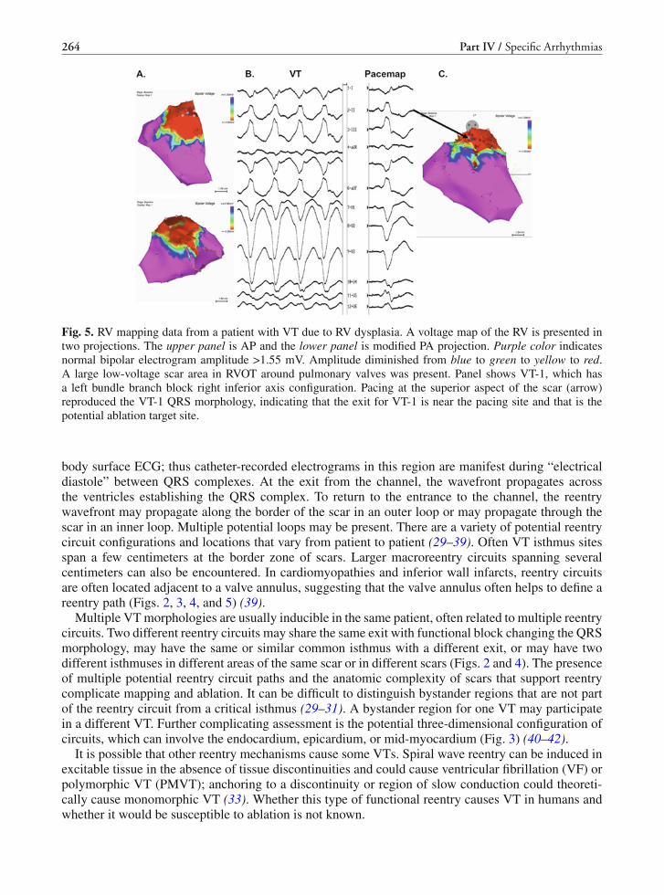
264 Part IV / Specific Arrhythmias
Fig. 5. RV mapping data from a patient with VT due to RV dysplasia. A voltage map of the RV is presented intwo projections. The upper panel is AP and the lower panel is modified PA projection. Purple color indicatesnormal bipolar electrogram amplitude >1.55 mV. Amplitude diminished from blue to green to yellow to red.A large low-voltage scar area in RVOT around pulmonary valves was present. Panel shows VT-1, which hasa left bundle branch block right inferior axis configuration. Pacing at the superior aspect of the scar (arrow)reproduced the VT-1 QRS morphology, indicating that the exit for VT-1 is near the pacing site and that is thepotential ablation target site.
body surface ECG; thus catheter-recorded electrograms in this region are manifest during “electricaldiastole” between QRS complexes. At the exit from the channel, the wavefront propagates acrossthe ventricles establishing the QRS complex. To return to the entrance to the channel, the reentrywavefront may propagate along the border of the scar in an outer loop or may propagate through thescar in an inner loop. Multiple potential loops may be present. There are a variety of potential reentrycircuit configurations and locations that vary from patient to patient (29–39). Often VT isthmus sitesspan a few centimeters at the border zone of scars. Larger macroreentry circuits spanning severalcentimeters can also be encountered. In cardiomyopathies and inferior wall infarcts, reentry circuitsare often located adjacent to a valve annulus, suggesting that the valve annulus often helps to define areentry path (Figs. 2, 3, 4, and 5) (39).
Multiple VT morphologies are usually inducible in the same patient, often related to multiple reentrycircuits. Two different reentry circuits may share the same exit with functional block changing the QRSmorphology, may have the same or similar common isthmus with a different exit, or may have twodifferent isthmuses in different areas of the same scar or in different scars (Figs. 2 and 4). The presenceof multiple potential reentry circuit paths and the anatomic complexity of scars that support reentrycomplicate mapping and ablation. It can be difficult to distinguish bystander regions that are not partof the reentry circuit from a critical isthmus (29–31). A bystander region for one VT may participatein a different VT. Further complicating assessment is the potential three-dimensional configuration ofcircuits, which can involve the endocardium, epicardium, or mid-myocardium (Fig. 3) (40–42).
It is possible that other reentry mechanisms cause some VTs. Spiral wave reentry can be induced inexcitable tissue in the absence of tissue discontinuities and could cause ventricular fibrillation (VF) orpolymorphic VT (PMVT); anchoring to a discontinuity or region of slow conduction could theoreti-cally cause monomorphic VT (33). Whether this type of functional reentry causes VT in humans andwhether it would be susceptible to ablation is not known.

Chapter 12 / Ablation for Ventricular Tachycardia 265
Fig. 6. Recordings from a patient with idiopathic ventricular fibrillation and electrical storm are shown. Pan-els a, b, c show recordings from the electrophysiology laboratory. Frequent premature ventricular contractionswere present. LV mapping identified sites with early potentials preceding the onset of the premature ventricularcontractions (arrow at the onset of endocardial signal). Ablation at these sites abolished ventricular ectopy andepisodes of ventricular fibrillation. Panel c: post-ablation ECG demonstrates widening of QRS complex, a resultof ablation of a part of His–Purkinje network in the LV. Panel d shows the position of the RF ablation catheterin the LV at the time of ablation.
Reentry in the Purkinje SystemReentry within the Purkinje fibers and the specialized conduction system causes ˜5% of all sus-
tained monomorphic VTs encountered in patients undergoing catheter ablation. Macroreentry throughthe bundle branches occurs in patients with slowed conduction through the His–Purkinje system and isusually associated with severe left ventricular (LV) dysfunction due to dilated cardiomyopathy, valvu-lar heart disease, and less often ischemic heart disease (Fig. 1d) (43).
Left ventricular intrafascicular verapamil-sensitive VT occurs in patients without structural heartdisease. The mechanism is reentry that appears to involve a portion of the LV Purkinje fibers, mostoften in the region of the left posterior fascicle, giving rise to a characteristic right bundle branchblock (RBBB) superior axis QRS configuration and a QRS duration that is only slightly prolonged(Fig. 1b) (44).
ELECTROPHYSIOLOGICAL BASIS FOR CATHETER ABLATION
The mechanism of VT is a key determinant for selection of mapping strategies to identify abla-tion target sites. For idiopathic VT, the focal origin or critical portion of the reentry path is usually

266 Part IV / Specific Arrhythmias
contained in a very small area such that discrete lesions can abolish VT; therefore, mapping targets aprecise region. For scar-related VTs, ablation is aimed at transecting the critical VT isthmus. Ventricu-lar tachycardia isthmuses may be narrow, allowing a discrete lesion to abolish VT, or broad, requiringlarger ablation areas. In addition, in patients with unmappable VTs and multiple VTs, larger abla-tion areas targeting putative critical reentry sites, often in or near the border zone of scars, are oftenemployed. In post-MI VT, selection of catheter ablation for an individual patient should consider risksand benefits that are determined by patient characteristics, as well as the availability of appropriatefacilities with technical expertise. In patients with structural heart disease, episodes of sustained VT area marker for increased mortality and reduce quality of life in patients who have implanted defibrillatorsand structural heart disease. Antiarrhythmic medications can reduce the frequency of ICD therapies,but have disappointing efficacy and side effects (1–4). Advances in technology and understanding ofVT substrates now allow ablation of multiple and unstable VTs with acceptable safety and efficacy,even in patients with advanced heart disease. In the past, ablation was often not considered until phar-macological options had been exhausted, often after the patient had suffered substantial morbidityfrom recurrent episodes of VT and ICD shocks. At this point of time catheter ablation for VT shouldgenerally be considered early in the treatment of patients with recurrent VT.
INDICATIONS FOR VT ABLATION
Patients with Structural Heart Diseases Including Prior MI, Dilated Cardiomyopathy,and Arrhythmogenic Right Ventricular Cardiomyopathy/Dysplasia (ARVC/D)
CATHETER ABLATION OF VT IS RECOMMENDED
1. for symptomatic sustained monomorphic VT (SMVT), including VT terminated by an ICD, that recursdespite antiarrhythmic drug therapy or when antiarrhythmic drugs are not tolerated or not desired;
2. for control of incessant SMVT or VT storm that is not due to a transient reversible cause;3. for patients with frequent PVCs, NSVTs, or VT that is presumed to cause ventricular dysfunction;4. for bundle branch reentrant or interfascicular VTs;5. for recurrent sustained polymorphic VT and VF that is refractory to antiarrhythmic therapy when there
is a suspected trigger that can be targeted for ablation (1).
CATHETER ABLATION SHOULD BE CONSIDERED
1. in patients who have one or more episodes of SMVT despite therapy with one of more class I or IIIantiarrhythmic drugs;
2. in patients with recurrent SMVT due to prior MI who have LV ejection fraction >0.30 and expectationfor >1 year of survival and is an acceptable alternative to amiodarone therapy;
3. in patients with hemodynamically tolerated SMVT due to prior MI who have reasonably preserved LVejection fraction (>0.35) even if they have not failed antiarrhythmic drug therapy.
Patients Without Structural Heart DiseaseCATHETER ABLATION OF VT IS RECOMMENDED FOR PATIENTS WITH IDIOPATHIC VT1. for monomorphic VT that is causing severe symptoms;2. for monomorphic VT when antiarrhythmic drugs are not effective, not tolerated, or not desired;3. for recurrent sustained polymorphic VT and VF (electrical storm) that is refractory to antiarrhythmic
therapy when there is a suspected trigger that can be targeted for ablation.
VT CATHETER ABLATION IS CONTRAINDICATED
1. in the presence of a mobile ventricular thrombus (epicardial ablation may be considered);
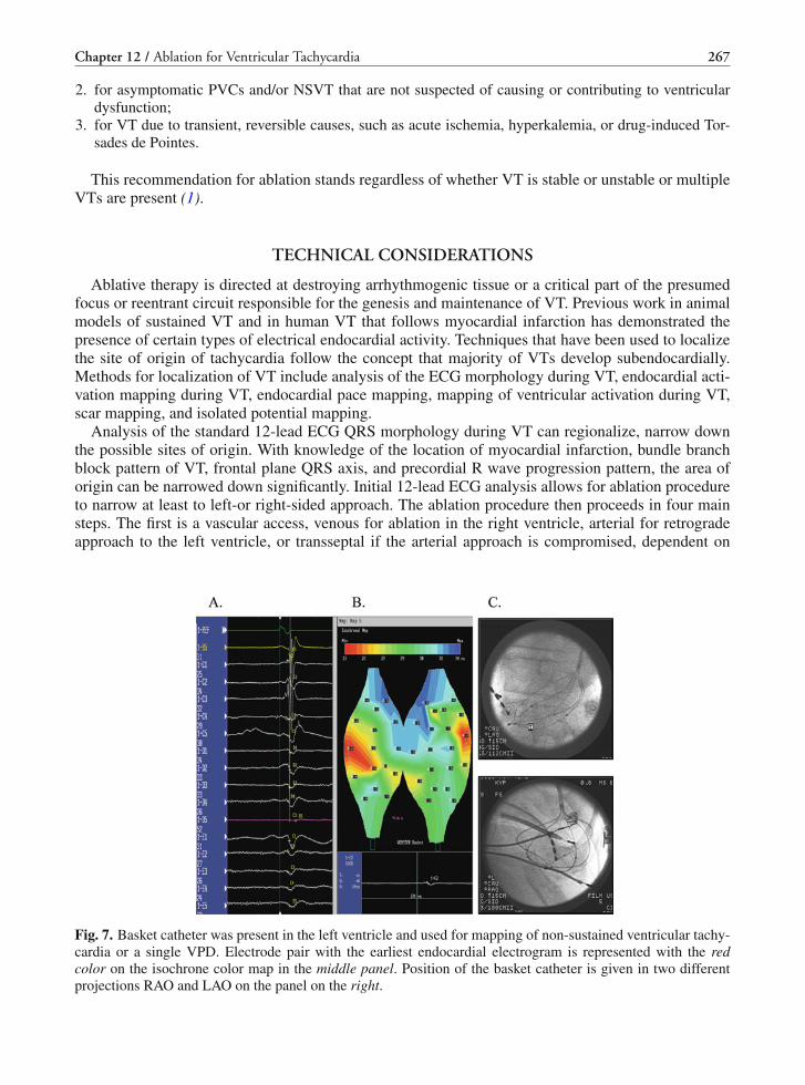
Chapter 12 / Ablation for Ventricular Tachycardia 267
2. for asymptomatic PVCs and/or NSVT that are not suspected of causing or contributing to ventriculardysfunction;
3. for VT due to transient, reversible causes, such as acute ischemia, hyperkalemia, or drug-induced Tor-sades de Pointes.
This recommendation for ablation stands regardless of whether VT is stable or unstable or multipleVTs are present (1).
TECHNICAL CONSIDERATIONS
Ablative therapy is directed at destroying arrhythmogenic tissue or a critical part of the presumedfocus or reentrant circuit responsible for the genesis and maintenance of VT. Previous work in animalmodels of sustained VT and in human VT that follows myocardial infarction has demonstrated thepresence of certain types of electrical endocardial activity. Techniques that have been used to localizethe site of origin of tachycardia follow the concept that majority of VTs develop subendocardially.Methods for localization of VT include analysis of the ECG morphology during VT, endocardial acti-vation mapping during VT, endocardial pace mapping, mapping of ventricular activation during VT,scar mapping, and isolated potential mapping.
Analysis of the standard 12-lead ECG QRS morphology during VT can regionalize, narrow downthe possible sites of origin. With knowledge of the location of myocardial infarction, bundle branchblock pattern of VT, frontal plane QRS axis, and precordial R wave progression pattern, the area oforigin can be narrowed down significantly. Initial 12-lead ECG analysis allows for ablation procedureto narrow at least to left-or right-sided approach. The ablation procedure then proceeds in four mainsteps. The first is a vascular access, venous for ablation in the right ventricle, arterial for retrogradeapproach to the left ventricle, or transseptal if the arterial approach is compromised, dependent on
Fig. 7. Basket catheter was present in the left ventricle and used for mapping of non-sustained ventricular tachy-cardia or a single VPD. Electrode pair with the earliest endocardial electrogram is represented with the redcolor on the isochrone color map in the middle panel. Position of the basket catheter is given in two differentprojections RAO and LAO on the panel on the right.

268 Part IV / Specific Arrhythmias
initial presentation of arrhythmia. The second is arrhythmia induction and the third is mapping andablation. The fourth is testing – attempt to reinduce the arrhythmia at the end of the procedure.
Mapping for Localization of VT OriginWhen VT is hemodynamically stable, mapping can be performed during VT. Many patients with
heart disease have VTs that are “unmappable” or “unstable” because they require immediate ter-mination because of hemodynamic intolerance, they cannot be reliably induced, or they frequentlychange from one VT to another. Substrate mapping or multielectrode mapping approaches have beendeveloped to guide ablation of these VTs (Figs. 6 and 7).
Mapping systems that recreate the geometry of the ventricles from point-by-point sampling whileproviding continuous display of catheter position are in common use (45, 46). Electrophysiologicaldata such as the activation time and electrogram amplitude are color coded for display (Figs. 2, 4,and 5). Cardiac motion introduces some error. Papillary muscles and other fine anatomic details arenot visible. Registration of pre-acquired computed tomographic or magnetic resonance images onmapping systems shows promise for improving anatomic definition.
Balloon or basket electrode arrays sample from multiple sites simultaneously, potentially allowingactivation to be defined from a single beat or brief run of VT (Fig. 7). A “noncontact” mapping systemmathematically reconstructs potentials from adjacent sites (45, 46).
ABLATION TECHNOLOGIES
The simplicity and safety of radiofrequency (RF) current make it the most common ablation energy.Solid 4- or 5-mm electrodes are used for ablation of idiopathic VTs, but scar-related VTs oftenrequire repeated RF applications and larger, deeper lesions that are facilitated by irrigated RF abla-tion or larger (e.g., 8 mm) electrodes. Large electrodes are convenient but reduce mapping preci-sion and have a greater risk of coagulum formation because of the greater temperature disparitiesacross the electrode compared with smaller electrodes (47, 48). Irrigated and large-tip electrodes areprone to deep heating within the tissue (Fig. 8), sometimes causing steam formation that can explodethrough the tissue (“steam pops”). Tamponade can occur but is rare during ablation of scar-relatedleft ventricular (LV) tachycardias. The risk is likely greater in the thin-walled right ventricle (RV)and with power >40 W. External irrigation has been suggested to reduce the risk of thrombus forma-tion, but administration of intravascular saline requires monitoring volume status and often diuresis(47–51).
Experience with catheter cryoablation is limited, but it may pose less risk of injury to adjacentcoronary arteries for epicardial ablation than RF ablation (52).
ENDOCARDIAL MAPPING AND ABLATION
Access to the LV for endocardial mapping is commonly achieved retrogradely across the aorticvalve. Damage to the aortic valve or coronary artery ostia is possible but rare (20). Significant vascularaccess complications, including arterial dissection, significant bleeding, and femoral atrioventricularfistulas, occurred in 2.1% of patients in one multicenter study (51). A transseptal approach to the leftatrium allows access to the LV through the mitral valve for patients with peripheral vascular diseaseor a mechanical aortic valve or for insertion of multiple mapping catheters.
Cerebral or systemic embolism is reported in 0–2.7% of patients (51). Echocardiography shouldbe performed before the procedure to detect mobile LV thrombus, which is a contraindication to LVmapping. If laminated thrombus is suspected, a several week period of anticoagulation with warfarinmay be considered in the hope of reducing the presence of friable thrombus, but no data exist assessingthis approach.

Chapter 12 / Ablation for Ventricular Tachycardia 269
During LV mapping and ablation, patients are systemically anticoagulated with heparin. After abla-tion, continued anticoagulation with aspirin or warfarin is recommended depending on the extentof ablation performed. Atrioventricular block can occur with either RV or LV ablation and may beunavoidable when VT originates from the septum.
EPICARDIAL MAPPING AND ABLATION
Endocardial ablation fails if VT originates from a deep intramural or epicardial source. Percutaneousepicardial mapping and ablation are performed as described by Sosa and colleagues (53, 54). Thepericardial space is entered with an epidural needle under fluoroscopic imaging with contrast injection,followed by placement of a guidewire and introducer sheath for the mapping catheter. In the absenceof adhesions, the catheter moves freely in the pericardial space for mapping (Fig. 3) (53). Epicardialablation with standard solid RF ablation electrodes can be successful, but absence of cooling fromcirculating blood often results in low-power heating with limited lesion creation, requiring irrigatedRF ablation (55, 56).
The risk of injury to epicardial coronary arteries is a major concern and is dependent on proximity tothe vessel, overlying fat, and vessel diameter, with smaller vessels more susceptible to thermal injury.Proximity to the coronary arteries is assessed from angiography while the ablation catheter is at thetarget site; ablation directly on major vessels is avoided (51, 56).
Pericardial bleeding can occur but rarely requires prolonged drainage or surgical treatment. Rarely,puncture of a subdiaphragmatic vessel can cause significant abdominal bleeding. The left phrenicnerve is potentially susceptible to injury as it courses down the lateral aspect of the LV but can beidentified by pacing from the ablation catheter. After ablation, symptoms of pericarditis are commonbut generally resolve within a few days.
Pericardial adhesions after cardiac surgery often prevent percutaneous access, although limitedaccess to the inferior wall may be possible. A direct surgical approach to the pericardial space viaa subxiphoid pericardial window or thoracotomy in the electrophysiology laboratory can be used formapping and ablation (57).
Fig. 8. One-week old radiofrequency lesion in a patient with nonischemic cardiomyopathy. Lesion was createdwith the “Chilli” closed irrigation catheter. There was a clear hemorrhagic delineation of the lesion that was8 mm deep and about 16 mm wide. There was some endocardial sparing as well.

270 Part IV / Specific Arrhythmias
ABLATION OF MONOMORPHIC VT IN STRUCTURAL HEART DISEASES
Patients with VT and structural heart diseases often have depressed ventricular function, heart fail-ure, and coronary artery disease. Induced VT is often poorly tolerated, requiring prompt restoration ofsinus rhythm, rendering VT unmappable with catheter techniques. Inability to tolerate long episodesof VT was a deterrent to ablation in the past but is now commonly addressed with substrate map-ping approaches or multielectrode catheter mapping. Even so, induction of VT is generally neededto confirm the type of VT and to assess the effect of ablation, placing the patient at risk of hemo-dynamic deterioration and ischemia during periods of tachycardia and hypotension. Assessment andmanagement of these risks begin before the procedure. Intravascular volume status should be opti-mized. A recent assessment of potential myocardial ischemia with perfusion imaging or angiographyis helpful to guide management decisions if hypotension or pulmonary edema occurs during the proce-dure. Before the procedure, it is desirable to discontinue antiarrhythmic drugs that may interfere withinitiation of VTs, but this may not be safe or necessary when VT occurs despite therapy.
Consideration of risks and benefits should be individualized. Procedural risks are likely to beincreased in the elderly and in patients with severe underlying heart disease. In many patients, VTrecurrences are acceptably reduced by antiarrhythmic drug therapy, and ablation is not required. Recur-rent VT episodes can be a marker for deterioration of heart failure and mortality risk despite an ICD.Some patients warrant assessment for cardiac transplantation or LV assist device placement so that theeligibility of these options is known in the event of further deterioration.
Scar-Related Sustained Monomorphic VTScar-related reentry is the most common cause of sustained monomorphic VT in patients with struc-
tural heart disease. Causes include old myocardial infarction, cardiomyopathies, and surgical incisions.Ventricular scars are composed of variable regions of dense fibrosis that create conduction block andsurviving myocyte bundles with interstitial fibrosis and diminished coupling, which produce circuitousslow-conduction paths that promote reentry (36, 37).
Many scar-related reentry circuits can be modeled as having an isthmus or channel composed ofa small mass of tissue that does not contribute to the surface ECG. The QRS complex is inscribedwhen the excitation wavefront emerges from an exit along the border of the scar and spreads acrossthe ventricles (Fig. 9). Reentry circuit configurations and locations vary from patient to patient. Largemacroreentry circuits spanning several centimeters are common causes of slow VTs. Scars causing VToften border a valve annulus that forms a border segment for part of the reentry circuit (35–39).
Repeated programmed stimulation typically induces >1 monomorphic VT. Multiple VTs can be dueto different circuits in widely disparate areas of scar, different exits from the same region of scar, orchanges in activation remote from the circuit due to functional regions of block. Ablation at one regionoften abolishes >1 VTs (Fig. 4) (59, 60).
QRS Morphology as a Guide to the VT ExitThe QRS morphology of VT is an indication of the location of the circuit exit. VTs with a left
bundle branch block-like configuration in lead V1 have an exit in the RV or interventricular septum;dominant R waves in V1 indicate an LV exit. The frontal plane axis indicates whether the exit is onthe inferior wall, which produces a superiorly directed axis, or on the anterior (cranial) wall, whichproduces an inferiorly directed axis. The mid precordial leads, V3 and V4, provide an indication ofexit location between the base and apex; apical exits generate dominant S waves; basal exits generatedominant R waves. VTs that originate in the subepicardium generally have a longer QRS duration andslower QRS upstrokes in the precordial leads than those with an endocardial exit (61, 62).

Chapter 12 / Ablation for Ventricular Tachycardia 271
Fig. 9. Schematics used for entrainment mapping in hemodynamically stable patient.
Mapping During VTWhen VT is stable for mapping, an isthmus can be targeted during VT (59, 61). In activation
sequence maps, the circuit exit and isthmus are identified from presystolic (before the QRS onset)and diastolic (earlier than presystolic) activation, respectively, and are confirmed with pacing maneu-vers such as entrainment (Figs. 2, 4, and 9) (29, 35, 60, 62).
Multielectrode and noncontact mapping systems identify endocardial exit regions of presystolicelectric activity in >90% of VTs. Some diastolic activity is identified in approximately two-thirds ofpatients, but complete reentry circuits are defined in <20% (29, 35, 60).
Because of heterogeneity of scars with multiple potential conduction paths and channels, electro-gram timing alone is not a reliable guide for targeting a specific reentry circuit isthmus (29, 35). Con-firmation that a site is involved in the reentry circuit can be obtained by pacing at the site (entrainmentmapping; Figs. 2, 4, and 9) or reproducible VT termination by mechanical pressure at the site (29, 35,62–64). It is not necessary to define the entire circuit if an isthmus can be identified for ablation.
Substrate MappingFor multiple and unstable VTs, substrate mapping can be used to identify regions of scar and
potential reentry circuit channels during stable sinus or paced rhythm. Voltage maps are created fromthree-dimensional anatomic plots of electrogram amplitude (Figs. 2, 3, and 4). Low-voltage regions(<0.55 mV in bipolar recordings) identify areas of scar (65–67).

272 Part IV / Specific Arrhythmias
The low-voltage region contains the reentry circuit but is usually too large for ablation of the entireregion or its circumference. Additional markers of the circuit exit or isthmus are sought for ablation.During sinus rhythm the exit can often be located by pace mapping along the scar border. Pacing inthe exit region replicates the QRS morphology of VT. (Figs. 2 and 4) (68, 69).
A potential isthmus or channel within low-voltage regions can also be identified during sinusrhythm, suggested by low-amplitude isolated potentials and late potentials inscribed after the endof the QRS complex. Pacing in a channel produces a QRS that emerges after a delay due to slowconduction through the channel (Figs. 2 and 4). When the stimulated wavefront propagates througha reentry circuit exit region, the paced QRS morphology resembles VT, strongly suggesting that thepacing site is in a reentry circuit isthmus. If the wavefront leaves the scar by another path, the pacedQRS morphology may differ from VT or resemble a different VT. Absence of channels may indicatethat functional block is present during VT or that the channels are not endocardial (69, 70).
Areas of dense fibrosis that form some reentry circuit borders can be detected as electrically unex-citable scar, where pacing does not capture (pacing threshold >10 mA at 2-ms pulse width). Markingelectrically unexcitable scar areas creates a visual map of potential channels, which can then be inves-tigated for ablation. Not all scars have large electrically unexcitable scar areas; narrow bands of normaltissue within fibrosis likely escape detection on the basis of pacing threshold. Areas of block can alsobe indicated by very low-amplitude regions on either side of a larger amplitude channel (66, 68–70).
Most recent ablation series include patients with multiple and unstable VTs targeted by these tech-niques. When VT is stable, substrate mapping during sinus rhythm can be used to identify regions forfurther evaluation during VT, minimizing the time spent in VT. Brief entrainment is often possible,even for unstable VTs, to confirm the location of a reentry circuit, potentially allowing ablation with asmaller number of RF lesions than when ablation is guided only by substrate mapping.
Inducible VTs and Ablation End PointsOn average, three different monomorphic VTs are usually inducible with repeated programmed
stimulation. VTs that have been observed to occur spontaneously are often referred to as “clinicalVTs”; the others are often designated “nonclinical,” implying that they are unlikely to occur sponta-neously. This distinction can be problematic; nonclinical VTs may subsequently occur spontaneously(71, 72). In many patients, the ECG morphology of spontaneous VT is not known because an ICDpromptly terminates VT.
Abolishing incessant VT and inducible clinical VT is generally the minimum end point that definesa successful procedure. Some centers attempt to abolish all inducible VTs or all inducible mappableVTs, aiming to reduce recurrences. These different end points have not been compared directly. Afterablation, at least one VT is no longer inducible in 73–100% of patients, and all inducible VTs are abol-ished in 38–95% of patients. Remaining inducible VTs are often faster and are induced by aggressivestimulation (72–75).
When the targeted VT remains inducible after ablation, the recurrence risk exceeds 60% (71, 74).Absence of inducible VT has been associated with a lower but still significant incidence of recurrence,ranging from <3 to 27% in single-center reports (73–75). Inducible, nonclinical VTs are associatedwith increased risk of recurrence in some studies (75). Healing of initial ablation lesions and reductionof antiarrhythmic medications likely contribute to recurrences, and some patients benefit from a repeatprocedure. Even when VT recurs, the frequency of episodes is often reduced. In a multicenter trial in146 patients, the immediate effect on inducible VT did not predict outcomes; VT recurred in 44% ofpatients who had no inducible VT and 46% of those who had inducible VT (51). The frequency ofspontaneous VT during short-term follow-up was reduced by >75% in the majority of patients.

Chapter 12 / Ablation for Ventricular Tachycardia 273
Purkinje System VTA diseased Purkinje system causes VT in 8% of patients with structural heart disease referred for
catheter ablation (13). VT can be due to macroreentry or focal automaticity that is catecholaminesensitive. VT has an ECG appearance of typical left or right bundle branch block and is often associatedwith severely depressed LV function.
In bundle branch reentrant VT, the circulating wavefront propagates up the left bundle branch andantegrade down the right bundle branch, producing VT with a QRS configuration of typical left bundlebranch block (Fig. 1d). Less frequently, the circuit revolves in the opposite direction, producing a rightbundle branch block configuration. Ablation of the right or left bundle branch is curative, but ablationof the right bundle is generally preferred to avoid hemodynamic consequences of left bundle branchblock. Associated infranodal conduction delay and the frequent presence of other, scar-related VTs inapproximately a third of patients warrant pacemaker or ICD implantation as well.
ABLATION IN SPECIFIC DISEASES
Prior Myocardial InfarctionPatients have generally been referred after failed drug therapy and ICD implantation. Ablation is
initially successful, abolishing one or more VTs in 77–95% of patients. During follow-up, previouslyineffective antiarrhythmic drugs, frequently amiodarone, are often continued. VT recurs in 19–50% ofpatients, although the frequency is reduced in the majority. Multiple morphologies of VT and unstableVTs are associated with a higher recurrence risk. Failure of endocardial ablation can be due to intra-mural or epicardial reentry circuits. Epicardial circuits are present in 10–30% of patients, more oftenin inferior wall as opposed to anterior wall infarctions (28, 41, 42).
Major complications occur in 5–10% of patients including cardiac tamponade, shock, stroke, aorticvalve injuries, and vascular injuries. Procedure mortality, 2.7% in one trial, is often due to failure tocontrol VT rather than complications (51).
The annual mortality after catheter ablation ranges from 5 to >20%, with death from progressiveheart failure being the most common cause (75, 76). The substantial mortality is consistent with theseverity of heart disease and association of spontaneous VT with mortality and heart failure even whenVT is treated effectively by an ICD (4). Older age and greater LV size and dysfunction increase mor-tality (75, 76). The potential for ablation to adversely affect LV function is cause for concern, althoughassessment of LV ejection fraction after ablation has not shown deterioration (77). Confining ablationlesions to regions of low-amplitude scar and attention to appropriate medical therapy beneficial topatients with LV dysfunction are prudent.
Dilated Cardiomyopathy Without Coronary Artery DiseaseSustained monomorphic VT is not common in nonischemic dilated cardiomyopathies, but 80%
of those that occur are due to scar-related reentry, with the remainder due to bundle branch reentryor a focal origin. Myocardial scar has been demonstrated with magnetic resonance imaging and isfrequently abutting a valve annulus (77, 78). Progressive replacement fibrosis is a likely cause. VTablation is often more difficult than in coronary artery disease, but recurrent arrhythmias are controlledin >60% of patients (23, 24). Reentry circuits require epicardial ablation in a third or more of patients(Figs. 3 and 8). Occasionally, incessant idiopathic VT or ventricular ectopy causes a tachycardia-induced cardiomyopathy that improves after ablation (79).

274 Part IV / Specific Arrhythmias
RV CardiomyopathiesScar-related RV tachycardias occur in idiopathic cardiomyopathy, arrhythmogenic RV dyspla-
sia/cardiomyopathy, and cardiac sarcoidosis. VT has a left bundle branch block configuration. Reentrycircuits are often in scars adjacent to the tricuspid or pulmonic annulus. Focal origin VTs or epicardialreentry with a focal endocardial breakthrough also occurs. Areas of scar associated with VT can alsobe identified from delayed enhancement on magnetic resonance imaging. Ablation is often successfulin the short term, but late arrhythmia recurrences are common, suggesting disease progression. Cardiacperforation is the major complication but is infrequent (55, 58, 61, 67, 79, 82, 84).
Repaired Congenital Heart DiseasesScar-related VT involving RV regions of repair also occur late after surgical correction of congenital
heart disease, notably tetralogy of Fallot. Catheter ablation is feasible and can reduce recurrences, butin a small series only 43% of 14 patients with tetralogy of Fallot remained free of VT (84). Densefibrosis or synthetic patches used in the repair may hinder ablation in some patients.
IDIOPATHIC MONOMORPHIC VTS
Idiopathic monomorphic VTs tend to present in young, healthy individuals, often precipitated byexertion or emotion. The most common forms have a focal origin in the RV or LV outflow regions (13,15, 17, 81–85). Up to 12% are due to reentry involving LV Purkinje fascicles. Initial evaluation shouldexclude structural heart disease, particularly arrhythmogenic RV dysplasia, cardiac sarcoidosis, andother cardiomyopathies. Any sinus rhythm ECG abnormality should increase suspicion for underly-ing structural disease. Areas of delayed enhancement consistent with fibrosis on magnetic resonanceimaging suggest scar-related VT. Magnetic resonance imaging in patients with idiopathic RV outflowtract (RVOT) VTs often shows focal areas of thinning, fatty infiltration, and diminished wall motionthat can be normal variants (83).
The prognosis is good and the risk of sudden death is remote provided that underlying heart diseaseis excluded. Rarely, an RVOT focus is the trigger for idiopathic ventricular fibrillation or polymorphicVT in the presence of an inherited arrhythmia syndrome (86). Incessant VT or extremely frequentectopy can cause depressed LV function that improves after the arrhythmia is controlled (80).
Fig. 10. Typical catheter positions for ablation of idiopathic ventricular tachycardia originating in RVOT.

Chapter 12 / Ablation for Ventricular Tachycardia 275
Long-term therapy with β-adrenergic blockers, calcium channel blockers, or both or with a mem-brane active antiarrhythmic drug is often effective. Catheter ablation is an accepted therapy for sus-tained or symptomatic arrhythmias when medications are ineffective or not desired (1).
Outflow-Type Idiopathic VTOutflow-type VTs have an inferiorly directed frontal plane axis (Figs. 1a and 10). Most originate
in the RV and have a left bundle branch block configuration in V1. VT is often induced with isopro-terenol infusion and burst pacing and can be terminated with high-dose adenosine, consistent withcAMP-mediated delayed afterdepolarizations and triggered activity as the mechanism (82, 89). Anacquired somatic cell mutation in the inhibitory G protein Gi2 has been found in the RV in somepatients (87).
Clinical presentations include paroxysmal exercise-induced VT, repetitive monomorphic VT, withbursts of non-sustained VT separated by one or more sinus beats, and frequent premature ventricularbeats. The location of the discrete VT focus is suggested by the ECG, but the close anatomic relationsof the RVOT, LV outflow tract, and their great vessels preclude definitive localization from the ECGalone (88). Success rates for catheter ablation range from 85 to 97% (82, 89). Long-term follow-up islimited, but recurrence rates are generally low.
The variety of potential locations makes it desirable to perform mapping and ablation during VT.Inability to initiate the arrhythmia in the laboratory, likely related to sedation and systemic absorptionof local anesthetic, is a major cause of ablation failure. The first step is to induce VT, confirm thediagnosis, and obtain a 12-lead ECG to allow pace mapping to guide ablation if VT becomes quiescent.
RVOT TachycardiaThe free wall of the RVOT is oriented diagonally from posterior to anterior as a catheter moves from
right to left. The side of the RVOT that is opposite the free wall sits anterior to the aorta and superiorto the LV outflow tract. VT originating from the RVOT has a left bundle branch block configurationin V1 with a transition to more positive QRS by V4. A free wall origin is suggested by QRS duration>140 ms and notches in the inferior leads (II, III, AVF). Deeper S waves in aVL than in aVR suggesta leftward superior focus, and this ratio decreases with sites located more rightward and inferior inRVOT (90, 91).
Precise localization relies on combined use of pace mapping and activation mapping. In mostpatients, a site with a pace map QRS that exactly matches that of VT can be identified at or veryclose to the VT focus, but excellent pace maps may be obtained several millimeters distant from theearliest activation (92). Earliest activation during VT or premature ventricular contractions from thefocus typically precedes the QRS onset by 15–45 ms, with a characteristic QS complex in the unipolarsignal (92). RF application often produces acceleration followed by termination of VT and renders itno longer inducible.
Successful ablation is achieved in 65–97% of patients (90, 93). Serious complications are infrequentbut include perforation of the RV free wall. Although the left main coronary artery can be in closeproximity to the posterior aspect of the RVOT (Fig. 10), we are not aware of any cases of recognizedcoronary injury (88). Infrequently, the focus is adjacent to the His bundle, where heart block is a riskof ablation (94).
Pulmonary Artery VTsVT can originate from sleeves of myocardium extending along the pulmonary artery, above the
pulmonary valve, requiring ablation from within the pulmonary artery (95, 96). VT typically has largeR waves in the inferior leads and greater R/S ratio in lead V2 than in RVOT VT. Ablation risks are notwell defined for these infrequent VTs.

276 Part IV / Specific Arrhythmias
LV Outflow Tract VTsLV outflow tract VT can originate from the superior basal region of the left interventricular septum
or LV free wall, aortic sinuses of Valsalva, or LV epicardium. The QRS has an inferior axis, but theprecordial transition to positive R waves is earlier, with prominent R waves often present in V1 or V2compared with RVOT tachycardia.
LV endocardial VTs can originate from foci below the aortic valve (97–99). Heart block and aorticvalve and coronary artery injury are potential risks for ablation but are reported rarely.
Aortic Cusp VTsVT originating from extension of ventricular myocardium above the aortic annulus, requiring abla-
tion from the left or right sinus of Valsalva, caused 21% of idiopathic VTs in one series (64). Pacemapping in the aortic sinus may require high output and may not exactly reproduce the VT QRS.Activation mapping is required and typically shows a two-component electrogram with the earliestdeflection preceding the QRS complex by an average of 39 ms (97, 98).
The potential for acute occlusion of the left main or right coronary arteries is a major risk con-sideration. Coronary angiography and intracardiac ultrasound imaging have been used to define theproximity of the coronary ostia to the ablation site. RF ablation has been performed safely at sites>8 mm below the coronary artery ostia with careful continuous monitoring of catheter position duringthe RF application. Standard RF ablation with tip temperature maintained at <55◦C has been suggestedto prevent aortic valve damage observed in animal studies (99).
Epicardial Outflow Tract VTsEpicardial foci cause VTs with a QRS onset that is often slurred, creating a pseudo-delta wave
appearance. The interval from the onset of the QRS to earliest maximal deflection in the precordialleads is delayed, consistent with late access of the wavefront to the endocardial Purkinje system (100).The VT focus may be adjacent to an epicardial vein that can be cannulated via the coronary sinus, andsuccessful ablation has been performed via this route as well as via percutaneous pericardial access.When proximity to a epicardial coronary artery precludes catheter ablation, a direct surgical approachcan be an option (100).
Mitral Annulus VTFocal VTs from the mitral annulus accounted for 5% of 352 idiopathic VTs in one series. VT has a
right bundle branch block or RS pattern in V1 and monophasic R or RS pattern in leads V2–V6. TheQRS axis depends on the location on the annulus. Endocardial ablation is usually successful; ablationfrom within the coronary sinus may be required occasionally (101).
LV Fascicular Idiopathic VTVerapamil-sensitive idiopathic LV tachycardia typically presents as exercise-related VT between the
ages of 15 and 40 years; 60–80% of patients are male (102–104). VT has a right bundle branch blockconfiguration, most commonly with a superiorly directed frontal plane axis (Fig. 1b).
VT is due to reentry involving fascicles of the left bundle and an abnormal region, possibly also inthe Purkinje system, with slow conduction that is sensitive to verapamil and that is depolarized earlyin diastole in VT and in sinus rhythm. More than 80% of these VTs involve the posterior fascicles ofthe left bundle, identified along the inferoseptal aspect of the LV (105, 106).
Ablation targets the anterograde Purkinje potentials or diastolic potentials during VT, avoiding theproximal Purkinje system to avoid causing left bundle branch block. Mechanical trauma from cathetermanipulation often terminates VT and prevents reinitiation. Ablation then targets the site of mechanical

Chapter 12 / Ablation for Ventricular Tachycardia 277
termination, low amplitude, or diastolic sinus rhythm potentials or creates a line of lesions through thisregion (102, 103). Efficacy is >80%. Complications are infrequent.
ABLATION FOR POLYMORPHIC VT AND VENTRICULAR FIBRILLATION
Recurrent polymorphic VT causing “electrical storm” not due to ongoing acute ischemia is rare butis seen in idiopathic ventricular fibrillation, long QT syndromes, Brugada syndrome, and myocardialinfarction (1, 86, 105–107). VT is often initiated by premature beats from one or a few foci that can beablated if they occur with enough frequency to be located (Fig. 6a–d). Most appear to originate fromthe Purkinje system and have sharp presystolic potentials recorded from the focus. Less frequently,an RVOT focus is a trigger (86). Successful ablation requires the presence of spontaneous ectopicbeats for mapping. Electrical storms can wax and wane with long periods of quiescence. Immediatetransport of the patient to the laboratory when the arrhythmia is active is warranted if ablation is to beattempted. In selected patients, excellent outcomes can be achieved, with 90% of patients free fromrecurrent arrhythmias during follow-up, but the number of patients reported is small (1, 86, 106).
Advances over the past decade now allow ablation of multiple, hemodynamically unstable, epi-cardial, and polymorphic VTs, formerly considered unmappable. Specific locations for the origins ofidiopathic VT outside of the RVOT have been defined that can be expected to improve overall successfor ablation of these arrhythmias. These procedures are often more difficult than ablation of manysupraventricular tachycardias and are best approached by experienced operators in experienced labo-ratories. The field continues to benefit from technological developments (Fig. 11). When the expertiseis available, catheter ablation should be considered earlier in the therapeutic armamentarium for treat-ment of recurrent VT.
Fig. 11. New and emerging catheter technologies may offer better visualization and contact within the mappingarea in the ventricles. This is the catheter that would allow direct visualization of the ventricular wall by injectingsmall stream of saline through the center of the small hood at the end of the catheter. The same saline streammay be used for ablation.
DISCLOSURES
Dr Kocovic has received speaking honoraria from Medtronic, Inc, Boston Scientific, Inc, St Jude,Inc, and is a consultant to and has received research support from Medtronic, Boston Scientific IncGuidant and St Jude.

278 Part IV / Specific Arrhythmias
REFERENCES
1. Aliot EM, Stevenson WG, Almendral-Garrote JM, Bogun F, Calkins CH, Delacretaz E, Della Bella P, Hindricks G,Jaïs P, Josephson ME, Kautzner J, Kay GN, Kuck KH, Lerman BB, Marchlinski F, Reddy V, Schalij MJ, Schilling R,Soejima K, Wilber D (2009) EHRA/HRS expert consensus on catheter ablation of ventricular arrhythmias: developedin a partnership with the European heart rhythm association (EHRA), a registered branch of the European societyof cardiology (ESC), and the heart rhythm society (HRS); in collaboration with the American college of cardiology(ACC) and the American heart association (AHA). European heart rhythm association (EHRA); registered branchof the European society of cardiology (ESC); heart rhythm society (HRS); American college of cardiology (ACC);American heart association (AHA). Heart Rhythm 6(6):886–933
2. Connolly SJ, Dorian P, Roberts RS, Gent M, Bailin S, Fain ES, Thorpe K, Champagne J, Talajic M, Coutu B, GronefeldGC, Hohnloser SH (2006) Comparison of beta-blockers, amiodarone plus beta-blockers, or sotalol for prevention ofshocks from implantable cardioverter defibrillators: the OPTIC Study: a randomized trial. JAMA 295:165–171
3. Schron EB, Exner DV, Yao Q, Jenkins LS, Steinberg JS, Cook JR, Kutalek SP, Friedman PL, Bubien RS, Page RL,Powell J (2002) Quality of life in the antiarrhythmics versus implantable defibrillators trial: impact of therapy andinfluence of adverse symptoms and defibrillator shocks. Circulation 105:589–594
4. Moss AJ, Greenberg H, Case RB, Zareba W, Hall WJ, Brown MW, Daubert JP, McNitt S, Andrews ML, Elkin AD(2004) Long-term clinical course of patients after termination of ventricular tachyarrhythmia by an implanted defibril-lator. Circulation 110:3760–3765
5. Brugada J, Berruezo A, Cuesta A, Osca J, Chueca E, Fosch X, Wayar L, Mont L (2003) Nonsurgical transtho-racic epicardial radiofrequency ablation: an alternative in incessant ventricular tachycardia. J Am Coll Cardiol 41:2036–2043
6. Bansch D, Oyang F, Antz M, Arentz T, Weber R, Val-Mejias JE, Ernst S, Kuck KH (2003) Successful catheter ablationof electrical storm after myocardial infarction. Circulation 108:3011–3016
7. Lopera G, Stevenson WG, Soejima K, Maisel WH, Koplan B, Sapp JL, Satti SD, Epstein LM (2004) Identificationand ablation of three types of ventricular tachycardia involving the His-Purkinje system in patients with heart disease.J Cardiovasc Electrophysiol 15:52–58
8. Haissaguerre M, Extramiana F, Hocini M, Cauchemez B, Jais P, Cabrera JA, Farre G, Leenhardt A, Sanders P, ScaveeC, Hsu LF, Weerasooriya R, Shah DC, Frank R, Maury P, Delay M, Garrigue S, Clementy J (2003) Mapping andablation of ventricular fibrillation associated with long-QT and Brugada syndromes. Circulation 108:925–928
9. Buxton AE, Waxman HL, Marchlinski FE, Simson MB, Cassidy D, Josephson ME (1983) Right ventricular tachycar-dia: clinical and electrophysiologic characteristics. Circulation 68:917–927
10. Ito S, Tada H, Naito S, Kurosaki K, Ueda M, Hoshizaki H et al (2003) Development and validation of an ECG algorithmfor identifying the optimal ablation site for idiopathic ventricular outflow tract tachycardia. J Cardiovasc Electrophysiol14:1280–1286
11. Kamakura S, Shimizu W, Matsuo K, Taguchi A, Suyama K, Kurita T et al (1998) Localization of optimal ablation siteof idiopathic ventricular tachycardia from right and left ventricular outflow tract by body surface ECG. Circulation98:1525–1533
12. Lerman BB, Belardinelli L, West GA, Berne RM, DiMarco JP (1986) Adenosine-sensitive ventricular tachycardia:evidence suggesting cyclic AMP-mediated triggered activity. Circulation 74:270–280
13. Lerman BB, Stein K, Engelstein ED, Battleman DS, Lippman N, Bei D et al (1995) Mechanism of repetitive monomor-phic ventricular tachycardia. Circulation 92:421–429
14. Sung RJ, Keung EC, Nguyen NX, Huycke EC (1988) Effects of beta-adrenergic blockade on verapamil-responsive andverapamil-irresponsive sustained ventricular tachycardias. J Clin Invest 81:688–699
15. Tada H, Tadokoro K, Miyaji K, Ito S, Kurosaki K, Kaseno K et al (2008) Idiopathic ventricular arrhythmias arisingfrom the pulmonary artery: prevalence, characteristics, and topography of the arrhythmia origin. Heart Rhythm 5:419–426
16. Antzelevitch C, Shimizu W (2002) Cellular mechanisms underlying the long QT syndrome. Curr Opin Cardiol 17:43–51
17. Lerman BB (2007) Mechanism of outflow tract tachycardia. Heart Rhythm 4:973–97618. Damle RS, Landers M, Kelly PA, Reiter MJ, Mann DE (1998) Radiofrequency catheter ablation of idiopathic left
ventricular tachycardia originating in the left anterior fascicle. Pacing Clin Electrophysiol 21:1155–115819. Huelsing DJ, Spitzer KW, Pollard AE (2003) Spontaneous activity induced in rabbit Purkinje myocytes during coupling
to a depolarized model cell. Cardiovasc Res 59:620–62720. Spitzer KW, Pollard AE, Yang L, Zaniboni M, Cordeiro JM, Huelsing DJ (2006) Cell-to-cell electrical interactions
during early and late repolarization. J Cardiovasc Electrophysiol 17(Suppl 1):S8–S1421. Belhassen B, Caspi A, Miller H, Shapira I, Laniado S (1984) Extensive endocardial mapping during sinus rhythm and
ventricular tachycardia in a patient with arrhythmogenic right ventricular dysplasia. J Am Coll Cardiol 4:1302–1306

Chapter 12 / Ablation for Ventricular Tachycardia 279
22. Delacretaz E, Stevenson WG, Ellison KE, Maisel WH, Friedman PL (2000) Mapping and radiofrequency catheter abla-tion of the three types of sustained monomorphic ventricular tachycardia in nonischemic heart disease. J CardiovascElectrophysiol 11:11–17
23. Eckart RE, Hruczkowski TW, Tedrow UB, Koplan BA, Epstein LM, Stevenson WG (2007) Sustained ventriculartachycardia associated with corrective valve surgery. Circulation 116:2005–2011
24. Ellison KE, Friedman PL, Ganz LI, Stevenson WG (1998) Entrainment mapping and radiofrequency catheter ablationof ventricular tachycardia in right ventricular dysplasia. J Am Coll Cardiol 32:724–728
25. Josephson ME, Horowitz LN, Farshidi A, Kastor JA (1978) Recurrent sustained ventricular tachycardia. 1. Mecha-nisms. Circulation 57:431–440
26. Josephson ME, Almendral JM, Buxton AE, Marchlinski FE (1987) Mechanisms of ventricular tachycardia. Circulation75(Pt 2):III41–III47
27. Koplan BA, Soejima K, Baughman K, Epstein LM, Stevenson WG (2006) Refractory ventricular tachycardia secondaryto cardiac sarcoid: electrophysiologic characteristics, mapping, and ablation. Heart Rhythm 3:924–929
28. Sosa E, Scanavacca M, D’Avila A, Piccioni J, Sanchez O, Velarde JL et al (1998) Endocardial and epicardial abla-tion guided by nonsurgical transthoracic epicardial mapping to treat recurrent ventricular tachycardia. J CardiovascElectrophysiol 9:229–239
29. Stevenson WG, Khan H, Sager P, Saxon LA, Middlekauff HR, Natterson PD et al (1993) Identification of reentry circuitsites during catheter mapping and radiofrequency ablation of ventricular tachycardia late after myocardial infarction.Circulation 88(Pt 1):1647–1670
30. Dillon SM, Allessie MA, Ursell PC, Wit AL (1988) Influences of anisotropic tissue structure on reentrant circuits inthe epicardial border zone of subacute canine infarcts. Circ Res 63:182–206
31. Downar E, Harris L, Mickleborough LL, Shaikh N, Parson ID (1988) Endocardial mapping of ventricular tachycardiain the intact human ventricle: evidence for reentrant mechanisms. J Am Coll Cardiol 11:783–791
32. Downar E, Kimber S, Harris L, Mickleborough L, Sevaptsidis E, Masse S et al (1992) Endocardial mapping of ven-tricular tachycardia in the intact human heart. II. Evidence for multiuse reentry in a functional sheet of survivingmyocardium. J Am Coll Cardiol 20:869–878
33. Kleber AG, Rudy Y (2004) Basic mechanisms of cardiac impulse propagation and associated arrhythmias. Physiol Rev84:431–488
34. Baba S, Dun W, Cabo C, Boyden PA (2005) Remodeling in cells from different regions of the reentrant circuit duringventricular tachycardia. Circulation 112:2386–2396
35. Kocovic DZ, Harada T, Friedman PL, Stevenson WG (1999) Characteristics of electrograms recorded at reentry circuitsites and bystanders during ventricular tachycardia after myocardial infarction. J Am Coll Cardiol 34:381–388
36. de Bakker JM, van Capelle FJ, Janse MJ, Wilde AA, Coronel R, Becker AE et al (1988) Reentry as a cause of ventricu-lar tachycardia in patients with chronic ischemic heart disease: electrophysiologic and anatomic correlation. Circulation77:589–606
37. de Bakker JM, van Capelle FJ, Janse MJ, Tasseron S, Vermeulen JT, de JN et al (1993) Slow conduction in the infarctedhuman heart. ‘Zigzag’ course of activation. Circulation 88:915–926
38. El-Sherif N, Mehra R, Gough WB, Zeiler RH (1983) Reentrant ventricular arrhythmias in the late myocardial infarctionperiod. Interruption of reentrant circuits by cryothermal techniques. Circulation 68:644–656
39. Hadjis TA, Stevenson WG, Harada T, Friedman PL, Sager P, Saxon LA (1997) Preferential locations for critical reen-try circuit sites causing ventricular tachycardia after inferior wall myocardial infarction. J Cardiovasc Electrophysiol8:363–370
40. Brugada P, Abdollah H, Wellens HJ (1985) Continuous electrical activity during sustained monomorphic ventriculartachycardia. Observations on its dynamic behavior during the arrhythmia. Am J Cardiol 55:402–411
41. Kaltenbrunner W, Cardinal R, Dubuc M, Shenasa M, Nadeau R et al (1991) Epicardial and endocardial mapping ofventricular tachycardia in patients with myocardial infarction. Is the origin of the tachycardia always subendocardiallylocalized? Circulation 84:1058–1071
42. de Bakker JM, Stein M, van Rijen HV (2005) Three-dimensional anatomic structure as substrate for ventricular tachy-cardia/ventricular fibrillation. Heart Rhythm 2:777–799
43. Caceres J, Jazayeri M, McKinnie J, Avitall B, Denker ST, Tchou P et al (1989) Sustained bundle branch reentry as amechanism of clinical tachycardia. Circulation 79:256–270
44. Nakagawa H, Beckman KJ, McClelland JH, Wang X, Arruda M, Santoro I et al (1993) Radiofrequency catheter abla-tion of idiopathic left ventricular tachycardia guided by a Purkinje potential. Circulation 88:2607–2617
45. Reddy VY, Malchano ZJ, Holmvang G, Schmidt EJ, d’Avila A, Houghtaling C, Chan RC, Ruskin JN (2004) Integra-tion of cardiac magnetic resonance imaging with three-dimensional electroanatomic mapping to guide left ventricularcatheter manipulation: feasibility in a porcine model of healed myocardial infarction. J Am Coll Cardiol 44:2202–2213
46. Della Bella P, Pappalardo A, Riva S, Tondo C, Fassini G, Trevisi N (2002) Non-contact mapping to guide catheterablation of untolerated ventricular tachycardia. Eur Heart J 23:742–752

280 Part IV / Specific Arrhythmias
47. Soejima K, Delacretaz E, Suzuki M, Brunckhorst CB, Maisel WH, Friedman PL, Stevenson WG (2001) Saline-cooled versus standard radiofrequency catheter ablation for infarct-related ventricular tachycardias. Circulation 103:1858–1862
48. Matsudaira K, Nakagawa H, Wittkampf FH, Yamanashi WS, Imai S, Pitha JV, Lazzara R, Jackman WM (2003) Highincidence of thrombus formation without impedance rise during radiofrequency ablation using electrode temperaturecontrol. Pacing Clin Electrophysiol 26:1227–1237
49. Yokoyama K, Nakagawa H, Wittkampf FH, Pitha JV, Lazzara R, Jackman WM (2006) Comparison of electrode coolingbetween internal and open irrigation in radiofrequency ablation lesion depth and incidence of thrombus and steam pop.Circulation 113:11–19
50. Cooper JM, Sapp JL, Tedrow U, Pellegrini CP, Robinson D, Epstein LM, Stevenson WG (2004) Ablation with aninternally irrigated radiofrequency catheter: learning how to avoid steam pops. Heart Rhythm 1:329–333
51. Calkins H, Epstein A, Packer D, Arria AM, Hummel J, Gilligan DM, Trusso J, Carlson M, Luceri R, KopelmanH, Wilber D, Wharton JM, Stevenson W (2000) Catheter ablation of ventricular tachycardia in patients with struc-tural heart disease using cooled radiofrequency energy: results of a prospective multicenter study. J Am Coll Cardiol35:1905–1914
52. Lustgarten DL, Bell S, Hardin N, Calame J, Spector PS (2005) Safety and efficacy of epicardial cryoablation in acanine model. Heart Rhythm 2:82–90
53. Sosa E, Scanavacca M, d’Avila A, Oliveira F, Ramires JA (2000) Nonsurgical transthoracic epicardial catheter ablationto treat recurrent ventricular tachycardia occurring late after myocardial infarction. J Am Coll Cardiol 35:1442–1449
54. Sosa E, Scanavacca M, D’Avila A, Antonio J, Ramires F (2004) Nonsurgical transthoracic epicardial approach inpatients with ventricular tachycardia and previous cardiac surgery. J Interv Card Electrophysiol 10:281–288
55. Soejima K, Stevenson WG, Sapp JL, Selwyn AP, Couper G, Epstein LM (2004) Endocardial and epicardial radiofre-quency ablation of ventricular tachycardia associated with dilated cardiomyopathy: the importance of low-voltagescars. J Am Coll Cardiol 43:1834–1842
56. Sosa E, Scanavacca M (2007) Percutaneous pericardial access for mapping and ablation of epicardial ventricular tachy-cardias. Circulation 115:e542–e544
57. Soejima K, Couper G, Cooper JM, Sapp JL, Epstein LM, Stevenson WG (2004) Subxiphoid surgical approach forepicardial catheter-based mapping and ablation in patients with prior cardiac surgery or difficult pericardial access.Circulation 110:1197–1201
58. Bansch D, Bocker D, Brunn J, Weber M, Breithardt G, Block M (2000) Clusters of ventricular tachycardias signifyimpaired survival in patients with idiopathic dilated cardiomyopathy and implantable cardioverter defibrillators. J AmColl Cardiol 36:566–573
59. de Chillou C, Lacroix D, Klug D, Magnin-Poull I, Marquie C, Messier M, Andronache M, Kouakam C, Sadoul N,Chen J, Aliot E, Kacet S (2002) Isthmus characteristics of reentrant ventricular tachycardia after myocardial infarction.Circulation 105:726–731
60. Soejima K, Suzuki M, Maisel WH, Brunckhorst CB, Delacretaz E, Blier L, Tung S, Khan H, Stevenson WG (2001)Catheter ablation in patients with multiple and unstable ventricular tachycardias after myocardial infarction: shortablation lines guided by reentry circuit isthmuses and sinus rhythm mapping. Circulation 104:664–669
61. Berruezo A, Mont L, Nava S, Chueca E, Bartholomay E, Brugada J (2004) Electrocardiographic recognition of theepicardial origin of ventricular tachycardias. Circulation 109:1842–1847
62. Delacretaz E, Stevenson WG (2001) Catheter ablation of ventricular tachycardia in patients with coronary heart disease,part I: mapping. Pacing Clin Electrophysiol 24:1261–1277
63. Bogun F, Good E, Han J, Tamirisa K, Reich S, Elmouchi D, Igic P, Lemola K, Oral H, Chugh A, Pelosi F, Morady F(2005) Mechanical interruption of postinfarction ventricular tachycardia as a guide for catheter ablation. Heart Rhythm2:687–691
64. El-Shalakany A, Hadjis T, Papageorgiou P, Monahan K, Epstein L, Josephson ME (1999) Entrainment/mapping criteriafor the prediction of termination of ventricular tachycardia by single radiofrequency lesion in patients with coronaryartery disease. Circulation 99:2283–2289
65. Marchlinski FE, Callans DJ, Gottlieb CD, Zado E (2000) Linear ablation lesions for control of unmappable ventriculartachycardia in patients with ischemic and nonischemic cardiomyopathy. Circulation 101:1288–1296
66. Arenal A, del Castillo S, Gonzalez-Torrecilla E, Atienza F, Ortiz M, Jimenez J, Puchol A, Garcia J, Almendral J(2004) Tachycardia-related channel in the scar tissue in patients with sustained monomorphic ventricular tachycardias:influence of the voltage scar definition. Circulation 110:2568–2574
67. Cesario DA, Vaseghi M, Boyle NG, Fishbein MC, Valderrabano M, Narasimhan C, Wiener I, Shivkumar K (2006)Value of high-density endocardial and epicardial mapping for catheter ablation of hemodynamically unstable ventric-ular tachycardia. Heart Rhythm 3:1–10
68. Kottkamp H, Wetzel U, Schirdewahn P, Dorszewski A, Gerds-Li JH, Carbucicchio C, Kobza R, Hindricks G (2003)Catheter ablation of ventricular tachycardia in remote myocardial infarction: substrate description guiding placementof individual linear lesions targeting noninducibility. J Cardiovasc Electrophysiol 14:675–681

Chapter 12 / Ablation for Ventricular Tachycardia 281
69. Arenal A, Glez-Torrecilla E, Ortiz M, Villacastin J, Fdez-Portales J, Sousa E, del Castillo S, Perez de Isla L, JimenezJ, Almendral J (2003) Ablation of electrograms with an isolated, delayed component as treatment of unmappablemonomorphic ventricular tachycardias in patients with structural heart disease. J Am Coll Cardiol 41:81–92
70. Soejima K, Stevenson WG, Maisel WH, Sapp JL, Epstein LM (2002) Electrically unexcitable scar mapping based onpacing threshold for identification of the reentry circuit isthmus: feasibility for guiding ventricular tachycardia ablation.Circulation 106:1678–1683
71. van der Burg AE, de Groot NM, van Erven L, Bootsma M, van der Wall EE, Schalij MJ (2002) Long-term follow-upafter radiofrequency catheter ablation of ventricular tachycardia: a successful approach? J Cardiovasc Electrophysiol13:417–423
72. Segal OR, Chow AW, Markides V, Schilling RJ, Peters NS, Davies DW (2005) Long-term results after ablation ofinfarct-related ventricular tachycardia. Heart Rhythm 2:474–482
73. Della Bella P, Riva S, Fassini G, Giraldi F, Berti M, Klersy C, Trevisi N (2004) Incidence and significance of pleo-morphism in patients with postmyocardial infarction ventricular tachycardia: acute and long-term outcome of radiofre-quency catheter ablation. Eur Heart J 25:1127–1138
74. O’Donnell D, Bourke JP, Furniss SS (2003) Standardized stimulation protocol to predict the long-term success ofradiofrequency ablation of postinfarction ventricular tachycardia. Pacing Clin Electrophysiol 26:348–351
75. Della Bella P, De Ponti R, Uriarte JA, Tondo C, Klersy C, Carbucicchio C, Storti C, Riva S, Longobardi M (2002)Catheter ablation and antiarrhythmic drugs for haemodynamically tolerated post-infarction ventricular tachycardia;long-term outcome in relation to acute electrophysiological findings. Eur Heart J 23:414–424
76. Nabar A, Rodriguez LM, Batra RK, Timmermans C, Cheriex E, Wellens HJ (2002) Echocardiographic predictors ofsurvival in patients undergoing radiofrequency ablation of postinfarct clinical ventricular tachycardia. J CardiovascElectrophysiol 13:S118–S121
77. Khan HH, Maisel WH, Ho C, Suzuki M, Soejima K, Solomon S, Stevenson WG (2002) Effect of radiofrequencycatheter ablation of ventricular tachycardia on left ventricular function in patients with prior myocardial infarction.J Interv Card Electrophysiol 7:243–247
78. Hsia HH, Callans DJ, Marchlinski FE (2003) Characterization of endocardial electrophysiological substrate in patientswith nonischemic cardiomyopathy and monomorphic ventricular tachycardia. Circulation 108:704–710
79. Nazarian S, Bluemke DA, Lardo AC, Zviman MM, Watkins SP, Dickfeld TL, Meininger GR, Roguin A, Calkins H,Tomaselli GF, Weiss RG, Berger RD, Lima JA, Halperin HR (2005) Magnetic resonance assessment of the substratefor inducible ventricular tachycardia in nonischemic cardiomyopathy. Circulation 112:2821–2825
80. Yarlagadda RK, Iwai S, Stein KM, Markowitz SM, Shah BK, Cheung JW, Tan V, Lerman BB, Mittal S (2005) Reversalof cardiomyopathy in patients with repetitive monomorphic ventricular ectopy originating from the right ventricularoutflow tract. Circulation 112:1092–1097
81. Miljoen H, State S, de Chillou C, Magnin-Poull I, Dotto P, Andronache M, Abdelaal A, Aliot E (2005) Electroanatomicmapping characteristics of ventricular tachycardia in patients with arrhythmogenic right ventricular cardiomyopa-thy/dysplasia. Europace 7:516–524
82. O’Donnell D, Cox D, Bourke J, Mitchell L, Furniss S (2003) Clinical and electrophysiological differences betweenpatients with arrhythmogenic right ventricular dysplasia and right ventricular outflow tract tachycardia. Eur Heart J24:801–810
83. Tandri H, Saranathan M, Rodriguez ER, Martinez C, Bomma C, Nasir K, Rosen B, Lima JA, Calkins H, BluemkeDA (2005) Noninvasive detection of myocardial fibrosis in arrhythmogenic right ventricular cardiomyopathy usingdelayed-enhancement magnetic resonance imaging. J Am Coll Cardiol 45:98–103
84. Morwood JG, Triedman JK, Berul CI, Khairy P, Alexander ME, Cecchin F, Walsh EP (2004) Radiofrequency catheterablation of ventricular tachycardia in children and young adults with congenital heart disease. Heart Rhythm 1:301–308
85. Tada H, Ito S, Naito S, Kurosaki K, Kubota S, Sugiyasu A, Tsuchiya T, Miyaji K, Yamada M, Kutsumi Y, Oshima S,Nogami A, Taniguchi K (2005) Idiopathic ventricular arrhythmia arising from the mitral annulus: a distinct subgroupof idiopathic ventricular arrhythmias. J Am Coll Cardiol 45:877–886
86. Noda T, Shimizu W, Taguchi A, Aiba T, Satomi K, Suyama K, Kurita T, Aihara N, Kamakura S (2005) Malignantentity of idiopathic ventricular fibrillation and polymorphic ventricular tachycardia initiated by premature extrasystolesoriginating from the right ventricular outflow tract. J Am Coll Cardiol 46:1288–1294
87. Lerman BB, Dong B, Stein KM, Markowitz SM, Linden J, Catanzaro DF (1998) Right ventricular outflow tract tachy-cardia due to a somatic cell mutation in G protein subunitalphai2. J Clin Invest 101:2862–2868
88. Vaseghi M, Cesario DA, Mahajan A, Wiener I, Boyle NG, Fishbein MC, Horowitz BN, Shivkumar K (2006) Catheterablation of right ventricular outflow tract tachycardia: value of defining coronary anatomy. J Cardiovasc Electrophysiol17:632–637
89. Iwai S, Cantillon DJ, Kim RJ, Markowitz SM, Mittal S, Stein KM, Shah BK, Yarlagadda RK, Cheung JW, Tan VR,Lerman BB (2006) Right and left ventricular outflow tract tachycardias: evidence for a common electrophysiologicmechanism. J Cardiovasc Electrophysiol 17:1052–1058

282 Part IV / Specific Arrhythmias
90. Vestal M, Wen MS, Yeh SJ, Wang CC, Lin FC, Wu D (2003) Electrocardiographic predictors of failure and recur-rence in patients with idiopathic right ventricular outflow tract tachycardia and ectopy who underwent radiofrequencycatheter ablation. J Electrocardiol 36:327–332
90. Tanner H, Hindricks G, Schirdewahn P, Kobza R, Dorszewski A, Piorkowski C, Gerds-Li JH, Kottkamp H (2005)Outflow tract tachycardia with R/S transition in lead V3: six different anatomic approaches for successful ablation.J Am Coll Cardiol 45:418–423
91. Yoshida Y, Hirai M, Murakami Y, Kondo T, Inden Y, Akahoshi M, Tsuda M, Okamoto M, Yamada T, Tsuboi N,Hirayama H, Ito T, Toyama J, Saito H (1999) Localization of precise origin of idiopathic ventricular tachycardia fromthe right ventricular outflow tract by a 12-lead ECG: a study of pace mapping using a multielectrode “basket” catheter.Pacing Clin Electrophysiol 22:1760–1768
92. Azegami K, Wilber DJ, Arruda M, Lin AC, Denman RA (2005) Spatial resolution of pacemapping and activa-tion mapping in patients with idiopathic right ventricular outflow tract tachycardia. J Cardiovasc Electrophysiol 16:823–829
93. Krittayaphong R, Sriratanasathavorn C, Dumavibhat C, Pumprueg S, Boonyapisit W, Pooranawattanakul S, Phrud-prisan S, Kangkagate C (2006) Electrocardiographic predictors of long-term outcomes after radiofrequency ablationin patients with right-ventricular outflow tract tachycardia. Europace 8:601–606
94. Yamauchi Y, Aonuma K, Takahashi A, Sekiguchi Y, Hachiya H, Yokoyama Y, Kumagai K, Nogami A, Iesaka Y, IsobeM (2005) Electrocardiographic characteristics of repetitive monomorphic right ventricular tachycardia originating nearthe His-bundle. J Cardiovasc Electrophysiol 16:1041–1048
95. Sekiguchi Y, Aonuma K, Takahashi A, Yamauchi Y, Hachiya H, Yokoyama Y, Iesaka Y, Isobe M (2005) Electrocar-diographic and electrophysiologic characteristics of ventricular tachycardia originating within the pulmonary artery.J Am Coll Cardiol 45:887–895
96. Timmermans C, Rodriguez LM, Crijns HJ, Moorman AF, Wellens HJ (2003) Idiopathic left bundle branch block-shaped ventricular tachycardia may originate above the pulmonary valve. Circulation 108:1960–1967
97. Ouyang F, Fotuhi P, Ho SY, Hebe J, Volkmer M, Goya M, Burns M, Antz M, Ernst S, Cappato R, Kuck KH (2002)Repetitive monomorphic ventricular tachycardia originating from the aortic sinus cusp: electrocardiographic charac-terization for guiding catheter ablation. J Am Coll Cardiol 39:500–508
98. Tada H, Naito S, Ito S, Kurosaki K, Ueda M, Shinbo G, Hoshizaki H, Oshima S, Taniguchi K, Nogami A (2004) Sig-nificance of two potentials for predicting successful catheter ablation from the left sinus of Valsalva for left ventricularepicardial tachycardia. Pacing Clin Electrophysiol 27:1053–1059
99. Hoachiya H, Aonuma K, Yamauchi Y, Igawa M, Nogami A, Iesaka Y (2002) How to diagnose, locate, and ablatecoronary cusp ventricular tachycardia. J Cardiovasc Electrophysiol 13:551–556
100. Daniels DV, Lu YY, Morton JB, Santucci PA, Akar JG, Green A, Wilber DJ (2006) Idiopathic epicardial left ventriculartachycardia originating remote from the sinus of Valsalva: electrophysiological characteristics, catheter ablation, andidentification from the 12-lead electrocardiogram. Circulation 113:1659–1666
101. Kumagai K, Yamauchi Y, Takahashi A, Yokoyama Y, Sekiguchi Y, Watanabe J, Iesaka Y, Shirato K, Aonuma K (2005)Idiopathic left ventricular tachycardia originating from the mitral annulus. J Cardiovasc Electrophysiol 16:1029–1036
102. Nogami A, Naito S, Tada H, Taniguchi K, Okamoto Y, Nishimura S, Yamauchi Y, Aonuma K, Goya M, Iesaka Y, HiroeM (2000) Demonstration of diastolic and presystolic Purkinje potentials as critical potentials in a macroreentry circuitof verapamil-sensitive idiopathic left ventricular tachycardia. J Am Coll Cardiol 36:811–823
103. Ouyang F, Cappato R, Ernst S, Goya M, Volkmer M, Hebe J, Antz M, Vogtmann T, Schaumann A, Fotuhi P, Hoffmann-Riem M, Kuck KH (2002) Electroanatomic substrate of idiopathic left ventricular tachycardia: unidirectional block andmacroreentry within the Purkinje network. Circulation 105:462–469
104. Arya A, Haghjoo M, Emkanjoo Z, Fazelifar AF, Dehghani MR, Heydari A, Sadr-Ameli MA (2004) Comparison ofpresystolic Purkinje and late diastolic potentials for selection of ablation site in idiopathic verapamil sensitive leftventricular tachycardia. J Interv Card Electrophysiol 11:135–141
105. Haissaguerre M, Shoda M, Jais P, Nogami A, Shah DC, Kautzner J, Arentz T, Kalushe D, Lamaison D, Griffith M,Cruz F, de Paola A, Gaita F, Hocini M, Garrigue S, Macle L, Weerasooriya R, Clementy J (2002) Mapping and ablationof idiopathic ventricular fibrillation. Circulation 106:962–967
106. Szumowski L, Sanders P, Walczak F, Hocini M, Jais P, Kepski R, Szufladowicz E, Urbanek P, Derejko P, BodalskiR, Haissaguerre M (2004) Mapping and ablation of polymorphic ventricular tachycardia after myocardial infarction.J Am Coll Cardiol 44:1700–1706
107. Marrouche NF, Verma A, Wazni O, Schweikert R, Martin DO, Saliba W, Kilicaslan F, Cummings J, Burkhardt JD,Bhargava M, Bash D, Brachmann J, Guenther J, Hao S, Beheiry S, Rossillo A, Raviele A, Themistoclakis S, NataleA (2004) Mode of initiation and ablation of ventricular fibrillation storms in patients with ischemic cardiomyopathy.J Am Coll Cardiol 43:1715–1720

13 Indications for ImplantableCardioverter-Defibrillators
Gustavo Lopera and Robert J. MyerburgCONTENTS
DEFIBRILLATION THERAPY
EVOLVING INDICATIONS OF IMPLANTABLE DEFIBRILLATORS
SECONDARY PREVENTION INDICATIONS
PRIMARY PREVENTION INDICATIONS IN HIGH-RISK POPULATIONS
POTENTIAL STRATIFYING TOOLS FOR ICD TRIALS
ICD INDICATIONS IN PATIENTS WITH INHERITED
ARRHYTHMOGENIC SYNDROMES
ICD INDICATIONS IN PATIENTS UNCOMMON WITH ACQUIRED
DISORDERS
FUTURE STRATEGIES FOR REFINING INDICATIONS FOR ICDSREFERENCES
Abstract
Ventricular fibrillation (VF) and hemodynamically unstable ventricular tachycardia (VT) likely remainthe most common initiating and potentially reversible mechanisms of out-of-hospital cardiac arrest andsudden cardiac death (SCD). Defibrillation is the only predictably effective therapy for reverting VF andpulseless VT to an effective rhythm for restoration of blood flow. Current indications for implantation ofimplantable cardioverter-defibrillators are the result of risk stratification strategies that can be divided asfollows: (1) secondary prevention indications; (2) primary prevention in high-risk groups with acquireddiseases; and (3) SCD prevention in selected patients with specific inherited arrhythmogenic syndromesor less common acquired diseases. Further improvement and understanding of different risk stratificationstrategies will eventually identify individuals at specific risk for SCD and, hence, determine the type ofpreventive intervention required for small, high-risk subsets of individuals, who are currently diluted withinlarger population groups or the general population that account for the majority of SCD victims.
Key Words: Sudden cardiac death; implantable cardioverter-defibrillators; indications; riskstratification.
Ventricular fibrillation (VF) and hemodynamically unstable ventricular tachycardia (VT) likelyremain the most common initiating and potentially reversible mechanisms of out-of-hospital cardiacarrest (CA) and sudden cardiac death (SCD). Data from King County, Washington, from 1990 to 1999identified VF or VT as the initial rhythm in 65% of witnessed CA compared to 28% in non-witnessedCA (1, 2). In European communities, VF was the reported rhythm in 42% (7758 of 18,105) of CAtreated by the Emergency Medical Systems (EMS) (3). Despite data from EMS systems suggesting
From: Contemporary Cardiology: Management of Cardiac ArrhythmiasEdited by: Gan-Xin Yan, Peter R. Kowey, DOI 10.1007/978-1-60761-161-5_13
C© Springer Science+Business Media, LLC 2011
283

284 Part IV / Specific Arrhythmias
that the proportion of CA caused by VF and VT is decreasing (61% in 1980 to 41% in 2000) (4), anundetermined number of events spontaneously evolve from VT/VF to asystole or pulseless electricalactivity (PEA) before EMS arrival.
Among US communities, reported survival rates range from 0% to 46%, clustering around 5–10%for most communities (1, 2). In European communities, the mean survival is 10.7% (1907/17,761)for all mechanisms of CA, and 21.2% (989/4668) for VF arrest treated by the EMS (3). Survivalfrom VF remains low despite programs of bystander cardiopulmonary resuscitation (CPR) and earlydefibrillation. The time to initiation of CPR and to defibrillation are both critical to patient survival.Delay of CPR for >10 min renders defibrillation ineffective and largely eliminates the potential benefitof prompt CPR (5, 6). Treatment delays of more than 10 min result in a 95% mortality (7) (Fig. 1).
Fig. 1. Expected survival according to the interval between collapse and the administration of the first shock bythe defibrillator. Modified from Ref. (7).
Because of the importance of time to defibrillation as a determinant of survival, the impact ofcommunity-based strategies on cumulative SCD statisitics is limited by delays in contacting EMS,unavailability for AEDs for early defibrillation, and arrival time delays because of traffic and loca-tions. Accordingly, while EMS responders are an important element for survival from unexpectedCAs in out-of-hospital settings, the continuous monitoring and responses provided by implantablecardioverter-defibrillators (ICDs) provide a higher order of protection based upon nearly “immediate”response times. The challenge is to identify specific individual candidates for the efficient and effectiveprescription of ICD therapy.
DEFIBRILLATION THERAPY
Defibrillation is the only predictably effective therapy for reverting VF and pulseless VT to aneffective rhythm for restoration of blood flow. The first documentation of successful termination ofVF in dogs with alternating current shock was reported in 1899 by Prevost and Batelli (8). Becket al. reported the first successful termination of VF with alternating current (AC) shock in humans.In this case, a 14-year-old boy developed VF during sternal resection for correction of severe funnelchest, termination of VF required direct application of AC to the open heart, a technique that wasdeveloped by Wiggers and Hooker (9). Zoll et al. reported the first successful termination of VF with

Chapter 13 / Indications for Implantable Cardioverter-Defibrillators 285
AC transthoracic countershock (10). Lown et al. reported in 1962 successful termination ventriculararrhythmias with transthoracic synchronized direct current (DC) shock (11), which is the preferredenergy configuration for cardioversion and defibrillation.
EVOLVING INDICATIONS OF IMPLANTABLE DEFIBRILLATORS
Current indications for implantation of ICD are the result of risk stratification strategies that can bedivided as follows: (1) secondary prevention indications; (2) primary prevention in high-risk groupswith acquired diseases; and (3) SCD prevention in patients with well-identified inherited arrhythmo-genic syndromes or less common acquired disease.
Current recommendations for indications for a diagnostic procedure, a particular therapy, or anintervention for management of patients with ventricular arrhythmias and prevention of sudden cardiacdeath (SCD) summarize both clinical evidence and expert opinion. A concise summary of the basis forthe ACC/AHA/ESC classification of recommendations and definitions of levels of supporting evidenceare provided in Table 1 (12).
Table 1Overview of Classifications of Recommendations and Levels of Evidence
Indicationclassification
Class I Class IIa Class IIb Class III
Benefit >>> Risk Benefit >> Risk Benefit ≥ Risk Risk ≥ BenefitProcedure/
treatmentshould beperformed/administered
It is reasonable toperformedprocedure/administertreatment
Procedure/treatment may beconsidered
Procedure/treatment shouldnot be performed/administered
Level of evidence Level A Level B Level CMultiple
populationsevaluated;
Limitedpopulationsevaluated;
Consensus opinionof experts based oncase studies,experimental data,and generallyaccepted practices
Data derived frommultiplerandomizedtrials
Data derived from asinglerandomized trialor multiplenonrandomizedtrials
Modified from Ref. (12)
SECONDARY PREVENTION INDICATIONS
ICD therapy was initially evaluated in survivors of VT/VF due to the known high risk of recurrenceof events in this patient population, with 3-year mortality as high as 50% (13). Three subsequentrandomized clinical trials evaluated the role of ICD therapy among VT/VF survivors. Based on theresults of these trials, ICD therapy became a class I indication in this patient population.
The AVID Trial was a multicenter, randomized clinical trial comparing anti-arrhythmic therapy vs.ICD among 1016 patients with a history of resuscitated VF, VT with syncope, or VT with LVEF 40%and symptoms suggesting severe hemodynamic compromise due to the arrhythmia (near-syncope,congestive heart failure, and angina). The mean follow-up was 18.2±12.2 months, the mean LVEFwas 32% in the ICD arm and 31% in anti-arrhythmic group. At 2 years follow-up, amiodarone andsotalol were used in 82.4% and 8.5% in the anti-arrhythmic group as compared to 9.3% and 3.1% in

286 Part IV / Specific Arrhythmias
Tab
le2
Seco
ndar
yPr
even
tion
Tria
ls
Tria
lSt
udy
grou
pTi
me
from
diag
nosi
sof
{f/u
anal
ysis
}D
efine
den
try
qual
ifyi
ngco
ndit
ion
toE
ject
ion
frac
tion
All
-cau
sem
orta
lity
(%)
Ben
efit(
%)
Year
publ
ishe
dcr
iter
iara
ndom
izat
ion
Enr
olle
dpa
tien
tsC
ontr
olIC
DR
elR
RA
bsR
R
AV
ID {2ye
aran
alys
is}
1997
VFi
b,V
Tw
ithsy
ncop
e,V
Tw
ithE
F≤40
%
Ent
rycr
iteri
on:U
ndefi
ned
Act
ual:
Not
repo
rted
EF:
3da
yspo
st-q
ualif
ying
even
t(m
edia
n)
32% (S
D=
±13%
)25
18–2
7–7
CID
S{2
year
anal
ysis
}20
00
VFi
b,ou
t-of
-hos
pita
lca
rdia
car
rest
due
toV
Fib
orV
T,V
Tw
ithsy
ncop
e,V
Tw
ithsy
mpt
oms
and
EF≤
35%
,un
mon
itore
dsy
ncop
ew
ithsu
bseq
uent
spon
tane
ous
orin
duce
dV
T
Ent
rycr
iteri
on:U
ndefi
ned
Act
ual:
Tim
efr
omqu
alif
ying
even
tto
rand
omiz
atio
nno
tre
port
ed.
Med
ian
time
from
rand
omiz
atio
nto
ICD
7da
ys(>
90%
in≤2
1da
ys)
EF:
Not
repo
rted
34% (S
D=
±14%
)21
15–3
0–6
CA
SH {9ye
aran
alys
is}
2000
VFi
b,V
TE
ntry
crite
ria:
Not
defin
edA
ctua
l:N
otre
port
edE
F:N
otre
port
ed
46% (S
D=
±18%
)44
36–2
3–8
EF
ejec
tion
frac
tion,
Abs
RR
abso
lute
risk
redu
ctio
n,R
elR
Rre
lativ
eri
skre
duct
ion,
f/u
follo
w-u
p,V
Fib
vent
ricu
lar
fibri
llatio
n,V
Tve
ntri
cula
rta
chyc
ardi
aFr
omR
ef.(
15),
with
perm
issi
onof
publ
ishe
r

Chapter 13 / Indications for Implantable Cardioverter-Defibrillators 287
the ICD group, respectively. Patients in the ICD arm achieved a 2-year relative risk reduction (RRR)of 27%, corresponding to an absolute mortality risk reduction (ARR) of 7% (14, 15) (Table 2).
In the AVID trial, the post-arrest EF and/or a history of congestive heart failure (CHF) significantlyinfluenced the patient outcomes. All-cause mortality was significantly greater (P<0.0001) in patientswith history of CHF compared with those without history of CHF. Survival in the group without priorCHF who received ICDs (16), or with EF greater than 35%, was no different than in those treatedwith anti-arrhythmic therapy. In contrast, patients with history of CHF or EFs less than 35% hada substantially better outcome when they received an ICD, compared with those treated with anti-arrhythmic therapy (P<0.0001) (16, 17).
The CIDS trial was randomized clinical study comparing ICD vs. amiodarone among 659 patientswith documented VF; documented sustained VT causing syncope; documented sustained VT at a rate150 beats/min, causing presyncope or angina in a patient with a LVEF 35%; or unmonitored syncopewith subsequent documentation of either spontaneous VT 10 sec or sustained (30 sec) monomorphicVT induced by programmed ventricular stimulation. The mean follow-up was 3 year, the mean agewas 63, and the mean LVEF was 34%. CIDS was stopped early after the results of AVID were reportedand therefore demonstrated only a nonsignificant reduction in the risk of death and arrhythmic deathin the ICD group, with a 2-year RRR of 30%, corresponding to an ARR of 6% (18), a trend similar tothe pattern observed in AVID (Table 2).
CASH trial was a randomized comparison of ICD vs. antiarrhythmic drugs among 288 patientsresuscitated from CA secondary to documented sustained ventricular arrhythmias. Patients were ran-domized to an ICD, amiodarone, propafenone, or metoprolol. The primary end point was all-causemortality. The propafenone arm was discontinued early due to a higher all-cause mortality rate thanthe ICD group. The mean follow-up was of 57±34 months, the mean age was 58 years, and the meanLVEF was 46% in the three groups. Therapy with an ICD was associated with a 23% (nonsignificant)reduction of all-cause mortality rates when compared with outcomes in the combined amiodarone andmetoprolol arms (19).
A meta-analysis of these trials showed a 28% RRR in all-cause mortality (P=0.0006) with ICDtherapy that was due almost entirely to a 50% RRR in arrhythmic death (P < 0.0001) (Fig. 2). Patientswith LVEF >35% had significantly less benefit from the ICD than those with LVEF ≤35% (P=0.011)
Fig. 2. Survival benefit of ICD therapy in secondary prevention trials. Definitions: RRR = relative risk reduction;ARR = absolute risk reduction.

288 Part IV / Specific Arrhythmias
Table 3Secondary Prevention Indications 2008 ACC/AHA/HRS Guidelines
Class I• ICD therapy is indicated in patients who are survivors of cardiac arrest due to VF or hemodynamically
unstable sustained VT after evaluation to define the cause of the event and to exclude any completelyreversible causes. (Level of Evidence: A)
• ICD therapy is indicated in patients with structural heart disease and spontaneous sustained VT,whether hemodynamically stable or unstable. (Level of Evidence: B)
• ICD therapy is indicated in patients with syncope of undetermined origin with clinically relevant,hemodynamically significant sustained VT or VF induced at electrophysiological study. (Level ofEvidence: B)
CLASS IIa• ICD implantation is reasonable for patients with unexplained syncope, significant LV dysfunction,
and nonischemic DCM.(Level of Evidence: C)• ICD implantation is reasonable for patients with sustained VT and normal or near-normal ventricular
function. (Level of Evidence:C)
CLASS IIb• ICD therapy may be considered in patients with syncope and advanced structural heart disease in
whom thorough invasive and noninvasive investigations have failed to define a cause. (Level ofEvidence: C)
CLASS III• ICD therapy is not indicated for patients who do not have a reasonable expectation of survival with an
acceptable functional status for at least 1 year, even if they meet ICD implantation criteria specified inthe Class I, IIa, and IIb recommendations above. (Level of Evidence: C)
• ICD therapy is not indicated for patients with incessant VT or VF. (Level of Evidence: C)• ICD therapy is not indicated in patients with significant psychiatric illnesses that may be aggravated
by device implantation or that may preclude systematic follow-up. (Level of Evidence: C)• ICD therapy is not indicated for syncope of undetermined cause in a patient without inducible
ventricular tachyarrhythmias and without structural heart disease. (Level of Evidence: C)• ICD therapy is not indicated when VF or VT is amenable to surgical or catheter ablation (e.g., RV or
LV outflow tract VT, idiopathic VT, or fascicular VT in the absence of structural heart disease). (Levelof Evidence: C)
• ICD therapy is not indicated for patients with ventricular tachyarrhythmias due to a completelyreversible disorder in the absence of structural heart disease (e.g., electrolyte imbalance, drugs, ortrauma). (Level of Evidence: B)
Modified from: Ref. (12)
(20). The recommendations from ACC/AHA/HRS 2008 Guidelines for ICD therapy for secondaryprevention are shown in Table 3 (12).
PRIMARY PREVENTION INDICATIONS IN HIGH-RISK POPULATIONS
Reliable identification of patients at risk of SCD remains challenging. Based on data from the Resus-citation Outcomes Consortium, 294,851 emergency medical services-treated out-of-hospital CA occurannually in the United States (21). The majority of potential SCD victims come from larger popula-tions groups, such as the general population, those with coronary risk factors without clinically evidentdisease, or those with known disease who either have either normal or mildly depressed LVEFs withoutfunctional limitations (see Fig. 3) (22). These subpopulations are characterized by low-risk calcula-tions, based on large denominators that dilute out large cumulative numbers in the denominators.

Chapter 13 / Indications for Implantable Cardioverter-Defibrillators 289
Fig. 3. Estimates of incidence and total annual population burden for general adult population and increasinglyhigh-risk subgroups. Modified from Ref. (15), with permission of publisher.
Risk stratification to identified potential SCD victims have been extensively studied in the past threedecades. Four distinct types of SCD risk stratifiers can be identified:
(1) Markers of abnormal substrate or structural heart disease: LVEF, nonsustained ventricular tachycardia(NSVT), frequent ventricular ectopy, QRS duration;
(2) Markers of abnormal repolarization or electrical instability: electrophysiologic studies (EPS), T wavealternans (TWA), signal averaged ECG (SAECG), QT dispersion;
(3) Markers of abnormal autonomic balance: heart rate variability (HRV) and baroreceptor sensitivity(BRS).
(4) Genetic markers
Incorporation of a risk marker into clinical practice will depend on its power to predict adverseclinical outcomes if positive (positive predictive value, PPV) and/or their power to predict lack ofadverse outcomes if negative (negative predictive value, NPV).
The practice of medicine is largely based on the application of a therapy or intervention in manyindividuals to prevent or avoid an adverse outcome in some. For example, data from 16 individualtrials of subjects treated with HMG-CoA reductase inhibitors (statin drugs) demonstrated a 28% RRRin cardiovascular mortality, but only a 1.7% decrease in ARR of cardiovascular mortality, during anaverage follow-up of 3.3 years. In the GISSI trial, thrombolytic therapy was associated with a RRR inmortality of 18%, but the ARR in mortality was 2.3% (23). Yet, both these therapies are well acceptedin the care of patients with cardiovascular disease, with lesser effects on mortality than most of theICD trials.
As predicted by Miroski and Mower, in addition to use in survivors of potentially fatal arrhythmias,ICD therapy has expanded to include the implantation of prophylactic ICDs in individuals with highrisk of SCD, but no prior history of VT/VF. At present, the results of seven major primary preven-tion trials have been reported. These trials used LVEF, NSVT, EPS, TWA, and SAECG as their riskstratification tools in different degrees as follow.
The Multicenter Automatic Defibrillator Implantation Trial (MADIT) I was a randomized clini-cal study among 196 patients in New York Heart Association functional class I, II, or III with priormyocardial infarction; LVEF < 35%; a documented episode of asymptomatic NSVT (a run of 3–30ventricular ectopic beats at a rate >120 beats/min); and inducible, nonsuppressible ventricular tach-

290 Part IV / Specific Arrhythmias
yarrhythmia on EPS. Patients were randomly assigned to ICD or medical therapy. The mean follow-upwas 27 months, the mean age was 63, and the mean LVEF was 26%. ICD therapy was associated witha 59% RRR and a 19% ARR in mortality (Table 4) (15, 24).
In the CABG PATCH trial 900 patients scheduled for coronary bypass surgery, LVEF < 36%, andabnormal SAECG (duration of the filtered QRS complex, >114 msec; root-mean-square voltage in theterminal 40 msec of the QRS complex, <20 μV; or duration of the terminal filtered QRS complex at<40 μV, >38 msec) were prospectively randomized to ICD or medical therapy. The primary end pointof the study was overall mortality. The mean follow-up was 32 months, the mean age was 63, and themean LVEF was 27%. There were no significant differences in overall mortality in the two groups(HR = 1.07, P = 0.64) (25).
In the Multicenter Unsustained Tachycardia Trial (MUSTT) 704 patients with coronary artery dis-ease, LEVF <40%, asymptomatic NSVT (≥ 3 beats and onset ≥ 4 days after myocardial infarctionor revascularization procedure), and inducible, sustained ventricular tachyarrhythmias were randomlyassigned to electrophysiologically guided therapy (drugs and/or ICD) or no antiarrhythmic therapy.Among the 351 patients assigned to electrophysiologically guided therapy, 158 (45%) were dischargedwith antiarrhythmic drugs (class I agents, 26%; amiodarone, 10%; and sotalol, 9%). After discharge,12% switched from antiarrhythmic drugs to ICD. The mean follow-up was 39 months, the mean agewas 66, and the mean LVEF was 30%. The primary end point of CA or death from arrhythmia were25% among those receiving electrophysiologically guided therapy and 32%, representing a reductionin risk of 27%. The lower rates of arrhythmic events among the patients assigned to electrophysiolog-ically guided therapy were largely attributable to the use of defibrillators. The 5-year rate of CA ordeath from arrhythmia was 9% among the patients assigned to electrophysiologically guided therapywho received an ICD, as compared with 37% among those in this group who did not receive an ICD(P<0.001). In other words, the ICD subgroup achieved a 58% RRR and 31 ARR of death over 5 yearsof observation (Table 4) (15, 26).
The Defibrillators in Non-Ischemic Cardiomyopathy Treatment Evaluation (DEFINITE) trial was aprospective randomized clinical trial of medical therapy for HF vs. medical therapy for HF and ICDamong 458 patients with nonischemic dilated cardiomyopathy, LVEF <36%, and premature ventricularcomplexes or NSVT. The mean follow-up was 29 months, the mean age was 58, and the mean LVEFwas 21%. There were 28 in the ICD group, as compared to 40 in the medical therapy group (HR=0.65,P=0.08). There were 17 sudden deaths from arrhythmia: 3 in the ICD group, as compared with 14 inthe medical therapy group (HR, 0.20; P=0.006) (28).
The Sudden Cardiac Death in Heart Failure Trial (SCD-HeFT) evaluated the role of medical therapyfor HF plus placebo vs. medical therapy plus amiodarone vs. medical therapy for HF plus shock-only,single-lead ICD on 2521 patients with ischemic and non-ischemic HF, New York Heart Associationclass II or III HF, and LVEF ≤ 35%. The primary end point was death from any cause. The meanfollow-up was 45 months, the mean age was 60 and the mean LVEF was 25%. There were 244 deaths(29%) in the placebo group, 240 (28%) in the amiodarone group, and 182 (22%) in the ICD group. Ascompared with placebo, amiodarone was associated with a similar risk of death (HR=1.06, P=0.53)and ICD therapy was associated with a RRR of death of 23% and an ARR in mortality of 7.2% after5 years in the overall population. Results did not vary according to either ischemic or nonischemiccauses of CHF. Among patients with NYHA class III CHF, there was a relative 44% increase in therisk of death among patients in the amiodarone group, as compared with those in the placebo group(HR=1.44) (29).
The Defibrillator in Acute Myocardial Infarction Trial (DINAMIT) was a randomized, open-labelcomparison of ICD therapy and no ICD therapy in 674 patients 6–40 days after a myocardial infarc-tion, LVEF ≤ 35%, and impaired cardiac autonomic function (manifested as depressed HRV, standarddeviation of normal-to-normal RR intervals of ≤70 msec, or an elevated average 24-h heart rate on

Chapter 13 / Indications for Implantable Cardioverter-Defibrillators 291T
able
4Pr
imar
yPr
even
tion
ICD
Tria
ls
Tria
lSt
udy
grou
pTi
me
from
diag
nosi
sof
{f/u
anal
ysis
}D
efine
den
try
qual
ifyi
ngco
ndit
ion
toE
ject
ion
frac
tion
All
-cau
sem
orta
lity
(%)
Ben
efit(
%)
Year
publ
ishe
dcr
iter
iara
ndom
izat
ion
Enr
olle
dpa
tien
tsC
ontr
olIC
DR
elR
RA
bsR
R
MA
DIT
{2ye
aran
alys
is}
1996
Prio
rM
I,E
F≤3
5%,N
-SV
T,In
duci
ble
VT,
Faile
dI.
V.P
A
Ent
rycr
iteri
on:≥
3w
eeks
Act
ual:
75%
≥6m
onth
sQ
ualif
ying
EF:
Inte
rval
not
repo
rted
26% (S
D=
±7%
)32
%13
%–5
9%19
%
CA
BG
-Pat
ch{2
year
anal
ysis
}19
97
Cor
onar
yby
pass
surg
ery,
EF<
36%
,SA
EC
G(+
)D
iagn
osis
ofC
AD
:Int
erva
lno
trep
orte
dQ
ualif
ying
EF:
Inte
rval
not
repo
rted
SAE
CG
:Day
ofra
ndom
izat
ion
27% (S
D=
±6%
)18
%18
%N
/AN
/A
MU
STT
{5ye
aran
alys
is}
1999
CA
D(P
rior
MI
˜95%
),E
F≤4
0%,N
-SV
T,In
duci
ble
VT
Qua
lifyi
ngN
-SV
T:≥
4da
ysfr
omM
IT
ime
from
MIl
:17%
≤1m
onth
50%
≥3ye
ars
Qua
lifyi
ngE
F:In
terv
alno
tre
port
ed
30% (2
1%,3
5%)
[med
ian
(25t
h,75
thpe
rcen
tile)
]
55%
24%
[E-P
guid
edar
m:
AA
D]
–58%
–31%
[vs.
ICD
@60
m]
MA
DIT
-II
{2ye
aran
alys
is}
2002
Prio
rM
I(>
1m
onth
),E
F≤3
0%E
ntry
crite
ria:
≥1m
onth
Act
ual:
88%
≥6m
onth
sQ
ualif
ying
EF:
Inte
rval
not
repo
rted
23% (S
D=
±5%
)22
%16
%–2
8%–6
%
DE
FIN
ITE
{21 / 2
year
anal
ysis
}20
04
Non
isch
emic
CM
,Hx
HF,
EF≤
35%
,≥10
PVC
s/h
orN
-SV
T
Hea
rtfa
ilure
onse
t[m
ean]
:–
Con
trol
s=
3.27
year
s–I
CD
grou
p=
2.39
year
s
21% (r
ange
=7–
35%
)
14%
8%–4
4%–6
%
DIN
AM
IT{2
1 / 2ye
aran
alys
is}
2004
Rec
entM
I(6
–40
days
),E
F≤3
5%,a
bnor
mal
HR
Vor
mea
n24
hear
tra
te≥8
0/m
in
Ent
rycr
iteri
a:6–
40da
ysA
ctua
l:M
ean
=18
days
28% (S
D=
±5%
)17
%19
%N
/AN
/A
SCD
-HeF
T{5
year
anal
ysis
}20
05
Cla
ssII
–III
CH
F,E
F≤35
%E
ntry
crite
ria:
Inte
rval
not
repo
rted
Qua
lifyi
ngE
F:In
terv
alno
tre
port
ed
25% (2
0%,3
0%)
[med
ian
(25t
h,75
thpe
rcen
tile)
]
36%
29%
–23%
–7%
MI
myo
card
iali
nfar
ctio
n,E
Fej
ectio
nfr
actio
n,A
bsR
Rab
solu
teri
skre
duct
ion,
Rel
RR
rela
tive
risk
redu
ctio
n,f/
ufo
llow
-up,
PV
Cs
prem
atur
eve
ntri
cula
rco
mpl
exes
,V
Tve
ntri
cula
rta
chyc
ardi
a,N
-Sno
n-su
stai
ned,
CH
Fco
nges
tive
hear
tfai
lure
,AA
Dan
tiarr
hyth
mic
drug
,E-P
elec
trop
hysi
olog
ical
ly,I
.V.P
Ain
trav
enou
spr
ocai
nam
ide,
SAE
CG
(+)
posi
tive
sign
al-a
vera
ged
EC
G,C
AD
coro
nary
arte
rydi
seas
e,C
Mca
rdio
myo
path
y,(C
)HF
(con
gest
ive)
hear
tfai
lure
,HR
Vhe
artr
ate
vari
abili
tyFr
omR
ef.(
15),
with
perm
issi
onof
publ
ishe
r

292 Part IV / Specific Arrhythmias
Holter monitoring, ≥80 beats/min). The primary outcome was mortality from any cause. Death fromarrhythmia was a secondary endpoint. The mean follow-up was 30 months, the mean age was 61, andthe mean LVEF was 28%. There was no difference in overall mortality between the two treatmentgroups (HR=1.08, P=0.66). There were 12 deaths due to arrhythmia in the ICD group, as comparedwith 29 in the control group (HR=0.42, P=0.009). This benefit was offset by an increased in nonar-rhythmic mortality in the ICD group (HR=1.75. P=0.02) (30).
Although the overall average HR of all 7 primary prevention trials depicted in Fig. 4 was 0.72,indicating a 28% reduction in the risk of death in the ICD-treated patients as compared with theconventionally treated patients, the overall absolute 2-year mortality reduction was 3% (17.3% inthe conventionally treated patients vs. 14.3% in the ICD-treated patients) (31). However, within thisgroup, CABG PATCH and DINAMIT, studies which had potentially confounding design features(anti-ischemic surgical intervention in CABG-Patch and earlier ICD intervention in DINAMITE) didnot show any significant mortality benefits.
Fig. 4. Survival benefit of ICD therapy in primary prevention trials. Definitions: RRR = relative risk reduction;ARR = absolute risk reduction.
POTENTIAL STRATIFYING TOOLS FOR ICD TRIALS
Left Ventricular Ejection FractionThe mean LVEF in the ICD trials for secondary prevention of SCD was 32% (AVID), 33%, (CIDS)
and 46% (CASH). Consequently, most patients in these trials had an LVEF >30% (14, 15, 18, 19).In these trials, an average relative risk reduction of death of 35% was observed, combined with anabsolute risk reduction of 7.5% in ICD-treated patients as compared to medical therapy. While a meta-analysis suggested that benefit clustered primarily among patients with EFs <35%, it is likely that thereare population subgroups within the overall subgroup of patients with LVEF >35% that achieve greaterbenefit. Nonetheless, present guidelines do not define indications for ICDs for secondary preventionon the basis of EF (Fig. 2) (31).
Primary prevention trials of ICD therapy have focused on depressed left ventricular ejection fraction(LVEF) as the most strongly emphasized risk stratification tool to identify individuals with a high riskof SCD (32, 33). However, risk stratification strategies based predominantly on severely depressedLVEF may fail to identify up to two-thirds of SCD victims who could potentially benefit from

Chapter 13 / Indications for Implantable Cardioverter-Defibrillators 293
prophylactic ICDs and/or other therapeutic interventions (33). In the Maastricht circulatory arrestregistry, 56.5% of the SCD victims had an LVEF >30 and 20% had a LVEF >50% (34).
In the Oregon Sudden Unexpected Death Study, the LVEF was severely (<35%) reduced in 36patients (30%), mildly to moderately (36–54% LVEF) reduced in 27 patients (22%), and normal in58 patients (48%). Those with normal LVEF were younger and had a lower prevalence of establishedcoronary artery disease (35).
The prognostic value of a depressed LVEF appears to be influenced by other factors. Analysis ofdata from 674 patients enrolled in the MUSTT study who did not receive antiarrhythmic or ICDtherapy revealed that other variables, such as functional class, history of heart failure, NSVT, age, leftventricular conduction abnormalities, inducible sustained ventricular tachycardia, and atrial fibrillationinfluence arrhythmic death and total mortality risk. Patients with an LVEF ≤30%, but no other riskfactors had a low predicted mortality risk. Patients with LVEF >30% and other risk factors may havea higher mortality and a higher risk of sudden death than some patients with LVEF ≤30%. Thus, therisk of SCD depends on multiple variables in addition to EF (36).
Subgroup analyses from primary prevention trials had reported greater benefits from ICD therapyamong subgroups with the more severely depressed LVEF and marginal or uncertain benefit from ICDtherapy among patients with LVEF close to the enrollment inclusion cutoff. This is partly due thedifferences between the entry criteria cutoff and the mean or median values for patients who wereactually enrolled in these trials. For example, patients with LVEF >30% in SCD-HeFT trial had noapparent benefit from ICD therapy (29). Similarly, in MADIT, patients with LVEF >26% had nosignificant benefit from ICD therapy (37, 38). Both of these examples are based on post hoc subgroupanalyses, and therefore still require future prospective confirmation.
Current primary prevention recommendations for are based on the upper limit entry criteria cutoffLVEF of patients enrolled in primary prevention trials (12) (Table 4). This is in part due to the inherentlimitations of subgroup analysis, which sometimes can lead to misleading interpretations. However,subgroup analysis do raise important clinical questions that should be validated in prospective clinicaltrials (39).
Programmed Electrical Stimulation Studies and Risk StratificationThe predictive value of inducing sustained VT/VF during EPS (positive EPS) was tested in two
large prospective multicenter trials (MADIT, MUSTT). These trials demonstrated that a positive EPSin patients with prior MI, LVEF ≤35% or ≤40%, and NSVT identifies populations with a substantialbenefit from prophylactic ICD therapy, with a 56% and 76% RRR of overall mortality and cardiacarrest and arrhythmic death, respectively (24, 26, 40).
More specifically, the prognostic value of positive EPS in 353 MUSTT patients who were random-ized not to receive antiarrhythmic therapy was compared to that of negative EPS among 1397 patientswho were followed in the MUSTT registry. The primary endpoint was CA or death from arrhythmia.The 2-year and 5-year rates of CA or death due to arrhythmia were 12% and 24%, respectively, amongthe patients in the registry, as compared with 18% and 32% among the patients with inducible tach-yarrhythmias who were assigned to no antiarrhythmic therapy (adjusted P< 0.001). Overall mortalityafter 5 years was 48% among the patients with inducible tachyarrhythmias, as compared with 44%among the patients in the registry (adjusted P = 0.005) (26). However, even patients without inducibletachyarrhythmias had a relatively high risk of death, indicating that patients with negative EPS stillhad high mortality rates and could potentially benefit from prophylactic ICD therapy.
The presence of inducible sustained ventricular tachycardia proved to be a relatively specific pre-dictor of death from arrhythmia in this group of patients. The proportion of deaths classified as dueto arrhythmia was greater among the patients with inducible tachyarrhythmias who were randomlyassigned to no antiarrhythmic therapy than among the patients in the registry in whom sustainedtachyarrhythmias could not be induced. In the MUSTT registry, the 2-year negative predictive value

294 Part IV / Specific Arrhythmias
of EPS for CA or death due to arrhythmia was 88%. However, the negative predictive value of EPSmay diminish with time as coronary artery disease and LV dysfunction worsen (40).
In the MADIT II study, 593 (82%) of 720 patients randomized to the ICD arm also underwentEPS. Inducible patients had a greater likelihood of experiencing ICD therapy for spontaneous VTthan non-inducible patients (P = 0.023). However, ICD therapy for spontaneous VF was less frequentin inducible patients. Therefore, the 2-year event rate for combined VT/VF was 29.4% for induciblepatients and 25.5% for noninducible patients. (P = 0.280). In other words, a positive EPS is a goodpredictor of VT, but not a good predictor of VF (41).
The combination of NSVT, positive EPS, and depressed LVEF in the MADIT I and MUSTT trialshowed that four ICDs needed to be implanted to save a life during the mean follow-up of 33 month,whereas in the MADIT II 18 ICDs would need to be implanted to save a life during the mean follow-up of 20 months. In other words, as the indications for prophylactic ICDs are expanded and the riskstratification strategies are simplified, the NNT to save a life increases (42, 43).
Today, EPS still has a role in the risk stratification of asymptomatic patients with NSVT, coronaryartery disease, and LVEF between ≤35%. It also has a potential role in risk stratification of coronaryheart disease patients with unexplained syncope and better preserved LVEFs.
Microvolt T Wave Alternans (TWA)Results from clinical studies suggest that TWA have similar prognostic value in ischemic and non-
ischemic cardiomyopathy, adds predictive value to the other risk markers (LVEF, EPS and NSVT)and could identify different populations at risk of arrhythmic events to those identified with LVEF,nonsustained VT, and EPS (44).
Results from the most recent and larger population studies confirm that result of a TWA test cannotidentify or exclude benefit from prophylactic ICD therapy among patients with LVEF ≤35% (44),but patients with negative TWA and LVEF ≤35% seems to have less benefit from prophylactic ICDtherapy. This could be clinically relevant when patients are informed about risks and benefits of pro-phylactic ICD therapy. Chow et al. reported that among MADIT II-like patients the results of TWAtesting could be used to determine future benefit of ICD therapy. They reported that the NNT with anICD for 2 years to save one life was 9 among non-negative TWA patients and 76 among negative TWApatients. However, this data is derived from a relative small population group (45).
The recommendations from ACC/AHA/HRS 2008 Guidelines for ICD therapy for primary pre-vention in patient with depressed LVEF are summarized in Table 5 (12). These guidelines state that“it would be best to have ICDs offered to patients with clinical profiles as similar to those includedin the trials as possible”. These guidelines also acknowledge that the determination of LVEF lacksa “gold standard” and that there may be variation among the commonly used clinical techniques ofLVEF determination. All clinical methods of LVEF determination lack precision and the accuracyof techniques varies amongst laboratories and institutions. Based on these considerations, this writ-ing committee recommends that the clinician use the LVEF determination that they feel is the mostclinically accurate and appropriate in their institution.
Signal Averaged ElectrocardiographyThe predictive value of SAECG was evaluated in 1268 patients (66%) out of 1925 patients enrolled
in the MUSTT trial who had a baseline SAECG. In subjects with an abnormal SAECG, the 5-year ratesof the primary endpoint of arrhythmic death or cardiac arrest (28% vs. 17%, P = 0.0001), cardiac death(37% vs. 25%, P = 0.0001), and total mortality (43% vs. 35%, P = 0.0001) were significantly higher(46).
In the CABG Patch Trial, 900 patients scheduled for elective coronary bypass surgery with an LVEF35%, and an abnormal SAECG were randomly assigned to therapy with an ICD (446 patients) or tothe control group (454 patients). The primary endpoint of the study was overall mortality. During an

Chapter 13 / Indications for Implantable Cardioverter-Defibrillators 295
Table 5Primary Prevention Indications 2008 ACC/AHA/HRS Guidelines
Class I• ICD therapy is indicated in patients with LVEF less than 35% due to prior MI who are at least 40 days
post-MI and are in NYHA functional Class II or III. (Level of Evidence: A)• ICD therapy is indicated in patients with nonischemic DCM who have an LVEF less than or equal to 35%
and who are in NYHA functional Class II or III. (Level of Evidence: B)• ICD therapy is indicated in patients with LV dysfunction due to prior MI who are at least 40 days post-MI,
have an LVEF less than 30%, and are in NYHA functional Class I. (Level of Evidence: A)• ICD therapy is indicated in patients with NSVT due to prior MI, LVEF < 40%, and inducible VF or
sustained VT at electrophysiological study. (Level of Evidence: B)
Class IIa• ICD implantation is reasonable for nonhospitalized patients awaiting transplantation. (Level of Evidence: C)
Class IIb• ICD therapy may be considered in patients with nonischemic heart disease who have an LVEF of less than
or equal to 35% and who are in NYHA functional Class I. (Level of Evidence: C)
Class III• ICD therapy is not indicated for patients who do not have a reasonable expectation of survival with an
acceptable functional status for at least 1 year, even if they meet ICD implantation criteria specified in theClass I, IIa, and IIb recommendations above.(Level of Evidence: C)
• ICD therapy is not indicated in patients with significant psychiatric illnesses that may be aggravated bydevice implantation or that may preclude systematic follow-up. (Level of Evidence: C)
• ICD therapy is not indicated for NYHA Class IV patients with drug-refractory congestive heart failure whoare not candidates for cardiac transplantation or CRT-D. (Level of Evidence: C)
Modified from Ref. (12)
average follow-up of 32±16 months, there were 101 deaths in the defibrillator group and 95 in thecontrol group (hazard ratio = 1.07; P = 0.64). Revascularization may have altered the immediateprognosis irrespective of the SAECG and EF. On the other hand, in many patients, EF may haveimproved after revascularization, or the substrate for arrhythmogenesis could have been substantiallyaltered. Further analysis of the CABG Patch study showed that ICD use reduced arrhythmic death by45%, although total mortality was not reduced because of an increase in nonarrhythmic deaths (25).
Observations from these studies are not necessarily contradictory, since they compared differ-ent patient populations and different definitions of an abnormal SAECG were used (filtered QRSduration >114 msec in MUSTT vs. filtered QRS duration >114 msec; root-mean-square voltage inthe terminal 40 msec of the QRS complex <20 μV; or duration of the terminal filtered QRS complexat <40 μV >38 msec in the CABG Patch Trial). Hence, the SAECG might still have a role in riskstratification for arrhythmic death.
ICD INDICATIONS IN PATIENTS WITH INHERITED ARRHYTHMOGENICSYNDROMES
In the general population, there is a subgroup of individuals that exhibit a great propensity to developlethal cardiac arrhythmias. These individuals are affected by channelopathies (long/short QT syn-drome, Brugada syndrome, early repolarization syndrome) or structural heart disease with preserveLVEF (hypertrophic cardiomyopathies, cardiac sarcoidosis, and arrhythmogenic right ventricular dys-plasia). The therapeutic alternatives are limited in this population and, often times, ICD represents theonly therapeutic alternative (Table 6).

296 Part IV / Specific Arrhythmias
Table 6ICD Indications in Genetic Disorders Associated with SCD Risk
Diagnosisguidelines
ICDindication
Primarysource of data Risk indicators Classification Evidence
HCM SecondarySCAprotection
Primary SCAprotection
Registries;cohorts
Registries;cohorts
Prior SCA, pulseless VTSustained VT, unexplained
syncopeLV thickness >30 mm, high
LV outflow gradient, familyhistory of SCD, N-S VT,blunted BP response toexercise
Class IClass II a
Class II a
Level BLevel C
Level C
ARVD/RVCM SecondarySCAprotection
Primary SCAprotection
Registry; caseseries
Registry; caseseries
Prior SCA, sustained VTUnexplained syncope
Induced VT, ambient N-SVT, extensive disease
Class IClass IIa
Class IIa
Level B,CLevel C
Level C
CongenitalLQT
SecondarySCAprotection
Primary SCAprotection
Registry;cohorts
Registry;cohorts
Prior SCA, symptomatic VT
VT or syncope on β-blocker,QTc >500 msec, familyhistory premature SCA(?)
Class I
Class IIa,b
Level B
Level B
Familial SQT SecondarySCAprotection
Primary SCAprotection
Small caseseries
Small caseseries
Prior SCA, “idiopathic” VF
Unknown; family history ofSCD (?)
Class I
Class IIb, III
Level C
Level C
Brugadasyndrome
SecondarySCAprotection
Primary SCAprotection
Case cohorts
Case cohorts
Prior SCA, pulseless VT
Symptomatic VT,unexplained syncope,family history of prematureSCA with Type I ECGpattern
Class I
Class IIa
Level B
Level C
CPVT/F SecondarySCAprotection
Primary SCAprotection
Small caseseries
Small caseseries
Prior SCA, pulseless VT
Syncope or VT whilereceiving β-blockers,family history prematureSCA(?)
Class I
Class II a
Level C
Level C
HCM hypertrophic cardiomyopathy, ARVD/RVCM arrhythmogenic right ventricular dysplasia/cardiomyopathy, LQTlong QT syndrome, SQT short QT syndrome, CPVT/F catecholaminergic polymorphic ventricular tachycardia/“idiopathic”ventricular fibrillation, SCA sudden cardiac arrest, VT ventricular tachycardia, N–S nonsustained, PVT polymorphic ventric-ular tachycardia, VA ventricular arrhythmias, (?)=uncertain
Guideline classifications and levels of evidence are derived from an amalgamation of narrative and tabular statements intwo recent Guideline documents (see references) with variations in the documents adjudicated by the authors. Definitionsare the standard usages provided in Guidelines documents
From Ref. (15), with permission of publisher

Chapter 13 / Indications for Implantable Cardioverter-Defibrillators 297
Long QT Syndrome (LQTS)LQTS is a genetic channelopathy with variable penetrance that is associated with increased propen-
sity for polymorphic ventricular tachyarrhythmias (torsades de pointes) and SCD. QT prolongationis the hallmark of LQTS and may arise from a reduction in the outward potassium current (“loss offunction”) or an augmented late entry of sodium or calcium ions (“gain of function”). Beta-blockersconstitute the mainstay therapy for LQTS, but beta-blockers appear to be less effective in LQT2 andare without value in LQT3.
The incidence of cardiac events is higher among LQT1, but lethality of events is higher in LQT3.During childhood, male patients display a higher event rate than females, whereas after this timeperiod, females exhibit higher event rates than males.
Data from the International LQTS Registry demonstrated genotype information do not contributesignificantly to outcome after adjustment for clinical risk factors. This may be related to variablepenetrance among patients carrying the same genotype. The same data also demonstrated that syncopeis the most powerful predictor of subsequent life-threatening events. Interestingly, the risk associatedwith a history of syncope is significantly higher in females than in males and most pronounced whenthe event occurred within the past 2 years.
Duration of QT prolongation has also been consistently demonstrated to be a major risk factor.Priori et al. reported a 70% cumulative incidence of first cardiac event from birth to age 40 in patientswith QTc >500 msec.
Data analysis from the International LQTS Registry among 1915 LQTS probands and first- andsecond-degree relatives from birth through age 40 years showed that sibling death is not significantlyassociated with an increase in the rate of subsequent life-threatening cardiac events (CA or SCD),which might also be related to variable penetrance among relatives.
Data analysis from the International LQTS Registry also indicated that the risk of cardiac eventsremains elevated after the age of 40 among LQTS subjects. In this population, QTc duration 470 msecwas associated with a twofold increase in the risk of CA or death compared with unaffected subjectswith QTc <440 msec.
Currently, clinical risk factors constitute the main tool for patient selection for ICD therapy. LQTSrisk groups are categorized as high (history of CA and/or documented torsades de pointes), intermedi-ate (history of syncope and/or QTc prolongation >0.50 sec), and low (No prior syncope and with QTcduration ≤0.50 sec) risk.
ICD therapy is recommended for the high-risk (secondary prevention) and the intermediate riskgroups (primary prevention). Early therapy with an ICD should also be considered in high-risk Jervelland Lange-Nielsen patients, because the efficacy of beta-blocker therapy was found to be limited inthis population (47–52).
Hypertrophic Cardiomyopathy (HCM)HCM is a complex disease that is relatively common among the genetic cardiac disorders (about
1:500 in the general adult population). There is a clear consensus that secondary prevention of SCDwith ICDs for those patients HCM who have survived a CA or documented sustained VT due totheir high risk of recurrence rates. Current risk stratification strategies for primary prevention uti-lizes five clinical markers that have been defined in several retrospective and observational studies(49–57). These primary-prevention high-risk factors are (1) family history of SCD; (2) history of syn-cope, inconsistent with neurocardiogenic syncope; (3) multiple and/or prolonged runs of nonsustainedVT on serial 24-h Holter monitoring at heart rates ≥120 beats/min; (4) hypotensive or attenuatedblood pressure response to exercise; and (5) massive left ventricular hypertrophy (maximum wallthickness ≥30 mm).

298 Part IV / Specific Arrhythmias
In largest cohort of patients with HCMP (N = 506) treated with ICDs, 35% of the primary preventionpatients who received appropriate device therapy for potentially lethal ventricular arrhythmias hadbeen implanted with ICDs based on the presence of single high-risk factor (58).
The routine use of prophylactic ICD after alcohol septal ablation for symptomatic obstructive HCMremains unclear. A recent prospective study in 123 patients with obstructive HCM who underwentalcohol septal ablation and had an ICD implanted for primary prevention of SCD for known riskfactors. During a mean follow-up of 2.9 years, septal ablation was not associated with an increasedrisk of ventricular arrhythmias (59).
The Brugada SyndromeThis entity is characterized by ST-segment elevation in the right precordial ECG leads and high
incidence of SCD. The syndrome typically manifests during adulthood, with a mean age of SCDof 41 years. The syndrome is estimated to be responsible for at least 20% of SCD in patients withstructurally normal hearts. The prevalence of the disease is estimated to be 5/10,000 inhabitants (60).Amiodarone and beta-blockers have been shown to be ineffective, and class I antiarrhythmic drugsare contraindicated (61). Currently, an ICD is the only proven effective treatment for the disease.The Second Consensus Conference on Brugada syndrome recommends ICD therapy for patients withtype 1 Brugada ECG (either spontaneously or after sodium channel blockade) with history of SCD orcardiac syncope (class I, secondary prevention).
EPS is recommended for asymptomatic patients with type 1 Brugada ECG (either spontaneouslyor after sodium channel blockade) and family history of SCD. EPS is also recommended if the type1 ECG occurs spontaneously, even in the absence of family history of SCD. If inducible for ventric-ular arrhythmia, then the patient should receive an ICD (class II, primary prevention). Asymptomaticpatients without family history of SCD who develop a type 1 ECG only after sodium channel blockadeshould be followed up closely (60).
Arrhythmogenic Right Ventricular Dysplasia (ARVD)ARVD is an inherited cardiomyopathy characterized by ventricular arrhythmias and structural
abnormalities of the right ventricle that result from progressive replacement of right ventricularmyocardium with fatty and fibrous tissue. The precise prevalence of ARVD in the United States hasbeen estimated to be 1 in 5000 of the general population. ARVD accounts for approximately 10% ofSCD in individuals <35 years of age (62).
Patients with ARVD who are at the highest risk for arrhythmic death include (1) history ofSCD/syncope, (2) younger age, (3) marked right ventricular involvement, and (4) left ventricularinvolvement (63, 64). EPS was not a good predictor of appropriate ICD shocks in two large clini-cal trials (65).
Centers with the largest experience recommend prophylactic ICD to all patients that meet the TaskForce criteria for ARVD/C due to their high risk for SCD.
At the present time, catheter ablation is best used as a palliative measure for patients with refractoryVT/frequent ICD shocks.
Short QT Syndrome (SQTS)SQTS is an apparently inherited channelopathy characterized by a short QT interval (QTc ≤320
msec) associated with atrial fibrillation, syncope, and/or SCD in patients with no underlying structuralheart disease. Five forms of SQTS are currently recognized, SQTS 1–3 are due to mutations causinggain of function on potassium currents and SQTS 4–5 are due to mutations causing loss of functionof calcium channels. ICD implantation is recommended in all patients due to the high risk of SCD.Quinidine can prolong the QT interval significantly in this patient population and can be used as an

Chapter 13 / Indications for Implantable Cardioverter-Defibrillators 299
alternative treatment when ICD implantation is not feasible, especially in very young patients (50,66, 67).
Early Repolarization Syndrome (ERS)Early repolarization is a common electrocardiographic finding that is generally considered to be
benign. The prevalence of ERS varies between 1% and 2%, especially in young men. Although con-sidered benign manifestation for long time, recent experimental studies suggest that that depressionof the epicardial action potential plateau creates a transmural gradient that manifests as a ST-segmentelevation which might be prone to development of large transmural dispersion of repolarization, thatmight be proarrhythmic (68). Haïssaguerre et al. reported a high incidence of ERS among 206 CAsurvivors due to idiopathic VF compared to controls (31% vs. 5%, P<0.001) (69).
Therefore, ERS is potentially relevant in patients with family history of SCD or syncope. Recently,a variant of KCNJ8/K ATP was reported in a patient with idiopathic VF and ERS (70). The role ofEPS for risk stratification is not clear in this patient population (65), Quinidine and isoproterenol dohave antiarrhythmic effects in this patient group in the chronic and acute setting, respectively (71).
There is no clear risk stratification strategy that could identify patient that could benefit fromimplantable defibrillator, except perhaps patients with family history of SCD or syncope.
Catecholaminergic Polymorphic Ventricular Tachycardia (CPVT)CPVT is a rare inherited disorder that predominantly affects childldren or adolescents with struc-
turally normal hearts. It is characterized by bidirectional VT, monomorphic, and polymorphic VTinduced by physical or emotional stress, and a high risk for sudden cardiac death (30–50% by age20–30 years). Mutations in genes encoding the cardiac ryanodine receptor 2 (RyR2) or calsequestrin2 in patients have been associated with this condition. Beta-blockers, nadolol in particular, are consid-ered the best therapeutic option in CPVT (72).
ICD therapy is indicated in patients with history of CA, patients with syncope or ventricular arrhyth-mias despite adequate beta-blocker therapy, and patient intolerant or not complain with beta-blockers.
Left cardiac sympathetic denervation could also be considered for patients having recurrent symp-toms despite blocker therapy, intolerant or not complain with beta-blockers, and patients with frequentICD shocks (73).
ICD INDICATIONS IN PATIENTS UNCOMMON WITH ACQUIRED DISORDERS
MyocarditisSubclinical myocarditis is common in benign viral infections, but it may be associated with cardiac
arrhythmias, including potentially lethal arrhythmias, with or without significant ventricular dysfunc-tion. SCD can occur in myocarditis in the absence of clinical evidence of left ventricular dysfunction.In a study of SCD among previously screened military recruits, myocarditis-observed post-mortemwas determined to be the cause of death in 42% of the victims, and in a study from Sweden, 68% ofSCDs due to myocarditis had no pre-mortem symptoms. However, among survivors of CA or VT dueto acute myocarditis, risk does not persist in the absence of chronic post-myocarditis cardiomyopathyand ICDs are generally not indicated. With chronic heart failure or markedly reduced EF after healing,however, indications follow the same pattern as other nonischemic cardiomyopathies (12, 74).
Infiltrative DisordersCardiac sarcoidosis is a multisystem disorder of unknown etiology characterized by the accu-
mulation of T lymphocytes, phagocytes, and noncaseating granulomas in involved tissue. Cardiac

300 Part IV / Specific Arrhythmias
involvement is usually manisfested with heart block, ventricular tachycardia, and left ventricular dys-function. ICD therapy is indicated in patients with cardiac sarcodosis and syncope or VT (sustainedand possibly nonsustained), and patients with LVEF <35%. EPS could be used for risk stratification inpatients with significant scars detected with MRI, high-grade PVCs, or LVEF >35%. Due to its pro-gressive progressive nature, ICD is also recommended in patients with cardiac sarcoidosis requiringpacing for heart block (75, 76).
Other infiltrative disorders have indication criteria that are variable and not well defined, as outlinedin the 2008 ACC/AHA/HRS guidelines (12). However, the data are not extensive, and to a significantdegree, indications are determined by life expectancy.
The recommendations from ACC/AHA/HRS 2008 Guidelines for ICD therapy in arrhythmogenicsyndromes are summarized in Table 6 (12).
FUTURE STRATEGIES FOR REFINING INDICATIONS FOR ICDs
The majority of potential SCD victims come from larger populations groups, such as those withcoronary risk factors or the general population who either have preserved LVEFs or unrecognized LVdysfunction (22). Future risk stratification strategies will probably concentrate in the identification ofindividuals with high risk among subjects with LVEF > 40%. However, risk stratification strategies inthe population are limited by the relative lower incidence of events in this population group. Hence,successful risk stratification strategies that could eventually identify most of the potential victims ofSCD probably require the combination of multiple risk stratification tools, such as TWA, EPS, andgenetic profiling.
The implementation of prophylactic ICD therapy and/or other therapeutic intervention in theselarger population groups is, at least partially, limited by their cost, risk-benefit ratio (NNT), impacton quality of life, the lack of reliable risk stratification tools in larger population groups, and lack ofscientific evidence that ICD therapy improves survival in these lower risk populations.
Large multicenter clinical trials are needed before this suggested approach could be implemented inclinical practice.
REFERENCES
1. Becker L, Gold LS, Eisenberg M, White L, Hearne T, Rea T (2008) Ventricular fibrillation in King County, Washington:a 30-year perspective. Resuscitation 79:22–27
2. Eisenberg MS, Mengert TJ (2001) Cardiac resuscitation. N Engl J Med 344:1304–13133. Atwood C, Eisenberg MS, Herlitz J, Rea TD (2005) Incidence of EMS-treated out-of-hospital cardiac arrest in Europe.
Resuscitation 67:75–804. Cobb LA, Fahrenbruch CE, Olsufka M, Copass MK (2002) Changing incidence of out-of-hospital ventricular fibrilla-
tion, 1980–2000. JAMA 288:3008–30135. Valenzuela TD, Roe DJ, Cretin S, Spaite DW, Larsen MP (1997) Estimating effectiveness of cardiac arrest interventions:
a logistic regression survival model. Circulation 96:3308–33136. Marenco JP, Wang PJ, Link MS, Homoud MK, Estes NA 3rd (2001) Improving survival from sudden cardiac arrest: the
role of the automated external defibrillator. JAMA 285:1193–12007. Weaver WD, Peberdy MA (2002) Defibrillators in public places – one step closer to home. N Engl J Med 347:1223–12248. Passman R, Kadish A (2007) Sudden death prevention with implantable devices. Circulation 116:561–5719. Beck CS, Pritchard WH, Feil HS (1947) Ventricular Fibrillation of Long Duration Abolished by Electric Shock. JAMA
135:985–98610. Zoll PM, Linenthal AJ, Gibson W, Paul MH, Norman LR (1956) Termination of ventricular fibrillation in man by
externally applied electric countershock. N Engl J Med 254:727–73211. Lown B, Amarasingham R, Neuman J (1962) New method for terminating cardiac arrhythmias. Use of synchronized
capacitor discharge. JAMA 182:548–55512. ACC/AHA/HRS (2008) Guidelines for Device-Based Therapy of Cardiac Rhythm Abnormalities: a report of the Amer-
ican College of Cardiology/American Heart Association Task Force on Practice Guidelines (Writing Committee to

Chapter 13 / Indications for Implantable Cardioverter-Defibrillators 301
Revise the ACC/AHA/NASPE 2002 Guideline Update for Implantation of Cardiac Pacemakers and AntiarrhythmiaDevices) developed in collaboration with the American Association for Thoracic Surgery and Society of Thoracic Sur-geons. J Am Coll Cardiol 51:e1–e62
13. Goldstein S, Landis JR, Leighton R, Ritter G, Vasu CM, Wolfe RA, Acheson A, VanderBrug Medendorp S (1985) Pre-dictive survival models for resuscitated victims of out-of-hospital cardiac arrest with coronary heart disease. Circulation71:873–880
14. A comparison of antiarrhythmic-drug therapy with implantable defibrillators in patients resuscitated from near-fatal ven-tricular arrhythmias (1997) The antiarrhythmics versus implantable defibrillators (AVID) investigators. N Engl J Med337:1576–1583
15. Myerburg RJ, Reddy V, Castellanos A (2009) Indications for implantable cardioverter-defibrillators based on evidenceand judgment. J Am Coll Cardiol 54:747–763
16. Brodsky MA, McAnulty J, Zipes DP, Baessler C, Hallstrom AP, AVID Investigators (2006) A history of heart failurepredicts arrhythmia treatment efficacy: data from the Antiarrythmics versus Implantable Defibrillators (AVID) study.Am Heart J 152:724–730
17. Domanski MJ, Sakseena S, Epstein AE, Hallstrom AP, Brodsky MA, Kim S, Lancaster S, Schron E (1999) Relativeeffectiveness of the implantable cardioverter-defibrillator and antiarrhythmic drugs in patients with varying degrees ofleft ventricular dysfunction who have survived malignant ventricular arrhythmias. AVID investigators. Antiarrhythmicsversus implantable defibrillators. J Am Coll Cardiol 34:1090–1095
18. Connolly SJ, Gent M, Roberts RS, Dorian P, Roy D, Sheldon RS, Mitchell LB, Green MS, Klein GJ, O′Brien B (2000)Canadian implantable defibrillator study (CIDS): a randomized trial of the implantable cardioverter defibrillator againstamiodarone. Circulation 101:1297–302
19. Kuck KH, Cappato R, Siebels J, Rüppel R (2000) Randomized comparison of antiarrhythmic drug therapy withimplantable defibrillators in patients resuscitated from cardiac arrest: the cardiac arrest study hamburg (CASH). Cir-culation 102(7):748–754
20. Connolly SJ, Hallstrom AP, Cappato R, Schron EB, Kuck KH, Zipes DP, Greene HL, Boczor S, Domanski M, FollmannD, Gent M, Roberts RS (2000) Meta-analysis of the implantable cardioverter defibrillator secondary prevention trials.AVID, CASH and CIDS studies. Antiarrhythmics vs implantable defibrillator study. Cardiac Arrest Study Hamburg .Canadian implantable defibrillator study. Eur Heart J 21:2071–2078
21. American Heart Association (2009) Heart disease and stroke statistics – 2009 update. American Heart Association,Dallas, Texas
22. Huikuri HV, Castellanos A, Myerburg RJ (2001) Sudden death due to cardiac arrhythmias. N Engl J Med 345:1473–1482
23. Myerburg RJ, Mitrani R, Interian A Jr, Castellanos A (1998) Interpretation of outcomes of antiarrhythmic clinical trials:design features and population impact. Circulation 97:1514–1s521
24. Moss AJ, Hall WJ, Cannom DS, Daubert JP, Higgins SL, Klein H, Levine JH, Saksena S, Waldo AL, Wilber D,Brown MW, Heo M (1996) Improved survival with an implanted defibrillator in patients with coronary disease at highrisk for ventricular arrhythmia. Multicenter automatic defibrillator implantation trial investigators. N Engl J Med 335:1933–1940
25. Bigger JT Jr (1997) Prophylactic use of implanted cardiac defibrillators in patients at high risk for ventricular arrhyth-mias after coronary-artery bypass graft surgery. Coronary Artery Bypass Graft (CABG) Patch Trial Investigators. N EnglJ Med 337:1569–1575
26. Buxton AE, Lee KL, Fisher JD, Josephson ME, Prystowsky EN, Hafley G (1999) A randomized study of the preventionof sudden death in patients with coronary artery disease. Multicenter unsustained tachycardia trial investigators. N EnglJ Med 341:1882–1890
27. Moss AJ, Zareba W, Hall WJ, Klein H, Wilber DJ, Cannom DS, Daubert JP, Higgins SL, Brown MW, Andrews ML(2002) Multicenter automatic defibrillator implantation trial II investigators prophylactic implantation of a defibrillatorin patients with myocardial infarction and reduced ejection fraction. N Engl J Med 346:877–883
28. Kadish A, Dyer A, Daubert JP, Quigg R, Estes NA, Anderson KP, Calkins H, Hoch D, Goldberger J, Shalaby A,Sanders WE, Schaechter A, Levine JH (2004) Defibrillators in Non-Ischemic Cardiomyopathy Treatment Evaluation(DEFINITE) Investigators. Prophylactic defibrillator implantation in patients with nonischemic dilated cardiomyopathy.N Engl J Med 350:2151–2158
29. Bardy GH, Lee KL, Mark DB et al (2005) for the Sudden Cardiac Deathin Heart Failure Trial (SCD-HeFT) Investigators.Amiodarone or an implantable cardioverter-defibrillator for congestive heart failure. N Engl J Med 352:225–237
30. Hohnloser SH, Kuck KH, Dorian P et al (2004) for the DINAMIT Investigators. Prophylactic use of an implantablecardioverter-defibrillator after acute myocardial infarction. N Engl J Med 351:2481–2488
31. Moss AJ (2005) Should everyone with an ejection fraction less than or equal to 30% receive an implantable cardioverter-defibrillator? Everyone with an ejection fraction < or = 30% should receive an implantable cardioverter-defibrillator.Circulation 111:2537–2549

302 Part IV / Specific Arrhythmias
32. Al-Khatib SM, Sanders GD, Mark DB et al (2005) Expert panel participating in a Duke Clinical Research Institute-sponsored conference. Implantable cardioverter defibrillators and cardiac resynchronization therapy in patients with leftventricular dysfunction: randomized trial evidence through 2004. Am Heart J 149:1020–1034
33. Kusmirek SL, Gold MR (2007) Sudden cardiac death: the role of risk stratification. Am Heart J 153(Suppl):25–3334. Gorgels AP, Gijsbers C, de Vreede-Swagemakers J, Lousberg A, Wellens HJ (2003) Out-of-hospital cardiac arrest–the
relevance of heart failure. The Maastricht Circulatory Arrest Registry. Eur Heart J 24:1204–120935. Stecker EC, Vickers C, Waltz J et al (2006) Population-based analysis of sudden cardiac death with and without left
ventricular systolic dysfunction: two-year findings from the Oregon sudden unexpected death study. J Am Coll Cardiol47:1161–1166
36. Buxton AE, Lee KL, Hafley GE et al (2007) Limitations of ejection fraction for prediction of sudden death risk inpatients with coronary artery disease: lessons from the MUSTT study. J Am Coll Cardiol 50:1150–1157
37. Myerburg RJ (2008) Implantable cardioverter-defibrillators after myocardial infarction. N Engl J Med 359:2245–225338. Moss AJ, Fadl Y, Zareba W, Cannom DS, Hall WJ (2001) Defibrillator Implantation Trial Research Group. Survival
benefit with an implanted defibrillator in relation to mortality risk in chronic coronary heart disease. Am J Cardiol88:516–520
39. Wang R, Lagakos SW, Ware JH, Hunter DJ, Drazen JM (2007) Statistics in medicine – reporting of subgroup analysesin clinical trials. N Engl J Med 357(21):2189–2194
40. Buxton AE, Lee KL, DiCarlo L, Gold MR, Greer GS, Prystowsky EN, O′Toole MF, Tang A, Fisher JD, Coromilas J,Talajic M, Hafley G (2000) Electrophysiologic testing to identify patients with coronary artery disease who are at riskfor sudden death. Multicenter unsustained tachycardia trial investigators. N Engl J Med 342:1937–1945
41. Daubert JP, Zareba W, Hall WJ et al (2006) MADIT II Study Investigators. Predictive value of ventricular arrhyth-mia inducibility for subsequent ventricular tachycardia or ventricular fibrillation in Multicenter automatic defibrillatorimplantation trial (MADIT) II patients. J Am Coll Cardiol 47:98–107
42. Lopera G, Curtis AB (2009) Risk stratification for sudden cardiac death: current approaches and predictive value. CurrCardiol Rev 5(1)
43. Hohnloser SH, Ikeda T, Bloomfield DM, Dabbous OH, Cohen RJ (2003) T-wave alternans negative coronary patientswith low ejection and benefit from defibrillator implantation. Lancet 362:125–126
44. Lopera G, Curtis AB (2009) Role of T wave alternans testing in risk stratification for sudden cardiac death. J Med2(1):34–40
45. Chow T, Kereiakes DJ, Bartone C et al (2007) Microvolt T-wave alternans identifies patients with ischemic cardiomy-opathy who benefit from implantable cardioverter-defibrillator therapy. J Am Coll Cardiol 49:50–58
46. Gomes JA, Cain ME, Buxton AE, Josephson ME, Lee KL, Hafley GE (2001) Prediction of long-term outcomes bysignal-averaged electrocardiography in patients with unsustained ventricular tachycardia, coronary artery disease, andleft ventricular dysfunction. Circulation 104:436–441
47. Goldenberg I, Moss AJ (2008) Long QT syndrome. J Am Coll Cardiol 51:2291–230048. Goldenberg I, Zareba W, Moss AJ (2008) Long QT syndrome. Curr Probl Cardiol 33:629–69449. Daubert JP, Zareba W, Rosero SZ, Budzikowski A, Robinson JL, Moss AJ (2007) Role of implantable cardioverter
defibrillator therapy in patients with long QT syndrome. Am Heart J 153(4 Suppl):53–5850. Zareba W, Cygankiewicz I (2008) Long QT syndrome and short QT syndrome. Prog Cardiovasc Dis 51:264–27851. Goldenberg I, Moss AJ, Bradley J, Polonsky S, Peterson DR, McNitt S, Zareba W, Andrews ML, Robinson JL, Acker-
man MJ, Benhorin J, Kaufman ES, Locati EH, Napolitano C, Priori SG, Qi M, Schwartz PJ, Towbin JA, Vincent GM,Zhang L (2008) Long-QT syndrome after age 40. Circulation 117:2192–2201
52. Priori SG, Schwartz PJ, Napolitano C, Bloise R, Ronchetti E, Grillo M, Vicentini A, Spazzolini C, Nastoli J, Bottelli G,Folli R, Cappelletti D (2003) Risk stratification in the long-QT syndrome. N Engl J Med 348:1866–1874
53. Maron BJ, Spirito P (2008) Implantable defibrillators and prevention of sudden death in hypertrophic cardiomyopathy.J Cardiovasc Electrophysiol 19:1118–1126
54. Begley DA, Mohiddin SA, Tripodi D, Winkler JB, Fananapazir L (2003) Efficacy of implantable cardioverter defibrilla-tor therapy for primary and secondary prevention of sudden cardiac death in hypertrophic cardiomyopathy. Pacing ClinElectrophysiol 26:1887–1896
55. Maron BJ, Shen WK, Link MS, Epstein AE, Almquist AK, Daubert JP, Bardy GH, Favale S, Rea RF, Boriani G, EstesNA 3rd, Spirito P (2000) Efficacy of implantable cardioverter-defibrillators for the prevention of sudden death in patientswith hypertrophic cardiomyopathy. N Engl J Med 342:365–373
56. Almquist AK, Montgomery JV, Haas TS, Maron BJ (2005) Cardioverter-defibrillator implantation in high-risk patientswith hypertrophic cardiomyopathy. Heart Rhythm 2:814–819
57. Maron BJ, McKenna WJ, Danielson GK, Kappenberger LJ, Kuhn HJ, Seidman CE, Shah PM, Spencer WH 3rd, SpiritoP, Ten Cate FJ, Wigle ED (2003) Task force on clinical expert consensus documents. American College of Cardiology;Committee for Practice Guidelines. European Society of Cardiology. American College of Cardiology/European Societyof Cardiology clinical expert consensus document on hypertrophic cardiomyopathy. A report of the American College

Chapter 13 / Indications for Implantable Cardioverter-Defibrillators 303
of Cardiology Foundation Task Force on Clinical Expert Consensus Documents and the European Society of CardiologyCommittee for Practice Guidelines. J Am Coll Cardiol 42:1687–1713
58. Maron BJ, Spirito P, Shen WK, Haas TS, Formisano F, Link MS, Epstein AE, Almquist AK, Daubert JP, Lawrenz T,Boriani G, Estes NA 3rd, Favale S, Piccininno M, Winters SL, Santini M, Betocchi S, Arribas F, Sherrid MV, BujaG, Semsarian C, Bruzzi P (2007) Implantable cardioverter-defibrillators and prevention of sudden cardiac death inhypertrophic cardiomyopathy. JAMA 298:405–412
59. Cuoco FA, Spencer WH 3rd, Fernandes VL, Nielsen CD, Nagueh S, Sturdivant JL, Leman RB, Wharton JM, GoldMR (2008) Implantable cardioverter-defibrillator therapy for primary prevention of sudden death after alcohol septalablation of hypertrophic cardiomyopathy. J Am Coll Cardiol 52:1718–1723
60. Antzelevitch C, Brugada P, Borggrefe M, Brugada J, Brugada R, Corrado D, Gussak I, LeMarec H, Nademanee K, PerezRiera AR, Shimizu W, Schulze-Bahr E, Tan H, Wilde A (2005) Brugada syndrome: report of the second consensusconference: endorsed by the Heart rhythm society and the European heart rhythm association. Circulation 111:659–670
61. Brugada J, Brugada R, Brugada P (1998) Right bundle-branch block and ST-segment elevation in leads V1 through V3:a marker for sudden death in patients without demonstrable structural heart disease. Circulation 97:457–460
62. Muthappan P, Calkins H (2008) Arrhythmogenic right ventricular dysplasia. Prog Cardiovasc Dis 51:31–4363. Wichter T, Paul M, Wollmann C, Acil T, Gerdes P, Ashraf O, Tjan TD, Soeparwata R, Block M, Borggrefe M, Scheld
HH, Breithardt G, Böcker D (2004) Implantable cardioverter/defibrillator therapy in arrhythmogenic right ventricu-lar cardiomyopathy: single-center experience of long-term follow-up and complications in 60 patients. Circulation109:1503–1508
64. Corrado D, Leoni L, Link MS, Della Bella P, Gaita F, Curnis A, Salerno JU, Igidbashian D, Raviele A, Disertori M,Zanotto G, Verlato R, Vergara G, Delise P, Turrini P, Basso C, Naccarella F, Maddalena F, Estes NA 3rd, Buja G, ThieneG (2003) Implantable cardioverter-defibrillator therapy for prevention of sudden death in patients with arrhythmogenicright ventricular cardiomyopathy/dysplasia. Circulation 108:3084–3091
65. Piccini JP, Dalal D, Roguin A, Bomma C, Cheng A, Prakasa K, Dong J, Tichnell C, James C, Russell S, Crosson J,Berger RD, Marine JE, Tomaselli G, Calkins H (2005) Predictors of appropriate implantable defibrillator therapies inpatients with arrhythmogenic right ventricular dysplasia. Heart Rhythm 2:1188–1194
66. Giustetto C, Di Monte F, Wolpert C, Borggrefe M, Schimpf R, Sbragia P, Leone G, Maury P, Anttonen O, Haissa-guerre M, Gaita F (2006) Short QT syndrome: clinical findings and diagnostic-therapeutic implications. Eur Heart J 27:2440–2447
67. Gaita F, Giustetto C, Bianchi F, Schimpf R, Haissaguerre M, Calò L, Brugada R, Antzelevitch C, Borggrefe M, WolpertC (2004) Short QT syndrome: pharmacological treatment. J Am Coll Cardiol 43:1494–1499
68. Gussak I, Antzelevitch C (2000) Early repolarization syndrome: clinical characteristics and possible cellular and ionicmechanisms. J Electrocardiol 33:299–309
69. Haïssaguerre M, Derval N, Sacher F, Jesel L, Deisenhofer I, de Roy L, Pasquié JL, Nogami A, Babuty D, Yli-MayryS, De Chillou C, Scanu P, Mabo P, Matsuo S, Probst V, Le Scouarnec S, Defaye P, Schlaepfer J, Rostock T, Lacroix D,Lamaison D, Lavergne T, Aizawa Y, Englund A, Anselme F, O′Neill M, Hocini M, Lim KT, Knecht S, Veenhuyzen GD,Bordachar P, Chauvin M, Jais P, Coureau G, Chene G, Klein GJ, Clémenty J (2008) Sudden cardiac arrest associatedwith early repolarization. N Engl J Med 358:2016–2023
70. Haïssaguerre M, Chatel S, Sacher F, Weerasooriya R, Probst V, Loussouarn G, Horlitz M, Liersch R, Schulze-BahrE, Wilde A, Kääb S, Koster J, Rudy Y, Le Marec H, Schott JJ (2009) Ventricular fibrillation with prominent earlyrepolarization associated with a rare variant of KCNJ8/KATP channel. J Cardiovasc Electrophysiol 20:93–98
71. Haïssaguerre M, Sacher F, Nogami A, Komiya N, Bernard A, Probst V, Yli-Mayry S, Defaye P, Aizawa Y, Frank R,Mantovan R, Cappato R, Wolpert C, Leenhardt A, de Roy L, Heidbuchel H, Deisenhofer I, Arentz T, Pasquié JL,Weerasooriya R, Hocini M, Jais P, Derval N, Bordachar P, Clémenty J (2009) Characteristics of recurrent ventricularfibrillation associated with inferolateral early repolarization role of drug therapy. J Am Coll Cardiol 53:612–619
72. Antzelevitch C (2007) Heterogeneity and cardiac arrhythmias: an overview. Heart Rhythm 4:964–97273. Collura CA, Johnson JN, Moir C, Ackerman MJ (2009) Left cardiac sympathetic denervation for the treatment of long
QT syndrome and catecholaminergic polymorphic ventricular tachycardia using video-assisted thoracic surgery. HeartRhythm 6:752–759
74. Diaz FJ, Loewe C, Jackson A (2006) Death caused by myocarditis in WayneCounty, Michigan: a 9-year retrospectivestudy. Am J Forensic Med Pathol 27:300–303
75. Soejima K, Yada H (2009) The work-up and management of patients with apparent or subclinical cardiac sarcoidosis:with emphasis on the associated heart rhythm abnormalities. J Cardiovasc Electrophysiol 20(5):578–583
76. Kim JS, Judson MA, Donnino R, Gold M, Cooper LT Jr, Prystowsky EN, Prystowsky S (2009) Cardiac sarcoidosis.Am Heart J 157(1):9–21

14 Bradyarrhythmias
Ernest Matthew Quin, J. Marcus Wharton,and Michael R. GoldCONTENTS
INTRODUCTION
SINUS NODE DYSFUNCTION
AV NODE DYSFUNCTION
PATHOPHYSIOLOGY OF SINUS AND ATRIOVENTRICULAR NODE
DYSFUNCTION
DIAGNOSTIC EVALUATION
INDICATIONS FOR PACEMAKER THERAPY
ACC/AHA/HRS GUIDELINES FOR DEVICE-BASED THERAPY
(2008)REFERENCES
Abstract
Bradyarrhythmias are due to a variety of disorders leading to a reduction of heart rate below that whichis required to meet physiologic demand. The limitation of heart rate can be related to impaired impulseformation, blocked impulses, or a combination of these. Bradycardia related to sinus node dysfunction mostfrequently results from a reduction in the number of pacemaker impulses, while bradycardia related to theatrioventricular node most often is due to impaired conduction. Regardless of the cause, the diagnosticalgorithm for bradycardia is straightforward and includes an electrocardiogram with use of ambulatoryelectrocardiographic monitoring when needed. For special situations, exercise testing or electrophysiologictesting can be useful. Therapy of bradyarrhythmias consists of removing aggravating factors and permanentpacemaker implantation when necessary.
Key Words: Chronotropy; dromotropy; sinoatrial node; atrioventricular node; chronotropic incompe-tence; atrioventricular node; sinus node dysfunction; sick sinus syndrome; pacemaker; AV delay; first-degree AV block (f); second-degree AV block type I (f); second-degree AV block type II (f); third-degreeAV block (f); pacemaker syndrome; high-grade AV block; junctional rhythm; ventricular rhythm; isorhyth-mic AV dissociation; congenital heart block; Lev’s disease; Lenègre’s disease; endocarditis; Lyme disease;infiltrative diseases; Chagas disease; neuromuscular disorders; obstructive sleep apnea; electrophysiologi-cal study (f); Holter monitor.
From: Contemporary Cardiology: Management of Cardiac ArrhythmiasEdited by: Gan-Xin Yan, Peter R. Kowey, DOI 10.1007/978-1-60761-161-5_14
C© Springer Science+Business Media, LLC 2011
305

306 Part IV / Specific Arrhythmias
INTRODUCTION
Bradyarrhythmias are due to either a decrease in the number (frequency) of impulses generated ora block of the impulse once generated. A reduction in the frequency of stimulus generation is calledreduced chronotropy; impairment in stimulus conduction is referred to as reduced dromotropy. Whilemany disorders affect both chronotropy and dromotropy, it is useful to discriminate between the twofor a discussion of mechanisms.
Pacemaker cells within the sinoatrial (SA) node are capable of spontaneous action potential gener-ation due to slow diastolic depolarization during phase 4 of the action potential, which is due largelyto an inward transmembrane current termed If, or “funny” current. Numerous mechanisms affect theslope of If; in particular, modulation of the cyclic AMP (cAMP) cascade by β (beta) blockers hasprovided a useful method for reducing phase 4 depolarization in the SA node to reduce heart rate.Regardless of cause, the clinical correlate of impaired chronotropy is bradycardia.
Unlike the mechanism of impaired chronotropy, reduced dromotropy has more complex and variedorigins. Conduction block occurs at sites that exhibit impaired or absent conduction, either functionaland/or structural in origin. Slowed, or decremental, conduction is due to entry of the action potentialinto tissue that has slower conduction. This property is normal and even protective in the atrioventric-ular (AV) node. The relative paucity of gap junctions and slowly activating calcium-dependent actionpotentials in this structure permits conduction of impulses at physiologic rates, but restricts conductionat high rates (such as during atrial fibrillation). Similar functional impairment of impulse conductioncan occur as a result of drug administration or endocrine abnormalities. Structural abnormalities withinmyocardium leading to fibrosis, as a consequence of infarction, aging, or other mechanisms, can leadto complete blockade of impulses at high or low rates. For example, impaired conduction out of the SAnode by fibrosis of nodal cells or by drugs can lead to failure of an SA nodal impulse to depolarize theatria, called sinoatrial block. Structural disease of the AV junction, AV node, and His-Purkinje systemscan lead to intermittent or complete block of atrial impulses to the ventricles (AV block).
By convention, the range of normal heart rates is from 60 to 100 beats per minute (bpm), with brady-cardia by definition less than 60 bpm, although population averages are slightly slower (1). The firstdistinction that must be made for heart rates in the bradycardic range is whether or not the etiologyrepresents a pathological state. There are numerous reports of well-conditioned athletes with averageheart rates in the 40s (2–4), and in the era of aggressive β blockade for ischemic heart disease, it iscommon to encounter heart rates between 40 and 60 bpm. The differentiating factor between normal
Table 1Causes of Bradycardia
Cause Affected site
Congenital AV nodePost-surgical SA node, AV nodeIschemic heart disease SA node, AV nodeAgeing SA node, AV nodeEndocarditis AV node, His-Purkinje systemHyperkalemia SA node, AV node, His-Purkinje systemHypothyroidism SA nodeNeurocardiogenic syndromes SA node, AV nodeNeuromuscular diseases AV nodeObstructive sleep apnea SA node, AV nodeInfiltrative diseases (amyloidosis, sarcoidosis) AV nodeDrugs SA node, AV node

Chapter 14 / Bradyarrhythmias 307
or functional bradycardia and pathological bradycardia is the presence of symptoms which reflect theinability to meet the metabolic need or cardiac output in that situation. Although the absolute heartrate is the most commonly used measure to determine bradycardia, a heart rate that is inappropriatelylow for a given level of exertion, or chronotropic incompetence, can also lead to functional limitation(5, 6). The differential diagnosis of bradyarrhythmia is broad, and can be logically divided into dys-function of the SA node or AV node. SA nodal disease or sick sinus syndrome encompasses a spectrumfrom sinus bradycardia and sinus pauses to sinoatrial exit block and junctional rhythm. Heart rate isregulated by modulation of the autonomic nervous system; high parasympathetic (or vagal) tone mayunderly episodes of sinus bradycardia or AV block. AV nodal dysfunction can be due to multiplecauses including congenital block, endocarditis, or fibrosis of the conduction system and/or surround-ing tissue due to aging or ischemic heart disease. Medical therapies, either pharmacologic or surgical,can affect both SA node and AV node (see Table 1).
SINUS NODE DYSFUNCTION
Symptomatic sinus bradycardia, often associated with sinus pauses, is related to impaired auto-maticity within the sinus node or to lack of conduction of sinus impulses to atrial tissue due to block inthe perinodal tissue. Typical symptoms include fatigue, exercise intolerance, dizziness, dyspnea, andsyncope in the most severely affected. Sinus node dysfunction predominantly affects the elderly, andis the primary diagnosis in approximately 48% of pacemaker implants (7). In the majority of cases, SAnodal dysfunction is caused by progressive fibrosis of SA nodal cells as a result of aging; however, anyprocess that results in damage to the SA node, such as collagen vascular diseases, atherosclerosis ofthe SA nodal artery, infiltrative diseases, or surgical trauma, can be a contributing factor (5, 8). Fibrotictissue in or around the damaged SA node may act to slow intrinsic activity, or can act as a barrier toprevent conduction of sinus impulses, leading to sinoatrial block (9, 10). Clinical “sinus pauses” mayactually represent episodes of sinoatrial block in a significant fraction of patients (11).
A subset of sick sinus syndrome associated with paroxysmal atrial fibrillation or other atrial tachy-cardia is the “tachy-brady” syndrome. Highly symptomatic patients are characterized by long recoverytimes for the sinus node after conversion of atrial fibrillation to sinus rhythm, due either to sinoa-trial block or overdrive suppression of the SA node (12, 13). Attempts to control tachycardia asso-ciated with atrial fibrillation are frequently frustrated by symptomatic bradyarrhythmias when atrialfibrillation is absent. In affected individuals permanent pacing is often indicated, either to amelio-rate resting bradycardia or to allow more aggressive titration of rate-controlling and antiarrhythmicmedications (5).
Some patients with asymptomatic resting bradycardia have exercise intolerance or lightheadednesswith activity due to a less than normal increase in heart rate with exertion. While diagnostic criteria areimperfect, a commonly held metric is that patients who achieve less than 80% of their age-predictedmaximum heart rate with maximal activity have chronotropic incompetence (5, 6). The best exerciseprotocol to assess chronotropic response remains controversial. For some patients with chrontopicincompetence, implanted pacemaker therapy is indicated and has been shown to mimic normal heartrate responses with activity, although objective improvements in quality of life have been more difficultto document in this situation (14, 15).
The clinical course of sick sinus syndrome is typically progressive, with 57% of patients experienc-ing symptoms over a 4 year period if untreated, and a 23% prevalence of syncope over the same timeframe (16). Among patients treated with pacemakers, mortality is determined by underlying comor-bidities rather than complications related to the bradyarrhythmia itself (17).
Whereas implanted pacemakers are currently the only therapy for symptomatic sinus node dysfunc-tion, the type of pacing therapy utilized remains varied. Atrial pacing only is safe and effective, but

308 Part IV / Specific Arrhythmias
the rate of development of high-grade AV block in patients with pacemakers for sinus node diseaseis 1.8% per year (18), prompting many to implant dual chamber systems in these patients. However,right ventricular pacing is associated with higher rates of atrial fibrillation, increased heart failure, anddecreased quality of life (19–21). Therefore, in sinus node dysfunction, minimizing or eliminatingventricular pacing is a key goal. It is interesting that with newer pacemaker algorithms, or atrial pacingonly, marked prolongation of the AV delay can occur with pacing. Whether there are limits where verylong AV delays are more deleterious than right ventricular pacing is an area of current debate.
AV NODE DYSFUNCTION
Disease of the AV node also exists along a spectrum. Mild degrees of impulse slowing in the AVnode leads to slight prolongation of the P–R segment, causing first-degree AV block (1st-degree AVB),which is defined as prolongation of the P–R interval to greater than 0.20 sec. More significant dysfunc-tion of the AV node can lead to second-degree AV block (2nd-degree AVB), which is subclassified asMobitz type I and type II. A distinctive electrocardiographic finding in patients with 2nd-degree AVBis “group beating”, in which QRS complexes tend to cluster (see Fig. 1). Mobitz type I 2nd-degreeAVB (Wenckebach block) demonstrates progressive P–R interval prolongation with an eventual non-conducted beat, with return to a shorter P–R interval on the first beat following the nonconducted beat.
a
b
c
Fig. 1. Spectrum of AV block. In Panel a, there is first-degree AV block with a PR interval of approximately 280msec (P waves are noted). Panel b shows second-degree AV block, with conduction block following the secondP wave. Panel c shows third-degree (complete) AV block, with dissociation between atrial and ventricular beatsand a wide complex ventricular escape rhythm.

Chapter 14 / Bradyarrhythmias 309
Mobitz type II 2nd-degree AVB is characterized by a fixed P–R interval before and after a droppedbeat. Type I 2nd-degree AVB is more commonly due to dysfunction of the AV node itself, whereas typeII 2nd-degree AVB usually implies disease of the His bundle (intra-Hisian block) or bundle branches(infra-Hisian block). Among patients exhibiting 2:1 AV block, identification of the type of block can-not be assumed; however, since type II 2nd-degree AVB involves the distal conduction system, it ismore commonly associated with QRS prolongation or bundle branch block. Advanced, or high-grade,2nd-degree AV block is present when there is more than one consecutive nonconducted beat; in thesetting of AV node disease, AV conduction is severely affected, but some degree of function is main-tained (an important exception is neurally mediated AV block in the setting of high parasympathetictone). The diagnosis of high-grade AV block should be considered when there are prolonged pausesin ventricular activation exceeding 5 sec during atrial fibrillation (5). Third-degree, or complete, AVblock (3rd-degree AVB) implies a complete lack of conduction from atrium to ventricle; usually anescape rhythm is present from the AV junction or within the ventricle. Electrocardiography in thissituation demonstrates dissociated atrial and ventricular activation, often with a wide QRS complex(Fig. 1). The lack of a consistent relationship between atrial and ventricular activity does not alwaysrepresent block, however. Atrioventricular dissociation (including isorhythmic AV dissociation) canoccur in the setting of accelerated junctional or ventricular rhythms, in which a lower focus paces theventricles at a rate faster than the sinus node; this does not typically represent block, as AV conductionis usually intact.
Disease of the cardiac conduction system can be detected, even without heart block, in the presenceof left or right bundle branch block. Poor conduction in both the right and the left bundle branches(e.g., concomitant right bundle branch block and left anterior fascicular block), or bifascicular block,is associated with increased risk of complete heart block. Alternating bundle branch block is diagnosedwhen disease of both bundle branches are present on serial ECG monitoring.
First-degree AVB is not associated with bradycardia except in the setting of sinus bradycardia. Whileit is commonly associated with damage and/or fibrosis of the AV node, it can also be physiologic inhealthy individuals and during sleep (2–4, 22). The prevalence of 1st-degree AVB is approximately1% in the general population (23). Mild 1st-degree AVB (less than 0.30 sec) is typically asymptomaticand does not warrant specific therapy. More severe AV block has been associated with loss of AVsynchrony and symptoms similar to pacemaker syndrome (24). Anecdotal reports indicate pacing canalleviate symptoms, and current guidelines support the use of dual chamber pacing in this setting,although there are no large randomized studies of pacing for marked first-degree AV block (5). Pacingfor 1st-degree AVB is a very unusual indication for pacemaker implantation.
Like 1st-degree AVB, type I 2nd-degree AVB can occur in normal individuals and is commonduring sleep in 4–6% (25). It may be particularly prevalent among patients with obstructive sleepapnea, in which therapy with positive airway pressure often alleviates the finding (26). Type I 2nd-degree AVB can lead to loss of AV synchrony, although this is typically not problematic unless asubstantial 1st-degree AVB is also present (5). Pacing is indicated in these patients in the settingof symptomatic bradycardia, and is reasonable when there is concomitant disease at the level ofthe His bundle or at infra-His levels (5). In select patients with symptoms suggesting more severeconduction system disease, electrophysiologic study may be warranted as infra-Hisian conductiondisease can occasionally be associated with a Wenckebach pattern (27); in the setting of bundlebranch block a Wenckebach pattern is related to infra-Hisian block and AV nodal block with similarfrequencies (28).
Type II 2nd-degree AV block is associated with disease of the conduction system below the AV node(28), and as such has a higher likelihood of progression to complete AV block (29, 30). The character-istic finding in patients with type II 2nd-degree AV block is prolonged conduction in the His-Purkinjesystem, which is demonstrated during electrophysiology study by an abnormally long H–V interval(Fig. 2). However, the surface electrocardiogram is usually sufficient to make a diagnosis and invasive

310 Part IV / Specific Arrhythmias
study is not often required. Exercise treadmill testing can be used diagnostically as well, since type I2nd-degree AVB generally improves with exercise, whereas type II 2nd-degree AVB often worsens.For patients with symptomatic 2nd-degree AVB, dual chamber pacemaker is warranted; pacemakerimplantation is reasonable for 2nd-degree AVB at His or infra-His levels in asymptomatic individu-als. Asymptomatic patients exhibiting type II 2nd-degree AVB who have atrial fibrillation and pausesin ventricular activation with 5 sec or longer pauses are also thought to benefit from dual chamberpacing (5).
High-grade AV block and complete heart block represent the most severe forms of AV nodal dis-ease, and are associated with substantial risk of syncope and death (31). The prevalence of 3rd-degreeAVB in the general population is low (0.04%), and usually associated with underlying heart disease(32). Electrocardiography shows independently activated atria and ventricles, with the ventricular rateslower than the atrial. The ventricular rate is dependent on the level of the pacemaker driving the ven-tricles. A junctional pacemaker typically leads to a narrow complex QRS with a ventricular rate from40 to 60 bpm; ventricular pacemakers result in a wide QRS complex with rates generally less than40 bpm. Individuals with high-grade AV block (not related to parasympathetic tone) and completeheart block often benefit from pacemaker implantation (see below).
Fig. 2. Prolonged H–V interval noted on electrophysiologic testing. From top to bottom are surface ECG leads Iand V1, followed by intracardiac electrograms from the right atrium (RA), proximal and distal His bundle (HISpand HISd, respectively), and the right ventricle (RV). The HV interval is 86 msec.
PATHOPHYSIOLOGY OF SINUS AND ATRIOVENTRICULAR NODEDYSFUNCTION
Although the causes of sinus and AV nodal dysfunction are quite diverse, a majority of instancesare explained by relatively few diagnoses. Ageing is the most common cause of bradyarrhythmia andis due to chronic fibrotic changes in the cardiac conduction system (9, 10). Progressive senescenceof the SA node leads to sick sinus syndrome; degeneration of the proximal bundle branches (Lev’sdisease) or distal bundle branches (Lenègre’s disease) is the most common cause of AV block (33). As

Chapter 14 / Bradyarrhythmias 311
these disorders are related to scarring and fibrosis of the cardiac connective tissue or degeneration ofHis-Purkinje cells, they are irreversible (34), and pacemaker therapy is often required.
The most common reversible cause of AV nodal disease remains myocardial ischemia. Brad-yarrhythmias, including complete heart block, are common in the setting of acute myocardial ischemia.Ischemia or infarction of the artery to the SA node can lead to chronic sinus bradycardia. Myocardialinfarction with ST-segment elevation is associated with a 30–40% rate of sinus bradycardia and heartblock in 6–14% (35). These bradyarrhythmias are usually transient, lasting 2–3 days, and progno-sis is determined by the degree of myocardial injury rather than the effects of AV block. However,in a minority of patients there is permanent damage to the conduction system. Pacemaker implanta-tion is indicated for patients with sustained 2nd- or 3rd-degree AVB in the setting of bilateral bundlebranch block and conduction block in the distal conduction system (within the His-Purkinje system),or for symptomatic patients with persistent 2nd- or 3rd-degree AVB regardless of the location of block(35). In general, heart block or bradycardia associated with an inferior myocardial infarction is morelikely to resolve, so permanent pacemaker implantation is delayed. Indications for pacing in chronicischemic heart disease follow general guidelines for pacemaker use in conduction system disease ofany type.
Congenital heart disease, both unrepaired and repaired, comprises an increasing number of patientswith bradyarrhythmias presenting to adult cardiology clinics. In patients with congenital complete AVblock who have remained asymptomatic to the age of 15 years, there is a 5% mortality risk beforethe age of 50 years, and 30% will experience syncope or heart failure over the same time frame (36).Symptoms may appear at any age, as there is a progressive decline in ventricular rates with increas-ing age. Permanent pacemaker implantation is now performed routinely typically as a teenager afterpuberty, but before the age of 18 rather than waiting for symptoms or slowing of mean heart rate.AV block in the setting of repaired congenital heart disease represents a particular problem, as AVblock occurs in 1–3%; greater than 90% of those with postoperative complete heart block do notrecover AV conduction (37, 38). Compounding the issue is the late occurrence of AV block, whichcan be delayed several years following surgical intervention (39). While recommendations for pace-maker implantation in adults with congenital heart disease generally mirror standard indications, thesepatients represent a high-risk group for development of AV conduction abnormalities and merit closefollow-up.
Endocarditis often affects the cardiac conduction system, although the sensitivity of ECG abnormal-ities to detect patients with endocarditis is poor. Defects of AV conduction often imply perivalvularextension of infection, and as such portend a poor prognosis; new 1st-degree AV or bundle branchblock imply high risk of complete heart block and sometimes warrant temporary pacing or surgery(40). Whereas the decision for permanent pacemaker therapy is made on the same clinical grounds asfor other diagnoses, implantation is made more difficult by the possibility of persistent infection at thetime of implant. Certain infectious etiologies, such as Lyme disease, have a predilection for cardiacinvolvement. Whereas only 4–10% of affected patients have cardiac involvement (41), of these 98%have 1st degree AVB, 40% have 2nd degree AVB (Mobitz type I), and temporary pacing for completeheart block is necessary in 38% (42). Although some patients with Lyme disease will require perma-nent pacing, a significant proportion of patients have return of normal conduction with antibiotic ther-apy (42, 43). In South America, both brady- and tachyarrhythmias are often associated with chronicChagasic myocarditis. Typically this occurs late in the course of disease at a stage tachyarrhythmiasoften dominate the clinical picture (44).
Metabolic disorders, endocrinopathies, infiltrative diseases, and collagen vascular disorders canalso affect cardiac conduction. While hyperkalemia is often associated with sinus bradycardia, pro-foundly high potassium levels impair AV conduction as well (45, 46). Hypothyroidism has beenimplicated in both sinus bradycardia and AV block (47). Cardiac involvement in sarcoidosis, amy-loidosis, and hemochromatosis has been associated with a variety of bradyarrhythmias (5, 8, 48).

312 Part IV / Specific Arrhythmias
Bradycardia or milder forms of conduction system disease has been found across the spectrum ofrheumatologic disorders, including Wegener’s granulomatosis (49), rheumatoid arthritis (50), systemicsclerosis (50, 51), systemic lupus erythematosus (50, 52), psoriatic arthritis (53), Reiter’s syndrome(54), ankylosing spondylitis (55), and polymyositis-dermatomyositis (56).
Neuromuscular diseases such as myotonic dystrophy and Emery Dreifuss muscular dystrophy havebeen associated with significant conduction system disease, including the sudden development of com-plete AV block (57). Kearns-Sayre syndrome, or chronic progressive external opthalmoplegia, is amitochondrial myopathy characterized by a clinical triad of progressive external opthalmoplegia, pig-mentary retinopathy, and AV block. As with the neuromuscular diseases, development of AV blockcan be sudden and devastating. In patients with these diagnoses, consideration should be given earlyto pacemaker placement in the setting of conduction system disease on ECG or electrophysiologictesting (5).
Finally, sinus bradycardia and 1st-degree AVB are common during sleep and are normal findings(25). Obstructive sleep apnea is associated with more profound bradyarrhythmias, including sinuspauses and type II 2nd-degree AVB (58, 59); fortunately, these rhythms respond favorably to standardtherapy with continuous positive airway pressure (60).
DIAGNOSTIC EVALUATION
Initial evaluation of patients suspected to have bradycardia begins with an electrocardiogram.Whereas sinus bradycardia and 1st-degree AVB are relatively apparent, higher degrees of block canbe intermittent, and may not be apparent on preliminary evaluation. Electrocardiographic clues cansuggest the possibility of more severe block, however; greater conduction impairment (as with severe1st-degree AVB, right or left bundle branch block) often suggests advanced block. In the setting ofsinus bradycardia with complete heart block, atrial and ventricular rates can be similar; diagnosis isfrequently aided by an extended rhythm strip so that a lack of relationship between atria and ventriclesis more easily recognized.
Sinus bradycardia often accompanies chronotropic incompetence, although resting heart rates canalso be normal. When clinical suspicion is high due to symptoms of exertional fatigue, exercise testingcan aid diagnosis. As noted previously, in the setting of poor exercise tolerance, the inability to raiseheart rate above 80% of the age predicted maximum strongly suggests chronotropic incompetence(61, 62).
For patients who have frequent symptoms that may be related to bradycardia, diagnoses can be facil-itated with ambulatory rhythm monitoring (Holter monitor), although the yield for definitive diagnosisis only approximately 7.5% (63). Patients should be encouraged to maintain a diary of symptoms; cor-relating symptoms with rhythm tracings can provide information useful for determination of cardiaccause in as many as 26% (64). Unfortunately, due to the poor sensitivity of this approach owing toinfrequency of symptoms, other diagnostic methods are often necessary (65).
When resting and ambulatory electrocardiography have been unrevealing, formal electrophysiologic(EP) testing is sometimes useful to assess conduction, although sensitivity is poor for unexplainedsyncope or near syncope in the setting of an otherwise normal evaluation (66). EP study is particularlyeffective in patients with some degree of underlying conduction system disease to assess the levelat which impaired conduction occurs (see Fig. 2 and Table 2) (67). In patients suspected of havingsinus node dysfunction, assessment of the sinus node can be performed; atrial, AV nodal, and His-Purkinje conduction times are easily assessed. In selected patients with high clinical suspicion ofsevere conduction disease or borderline findings on EP testing, pharmacologic challenge with a classI antiarrhythmic drug, such as procainamide, can improve sensitivity (68).

Chapter 14 / Bradyarrhythmias 313
Table 2Normal Conduction Times
Interval Range of normal (msec)
SNRT <1500cSNRT <550PA 25–55AH 55–125HV 35–55PR 120–200QRS ≤ 100R–R ≤ 1000
SNRT sinus node recovery time, cSNRT corrected SNRT, PAinterval is time from earliest measured atrial activity to atrial depo-larization noted on His catheter, AH interval is time from atrial toHis depolarization on His catheter, HV is time from His depolariza-tion to earliest recorded ventricular depolarization
Patients who experience only intermittent symptoms and/or have had nondiagnostic testing withthe above modalities are candidates for long-term rhythm monitoring with a cutaneous or implantedrhythm monitor, also known as an implantable loop recorder (ILR). ILRs are placed entirely subcuta-neously and can provide continuous assessment of heart rate for more than 2 years. Several manufac-turers have models available; with some vendors the option of physician alerts for significant eventsis available. In selected patients experiencing intermittent syncope, use of an implanted rhythm mon-itor results in definitive diagnosis in 45–50% (69). Although surgical implantation is necessary, thesedevices have proven not only to be clinically useful but cost-effective as well (70).
INDICATIONS FOR PACEMAKER THERAPY
As mentioned above, indications for pacemaker therapy with sinus node dysfunction hinge on thepresence of symptoms. Given the risk of abrupt and life-threatening bradycardia or asystole with AVnodal disease, the decision for permanent pacing relies not only on symptoms but also on the resultsof diagnostic testing. The decision to implant a pacemaker is a complex one, and should involve thepatient in a discussion of the risks of implantation as well as the long-term risks of device malfunc-tion, infection, and pulse generator replacement. In general, however, a reasonable approach suggestsabsolute and relative indications for pacing as well as situations in which pacing is not likely to behelpful. Pacing indications are summarized in Table 3, and guidelines for pacemaker implantation asset forth by the joint committee representing the American College of Cardiology, American HeartAssociation, and Heart Rhythm Society are included below as well (5).
ACC/AHA/HRS GUIDELINES FOR DEVICE-BASED THERAPY (2008)
Recommendations for Permanent Pacing in Sinus Node DysfunctionCLASS I1. Permanent pacemaker implantation is indicated for sinus node dysfunction with documented symp-
tomatic bradycardia, including frequent sinus pauses that produce symptoms.2. Permanent pacemaker implantation is indicated for symptomatic chronotropic incompetence.

314 Part IV / Specific Arrhythmias
Table 3Indications for Implanted Pacemaker
Absolute Relative
Sinus node dysfunction• Symptomatic bradycardia• Chronotropic incompetence• Symptomatic bradycardia due to required drug
therapy
Sinus node dysfunction• Symptoms consistent with bradycardia and HR
< 40 bpm• Syncope presumed to be due to sinus
bradycardia
AV node dysfunction• Symptomatic 2nd- or 3rd-degree AV block• 2nd- or 3rd-degree block and escape rate
< 40 bpm• 2nd- or 3rd-degree AV block with atrial
fibrillation and ventricular pauses ≥5 sec• 2nd- or 3rd-degree AV block associated with
neuromuscular diseases• 2nd-degree AV block with symptomatic
bradycardia• 3rd-degree AV block with structural heart disease• Exercise-induced 2nd- or 3rd-degree AV block• 2nd-degree AV block with bifascicular block• Mobitz II 2nd-degree AV block with bifascicular
block• Alternating bundle branch
block• Carotid hypersensitivity with pauses > 3.0 sec
AV node dysfunction• 3rd-degree AV block with heart rate > 40 bpm• Asymptomatic 2nd-degree AV block at level of
His or lower• Syncope with bifascicular block and no other
identified cause• HV interval ≥ 5.0 sec with bifascicular block• Neuromuscular diseases with any degree of
AV block• Neuromuscular diseases with bifascicular
block
3. Permanent pacemaker implantation is indicated for symptomatic sinus bradycardia that results fromrequired drug therapy for medical conditions.
CLASS IIA1. Permanent pacemaker implantation is reasonable for sinus node dysfunction with heart rate less than
40 bpm when a clear association between significant symptoms consistent with bradycardia and theactual presence of bradycardia has not been documented.
2. Permanent pacemaker implantation is reasonable for syncope of unexplained origin when clinically sig-nificant abnormalities of sinus node function are discovered or provoked in electrophysiological studies.
CLASS IIB1. Permanent pacemaker implantation may be considered in minimally symptomatic patients with chronic
heart rate less than 40 bpm while awake.
CLASS III1. Permanent pacemaker implantation is not indicated for sinus node dysfunction in asymptomatic patients.2. Permanent pacemaker implantation is not indicated for sinus node dysfunction in patients for whom
the symptoms suggestive of bradycardia have been clearly documented to occur in the absence ofbradycardia.
3. Permanent pacemaker implantation is not indicated for sinus node dysfunction with symptomatic brady-cardia due to nonessential drug therapy.

Chapter 14 / Bradyarrhythmias 315
Recommendations for Acquired Atrioventricular Block in AdultsCLASS I
1. Permanent pacemaker implantation is indicated for third-degree and advanced second-degree AV blockat any anatomic level associated with bradycardia with symptoms (including heart failure) or ventric-ular arrhythmias presumed to be due to AV block.
2. Permanent pacemaker implantation is indicated for third-degree and advanced second-degree AV blockat any anatomic level associated with arrhythmias and other medical conditions that require drug ther-apy that results in symptomatic bradycardia.
3. Permanent pacemaker implantation is indicated for third-degree and advanced second-degree AV blockat any anatomic level in awake, symptom-free patients in sinus rhythm, with documented periods ofasystole greater than or equal to 3.0 sec or any escape rate less than 40 bpm, or with an escape rhythmthat is below the AV node.
4. Permanent pacemaker implantation is indicated for third-degree and advanced second-degree AV blockat any anatomic level in awake, symptom-free patients with atrial fibrillation and bradycardia with 1 ormore pauses of at least 5 sec or longer.
5. Permanent pacemaker implantation is indicated for third-degree and advanced second-degree AV blockat any anatomic level after catheter ablation of the AV junction.
6. Permanent pacemaker implantation is indicated for third-degree and advanced second-degree AV blockat any anatomic level associated with postoperative AV block that is not expected to resolve aftercardiac surgery.
7. Permanent pacemaker implantation is indicated for third-degree and advanced second-degree AV blockat any anatomic level associated with neuromuscular diseases with AV block, such as myotonic muscu-lar dystrophy, Kearns-Sayre syndrome, Erb dystrophy (limb-girdle muscular dystrophy), and peronealmuscular atrophy, with or without symptoms.
8. Permanent pacemaker implantation is indicated for second-degree AV block with associated symp-tomatic bradycardia regardless of type or site of block.
9. Permanent pacemaker implantation is indicated for asymptomatic, persistent, third-degree AV blockat any anatomic site with average awake ventricular rates of 40 bpm or faster if cardiomegaly or LVdysfunction is present or if the site of block is below the AV node.
10. Permanent pacemaker implantation is indicated for second- or third-degree AV block during exercisein the absence of myocardial ischemia.
CLASS IIA1. Permanent pacemaker implantation is reasonable for persistent third-degree AV block with an escape
rate greater than 40 bpm in asymptomatic adult patients without cardiomegaly.2. Permanent pacemaker implantation is reasonable for asymptomatic second-degree AV block at intra- or
infra-His levels found at electrophysiological study.3. Permanent pacemaker implantation is reasonable for first- or second-degree AV block with symptoms
similar to those of pacemaker syndrome or hemodynamic compromise.4. Permanent pacemaker implantation is reasonable for asymptomatic type II second-degree AV block with
a narrow QRS. When type II second-degree AV block occurs with a wide QRS, including isolated rightbundle-branch block, pacing becomes a class I recommendation.
CLASS IIB1. Permanent pacemaker implantation may be considered for neuromuscular diseases such as myotonic
muscular dystrophy, Erb dystrophy (limb-girdle muscular dystrophy), and peroneal muscular atrophywith any degree of AV block (including first-degree AV block), with or without symptoms, becausethere may be unpredictable progression of AV conduction disease.

316 Part IV / Specific Arrhythmias
2. Permanent pacemaker implantation may be considered for AV block in the setting of drug use and/ordrug toxicity when the block is expected to recur even after the drug is withdrawn.
CLASS III1. Permanent pacemaker implantation is not indicated for asymptomatic first-degree AV block.2. Permanent pacemaker implantation is not indicated for asymptomatic type I second-degree AV block at
the supra-His (AV node) level or that which is not known to be intra- or infra-Hisian.3. Permanent pacemaker implantation is not indicated for AV block that is expected to resolve and is
unlikely to recur (e.g., drug toxicity, Lyme disease, or transient increases in vagal tone or during hypoxiain sleep apnea syndrome in the absence of symptoms).
Recommendations for Permanent Pacing in Chronic Bifascicular BlockCLASS I1. Permanent pacemaker implantation is indicated for advanced second-degree AV block or intermittent
third-degree AV block.2. Permanent pacemaker implantation is indicated for type II second-degree AV block.3. Permanent pacemaker implantation is indicated for alternating bundle branch block.
CLASS IIA1. Permanent pacemaker implantation is reasonable for syncope not demonstrated to be due to AV block
when other likely causes have been excluded, specifically ventricular tachycardia.2. Permanent pacemaker implantation is reasonable for an incidental finding at electophysiological study
of a markedly prolonged HV interval (greater than or equal to 100 msec) in asymptomatic patients.3. Permanent pacemaker implantation is reasonable for an incidental finding at electrophysiological study
of pacing-induced infra-His block that is not physiological.
CLASS IIB1. Permanent pacemaker implantation may be considered in the setting of neuromuscular diseases such as
myotonic muscular dystrophy, Erb dystrophy (limb-girdle muscular dystrophy), and peroneal muscularatrophy with bifascicular block or any fascicular block, with or without symptoms.
CLASS III1. Permanent pacemaker implantation is not indicated for fascicular block without AV block or symptoms.2. Permanent pacemaker implantation is not indicated for fascicular block with first-degree AV block with-
out symptoms.
Recommendations for Permanent Pacing After the Acute Phase of MyocardialInfarction
CLASS I1. Permanent ventricular pacing is indicated for persistent second-degree AV block in the His-Purkinje
system, with alternating bundle branch block or third-degree AV block within or below the His-Purkinjesystem after ST-segment elevation MI.
2. Permanent ventricular pacing is indicated for transient advanced second- or third-degree infranodal AVblock and associated bundle-branch block. If the site of block is uncertain, electrophysiological studymay be necessary.
3. Permanent ventricular pacing is indicated for persistent and symptomatic second- or third-degree AVblock.

Chapter 14 / Bradyarrhythmias 317
CLASS IIA1. Permanent ventricular pacing may be considered for persistent second- or third-degree AV block at the
AV node level, even in the absence of symptoms.
CLASS III1. Permanent ventricular pacing is not indicated for transient AV block in the absence of intraventricular
conduction defects.2. Permanent ventricular pacing is not indicated for transient AV block in the presence of isolated left
anterior fascicular block.3. Permanent ventricular pacing is not indicated for new bundle-branch block or fascicular block in the
absence of AV block.4. Permanent ventricular pacing is not indicated for persistent asymptomatic first-degree AV block in the
presence of bundle-branch or fascicular block.
REFERENCES
1. Spodick DH, Raju P, Bishop RL et al (1992) Operational definition of normal sinus heart rate. Am J Cardiol69:1245–1246
2. Viitasalo MT, Kala R, Eisalo A (1982) Ambulatory electrocardiographic recording in endurance athletes. Br Heart J47:213–220
3. Northcote RJ, Canning GP, Ballantyne D (1989) Electrocardiographic findings in male veteran endurance athletes. BrHeart J 61:267–278
4. Link MS, Homoud MK, Wang PJ et al (2002) Cardiac arrhythmias in the athlete: the evolving role of electrophysiology.Curr Sports Med Rep 1:75–85
5. Epstein AE, DiMarco JP, Estes NA 3rd et al (2008) ACC/AHA/HRS 2008 Guidelines for device-based therapy ofcardiac rhythm abnormalities. J Am Coll Cardiol 51:e1–e62
6. Katritsis D, Camm AJ (1993) Chronotropic incompetence: a proposal for definition and diagnosis. Br Heart J70:400–402
7. Bernstein AD, Parsonnet V (2001) Survey of cardiac pacing and implanted defibrillator practice patterns in the UnitedStates in 1997. Pacing Clin Electrophysiol 24:842–855
8. Mangrum JM, DiMarco JP (2000) The evaluation and management of bradycardia. N Engl J Med 342:703–708
9. Kohl P, Camelliti P, Burton FL, Smith GL (2005) Electrical coupling of fibroblasts and myocytes: relevance for cardiacpropagation. J Electrocardiol 38(Suppl 4):45–50
10. Jones SA, Lancaster MK, Boyett MR (2004) Ageing-related changes of connexins and conduction within the sinoatrialnode. J Physiol 560:429–437
11. Thery C, Gosselin B, LeKieffre J, Warembourg H (1977) Pathology of the sinoatrial node: correlations with electrocar-diographic findings in 111 patients. Am Heart J 93:735–740
12. Asseman P, Berzin B, Desry D et al (1983) Persistent sinus nodal electrograms during abnormally prolonged postpacingatrial pauses in sick sinus syndrome in humans: sinoatrial block vs. overdrive suppression. Circulation 68:33–41
13. Wu D, Yeh SJ, Lin FC et al (1992) Sinus automaticity and sinoatrial conduction in severe symptomatic sick sinussyndrome. J Am Coll Cardiol 19:355–364
14. Israel CW, Hohnloser SH (2000) Current status of dual-sensor pacemaker systems for correction of chronotropic incom-petence. Am J Cardiol 86:86 K–94 K
15. Lamas GA, Knight JD, Sweeney MO et al (2007) Impact of rate-modulated pacing on quality of life and exer-cise capacity—evidence from the advanced elements of pacing randomized controlled trial (ADEPT). Heart Rhythm4:1125–1132
16. Menozzi C, Brignole M, Alboni P et al (1998) The natural course of untreated sick sinus syndrome and identification ofthe variables predictive of unfavorable outcome. Am J Cardiol 82:1205–1209
17. Simon AB, Janz N (1982) Symptomatic bradyarrhythmias in the adult: natural history following ventricular pacemakerimplantation. Pacing Clin Electrophysiol 5:372–383
18. Brandt J, Anderson H, Fahraeus T, Schuller H (1992) Natural history of sinus node disease treated with atrial pacing in213 patients: implications for selection of stimulation mode. J Am Coll Cardiol 20:633–639
19. Sweeney MO, Bank AJ, Nsah E et al (2007) Minimizing ventricular pacing to reduce atrial fibrillation in sinus-nodedisease. N Engl J Med 357:1000–1008

318 Part IV / Specific Arrhythmias
20. Lamas GA, Lee KL, Sweeney MO et al (2002) Ventricular pacing or dual-chamber pacing for sinus-node dysfunction.N Engl J Med 346:1854–1862
21. Andersen HR, Nielsen JC, Thomsen PE et al (1997) Long-term follow-up of patients from a randomized trial of atrialversus ventricular pacing for sick-sinus syndrome. Lancet 350:1210–1216
22. Durham D, Worthley LI (2002) Cardiac arrhythmias: diagnosis and management. The bradycardias. Crit Care Resusc4:54–60
23. Bexton RS, Camm AJ (1984) First degree atrioventricular block. Eur Heart J 5(Suppl A):107–10924. Hayes DL (1999) Evolving indications for permanent pacing. Am J Cardiol 83:161D–165D25. Adlakha A, Shepard JW (1998) Cardiac arrhythmias during normal sleep and in obstructive sleep apnea syndrome.
Sleep Med Rev 2:45–6026. Somers VK, White DP, Amin R et al (2008) Sleep apnea and cardiovascular disease: an American heart associa-
tion/American college of cardiology foundation scientific statement from the American heart association council forhigh blood pressure research professional education committee, council on clinical cardiology, stroke council, and coun-cil on cardiovascular nursing. J Am Coll Cardiol 52:686–717
27. Zipes DP (1979) Second-degree atrioventricular block. Circulation 60:465–47228. Puech P (1975) Contribution of the his bundle recording to the diagnosis of bilateral bundle branch conduction defects.
Adv Cardiol 14:178–18829. Dhingra RC, Denes P, Wu D et al (1974) The significance of second degree atrioventricular block and bundle branch
block. Observations regarding site and type of block. Circulation 49:638–64630. Bexton RS, Camm AJ (1984) Second degree atrioventricular block. Eur Heart J 5:111–11431. Edhag O, Swahn A (1976) Prognosis of patients with complete heart block or arrhythmic syncope who were not treated
with artificial pacemakers. A long-term follow-up study of 101 patients. Acta Med Scand 200:457–46332. Kojic EM, Hardarson T, Sigfusson N, Sigvaldason H (1999) The prevalence and prognosis of third-degree atrioventric-
ular conduction block: the Reykjavik study. J Intern Med 246:81–8633. Davies MJ (1976) Pathology of chronic A-V block. Acta Cardiol Suppl 21:19–3034. Katz AM (2006) Arrhythmias. In: Physiology of the heart, 4th edn. Lippincott Williams & Wilkins, Philadelphia, PA,
pp 483–48935. Antman EM, Anbe DT, Armstrong PW et al (2004) ACC/AHA guidelines for the management of patients with ST-
elevation myocardial infarction: a report of the American college of cardiology/American heart association task forceon practice guidelines (committee to revise the 1999 guidelines for the management of patients with acute myocardialinfarction). J Am Coll Cardiol 44:E1–E211
36. Michaelsson M, Riesenfeld T, Jonzon A (1997) Natural history of congenital complete atrioventricular block. PacingClin Electrophysiol 20:2098–2101
37. Gross GJ, Chiu CC, Hamilton RM et al (2006) Natural history of postoperative heart block in congenital heart disease:implications for pacing intervention. Heart Rhythm 3:601–604
38. Batra AS, Wells WJ, Hinoki KW et al (2003) Late recovery of atrioventricular conduction after pacemaker implan-tation for complete heart block associated with surgery for congenital heart disease. J Torac Cardiovasc Surg 125:1291–1293
39. Liberman L, Pass RH, Hordof AJ, Spotnitz HM (2008) Late onset of heart block after open heart surgery for congenitalheart disease. Pediatr Cardiol 29:56–59
40. Carpenter JL (1991) Perivalvular extension of infection in patients with infectious endocarditis. Rev Infect Dis13:127–138
41. Haywood GA, O’Connell S, Gray HH (1993) Lyme carditis: a United Kingdom perspective. Br Heart J 70:15–1642. van der Linde MR, Crijns JH, de Koning J et al (1990) Range of atrioventricular conduction disturbances in Lyme
borreliosis: a report of four cases and review of other published reports. Br Heart J 63:162–16843. McAlister HF, Klementowicz PT, Andrews C et al (1989) Lyme carditis: an important cause of reversible heart block.
Ann Intern Med 110:339–34544. Elizari MV (2002) Arrhythmias associated with Chagas’ heart disease. Card Electrophysiol Rev 6:115–11945. Noble K, Isles C (2006) Hyperkalaemia causing profound bradycardia. Heart 92:106346. Michaeli J, Bassan MM, Brezis M (1986) Second degree type II and complete atrioventricular block due to hyper-
kalemia. J Electrocardiol 19:393–39647. Gammage M, Franklyn J (1997) Hypothyroidism, thyroxine treatment, and the heart. Heart 77:189–19048. Prochorec-Sobieszek M, Szufladowicz E, Szaroszyk W et al (2005) Clinical and morphological diagnosis of cardiac AL
amyloidosis: a review of findings and difficulties as illustrated in one case. Med Sci Monit 11:CS45–CS4849. Ghaussy NO, Du Clos TW, Ashley PA (2004) Limited Wegener’s granulomatosis presenting with complete heart block.
Scand J Rheumatol 33:115–11850. Seferovic PM, Ristic AD, Maksimovic R et al (2006) Cardiac arrhythmias and conduction disturbances in autoimmune
rheumatic diseases. Rheumatology (Oxford) 45:iv39–iv42

Chapter 14 / Bradyarrhythmias 319
51. Janosik DL, Osborn TG, Moore TL et al (1989) Heart disease in systemic sclerosis. Semin Arthritis Rheum 19:191–20052. Huo AP, Su KC, Liao HT et al (2005) Second-degree atrioventricular block as an early manifestation of adult systemic
lupus erythematosis. J Microbiol Immunol Infect 38:296–29953. Feld J, Weiss G, Rosner I et al (2008) Electrocardiographic findings in psoriatic arthritis: a case-controlled study.
J Rheumatol 35:2379–238254. Haverman JF, van Albada-Kuipers GA, Dohmen HJ et al (1988) Atrioventricular conduction disturbance as an early
feature of Reiter’s syndrome. Ann Rheum Dis 47:1017–102055. Alves MG, Espirito-Santo J, Queiroz MV et al (1988) Cardiac alterations in ankylosing spondylitis. Angiology
39:567–57156. Lundberg IE (2006) The heart in dermatomyositis and polymyositis. Rheumatology (Oxford) 45:iv18–iv2157. Lazarus A, Varin J, Babuty D et al (2002) Long-term follow-up of arrhythmias in patients with myotonic dystrophy
treated by pacing: a multicenter diagnostic pacemaker study. J Am Coll Cardiol 40:1645–165258. Guilleminault C, Connolly SJ, Winkle RA (1983) Cardiac arrhythmia and conduction disturbances during sleep in 400
patients with sleep apnea syndrome. Am J Cardiol 52:490–49459. Becker HF, Koehler U, Stammnitz A, Peter JH (1998) Heart block in patients with sleep apnoea. Thorax 53:S29–S3260. Grimm W, Koehler U, Fus E et al (2000) Outcome of patients with sleep apnea-associated severe bradyarrhythmias after
continuous positive airway pressure therapy. Am J Cardiol 86:688–69261. Brubaker PH, Kitzman DW (2007) Prevalence and management of chronotropic incompetence in heart failure. Curr
Cardiol Rep 9:229–23562. Wilkoff BL, Miller RE (1992) Exercise testing for chronotropic assessment. Cardiol Clin 10:705–71763. Maloney JD (1980) An approach to the management of outpatients with cardiac arrhythmias. Cardiovas Clin 10:19–3264. Ward DE, Camm AJ, Darby N (1980) Assessment of the diagnostic value of 24-hour ambulatory electrocardiographic
monitoring. Biotelem Patient Monit 7:57–6665. DiMarco JP, Philbrick JT (1990) Use of ambulatory electrocardiographic (Holter) monitoring. Ann Intern Med
113:53–6866. Gulamhusein S, Naccarelli GV, Ko PT et al (1982) Value and limitations of clinical electrophysiologic study in assess-
ment of patients with unexplained syncope. Am J Med 73:700–70567. Hammill SC (1989) Overview of the clinical electrophysiology study. J Electrocardiol 22(Suppl):209–21768. Englund A, Bergfeldt L, Rosenqvist M (1998) Pharmacological stress testing of the His-Purkinje system in patients with
bifascicular block. Pacing Clin Electrophysiol 21:1979–198769. Pezawas T, Stix G, Kastner J et al (2008) Implantable loop recorder in unexplained syncope: classification, mechanism,
transient loss of consciousness and role of major depressive disorder in patients with and without structural heart disease.Heart 94:e17
70. Krahn AD, Klein GJ, Skanes AC, Yee R (2004) Insertable loop recorder use for detection of intermittent arrhythmias.Pacing Clin Electrophysiol 27:657–664

V ARRHYTHMIAS IN SPECIFIC POPULATIONS

15 Arrhythmias in the Athlete
John A. Kalin, Mark S. Link,and N.A. Mark Estes IIICONTENTS
BRADYARRHYTHMIAS
HEART BLOCK
SUPRAVENTRICULAR TACHYCARDIAS
ATRIOVENTRICULAR NODAL REENTRANT TACHYCARDIA (AVNRT)ATRIOVENTRICULAR REENTRANT TACHYCARDIA (AVRT)ATRIAL TACHYCARDIA
ATRIAL FIBRILLATION AND FLUTTER
PRE-PARTICIPATION SCREENING
EVALUATION OF THE ATHLETE
TREATMENT
CONCLUSION
REFERENCES
Abstract
While athletes are symbols of the healthiest segments of our society, they are occasionally affectedby cardiovascular conditions and arrhythmias that come to the attention of the clinician. Evaluation andmanagement of the athlete with cardiovascular disease and arrhythmias represent a unique challenge.Despite the best available evaluation and testing diagnostic uncertainty occurs with considerable fre-quency. The cardiovascular conditions predisposing to arrhythmias and the arrhythmias that can occurin the absence of any structural heart disease in the athlete are reviewed in this chapter from multipleperspectives. These include clinical evaluation of the athlete with arrhythmias, screening strategies for car-diovascular conditions that predispose to arrhythmias and guidelines for athletic participation.
Key Words: Arrhythmia; athlete; athletics; supraventricular arrhythmia; ventricular arrhythmia;bardycardia; exercise.
While athletes are symbols of the healthiest segments of our society, they are occasionally affectedby cardiovascular conditions and arrhythmias that come to the attention of the clinician. Evaluationand management of the athlete with cardiovascular disease and arrhythmias represents a unique chal-lenge. Despite the best available evaluation and testing, diagnostic uncertainty occurs with consider-able frequency. Clinical judgment commonly is the basis for recommendations for therapy and athleticparticipation. In this setting, there is the real risk of failing to detect an arrhythmia or cardiac conditionthat may result in serious or even life-threatening consequences. At the same time, there is the risk ofunnecessarily treating and restricting athletic participation in an athlete misdiagnosed as having a
From: Contemporary Cardiology: Management of Cardiac ArrhythmiasEdited by: Gan-Xin Yan, Peter R. Kowey, DOI 10.1007/978-1-60761-161-5_15
C© Springer Science+Business Media, LLC 2011
323
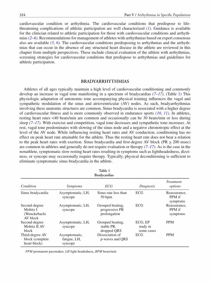
324 Part V / Arrhythmias in Specific Populations
cardiovascular condition or arrhythmia. The cardiovascular conditions that predispose to life-threatening complications of athletic participation are well characterized (1). Guidance is availablefor the clinician related to athletic participation for those with cardiovascular conditions and arrhyth-mias (2–6). Recommendations for management of athletes with arrhythmias based on expert consensusalso are available (5, 6). The cardiovascular conditions predisposing to arrhythmias and the arrhyth-mias that can occur in the absence of any structural heart disease in the athlete are reviewed in thischapter from multiple perspectives. These include clinical evaluation of the athlete with arrhythmias,screening strategies for cardiovascular conditions that predispose to arrhythmias and guidelines forathletic participation.
BRADYARRHYTHMIAS
Athletes of all ages typically maintain a high level of cardiovascular conditioning and commonlydevelop an increase in vagal tone manifesting in a spectrum of bradycardias (7–17). (Table 1) Thisphysiologic adaptation of autonomic tone accompanying physical training influences the vagal andsympathetic modulation of the sinus and atrioventricular (AV) nodes. As such, bradyarrhythmiasinvolving these anatomic structures are common. Sinus bradycardia is associated with a higher degreeof cardiovascular fitness and is more commonly observed in endurance sports (10, 11). In athletes,resting heart rates <40 beats/min are common and occasionally can be 30 beats/min or less duringsleep (7–17). With exercise and competition, vagal tone decreases and sympathetic tone increases. Atrest, vagal tone predominates with slowing of the sinus node and a negative chronotropic effect at thelevel of the AV node. While influencing resting heart rates and AV conduction, conditioning has noeffect on peak heart rate attainable for the athlete. Thus the resting heart rate does not bear a relationto the peak heart rates with exertion. Sinus bradycardia and first-degree AV block (PR > 200 msec)are common in athletes and generally do not require evaluation or therapy (7–17). As is the case in thenonathlete, symptomatic slow resting heart rates resulting in symptoms such as lightheadedness, dizzi-ness, or syncope may occasionally require therapy. Typically, physical deconditioning is sufficient toeliminate symptomatic sinus bradycardia in the athlete.
Table 1Bradycardias
Condition Symptoms ECG DiagnosisTreatment
options
Sinus bradycardia Asymptomatic, LH,syncope
Sinus rate less than50 bpm
ECG Reassurance,PPM ifsymptoms
Second-degreeMobitz I(Wenckebach)AV block
Asymptomatic, LH,syncope
Grouped beating,progressive PRprolongation
ECG Reassurance,PPM ifsymptoms
Second-degreeMobitz II AVblock
Asymptomatic, LH,syncope
Grouped beating,stable PR,dropped QRS
ECG, EPstudy insome cases
PPM
Third-degree AVblock (completeheart block)
Asymptomatic,fatigue, LH,syncope
Dissociation ofp-waves and QRS
ECG PPM
PPM permanent pacemaker, LH light headedness, BPM beats/min

Chapter 15 / Arrhythmias in the Athlete 325
Generally, maximum heart rates attainable are approximately 220 minus the individual’s age. Ath-letes should be able to achieve their predicted maximal heart rate for age although it may require longerduration and intensity of exercise compared to the nonathlete. Chronotropic incompetence, which bydefinition is the inability to raise the heart rate appropriately with exercise, is rare in the younger ath-lete (aged less than 35), but can be seen in older athletes with primary sinus node dysfunction (7–17).Invasive electrophysiologic studies are of no clinical utility in patients with this sinus node dysfunc-tion as such testing is neither sensitive nor specific for bradyarrhythmias. Evaluation and managementof bradycardias are guided by clinical assessment of symptoms and their relationship to clinicallydocumented arrhythmias.
At present, there are no medications that effectively treat sinus node dysfunction on a long-termbasis. The definitive therapy for symptomatic bradycardia is a permanent pacemaker. It is evident thatdiscontinuation of pharmacologic agents that slow sinus node function should be the initial approachto any athlete or other individual with symptomatic sinus bradycardia. Rarely it becomes necessaryto implant a permanent pacemaker. Virtually all available pacemakers have the ability to enhanceheart rate based on sensed activity. With the placement of a permanent pacemaker, there are sportspecific recommendations regarding participation. Contact sports with risk of impact are not advisablein patients with a permanent pacemaker (5, 6). Activities that involve repetitive movement of theupper extremity and movement of the clavicle over the ribs, like weightlifting, freestyle swimming,and pitching in baseball, are also associated with a greater risk of damage to leads. Accordingly, theyshould be avoided.
HEART BLOCK
Like sinus bradycardia, 2nd-degree heart block Mobitz type I (Wenckebach) is a common find-ing in athletes at rest due to the influence of enhanced vagal tone (7–17). With exercise, vagal toneis withdrawn and Wenckebach generally resolves, as does sinus bradycardia. Wenckebach type AVblock that occurs during sleep does not warrant treatment. Symptoms of syncope or lightheadedness,however, are concerning in the athlete with Wenckebach rhythm. Wenckebach not resolving with exer-cise is abnormal and suggests underlying pathology of the AV node. Under such circumstances, likesymptomatic sinus bradycardia, a permanent pacemaker is the only effective treatment (18).
More advanced heart block such as 2nd-degree Mobitz type II heart block or 3rd-degree heart block(complete heart block) are generally considered abnormal, even in the highly trained athlete. Findingsof Mobitz II or complete heart block indicate that the site of block is below the AV node and notunder vagal influence. Generally, Mobitz II and complete heart block are an indication for permanentpacemaker, particularly when occurring in the setting of structural heart disease or symptoms (18).
Occasionally, 2:1 AV block is seen with two P-waves for every QRS complex. It can be difficult todetermine whether the block is at the level of the AV node or below. This distinction assumes clinicalimportance, as the former is typically benign and not treated. By contrast, the latter typically warrantsa pacemaker as it commonly progresses to a higher degree of heart block. The width of the QRScomplex is useful in making assumptions regarding the level of block when 2:1 AV conduction isnoted clinically. A narrow QRS complex (<120 msec) suggests block at the level of the AV node andan intact His-Purkinje system. A wide QRS complex (>120 msec) more commonly signifies diseasein the His-Purkinje system. In patients in whom the level of block cannot be determined, invasiveelectrophysiologic studies can provide useful prognostic information and guide decisions regardingthe need for a pacemaker. Block at the level of the AV node (Wenckebach) and is generally benign andnot progressive. Block in the His-Purkinje system or below generally requires permanent pacing (18).

326 Part V / Arrhythmias in Specific Populations
SUPRAVENTRICULAR TACHYCARDIAS
Supraventricular tachycardia (SVT) typically occurs with the same frequency in athletes as inthe general population. Classification of SVTs is based on the electrophysiologic mechanism of thearrhythmia as noted in Table 2 (19, 20). With understanding of the fundamental mechanisms of SVTand development of techniques of mapping and ablation of most SVTs, it is now possible to cure mostathletes with this spectrum of arrhythmias.
Table 2Supraventricular Tachycardia
Condition Symptoms ECG Diagnosis Treatment
APCs Palpitations Often normal Monitor Reassurance, BB orCCB if highlysymptomatic
Atrial fibrillation Palpitations Often normal Monitor Rate control,anticoagulation,antiarrhythmics,ablation
Atrial flutter Palpitations Often normal Monitor Rate control,anticoagulation,antiarrhythmics,ablation
AVNRT Palpitations, LH,syncope
Often Normal Monitor, EPstudy
BB or CCB,digoxin, RFA
AVRT (WPW) Asymptomatic Short PR, deltawave
ECG, EP study No therapy,ablation if highrisk
AVRT (WPW) Symptomatic(palpitations,syncope, LH)
Short PR, deltawave
ECG, EP study Antiarrhythmics,ablation
ECG electrocardiogram, APC atrial premature complex, AVNRT atrioventricular nodal reentrant tachycardia, AVRTatrioventricular reentrant tachycardia, BB beta blockers, CCB calcium channel blockers, WPW Wolfe-Parkinson-Whitesyndrome
A common tachycardia in athletes is sinus tachycardia that is a normal response of the sinus node tophysiologic demands. Sympathetic tone increases and vagal tone is withdrawn with exercise, fear, painand other physiologic states of increased oxygen demand. Sinus tachycardia can occasionally mimicother SVTs, particularly in young athletes who are capable of high sinus rates. Typically sinus tachy-cardia has a gradual onset and termination in contrast to atrioventricular nodal reentrant tachycardia(AVNRT) or atrioventricular reciprocating tachycardia (AVRT), discussed in prior chapters. Character-istically the P-wave morphology on the ECG is similar to that at rest with the P-wave vector orientedinferiorly and leftward. Sinus tachycardia, however, is a normal response to physiologic stimuli anddoes not require therapy. Atrial premature contractions (APCs) are common in adults including trainedathletes and also do not require therapy (17). In the minority of patients, these APCs may be symp-tomatic, manifesting as palpitations, skipped beats, or extra beats in the heart. APCs can be treatedwith medications such as beta-blockers which may suppress the extra-systoles and improve symp-toms. Typically beta-blocker therapy is not well tolerated with side effects of fatigue and a decreasein exercise capacity noted by many athletes. Additionally, beta-blockers are banned in some types ofathletic competition.

Chapter 15 / Arrhythmias in the Athlete 327
ATRIOVENTRICULAR NODAL REENTRANT TACHYCARDIA (AVNRT)
AVNRT is the most common SVT representing approximately 60–70% of all SVTs. Mechanisticallyit occurs when a reentrant circuit is present involving the AV node (19, 20). The circuit typicallyrequires dual AV nodal physiology with an anterior “fast” pathway and a posterior “slow” pathway.With the common type of AVNRT, the antegrade refractory period of the fast pathway is longer thanthat of the slow pathway. An appropriately timed atrial premature beat can block antegrade in thefast pathway, travel through the slow pathway and “echo” retrograde via the fast pathway to initiateand perpetuate this tachycardia. Under the proper conditions, a reentrant arrhythmia can be sustainedwith antegrade conduction via the slow pathway and retrograde conduction via the fast. AVNRT ischaracterized by rates between 130 and 240 beats/min with abrupt onset and termination (19, 20). Theresting ECG is generally normal. The ECG during the tachycardia usually demonstrates a narrow QRScomplex tachycardia that is regular with atrial activity manifesting with a small R′ wave in lead V1and shallow S waves in leads II, III, and aVF. This rhythm can occasionally be terminated with vagalmaneuvers. However, definitive acute therapy is intravenous adenosine. Preventative pharmacologictherapy involves the use of beta-blockers, calcium channel blockers, or other antiarrhythmic agents.However, AVNRT is safely and successfully cured in the vast majority of patients with catheter-basedablation of the slow pathway. Ablation is considered to be first-line therapy over medications for thisreason (19, 20).
ATRIOVENTRICULAR REENTRANT TACHYCARDIA (AVRT)
Wolfe-Parkinson-White syndrome (WPW) was first described in the 1930s in young, otherwisehealthy subjects with abnormal ECG findings of a short PR interval and a widened QRS complex andparoxysmal tachycardia. This syndrome is now well understood to be cause by accessory pathwaysof myocardial tissue bridging the atrioventricular groove. This accessory pathway, or bundle of Kent,allows conduction between the atrium and ventricle. Accessory pathways, also known as a bypass tract,typically do not have decremental conduction like the AV node. When bypass tracts have the abilityto conduct antegrade and result in a pattern of short PR interval with a delta wave on the ECG theyare classified as “manifest” pre-excitation (19, 20). Commonly, bypass tracts only have the ability toconduct retrograde and allow conduction between the ventricle and atrium during AVRT. Such bypasstracts that only conduct retrograde cannot be identified on the resting ECG in sinus rhythm and arelabeled “concealed” (19, 20).
Both manifest and concealed bypass tracts can serve as the anatomic substrate for a reentrant circuitthat involving the AV node antegrade and the bypass tract retrograde. This reentrant arrhythmia isthe second most common type of SVT and is classified as orthodromic reciprocating tachycardia (19,20). Less commonly, the reentrant circuit involves antegrade conduction via a manifest bypass tractand retrograde conduction via the AV node. This results in a wide complex tachycardia classified asantedromic reciprocating tachycardia. While antiarrhythmic agents can reduce symptoms by slowingor blocking conduction in the bypass tract or AV node in many patients, the preferred approach to curewith radiofrequency ablation of the bypass tract. When an athlete has symptomatic WPW, ablationtherapy is curative and should be the recommended first-line therapy. Restriction from athletics isreasonable for a period of several weeks to allow vascular heeling (5, 6).
Manifest bypass tracts typically lack decremental conduction and atrial fibrillation can be transmit-ted to the ventricle at rapid rates. Very rarely, individuals may be at risk of sufficiently high rates ofventricular response to cause hemodynamic instability or ventricular fibrillation. This unusual clinicalcircumstance occurs more commonly with physical exertion. WPW is commonly detected in asymp-tomatic athletes on ECG, as the condition is present in approximately 1/3000 individuals. Athleteswho present with evidence of preexcitation on ECG but without symptoms have a very low risk of

328 Part V / Arrhythmias in Specific Populations
developing life-threatening arrhythmias. Based on this consideration, most experts do not recommendroutine electrophysiologic evaluation of asymptomatic patients with ventricular preexcitation (5, 6).Some experts take the approach of performing electrophysiologic evaluation of asymptomatic athleteswith preexcitation to characterize the properties and location of the bypass tract. If there are indica-tions of high risk, such as multiple bypass tracts, antegrade refractory period of the bypass tract < 250msec, or induction of sustained arrhythmias, ablation is felt to be reasonable (5, 6). For asymptomaticpatients with preexcitation, and those symptomatic or those asymptomatic patients who have under-gone successful ablation of their bypass tract, there are no limitations on competitive sports once thereis an appropriate period of recovery from the procedure (5, 6).
ATRIAL TACHYCARDIA
Another common SVT is atrial tachycardia, which refers to an area in the atrium that has enhancedautomaticity (19, 20). Typically, the sinus node acts to initiate the cardiac cycle through a property ofautomaticity that produces regular impulses from that are of the myocardium. In atrial tachycardia, anabnormal tract of atrial myocardium will depolarize and initiate localized and progressive activationof the myocardium. The rates of atrial tachycardia vary, but typically are fast enough to suppresssinus node activity and can be highly symptomatic, typically manifesting as palpitations. This SVTfrequently has a “warm-up” period with progressively shortening cycle lengths with each beat and onECG or electrocardiographic monitoring, this appears as a narrow complex tachycardia that is fasterwith each beat until the maximum tachycardia rate is reached. Atrial tachycardia can be suppressedby use of beta-blockers or calcium channel blockers, but is also amenable to catheter-based ablation,which is typically curative treatment.
ATRIAL FIBRILLATION AND FLUTTER
Atrial fibrillation likely occurs more commonly in athletes when compared to non-athletes dueto adaptation of the autonomic nervous system with heightened vagal tone (21–26). This typeof vagally mediated atrial fibrillation frequently occurs in the setting of nocturnal bradycardia(21–26). Bradycardia may allow for altered dispersion of atrial repolarization creating the electro-physiologic substrate for atrial fibrillation (21–26). Atrial fibrillation is a common arrhythmia inadults who are not athletes and is commonly related to systemic hypertension, congestive heart fail-ure or coronary artery disease (27). In the athlete without these predisposing factors, atrial fibrilla-tion is likely related to heightened vagal tone or the result of another SVT degenerating into atrialfibrillation (21–26).
Paroxysmal atrial fibrillation generally originates in the muscular sleeves that extend into the pul-monary veins from the left atrium and form a continuous transition between the left atrial tissue andthe endothelium of the pulmonary veins (21–26). Electrophysiologic observations related to ablationof the tissue in the pulmonary veins support the notion that this location is the origin of most parox-ysmal atrial. The principles of evaluation and management of atrial fibrillation in the athlete are sim-ilar to those that guide evaluation and management of this arrhythmia in the non-athlete (5, 6, 27).Every patient with atrial fibrillation should have a complete history, physical examination, ECG, andechocardiogram. Routine laboratory test should include a TSH to exclude hyper or hypothyroidism.Evaluation includes determination of the ventricular response during athletic activity or an exercisetest comparable to the intended athletic competition.
The decision to take a rate control versus rhythm control strategy is largely driven by symptoms(27). Typically, when the arrhythmia is symptomatic, a rhythm control strategy is used with antiar-rhythmic agents selected based on the presence or absence of structural heart disease (27). Curative

Chapter 15 / Arrhythmias in the Athlete 329
approach with catheter ablation is typically reserved for symptomatic individuals who have failed atleast one antiarrhythmic drug (27–29). Based on risks, benefits, and patient preference, it is reason-able to consider AF ablation as first-line therapy in selected symptomatic athletes (27–29). In lesssymptomatic or asymptomatic athletes, rate control with beta-blockers or calcium channel blockersis generally recommended (27, 28). Anticoagulation with aspirin or warfarin is selectively initiatedbased on the athlete’s risk of thromboembolic events (27, 28). It should be noted that in athletes orother individuals undergoing a curative approach to atrial fibrillation with ablation, anticoagulation isrecommended long term if the patient’s CHADS2 score is 2 or greater even if there is an apparent cureof the arrhythmia (27, 28).
Recommendations regarding participation in athletics for individuals with atrial fibrillation arebased on the best available data and expert opinion (5, 6). In the absence of WPW syndrome, ath-letes with asymptomatic atrial fibrillation without structural heart disease who maintain a ventricularrate that increases and slows appropriately and is comparable to that of a normal sinus response inrelation to the level of activity, while receiving no therapy or therapy with AV nodal blocking drugs,can participate in all competitive sports (5, 6). Athletes who have atrial fibrillation in the presenceof structural heart disease who maintain a ventricular rate comparable to that of an appropriate sinustachycardia during physical activity while receiving no therapy or therapy with AV nodal-blockingdrugs can participate in sports consistent with the limitations of the structural heart disease (5, 6). It isevident that athletes who require anticoagulation should not participate in sports with danger of bodilycollision (5, 6). Athletes without structural heart disease who have elimination of atrial fibrillation byan ablation technique, including surgery, may participate in all competitive sports after several weekswith an arrhythmia recurrence (5, 6).
Atrial flutter is another form of SVT that results from a macro-reentrant circuit in the right atrialmyocardium (30). In the absence of an acute, limiting illness, sustained atrial flutter is an uncommonrhythm disturbance in athletes without structural heart disease. This arrhythmia typically rotates in acounterclockwise direction through the isthmus of tissue between the os of the coronary sinus and theannulus of the tricuspid valve, along the intra-atrial septum, and over the roof and lateral wall of theright atrium (30). Athletes developing atrial flutter should have the same standard evaluation as thosepresenting with atrial fibrillation. The typical form of atrial flutter manifests with the classic saw-toothed pattern with inverted flutter waves in the inferior leads of the ECG and a variable ventricularresponse (30).
Like atrial fibrillation, therapy for atrial flutter is guided by the presence or absence of symptoms(27). Because the potential for very rapid ventricular rates exists if the atrial flutter conducts 1:1 tothe ventricles, ECG determination of the ventricular response during an exercise test or athletic eventduring treatment is essential (5, 6). Antiarrhythmic drugs can be effective in restoring and maintainingnormal sinus rhythm (27). However, given the high efficacy of flutter ablation by creating a line ofblock along the cavo-tricuspid isthmus, many experts consider ablation to be the first-line approach(5, 6, 27). This ablation procedure is also accompanied by a very low complication rate given theright atrial location (29). When the strategy of rate control is taken, beta-blockers or calcium channelblockers can be used (27). Anticoagulation is selectively used based on the CHADS2 score in a fashionsimilar to atrial fibrillation (27, 28). In some patients atrial flutter degenerates to atrial fibrillation. Insuch case, patient’s ablation of the atrial flutter can prevent development of this secondary arrhythmia.
Athletes with atrial flutter in the absence of structural heart disease who maintain a ventricular ratethat increases and slows appropriately comparable to that of a normal sinus response in relation to thelevel of activity, while receiving no therapy or therapy with AV nodal blocking drugs, can participatein competitive sports with the warning that rapid 1:1 conduction still may occur (5, 6). However, fullparticipation in all competitive sports should not be allowed unless the athlete has been without atrialflutter for two to three months with or without drug treatment (5, 6). It is recommended that athleteswith structural heart disease who have atrial flutter participate in competitive sports only after two to

330 Part V / Arrhythmias in Specific Populations
four weeks have elapsed without an episode of atrial flutter (5, 6). Athletes without structural heartdisease who have elimination of the atrial flutter by an ablation technique or surgery can participate inall competitive sports after two to four weeks without a recurrence, or in several days after an electro-physiologic study showing noninducibility of the atrial flutter in the presence of bidirectional isthmusblock (5, 6, 30). As with atrial fibrillation, athletes in whom anticoagulation is deemed necessarycannot participate in competitive sports where the danger of bodily collision is present (5, 6).
PRE-PARTICIPATION SCREENING
Most athletes at risk for sudden cardiac death (SCD) with athletic activity have preexisting andunderlying heart disease (1). The conditions associated with an increased risk of SCD in associationwith exercise have been identified and many of them can be detected by cardiovascular screening(Table 3). It has been determined that systematic screening for these underlying cardiovascular con-ditions allows for identification of athletes at risk for SCD (2, 3, 31–33). The strategy of withdrawingthose identified at high risk for SCD from vigorous training or competition is felt to reduce the risk ofSCD (31–33). Most experts accept the notion that the risk for SCD increases with vigorous training(31–33). The best available evidence for the efficacy of screening athletes and selectively restrictingathletic participation comes from Italy (31–33). Through a national program all athletes engaged inorganized sports must undergo yearly screening. This systematic approach served as the impetus for aproposal in 2005 by the European Society of Cardiology (ESC) for a common European protocol forpre-participation athlete screening (31–33).
Fundamental to these recommendations are pre-participation athletic screenings by specially trainedsports medicine physicians (31–33). This evaluation consists of personal and family history, physical
Table 3Cardiovascular Conditions Associated with Sudden Death
in the Athlete
Structural heart diseases
Hypertrophic cardiomyopathyCoronary artery anomaliesLeft ventricular hypertrophy of indeterminate causationMyocarditisRuptured aortic aneurysm (Marfan’s syndrome)Arrhythmogenic right ventricular cardiomyopathyTunneled (bridged) coronary arteryAortic-valve stenosisAtherosclerotic coronary artery diseaseDilated cardiomyopathyMyxomatous mitral-valve degenerationAsthma (or other pulmonary condition)Heat stroke
Channelopathies
Long QT syndromeShort QT SyndromeCatecholaminergic polymporphic ventricular tachycardiaBrugada syndromeIdiopathic ventricular fibrillation

Chapter 15 / Arrhythmias in the Athlete 331
examination, and 12-lead electrocardiogram (31–33). These European guidelines recommend repeatedscreening for the duration of exposure to athletic activity every 2 years to identify any cardiovascu-lar conditions that may manifest following the previous evaluation (31–33). Selective use of moreadvanced cardiovascular testing is utilized as appropriate. If cardiovascular conditions that are asso-ciated with an increased risk of SCD are detected, recommendations for exclusion of the individualfrom athletic activity are similar to the Bethesda 36 guidelines (5, 6, 31–33).
In the United States, with a heterogeneous and diverse population, pre-participation evaluation ofhigh school and college athletes has traditionally included only a history and physical examination(33). This screening has been traditionally limited to a history and physical exam reflecting the largerand more diverse background of American athletes and the broader spectrum of normal ECGs that canbe difficult to distinguish from abnormal (33). Athletes in the United Stated are typically screened atthe high school and collegiate level, and routine evaluation does not involve an ECG based on the bestavailable data which remains incomplete (33).
A study in 2005 suggested that this standard screening at US colleges may have a low sensitivitywith regards to SCD (33). A 2000 survey of 879 American colleges and universities reported that97% did participate in screening of their student-athletes, but only 25% used at least 9 of 12 AHArecommended screening guidelines and 24% included less than or equal to 4 of 12 (33). Screening isperformed by a range of providers depending on the individual institution and to date, the screeningsystem for competitive athletes in the United States remains highly variable and inconsistent. Whilemany have called for a more uniform and standardized approach to American athletes, the complexityof the American health care system makes this a challenging endeavor. A wide range of complexmedical, legal, and ethical issues remain unresolved related to athletic screening for cardiovascularconditions and athletic restrictions (33, 34).
The American College of Cardiology and the American Heart Association recommend screeninghigh school athletes every 2 years and college athletes initially and then every 4 years (33, 34). Rec-ommended aspects of this screening included personal and familial historical data and a physicalexamination. ECGs and echocardiograms are not recommended in all individuals, but rather that theybe used selectively based on symptoms, family history, or findings on physical exam (33, 34).
Whether ECGs should be performed in all athletes remains a controversial issue. Electrocardio-grams are frequently abnormal in the competitive athlete and distinguishing normal variants fromabnormal is not possible without further cardiovascular evaluation in many athletes (Table 4). Upto 50% of athletes, mostly men and endurance athletes, will have some abnormal finding on ECG(35–38). Early repolarization patterns, deep Q-waves, high QRS voltage, and T-Wave inversions aremost common. Endurance athletes may have findings on ECG of increases P-wave amplitude, incom-plete right bundle branch block, and high QRS voltages suggestive of left and right ventricular hyper-trophy (35–38). Some elite athletes have highly abnormal or bizarre findings that in the presence ofa negative workup are felt to be the limit of physiological change with the athlete’s heart (35–38).Criteria for LV hypertrophy and even ST changes consistent with ischemia frequently are present inthe well-trained athlete (35–38).
The cardiac diseases that can be identified or suspected by an ECG include HCM, the long QTsyndromes, ARVD, and WPW syndrome. Hypertrophic cardiomyopathy can be suspected by inter-ventricular conduction delays, septal or inferior Q waves, LV hypertrophy, and ST abnormalities ona 12-lead ECG (35–38). With the LQTS, the diagnosis is made by the ECG, but up to 6% of genecarriers will have a QTc of 0.44 or less. Patients with ARVD may have RV conduction delays (epsilonwave), right bundle-branch blocks, and inverted T waves in the pre-cordial leads. Unfortunately, noneof these ECG findings (other than a long QT) are sensitive or specific for ascertaining abnormal fromnormal findings in the athlete. Whether ECGs will become standard in the United States for screeningathletes is not yet determined; however, at present, the American Heart Association guidelines do notadvocate an ECG in the routine screening of athletes (33, 34).

332 Part V / Arrhythmias in Specific Populations
Table 4ECG Abnormalities
Diagnosis of heart disease ECG abnormalities
Hypertrophic cardiomyopathy Left ventricular hypertrophyAnterior pseudoinfarct, Q-wavesRarely normal
Arrhythmogenic right ventricular dysplasia Anterior T-wave inversionsEpsilon waveRBBB (complete or incomplete)Rarely normal
Wolff–Parkinson–White Short PRDelta WavesPseudoinfarct patterns
Long QT syndrome Prolonged QTAbnormal ST segment
Short QT syndrome Shortened QTTall T-waves
Catecholaminergic polymorphic ventriculartachycardia
Usually normalProminent U-wave
Idiopathic dilated cardiomyopathy LBBBProlonged QTCan be normal
Anomalous coronary artery Usually normalBrugada syndrome RBBB (complete or incomplete)
ST elevation anteriorlyVariable ECG
RBBB right bundle branch block, LBBB left bundle branch block
EVALUATION OF THE ATHLETE
The evaluation of the athlete with suspected cardiac arrhythmia is fundamentally different from rou-tine screening for athletes recommended by the American Heart Association. The evaluation of theseathletes is, however, similar to those members of the general population with suspected arrhythmia.The key elements of the evaluation include the history with detailed evaluation of symptoms. Syncope,while less serious than previous sudden death, is more ominous when occurring suddenly or occurringin the setting of intense exercise. Sudden syncope may be associated with significant bodily injury asit may have no premonitory symptoms to warn the patient. Presyncope and lightheadedness are fre-quent presentations, but are less likely to be associated with malignant ventricular arrhythmia than truesyncope though they still require investigation Many of the cardiac syndromes associated with SCDhave a genetic component including HCM, long QT, idiopathic cardiomyopathy, and ARVD to somedegree. Accordingly, a detailed family history is warranted.
Initial evaluation following a thorough history and physical exam should include 12-lead ECG andtransthoracic echocardiogram. In those patients over age 35, an evaluation for cardiac ischemia is war-ranted with an exercise nuclear stress test. Outpatient electrocardiographic monitoring including Holtermonitors, or loop type of ambulatory monitoring in patients with infrequent or intermittent symptoms,aid in the evaluation by capturing the clinical arrhythmia in various settings including sleep, exercise,and varied times through the day. In patients with high risk features such as sudden syncope, additionalimaging testing such as cardiac CT or MRI should be selectively done as appropriate to best define thepresence or absence of structural heart disease. Invasive testing with cardiac catheterization, coronaryangiography, and electrophysiologic study should be pursued selectively as clinically indicated (36).

Chapter 15 / Arrhythmias in the Athlete 333
TREATMENT
The presence or absence of structural heart disease and symptoms related to ventricular arrhythmiasare two key factors guiding evaluation and management of the athlete with ventricular arrhythmias(Table 5). The justification for and benefits of treatment of ventricular arrhythmias in the athlete arereducing symptoms or reducing the risk of sudden death. In those patients without structural heartdisease, the risk of sudden death is generally considered to be low.
In athletes with documented ventricular arrhythmias without structural heart disease, exercise-induced or resting non-sustained ventricular tachycardia and frequent premature ventricular contrac-tions generally do not increase including the risk of sudden death (5, 6, 36). If the athlete’s heart iscompletely normal neither treatment nor restriction from athletics is warranted (5, 6, 36). Treatmentis reasonable for clinically significant symptoms such as palpitations. Beta-blockers are the preferredinitial agent. Treatment options for symptomatic ventricular arrhythmias in athletes without structuralheart disease include beta-blockers, antiarrhythmic agents, and ablation. In athletes without structuralheart disease, symptomatic sustained ventricular tachycardias originating from the right or left ventric-
Table 5Ventricular Arrhythmia in the Athlete
Condition Symptoms ECGVT
morphologyTreatment
optionsCompetitive
athletics
HCM Palpitations,syncope, SCD
LVH, anteriorQ-waves
PMVT, VF BB, AAD,myomectomy,ICD
Low intensity
ARVD Palpitations,syncope, SCD
AnteriorT-inversions,RBBB,Epsilon wave
Left bundle,inferior axis
Sotolol, Amio,ICD, RFA
Only lowintensity
CAD Palpitations,syncope, SCD
Infarcts,ischemic STchanges
RB or LB Amio, ICD Only lowintensity
IDCM Palpitations,syncope, SCD
Often LBBB RB or LB Amio, ICD Only lowintensity
LQTS Palpitations,syncope, SCD
Long QTc PMVT BB, PPM, ICD Only lowintensity
Anomalouscoronary
Exertional chestpain, syncope,SCD
Normal VF CABG No restrictionspost CABGwith negativestress
IdiopathicRVOT VT
Palpitations, LH,syncope
Normal LB, inferioraxis
RFA No restrictionspost-RFA
IdiopathicLV VT
Palpitations, LH,syncope
Normal RB, left axis RFA No restrictionspost-RFA
IdiopathicVF
Syncope, SCD Normal VF ICD Only Lowintensity
ECG electrocardiogram, VT ventricular tachycardia, LV left ventricle, LH lightheadedness, Nl normal, RB right bundle,RFA radiofrequency ablation, RVOT right ventricle outflow tract VT, LB left bundle, HCM hypertrophic cardiomyopathy,SCD sudden cardiac death, BB beta-blockers, AAD antiarrhythmic drugs, ICD implantable cardioverter defibrillator, ARVDarrhythmogenic right ventricular dysplasia, RBBB right bundle branch block, LBBB left bundle branch block, sot sotalol,amio amiodarone, PPM permanent pacemaker, CABG coronary artery bypass surgery, PMVT polymorphic VT (torsadesde point)

334 Part V / Arrhythmias in Specific Populations
ular outflow tracts or the left posterior fascicle of the left bundle can be safely and successfully curedwith mapping and radiofrequency ablation (5, 6, 36) (Table 5). Once the ablation has been successfullyperformed, such athletes can return to athletic activities without restrictions (5, 6, 36). When structuralheart disease is identified in the athlete, recommendations for treatment and athletic restriction areconsiderably different from those that are relevant to athletes without structural heart disease (5, 6, 36)(Table 3). With most types of structural heart disease that predisposes to sudden death the presence orabsence of symptoms is not a factor in clinical decision regarding treatment or athletic restriction (5,6, 36) (Table 5).
The Bethesda Guidelines related to ventricular arrhythmias provide the best available guidance forevaluation, management, and athletic restriction of athletes with ventricular arrhythmias (4, 5). Ath-letes without structural heart disease who have premature ventricular complexes at rest and duringexercise, including exercise testing that is comparable to the sport in which they compete, can partici-pate in all competitive sports without restrictions (5). If the premature ventricular complexes increaseduring with exercise testing and produce symptoms produce symptoms of impaired consciousness, sig-nificant fatigue, or dyspnea, the athlete can participate low intensity sports such as bowling, billiards orgolf (4–6, 36). Athletes with structural heart disease who are in high-risk groups based on the presenceof structural heart disease that predisposes to sudden death as noted in Table 3 and have premature ven-tricular complexes should only participate in low intensity sports regardless of treatment (5). It is alsorecommended that such athletes with premature ventricular complexes that are suppressed by drugtherapy as assessed by ambulatory ECG recordings during participation in the sport only participate inlow-intensity sports (4–6, 36).
Nonsustained or sustained monomorphic or polymorphic VT requires careful evaluation and shouldbe considered a potentially serious occurrence particularly when occurring the setting of structuralheart diseases that predispose to sudden death (4–6, 36). Routinely, noninvasive tests should be per-formed including an ECG, exercise test, and echocardiogram (5, 36). Ambulatory monitoring may beappropriate in selected patients. Cardiac catheterization and electrophysiologic study should be con-sidered to exclude otherwise undetected heart disease and establish the location and mechanism VT.Patients with accelerated idioventricular rhythm in whom the arrhythmia rate is similar to the sinusrate should be treated in a fashion similar to athletes with premature ventricular complexes (5, 36).
Athletes with a structurally normal heart and monomorphic nonsustained or sustained VT that canbe localized to a specific site are candidates for a catheter ablation procedure that may potentiallyoffer a cure (5, 36). Following such a successful ablation procedure, with subsequent failure to induceVT during electrophysiologic study (EPS) with/without isoproterenol when the VT was reproduciblyinduced before ablation, the athlete can resume full competitive activity within a few weeks (4–6, 36).
A more conservative approach is recommended for the athlete in whom antiarrhythmic drug sup-pression is used as therapy because catecholamines released during athletic activity can counter thesuppressive effects of the drug, and the VT can reemerge (5, 36). It is recommended that the athleteshould not compete in any sports for at least two to three months after the last VT episode (5–6, 36). Inthe absence of clinical recurrences, and inducibility of VT by testing and EPS, all competitive sportsmay be permitted in the athlete with no structural heart disease (5). Occasionally, deconditioning canresult in the loss or lessening of ventricular arrhythmias (4–6, 36). Accordingly, a period of decondi-tioning and reevaluation is reasonable in some athletes.
For the athlete with structural heart disease and VT, moderate- and high-intensity competition iscontraindicated regardless of whether the VT is suppressed or ablated. Generally, only low-intensitysports are sports are permitted for such athletes (4–6, 36). An exception to this general recommendationis the asymptomatic athlete with brief episodes of nonsustained monomorphic VT (generally less than8–10 consecutive ventricular beats), rates generally less than 150 beats/min, and no structural heartdisease (5, 36). Such athletes do not appear to be at increased risk for sudden cardiac death (5). If

Chapter 15 / Arrhythmias in the Athlete 335
exercise testing and ambulatory ECG recording during the specific competitive activity demonstratessuppression of the VT, there is no need for athletic restriction (4–6, 36).
The Bethesda Guidelines specify that the desire of the athlete to continue athletic competition shouldnot represent the primary indication for use of an implantable cardioverter-defibrillator (ICD) (5). Theefficacy with which these devices will terminate a potentially lethal arrhythmia under the extremeconditions of competitive sports, with the associated metabolic and autonomic changes, and possiblemyocardial ischemia, is unknown (5, 6, 36). In addition, sports with physical contact may result indamage to the ICD and/or lead, preventing normal function (5, 6, 36). For athletes with ICDs, allmoderate and high-intensity sports are contraindicated. Class IA sports are permitted (4–6, 36).
Athletes with cardiovascular disease that result in a predisposition to a cardiac arrest in the presenceor absence of structural heart disease generally are treated with an ICD and cannot participate in anymoderate or high-intensity competitive sports (4–6, 36). However, athletes with ICDs and who havehad no episodes of ventricular flutter or ventricular fibrillation requiring device therapy for six monthsmay engage in low-intensity competitive sports (4–6, 36).
CONCLUSION
Evaluation and management of the athlete with cardiovascular conditions including structural heartdisease and inherited arrhythmia syndromes represents a challenge for the clinician. Among the fac-tors that need to be carefully considered are the presence or absence of symptoms or cardiovascularconditions predisposing to sudden cardiac death. Since screening strategies for detecting and prevent-ing sudden death on the athletic field have not been shown to have sufficient sensitivity, specificity,and cost-effectiveness in the United States, it is likely that initial manifestation of cardiovascular con-ditions predisposing to sudden death will continue to be cardiac arrest in a considerable number ofathletes. Based on this, recommendations for automatic external defibrillation, training in cardiopul-monary resuscitation, and an emergency response planning have been developed for athletic activities(39). With ongoing research, it is likely that evidence based medicine will guide strategies for iden-tification, evaluation, and therapy of athletes with cardiac arrhythmia in the future. In the meantime,the clinician should consider the best available information and expert opinion and consensus to guideclinical management.
REFERENCES
1. Maron BJ (2003) Sudden death in young athletes. N Engl J Med 349:1064–10752. Heidbüchel H, Corrado D, Biffi A, Hoffmann E, Panhuyzen-Goedkoop N et al (2006) Recommendations for participa-
tion in leisure-time physical activity and competitive sports of patients with arrhythmias and potentially arrhythmogenicconditions. Part II: ventricular arrhythmias, channelopathies and implantable defibrillators. Study group on sports car-diology of the European association for cardiovascular prevention and rehabilitation. Eur J Cardiovasc Prev Rehabil13(5):676–686
3. Maron BJ, Chaitman BR, Ackerman MJ, Bayés de Luna A, Corrado D et al (2004) Recommendations for physicalactivity and recreational sports participation for young patients with genetic cardiovascular diseases. Working groupsof the American heart association committee on exercise, cardiac rehabilitation, and prevention; councils on clinicalcardiology and cardiovascular disease in the young. Circulation 109(22):2807–2816
4. Maron BJ, Zipes DP et al (2005) 36th Bethesda conference: eligibility recommendations for competitive athletes withcardiovascular abnormalities. J Am Coll Cardiol 45:1318–1321
5. Zipes DP, Ackerman MJ, Estes NA 3rd, Grant AO, Myerburg RJ, Van Hare G (2005) Task Force 7: arrhythmias. J AmColl Cardiol 45:1354–1363
6. Estes NA 3rd, Link MS, Cannom D et al (2001) Report of the NASPE policy conference on arrhythmias and the athlete.J Cardiovasc Electrophysiol 12:1208–1219
7. Balady GJ, Cadigan JB, Ryan TJ (1984) Electrocardiogram of the athlete: an analysis of 289 professional footballplayers. Am J Cardiol 53:1339–1343

336 Part V / Arrhythmias in Specific Populations
8. Smith ML, Hudson DL, Graitzer HM, Rave PB (1989) Exercise training bradycardia: the role of autonomic balance.Med Sci Sports Exerc 21:40–44
9. Foote CB, Michaud G (1998) The athletes electrocardiogram: distinguishing normal from abnormal. In: Estes NI, SalemD, Wang PJ (eds) Sudden cardiac death in the athlete. Futura Publishing Company, Armonk, NY, pp 101–114
10. Baldesberger S, Bauersfeld U, Candinas R, Seifert B, Zuber M et al (2008) Sinus node disease and arrhythmias in thelong-term follow-up of former professional cyclists. Eur Heart J 29(1):71–78
11. Bjørnstad HH, Bjørnstad TH, Urheim S, Hoff PI, Smith G, Maron BJ (2009) Long-term assessment of electrocardio-graphic and echocardiographic findings in Norwegian elite endurance athletes. Cardiology 112(3):234–241
12. Zehender M, Meinertz T, Keul J, Just H (1990) ECG variants and cardiac arrhythmias in athletes: clinical relevance andprognostic importance. Am Heart J 119:1378–1391
13. Gillette PC, Shannon C, Garson A et al (1983) Pacemaker treatment of sick sinus syndrome in children. J Am CollCardiol 1:1325–1329
14. Zeppilli P, Fenici R, Sassasra M et al (1980) Wenckebach second degree AV block in top-ranking athletes: an oldproblem revisited. Am Heart J 100:281–294
15. Crouse SF, Meade T, Hansen BE, Green JS, Martin SE. Electrocardiograms of collegiate football athletes. Clin Cardiol.2009 Jan; 32(1):37–42
16. Serra-Grima R, Puig T, Doñate M, Gich I, Ramon J. Long-term follow-up of bradycardia in elite athletes. Int J SportsMed. 2008 Nov;29(11):934–7
17. Talan DA, Bauernfeind RA, Ashley WW et al (1982) Twenty-four hour continuous ECG recordings in long-distancerunners. Chest 82:19–24
18. Epstein AE, DiMarco JP, Ellenbogen KA, Estes NA 3rd, Freedman RA et al (2008) ACC/AHA/HRS 2008 guidelinesfor device-based therapy of cardiac rhythm abnormalities: a report of the American College of Cardiology/AmericanHeart Association Task Force on Practice Guidelines (Writing Committee to Revise the ACC/AHA/NASPE 2002Guideline Update for Implantation of Cardiac Pacemakers and Antiarrhythmia Devices): developed in collabora-tion with the American Association for Thoracic Surgery and Society of Thoracic Surgeons. American Collegeof Cardiology/American Heart Association Task Force on Practice Guidelines (Writing Committee to Revise theACC/AHA/NASPE 2002 Guideline Update for Implantation of Cardiac Pacemakers and Antiarrhythmia Devices);American Association for Thoracic Surgery; Society of Thoracic Surgeons. Circulation 117(21):e350–e408
19. Manolis AS, Wang PJ, Estes NA 3rd (1994) Radiofrequency catheter ablation for cardiac tachyarrhythmias. Ann InternMed 121(6):452–461
20. Naccarelli GV, Shih H, Jalal S (1995) Catheter ablation for the treatment of paroxysmal supraventricular tachycardia.J Cardiovasc Electrophysiol 6:951–961
21. Pelliccia A, Maron BJ, Di Paolo FM, Biffi A, Quattrini FM, Pisicchio C, Roselli A, Caselli S, Culasso F (2005)Prevalence and clinical significance of left atrial remodeling in competitive athletes. J Am Coll Cardiol 46(4):690–696
22. Mont L, Elosua R, Brugada J (2009) Endurance sport practice as a risk factor for atrial fibrillation and atrial flutter.Europace 11(1):11–17
23. Aizer A, Gaziano JM, Cook NR, Manson JE, Buring JE, Albert CM (2009) Relation of vigorous exercise to risk of atrialfibrillation. Am J Cardiol 103(11):1572–1577
24. Furlanello F, Bertoldi A, Dallago M, Galassi A, Fernando F, Biffi A, Mazzone P, Pappone C, Chierchia S (1998) Atrialfibrillation in elite athletes. J Cardiovasc Electrophysiol 9(8 Suppl):S63–S68
25. Coumel P (1996) Autonomic influences in atrial tachyarrhythmias. J Cardiovasc Electrophysiol 7:999–100726. Haïssaguerre M, Jaïs P, Shah DC, Takahashi A, Hocini M, Quiniou G, Garrigue S, Le Mouroux A, Le Métayer P,
Clémenty J (1998) Spontaneous initiation of atrial fibrillation by ectopic beats originating in the pulmonary veins. NewEngl J Med 339(10):659–666
27. Fuster V, Rydén LE, Cannom DS, Crijns HJ, Curtis AB et al (2006) ACC/AHA/ESC 2006 Guidelines for the manage-ment of patients with atrial fibrillation: a report of the American College of Cardiology/American Heart AssociationTask Force on Practice Guidelines and the European Society of Cardiology Committee for Practice Guidelines (Writ-ing Committee to Revise the 2001 Guidelines for the Management of Patients With Atrial Fibrillation): developed incollaboration with the European Heart Rhythm Association and the Heart Rhythm Society. American College of Cardi-ology/American Heart Association Task Force on Practice Guidelines; European Society of Cardiology Committee forPractice Guidelines; European Heart Rhythm Association; Heart Rhythm Society. Circulation 114(7):e257–354
28. Calkins H, Brugada J, Packer DL, Cappato R, Chen SA et al (2007) HRS/EHRA/ECAS expert consensus statementon catheter and surgical ablation of atrial fibrillation: recommendations for personnel, policy, procedures and follow-up. A report of the Heart Rhythm Society (HRS) Task Force on Catheter and Surgical Ablation of Atrial Fibrillationdeveloped in partnership with the European Heart Rhythm Association (EHRA) and the European Cardiac Arrhyth-mia Society (ECAS); in collaboration with the American College of Cardiology (ACC), American Heart Association(AHA), and the Society of Thoracic Surgeons (STS). Endorsed and approved by the governing bodies of the American

Chapter 15 / Arrhythmias in the Athlete 337
College of Cardiology, the American Heart Association, the European Cardiac Arrhythmia Society, the European HeartRhythm Association, the Society of Thoracic Surgeons, and the Heart Rhythm Society. Heart Rhythm Society; Euro-pean Heart Rhythm Association; European Cardiac Arrhythmia Society; American College of Cardiology; AmericanHeart Association; Society of Thoracic Surgeons. Europace 9(6):335–379
29. Haissaguerre M, Jais P, Shah DC et al (2000) Electrophysiological endpoint for catheter ablation of atrial fibrillationinitiated from multiple pulmonary venous foci. Circulation 101:1409–1417
30. Fischer B, Haissaguerre M, Garrigues S et al (1995) Radiofrequency catheter ablation of common atrial flutter in80 patients. J Am Coll Cardiol 25:1365–1372
31. Corrado D, Basso C, Schiavon M, Thiene G (1998) Screening for hypertrophic cardiomyopathy in young athletes.N Engl J Med 339(6):364–369
32. Corrado D, Pelliccia A, Bjørnstad HH et al (2005) Cardiovascular pre-participation screening of young competitiveathletes for prevention of sudden death: proposal for a common European protocol: consensus statement of the studygroup of sport cardiology of the working group of cardiac rehabilitation and exercise physiology and the working groupof myocardial and pericardial diseases of the European society of cardiology. Eur Heart J 26(5):516–524
33. Pfister GC, Puffer JC, Maron BJ (2000) Preparticipation cardiovascular screening for US collegiate student-athletes.JAMA 283:1597–1599
34. Paterick TE, Paterick TJ, Fletcher GF, Maron BJ (2005) Medical and legal issues in the cardiovascular evaluation ofcompetitive athletes. JAMA 294(23):3011–3018
35. Estes NAM III, Link MS, Homoud MK, Wang PJ (2000) Electrocardiographic variants and cardiac rhythm and con-duction disturbances in the athlete. In: Thompson PD (eds) Exercise and sports cardiology. McGraw Hill, New York,NY
36. Link MS, Wang PJ, Estes NAM III (1999) Cardiac arrhythmias and electrophysiologic observations in the athlete. In:Williams RA (eds) The athlete and heart disease: diagnosis, evaluation & management. Lippincott Williams & Wilkins,Philadelphia, PA, p 2196
37. Serra-Grima R, Estorch M, Carrio I, Subirana M, Berna L, Prat T (2000) Marked ventricular repolarization abnormalitiesin highly trained athletes’ electrocardiograms: clinical and prognostic implications. J Am Coll Cardiol 36:1310–1316
38. Biffi A, Pelliccia A, Verdile L et al (2002) Long-term clinical significance of frequent and complex ventricular tach-yarrhythmias in trained athletes. J Am Coll Cardiol 40:446–452
39. Myerburg RJ, Estes NA 3rd, Fontaine JM, Link MS, Zipes DP (2005) Task force 10: automated external defibrillators.J Am Coll Cardiol 45(8):1369–1371

16 Arrhythmias in Pregnancy and Postpartum
Kristen K. Patton and Richard L. Page
CONTENTS
INTRODUCTION
PHYSIOLOGIC ADAPTATIONS OF PREGNANCY
EPIDEMIOLOGY
THERAPY OF ARRHYTHMIAS DURING PREGNANCY
PHARMACOLOGIC CHANGES OF PREGNANCY
SPECIFIC DRUGS IN PREGNANCY BY VAUGHN WILLIAMS
CLASSIFICATION
MISCELLANEOUS ANTIARRHYTHMIC AGENTS: DIGOXIN
AND ADENOSINE
CLASS I AGENTS (SODIUM CHANNEL BLOCKADE)CLASS II (BETA-ADRENERGIC BLOCKING AGENTS)CLASS III AGENTS (PROLONGATION OF REPOLARIZATION)CLASS IV (CALCIUM CHANNEL BLOCKING AGENTS)NONPHARMACOLOGIC THERAPY OF ARRHYTHMIAS DURING
PREGNANCY
LABOR AND DELIVERY
POSTPARTUM AND LACTATION
CONCLUSION
REFERENCES
Abstract
The significant physiologic adaptations of pregnancy predispose to arrhythmias, while concurrentchanges in pharmacokinetics and concerns for fetal health and well-being complicate management. Anincreased incidence of both atrial and ventricular arrhythmias has been reported in population-based Holterstudies, yet most incident pregnancy-related arrhythmias are benign and do not require intervention. Phar-macologic absorption, distribution, metabolism, and elimination are altered, as are gut absorption, proteinbinding, and hepatic and renal blood flow. This chapter will review antiarrhythmic drug therapy by VaughnWilliams classification, nonpharmacologic arrhythmia therapy (cardiovascular implanted electronic devicesand ablation), as well as management of specific cardiovascular conditions during pregnancy, labor, andpostpartum.
Key Words: Pregnancy; hemodynamic; adaptation; cardiac output; arrhythmia; pharmacologic; pharma-cokinetics; antiarrhythmic drugs; digoxin; adenosine; quinidine; procainamide; disopyramide; lidocaine;mexiletine; flecainide; propafenone; beta-blockers; sotalol; dofetalide; ibutilide; amiodarone; verapamil;diltiazem; cardiversion; cardiac arrest; cardiopulmonary resuscitation; pacemaker; implantable cardioverter
From: Contemporary Cardiology: Management of Cardiac ArrhythmiasEdited by: Gan-Xin Yan, Peter R. Kowey, DOI 10.1007/978-1-60761-161-5_16
C© Springer Science+Business Media, LLC 2011
339

340 Part V / Arrhythmias in Specific Populations
defibrillator; radiofrequency ablation; premature atrial contractions; premature ventricular contractions;atrioventricular nodal reentrant tachycardia; atrioventricular reentrant tachycardia; atrial tachycardia; atrialflutter; atrial fibrillation; ventricular tachycardia; congenital heart disease; long QT syndrome; Wolff-Parkinson-White syndrome; hypertrophic cardiomyopathy; structural heart disease; syncope; Marfan syn-drome; labor and delivery; postpartum; and lactation.
INTRODUCTION
Pregnancy causes remarkable physiologic alterations, resulting in hemodynamic changes that pre-dispose to arrhythmia generation. These adaptations occur in the setting of pregnancy-induced changesin maternal drug metabolism and concerns regarding the effect of antiarrhythmic therapies on thedeveloping fetus. As a consequence, management of arrhythmias in pregnancy is complex, particu-larly in the setting of maternal heart disease.
PHYSIOLOGIC ADAPTATIONS OF PREGNANCY
The massive increase in total body water (average of 7 L) sustained during pregnancy results in acondition comparable to chronic volume overload (1, 2). Sodium and water retention is mediated byarginine vasopressin and the renin-angiotensin-aldosterone system, and results in a 30–50% increasein cardiac output and a dilutional anemia of pregnancy (1, 3, 4). The marked increase in cardiac outputfrom 5 L/min to 7.3 L/min is due to an increase in both heart rate and stroke volume. Although thereis some variation in human studies, the rise in cardiac output begins early, and by 12 weeks is nearly40% of pre-gravid levels (5, 6). Cardiac output appears to peak between 25 and 30 weeks. Maternalheart rate also begins to rise early in pregnancy, paralleling the rise in cardiac output, and peaks at15–20 beats/min above the pre-pregnancy state (1, 7). Stroke volume begins its rise slightly later, atapproximately 8 weeks, reaching a peak 20–30% above pre-pregnancy levels at 20 weeks. In laterpregnancy, cardiac output is dependent on maternal position. Blood return and stroke volume arereduced by both standing and lying supine; in the latter position, this is caused by uterine compressionof the inferior vena cava (8).
Despite significantly increased cardiac output during pregnancy, maternal blood pressure fallsdue to a decrease in systemic vascular resistance (9). This fall occurs despite renin-angiotensin-aldosterone system activation in pregnancy; in normal pregnancy, the gravid woman is less responsiveto angiotensin II-mediated vasoconstriction due to increased nitric oxide production and gestationalhormones (10). Blood pressure falls to a nadir at mid-pregnancy and gradually increases to near pre-pregnancy levels at term (9).
Cardiac hypertrophy occurs during pregnancy; ultrasound studies have shown an increase in leftventricular mass of 52%, as well as an increase in left ventricular end-diastolic and end-systolic diam-eters (12 and 20%, respectively) (11). Left ventricular diastolic function, as demonstrated by changesin mitral valve flow velocities, increases in the first two trimesters but declines in the third trimester(12). Left atrial dimension also gradually increases during pregnancy, peaking at 40% at 30 weeksgestation (5).
During labor, cardiac output increases even further (7–10.6 L/min) (1), resulting from the increasedstroke volume and the increased venous return that occurs during uterine contractions. Mean arterialpressure also increases in labor and during contractions. Cardiac output reaches its peak immediatelypost-partum and returns to pre-pregnancy levels in 2–4 weeks (5).
EPIDEMIOLOGY
An increase in arrhythmia during pregnancy has been documented in the literature. Holter stud-ies showed a similar and high prevalence of premature atrial and ventricular contractions among

Chapter 16 / Arrhythmias in Pregnancy and Postpartum 341
both symptomatic and asymptomatic pregnant subjects (56% and 59%, respectively) (13). The fre-quency of ventricular arrhythmias was higher in symptomatic patients (3235 ± 6397 vs. 678 ± 3358beats/24 h p <0.05). Follow-up Holter monitoring in a subset of patients with frequent prematureventricular contractions during pregnancy revealed a substantial reduction in arrhythmia burden post-partum (9073 ± 9210/24 h to 1345 ± 1997/24 h (p <0.05)) (13). In asymptomatic pregnant patients, ahigh incidence of arrhythmias was reported (13).
A large retrospective review of over 136,000 pregnancy-related admissions to Parkland hospital inDallas, TX, revealed 226 admissions with concurrent arrhythmia diagnosis (0.17%) (14). The mostfrequently reported arrhythmias (sinus tachycardia, sinus bradycardia, and sinus arrhythmia) werebenign and did not require intervention. Premature atrial and ventricular contractions were the secondmost common arrhythmia seen. Supraventricular tachycardia was less commonly noted (24/100,000),and either terminated spontaneously or responded well to adenosine. Episodes associated with Wolff-Parkinson-White syndrome responded to procainamide. The vast majority of episodes were the firstpresentation with an arrhythmia. Serious arrhythmias, such as high-degree atrioventricular block, ven-tricular arrhythmias, and cardiac arrest, were very rare (Fig. 1) (14).
Fig. 1. Distribution of arrhythmias diagnosed during pregnancy (expressed as the percentage of total arrhythmiaoccurrence) (from Li et al., with permission) (14). AF: atrial fibrillation; AFL: atrial aflutter; AVB: atrioventric-ular block; PAC: premature atrial contraction; PVC: premature ventricular contraction; SA: sinus arrhythmia;SB: sinus bradycardia; ST: sinus tachycardia; SVT: supraventricular tachycardia; VF: ventricular fibrillation;VT: ventricular tachycardia.
THERAPY OF ARRHYTHMIAS DURING PREGNANCY
Conservative, nonpharmacologic measures are generally preferred since antiarrhythmic therapy iscomplicated by concerns for potential risk to the fetus. Teratogenic risk is highest early in the firsttrimester; after eight weeks, organogenesis is largely completed (15). Nonsustained episodes withmild symptomatology that do not compromise fetal well-being may best be treated with reassurance.Sustained episodes of supraventricular tachycardia may terminate with vagal maneuvers. Triggers ofspecific episodes may be avoided by hydration, avoidance of caffeine, and rest.
PHARMACOLOGIC CHANGES OF PREGNANCY
The substantial physiologic adaptions of pregnancy result in changes to each aspect of drug phar-macokinetics: absorption, distribution, metabolism, and elimination (16). Determining antiarrhythmicdosage requirements of a pregnant patient is complex: changes in clearance affect the maintenance

342 Part V / Arrhythmias in Specific Populations
dose; increases in the volume of distribution modify loading dose; and changes in metabolism affecthalf-life, varying the time to steady state and the dosage interval. Drug concentrations are also affectedby gut absorption, decreased protein binding, and changes in hepatic and renal blood flow, all of whichvary throughout the course of pregnancy. Hepatic and intestinal CYP3A and renal P-glycoproteinactivity are both increased during pregnancy (17). Creatinine clearance increases by 45% during thefirst trimester, but returns to normal by term (18).
SPECIFIC DRUGS IN PREGNANCY BY VAUGHN WILLIAMS CLASSIFICATION
Lidocaine and sotalol are category B: “Either animal-reproduction studies have not demonstrated afetal risk but there are no controlled studies in pregnant women, or animal-reproduction studies haveshown an adverse effect (other than a decrease in fertility) that was not confirmed in controlled studiesin women in the first trimester (and there is no evidence of a risk in later trimesters). Most antiar-rhythmic drugs are in Food and Drug Administration pregnancy category C; which states: “Animalreproduction studies have shown an adverse effect on the fetus and there are no adequate and well-controlled studies in humans, but potential benefits may warrant the use of the drug in pregnant womendespite potential risks.” Amiodarone, atenolol, and phenytoin are labeled pregnancy class D: “There ispositive evidence of human fetal risk based on adverse reaction data from investigational or marketingexperience or studies in humans, but potential benefits may warrant use of the drug in pregnant womendespite potential risks” (Table 1).
Table 1Antiarrhythmic Drugs During Pregnancy and Lactation
VaughnWilliamsclassification Drug FDA Comments
Potential adverseeffects
AAP lactationcompatible
IA Quinidine C Long history of use inpregnancy, now lesscommon due to theavailability ofalternative agents
Mild uterinecontractions,premature labor, fetalthrombocytopenia,and cranial nerveVIII toxicity
Yes
IA Procainamide C Used for maternal andfetal tachyarrhythmia
Lupus-like syndrome Yes
IA Disopyramide C Negative inotrope,anticholinergic sideeffects
Uterine contractions,pre-term labor,abruptio placentae
Yes
IB Lidocaine B Common anestheticagent
Fetal acidosis Yes
IB Mexiletine C Limited data available Fetal bradycardia,small size forgestational age, andlow Apgar scores
Yes
IC Flecainide C Most commonly usedIC agent inpregnancy. Used forfetal arrhythmia.
Yes
(Continued)

Chapter 16 / Arrhythmias in Pregnancy and Postpartum 343
Table 1(Continued)
VaughnWilliamsclassification Drug FDA Comments
Potential adverseeffects
AAPlactation
compatible
IC Propafenone C Limited data available UnknownII General class
effectC Commonly used for
maternalhypertension
Intrauterine growthretardation, fetalbradycardia, apnea,hypoglycemia,hyperbilirubinemia,premature labor
II Propranolol C YesII Atenolol D Still commonly used in
pregnancyHigher prevalence of
pre-term delivery andsmall-for-gestationalage babies
Caution –cyanosis,bradycar-dia
II Metoprolol C YesIII Ibutilide C Teratogenic in
supraclinical dosagesin animal models
Unknown
III Sotalol B May cause adverseeffects similar tobeta-blockers, inaddition to class IIIeffects
Potential teratogen,fetal death
Yes
III Amiodarone D Used for maternal andfetal tachycardias
Thyroid dysfunction,prolongation of theQT interval,bradycardia,neurodevelopmentaldelay, and bothintrauterine andpost-natal growthdefects
Of concern –possiblehypothy-roidism
IV Verapamil C Maternal and fetalsupraventriculartachycardia
Fetal bradycardia, heartblock, hypotension,death
Yes
IV Diltiazem C Maternal and fetalsupraventriculartachycardia.Teratogenic in animalmodels
Limited data, likelysimilar to verapamil
Yes
Misc Adenosine C Transient fetalbradycardia
Unknown
Misc Digoxin C Used for maternal andfetal tachyarrhythmia.Long history of use inpregnancy
Yes
AAP: American Academy of Pediatrics (149)

344 Part V / Arrhythmias in Specific Populations
MISCELLANEOUS ANTIARRHYTHMIC AGENTS: DIGOXIN AND ADENOSINE
Adenosine has been shown to be safe and well tolerated in pregnancy for termination of supraven-tricular arrhythmias dependent on atrioventricular nodal conduction (19, 20). Persistent adverse effectson either mother or fetus have not been described, although transient fetal bradycardia has beenreported. It is considered the treatment of choice for acute termination of persistent supraventricu-lar tachycardia refractory to vagal maneuvers (15, 21, 22).
Digoxin has perhaps the longest history of use in pregnancy, and is considered safe, despite FDAcategory C classification (23). Digoxin crosses the placenta, but is generally found in lower levels incord blood than in maternal blood, and has not been reported to be teratogenic (24). Digoxin clear-ance is increased during pregnancy, due to increased renal clearance; and levels may be confusing tointerpret, due to the increase in circulating digoxin-like substances late in the third trimester (25, 26).Digoxin also is secreted in breast milk, but is detected in only minute amounts in the infant blood-stream (26, 27).
CLASS I AGENTS (SODIUM CHANNEL BLOCKADE)
Quinidine has a long record of safe use during pregnancy both for arrhythmia and for the treatmentof malaria (28, 29). Reported adverse events include mild uterine contractions, premature labor, throm-bocytopenia, and fetal cranial nerve VIII toxicity (15, 30). Quinidine is 80% protein-bound; changesin protein concentrations during pregnancy increase complexity when monitoring dosage (26).
Procainamide has the advantage of intravenous dosing, and appears to be safe and effective inarrhythmia management during pregnancy (31). Because of its effectiveness in treatment for bothsupraventricular and ventricular tachycardias, it is a particularly appropriate choice for the manage-ment of undiagnosed wide-complex tachycardias. Procainamide crosses the placenta, but has notbeen reported to cause adverse effects in the fetus; however, the association with a lupus-like syn-drome lessens its usefulness. Although both procainamide and its less active metabolite, N-acetyl-procainamide, are secreted in breast milk, the amount that would be ingested by an infant is low, andthe drug is considered safe for use during lactation (32).
Disopyramide crosses the placenta, and is found in concentrations of about 40% of maternal levelsin the fetus (33). Several reports have linked its use during pregnancy to uterine contractions (34),pre-term labor (35), and in one case report, placental abruption (36). It is therefore considered rela-tively unappealing for use during pregnancy (35). Disopyramide is secreted in breast milk (26), but isconsidered compatible for use during breastfeeding.
Lidocaine, used not just for therapy of life-threatening ventricular arrhythmias but for epidural andlocal anesthesia during labor and delivery, appears to be safe for use in pregnancy (22). It rapidlycrosses the placenta, and is found in concentrations of up to 60% of maternal levels (37). Case reportsof adverse events have included neonatal toxicity (38, 39); transiently low APGAR scores have beenassociated with high levels (40). Of particular concern is lidocaine use in cases of fetal stress andhypoxia, since acidosis will increase fetal lidocaine levels (41).
There are few reports concerning the use of mexiletine in pregnancy and postpartum, but it is clearthe drug liberally crosses the placenta and is secreted in breast milk (42–44). No teratogenic effectshave been reported, although fetal bradycardia, small size for gestational age, and low Apgar scoreshave been documented (42–44).
Flecainide has been used successfully in pregnancy for the treatment of both supraventricular andventricular tachycardia (45–48). It actively crosses the placenta, and has been effectively used to treatfetal arrhythmias (49, 50). Due to good fetal outcomes, flecainide is considered quite safe for use inpregnancy, although data are limited. The drug is excreted in breast milk, without deleterious effectshaving been described (51).

Chapter 16 / Arrhythmias in Pregnancy and Postpartum 345
Very few case reports have been published regarding the use of propafenone in pregnancy (21). It hasbeen used successfully in later pregnancy for the control of ventricular arrhythmia, and is transferredacross the placenta, as well as into maternal milk at low concentrations (52, 53).
CLASS II (BETA-ADRENERGIC BLOCKING AGENTS)
There are extensive data regarding the use of both selective and nonselective beta-blockers duringpregnancy, due to their use in the treatment of hypertension, arrhythmia, hyperthyroidism, hypertrophiccardiomyopathy, and the long QT syndrome (21, 54). In general, beta-blockers are considered safe, yetreports of intrauterine growth retardation, fetal bradycardia, apnea, hypoglycemia, hyperbilirubinemia,and early labor have been linked to their use (15). Through effects on beta-2 adrenergic receptors, beta-blockers reduce umbilical blood flow, increase uterine contractility, and cause peripheral vasodilation,which has led to the suggestion that beta-1 selective drugs may be preferred agents in this class (54).Adverse events in the fetus may occur most commonly in the stressed fetus due to impairment of thefetal response to stress by beta-blockade (54).
Propranolol is the most widely used beta-blocker in pregnancy, and is FDA category C, as are themajority of beta-blockers. Acetabulol and pindolol are FDA category B; atenolol is FDA categoryD due to a study that demonstrated an increased prevalence of pre-term delivery and infants thatwere small-for-gestational age (55). Although several studies have not shown a connection betweenbeta-blockers and fetal growth retardation, there is evidence that the use of beta-blockers in the firsttrimester may increase the risk of this adverse effect (55, 56). Due to pharmacodynamic changesduring pregnancy, the dosage of beta-blocker often needs to be significantly increased (18). Most beta-blockers, which are weak bases, do accumulate in breast milk, which is slightly acidic; however, theamount of drug ingested by an infant is usually clinically insignificant (57, 58).
CLASS III AGENTS (PROLONGATION OF REPOLARIZATION)
Sotalol is a class III agent with nonselective beta-blocking properties, and is FDA category B. Smallstudies in women with hypertension have shown that like other beta-blockers, sotalol crosses the pla-centa and concentrates in breast milk. Of 12 deliveries where sotalol was used from the mid-secondtrimester of pregnancy, 8 were uncomplicated, 2 died from congenital anomalies not attributed tosotalol: 1 died of asphyxia, and 1 had transient hypoglycemia (59–61).
Clinical use of dofetilide (FDA category C) during pregnancy has not been reported. Studies inrat models suggest teratogenic potential in early pregnancy, although this does not differ from otherclass III agents, which after long-term dosing have been shown to produce malformations due tobradycardia/arrhythmia-induced hypoxia (62, 63).
Ibutilide, also FDA category C, prolongs repolarization. It has been described in case reports to havesuccessfully terminated both atrial flutter and atrial fibrillation during pregnancy (64, 65). Chemicalconversion allowed avoidance of the sedation necessary for direct current cardioversion in a hypoten-sive patient (64). No adverse maternal or fetal effects were noted, and the half-life of the drug is short(2–12 h). There are concerns regarding potential toxicity of ibutilide, however, due to animal studiesin other class III agents (62, 66).
Amiodarone, FDA category D, has mixed class I, II, III, and IV effects and has been used duringall stages of pregnancy for both supraventricular and ventricular tachycardia (67–72). Due to the highiodine content of the drug, the most common adverse effect described in the fetus is thyroid dysfunc-tion; most commonly, this is transient hypothyroidism (9–17%), although goiter and hyperthyroidismhave been described (71, 73–75). Both amiodarone and its metabolite, desethyamiodarone, cross the

346 Part V / Arrhythmias in Specific Populations
placenta, with concentrations of 10 and 25% that of maternal plasma, respectively (76), although thelevels in placental tissue are higher than in maternal plasma (71).
Other reported adverse effects include prolongation of the QT interval (69), bradycardia (69, 73),neurodevelopmental delay (73, 74), and both intrauterine and post natal growth defects (73). In aggre-gate, it is clear that the use of amiodarone during pregnancy poses potential risk to the fetus. Therefore,its use should be restricted to tachyarrhythmias that are resistant to other drugs or life-threatening.
A relatively high concentration of amiodarone is found in breast milk. There are no reports ofadverse consequences from breastfeeding, but it is reasonable to use as low of a dose as possible andto continue to monitor maternal and neonatal thyroid function (70, 71).
CLASS IV (CALCIUM CHANNEL BLOCKING AGENTS)
Verapamil has been used to acutely convert maternal supraventricular tachycardia, and for chronictreatment for supraventricular and ventricular arrhythmia, fetal arrhythmia, premature labor, andpreeclampsia (77–82). A limited number of case reports document its safety and efficacy; however,maternal hypotension has been described, and verapamil crosses the placenta (78, 82). Verapamil iseffective for treatment of fetal arrhythmia, yet its use has been associated with fetal bradycardia, heartblock, hypotension, and even death (80, 83–85). Verapamil is excreted in breast milk in varying con-centrations (77).
Little has been published regarding the use of diltiazem during pregnancy (86). In a small seriesof women with chronic renal disease, diltiazem had beneficial effects in pregnancy, which includeddecreased maternal proteinuria, fetal growth restriction, and need for labor induction (87).
NONPHARMACOLOGIC THERAPY OF ARRHYTHMIAS DURING PREGNANCY
Cardioversion has been reported to be safe at all trimesters of pregnancy; with standard pad place-ment the uterus is not within the current pathway, and the fetal fibrillation threshold is high (88–93).Transient fetal bradycardia and distress due to provocation of uterine contraction in the third trimesterhas been described, and it is recommended that the procedure be performed with fetal rhythm moni-toring (94).
Cardiopulmonary resuscitation for the treatment of cardiac arrest during pregnancy is complicatedin later trimesters by compression of the inferior vena cava and aorta by the gravid uterus (15, 95–98).The 20-week or later gravid woman is best placed in a left lateral decubitus position, often accom-plished by a wedge, or by resting the patient on the knees of a rescuer (99). Airway management andresuscitation pad position can also be complicated by pregnancy. Pregnant patients develop hypoxemiarapidly because of decreased functional residual capacity and increased oxygen demand. Energy andmedication dosage recommendations are not changed by the gravid state (98, 100). The success ofresuscitation for both the mother and the fetus may depend on emergent caesarean section (95–98).
Successful pregnancies in the setting of a previously implanted pacemaker have been well described(101–106), even when the generator is implanted in the abdominal wall (107). One reported case ofrecurrent pulmonary emboli during pregnancy was due to pacer wire thrombus (108). Managementof a first presentation of complete heart block occurring during pregnancy has varied from closeobservation without need for pacemaker support, to insertion of a permanent pacemaker system inearly pregnancy, to the prolonged use of a temporary wire for pacing through the third trimester andlabor (101–103, 109). Implantations of permanent pacemaker systems for symptomatic bradycardiaduring pregnancy by echocardiographic guidance without the use of fluoroscopy have been reported(110–112).

Chapter 16 / Arrhythmias in Pregnancy and Postpartum 347
The presence of an implantable defibrillator, and even ICD shocks, have been reported without com-plications during pregnancy (113–116). Insertion of defibrillators using minimal fluoroscopy (0.9s)or echocardiographic guidance has also been reported (117, 118). The largest retrospective series ofdefibrillator patients during pregnancy reported an 18% rate of device-related complications, includingpocket tenderness, generator migration, and pericarditis (119). The majority of systems were abdom-inal, 23% of patients received a shock during pregnancy, and 89% of the babies were born healthy.One infant was stillborn, two were small for gestational age, one had transient hypoglycemia, and onetherapeutic abortion occurred, unrelated to the defibrillator (119).
Radiofrequency ablation has been performed during pregnancy in cases of supraventricular arrhyth-mias refractory to drug management (120–124). A case series of three patients reported a calculatedX-ray exposure of the fetus ranging from 118.3 to 515.0 mrad, not taking into account the leadedabdominal protection (123). Two of the three infants were normal at 2 years, and the child withthe least amount of radiation exposure (procedure performed at 29 weeks gestation) showed cranialgrowth retardation at 4 months, despite sonographic normal growth during the pregnancy (123). Inanother report, successful ablation using intracardiac echocardiographic guidance at 10 weeks gesta-tion resulted in a normal pregnancy (120). Future development of mapping systems limiting radiationexposure will enhance the safety of this therapeutic strategy.
Management of Specific Arrhythmias During PregnancyAtrial and ventricular premature beats occurring in the setting of a structurally normal heart are
benign, and anxiety related to their existence often responds well to reassurance. Counseling to avoidtriggering conditions and exacerbating factors, such as dehydration, caffeine, and sleep deprivation,can also be helpful. If ectopy does not respond to these measures, and is quite bothersome, beta-blockers may be useful.
Atrioventricular node-dependent tachycardias and atrial tachycardia will often respond to nonphar-macologic vagal maneuvers, and drug therapy is only indicated in patients with symptoms refractoryto this and avoidance of precipitating factors. Intravenous adenosine or cardioversion can be used foracute management. Digoxin and beta-blockers are the first-line agents for prevention of recurrence;flecainide and propafenone may be useful if these agents fail. Procainamide may be particularly usefulacutely for arrhythmias associated with the Wolff-Parkinson-White syndrome.
Atrial flutter and atrial fibrillation can be managed with a rate or rhythm control strategy, similarto in the nonpregnant state, although rate control agents are more appealing for use in pregnancy dueto the safety record of digoxin and beta-blockers. As noted above, class IC agents are second-lineoptions.
Ventricular arrhythmia management depends on the underlying heart condition. In patients witha structurally normal heart and idiopathic ventricular tachycardia (VT), beta-blockers are first-linetherapy, with class IC agents or sotalol reserved as second-line agents.
In patients with acquired or congenital heart disease, the prognosis can be serious. Patients with longQT or hypertrophic cardiomyopathy may also present during pregnancy with ventricular arrhythmias.Sustained VT may be the initial presentation of peripartum cardiomyopathy or vasospastic coronaryartery disease, and requires evaluation after clinical stability is attained. Hemodynamically unsta-ble VT should be treated with emergent cardioversion. If the VT is well tolerated, lidocaine is thefirst immediate option. Sustained or recurrent VT may require procainamide, flecainide, propafenone,sotalol, or even amiodarone. As noted above, ICD placement and therapy is tolerated in pregnancy.
Asymptomatic bradycardia is best managed conservatively. As noted previously, temporary or per-manent pacemakers may be safely inserted during pregnancy, either with minimal radiation and leadabdominal shielding, or echocardiographic guidance.

348 Part V / Arrhythmias in Specific Populations
Management of Specific Conditions During PregnancyWolff-Parkinson-White Syndrome: The incidence of symptomatic arrhythmias associated with manifest pre-
excitation appears to increase during pregnancy (125, 126). It is therefore reasonable to offer prophylacticelectrophysiology study and ablation to even asymptomatic patients who intend to become pregnant inthe future.
Long QT Syndrome: Patients with familial long QT syndrome type 1 (LQT1) have an increased risk ofarrhythmic events triggered by adrenergic stimuli; in LQT2, events are often triggered by startle, arousal,or emotional stimuli; and in LQT3 by sleep (127). A retrospective review of the 422 women enrolledin the Long QT Syndrome Registry who had one or more pregnancies revealed no association withcardiac events during pregnancy for probands or first-degree relatives, but an independent associationwith cardiac events for probands during the postpartum interval (OR, 40.8; 95% CI, 3.1 to 540; p=0.01)(128). Treatment with beta-adrenergic blockers was independently associated with a decrease in therisk for cardiac events among probands (128). A later analysis of 391 patients showed that pregnancyitself was associated with a reduced risk of cardiac events, whereas the 9-month postpartum time hadan increased risk of cardiac events (129). After the 9-month postpartum period, the risk was similar tothe period before the first conception. Genotype analysis of a subset showed that women with the LQT2genotype were more likely to experience a cardiac event than women with the LQT1 or LQT3 genotype,and confirmed that the cardiac event risk during the high-risk postpartum period was reduced amongwomen using beta-blocker therapy (129).
Hypertrophic Cardiomyopathy: The majority of patients with hypertrophic cardiomyopathy have unevent-ful pregnancies (130), although sudden death has been reported (131). In a series of 199 live births, thetotal mortality rate in women with hypertrophic cardiomyopathy was 10 per 1000 live births. The twodeaths were in particularly high-risk patients that had been advised to forgo pregnancy. Neither patienthad a defibrillator; one died of witnessed sudden death, and one died of arrhythmia during labor (132).Arrhythmias and symptoms other than heart failure were not common during pregnancy: sustained ven-tricular arrhythmias did not occur in any other of the patients; paroxysmal atrial fibrillation occurredin one; and syncope occurred in one. Patients in these last two categories had also experienced simi-lar and recurrent events before pregnancy (132). In another analysis of 271 pregnancies in 127 patientswith hypertrophic cardiomyopathy, no arrhythmias or maternal deaths occurred (130). During pregnancy,7.1% experienced palpitations and 1.6% reported syncope. These symptoms had occurred prior to preg-nancy in all of these patients (130).
Structural Heart Disease: Due to technological developments and improved care over the past severaldecades, the number of adult women with congenital and acquired heart disease is increasing (133). Sur-veys of outcomes of pregnancies in affected women reveal a low overall risk of maternal or neonatalmortality, yet significant cardiac and neonatal morbidity (134). A prior history of arrhythmia has beenfound to be predictive of maternal cardiac complications (134, 135). In women with a history of arrhyth-mias, the recurrence rate is high (136). In one analysis, 44% (36 of 81 pregnancies) of women developedrecurrences of tachyarrhythmias during pregnancy or in the early postpartum period. The recurrencerates in women with a history of supraventricular tachycardia, paroxysmal atrial fibrillation or flutter,and ventricular tachycardia were 50%, 52%, and 27%, respectively. Adverse fetal events occurred morecommonly in women who developed antepartum arrhythmias, independent of other maternal and fetalrisk factors (136).
The incidence of arrhythmia during pregnancy in patients with repaired congenital heart disease iseven higher (137). Of 29 pregnancies in 27 women after cardiac repair, supraventricular tachycardia wasidentified in 15, ventricular tachycardia in 9, high-grade atrioventricular block in 4, and sick sinus syn-drome in 3 (137). Analysis of autonomic function during pregnancy in repaired congenital heart diseasepatients has revealed a significantly higher incidence of tachyarrhythmia compared with controls andpostpartum, coupled with suppression of heart rate variability indices (138). Arrhythmia is a particularlycommon and important complication during pregnancy in patients after atrial repair for transposition ofthe great arteries and tetralogy of Fallot (139–141).

Chapter 16 / Arrhythmias in Pregnancy and Postpartum 349
Syncope: Syncope is not uncommon during pregnancy. In a survey of normal pregnancies, 4.6% reportedexperiencing syncope and 10.3% reported recurrent presyncopal episodes sufficient to cause a changein activity or lifestyle (142). Events were not associated with age, race, marital status, hemoglobin lev-els, blood pressure, occurrence of pregnancy complications, or presence of other underlying medicalconditions (142). However, symptoms such as palpitations, dizziness, and syncope are most commonlyunrelated to arrhythmia, and the cause for symptoms in most episodes is unclear (13). A comparison ofsymptomatic pregnant patients to an asymptomatic control group found a higher incidence of frequentventricular ectopic activity in the symptomatic group, although correlation of symptoms and arrhythmiawas seen in only 10% of episodes (13).
Marfan Syndrome: The risk of aortic dissection associated with Marfan syndrome, an inherited connectivetissue disorder, has been shown to increase during pregnancy as well as in labor and delivery in high-riskpatients (143, 144). In long-term follow-up, sudden arrhythmic death despite beta-blocker therapy hasbeen described in patients with mitral valve prolapse, left ventricular dilatation, and ventricular coupletsor tachycardia on Holter monitoring (145). Because of the increased incidence of arrhythmias duringpregnancy (13), some have recommended consideration of defibrillator placement in high-risk Marfansyndrome patients prior to conception (143).
LABOR AND DELIVERY
Labor and delivery are associated with significant hemodynamic changes, pain, and anxiety, all ofwhich may increase the risk of arrhythmias. One Holter study of normal, healthy subjects showedthat, excluding sinus rhythm variations, only a slight majority of the study subjects had arrhyth-mia at all; while only 2% had more complex arrhythmias, none of which had any clinical con-sequences for the mother or the newborn (146). Another similar study revealed a high incidenceof isolated atrial premature beats (90%) and ventricular premature beats (50%), which was sim-ilar to a comparable group of nonpregnant women in another study, but no sustained arrhythmias(147).
POSTPARTUM AND LACTATION
The actual dose of a particular drug an infant receives when breastfeeding is dependent on severalfactors: the amount excreted into the breast milk, the daily volume of milk ingested, and the averageplasma concentration in the mother (148). Often, the only property known about a particular drugis protein binding. An analysis of infant drug concentration resulting from breastfeeding exposurerevealed that for drugs that were at least 85% protein bound, measurable concentrations of drug in theinfant did not occur if there was no placental exposure immediately prior to or during delivery. Thissuggests that knowledge of the protein binding properties of a drug can provide a convenient tool toestimate exposure of an infant to medication from breastfeeding (148).
CONCLUSION
The treatment of arrhythmias in pregnancy is complicated by dynamic changes in maternal phys-iology and concern for fetal well-being. All antiarrhythmic drugs should be considered poten-tially toxic to the fetus, and whenever possible, nonpharmacologic measures should be counseled.Drug therapy is indicated only for intolerable symptoms, or if maternal or fetal well-being iscompromised.
350 Part V / Arrhythmias in Specific Populations
REFERENCES
1. Gordon MC (2007) Chapter 3 – maternal physiology. In: Obstetrics: normal and problem pregnancies, 5th edn.Churchill Livingstone, an imprint of Elsevier, Philadelphia
2. Theunissen IM, Parer JT (1994) Fluid and electrolytes in pregnancy. Clin Obstet Gynecol 37(1):3–153. Lindheimer MD, Davison JM (1995) Osmoregulation, the secretion of arginine vasopressin and its metabolism during
pregnancy. Eur J Endocrinol 132(2):133–1434. Miyamoto S, Shimokawa H, Sumioki H, Touno A, Nakano H (1988) Circadian rhythm of plasma atrial natriuretic
peptide, aldosterone, and blood pressure during the third trimester in normal and preeclamptic pregnancies. Am JObstet Gynecol 158(2):393–399
5. Duvekot JJ, Peeters LL (1994) Maternal cardiovascular hemodynamic adaptation to pregnancy. Obstet Gynecol Surv49(12 Suppl):S1–S14
6. van Oppen AC, Stigter RH, Bruinse HW (1996) Cardiac output in normal pregnancy: a critical review. Obstet Gynecol87(2):310–318
7. Hunter S, Robson SC (1992) Adaptation of the maternal heart in pregnancy. Br Heart J 68(6):540–5438. Clark SL, Cotton DB, Pivarnik JM et al (1991) Position change and central hemodynamic profile during normal third-
trimester pregnancy and post partum. Am J Obstet Gynecol 164(3):883–8879. Clapp JF 3rd, Seaward BL, Sleamaker RH, Hiser J (1988) Maternal physiologic adaptations to early human pregnancy.
Am J Obstet Gynecol 159(6):1456–146010. Granger JP (2002) Maternal and fetal adaptations during pregnancy: lessons in regulatory and integrative physiology.
Am J Physiol Regul Integr Comp Physiol 283(6):R1289–R129211. Kametas NA, McAuliffe F, Hancock J, Chambers J, Nicolaides KH (2001) Maternal left ventricular mass and diastolic
function during pregnancy. Ultrasound Obstet Gynecol 18(5):460–46612. Zentner D, du Plessis M, Brennecke S, Wong J, Grigg L, Harrap SB (2009) Deterioration in cardiac systolic and
diastolic function late in normal human pregnancy. Clin Sci (Lond) 116(7):599–60613. Shotan A, Ostrzega E, Mehra A, Johnson JV, Elkayam U (1997) Incidence of arrhythmias in normal pregnancy and
relation to palpitations, dizziness, and syncope. Am J Cardiol 79(8):1061–106414. Li JM, Nguyen C, Joglar JA, Hamdan MH, Page RL (2008) Frequency and outcome of arrhythmias complicating
admission during pregnancy: experience from a high-volume and ethnically-diverse obstetric service. Clin Cardiol31(11):538–541
15. Joglar JA, Page RL (1999) Treatment of cardiac arrhythmias during pregnancy: safety considerations. Drug Saf20(1):85–94
16. Walbrandt-Pigarelli DL, Kraus CK, Potter BE (2008) Chapter 81. Pregnancy and lactation: therapeutic considerations.In: Pharmacotherapy: a pathophysiologic approach, 7th edn. McGraw-Hill
17. Hebert MF, Easterling TR, Kirby B et al (2008) Effects of pregnancy on CYP3A and P-glycoprotein activities asmeasured by disposition of midazolam and digoxin: a University of Washington specialized center of research study.Clin Pharmacol Ther 84(2):248–253
18. Hebert MF, Carr DB, Anderson GD et al (2005) Pharmacokinetics and pharmacodynamics of atenolol during preg-nancy and postpartum. J Clin Pharmacol 45(1):25–33
19. Elkayam U, Goodwin TM (1995) Adenosine therapy for supraventricular tachycardia during pregnancy. Am J Cardiol75(7):521–523
20. Hagley MT, Cole PL (1994) Adenosine use in pregnant women with supraventricular tachycardia. Ann Pharmacother28(11):1241–1242
21. Joglar JA, Page RL (2001) Antiarrhythmic drugs in pregnancy. Curr Opin Cardiol 16(1):40–4522. Page RL (1995) Treatment of arrhythmias during pregnancy. Am Heart J 130(4):871–87623. Warnes CA (2007) Chapter 77 – pregnancy and heart disease. In: Braunwald’s heart disease: a textbook of cardiovas-
cular medicine, 8th edn. Philadelphia24. Rogers MC, Willerson JT, Goldblatt A, Smith TW (1972) Serum digoxin concentrations in the human fetus, neonate
and infant. N Engl J Med 287(20):1010–101325. Anderson GD (2005) Pregnancy-induced changes in pharmacokinetics: a mechanistic-based approach. Clin Pharma-
cokinet 44(10):989–100826. Lees KR, Rubin PC (1987) Treatment of cardiovascular diseases. Br Med J (Clin Res Ed) 294(6568):358–36027. Chan V, Tse TF, Wong V (1978) Transfer of digoxin across the placenta and into breast milk. Br J Obstet Gynaecol
85(8):605–60928. Wong RD, Murthy AR, Mathisen GE, Glover N, Thornton PJ (1992) Treatment of severe falciparum malaria during
pregnancy with quinidine and exchange transfusion. Am J Med 92(5):561–56229. Hill LM, Malkasian GD Jr (1979) The use of quinidine sulfate throughout pregnancy. Obstet Gynecol 54(3):
366–368

Chapter 16 / Arrhythmias in Pregnancy and Postpartum 351
30. Meyer J, Lackner J, Schochet S (1930) Paroxysmal tachycardia in pregnancy. JAMA 94:1901–190431. Allen NM, Page RL (1993) Procainamide administration during pregnancy. Clin Pharm 12(1):58–6032. Wilson JT, Brown RD, Cherek DR et al (1980) Drug excretion in human breast milk: principles, pharmacokinetics and
projected consequences. Clin Pharmacokinet. 5(1):1–6633. Shaxted EJ, Milton PJ (1979) Disopyramide in pregnancy: a case report. Curr Med Res Opin 6(1):70–7234. Leonard RF, Braun TE, Levy AM (1978) Initiation of uterine contractions by disopyramide during pregnancy. N Engl
J Med 299(2):84–8535. Tadmor OP, Keren A, Rosenak D et al (1990) The effect of disopyramide on uterine contractions during pregnancy.
Am J Obstet Gynecol 162(2):482–48636. Abbi M, Kriplani A, Singh B (1999) Preterm labor and accidental hemorrhage after disopyramide therapy in pregnancy.
A case report. J Reprod Med 44(7):653–65537. Brown WU, Bell GC, Lurie AO, Weiss B, Scanlon JW, Alper MH (1975) Newborn blood levels of lidocaine and
mepivacaine in the first postnatal day following maternal epidural anesthesia. Anesthesiology 42(6):698–70738. De Praeter C, Vanhaesebrouck P, De Praeter N, Govaert P, Bogaert M, Leroy J (1991) Episiotomy and neonatal lido-
caine intoxication. Eur J Pediatr 150(9):685–68639. Kim WY, Pomerance JJ, Miller AA (1979) Lidocaine intoxication in a newborn following local anesthesia for epi-
siotomy. Pediatrics 64(5):643–64540. Shnider SM, Way EL (1968) Plasma levels of lidocaine (Xylocaine) in mother and newborn following obstetrical
conduction anesthesia: clinical applications. Anesthesiology 29(5):951–95841. Brown WU Jr, Bell GC, Alper MH (1976) Acidosis, local anesthetics, and the newborn. Obstet Gynecol 48(1):27–3042. Gregg AR, Tomich PG (1988) Mexilitene use in pregnancy. J Perinatol 8(1):33–3543. Lownes HE, Ives TJ (1987) Mexiletine use in pregnancy and lactation. Am J Obstet Gynecol 157(2):446–44744. Timmis AD, Jackson G, Holt DW (1980) Mexiletine for control of ventricular dysrhythmias in pregnancy. Lancet
2(8195 Pt 1):647–64845. Villanova C, Muriago M, Nava F (1998) Arrhythmogenic right ventricular dysplasia: pregnancy under flecainide treat-
ment. G Ital Cardiol 28(6):691–69346. Ahmed K, Issawi I, Peddireddy R (1996) Use of flecainide for refractory atrial tachycardia of pregnancy. Am J Crit
Care 5(4):306–30847. Connaughton M, Jenkins BS (1994) Successful use of flecainide to treat new onset maternal ventricular tachycardia in
pregnancy. Br Heart J 72(3):29748. Doig JC, McComb JM, Reid DS (1992) Incessant atrial tachycardia accelerated by pregnancy. Br Heart J 67(3):
266–26849. Gerli S, Clerici G, Mattei A, Di Renzo GC (2006) Flecainide treatment of fetal tachycardia and hydrops fetalis in a
twin pregnancy. Ultrasound Obstet Gynecol 28(1):11750. Allan LD, Chita SK, Sharland GK, Maxwell D, Priestley K (1991) Flecainide in the treatment of fetal tachycardias.
Br Heart J 65(1):46–4851. Wagner X, Jouglard J, Moulin M, Miller AM, Petitjean J, Pisapia A (1990) Coadministration of flecainide acetate and
sotalol during pregnancy: lack of teratogenic effects, passage across the placenta, and excretion in human breast milk.Am Heart J 119(3 Pt 1):700–702
52. Braverman AC, Bromley BS, Rutherford JD (1991) New onset ventricular tachycardia during pregnancy. Int J Cardiol33(3):409–412
53. Libardoni M, Piovan D, Busato E, Padrini R (1991) Transfer of propafenone and 5-OH-propafenone to foetal plasmaand maternal milk. Br J Clin Pharmacol 32(4):527–528
54. Frishman WH, Chesner M (1988) Beta-adrenergic blockers in pregnancy. Am Heart J 115(1 Pt 1):147–15255. Lydakis C, Lip GY, Beevers M, Beevers DG (1999) Atenolol and fetal growth in pregnancies complicated by hyper-
tension. Am J Hypertens 12(6):541–54756. Lip GY, Beevers M, Churchill D, Shaffer LM, Beevers DG (1997) Effect of atenolol on birth weight. Am J Cardiol
79(10):1436–143857. Beardmore KS, Morris JM, Gallery ED (2002) Excretion of antihypertensive medication into human breast milk: a
systematic review. Hypertens Pregnancy 21(1):85–9558. Lindeberg S, Sandstrom B, Lundborg P, Regardh CG (1984) Disposition of the adrenergic blocker metoprolol in the
late-pregnant woman, the amniotic fluid, the cord blood and the neonate. Acta Obstet Gynecol Scand Suppl 118:61–6459. O’Hare MF, Leahey W, Murnaghan GA, McDevitt DG (1983) Pharmacokinetics of sotalol during pregnancy. Eur J
Clin Pharmacol 24(4):521–52460. O’Hare MF, Murnaghan GA, Russell CJ, Leahey WJ, Varma MP, McDevitt DG (1980) Sotalol as a hypotensive agent
in pregnancy. Br J Obstet Gynaecol 87(9):814–82061. O’Hare MF, Russell CJ, Leahey WJ, Varma MP, Murnaghan GA, McDevitt DG (1979) Sotalol in the management of
hypertension complicating pregnancy [proceedings]. Br J Clin Pharmacol 8(4):390P–391P

352 Part V / Arrhythmias in Specific Populations
62. Danielsson BR, Skold AC, Azarbayjani F (2001) Class III antiarrhythmics and phenytoin: teratogenicity due to embry-onic cardiac dysrhythmia and reoxygenation damage. Curr Pharm Des 7(9):787–802
63. Webster WS, Brown-Woodman PD, Snow MD, Danielsson BR (1996) Teratogenic potential of almokalant, dofetilide,and d-sotalol: drugs with potassium channel blocking activity. Teratology 53(3):168–175
64. Burkart TA, Kron J, Miles WM, Conti JB, Gonzalez MD (2007) Successful termination of atrial flutter by ibutilideduring pregnancy. Pacing Clin Electrophysiol 30(2):283–286
65. Kockova R, Kocka V, Kiernan T, Fahy GJ (2007) Ibutilide-induced cardioversion of atrial fibrillation during pregnancy.J Cardiovasc Electrophysiol 18(5):545–547
66. Marks TA, Terry RD (1996) Developmental toxicity of ibutilide fumarate in rats after oral administration. Teratology54(3):157–164
67. McKenna WJ, Harris L, Rowland E, Whitelaw A, Storey G, Holt D (1983) Amiodarone therapy during pregnancy. AmJ Cardiol 51(7):1231–1233
68. Robson DJ, Jeeva Raj MV, Storey GC, Holt DW (1985) Use of amiodarone during pregnancy. Postgrad Med J 61:75–77
69. Foster CJ, Love HG (1988) Amiodarone in pregnancy. Case report and review of the literature. Int J Cardiol 20(3):307–316
70. Strunge P, Frandsen J, Andreasen F (1988) Amiodarone during pregnancy. Eur Heart J 9(1):106–10971. Plomp TA, Vulsma T, de Vijlder JJ (1992) Use of amiodarone during pregnancy. Eur J Obstet Gynecol Reprod Biol
43(3):201–20772. Valensise H, Civitella C, Garzetti GG, Romanini C (1992) Amiodarone treatment in pregnancy for dilatative car-
diomyopathy with ventricular malignant extrasystole and normal maternal and neonatal outcome. Prenat Diagn 12(9):705–708
73. Magee LA, Downar E, Sermer M, Boulton BC, Allen LC, Koren G (1995) Pregnancy outcome after gestational expo-sure to amiodarone in Canada. Am J Obstet Gynecol 172(4 Pt 1):1307–1311
74. Bartalena L, Bogazzi F, Braverman LE, Martino E (2001) Effects of amiodarone administration during pregnancy onneonatal thyroid function and subsequent neurodevelopment. J Endocrinol Invest 24(2):116–130
75. Widerhorn J, Bhandari AK, Bughi S, Rahimtoola SH, Elkayam U (1991) Fetal and neonatal adverse effects profile ofamiodarone treatment during pregnancy. Am Heart J 122(4 Pt 1):1162–1166
76. Pitcher D, Leather HM, Storey GC, Holt DW (1983) Amiodarone in pregnancy. Lancet 1(8324):597–59877. Tan HL, Lie KI (2001) Treatment of tachyarrhythmias during pregnancy and lactation. Eur Heart J 22(6):458–46478. Byerly WG, Hartmann A, Foster DE, Tannenbaum AK (1991) Verapamil in the treatment of maternal paroxysmal
supraventricular tachycardia. Ann Emerg Med 20(5):552–55479. Cleary-Goldman J, Salva CR, Infeld JI, Robinson JN (2003) Verapamil-sensitive idiopathic left ventricular tachycardia
in pregnancy. J Matern Fetal Neonatal Med 14(2):132–13580. Rey E, Duperron L, Gauthier R, Lemay M, Grignon A, LeLorier J (1985) Transplacental treatment of tachycardia-
induced fetal heart failure with verapamil and amiodarone: a case report. Am J Obstet Gynecol 153(3):311–312
81. Rimar JM (1985) Verapamil for fetal supraventricular tachycardia. MCN Am J Matern Child Nurs 10(5):34582. Klein V, Repke JT (1984) Supraventricular tachycardia in pregnancy: cardioversion with verapamil. Obstet Gynecol
63(3 Suppl):16S–18S83. Owen J, Colvin EV, Davis RO (1988) Fetal death after successful conversion of fetal supraventricular tachycardia with
digoxin and verapamil. Am J Obstet Gynecol 158(5):1169–117084. Truccone N, Mariona F (1985) Intrauterine conversion of fetal supraventricular tachycardia with combination of
digoxin and verapamil. Pediatr Pharmacol (New York) 5(2):149–15385. Lilja H, Karlsson K, Lindecrantz K, Sabel KG (1984) Treatment of intrauterine supraventricular tachycardia with
digoxin and verapamil. J Perinat Med 12(3):151–15486. Lubbe WF (1987) Use of diltiazem during pregnancy. N Z Med J 100(818):12187. Khandelwal M, Kumanova M, Gaughan JP, Reece EA (2002) Role of diltiazem in pregnant women with chronic renal
disease. J Matern Fetal Neonatal Med 12(6):408–41288. Cullhed I (1983) Cardioversion during pregnancy. A case report. Acta Med Scand 214(2):169–17289. Finlay AY, Edmunds V (1979) D.C. cardioversion in pregnancy. Br J Clin Pract 33(3):88–9490. Klepper I (1981) Cardioversion in late pregnancy. The anaesthetic management of a case of Wolff-Parkinson-White
syndrome. Anaesthesia 36(6):611–61691. Ogburn PL Jr, Schmidt G, Linman J, Cefalo RC (1982) Paroxysmal tachycardia and cardioversion during pregnancy.
J Reprod Med 27(6):359–36292. Schroeder JS, Harrison DC (1971) Repeated cardioversion during pregnancy. Treatment of refractory paroxysmal atrial
tachycardia during 3 successive pregnancies. Am J Cardiol 27(4):445–44693. Rosemond RL (1993) Cardioversion during pregnancy. JAMA 269(24):3167

Chapter 16 / Arrhythmias in Pregnancy and Postpartum 353
94. Barnes EJ, Eben F, Patterson D (2002) Direct current cardioversion during pregnancy should be performed with facil-ities available for fetal monitoring and emergency caesarean section. BJOG 109(12):1406–1407
95. Atta E, Gardner M (2007) Cardiopulmonary resuscitation in pregnancy. Obstet Gynecol Clin North Am 34(3):585–597
96. Morris S, Stacey M (2003) Resuscitation in pregnancy. BMJ 327(7426):1277–127997. Mallampalli A, Guy E (2005) Cardiac arrest in pregnancy and somatic support after brain death. Crit Care Med 33(10
Suppl):S325–33198. Part 10.8: Cardiac Arrest Associated With Pregnancy. Circulation. December 13, 2005; 112 (24_suppl):IV-150–IV-15399. Goodwin AP, Pearce AJ (1992)The human wedge. A manoeuvre to relieve aortocaval compression during resuscitation
in late pregnancy. Anaesthesia 47(5):433–434100. Nanson J, Elcock D, Williams M, Deakin CD (2001) Do physiological changes in pregnancy change defibrillation
energy requirements? Br J Anaesth 87(2):237–239101. Suri V, Keepanasseril A, Aggarwal N, Vijayvergiya R, Chopra S, Rohilla M (2009) Maternal complete heart block in
pregnancy: analysis of four cases and review of management. J Obstet Gynaecol Res 35(3):434–437102. Minassian VA, Jazayeri A (2002) Favorable outcome in a pregnancy with complete fetal heart block and severe brady-
cardia. Obstet Gynecol 100(5 Pt 2):1087–1089103. Dalvi BV, Chaudhuri A, Kulkarni HL, Kale PA (1992) Therapeutic guidelines for congenital complete heart block
presenting in pregnancy. Obstet Gynecol 79(5 Pt 2):802–804104. Bemiller CR, Forker AD, Morgan JR (1970) Complete heart block, prosthetic aortic valve, and successful pregnancy.
JAMA 214(5):915105. Ginns HM, Hollinrake K (1970) Complete heart block in pregnancy treated with an internal cardiac pacemaker. J Obstet
Gynaecol Br Commonw 77(8):710–712106. Schonbrun M, Rowland W, Quiroz AC (1966) Complete heart block in pregnancy. Successful use of an intravenous
pacemaker in 2 patients during labor. Obstet Gynecol 27(2):243–246107. Middleton EB, Lee YC (1971) Pregnancy associated with cardiac pacemaker generator implanted in abdominal wall.
A case report. Obstet Gynecol 38(2):272–275108. Bilge M, Guler N, Eryonucu B (1999) Recurrent pulmonary emboli and thrombus attached to a permanently implanted
pacemaker wire in pregnancy. Acta Cardiol 54(2):97–99109. Sharma JB, Malhotra M, Pundir P (2000) Successful pregnancy outcome with cardiac pacemaker after complete heart
block. Int J Gynaecol Obstet 68(2):145–146110. Pedrinazzi C, Gazzaniga P, Durin O, Tovena D, Inama G (2008) Implantation of a permanent pacemaker in a preg-
nant woman under the guidance of electrophysiologic signals and transthoracic echocardiography. J Cardiovasc Med(Hagerstown) 9(11):1169–1172
111. Antonelli D, Bloch L, Rosenfeld T (1999) Implantation of permanent dual chamber pacemaker in a pregnant womanby transesophageal echocardiographic guidance. Pacing Clin Electrophysiol 22(3):534–535
112. Gudal M, Kervancioglu C, Oral D, Gurel T, Erol C, Sonel A (1987) Permanent pacemaker implantation in a pregnantwoman with the guidance of ECG and two-dimensional echocardiography. Pacing Clin Electrophysiol 10(3 Pt 1):543–545
113. Piper JM, Berkus M, Ridgway LE 3rd (1992) Pregnancy complicated by chronic cardiomyopathy and an automaticimplantable cardioverter defibrillator. Am J Obstet Gynecol 167(2):506–507
114. Isaacs JD, Mulholland DH, Hess LW, Allbert JR, Martin RW (1993) Pregnancy in a woman with an automaticimplantable cardioverter-defibrillator. A case report. J Reprod Med 38(6):487–488
115. Bonini W, Botto GL, Broffoni T, Dondina C (2000) Pregnancy with an ICD and a documented ICD discharge. Europace2(1):87–90
116. Olufolabi AJ, Charlton GA, Allen SA, Mettam IM, Roberts PR (2002) Use of implantable cardioverter defibrillatorand anti-arrhythmic agents in a parturient. Br J Anaesth 89(4):652–655
117. Abello M, Peinado R, Merino JL et al (2003) Cardioverter defibrillator implantation in a pregnant woman guided withtransesophageal echocardiography. Pacing Clin Electrophysiol 26(9):1913–1914
118. Doyle NM, Monga M, Montgomery B, Dougherty AH (2005) Arrhythmogenic right ventricular cardiomyopathy withimplantable cardioverter defibrillator placement in pregnancy. J Matern Fetal Neonatal Med 18(2):141–144
119. Natale A, Davidson T, Geiger MJ, Newby K (1997) Implantable cardioverter-defibrillators and pregnancy: a safecombination? Circulation 96(9):2808–2812
120. Bongiorni MG, Di Cori A, Soldati E et al (2008) Radiofrequency catheter ablation of atrioventricular nodal recipro-cating tachycardia using intracardiac echocardiography in pregnancy. Europace 10(8):1018–1021
121. Kanjwal Y, Kosinski D, Kanj M, Thomas W, Grubb B (2005) Successful radiofrequency catheter ablation ofleft lateral accessory pathway using transseptal approach during pregnancy. J Interv Card Electrophysiol 13(3):239–242

354 Part V / Arrhythmias in Specific Populations
122. Pagad SV, Barmade AB, Toal SC, Vora AM, Lokhandwala YY (2004) ‘Rescue’ radiofrequency ablation for atrialtachycardia presenting as cardiomyopathy in pregnancy. Indian Heart J 56(3):245–247
123. Bombelli F, Lagona F, Salvati A, Catalfamo L, Ferrari AG, Pappone C (2003) Radiofrequency catheter ablation in drugrefractory maternal supraventricular tachycardias in advanced pregnancy. Obstet Gynecol 102(5 Pt 2):1171–1173
124. Dominguez A, Iturralde P, Hermosillo AG, Colin L, Kershenovich S, Garrido LM (1999) Successful radiofrequencyablation of an accessory pathway during pregnancy. Pacing Clin Electrophysiol 22(1 Pt 1):131–134
125. Gleicher N, Meller J, Sandler RZ, Sullum S (1981) Wolff-Parkinson-White syndrome in pregnancy. Obstet Gynecol58(6):748–752
126. Kounis NG, Zavras GM, Papadaki PJ, Soufras GD, Kitrou MP, Poulos EA (1995) Pregnancy-induced increase ofsupraventricular arrhythmias in Wolff-Parkinson-White syndrome. Clin Cardiol 18(3):137–140
127. Schwartz PJ, Priori SG, Spazzolini C et al (2001) Genotype-phenotype correlation in the long-QT syndrome: gene-specific triggers for life-threatening arrhythmias. Circulation 103(1):89–95
128. Rashba EJ, Zareba W, Moss AJ et al (1998) Influence of pregnancy on the risk for cardiac events in patients withhereditary long QT syndrome. LQTS investigators. Circulation 97(5):451–456
129. Seth R, Moss AJ, McNitt S et al (2007) Long QT syndrome and pregnancy. J Am Coll Cardiol 49(10):1092–1098130. Thaman R, Varnava A, Hamid MS et al (2003) Pregnancy related complications in women with hypertrophic car-
diomyopathy. Heart 89(7):752–756131. Pelliccia F, Cianfrocca C, Gaudio C, Reale A (1992) Sudden death during pregnancy in hypertrophic cardiomyopathy.
Eur Heart J 13(3):421–423132. Autore C, Conte MR, Piccininno M et al (2002) Risk associated with pregnancy in hypertrophic cardiomyopathy. J Am
Coll Cardiol 40(10):1864–1869133. Proceedings of the 32ND Bethesda Conference (2001) Care of the adult with congenital heart disease. J Am College
Cardiol 37:1161–1198134. Siu SC, Sermer M, Harrison DA et al (1997) Risk and predictors for pregnancy-related complications in women with
heart disease. Circulation 96(9):2789–2794135. Siu SC, Sermer M, Colman JM et al (2001) Prospective multicenter study of pregnancy outcomes in women with heart
disease. Circulation 104(5):515–521136. Silversides CK, Harris L, Haberer K, Sermer M, Colman JM, Siu SC (2006) Recurrence rates of arrhythmias dur-
ing pregnancy in women with previous tachyarrhythmia and impact on fetal and neonatal outcomes. Am J Cardiol97(8):1206–1212
137. Tateno S, Niwa K, Nakazawa M, Akagi T, Shinohara T, Yasuda T (2003) Arrhythmia and conduction disturbances inpatients with congenital heart disease during pregnancy: multicenter study. Circ J 67(12):992–997
138. Niwa K, Tateno S, Akagi T et al (2007) Arrhythmia and reduced heart rate variability during pregnancy in women withcongenital heart disease and previous reparative surgery. Int J Cardiol 122(2):143–148
139. Drenthen W, Pieper PG, Ploeg M et al (2005) Risk of complications during pregnancy after Senning or Mustard (atrial)repair of complete transposition of the great arteries. Eur Heart J 26(23):2588–2595
140. Megerian G, Bell JG, Huhta JC, Bottalico JN, Weiner S (1994) Pregnancy outcome following Mustard procedure fortransposition of the great arteries: a report of five cases and review of the literature. Obstet Gynecol 83(4):512–516
141. Meijer JM, Pieper PG, Drenthen W et al (2005) Pregnancy, fertility, and recurrence risk in corrected tetralogy of Fallot.Heart 91(6):801–805
142. Gibson PS, Powrie R, Pipert J (2001) Prevalence of syncope and recurrent presyncope during pregnancy. ObstetGynecol Surv 97:S41–S42
143. Goland S, Elkayam U (2009) Cardiovascular problems in pregnant women with marfan syndrome. Circulation119(4):619–623
144. Meijboom LJ, Vos FE, Timmermans J, Boers GH, Zwinderman AH, Mulder BJ (2005) Pregnancy and aortic rootgrowth in the Marfan syndrome: a prospective study. Eur Heart J 26(9):914–920
145. Yetman AT, Bornemeier RA, McCrindle BW (2003) Long-term outcome in patients with Marfan syndrome: is aorticdissection the only cause of sudden death? J Am Coll Cardiol 41(2):329–332
146. Berlinerblau R, Yessian A, Lichstein E, Haberman S, Oruci E, Jewelewicz R (2001) Maternal arrhythmias of normallabor and delivery. Gynecol Obstet Invest 52(2):128–131
147. Romem A, Romem Y, Katz M, Battler A (2004) Incidence and characteristics of maternal cardiac arrhythmias duringlabor. Am J Cardiol 93(7):931–933
148. Anderson GD (2006) Using pharmacokinetics to predict the effects of pregnancy and maternal-infant transfer of drugsduring lactation. Expert Opin Drug Metab Toxicol 2(6):947–960
149. American Academy of Pediatrics Committee on Drugs (2001) The Transfer of Drugs and Other Chemicals into HumanMilk. Pediatrics 108(3):776–789

17 Arrhythmias in Children
Bhavya Trivedi and Ronald Kanter
CONTENTS
INTRODUCTION
SECTION I: CONGENITAL ABNORMALITIES OF THE SPECIALIZED
CONDUCTION SYSTEM
SECTION II: ARRHYTHMIAS ASSOCIATED WITH COMMON
CONGENITAL HEART DEFECTS
SECTION III: ARRHYTHMIAS FOLLOWING CONGENITAL HEART
SURGERY
SECTION IV: SPECIFIC ARRHYTHMIAS IN CHILDREN AND
TEENAGERS
SECTION V: THERAPIES FOR PEDIATRIC ARRHYTHMIAS
REFERENCES
Abstract
Most cardiac arrhythmias may occur in the fetus, infant, child, and adolescent as a result of congenitalarrhythmia substrates or in response to hemodynamic perturbations related to structural defects or sub-sequent surgical treatments. Unrelated to surgery, arrhythmias are especially prevalent in persons havingEbstein anomaly of the tricuspid valve, congenitally corrected transposition, or heterotaxy. Atrial arrhyth-mias are important following the older atrial redirection operations for d-transposition of the great arteriesand following the Fontan operation for single ventricle physiology. Catheter ablation of these substratesis now possible using electroanatomic mapping systems and high output radiofrequency generators. Ven-tricular tachycardia and sudden death are of concern in some patients following surgical repair of tetral-ogy of Fallot and left ventricular outflow tract obstruction. Among children having structurally normalhearts, atrioventricular reciprocating tachycardia and atrioventricular nodal reentrant tachycardia comprisethe majority of tachyarrhythmias in infants and teenagers, respectively. Catheter ablation has largely sup-planted pharmacological therapy for supraventricular tachycardias in children older than about 5 years, withresults comparable to those in adults. Risk of collateral damage to structures in the growing heart makescatheter ablation less attractive as primary therapy in smaller children. Indications for device implantationare being established for the young and may be applied down to newborns (pacemakers) and older infants(implantable cardioverter-defibrillators). Planning such implantations in children and in patients havingcongenital heart disease requires careful consideration of somatic growth, cardiac anatomy, and venouscaliber.
Key Words: L-transposition of the great arteries; Tricuspid atresia; Ebstein anomaly of the tri-cuspid valve; Wolff-Parkinson-White syndrome; Atrioventricular septal defect; Heterotaxy; Asplenia;Polysplenia; Atrial septal defect; Ventricular septal defect; Pulmonic stenosis; Aortic stenosis; Sinoatrialnode dysfunction; D-transpoistion of the great arteries; Mustard operation; Senning operation; Fontan
From: Contemporary Cardiology: Management of Cardiac ArrhythmiasEdited by: Gan-Xin Yan, Peter R. Kowey, DOI 10.1007/978-1-60761-161-5_17
C© Springer Science+Business Media, LLC 2011
355

356 Part V / Arrhythmias in Specific Populations
operation; Intraatrial reentry tachycardia; Ventricular tachycardia; Right bundle branch block; Cox III oper-ation; Tetralogy of Fallot; Congenital atrioventricular block; Acquired atrioventricular block; Nkx2.5 tran-scription factor; Tbx5 transciption factor; Holt–Oram syndrome; Sudden cardiac death; Atrioventricularreciprocating tachycardia; Atrioventricular nodal reentrant tachycardia; Chaotic atrial tachycardia; Congen-ital junctional ectopic tachycardia; Post-operative junctional ectopic tachycardia; Radiofrequency catheterablation; Cryoablation; Electroanatomic mapping; Prospective Assessment after Pediatric Catheter Abla-tion (PAPCA); Pacemaker; Implantabale cardioverter-defibrillator.
INTRODUCTION
Arrhythmias can occur in children with or without associated congenital heart disease (CHD). Basedupon emergency room visits, among children with structurally normal hearts, 0.05% will experiencean arrhythmia before the age of 18 (1). Congenital heart defects occur in 0.8% of live births, andcertain structural abnormalities are associated with arrhythmias. After surgical repair of many typesCHD, fibrosis, chronic hypoxia, hypertrophy, chamber dilation, patches, and suture lines can create asubstrate for arrhythmias.
Over the past 20 years, improved awareness of normal children with arrhythmias as well as theincreasing prevalence of tachyarrhythmias in children with CHD (due to improved survival/surgicaltechniques) has resulted in improved medical treatment and the emergence of ablative techniques.Pacemaker and ICD devices are also widely utilized in the management of children and adult congeni-tal patients with arrhythmias. The approach to arrhythmias in children must be framed in the context ofsomatic growth, cardiac developmental changes, maturation of ion channel expression, and autonomicnervous system influences.
This chapter reviews the most common arrhythmias in children, electrophysiological abnormalitiesassociated with specific congenital heart malformations, post-surgical arrhythmias in patients of allages having CHD, and pharmacologic and non-pharmacologic therapies for pediatric arrhythmias.
SECTION I: CONGENITAL ABNORMALITIES OF THE SPECIALIZEDCONDUCTION SYSTEM
In the primitive heart tube, all cardiomyocytes have automaticity and are capable of initiatingthe heart beat. During development, most cardiomyocytes mature to become contractile workingmyocardium, while the cells in the four intersegmental zones retain some of their “primitive” char-acteristics and later differentiate into the specialized conduction system including the sinoatrial node(SAN), atrioventricular node (AVN), and His–Purkinje network (see Fig. 1) (2). While this “four ringtheory” remains controversial, immunohistochemical and molecular markers are beginning to unravelthe roles that multiple families of transcription factors play in conduction system development in thoseanatomic zones, especially the T-box (Tbx2 and Tbx3) and homeodomain (Nkx2.5) families. There isadditional contribution of neural crest and epicardium-derived cells, especially to the His bundle andPurkinje fibers (2). The anatomic location and often the function of these structures depend upon thepattern of development of adjacent structures including atrial situs, ventricular looping, and septation.As the right side of the primitive atrium joins the bulbus cordis, a portion of this ring of conductiontissue translates rightward and superiorly effectively positioning the compact AVN adjacent to the cen-tral fibrous body above the AV valves (arrow in Fig. 1). The position of the AVN will influence thelength and course of the penetrating bundle.

Chapter 17 / Arrhythmias in Children 357
Fig. 1. Contemporary theory of development of the specialized conduction system. (a) Paired cardiogenic platesderived from splanchnic mesoderm; (b) initially straight heart tube formed from fusion of the cardiogenic plates;(c) normal rightward (“dextro”) looping of the heart tube; (d) following initiation of looping, rings at transi-tional zones appear; (e) during later development and early septation, these rings appear at the putative junctionsbetween chambers: SAR=sinoatrial ring (teal), AVR=atrioventricular ring (blue), PR=primary ring at the bul-boventricular foramen (yellow), and ventriculoarterial ring=VAR (green); (f) position of rings in the developedheart. ANT: anterior; AP: arterial pole; AS: aortic sac; PA: primitive atrium; POST: posterior; SV: sinus venosus;VIS: ventricular inlet segment; VOS: ventricular outlet segment. Adapted with permission from Ref. (2).
Levotransposition of the Great ArteriesLevotransposition of the great arteries (L-TGA) is also known as congenitally corrected transposi-
tion, since there are both atrioventricular and ventriculoarterial discordance. This results in systemicvenous return ultimately being directed to the pulmonary arteries and pulmonary venous return to theaorta. The AV valves form in association with their usual morphological ventricle, resulting in a sys-temic venous mitral valve and pulmonary venous tricuspid valve. The most common associated struc-tural heart defects include ventricular septal defect (VSD) and pulmonic stenosis. Due to l-loopingof the ventricles, the AVN becomes situated anteriorly in the right atrium just lateral to the area ofmitral–pulmonary valve continuity. This creates a longer than normal His bundle that courses anteriorto the pulmonary valve annulus and then bifurcates into the bundle branches at the crest of the muscu-lar septum. In the presence of a VSD, the His bundle is located at the anterior–superior margin of thedefect (see Fig. 2). Dual AVNs may be present with a second, often diminutive node anterior to thecoronary sinus (CS) ostium. In rare circumstances, this secondary AVN also connects to the ventricularmyocardium via its own penetrating bundle or can have a sling of conduction tissue connecting to theprimary anterior AVN (Mönckeberg sling).
The anatomic orientation of the conduction tissue in L-TGA creates an ECG pattern with a QS orqR in V1 and a rS or RS in V6 (see Fig. 3). There is a greater than expected incidence of mirror-image

358 Part V / Arrhythmias in Specific Populations
Fig. 2. Congenitally corrected transposition of the great arteries (l-TGA) showing the relationship of the special-ized atrioventricular (AV) conduction system to a large ventricular septal defect (VSD).
Fig. 3. 12-lead ECG from a 3-year-old having congenitally corrected transposition (l-TGA) and mild pulmonicstenosis, illustrating a septal q wave in V1, absent q wave in V6, and hypertrophy of the right-sided ventricle.
situs inversus with congenitally corrected transposition. In that case, the AVN is located normally atthe apex of the triangle of Koch, since the ventricles are d-looped.
The AVN and penetrating bundle are not only malpositioned in l-TGA, but histopathologic stud-ies have demonstrated fibrous infiltration of the elongated His bundle. This may result in complete

Chapter 17 / Arrhythmias in Children 359
electrical disruption and AV block. Accordingly, 3.7–18.2% of newborns with l-TGA have congenitalcomplete heart block (CHB) and the remainder carry a 2% per year risk of development of CHB (3).Prophylactic pacemaker placement is not necessary, unless there is significant associated structuralabnormalities, or – in a child – if the baseline heart rate is less than 50 beats/min. The postoperativerisk of CHB in children undergoing VSD repair is significantly higher in those with l-TGA than inthose with ventricular d-looping.
Paroxysmal supraventricular tachycardia (PSVT), usually atrioventricular tachycardia (AVRT), isalso more frequent in children with l-TGA compared to normal children. The substrate for AVRT isusually single or multiple accessory AV pathways associated with the left-sided tricuspid valve. Thisvalve may also be malformed, so-called Ebsteinoid malformation. This may lead to significant tricus-pid valve regurgitation, atrial enlargement, and an increased risk of atrial tachyarrhythmias in olderpatients. Adult congenital patients with l-TGA are also at increased risk for symptomatic ventriculartachycardia (VT) (4, 5).
Tricuspid AtresiaIn tricuspid atresia, the tricuspid valve does not form, leaving a muscular floor between the right
atrium and the right ventricle. In this single ventricle physiology situation, a large atrial septal com-munication is required for survival. Although these patients cannot be staged to a two-ventricle repair,there may be a right ventricle and outflow tract, depending upon the size of the commonly associatedVSD. The AVN is located posteroinferiorly, related to both the muscular plate at the floor of the rightatrium and the coronary sinus ostium.
The surface ECG is influenced primarily by the anatomy and hemodynamics, but there typicallyis right atrial enlargement and minimal right- and prominent left-sided forces. Left axis deviationis present in two-thirds of patients, owing to the course of the specialized conduction system andits relationship to the VSD (see Fig. 4). In 0.29–0.51% there is the appearance of preexcitation, butonly 10% of these patients have a true accessory pathway. The term pseudopreexcitation is appliedto those without an accessory pathway. The true accessory pathways are usually right sided and canbe septal. With an older type of Fontan operation involving right atrial appendage to right ventricularoutflow connection (Bjork modification), an acquired electrical communication may occur across thesuture line, resulting in preexcitation and even AVRT (6). This can be treated with catheter ablationtechniques.
Ebstein Anomaly of the Tricuspid ValveIn Ebstein anomaly, there is incomplete delamination of the septal and posterior leaflets of the tricus-
pid valve from underlying right ventricular myocardium. The combination of leaflet malformation andapical displacement of leaflet coaptation results in tricuspid regurgitation. The functional right atriummay be massively dilated, because it includes the thin-walled, poorly contractile “atrialized” portionof the right ventricle (the region between the leaflet coaptation plane and the normally located plane ofthe valve annulus) plus the anatomical right atrium. Twenty percent of patients have an accessory AVpathway, which may be manifest. The WPW pattern may be subtle due to relatively slow conductionin atrial myocardium and the inlet portion of the right ventricle. In the absence of WPW pattern, thesurface ECG reflects hemodynamic abnormalities and probable hypoplasia of the right bundle branch,with right bundle branch block (RBBB, often with a complex RSR’S’ pattern) being a common findingeven in those who have had no surgical intervention (see Fig. 5).
Patients with Ebstein anomaly have a high incidence of arrhythmias related to both hemodynamicabnormalities and their accessory connections. Common presentations in children include shortness ofbreath, cyanosis, and exercise intolerance. In contrast, the most common presenting symptom (42%)in teenagers and adults is arrhythmia. Wolff–Parkinson–White pattern does not seem to contribute

360 Part V / Arrhythmias in Specific Populations
Fig. 4. 12-lead ECG from a 6-month old having tricuspid atresia. Note the relative absence of right-sided forces(rS in V1), short PR interval, right atrial enlargement, and left axis deviation.
Fig. 5. ECG from a 19-year old having Ebstein anomaly of the tricuspid valve. Note the right atrial enlargementand the complex right bundle branch block pattern (rSR’S’ in V1).

Chapter 17 / Arrhythmias in Children 361
to an increased risk of sudden death. Accessory connections are almost always right sided and 50%of affected patients have multiple bypass tracts (7), including the atriofascicular variety of Mahaimfibers. Most pathways are associated with the inferior half of the tricuspid valve annulus (rangingfrom right posteroseptal to right posterolateral by traditional nomenclature). Catheter ablation is thestandard of care in treatment of these accessory pathways with acute success rates of 85% (7) butwith a higher than usual recurrence rate. The major difficulty with ablative therapy is identificationof the true tricuspid valve annulus due to loss of typical anatomic landmarks and presence of frac-tionated electrograms within the peri-annular right ventricle. Such pacing maneuvers as placementof premature atrial beats into AVRT may be employed to discriminate electrograms. Right coronaryangiography can help locate the AV groove and identify the true tricuspid valve annulus. Operatorshave also reported placement of 2.5 Fr electrode catheters into the right coronary artery to aid inmapping (8).
Atrioventricular Septal DefectIn all variations of atrioventricular septal defect (AVSD), there is no true central fibrous body. Hence,
the specialized conduction tissue is always posteroinferiorly displaced from its normal location atthe junction of the atrial and ventricular septa, and the AVN is found close to the CS ostium. Theelongated penetrating bundle courses inferior to the VSD rim, and there is relative underdevelopmentof the left bundle branch, creating the left axis deviation and counterclockwise frontal QRS vector onthe surface ECG (see Fig. 6). WPW rarely coexists with AVSD with most accessory pathways beingposteroseptal.
Fig. 6. Composite figure illustrating features of atrioventricular septal defect (AVSD). Left: Surgeon’s view of thesingle AV valve from within the open right atrium, demonstrating the location of the specialized AV conductionsystem. The inferiorly positioned AV node near the coronary sinus ostium gives rise to the penetrating bundlealong the margin of the inlet ventricular septal defect. Right: 12-lead ECG showing superior QRS axis and rightventricular hypertrophy.
HeterotaxyHeterotaxy is a broad term encompassing abnormalities of visceral and vascular situs. Although
multiple variations in thoracic and abdominal lateralization can exist, two general patterns of mal-formations are most commonly described; the cardiac-oriented euphemisms, right atrial isomerism(“asplenia”), and left atrial isomerism (“polysplenia”) are applied to these. Important cardiac defectscoexist with both major categories of heterotaxy (see Fig. 7). Since differentiation of the SAN relies

362 Part V / Arrhythmias in Specific Populations
Fig. 7. Composite figure illustrating morphologic and electrocardiologic features of the heterotaxy. Right iso-merism, top: 1-bilateral eparterial bronchi; 2-bilaterial trilobed lungs; 3-bilateral right atrial appendages withtwin sinoatrial nodes (arrowheads); 4-total anomalous pulmonary venous connection; 5-AV septal defect withpulmonary atresia; 6-intact inferior vena cava with bilateral superior vena cavae; 7-transverse liver, absent spleen,and abnormal visceral situs. Right isomerism, bottom: Continuous rhythm strip showing varying sinus pace-maker, followed by varying PR interval and QRS morphology, representing duplication of the specialized AVconduction system. Left isomerism, top: 1-bilateral hyparterial bronchi; 2-bilateral bilobed lungs; 3-bilateral leftatrial appendages with absence of sinoatrial node tissue; 4 and 6-ipsilateral pulmonary venous return, inter-rupted inferior vena cava with azygos continuation to superior vena cava, bilateral superior vena cavae; 5-AVseptal defect; 7-transverse liver, abnormal visceral situs; 8-multiple spleens. Left isomerism, bottom: 12-leadECG showing junctional rhythm. (Adapted with permission from: McGraw-Hill, New York, NY. In: Kanter RJ,Knilans T (eds) Pediatric arrhythmias. Hurst’s the heart. 2008. pp 1121–1142).
upon formation of the superior caval-to-right atrial junction, patients with polysplenia often have lowatrial rhythm, junctional rhythm, and other evidence for sinus node dysfunction. In addition, patientshaving polysplenia may have congenital or develop acquired heart block, independent of other cardiacanomalies such as AVSD or VSD. This is more common in the presence of ventricular l-looping. Con-versely, patients having asplenia may have duplication of sinus node tissue with alternating P wavemorphologies, each having an inferior axis. In both categories of heterotaxy, the unusually preva-lent combination of double outlet right ventricle and AVSD may be associated with duplication ofthe AV conduction system, even to the extent of double AV nodes, His bundles, and bundle branchsystems. This may manifest as gradual shifts in rhythm, involving different P waves, PR intervals,and QRS morphologies. These patients may also have a paroxysmal reentrant tachycardia, utilizingone AV node in the antegrade direction and the other in the retrograde direction, so-called nodo-nodal reentrant tachycardia. This can be treated by catheter ablation of the retrogradedly conductingAV node.

Chapter 17 / Arrhythmias in Children 363
SECTION II: ARRHYTHMIAS ASSOCIATED WITH COMMON CONGENITALHEART DEFECTS
Atrial Septal DefectIsolated ostium secundum atrial septal defect (ASD) is the second most common congenital heart
defect, excluding bicuspid aortic valve. If left unrepaired into adulthood, chronic left to right shuntingwith consequent right atrial and right ventricular dilatation leads to an increased incidence of atrialarrhythmias. Sinus node dysfunction has been reported in 22–65% in adults with unrepaired ASDs(9, 10), and almost all patients have ECG evidence of right ventricular conduction delay. For atrialtachyarrhythmias, the primary risk factors are older age at presentation, older age at surgery, higherratio of pulmonary to systemic blood flow, elevated mean pulmonary artery pressure, pre- or immediatepostoperative atrial arrhythmia, and occurrence of immediate postoperative junctional rhythm (11).Atrial flutter and/or atrial fibrillation is seen in 50% of patients who present over age 50 and 80%of patients who present over age 80 (12). Surgical repair or device closure beyond early adulthooddoes not appear to decrease the risk for atrial flutter or atrial fibrillation. In contrast, only 4% ofchildren will experience an atrial tahcyarrhythmia if they undergo ASD repair under age 11. Catheterablation of atrial flutter and other macroreentrant atrial tachycardias is effective in nearly all cases withlow recurrence rates. The cavotricuspid isthmus, right atriotomy site, and, less commonly, the tissuesurrounding the ASD patch are regions of interest to the ablationist.
Since the late 1990s, an ever increasing number of ASDs have been closed with transcatheterdevices. In these procedures, transient AV block has been reported in 6% with permanent AV blockrequiring pacemaker placement in only 0.1% (13, 14). These numbers vary greatly depending on thedevice (Amplatzer, Helex, Cardioseal). Device closure does not alter the risk of atrial tachyarrhyth-mias, although the device-related risk of arrhythmia is very low (15).
Ventricular Septal Defect (VSD)Isolated VSD is the most common congenital heart defect, again, excluding bicuspid aortic valve.
The pathophysiology involves volume overload of the right ventricle, pulmonary arteries, left atrium,and left ventricle, proportionate to the amount of left to right shunting. In the case of large defects, ifleft unrepaired, irreversible pulmonary hypertension is the result; this is so-called Eisenmenger phys-iology. Among patients having a VSD and enrolled in the Second Natural History Study, there was a15.3% incidence of SVT (versus 5% of controls) and an even higher incidence of premature ventricularbeats (16). More serious ventricular arrhythmias including ventricular couplets, multiform PVCs, andventricular tachycardia also were observed. Table 1 summarizes the incidence and associated risk fac-tors (17) in this patient group. Arrhythmia risk in adults with VSDs has been shown to be independentof VSD size. The causal relationship between VSD and sudden unexplained death (4% incidence) isunclear but is higher than age-matched controls (Table 1), thus warranting close surveillance of patientswho have significant hemodynamic alterations. Experience with catheter-delivered device closure ofVSDs in older patients is limited but does not appear to be associated with long-term arrhythmias orconduction defects (18, 19). In infants, such devices for perimembranous defects may be associatedwith AV conduction system damage (20).
Pulmonic StenosisThe long-term risk of arrhythmia in patients with pulmonic stenosis was also investigated in the
Second Natural History Study. Right ventricular outflow obstruction creates RV hypertension andhypertrophy. After surgical or balloon valvotomy, right ventricular volume overload and dilatationfrom pulmonary insufficiency may occur. The specific effects of these hemodyanamic alterations onarrhythmogenesis in patients having isolated pulmonic stenosis are largely unexplored, in contrast to

364 Part V / Arrhythmias in Specific Populations
Table 1Serious Ventricular Arrhythmias in Patients Having Ventricular Septal Defect, Pulmonic Stenosis,
or Aortic Stenosis
Serious ventricular arrhythmias1Sudden unexplained
death
Congenital heartdefect Incidence (%) Independent risk factors Odds ratio Incidence (%)
Ventricular septaldefect
31.4 Main pulmonary arterypressure2
1.49 4.0
Age3 1.51NYHA class3 8.53Cardiomegaly3 2.79
Pulmonic stenosis 29.7 Age2 1.05 0.5Age3 1.04NYHA Class3 7.93Cardiomegaly3 3.21
Aortic stenosis 44.8 Left ventricularend-diastolic pressure2
2.02 5.4
Aortic regurgitation3 11.70Gender3 4.10Prior aortic valve
replacement34.80
1Defined as ventricular couplets, multiform ventricular premature beats, or ventricular tachycardia.2Upon entry into First Natural History Study.3Upon entry into Second Natural History Study (17).NYHA New York Heart Association.With permission from Fuster et al. (119), Table 47-1, p 1125.
the extensive data accrued from the pathophysiology resulting from tetralogy of Fallot “repair.” We doknow that SVT occurred in 18.9% versus 5% of controls in the Second Natural History Study, and thiswas not related to prior valvotomy (21). More frequent PVCs were associated with prior valvotomy.Serious ventricular arrhythmias, other than ventricular tachycardia, also occurred with high incidence(see Table 1) and were associated with certain preexisting hemodynamic alterations (higher NYHAclass, cardiomegaly) but not to degree of stenosis. The incidence of ventricular tachycardia was thesame as in controls (2%).
Aortic StenosisAortic stenosis results in left ventricular hypertrophy, reduced left ventricular compliance, and left
atrial hypertension. If there is significant associated aortic insufficiency (before or after palliativevalvotomy), subendocardial ischemia results from increased wall tension and reduced coronary per-fusion gradient. The Second Natural History Study reported a 24.8% incidence of SVT versus 5%of controls independent of prior valvotomy and 13% incidence of PVCs in patients who had under-gone valvotomy. There was a very high incidence of serious ventricular arrhythmias (see Table 1)with a 13% incidence of ventricular tachycardia. Unlike pulmonic stenosis, in aortic stenosis the riskof ventricular arrhythmias was associated with severity of obstruction. Concomitant aortic regurgita-tion appeared to greatly increase the risk of ventricular arrhythmia. These patients have an importantincidence of sudden unexplained death (see Table 1). In fact, in Silka’s analysis of sudden death risk

Chapter 17 / Arrhythmias in Children 365
among adults having all forms of congenital heart disease, aortic stenosis conferred the highest suchrisk, 5.4/1000 pt-yrs (22).
SECTION III: ARRHYTHMIAS FOLLOWING CONGENITAL HEART SURGERY
Sinoatrial Node Dysfunction (SAND)Although SAND may occur after any surgery involving an atriotomy, it is most common after Mus-
tard or Senning repair for d-transposition of the great arteries (d-TGA) and after Fontan palliationfor patients having a single ventricle. Because the Mustard and Senning procedures were supplantedby the arterial switch operation starting in the mid-1980s, most information regarding SAND is fromolder studies on adults having undergone these repairs. Most of these patients are now in their third,fourth, and even fifth decades. The Mustard and Senning operations involve creation of atrial bafflesusing prosthetic material or pericardium (Mustard) or the atrial wall itself (Senning) to redirect thesystemic venous return to the sub-pulmonic left ventricle and the pulmonary venous return to the sub-aortic right ventricle. The extensive atrial suture lines may damage the sinus node, perinodal tissues, orthe blood supply to these structures (see Fig. 8). The Fontan-style operations have undergone multipleiterations, but all are designed to direct systemic venous return to the pulmonary arteries without aninterposed ventricle. Most currently adult-aged “Fontan patients” had undergone an atriopulmonaryconnection, in which the entire right atrium is included in the systemic venous-to-pulmonary arterycircuit (see Fig. 9). These atria become massively dilated, hypertrophic, and fibrotic in response toelevated systemic venous pressure. In most centers, the present surgical strategy in children with sin-gle ventricle is to circumvent the atrial mass entirely with an “extracardiac conduit” from the inferior
Fig. 8. General anatomic features and anatomic locations of sinoatrial (SAN, black stippled areas) and AVnodes (AVN within anterior atrial septal remnant) following Mustard and Senning operations for d-transpositionof the great arteries. Mustard operation: 1-superior vena cava return; 2-inferior vena cava return; 3-pulmonaryvenous return (white arrow implies direction of flow to tricuspid valve); 4-tricupid valve. ///////, systemic venousbaffled region; gray MV, mitral valve. Senning operation: a-superior vena cava; b-inferior vena cava; c-incisionrightward of right pulmonary veins (black arrow implies direction of flow to tricuspid valve); d-tricuspid valve.RAFW, right atrial free wall, representing roof over intercaval tube. Open arrows represent anterior suture lineof right atrial free wall flap to anterior atrial septal remnant. (Right-sided figure adapted with permission from:Elsevier, New York, NY. In: Ref. (33)).

366 Part V / Arrhythmias in Specific Populations
vena cava to the pulmonary arteries and with a bidirectional Glenn procedure (superior vena cava topulmonary artery).
Electrophysiologic testing for sinus node function has fallen out of favor. Among Mustard andFontan patients, older studies showed prolonged sinus node recovery times in >50% and prolongedsinoatrial conduction times in 33–50% of these patients (23).
Patients with SAND may present with fatigue, exercise intolerance, presyncope, or syncope.Asymptomatic patients with signs of sinus node dysfunction on ECG should undergo further non-invasive evaluation with 24-h ambulatory ECGs and all patients who have had extensive atrial surgeryshould be monitored with 24-h Holters every 2–5 years. Exercise testing is indicated in those withexertional fatigue, syncope, or other symptoms. Sudden, postexercise sinus bradycardia has been welldescribed in Fontan patients. Invasive EP testing is usually not indicated in patients with isolated signsor symptoms of SAND, but those with syncope, presyncope, palpitations, or other paroxysmal eventsthat could also be due to a tachyarrhythmia should undergo formal EP studies to evaluate for VT, SVT,or atrial tachyarrhythmias (see below).
Management of clinically important SAND is with pacemaker implantation. The AHA/ACC/HRSJoint Committee on Pacing (24) recommends pacemaker placement in patients with congenitalheart disease if they have (1) SAND with correlation of symptoms during age-inappropriate brady-cardia (Class I indication); (2) resting heart rate less than 40 bpm or pauses in ventricular ratelonger than 3 sec (class IIa indication; class IIb if after two-ventricle repair of congenital heartdisease); or (3) impaired hemodynamics due to sinus bradycardia or loss of AV synchrony (classIIa indication). These recommendations are only guidelines, and many children with less well-defined symptoms such as daytime somnolence and morning headaches may also benefit from atrialpacing.
Fig. 9. Diagrams of older (left) and newer (right) styles of Fontan operations, designed to direct systemic venousreturn to the pulmonary arteries without an interposed ventricle.
Atrial TachycardiasPatients who have undergone extensive atrial surgery such as the Mustard, Senning, or Fontan are
also at risk for atrial tachycardias, most commonly intraatrial reentry tachycardia (IART). With over 10

Chapter 17 / Arrhythmias in Children 367
years of follow-up, the incidence of IART or typical atrial flutter is as high as 48% in Mustard patientsand 57% in Fontan patients. Late sudden death may also occur in Mustard or Senning patients andmay be related to 1:1 conduction during atrial flutter. The presence of atrial flutter is associated with anincreased risk of sudden death by 4.7-fold (p<0.01) (25). With regard to Fontan operations, studies onarrhythmia prevalence must be interpreted in the context of each particular style of Fontan procedure.The incidence of atrial arrhythmias following the lateral tunnel and extracardiac conduit style of Fontanoperations does appear to be lower (0–26%) compared to the classic atriopulmonary-type Fontan(12–57%) (26–30). The degree of right atrial enlargement is clearly a risk factor, as are duration offollow-up, history of AV valve surgery, and history of immediate postoperative arrhythmias. The cur-rent surgical epoch of the extracardiac conduit Fontan operation partially evolved in order to avoidatrial dilatation and potentially traumatic atrial suture lines. This appears to result in lower incidenceof atrial tachycardias and improved hemodynamics.
Because IART may be associated with an increased risk of sudden death in Mustard and Senningpatients, therapy should be directed at complete atrial suppression. During an active episode of IART,initial management may involve a transesophageal echocardiogram to evaluate for atrial thrombi,although the incidence of atrial thrombus is low in this patient population. Cardioversion may beeffected by either transesophageal or transvenous overdrive pacing termination or by DC cardiover-sion. Due to the higher risk of thrombus formation in classic and lateral tunnel Fontan patients (31),they should always undergo TEE evaluation prior to rhythm conversion. Also, IART in these patientsis less likely to respond to esophageal overdrive pacing.
Catheter ablation of IART is a potentially curative procedure with long-term success rates of upto 80% (32, 33). Antiarrhythmic agents, including sotalol, amiodarone, and dofetilide, are of variablevalue. Class Ia agents in combination with an AV node blocking drug have fallen out of favor due tocardiac and non-cardiac side effects. Class Ic agents are mostly avoided due to their proarrhythmicrisk in this patient group. Antibradycardia pacing may become necessary due to sinus node suppres-sion from any of these drugs. Antitachycardia pacing with a pacemaker or ICD may be efficacious,especially in the Mustard/Senning patients, but inadvertant IART acceleration is a particular hazard inall of these patients.
A surgical approach to IART management in Fontan patients has been championed by sev-eral groups. The surgical atrial maze procedure is combined with surgical conversion of theatriopulmonary-style Fontan to an extracardiac conduit and right atrial reduction. Specific lines ofconduction block using cryo-energy are created, customized to the various intrinsic anatomies encoun-tered and must include a left atrial maze (Cox III), if the patient has a history of atrial fibrillation(Fig. 10). The anatomic “conversion” component of this operation results in improved hydrodynam-ics and, eventually, measureable functional status. Antibradycardia pacing is nearly always requiredin these patients. In several surgical series, at 17–43 months follow-up, IART recurrence is still inthe 8–30% range. The operative mortality rate is reported at <10% (34–38). There remains debateamong members of the electrophysiology community between transvenous catheter ablation and sur-gical maze in many of these patients. In general, patients having very large right atria and NYHA classworse than I are best served by the surgical approach.
The catheter ablation technique in Mustard and Senning patients with IART has been described(33, 39). As in anatomically normal hearts, the cavotricuspid isthmus (CTI) is the most common sub-strate. However, in these patients, this structure is bisected by the inferior portion of the surgicallyplaced baffle. Catheter access to the pulmonary venous side of the CTI may be required for successfulablation and may be achieved by a retroaortic, retro-tricuspid valve approach or by baffle perforation(see Fig. 11).
The ablation procedure is more challenging in Fontan patients due to the presence of multiple tachy-cardia circuits, difficulty maintaining good catheter contact in massively dilated chambers, and often

368 Part V / Arrhythmias in Specific Populations
Fig. 10. Typical cryolesions used for the intraoperative maze procedure to treat atrial intraatrial reentry tachycar-dia arising in the right atrium (top) and atrial fibrillation in the left atrium (bottom) in patients who had undergonean older Fontan-style operation, at the time of surgical conversion to an extracardiac conduit from inferior venacava to pulmonary arteries (reproduced with permission from Mavroudis et al. (120), Elsevier, New York, NY).
very thick atrial walls (40). A history of atrial fibrillation and failure to eliminate all tachycardia sub-strates are associated with reduced rates of success at follow-up (39). Acutely successful ablation ofall substrates, the use of irrigated-tipped catheters, and use of electroanatomic mapping systems areassociated with clinical success at follow-up (39). Compared with IART in other forms of CHD, iden-tification of ideal ablation targets is more difficult (41). Location of diastolic potentials and concealedentrainment mapping may not be helpful due to lack of specificity, further complicating the ablationprocedure. With the atriopulmonary-type Fontan, IART circuits are most often related to the atriopul-monary connection at the right atrial appendage, the cavotricuspid isthmus, or the atriotomy scar (42).

Chapter 17 / Arrhythmias in Children 369
Fig. 11. Left: Successful radiofrequency catheter ablation locations for supraventricular tachycardia substratesfollowing Mustard operation for d-transposition of the great arteries. Gray region, systemic venous baffle; stip-pled region, cavotricuspid isthmus; black rectangles, successful zones in systemic venous atrium for intraatrialreentry tachycardia (IART); gray rectangles, successful zones in pulmonary venous atrium for IART; blackcircle, successful location in systemic venous atrium for focal atrial tachycardia (FAT); gray-hatched circle, suc-cessful location in pulmonary venous atrium for FAT; gray-stippled circles, successful location in pulmonaryvenous atrium for AV nodal reentry tachycardia (AVNRT). Right: Posteroanterior (PA) and lateral (Lat) radio-graphs showing electrode catheter locations during successful radiofrequency ablation of slow inputs to AV nodefor AVNRT in a Mustard patient. Asterisk, tip of ablation catheter; Abl, ablation catheter; Baff, multipole catheterin systemic venous baffle; HLA, high left atrial catheter; LV, left ventricular catheter; PSRA, posteroseptal rightatrial catheter (both figures adapted with permission from Elsevier, New York, NY. In: Ref. (33)).
Despite the challenges, acute success rates of up to 85% have been reported (39) but there is a highlikelihood of late recurrence (43). Patients with newer types of Fontans such as the lateral tunnel orextracardiac conduit present a new challenge since venous access to the atria is limited. Transthoracicpuncture has been employed as an ablation approach in these patients (44). Irrespective of the methodof IART ablation, adjunctive bradycardia pacing and antiarrhythmic drug therapy are still required ina large proportion of these challenging patients.
Ventricular TachyarrhythmiasPatients who have undergone any type of ventricular surgery are at risk for ventricular arrhyth-
mias. Sudden death has been correlated with ventricular tachycardia in patients following surgery fortetralogy of Fallot and left ventricular outflow tract obstruction, with an overall risk for long-term sud-den cardiac death 25–100 times greater than age-matched controls (22). Ventricular tachycardia hasbeen demonstrated in up to 8% of patients having undergone Mustard operation for d-TGA, especiallyamong those having undergone concomitant VSD closure; this probably accounts for at least a portionof the increased sudden death risk in this patient group, as well.
Most data on ventricular arrhythmias following congenital heart surgery are from patients withrepaired tetralogy of Fallot, although the results from this patient group can almost certainly be applied

370 Part V / Arrhythmias in Specific Populations
to patients having undergone classic Rastelli operation for d-TGA, PS, and VSD; repair of truncusarteriosus; and repair of pulmonary atresia and VSD. Older studies identified older age at repair, priorperformance of certain palliative procedures (especially the Potts anastamosis: descending aorta-to-leftpulmonary artery anasatamosis), duration since repair, residual outflow tract obstruction, and sponta-neous high-grade ventricular ectopy (especially during exercise) as risk factors for clinically importantsustained ventricular arrhythmias and sudden death. By the mid-1990s, the advent of catheter ablationand transvenous ICD implantation heralded intensified interest in scientific approaches to risk assess-ment, so that primary prevention of sudden death could be offered. Perhaps excepting only sponta-neous ventricular ectopy, the above risk factors are still relevant, but right and left ventricular systolicdysfunction is now recognized as major risk factors. In many individuals, this results from severe pul-monary valve insufficiency. In those patients, right ventricular dilation combined with right bundlebranch block (RBBB) appears to create a vulnerable substrate for ventricular tachycardia and fibril-lation. They manifest “electromechanical interaction,” with an increased cardiothoracic ratio parallel-ing certain electrocardiographic changes. Hence, such electrocardiographic features as QRS duration≥180 msec (45, 46), and rapid rate of QRS duration increase have good predictive value for suddendeath risk. Irrespective of ECG findings, significantly reduced right or left ventricular ejection fractionis considered worrisome, as well.
If ventricular function and “electromechanical interactions” are acceptable, asymptomatic patientswho have high-grade ventricular ectopy during ambulatory monitoring may simply be followed closelyor referred for surgical correction of any hemodynamic abnormality (such as compensated pulmonaryvalve insufficiency). Patients with minor symptoms may be evaluated with event recorders, 24-h ECG,and exercise tests, but invasive electrophysiologic testing is warranted in patients with poor hemo-dynamics, markedly prolonged QRS duration, concerning paroxysmal palpitations, or syncope. In amulticenter study, inducible ventricular tachycardia during invasive testing has been validated as animportant prognostic indicator in repaired tetralogy of Fallot patients (46). Polymorphic ventriculartachycardia had an even worse prognosis than monomorphic. More recently, the same authors showeda similar rate of appropriate ICD discharge in groups implanted for either primary or secondary pre-vention (47). Hence, as a medical community, we are getting closer to accurate risk assessment inthese patients. In patients with inducible or documented sustained monomorphic ventricular tachycar-dia, radiofrequency catheter ablation and/or ICD placement is indicated. With electroanatomic map-ping and standard entrainment techniques, macroreentrant circuits can be identified and successfullyablated. Follow-up electrophysiologic testing at intervals is mandatory, if an ICD is not implanted.We have found a higher than expected recurrence rate in patients having more complex anatomy witheven moderately elevated right ventricular systolic pressure, especially when the anatomy involves aright ventricle-to-pulmonary artery conduit. Antiarrhythmic drugs that have historically demonstratedefficacy such as propranolol, phenytoin, mexiletine, sotalol, and amiodarone are now consideredadjunctive therapy, in order to limit ICD discharges. ICD implantation is required for patients whohave survived resuscitated sudden death, in those who have inducible polymorphic ventricular tachy-cardia, and, probably, in those having monomorphic ventricular tachycardia and reduced ventricularfunction.
Postoperative AV BlockIrrespective of initial anatomic abnormality, approximately 90% of patients who undergo right
ventricular outflow tract patch or conduit plus VSD repair have residual right bundle branch block(RBBB). Most data regarding RBBB and future conduction abnormalities as well as long-term func-tional consequences from dyssynchronous ventricular activation are from the cohort of patients withtetralogy of Fallot repair. This repair involves patch closure of a membranous VSD and right ventricu-lar muscle bundle resection to enlarge the infundibulum. Although modern surgical techniques utilize

Chapter 17 / Arrhythmias in Children 371
limited ventriculotomies and even transatrial approaches, adults having undergone this repair in child-hood had generally received a very generous ventriculotomy. In this population, 80% have RBBB; 11%have RBBB with left axis deviation; and 3% have RBBB, left axis deviation, and first-degree AV block.Permanent AV block occurs in less than 1% (48), but some patients may have transient complete AVblock which is also associated with a risk of late sudden death (49). Occasionally patients with RBBBwill develop LBBB later in life and result in complete AV block. Functionally, there is growing con-cern that dyssynchronous ventricular activation from RBBB may lead to global LV dysfunction (50),a higher risk of ventricular arrhythmias, and reduced exercise capacity (51). Investigations to assessthe benefits of right ventricular resynchronization are underway.
Patients with lower conduction system disease may experience symptoms such as presyncope orsyncope from transient AV block. Evaluation usually involves ambulatory event recorders but invasiveelectrophysiologic testing is also helpful for prognosis and decision making. If there is significantHV prolongation or any evidence of block below the His at atrial paced rates less than 120/min thena pacemaker is indicated. In the absence of symptoms, bifascicular or trifascicular block may alsorequire permanent pacing if there is type II second-degree AV block during sinus rhythm, completeAV block, or marked HV prolongation (>100 msec). Alternatively, patients may be followed closelywith 24-h ambulatory ECG and exercise testing. With transient postoperative AV block, permanentpacing is indicated in any child with type II second-degree AV block or complete AV block lastingmore than 7 days following surgery (class I indication) (24).
SECTION IV: SPECIFIC ARRHYTHMIAS IN CHILDREN AND TEENAGERS
Congenital Atrioventricular Block (CAVB)Congenital atrioventricular block occurs in 1 in 18,000–22,000 lives births. Approximately two-
thirds occur secondary to transplacental passage of anti-ribonucleoprotein antibodies (anti-SSA, Ro; oranti-SSB, La) from women who are ANA positive, though only a minority of these women have activesystemic lupus erythematosis, mixed connective tissue disease, or Sjögren syndrome. These antibodiesresult in an immune-mediated fibroelastic destructive process starting at about 18 weeks of gestationand resulting in third-degree AV block (>90%) and/or sinoatrial node dysfunction (10%). New dataidentify L-type calcium channels as being targets for these antibodies (52). About 2% of pregnanciesin women having these antibodies result in CAVB, but after a first affected infant, that incidenceincreases to about 15% for subsequent pregnancies. Affected newborns may also have transient rashesor cytopenias. Rarely, AV block does not occur until weeks to a few months post-partum.
Congenital atrioventricular block occurs with a higher than expected incidence in fetuses hav-ing certain structural congenital heart defects, especially congenitally corrected transposition andsome forms of heterotaxy (particularly, left atrial isomerism or “polysplenia”). Uncommon geneticetiologies, which may or may not have associated structural defects, include mutations in the Nkx2.5transcription factor gene (53) or the Tbx5 transcription factor gene. When the latter is associated withHolt–Oram syndrome, sinoatrial node dysfunction may coexist or predominate (54).
Congenital atrioventricular block is a morbid diagnosis. Considering all fetuses with this diagnosis,the mortality rate is 19–31%, and in the presence of hydrops, 73–100%. The diagnosis of congenitalheart block is usually made during fetal ultrasound, and once identified, patients should be observedclosely in utero for signs of heart failure and hydrops fetalis. Early delivery and immediate post-birthpacing may be indicated and weighed against the risks of prematurity. Several small studies havesuggested failure of progression or even reversal of first- or second-degree AV block with maternallyadministered steroids such as dexamethasone (55–57), but results are mixed (58–60). Randomizedstudies of steroid administration with longer term follow-up to determine if progression of AV block isalso prevented are currently ongoing. These agents are not helpful once AV block becomes complete.

372 Part V / Arrhythmias in Specific Populations
For fetuses with CAVB who have signs of hydrops or an excessively slow rate (<55 bpm) and inwhom early delivery is felt to be highly undesirable, maternal administration of the beta-adrenergicagent, ritodrine, has been reported to be helpful (61).
Once an infant with heart failure is born, aggressive therapy is required for all other aggravating fac-tors, including pleural and pericardial effusions, lung disease of prematurity, and coexisting structuralheart defects. Duration of temporary transcutaneous pacing is limited by skin fragility, so placementof temporary transvenous or epicardial wires in the intensive care nursery may be necessary.
Specific indications for permanent pacing in newborns and in children with congenital completeheart block are included in the 2008 guidelines for device-based therapy of cardiac rhythm abnormali-ties and appear in Table 2 (62) . Even asymptomatic adults with congenital complete heart block havea 5% incidence of sudden death at long-term follow-up (63). It has therefore become standard practiceto permanently pace all such patients by their late teenage years.
Among patients with congenital complete heart block who require pacing, perhaps up to 10% willdevelop significant ventricular dysfunction (64). Whether this is due to intrinsic muscle disease relatedto the initial immune-mediated process, or whether this is purely due to dyssynchronous ventriculardepolarization is unknown. Ventricular function benefits from ventricular resynchronization pacing inthis patient group (65).
Supraventricular Tachycardia (SVT) and PreexcitationThe ontogeny of SVT subtypes in the pediatric age range indicates that atrioventricular reciprocating
tachycardia (AVRT) accounts for 80% of cases in neonates and infants. By the teenage years, AVNRTis as frequent as AVRT. Focal atrial tachycardia and atrial ectopic tachycardia seem to have a constantand low combined incidence of 5–10% of SVT across the entire age spectrum. These data are basedupon esophageal EP studies, primarily using the V–A interval during SVT as the major diagnosticcriterion.
Antegrade dual AV nodal physiology (based on criteria established in adults) is seen in 50% orfewer pediatric patients with AVNRT (66) and may be almost as frequently seen in control patients(67). During diagnostic intracardiac studies, the observation that the AH interval exceeds the pacingcycle length during incremental atrial pacing appears to have better functional correlation with theexistence of the typical variety of AVNRT (68).
After the newborn period, the peak age for first occurrence of childhood SVT is about 8 years. Withregard to SVT due to AVRT, age at presentation is an important natural history factor. In neonateswith WPW, 25–35% will have spontaneous disappearance of their delta wave by 1 year of age, thoughthis does not always predict loss of retrograde accessory pathway function. Indeed, neonates whoseSVT has seemingly spontaneously resolved by 1 year of age may have SVT recurrences later in 1/3 ofcases. We routinely offer esophageal electrophysiological testing to all 1- to 2-year-old infants whoseECGs have lost preexcitation and who have had no SVT recurrences. We believe that this helps guidecontinuation of antiarrhythmic drug therapy and prognosis. In contrast, older children with SVT arevery unlikely to have spontaneous resolution of their arrhythmia substrate.
Management of children having asymptomatic preexcitation remains controversial. One recentprospective study in children found that 51 of 184 patients (28%) had an arrhythmic event after 20months of follow-up, with 19 of these cases being potentially “life threatening,” including 3 car-diac arrests (69). In this series, independent predictors of life-threatening arrhythmias were antegraderefractory period of accessory pathways (APERP) ≤240 msec and presence of multiple accessorypathways. The shortest preexcited R–R interval during induced atrial fibrillation <220 msec is themost robust predictor of catastrophic events (70). We now routinely recommend esophageal elec-trophysiological testing in asymptomatic 6- to 10-year-olds having preexcitation, before consentingto allow competitive sports participation. Just as in adults, digoxin therapy may increase the risk of
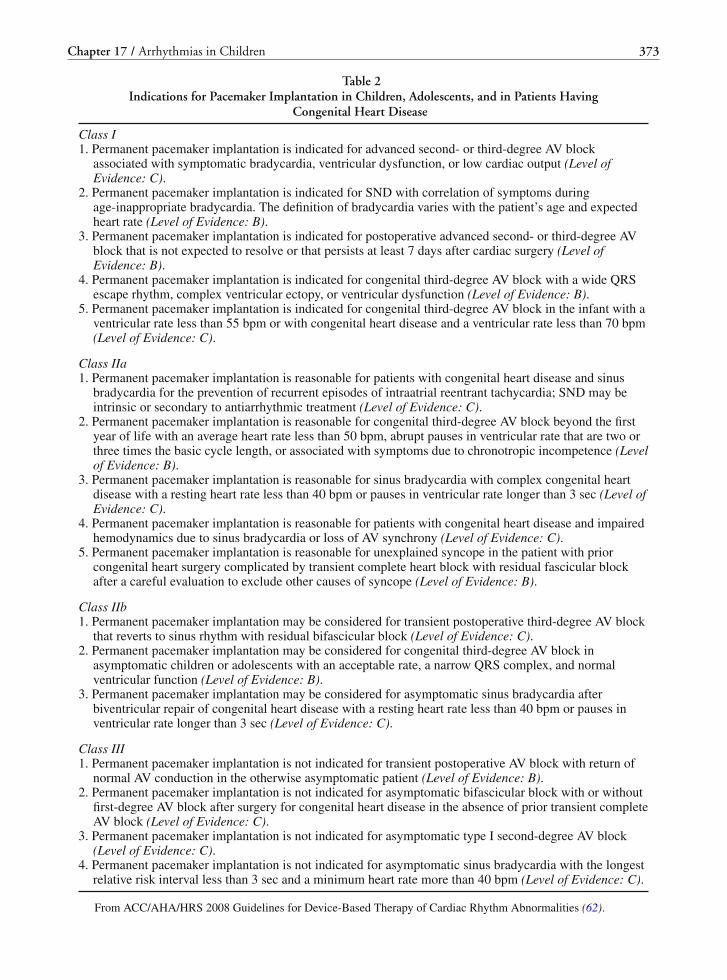
Chapter 17 / Arrhythmias in Children 373
Table 2Indications for Pacemaker Implantation in Children, Adolescents, and in Patients Having
Congenital Heart Disease
Class I1. Permanent pacemaker implantation is indicated for advanced second- or third-degree AV block
associated with symptomatic bradycardia, ventricular dysfunction, or low cardiac output (Level ofEvidence: C).
2. Permanent pacemaker implantation is indicated for SND with correlation of symptoms duringage-inappropriate bradycardia. The definition of bradycardia varies with the patient’s age and expectedheart rate (Level of Evidence: B).
3. Permanent pacemaker implantation is indicated for postoperative advanced second- or third-degree AVblock that is not expected to resolve or that persists at least 7 days after cardiac surgery (Level ofEvidence: B).
4. Permanent pacemaker implantation is indicated for congenital third-degree AV block with a wide QRSescape rhythm, complex ventricular ectopy, or ventricular dysfunction (Level of Evidence: B).
5. Permanent pacemaker implantation is indicated for congenital third-degree AV block in the infant with aventricular rate less than 55 bpm or with congenital heart disease and a ventricular rate less than 70 bpm(Level of Evidence: C).
Class IIa1. Permanent pacemaker implantation is reasonable for patients with congenital heart disease and sinus
bradycardia for the prevention of recurrent episodes of intraatrial reentrant tachycardia; SND may beintrinsic or secondary to antiarrhythmic treatment (Level of Evidence: C).
2. Permanent pacemaker implantation is reasonable for congenital third-degree AV block beyond the firstyear of life with an average heart rate less than 50 bpm, abrupt pauses in ventricular rate that are two orthree times the basic cycle length, or associated with symptoms due to chronotropic incompetence (Levelof Evidence: B).
3. Permanent pacemaker implantation is reasonable for sinus bradycardia with complex congenital heartdisease with a resting heart rate less than 40 bpm or pauses in ventricular rate longer than 3 sec (Level ofEvidence: C).
4. Permanent pacemaker implantation is reasonable for patients with congenital heart disease and impairedhemodynamics due to sinus bradycardia or loss of AV synchrony (Level of Evidence: C).
5. Permanent pacemaker implantation is reasonable for unexplained syncope in the patient with priorcongenital heart surgery complicated by transient complete heart block with residual fascicular blockafter a careful evaluation to exclude other causes of syncope (Level of Evidence: B).
Class IIb1. Permanent pacemaker implantation may be considered for transient postoperative third-degree AV block
that reverts to sinus rhythm with residual bifascicular block (Level of Evidence: C).2. Permanent pacemaker implantation may be considered for congenital third-degree AV block in
asymptomatic children or adolescents with an acceptable rate, a narrow QRS complex, and normalventricular function (Level of Evidence: B).
3. Permanent pacemaker implantation may be considered for asymptomatic sinus bradycardia afterbiventricular repair of congenital heart disease with a resting heart rate less than 40 bpm or pauses inventricular rate longer than 3 sec (Level of Evidence: C).
Class III1. Permanent pacemaker implantation is not indicated for transient postoperative AV block with return of
normal AV conduction in the otherwise asymptomatic patient (Level of Evidence: B).2. Permanent pacemaker implantation is not indicated for asymptomatic bifascicular block with or without
first-degree AV block after surgery for congenital heart disease in the absence of prior transient completeAV block (Level of Evidence: C).
3. Permanent pacemaker implantation is not indicated for asymptomatic type I second-degree AV block(Level of Evidence: C).
4. Permanent pacemaker implantation is not indicated for asymptomatic sinus bradycardia with the longestrelative risk interval less than 3 sec and a minimum heart rate more than 40 bpm (Level of Evidence: C).
From ACC/AHA/HRS 2008 Guidelines for Device-Based Therapy of Cardiac Rhythm Abnormalities (62).

374 Part V / Arrhythmias in Specific Populations
severe events in patients who develop atrial fibrillation and who also have ventricular preexcitationand is therefore contraindicated in children with SVT and WPW.
Chaotic Atrial TachycardiaThis uncommon arrhythmia is usually referred to as multifocal atrial tachycardia in adults, and
among children it occurs mostly in infants less than 1 year of age. One-third of patients tend to presentwith coexisting respiratory infections and one-third have structural heart disease (71). Diagnosis isbased on ECG criteria requiring: (1) more than three ectopic P wave morphologies; (2) irregular P–Pintervals; (3) isoelectric baseline between P waves; and (4) rapid atrial and ventricular rate. Some cri-teria also include absence of a dominant atrial pacemaker. Atrial rates vary from 250 to 600 beats/minwhile ventricular rates are from 110 to 250 beats/min. Congestive heart failure is not a common pre-sentation, but one-fourth of patients present with tachycardia-induced cardiomyopathy as evidencedby echocardiographic criteria (71). Chaotic atrial tachycardia is not responsive to adenosine, directcurrent cardioversion, and overdrive pacing suggesting that the mechanism is not reentry. It is thoughtto be a triggered mechanism but calcium channel blockers have not been shown to be efficacious. Per-haps the best explanation is rapid automaticity with various exit points and multiple reentry waveletssimilar to focal atrial fibrillation in adults.
Prognosis for infants with chaotic atrial tachycardia is generally good with most having completeresolution by 1 year of age. Pharmacologic therapy is aimed at control of the ventricular rate withdigoxin or an oral calcium channel blocker. Beta blocker therapy may be useful in some patientsin whom the ventricular rate cannot be otherwise controlled. Conversion to sinus rhythm may beattempted with agents such as amiodarone, flecainide, or, as is our preference, propafenone. Once insinus rhythm, therapy may be discontinued after 1 year of age with close follow-up of ambulatoryECGs.
Congenital Junctional Ectopic TachycardiaJunctional ectopic tachycardia (JET) that begins in the first year of life is considered “congenital.”
Most cases of congenital JET are sporadic, though a familial predisposition has been demonstrated insome patients (72). Congenital JET may also result from anti-Ro or anti-La antibodies and may evenevolve to CAVB (73). Since the ventricular rate varies from 140 to 370 beats/min in this incessanttachycardia, most infants present with heart failure due to tachycardia-induced cardiomyopathy. TheECG diagnostic criteria are (1) narrow QRS tachycardia; (2) AV dissociation; and (3) ventricular rategreater than the atrial rate. JET usually does not terminate with adenosine or cardioversion and thusthe mechanism is enhanced automaticity of the AV junction above the bundle branches. Therapy isaimed at control of the ventricular rate, generally to <150 beats/min, which should allow recovery ofventricular function. Pharmacologic therapy with digoxin has been shown to be ineffective and agentssuch as sotalol, flecainide, propranolol, or propafenone may have some efficacy. However, the mostuseful medication is amiodarone, which has become the mainstay of therapy for congenital JET. Forintractable cases in which pharmacologic therapy fails to control the ventricular rate, catheter ablationof the AV junction with preservation of normal AV conduction has been successfully performed, evenin infants (74, 75).
Postoperative Junctional Ectopic TachycardiaJunctional ectopic tachycardia is the most common postoperative arrhythmia after congenital heart
surgery with an incidence of up to 8–10%. It is most commonly associated with repair of tetralogyof Fallot, VSD, or AVCD, though it may be seen after nearly any type of cardiac surgery (76). Themechanisms are thought to be related to transient edema or inflammatory injury to the His bundle fromnearby suture lines or to direct traction. When it occurs following Fontan-style operations or repair of

Chapter 17 / Arrhythmias in Children 375
total anomalous pulmonary venous return, it almost certainly occurs due to traction from elevatedcavitary pressures, as might be related to higher filling pressure or pulmonary hypertension. Longercardiopulmonary bypass, longer aortic cross clamp times (77), and the level of inotropic support (78)are risk factors.
Postoperative JET usually occurs within the first 24 h after surgery and can have serious hemody-namic consequences due to loss of AV synchrony and limited diastolic filling time. Previously reportedmortality rates have been as high as 50%, but with early recognition and improvements in management,the mortality rate can be significantly reduced. The aim of therapy is to bring the ventricular rate below180 in infants and less than 150 in older children. The initial steps in management include correctionof electrolyte abnormalities, especially hypokalemia, and reduction in inotropic support as can be tol-erated hemodynamically. Moderate hypothermia (32–34◦C) has some efficacy, but it requires paralyticdrugs and often causes metabolic acidosis. Our institutional policy has been to cool the patient to mildhypothermia (35–36◦C) when the initial management fails. The drugs most frequently reported to beefficacious for postoperative JET are procainamide, propafenone, and amiodarone. Although amio-darone is the most widely used agent for the management of JET, practitioners must be aware of therisk of significant hypotension with IV administration. The hypotension can sometimes be managedwith administration of IV fluids and calcium. In our opinion, the rate of bolus administration shouldnot be faster than 5 mg/kg over 45 min. Maintenance dosing is then begun. Once the ventricular rateis reduced to less than 180, sequential AV pacing at a slightly faster rate may be initiated for AV syn-chrony. The older therapy of ventricular “double pacing” is now considered largely antiquated due tothe risk of causing ventricular fibrillation. The rhythm will gradually slow to less than the ambientsinus rate over 24–96 h, in most cases. With appropriate management, prognosis is generally good,though patients with JET do have a higher reported postoperative mortality (13.5%) compared to con-trols (1.7%) (78). Intractable JET may require aggressive support with ECMO or ventricular assistdevices and in some extreme cases, ablation of the AV node followed by placement of a permanentdual-chamber pacemaker.
SECTION V: THERAPIES FOR PEDIATRIC ARRHYTHMIAS
Pharmacologic TherapiesDrug therapy has an important role in pediatric arrhythmias despite the primary treatment of
catheter ablation for most arrhythmias. Some indications for pharmacologic management include fetaltachyarrhythmias and infant SVT in which catheter ablation carries increased risk, and which mayeventually resolve spontaneously. Ventricular arrhythmias in infants and young children with or with-out congenital heart disease often require antiarrhythmic medications.
Table 3 summarizes the major antiarrhythmic medications and includes information on dosing, phar-macology, and metabolism specific to the pediatric (and in some cases, fetal) population. As with allmedications used in children, the bioavailability must be taken into consideration with an understand-ing of gastrointestinal and hepatic physiology in the young. In general, drugs are less bioavailable inchildren compared to adults due to relative achlorhydria (until 3 years of age), slower gastric emptying(until 6–8 months of age), and reduced gut motility (until 4 years of age). Most hepatic enzyme sys-tems are immature until at least 6 months of age. This causes reduced biotransformation and first-passelimination of drugs such as propranolol, lidocaine, amiodarone, and verapamil; thus, their half-livesare longer than in older children and adults. Most antiarrhythmic medications are renally excreted, andthis system is also immature until 1 year of age. Pathologic processes in the gut (including mucosaledema), liver, or kidneys due to congestive heart failure, low cardiac output, congenital anomalies, orother causes of organ dysfunction affect drug metabolism similarly in children and adults.

376 Part V / Arrhythmias in Specific Populations
Tab
le3
Ant
iarr
hyth
mic
Dru
gU
sein
Chi
ldre
n
Dru
gH
alf-
life
Intr
aven
ous
dosi
ngO
rald
osin
gTr
ansp
lace
ntal
pass
age
(%)
Pre
gnan
cyca
tego
ry/s
afet
yfo
rfe
tus
Pass
age
inbr
east
mil
kIn
dica
tion
s
Cla
ssIa
Dis
opyr
amid
e4.
5–7.
8h
Not
avai
labl
e10
–30
mg/
kg/d
(inf
ants
);10
–20
mg/
kg/d
(chi
ldre
n);
6–15
mg/
kg/d
(ado
lesc
ents
);40
0–80
0m
g/d
(adu
lts)
q6
h(r
egul
ar),
q12
h(s
usta
ined
rele
ase)
;m
axim
um:1
600
mg/
d)
39C
50–9
0%m
ater
nal
bloo
dle
vel;
safe
for
infa
nt
PSV
T;A
F1;
AF;
VT
inC
HD
with
good
vent
ricu
lar
func
tion
Qui
nidi
ne30
–90
min
(sul
fate
);3.
4–4.
0h
(glu
cona
te)
Not re
com
men
ded
30–6
0m
g/kg
/d(4
50–9
00m
g/m
2);
q4–
6h;
10m
g/kg
/d(a
dult)
;20%
high
erif
gluc
onat
e
24–9
4C
Rep
orte
dto
occu
r;sa
fefo
rin
fant
Rar
ely
used
due
to“q
uini
dine
sync
ope”
;PS
VT;
AFI
;A
F;V
Tin
CH
DPr
ocai
nam
ide
1.7
h(c
hild
ren)
;2.
5–4.
7h
(adu
lts)
(sho
rtac
ting)
6–7
h(s
low
rele
ase)
;sh
orte
rin
child
ren;
long
erin
neon
ates
10–1
5m
g/kg
over
20m
in;
mai
nten
ance
:30
–80
μg/
kg/m
in
50–1
00m
g/kg
/d25
–100
CR
epor
ted
tooc
cur;
safe
for
infa
ntPS
VT;
AF1
;A
F;A
ET;
VT
inC
HD
;po
stop
era-
tive
JET
Cla
ssIb
Mor
iciz
ine
1.5–
3.5
h;bi
olog
icT
1/2
48h
Not
avai
labl
e20
0m
g/m
2/d
(ini
tial)
incr
ease
to60
0m
g/m
2/d
;q8
h
Not
know
nB
Not
know
nA
ET;
VT;
PSV
T

Chapter 17 / Arrhythmias in Children 377
Lid
ocai
ne3.
2h
(neo
nate
);1.
5–2
h(c
hild
ren
and
adul
ts)
1m
g/kg
,may
repe
attw
ice;
mai
nten
ance
:20
–50
μg
/kg/
min
Not
effe
ctiv
e50
B(f
etal
brad
ycar
dia
repo
rted
;ge
nera
llysa
fefo
rfe
tus)
40%
mat
erna
lbl
ood
leve
l;sa
fefo
rin
fant
VT,
espe
cial
lyis
chem
ic
Mex
iletin
e6.
3–11
.8h
Not
avai
labl
eIn
fant
s:8–
25m
g/kg
/d;
child
ren:
4.5–
15m
g/kg
/d;
max
imum
:60
0–12
00m
g/d;
q8
h
100
(equ
ival
ent
tom
ater
nal
bloo
dle
vel)
C10
0%m
ater
nal
bloo
dle
vel;
safe
for
infa
nt
VT
Phen
ytoi
n75
±64
h(p
rem
atur
e);
21±
12h
(ter
m);
;8
±4
h;(1
mon
th);
22±
7h
(chi
ld–a
dult)
10–1
5m
g/kg
over
1h
Loa
d:15
mg/
kg/d
q6
h(d
ay1)
,7.5
mg/
kg/d
q6
h(d
ay2)
;m
aint
enan
ce:0
–2w
eeks
:4–8
mg/
kg/d
q12
h;2
wee
ks–2
year
s:8–
12m
g/kg
/dq
8h;
3–12
year
s:5–
6m
g/kg
/dq
12h;
>12
year
s:4.
5m
g/kg
/dq
12h
Exc
elle
ntD
(11%
have
“fet
alhy
dant
oin
synd
rom
e”;
31%
have
less
erm
ani-
fest
atio
ns)
Safe
for
infa
ntV
T
Toca
inid
e15
h(a
dult)
Not
avai
labl
eC
hild
ren:
350–
700
mg/
m2/d
Not
know
nC
Not
know
nV
T
Cla
ss1c
Flec
aini
de29
h(n
ewbo
rn);
11–1
2h
(inf
ant)
;8h
(chi
ldre
n)12
–27
h(a
dult)
1–2
mg/
kgov
er5–
10m
in1–
6m
g/kg
/dor
50–1
50m
g/m
2/d
q8
h(c
hild
ren)
&q
12h
(new
born
,inf
anta
ndad
ult)
;max
imum
:40
0m
g/d;
avoi
dad
min
istr
atio
nw
ithm
ilk-b
ased
form
ulas
inin
fant
s
100
(equ
ival
ent
tom
ater
nal
bloo
dle
vel)
CC
once
ntra
ted
(2.3
–3.7
:1);
safe
for
infa
nt
PSV
T;A
F1;
AF;
PJR
T;V
T;fe
tal
tach
ycar
-di
as;
JET (C
ontin
ued)

378 Part V / Arrhythmias in Specific Populations
Tab
le3
(Con
tinu
ed)
Dru
gH
alf-
life
Intr
aven
ous
dosi
ngO
rald
osin
gTr
ansp
lace
ntal
pass
age
(%)
Pre
gnan
cyca
tego
ry/
safe
tyfo
rfe
tus
Pass
age
inbr
east
mil
kIn
dica
tion
s
Prop
afen
one
4.7
±1.
3h
(ext
ensi
vem
etab
oliz
er);
16.8
±10
.6h
(slo
w=
10%
popu
latio
n);↑
with
time
inbo
thgr
oups
0.2
mg/
kgq
10m
into
effe
ct(m
ax=
2m
g/kg
);m
aint
enan
ce:
4–7
μg
/kg/
min
)
200–
600
mg/
m2/d
q6–
8h;
max
imum
:90
0m
g/d
Not
know
nC
Not
know
nPS
VT;
AF1
;A
F;PJ
RT;
VT;
infa
ntile
CA
T;JE
T
Cla
ssII
Ate
nolo
l16
–35
h(n
ewbo
rn);
3.5–
7h
(chi
ldre
n);
6–9
h(a
dults
)
No
pedi
atri
cda
ta0.
8–1.
5m
g/kg
/dq
12–2
4h;
whe
nus
edfo
rpo
tent
ially
life-
thre
aten
ing
arrh
ythm
ia(L
QT
S),
q12
hre
com
men
ded
100
mat
erna
lbl
ood
leve
lD
Con
cent
rate
d(1
.5–6
:8:1
);fe
tal
brad
ycar
dia
PSV
T;ra
teco
ntro
lin
AF1
and
AF;
VT
inno
rmal
hear
t;L
QT
SE
smol
ol4.
5±
2.1
min
(chi
ldre
n);
9m
in(a
dults
)
Loa
d:50
0μ
g/kg
over
1m
in;
mai
nten
ance
:50
–600
μg
/kg/
min
Not
appl
icab
leN
otkn
own
CN
otkn
own
PSV
T;ra
teco
ntro
lin
AF1
and
AF
Nad
olol
20–2
4h
(adu
lts);
shor
ter
inin
fant
san
dch
ildre
n
Not
avai
labl
e1–
2m
g/kg
/dq
day
Som
epa
ssag
eoc
curs
C(h
ypog
lyce
mia
;sy
mpt
omat
icbr
adyc
ardi
a)
Con
cent
rate
d(3
:1);
feta
lbr
adyc
ardi
a
PSV
T;ra
teco
ntro
lin
AF1
and
AF;
VT
inno
rmal
hear
t;L
QT
S

Chapter 17 / Arrhythmias in Children 379
Prop
rano
lol
3.9–
6.4
h(c
hild
ren)
;4–
6h
(adu
lts)
0.01
–0.1
5m
g/kg
over
5m
in1–
5m
g/kg
/dq
6–8
h(s
hort
actin
g);q
12–2
4h
(tim
ere
leas
e);w
hen
used
for
pote
ntia
llylif
e-th
reat
enin
gar
rhyt
hmia
(LQ
TS)
,q12
hre
com
men
ded
100
mat
erna
lbl
ood
leve
lC
(IU
GR
;hyp
o-gl
ycem
ia;
resp
irat
ory
depr
essi
on)
64%
mat
erna
lpl
asm
ale
vel;
safe
for
infa
nt
PSV
T;ra
teco
ntro
lin
AF1
and
AF;
VT
inno
rmal
hear
t;L
QT
S
Cla
ssII
I
Am
ioda
rone
8–10
7d;
biph
asic
elim
inat
ion
(pla
sma
leve
l50
%af
ter
10d;
rebo
unds
days
12–2
1;fa
llsag
ain)
Loa
d:2–
5m
g/kg
over
20–4
5m
in(r
epea
tas
need
ed);
then
0.4–
1.0
mg/
kg/h
Loa
d:10
mg/
kg/d
q8–
12h
(up
to60
mg/
kg/d
x≤
3d,
ifne
cess
ary)
;the
n,5
mg/
kg/d
×2
mon
ths;
then
,wea
nto
2.5
mg/
kg/d
10–2
5m
ater
nal
bloo
dle
vel
D(p
rem
atur
ity12
%;I
UG
R21
%;
hypo
thy-
roid
ism
9%)
Exc
eeds
mat
erna
lpl
asm
ale
vel;
safe
for
infa
nt
PSV
T;A
F1;
AF;
VT;
JET;
AE
T
Sota
lol
9.5
h(c
hild
ren)
;7–
18h
(adu
lt);
beta
-blo
ckin
gef
fect
long
er
Not
avai
labl
e2–
8m
g/kg
/dor
40–2
50m
g/m
2/d
q8–
12h;
max
imum
dose
640
mg/
d;us
ual
adul
tsta
rtin
gdo
se:
160
mg/
d
5–10
0m
ater
nal
bloo
dle
vel
B(a
sym
ptom
atic
brad
ycar
dia)
Con
cent
rate
d(5
:4:1
);m
ayno
tbe
impo
rtan
t
PSV
T;A
F1;
AF;
VT
Cla
ssIV
Ver
apam
il2.
5–7
h(i
nfan
cy);
5–12
hw
ithch
roni
cus
e
0.1
mg/
kgov
er2
min
(max
imum
10m
g);
infu
sion
:5
μ/k
g/m
in;n
ot<
1ye
arof
age
3–10
mg/
kg/d
q8
h(s
hort
actin
g);q
day
(tim
ere
leas
e)
15–5
0m
ater
nal
leve
lC
23–1
00%
mat
erna
lblo
odle
vel;
safe
for
infa
nt
PSV
T;ra
teco
ntro
lin
AF1
and
AF;
feta
lPS
VT;
occa
sion
alfo
rms
ofV
Tin
norm
alhe
art
(Con
tinue
d)

380 Part V / Arrhythmias in Specific PopulationsT
able
3(C
onti
nued
)
Dru
gH
alf-
life
Intr
aven
ous
dosi
ngO
rald
osin
gTr
ansp
lace
ntal
pass
age
(%)
Pre
gnan
cyca
te-
gory
/saf
ety
for
fetu
sPa
ssag
ein
brea
stm
ilk
Indi
cati
ons
Dig
oxin
Dig
oxin
61–1
70h
(pre
mat
ure)
;35
–45
h(t
erm
);18
–25
h(i
nfan
t);3
5h
(chi
ld);
38–4
8h
(adu
lt)
Ora
lloa
d(t
hree
dose
s/16
–24
h):
30μ
g/kg
(pre
mat
ure)
,35
(ter
m),
40–5
0(<
2ye
ars)
,30–
40(>
2ye
ars)
;m
aint
enan
ce:
1 / 4lo
ad/q
12h;
Intr
aven
ous
dose
:75–
80%
oral
Sim
ilar
tom
ater
nall
evel
CSi
mila
rto
mat
erna
lbl
ood
leve
l;sa
fefo
rin
fant
PSV
T;fe
tal
PSV
T
Ade
nosi
ne
Ade
nosi
ne<
5se
c50
–300
μg/
kgra
pidl
yN
otap
plic
able
Not
appl
icab
leN
otap
plic
able
Not
appl
icab
lePS
VT;
SAN
RT;
som
eFA
T;so
me
norm
alhe
artV
T
AE
Tat
rial
ecto
pic
tach
ycar
dia;
AF
atri
alfib
rilla
tion;
AF
1at
rial
flutte
r;C
AT
chao
ticat
rial
tach
ycar
dia;
CH
Dco
ngen
italh
eart
dise
ase;
FAT
foca
latr
ialt
achy
card
ia;J
ET
junc
tiona
lect
opic
tach
ycar
dia;
LQ
TS
long
QT
synd
rom
e;P
JRT
perm
anen
tfor
mof
junc
tiona
lrec
ipro
catin
gta
chyc
ardi
a;P
SVT
paro
xysm
alsu
prav
entr
icul
arta
chyc
ardi
a;SA
NR
Tsi
noat
rial
node
reen
try
tach
ycar
dia;
VT
vent
ricu
lar
tach
ycar
dia;
Uni
ted
Stat
esFD
APr
egna
ncy
Cat
egor
ies:
AC
ontr
olle
dst
udie
sin
wom
enfa
ilto
dem
onst
rate
ari
skto
the
fetu
sin
the
first
trim
este
r,an
dth
epo
ssib
ility
offe
tal
harm
appe
ars
rem
ote.
BA
nim
alst
udie
sdo
not
indi
cate
ari
skto
the
fetu
san
dth
ere
are
noco
ntro
lled
hum
anst
udie
s,or
anim
alst
udie
sdo
show
anad
vers
eef
fect
onth
efe
tus
but
wel
l-co
ntro
lled
stud
ies
inpr
egna
ntw
omen
have
faile
dto
dem
onst
rate
ari
skto
the
fetu
s.C
Stud
ies
have
show
nth
atth
edr
ugex
erts
anim
alte
rato
geni
cor
embr
yoci
dale
ffec
ts,b
utth
ere
are
noco
ntro
lled
stud
ies
inw
omen
,or
nost
udie
sar
eav
aila
ble
inei
ther
anim
als
orw
omen
.DPo
sitiv
eev
iden
ceof
hum
anfe
talr
isk
exis
ts,b
utbe
nefit
sin
cert
ain
situ
atio
ns(e
.g.,
life-
thre
aten
ing
situ
atio
nsor
seri
ous
dise
ases
for
whi
chsa
fer
drug
sca
nnot
beus
edor
are
inef
fect
ive)
may
mak
eus
eof
the
drug
acce
ptab
lede
spite
itsri
sks,
XSt
udie
sin
anim
als
orhu
man
sha
vede
mon
stra
ted
feta
labn
orm
aliti
esor
ther
eis
evid
ence
offe
talr
isk
base
don
hum
anex
peri
ence
,or
both
,and
the
risk
clea
rly
outw
eigh
san
ypo
ssib
lebe
nefit
.A
dapt
edan
dw
ithpe
rmis
sion
from
:Fus
ter
etal
.(11
9),T
able
47-3
,pp
1134
–113
6.

Chapter 17 / Arrhythmias in Children 381
Catheter AblationIn many communities, catheter ablation has become first-line therapy for most tachyarrhythmia sub-
strates in older children and teenagers. Radiofrequency catheter ablation (RFA) of cardiac dysrrhyth-mia substrates in humans has been available for over 20 years, and its efficacy and safety was welldefined in children by the Prospective Assessment of Pediatric Catheter Ablation (PAPCA) project(79). Cryoablation has been used for over 10 years and has also been shown to be effective and safein children with increasing popularity as first-line treatment for dysrhythmia substrates located in theatrial paraseptal region (80). The indications for radiofrequency catheter ablation in pediatric patientsbased on the 2002 Expert Consensus Conference report are summarized in Table 4 (81).
Due to smaller patient size, there are special considerations when considering catheter ablation inchildren. For example, in the 3-year-old child, the triangle of Koch is 3–5 mm at its base and 3–6 mm inlength, which is not much larger than the lesion size generated by even the smallest available (3 mm tip)radiofrequency ablation catheters. Hence, ablation of septal accessory pathways or AVNRT presents asignificant risk in these smaller patients and is generally not electively undertaken. Another anatomicconsideration is cardiac wall thickness at the annulus fibrosis, representing the theoretical distancebetween the epicardial coronary arteries and the endocardial ablation catheter tip. Based upon autopsyspecimens, among children less than 7 years of age, this distance is <4 mm for 76% of measured sitesand <5 mm for 96% (82). As observed in adults, the shortest distance is just within the coronary sinusostium. There are few reports of damage to coronary arteries with catheter ablation in children, thoughit may go unrecognized more frequently than previously thought (83, 84). Cryoablation may be saferthan radiofrequency ablation with regard to coronary injury (85).
If necessary, invasive electrophysiologic testing and catheter ablation can be safely performed invery small patients including infants (86). Even with small femoral vein size, 7 Fr sheaths can beplaced and multiple 2 Fr quadripolar electrode catheters can be advanced or a single catheter maybe used for both His and right ventricular pacing/recording and for lateral right atrial and coro-nary sinus pacing/recording. Thus, femoral venous capacity is usually not a limiting factor, sinceeach vein can accommodate up to 5 mm circumference (5 Fr) hardware in children >2.0 kg, 6 Frif > 3.0 kg, 7 Fr if >5.0 kg, 9 Fr if >10 kg, 11 Fr if >25 kg, and 14 Fr for larger patients. Atrialsensing and pacing may also be performed from the esophagus. Arterial access requires differentconsiderations but is generally avoided with the use of transseptal access to the left side of theheart.
With regard to radiofrequency energy delivery, there does not appear to be a correlation betweenpatient size and impedance load. Thus, the applied power is the same as in adults. Longer durationof radiofrequency lesion application has been shown to correlate with higher complication rates insmaller patients and careful judgment must be employed weighing the risks of ablation and severityof the arrhythmia (87). Growing experience with irrigated-tipped catheters suggest they are also safeand perhaps more effective in congenital heart disease patients with arrhythmia substrates in theirthickened atrial walls (88).
Catheter ablation in patients with complex congenital heart disease presents further challenges. Priorto the procedure, review of surgical reports is a prerequisite, and the anatomy must be clearly definedwith imaging modalities such as transthoracic/transesophageal echocardiography, CT, and/or MRI. Atthe time of the procedure, angiography of the vascular and heart structures may be required prior toplacement of the electrode catheters. Reduced or altered venous access to the heart should be antic-ipated due to occluded iliofemoral veins, congenitally interrupted IVC with azygos continuation, orsurgically created venous bypass pathways (e.g., bidirectional Glenn procedure, Fontan operation).In patients with inferior vena caval interruption, successful catheter ablation may be performed viahepatic vein cannulation or by the azygos vein (89, 90). Anticipatory knowledge of unusual anatomiclocations of important conduction system structures in various congenital heart malformations should

382 Part V / Arrhythmias in Specific Populations
Table 4Indications for Radiofrequency Catheter Ablation in Pediatric Patients [from the 2002 Expert Consensus
Conference Report (80)]
Indications for radiofrequency catheter ablation in pediatric patients
Class I: There is consistent agreement and/or supportive data that catheter ablation is likely to bemedically beneficial or helpful for the patient.
1. Wolff–Parkinson–White (WPW) syndrome following an episode of aborted sudden cardiac death.2. The presence of WPW syndrome associated with syncope when there is a short preexcited R–R
interval during atrial fibrillation (preexcited R–R interval, 250 ms) or the antegrade effectiverefractory period of the AP measured during programmed electrical stimulation is <250 ms.
3. Chronic or recurrent supraventricular tachycardia (SVT) associated with ventricular dysfunction.4. Recurrent VT that is associated with hemodynamic compromise and is amenable to catheter
ablation.
Class IIA: There is a divergence of opinion regarding the benefit or medical necessity of catheterablation; the majority of opinions/data are in favor of the procedure.
1. Recurrent and/or symptomatic SVT refractory to conventional medical therapy and age >4 years.2. Impending congenital heart surgery when vascular or chamber access may be restricted following
surgery.3. Chronic (occurring for >6 to 12 months following an initial event) or incessant SVT in the presence
of normal ventricular function.4. Chronic or frequent recurrences of intraatrial reentrant tachycardia (IART).5. Palpitations with inducible sustained SVT during electrophysiological testing.
Class IIB: There is clear divergence of opinion regarding the need for the procedure.1. Asymptomatic preexcitation (WPW pattern on an electrocardiograph), age >5 years, with no
recognized tachycardia, when the risks and benefits of the procedure and arrhythmia have beenclearly explained.
2. SVT, age >5 years, as an alternative to chronic antiarrhythmic therapy which has been effective incontrol of the arrhythmia.
3. SVT, age <5 years (including infants), when antiarrhythmic medications, including sotalol andamiodarone, are not effective or associated with intolerable side effects.
4. IART, one to three episodes per year, requiring medical intervention.5. AVN ablation and pacemaker insertion as an alternative therapy for recurrent or intractable IART.6. One episode of VT associated with hemodynamic compromise and which is amenable to catheter
ablation.
Class III: There is an agreement that catheter ablation is not medically indicated and/or the risk of theprocedure may be greater than the benefit for the patient.
1. Asymptomatic WPW syndrome, age <5 years.2. SVT controlled with conventional antiarrhythmic medications, age <5 years.3. Nonsustained, paroxysmal VT, which is not considered incessant (i.e., present on monitoring for
hours at a time or on nearly all strips recorded during any 1-h period) and where no concomitantventricular dysfunction exists.
4. Episodes of nonsustained SVT that do not require other therapy and/or are minimally symptomatic.
be applied to meticulous electroanatomic mapping. Newer technologies such as intracardiac ultra-sound, image fusion, and non-contact balloon array mapping are emerging as useful techniques inpatients with congenital heart disease (32). Three-dimensional electroanatomic mapping has beendemonstrated to improve success rates for ablation of atrial tachyarrhythmia substrates following con-genital heart surgery (39) and is now considered requisite technology (see Fig. 12). It may also reduce

Chapter 17 / Arrhythmias in Children 383
Fig. 12. Electroanatomic maps used to direct catheter ablation of supraventricular tachycardia substrates fol-lowing complex congenital heart disease. Left: Right posterolateral projection of the systemic venous bafflefollowing Senning operation for d-transposition of the great arteries. The earliest focus of focal atrial tachycar-dia is identified adjacent to baffle suture line from this bipolar isochronal map (“activation map”). Red repre-sents earliest activity and violet, latest. Right: Right posterolateral projection of a dilated right atrium followingatriopulmonary-style Fontan operation. In this bipolar voltage map, electrically inactive regions (“scar”) arerepresented by gray and viable tissue by colors ranging from red (lowest voltage) to violet (highest). Catheterablation of narrow channels (1–3) of conducting tissue rendered this patient’s intraatrial reentry tachycardianon-inducible. IVC, inferior vena cava; RF, radiofrequency lesion; SVC, superior vena cava (right-sided figureadapted with permission from: Lippincott, Williams, & Wilkins, Philadelphia, PA. In: Ref. (41)).
or, in some cases, eliminate the need for radiation exposure (91) which is always a matter of concernin pediatric patients.
Most catheter ablation procedures in children are performed under general anesthesia. Anestheticagents such as propofol and isoflurane are commonly used and generally do not suppress arrhyth-mia inducibility (92). There are minor effects on conduction and refractoriness intervals, which areclinically unimportant (92). Among youngsters having tachycardias using enhanced automaticity asthe mechanism (especially AET and some “normal heart VTs”), general anesthesia may render thetachycardia uninducible. Of course, the selection of sedation versus anesthesia needs to be individ-ualized with awareness of coexistent issues such as structural heart disease, other medical issues,and potential airway obstruction. In particular, patients whose heart conditions are especially preloaddependent, such as those having cardiomyopathy or “Fontan physiology” may be especially proneto hypotension during general anesthesia. Primary agents such as ketamine may be useful in thoseinstances. Prior to the procedure, age-appropriate reading material, play therapy, and opportunities tonetwork with other children and teenagers who had previously undergone the procedure may be useful.
Overall, the results of catheter ablation in pediatric patients are comparable to results in adultpatients. Results from the PAPCA project comprise a large prospective analysis of AV reciprocatingtachycardia and AVNRT in patients aged 0–21 years, excluding patients with congenital heart disease(93). The cohort included 481 patients and the acute success rate was 95.7%. For left free wall andright free wall accessory pathways, the success rate was 97.8 and 90.8%, respectively. The proceduralcomplication rate was 4%. Patients were followed for 12 months, and the arrhythmia substrate recur-rence rate was 7.0, 9.2, and 10.7% at 2, 6, and 12 months, respectively (94). For individual arrhythmia

384 Part V / Arrhythmias in Specific Populations
substrates, the recurrence rates were as follows: 24.6% (right septal pathways), 15.8% (right free wallpathways), 9.3% (left free wall pathways), 4.3% (left septal pathways), and 4.8% (AVNRT). Similarresults were reported in other studies of radiofrequency ablation in pediatric patients (95, 96). Cryoab-lation is becoming more widely applied for pediatric catheter ablation due to relative safety whentargeting AVNRT or septal pathways. Reported acute success rate of cryoablation for AVNRT rangesbetween 83–96% and 60–92% for accessory pathways. Short-term follow-up studies have demon-strated an 8–45% recurrence rate with cryoablation (97–102). Longer applications of cryoenergy mayimprove success rates (103) and successful ablation of ventricular tachycardia has also been reportedwith cryoablation (104).
Pacemakers in Children and in Congenital Heart DiseaseThe indications for permanent pacing in pediatric children have been summarized in Table 2 (62).
As with catheter ablation, there are size, developmental, and disease-related considerations with regardto implantable cardiac rhythm devices in pediatric patients. Advances in device and lead design havemade it possible to provide rhythm management in the smallest of patients with epicardially placedsystems. Several factors have reversed the trend of transvenous lead placement in small children:Steroid-eluting, passive fixation epicardial leads have an excellent long-term track record; newer sur-gical techniques, including thoracoscopic approaches, have made epicardial lead placement less inva-sive than in the past; and there is now greater recognition of the long-term adverse impact of failedtransvenous leads in patients who still have decades of pacing ahead of them. Indeed, most pediatricelectrophysiologists now reserve transvenous lead implantation for children weighing at least 25 kg.
Among patients having repaired or unrepaired congenital heart disease and who require pacing,safe venous access to the heart may be limited by the presence of single ventricle physiology, residualpotential right–left shunts, and venous obstruction from prior procedures. As examples, following theMustard operation, the operator must be certain that there is no superior vena caval baffle obstruc-tion, and that there are no baffle “leaks” (potentiating paradoxical embolization) before placing leads;following the atriopulmonary type of Fontan operation, the operator must be concerned that a newatrial lead may stimulate thrombus formation due to the potential procoagulant state so common inthose patients; and following a Rastelli type of operation (right ventricle-to-pulmonary artery conduitand VSD closure as repair of d-TGA, VSD, and pulmonic stenosis; pulmonary atresia and VSD; ortruncus arteriosus), even a small residual VSD should make the operator concerned about paradoxicalembolization, as the conduit narrows over time, resulting in right ventricular hypertension. “Hybridprocedures” may be performed, in which one lead is transvenous and the other epicardial. Althoughthis was once mostly limited to atriopulmonary-style Fontan patients who had heart block and requiredan epicardial ventricular lead, this approach is now performed in an effort to provide ventricular resyn-chronization in young patients having ventricular dysfunction who require a systemic ventricular lead(see Fig. 13).
Ventricular resynchronization pacing is now being applied to certain patients having congenitalheart disease and ventricular dysfunction. In the immediate postoperative period (mostly in infants),there is a clear benefit of biventricular pacing using temporary pacing wires for children having poorsystemic ventricular function and flagging hemodynamic parameters (105). Evidence-based indica-tions for chronic application of this technology are more difficult to establish than in adults having twoanatomically and positionally normal ventricles. Initial multi-institution series of patients having het-erogeneous congenital heart defects have shown promising results (106). Those patients having a fail-ing systemic right ventricle and a subpulmonic left ventricle (both d-TGA following Mustard/Senningand l-TGA) appear to benefit with improved indices of conduction synchronization, ventricular func-tion, and NYHA class (107). Use of three-dimensional tissue Doppler techniques are currently being

Chapter 17 / Arrhythmias in Children 385
Fig. 13. Posteroanterior chest radiograph from an 11-year-old boy with congenitally corrected transposition(l-TGA) and acquired complete AV block, who is status-post left-sided tricuspid valve replacement and whodeveloped severely diminished systemic ventricular dysfunction 6 years after placement of a transvenous dualchamber pacing system. This figure illustrates a hybrid dual chamber, biventricular pacing system, with an epi-cardial bipolar lead (black arrows) attached to the posterolateral base of the left-sided ventricle and tunneledto the left infraclavicular pacemaker pocket. White arrow represents a “spare” unipolar epicardial lead attachedto the left-sided ventricle; black arrowhead represents the original right atrial lead; and white arrowhead, theoriginal right-sided ventricular lead.
applied to patients having a poorly functioning single ventricle in an effort to assess the role of multi-site pacing (108).
In general, children and adolescents adapt well to life with an implanted device and can maintaina healthy self-image given appropriate support from family members and medical professionals. Nor-malization of lifestyle helps greatly in psychosocial well-being. This includes permission to participatein sports activities as deemed appropriate for the disease. We allow participation in high enduranceactivities such as basketball, soccer, track, and swimming in patients who are not “pacemaker depen-dent” while keeping the families informed about potential damage to transvenous leads from excessiveshoulder movement. There is general agreement that these patients should be restricted from contactsports.
Implantable Cardioverter/Defibrillators (ICDs)Implantable cardioverter-defibrillators have been implanted in children and teenagers for preven-
tion of sudden cardiac death over the last 20 years. Silka’s original report substantiating the efficacy ofICDs in young patients having cardiomyopathy, primary electrical disease, or congenital heart disease,was primarily concerned with secondary prevention (109). Progress in sudden death risk identificationin the patient groups described above has shifted the balance toward implantation for primary preven-tion. This philosophy has been facilitated by downsizing of hardware and creative and less invasivemethods in conductor (sense/pace lead, high-voltage conductors) implantation. However, for childrenand adults having congenital heart disease, there is no evidence-based reservoir analogous to MADITor SCD-HeFT from which to “mine” data. Hence, practitioners largely base decisions upon single- or

386 Part V / Arrhythmias in Specific Populations
multi-institution retro- or ambispective studies (46, 110) or as a “best fit” with adult series of patientshaving cardiomyopathy or coronary artery disease. Therefore, implantable cardioverter-defibrillatorimplantation now appears somewhere in every treatment algorithm for pediatric patients having hyper-trophic cardiomyopathy, restrictive cardiomyopathy, dilated cardiomyopathy, left ventricular non-compaction, arrhythmogenic right ventricular cardiomyopathy, Brugada syndrome, catecholaminergicpolymorphic ventricular tachycardia, long QT syndrome; and in all patients having congenital heartdisease with ventricular tachyarrhythmias and/or low ejection fraction (<35%?). The efficacy of ICDimplantation has also been demonstrated as part of a “bridge” to heart transplantation in some patientswith severe cardiac failure and life-threatening arrhythmias (111). The 2008 guidelines for device-based therapy of cardiac rhythm abnormalities include a section on indications for ICD implantationin children and in patients with congenital heart disease (see Table 5) (62).
Table 5Indications for Implantable Cardioverter-Defibrillators in Children, Adolescents, and in Patients Having
Congenital Heart Disease
Class I1. ICD implantation is indicated in the survivor of cardiac arrest after evaluation to define the cause of
the event and to exclude any reversible causes (Level of Evidence: B).2. ICD implantation is indicated for patients with symptomatic sustained VT in association with
congenital heart disease who have undergone hemodynamic and electrophysiological evaluation.Catheter ablation or surgical repair may offer possible alternatives in carefully selected patients(Level of Evidence: C).
Class IIaICD implantation is reasonable for patients with congenital heart disease with recurrent syncope of
undetermined origin in the presence of either ventricular dysfunction or inducible ventriculararrhythmias at electrophysiological study (Level of Evidence: B).
Class IIbICD implantation may be considered for patients with recurrent syncope associated with complex
congenital heart disease and advanced systemic ventricular dysfunction when thorough invasive andnoninvasive investigations have failed to define a cause (Level of Evidence: C).
Class IIIAll Class III recommendations that appear in the general document as “indications for implantable
cardioverter-defibrillator therapy,” apply to pediatric patients and patients with congenital heartdisease, and ICD implantation is not indicated in these patient populations (Level of Evidence: C).
From ACC/AHA/HRS 2008 Guidelines for Device-Based Therapy of Cardiac Rhythm Abnormalities (62).
The mitigating factors of small patient size and lack of safe vascular access to the heart mentionedabove for pacemakers are amplified for ICDs. Every system requires attachment of a bipolar ventricularlead to the heart for purposes of sensing (mandatory) and pacing (usually desirable) and a conductorcapable of delivering high-voltage energy. All of these functions are incorporated into the standardtransvenous ventricular lead used with ICDs. When this is not possible, placement of subcutaneousarrays, pericardial patches, and/or coils into a subcutaneous location surrounding the ventricular massor affixing a transvenous ventricular lead to the pericardium within the pericardial space have proveneffective as one (or more) components of the shock vector, with the ICD device as another (see Fig. 14)

Chapter 17 / Arrhythmias in Children 387
Fig. 14. Posteroanterior chest radiograph from an 8-month-old boy having infant Brugada syndrome and pre-senting with rapid ventricular tachycardia. The superior aspect of an implantable cardioverter-defibrillator isindicated by the black arrowhead. The black arrow represents a bipolar, epicardial pace/sense lead attachedto the right ventricle. The white arrow represents a transvenous-style ventricle lead with coil, which has beenattached by its active fixation screw to the posterior pericardium. Due to unacceptably elevated defibrillationthreshold at implantation, a pericardial-style patch was placed into the subcutaneous tissue anterior to the heart(asterisk). Using the patch as a second conductor, an acceptable defibrillation threshold was achieved.
(112). Using this strategy, we have implanted devices in infants weighing as little as 5 kg. Patientsweighing more than 25–30 kg can usually receive transvenous leads and a subclavicular device.
Complications of ICD use in this patient population include lead or high-voltage conductor fail-ure and inappropriate shocks, especially when they are not in standard anatomic locations. Hardwarefracture results from repetitive body motion by active youngsters, and somatic growth may shift theICD device from its original position, potentially altering the shock vector and increasing the defibril-lation threshold or even potentiating electrical “shorts.” Although retrospective studies have demon-strated a high rate of appropriate ICD discharge (113), the rate of inappropriate shocks in pediatricpatients is higher than in adults, ranging from 20 to 50% in published series with mean follow-up of29–51 months (114–117). Inappropriate discharges result from appropriate sensing of sinus tachycar-dia or SVT, T wave oversensing, and from conductor damage. Although children having ICDs seemto be generally emotionally resilient (118), at least one study has suggested an increased incidenceof anxiety, depression, and reduced activity; and this incidence appears to increase with the numberof inappropriate shocks (113). The decision to implant an ICD in a pediatric patient requires opendiscussion with the family with careful consideration of the risks associated with the cardiac diseaseversus the risks of inappropriate shocks, device complications, and behavioral factors. With carefulpatient-specific device programming, pharmacologic therapy, and appropriate atrial and QRS discrim-ination algorithms the rate of inappropriate discharges from due to ST, atrial arrhythmias, or T waveoversensing can be greatly reduced.

388 Part V / Arrhythmias in Specific Populations
REFERENCES
1. Sacchetti A, Moyer V, Baricella R, Cameron J, Moakes ME (1999) Primary cardiac arrhythmias in children. PediatrEmerg Care 15:95–98
2. Jongbloed MR, Mahtab EA, Blom NA, Schalij MJ, Gittenberger-de Groot AC (2008) Development of the cardiacconduction system and the possible relation to predilection sites of arrhythmogenesis. Scientific World J 8:239–269
3. Huhta JC, Maloney JD, Ritter DG, Ilstrup DM, Feldt RH (1983) Complete atrioventricular block in patients withatrioventricular discordance. Circulation 67:1374–1377
4. Malhotra S, Patel RN, Mandawat M (2007) A case of congenitally corrected transposition of the great arteries withrare but life-threatening ventricular tachycardia and a coincidental single coronary ostium. J Invasive Cardiol 19:E139–E141
5. Baral VR, Veldtman GR, Yue AM, Duke A, Morgan JM (2004) Successful percutaneous ablation of ventriculartachycardia in congenitally corrected transposition of the great arteries, a case report. J Interv Card Electrophysiol11:211–215
6. Hager A, Zrenner B, Brodherr-Heberlein S, Steinbauer-Rosenthal I, Schreieck J, Hess J (2005) Congenital and surgi-cally acquired Wolff-Parkinson-White syndrome in patients with tricuspid atresia. J Thorac Cardiovasc Surg 130:48–53
7. Reich JD, Auld D, Hulse E, Sullivan K, Campbell R (1998) The pediatric radiofrequency ablation registry’s experiencewith Ebstein’s anomaly. Pediatric electrophysiology society. J Cardiovasc Electrophysiol 9:1370–1377
8. Shah MJ, Jones TK, Cecchin F (2004) Improved localization of right-sided accessory pathways with microcatheter-assisted right coronary artery mapping in children. J Cardiovasc Electrophysiol 15:1238–1243
9. Walker RE, Mayer JE, Alexander ME, Walsh EP, Berul CI (2001) Paucity of sinus node dysfunction following repairof sinus venosus defects in children. Am J Cardiol 87:1223–1226
10. Roos-Hesselink JW, Meijboom FJ, Spitaels SEC et al (2003) Excellent survival and low incidence of arrhythmias,stroke and heart failure long-term after surgical ASD closure at young age – a prospective follow-up study of 21–33years. Eur Heart J 24:190–197
11. Magnin-Poll I, De Chillou C, Miljoen H, Andronache M, Aliot E (2005) Mechanisms of right atrial tachycardia occur-ring late after surgical closure of atrial septal defects. J Cardiovasc Electrophysiol 16:681–687
12. Oliver JM, Gallego P, Gonzalez A, Benito F, Mesa JM, Sobrino JA (2002) Predisposing conditions for atrial fibrillationin atrial septal defect with and without operative closure. Am J Cardiol 89:39–43
13. Suda K, Raboisson MJ, Piette E, Dahdah NS, Miro J (2004) Reversible atrioventricular block associated with closureof atrial septal defects using the amplatzer device. J Am College Cardiol 43:1677–1682
14. Chessa M, Carminati M, Butera G et al (2002) Early and late complications associated with transcatheter occlusion ofsecundum atrial septal defect. J Am College Cardiol 39:1061–1065
15. Silversides CK, Haberer K, Siu SC et al (2008) Predictors of atrial arrhythmias after device closure of secundum typeatrial septal defects in adults. Am J Cardiol 101:683–687
16. Kidd L, Driscoll DJ, Gersony WM et al (1993) Second natural history study of congenital heart defects. Results oftreatment of patients with ventricular septal defects. Circulation 87:I38–I51
17. Wolfe RR, Driscoll DJ, Gersony WM et al (1993) Arrhythmias in patients with valvar aortic stenosis, valvar pulmonarystenosis, and ventricular septal defect. Results of 24-hour ECG monitoring. Circulation 87:I89–I101
18. Kalra GS, Verma PK, Dhall A, Singh S, Arora R (1999) Transcatheter device closure of ventricular septal defects:immediate results and intermediate-term follow-up. Am Heart J 138:339–344
19. Thanopoulos BD, Rigby ML (2005) Outcome of transcatheter closure of muscular ventricular septal defects with theAmplatzer ventricular septal defect occluder. Heart 91:513–516
20. 2Yip WCL, Zimmerman F, Hijazi ZM (2005) Heart block and empirical therapy after transcatheter closure of per-imembranous ventricular septal defect. Catheterization Cardiovasc Interventions 66:436–441
21. Hayes CJ, Gersony WM, Driscoll DJ et al (1993) Second natural history study of congenital heart defects. Results oftreatment of patients with pulmonary valvar stenosis. Circulation 87:I28–I37
22. Silka MJ, Hardy BG, Menashe VD, Morris CD (1998) A population-based prospective evaluation of risk of suddencardiac death after operation for common congenital heart defects. J Am Coll Cardiol 32:245–251
23. Khairy P, Landzberg MJ, Lambert J, O’Donnell CP (2004) Long-term outcomes after the atrial switch for surgi-cal correction of transposition: a meta-analysis comparing the Mustard and Senning procedures. Cardiol Young 14:284–292
24. Smith SC, Epstein AE, DiMarco JP et al (2008) ACC/AHA/HRS 2008 guidelines for device-based therapy of cardiacrhythm abnormalities: executive summary. Heart Rhythm 5:934–955
25. Gewillig M, Cullen S, Mertens B, Lesaffre E, Deanfield J (1991) Risk factors for arrhythmia and death after Mustardoperation for simple transposition of the great arteries. Circulation 84:III187–III192
26. Sugimoto S, Takagi N, Hachiro Y, Abe T (2001) High frequency of arrhythmias after Fontan operation indicates earlieranticoagulant therapy. Int J Cardiol 78:33–39

Chapter 17 / Arrhythmias in Children 389
27. Ghai A, Harris L, Harrison DA, Webb GD, Siu SC (2001) Outcomes of late atrial tachyarrhythmias in adults after theFontan operation. J Am College Cardiol 37:585–592
28. Stamm C, Friehs I, Mayer JE et al (2001) Long-term results of the lateral tunnel Fontan operation. J Thoracic Cardio-vasc Surg 121:28–41
29. Nurnberg JH, Ovroutski S, Alexi-Meskishvili V, Ewert P, Hetzer R, Lange PE (2004) New onset arrhythmias afterthe extracardiac conduit Fontan operation compared with the intraatrial lateral tunnel procedure: early and midtermresults. Ann Thoracic Surg 78:1979–1988
30. Ovroutski S, Dahnert I, Alexi-Meskishvili V, Nurnberg JH, Hetzer R, Lange PE (2001) Preliminary analysis of arrhyth-mias after the Fontan operation with extracardiac conduit compared with intra-atrial lateral tunnel. Thoracic CardiovascSurgeon 49:334–337
31. Varma C, Warr MR, Hendler AL, Paul NS, Webb GD, Therrien J (2003) Prevalence of "silent" pulmonary emboli inadults after the Fontan operation. J Am College Cardiol 41:2252–2258
32. Walsh EP, Cecchin F (2007) Arrhythmias in adult patients with congenital heart disease. Circulation 115:534–54533. Kanter RJ, Papagiannis J, Carboni MP, Ungerleider RM, Sanders WE, Wharton JM (2000) Radiofrequency catheter
ablation of supraventricular tachycardia substrates after mustard and senning operations for d-transposition of the greatarteries. J Am Coll Cardiol 35:428–441
34. Agnoletti G, Borghi A, Vignati G, Crupi GC (2003) Fontan conversion to total cavopulmonary connection and arrhyth-mia ablation: clinical and functional results. Heart 89:193–198
35. Sheikh AM, Tang AT, Roman K et al (2004) The failing Fontan circulation: successful conversion of atriopulmonaryconnections. J Thorac Cardiovasc Surg 128:60–66
36. Mavroudis C, Deal BJ, Backer CL (2004) Surgery for arrhythmias in children. Int J Cardiol 97(Suppl 1):39–5137. Weinstein S, Chan D (2005) Extracardiac Fontan conversion, cryoablation, and pacemaker placement for patients with
a failed Fontan. Semin Thorac Cardiovasc Surg 17:170–17838. Morales DL, Dibardino DJ, Braud BE et al (2005) Salvaging the failing Fontan: lateral tunnel versus extracardiac
conduit. Ann Thorac Surg 80:1445–1451; discussion 1451–145239. Triedman JK, Alexander ME, Love BA et al (2002) Influence of patient factors and ablative technologies on outcomes
of radiofrequency ablation of intra-atrial re-entrant tachycardia in patients with congenital heart disease. J Am CollegeCardiol 39:1827–1835
40. Triedman JK, Alexander ME, Berul CI, Bevilacqua LM, Walsh EP (2001) Electroanatomic mapping of entrainedand exit zones in patients with repaired congenital heart disease and intra-atrial reentrant tachycardia. Circulation103:2060–2065
41. Nakagawa H, Shah N, Matsudaira K et al (2001) Characterization of reentrant circuit in macroreentrant right atrialtachycardia after surgical repair of congenital heart disease – isolated channels between scars allow "focal" ablation.Circulation 103:699–709
42. Collins KK, Love BA, Walsh EP, Saul JP, Epstein MR, Triedman JK (2000) Location of acutely successful radiofre-quency catheter ablation of intraatrial reentrant tachycardia in patients with congenital heart disease. Am J Cardiol86:969–74
43. Kannankeril PJ, Anderson ME, Rottman JN, Wathen MS, Fish FA (2003) Frequency of late recurrence of intra-atrialreentry tachycardia after radiofrequency catheter ablation in patients with congenital heart disease. Am J Cardiol92:879–881
44. Nehgme RA, Carboni MP, Care J, Murphy JD (2006) Transthoracic percutaneous access for electroanatomic mappingand catheter ablation of atrial tachycardia in patients with a lateral tunnel Fontan. Heart Rhythm 3:37–43
45. Gatzoulis MA, Balaji S, Webber SA et al (2000) Risk factors for arrhythmia and sudden cardiac death late after repairof tetralogy of Fallot: a multicentre study. Lancet 356:975–981
46. Khairy P, Landzberg MJ, Gatzoulis MA et al (2004) Value of programmed ventricular stimulation after tetralogy ofFallot repair – a multicenter study. Circulation 109:1994–2000
47. Khairy P, Harris L, Landzberg MJ et al (2008) Implantable cardioverter-defibrillators in tetralogy of Fallot. Circulation117:363–370
48. Andersen HO, de Leval MR, Tsang VT, Elliott MJ, Anderson RH, Cook AC (2006) Is complete heart block aftersurgical closure of ventricular septum defects still an issue? Ann Thoracic Surg 82:948–957
49. Hokanson JS, Moller JH (2001) Significance of early transient complete heart block as a predictor of sudden death lateafter operative correction of tetralogy of Fallot. Am J Cardiol 87:1271–1277
50. Abd El Rahman MY, Hui W, Yigitbasi M et al (2005) Detection of left ventricular asynchrony in patients with rightbundle branch block after repair of tetralogy of Fallot using tissue-Doppler imaging-derived strain. J Am CollegeCardiol 45:915–921
51. D’Andrea A, Caso P, Sarubbi B et al (2004) Right ventricular myocardial dysfunction in adult patients late after repairof tetralogy of Fallot. Int J Cardiol 94:213–220
52. Qu Y, Xiao GQ, Chen L, Boutjdir M (2001) Autoantibodies from mothers of children with congenital heart blockdownregulate cardiac L-type Ca channels. J Mol Cell Cardiol 33:1153–1163

390 Part V / Arrhythmias in Specific Populations
53. Jay PY, Harris BS, Buerger A et al (2004) Function follows form: cardiac conduction system defects in Nkx2–5mutation. Anat Rec A Discov Mol Cell Evol Biol 280:966–972
54. Bossert T, Walther T, Gummert J, Hubald R, Kostelka M, Mohr FW (2002) Cardiac malformations associated with theHolt-Oram syndrome–report on a family and review of the literature. Thorac Cardiovasc Surg 50:312–314
55. Kleinman CS, Nehgme RA (2004) Cardiac arrhythmias in the human fetus. Pediatric Cardiol 25:234–25156. Raboisson MJ, Fouron JC, Sonesson SE, Nyman M, Proulx F, Gamache S (2005) Fetal Doppler echocardiographic
diagnosis and successful steroid therapy of Luciani-Wenckebach phenomenon and endocardial fibroelastosis related tomaternal anti-Ro and anti-La antibodies. J Am Soc Echocardiogr 18:375–380
57. Jaeggi ET, Silverman ED, Yoo SJ, Kingdom J (2004) Is immune-mediated complete fetal atrioventricular blockreversible by transplacental dexamethasone therapy? Ultrasound Obstetrics Gynecol 23:602–605
58. Fesslova V, Mannarino S, Salice P et al (2003) Neonatal lupus: fetal myocarditis progressing to atrioventricular blockin triplets. Lupus 12:775–778
59. Askanase AD, Friedman DM, Copel J et al (2002) Spectrum and progression of conduction abnormalities in infantsborn to mothers with anti-SSA/Ro-SSB/La antibodies. Lupus 11:145–151
60. Breur J, Visser GHA, Kruize AA, Stoutenbeek P, Meijboom EJ (2004) Treatment of fetal heart block with maternalsteroid therapy: case report and review of the literature. Ultrasound Obstetrics Gynecol 24:467–472
61. Matsubara S, Morimatsu Y, Shiraishi H et al (2008) Fetus with heart failure due to congenital atrioventricular blocktreated by maternally administered ritodrine. Arch Gynecol Obstet 278:85–88
62. Epstein AE, DiMarco JP, Ellenbogen KA et al (2008) ACC/AHA/HRS 2008 guidelines for device-based therapy ofcardiac rhythm abnormalities: a report of the American college of cardiology/American heart association task force onpractice guidelines (writing committee to revise the acc/aha/naspe 2002 guideline update for implantation of cardiacpacemakers and antiarrhythmia devices) developed in collaboration with the American association for thoracic surgeryand society of thoracic surgeons. J Am Coll Cardiol 51:e1–e62
63. Michaelsson M, Jonzon A, Riesenfeld T (1995) Isolated congenital complete atrioventricular block in adult life. Aprospective study. Circulation 92:442–449
64. Moak JP, Barron KS, Hougen TJ et al (2001) Congenital heart block: development of late-onset cardiomyopathy,a previously underappreciated sequela. J Am Coll Cardiol 37:238–242
65. Cecchin F, Frangini PA, Brown DW et al (2009) Cardiac resynchronization therapy (and multisite pacing) inpediatrics and congenital heart disease: five years experience in a single institution. J Cardiovasc Electrophysiol20:58–65
66. Lee PC, Chen SA, Chiang CE, Tai CT, Yu WC, Hwang B (2003) Clinical and electrophysiological characteristics inchildren with atrioventricular nodal reentrant tachycardia. Pediatr Cardiol 24:6–9
67. Blurton DJ, Dubin AM, Chiesa NA, Van Hare GF, Collins KK (2006) Characterizing dual atrioventricular nodalphysiology in pediatric patients with atrioventricular nodal reentrant tachycardia. J Cardiovasc Electrophysiol 17:638–644
68. Kannankeril PJ, Fish FA (2006) Sustained slow pathway conduction: superior to dual atrioventricular node physiologyin young patients with atrioventricular nodal reentry tachycardia? Pacing Clin Electrophysiol 29:159–163
69. Santinelli V, Radinovic A, Manguso F et al (2009) The natural history of asymptomatic ventricular pre-excitation along-term prospective follow-up study of 184 asymptomatic children. J Am Coll Cardiol 53:275–280
70. Bromberg BI, Lindsay BD, Cain ME, Cox JL (1996) Impact of clinical history and electrophysiologic characterizationof accessory pathways on management strategies to reduce sudden death among children with Wolff-Parkinson-Whitesyndrome. J Am Coll Cardiol 27:690–695
71. Bradley DJ, Fischbach PS, Law IH, Serwer GA, Dick M (2001) The clinical course of multifocal atrial tachycardia ininfants and children. J Am College Cardiol 38:401–408
72. Sarubbi B, Musto B, Ducceschi V et al (2002) Congenital junctional ectopic tachycardia in children and adolescents:a 20 year experience based study. Heart 88:188–190
73. Collins KK, Van Hare GF, Kertesz NJ et al (2009) Pediatric nonpost-operative junctional ectopic tachycardia medicalmanagement and interventional therapies. J Am Coll Cardiol 53:690–697
74. Fukuhara H, Nakamura Y, Ohnishi T (2001) Atrial pacing during radiofrequency ablation of junctional ectopic tachy-cardia – a useful technique for avoiding atrioventricular bloc. Jpn Circ J 65:242–244
75. Bae EJ, Kang SJ, Noh CI, Choi JY, Yun YS (2005) A case of congenital junctional ectopic tachycardia: diagnosis andsuccessful radiofrequency catheter ablation in infancy. Pacing Clin Electrophysiol 28:254–257
76. Batra AS, Chun DS, Johnson TR et al (2006) A prospective analysis of the incidence and risk factors associated withjunctional ectopic tachycardia following surgery for congenital heart disease. Pediatr Cardiol 27:51–55
77. Delaney JW, Moltedo JM, Dziura JD, Kopf GS, Snyder CS (2006) Early postoperative arrhythmias after pediatriccardiac surgery. J Thorac Cardiovasc Surg 131:1296–300
78. Andreasen JB, Johnsen SP, Ravn HB (2008) Junctional ectopic tachycardia after surgery for congenital heart diseasein children. Intensive Care Med 34:895–902

Chapter 17 / Arrhythmias in Children 391
79. Van Hare GF, Carmelli D, Smith WM et al (2002) Prospective assessment after pediatric cardiac ablation: design andimplementation of the multicenter study. Pacing Clin Electrophysiol 25:332–341
80. Perry JC (2006) State-of-the-art pediatric interventional electrophysiology: transvenous cryoablation establishes itsniche. Heart Rhythm 3:259–260
81. Friedman RA, Walsh EP, Silka MJ et al (2002) NASPE expert consensus conference: radiofrequency catheter ablationin children with and without congenital heart disease. Report of the writing committee. North American society ofpacing and electrophysiology. Pacing Clin Electrophysiol 25:1000–1017
82. Al-Ammouri I, Perry JC (2006) Proximity of coronary arteries to the atrioventricular valve annulus in young patientsand implications for ablation procedures. Am J Cardiol 97:1752–175
83. Weiss C, Becker J, Hoffmann M, Willems S (2002) Can radiofrequency current isthmus ablation damage the rightcoronary artery? Histopathological findings following the use of a long (8 mm) tip electrode. Pacing Clin Electrophysiol25:860–862
84. Sassone B, Leone O, Martinelli GN, Di Pasquale G (2004) Acute myocardial infarction after radiofrequency catheterablation of typical atrial flutter: histopathological findings and etiopathogenetic hypothesis. Ital Heart J 5:403–407
85. Lustgarten DL, Bell S, Hardin N, Calame J, Spector PS (2005) Safety and efficacy of epicardial cryoablation ina canine model. Heart Rhythm 2:82–90
86. Kolditz DP, Blom NA, Bokenkamp R, Schalij MJ (2005) Low-energy radiofrequency catheter ablation as therapy forsupraventricular tachycardia in a premature neonate. Eur J Pediatr 164:559–562
87. Blaufox AD, Paul T, Saul JP (2004) Radiofrequency catheter ablation in small children: relationship of complicationsto application dose. Pacing Clin Electrophysiol 27:224–229
88. Blaufox AD, Numan MT, Laohakunakorn P, Knick B, Paul T, Saul JP (2002) Catheter tip cooling during radiofre-quency ablation of intra-atrial reentry: effects on power, temperature, and impedance. J Cardiovasc Electrophysiol13:783–787
89. Emmel M, Sreeram N, Pillekamp F, Boehm W, Brockmeier K (2006) Transhepatic approach for catheter interventionsin infants and children with congenital heart disease. Clin Res Cardiol 95:329–333
90. Kilic A, Amasyali B, Kose S, Aytemir K, Kursaklioglu H, Lenk MK (2005) Successful catheter ablation of a right-sided accessory pathway in a child with interruption of the inferior vena cava and azygos continuation. Int HeartJ 46:537–541
91. Drago F, Silvetti MS, Di Pino A, Grutter G, Bevilacqua M, Leibovich S (2002) Exclusion of fluoroscopy duringablation treatment of right accessory pathway in children. J Cardiovasc Electrophysiol 13:778–782
92. Erb TO, Kanter RJ, Hall JM, Gan TJ, Kern FH, Schulman SR (2002) Comparison of electrophysiologic effects ofpropofol and isoflurane-based anesthetics in children undergoing radiofrequency catheter ablation for supraventriculartachycardia. Anesthesiology 96:1386–1394
93. Van Hare GF, Javitz H, Carmelli D et al (2004) Prospective assessment after pediatric cardiac ablation: demographics,medical profiles, and initial outcomes. J Cardiovasc Electrophysiol 15:759–770
94. Van Hare GF, Javitz H, Carmelli D et al (2004) Prospective assessment after pediatric cardiac ablation: recurrence at1 year after initially successful ablation of supraventricular tachycardia. Heart Rhythm 1:188–196
95. Bae EJ, Ban JE, Lee JA et al (2005) Pediatric radiofrequency catheter ablation: results of initial 100 consecutive casesincluding congenital heart anomalies. J Korean Med Sci 20:740–746
96. Celiker A, Kafali G, Karagoz T, Ceviz N, Ozer S (2003) The results of electrophysiological study and radio-frequencycatheter ablation in pediatric patients with tachyarrhythmia. Turk J Pediatr 45:209–216
97. Papez AL, Al-Ahdab M, Dick M 2nd, Fischbach PS (2006) Transcatheter cryotherapy for the treatment of supraven-tricular tachyarrhythmias in children: a single center experience. J Interv Card Electrophysiol 15:191–196
98. Miyazaki A, Blaufox AD, Fairbrother DL, Saul JP (2005) Cryo-ablation for septal tachycardia substrates in pediatricpatients: mid-term results. J Am Coll Cardiol 45:581–588
99. Kirsh JA, Gross GJ, O’Connor S, Hamilton RM (2005) Transcatheter cryoablation of tachyarrhythmias in children:initial experience from an international registry. J Am Coll Cardiol 45:133–136
100. Kriebel T, Broistedt C, Kroll M, Sigler M, Paul T (2005) Efficacy and safety of cryoenergy in the ablation of atrioven-tricular reentrant tachycardia substrates in children and adolescents. J Cardiovasc Electrophysiol 16:960–966
101. Drago F, De Santis A, Grutter G, Silvetti MS (2005) Transvenous cryothermal catheter ablation of re-entry circuitlocated near the atrioventricular junction in pediatric patients: efficacy, safety, and midterm follow-up. J Am CollCardiol 45:1096–1103
102. Bar-Cohen Y, Cecchin F, Alexander ME, Berul CI, Triedman JK, Walsh EP (2006) Cryoablation for accessory path-ways located near normal conduction tissues or within the coronary venous system in children and young adults. HeartRhythm 3:253–258
103. Drago F, Silvetti MS, De Santis A, Grutter G, Andrew P (2006) Lengthier cryoablation and a bonus cryoapplicationis associated with improved efficacy for cryothermal catheter ablation of supraventricular tachycardias in children.J Interv Card Electrophysiol 16:191–198

392 Part V / Arrhythmias in Specific Populations
104. Moniotte S, Triedman JK, Cecchin F (2008) Successful cryoablation of ventricular tachycardia arising from the prox-imal right bundle branch in a child. Heart Rhythm 5:142–144
105. Janousek J, Vojtovic P, Hucin B et al (2001) Resynchronization pacing is a useful adjunct to the management of acuteheart failure after surgery for congenital heart defects. Am J Cardiol 88:145–152
106. Dubin AM, Janousek J, Rhee E et al (2005) Resynchronization therapy in pediatric and congenital heart disease patients– an international multicenter study. J Am College Cardiol 46:2277–2283
107. Jauvert G, Rousseau-Paziaud J, Villain E et al (2009) Effects of cardiac resynchronization therapy on echocardio-graphic indices, functional capacity, and clinical outcomes of patients with a systemic right ventricle. Europace11:184–190
108. Zimmerman FJ, Starr JP, Koenig PR, Smith P, Hijazi ZM, Bacha EA (2003) Acute hemodynamic benefit of multisiteventricular pacing after congenital heart surgery. Ann Thorac Surg 75:1775–1780
109. Silka MJ, Kron J, Dunnigan A, Dick M 2nd (1993) Sudden cardiac death and the use of implantable cardioverter-defibrillators in pediatric patients. The pediatric electrophysiology society. Circulation 87:800–807
110. Spirito P, Bellone P, Harris KM, Bernabo P, Bruzzi P, Maron BJ (2000) Magnitude of left ventricular hypertrophy andrisk of sudden death in hypertrophic cardiomyopathy. N Engl J Med 342:1778–1785
111. Dubin AM, Berul CI, Bevilacqua LM et al (2003) The use of implantable cardioverter-defibrillators in pediatric patientsawaiting heart transplantation. J Card Fail 9:375–379
112. Stephenson EA, Batra AS, Knilans TK et al (2006) A multicenter experience with novel implantable cardioverterdefibrillator configurations in the pediatric and congenital heart disease population. J Cardiovasc Electrophysiol17:41–46
113. Blom NA (2008) Implantable cardioverter-defibrillators in children. Pacing Clin Electrophysiol 31(Suppl 1):S32–S34114. Ten Harkel AD, Blom NA, Reimer AG, Tukkie R, Sreeram N, Bink-Boelkens MT (2005) Implantable cardioverter
defibrillator implantation in children in The Netherlands. Eur J Pediatr 164:436–441115. Gradaus R, Wollmann C, Kobe J et al (2004) Potential benefit from implantable cardioverter-defibrillator therapy in
children and young adolescents. Heart 90:328–329116. Stefanelli CB, Bradley DJ, Leroy S, Dick M 2nd, Serwer GA, Fischbach PS (2002) Implantable cardioverter defibril-
lator therapy for life-threatening arrhythmias in young patients. J Interv Card Electrophysiol 6:235–244117. Korte T, Koditz H, Niehaus M, Paul T, Tebbenjohanns J (2004) High incidence of appropriate and inappropriate
ICD therapies in children and adolescents with implantable cardioverter defibrillator. Pacing Clin Electrophysiol27:924–932
118. DeMaso DR, Lauretti A, Spieth L et al (2004) Psychosocial factors and quality of life in children and adolescents withimplantable cardioverter-defibrillators. Am J Cardiol 93:582–587
119. Fuster V, O’Rourke RA, Walsh RA, Poole-Wilson P (eds) (2008) Hurst’s the heart, 12th edn. McGraw-Hill, New York,NY
120. Mavroudis C, Backer CL, Deal BJ, Johnsrude C, Strasburger J (2001) Total cavopulmonary conversion and mazeprocedure for patients with failure of the Fontan operation. J Thorac Cardiovasc Surg 122:863–871

VI SPECIFIC SYNDROMES

18 Syncope
Ilknur Can and David G. Benditt
CONTENTS
ESSENTIAL FEATURES OF SYNCOPE
INITIAL EVALUATION
DOES MANAGEMENT REQUIRE ADMISSION TO HOSPITAL?SYNCOPE MANAGEMENT UNITS (SMUs)CLASSIFICATION OF THE CAUSES OF SYNCOPE
SPECIFIC DIAGNOSTIC TESTING PROCEDURES
TREATMENT
PRIMARY CARDIAC ARRHYTHMIAS
CONCLUSION
POTENTIAL CONFLICTS OF INTEREST
REFERENCES
Abstract
Syncope is a syndrome characterized by a relatively sudden, temporary, and self-terminating loss of con-sciousness. Possible causes of syncope are numerous, but the unifying feature is the underlying pathophys-iology; specifically, a temporary inadequacy of cerebral nutrient flow most often due to a fall in systemicarterial pressure. Delineating the underlying etiology of syncope in a given patient is often challenging butis important, since syncope, while even if often relatively benign from a mortality perspective, tends torecur. An inaccurate diagnosis and an incorrect treatment leave the affected individual at risk of physicalinjury, diminished quality-of-life, and possible restriction from employment or avocation; furthermore, alife-threatening problem may be missed (e.g., cardiac syncope, channelopathies). In the majority of patientspresenting with syncope a careful history, physical examination, ECG, and possibly an echocardiogram aresufficient to establish the diagnosis with substantial certainty. The most common example is when the his-tory is indicative of one of the neurally mediated reflex faints (i.e., vasovagal, carotid sinus, or situationalsyncope). However, although the medical history is the physician’s most valuable tool in the initial syncopeassessment, its value is undermined if it is inadequate in detail and/or the patient cannot provide a reliabledescription of events (e.g., children, patients with cognitive impairment). In these latter cases or if the initialevaluation is inconclusive, selective application of further diagnostic tests (e.g., ambulatory ECG monitor-ing, head-up tilt test) is needed. As a rule assessment should be lead by physicians who take a particularinterest in the syncope evaluation. In this regard, more widespread development of syncope managementunits (SMUs) and rapid access syncope/falls clinics is strongly encouraged; together, they have been shownto reduce both hospital costs and number of undiagnosed cases.
Key Words: Syncope; carotid sinus syndrome; fainting; neural reflex syncope; vasodepressor;arrhythmia; vasodilatation; hypotension; cerebral hypoperfusion; orthostatic; tachycardia; vasovagal
From: Contemporary Cardiology: Management of Cardiac ArrhythmiasEdited by: Gan-Xin Yan, Peter R. Kowey, DOI 10.1007/978-1-60761-161-5_18
C© Springer Science+Business Media, LLC 2011
395

396 Part VI / Specific Syndromes
syncope; pseudosyncope; bradycardia; ECG monitors; transient loss of consciousness (TLOC); fludrocor-tisone; loop-recorders; tilt-table; midodrine; beta-blocker.
Syncope is a syndrome characterized by a relatively sudden, temporary, and self-terminating lossof consciousness. Possible causes of syncope are numerous, but the unifying feature is the underlyingpathophysiology; specifically, a temporary inadequacy of cerebral nutrient flow most often due to afall in systemic arterial pressure below the minimum needed to sustain cerebral blood flow.
Despite the fact that syncope is relatively common (1–3), it is only one of many possible expla-nations for suspected episodic transient loss of consciousness (TLOC) (Fig. 1). Consequently, thediagnostic evaluation of a patient who presents with an apparent self-terminating collapse or “black-out” should start not with assuming that it was “syncope”, but should be initiated more broadly withconsideration of a range of possible causes for real (e.g., seizures and concussion) or seemingly real(e.g., narcolepsy and certain psychogenic disturbances) TLOC (1) (Table 1).
Fig. 1. Classification of syncope and approximate frequency of occurrence in published studies. As a rule thecauses on the left side of the figure are most frequent and the likelihood of possible cause of syncope decreasesas one move towards the right side of the figure.
Table 1Pathophysiological Differences Among Causes of Real or Seemingly Real TLOC
Condition Basic pathophysiology
Syncope Transient self-terminating insufficiency of cerebral nutrient perfusionConcussion Head traumaEpilepsy Primary electrical disturbance of cerebral functionIntoxication Self-terminating depression of cerebral function by excess of specific agent (e.g., alcohol,
psychotropic drugs)Metabolic Self-terminating depression of cerebral function by metabolic disturbance (e.g.,
hypoglycemia)Psychogenic Not true TLOC, mechanisms unknown (pseudosyncope, pseudoseizure)

Chapter 18 / Syncope 397
ESSENTIAL FEATURES OF SYNCOPE
Loss of ConsciousnessTrue loss of consciousness is the “sine qua non” of syncope. Its occurrence can only be derived
from the history taken from the patient and/or from those who witnessed the episode(s). In this regard,“consciousness” is a complex concept that lies beyond the scope of this chapter. However, physiciansusually have a “working” definition that suffices for most clinical purposes. In essence, loss of con-sciousness implies not only loss of awareness and appropriate responsiveness to external stimuli butalso loss of postural tone. Consequently, if standing, the fainter falls down; if seated he or she slumpsover. Only rarely does syncope occur with the patient supine or prone; such cases suggest that a hemo-dynamically serious tachy- or bradyarrhythmia has occurred.
Occasionally, symptoms may suggest that “syncope” is imminent, but the full TLOC picture doesnot evolve at that time. This circumstance, which physicians may consider as being a “near-syncope”,is usually described by patients as “dizziness” or “lightheadedness”. However, these latter complaints(especially in the elderly) are nonspecific and may be due to an ill-defined functional cerebral distur-bance triggered by a less severe transient hypotension than that needed to cause a complete faint. Oneshould not assume that all complaints of “lightheadedness” are “near-syncope”. Distinguishing tran-sient functional disturbances of cerebral function due to modest hypoperfusion from “lightheadeness”of non-specific origin is often difficult.
If the history indicates that there has not been loss of consciousness associated with the “spell”,then “true” syncope is excluded. On the other hand, one can be misled. Due to lack of recall, cognitiveimpairment (especially in elderly “fallers”) or embarrassment, the patient may deny having lost con-sciousness. Reports from witnesses may prove essential to clarify the story. Often, however, despitebest efforts, whether true loss of consciousness has occurred remains unclear, and it is not possible todistinguish a faint from other conditions (e.g., an accidental fall).
Onset is Relatively RapidThe timing of events surrounding an apparent syncope is unreliable, as many fainters either do
not experience or have no recall of premonitory symptoms, or are incapable of assessing the passageof time accurately. Nonetheless, true syncope tends to be characterized by a relatively abrupt onset,perhaps within 10–20 sec of warning symptoms.
Recovery is Spontaneous, Complete, and Usually PromptA spontaneous, complete, and prompt recovery from the faint excludes a number of nonsyncope
conditions that may cause TLOC, but which do not self-terminate promptly or require medical inter-vention. Examples include coma, intoxicated states, and stroke (which may cause loss of consciousnessin some instances). On the other hand, in certain forms of syncope, particularly the vasovagal faint,recovery while rapid, may be accompanied by fatigue, and a general sense of diminished energy for alengthy period of time (often hours in duration).
Underlying Mechanism is Transient Global Cerebral HypoperfusionCerebral hypoperfusion differentiates “true syncope” from loss of consciousness due to trauma (e.g.,
concussion), seizures (epilepsy), intoxications, or metabolic disturbances. Both trauma and epilepsymay lead to loss of consciousness with complete and spontaneous recovery, but their origins are notinadequacy of cerebral perfusion. With regard to differentiating syncope from epilepsy, the aspectthat causes most confusion is abnormal motor activity. Jerky movements of the arms and legs fora brief period of time (sometimes referred to as “convulsive syncope”) is not unusual in syncope;non-expert bystanders may incorrectly interpret these movements as a “seizure” or a “fit”. However,

398 Part VI / Specific Syndromes
jerky movements during a faint differ from those accompanying epilepsy; they are of shorter duration,tend to occur after rather than before collapse, do not have tonic-clonic features, and are briefer induration.
INITIAL EVALUATION
Among patients in whom TLOC is deemed to be due to “true” syncope, the next step is thoroughevaluation of the underlying cause (Fig. 2). The goal should be to determine the cause of syncopewith sufficient confidence to provide a reliable assessment of prognosis, recurrence risk, and treatmentoptions.
Fig. 2. Initial evaluation and further management strategy of syncope based on European Society of CardiologyTask Force Guidelines (1). ECG, electrocardiogram; BP; blood pressure; AECG, ambulatory electrocardiogram;ILR, implantable loop recorder; EP, electrophysiologic.
The initial evaluation alone will permit an experienced physician to determine, with a high level ofconfidence, the cause of syncope in a substantial proportion of patients. As a rule, the initial evaluationbegins with a comprehensive medical history including detailed description of symptomatic events.We recommend beginning with obtaining details of the most recent episode, and then going backwardone-by-one with the expectation that a pattern will emerge. Additionaly, careful note should be madeof patient’s co-morbidities (for example diabetic neuropathy, autonomic dysfunction), drug therapy(including recent dose changes), and family history. A pre-prepared patient questionnaire may provehelpful to save time and still acquire the needed details. Additional steps at this stage include physicalexamination, electrocardiogram (ECG), and often an echocardiogram.

Chapter 18 / Syncope 399
Although the medical history is the physician’s most valuable tool in the initial syncope assessment,its value is undermined if the patient cannot provide a reliable description of events. Elderly patientsand those with cognitive impairment may not remember or accurately relate crucial details. In othercases, patients may not volunteer information because of risk of losing jobs, avocations, or drivingprivileges. Consequently, family members, friends, and witnesses to symptomatic events should beincluded in the history-taking process, whenever possible.
Not infrequently the history alone is diagnostic of the cause of syncope and no further testing isneeded. The most common example is when the history is indicative of a “classic” vasovagal faintor one of the so-called “situational” neurally mediated reflex faints. In such cases, an experiencedphysician can feel comfortable that the basis for symptoms has been established, and can proceed withappropriate treatment steps.
Among the most important factors to identify in the patient with suspected syncope is whetherhe/she has a history of or physical findings suggesting the presence of underlying structural heart dis-ease. In this regard, the inclusion of an echocardiogram as part of the initial evaluation of suspectedsyncope patients can be valuable. The presence of heart disease is an independent predictor of a “car-diac cause” for syncope (i.e., a primary arrhythmic cause or a cause based on a structural cardiacabnormality leading to a transient hemodynamic disturbance), with a sensitivity of 95% and a speci-ficity of 45%; by contrast, the absence of heart disease excludes a cardiac cause of syncope in 97% ofthe patients (4).
Syncope in conjunction with exertion raises special concerns. In particular, if the faint occurs in“full flight”, one must consider the possibility of structural and/or dynamic heart lesions producinga relatively “fixed” cardiac output in the setting of vascular dilatation (e.g., severe aortic or mitralvalvular stenosis, hypertrophic cardiomyopathy). However, syncope during or early following exercise(even moderate exertion such as climbing stairs) can also occur in patients with severe autonomicdysfunction (e.g., pure autonomic failure) in whom vascular control is unable to maintain adequatecerebral perfusion pressure (the presence of neck and shoulder pain in a “clothes hanger distribution” isan uncommon but important suggestion of autonomic failure syndrome). In addition, on rare occasionssyncope accompanying exertion may occur as a consequence of a neurally mediated reflex faint (i.e.,post-exertional variant of vasovagal faint); however, in these latter cases the faint typically occursshortly after completion of, not during, the exercise.
DOES MANAGEMENT REQUIRE ADMISSION TO HOSPITAL?
Even after the emergency departmnent (ED) physician or general practitioner has concluded that“syncope” was the most likely cause of TLOC, clinical assessment may remain a challenge for severalreasons. First, the affected individual is usually asymptomatic on arrival for medical attention, andas a result the physician has no direct “acute event observations” to work with. Second, the victim(especially if in an older age group) may not be able to provide a detailed history of the circum-stances. Third, the event(s) may not have been witnessed; or if witnessed, the observer may not beable to recollect sufficient detail. Finally, as noted earlier syncope has many possible causes rang-ing from relatively benign to potentially life-threatening arrhythmias. Sorting through the possibilitiesmay be time-consuming, and consequently not feasible given time limitations in an urgent care set-ting. Therefore, the physician almost always must determine whether the affected individual needsin-hospital evaluation or can be referred to an outpatient syncope evaluation clinic; in general, theissue that determines the answer to this question is most often concern regarding the individual’simmediate mortality risk, potential for physical injury (e.g., falls risk), the patient’s ability to care forhim/herself, and to a lesser extent the issue of whether certain treatments inherently require in-hospitalinitiation.

400 Part VI / Specific Syndromes
The outcome of the “hospitalization versus outpatient management” decision has many implica-tions, including life-style and economic concerns for the patient, health care system managementissues (e.g. bed availability, hospital costs, and laboratory utilization), and ultimately impact on thebroader healthcare system. In this regard utilization of syncope management units, guideline-basedsoftwares, and personnel training has been shown to reduce hospitalization rates, in-hospital stays,number of tests performed, and mean cost per diagnosis (5, 6).
In instances when the etiology of syncope has been diagnosed after initial clinical evaluation, needfor hospitalization depends in part on the immediate risk posed by the underlying problem, and inaddition on the treatment proposed. Thus, for example, patients with syncope accompanying com-plete heart block, ventricular tachycardia, acute aortic dissection, or pulmonary embolism should beadmitted to the hospital and preferably to an ECG monitored unit. On the other hand, most vasovagalfainters can be sent home after careful discussion of the nature of the problem and simple preventativemaneuvers (e.g., hydration, avoidance of hot crowded environments, etc.); syncope evaluation clinicfollow-up suffices in most of these cases.
The following provides an overview of common circumstances for which hospitalization is recom-mended or conversely is usually not needed.
Patients at High RiskSeveral markers identify syncope patients who should be considered for in-hospital evaluation. Syn-
cope associated with symptoms suggestive of acute myocardial ischemia or acute aortic dissection,signs or biochemical evidence (i.e., elevated brain natriuretic peptide, BNP) of congestive heart fail-ure, and/or suspicion of hemodynamically concerning underlying structural heart disease (e.g., valvu-lar aortic stenosis, pulmonary hypertension) have the highest immediate mortality risk. At similarhigh risk are the syncope patients with certain electrocardiographic (ECG) abnormalities, includinghigh-grade AV block, cardiac pauses >3 sec, preexcitation syndromes (e.g., Wolff-Parkinson-WhiteSyndrome), or suspected ventricular tachycardia including torsade de pointes (i.e., marked QT inter-val prolongation). Patients with syncope during exercise, and syncope causing motor vehicle or majorwork accidents or severe injury, should also be evaluated in the hospital.
Patients with Intermediate RiskSyncope associated at age >50 years, history of structural heart disease but without signs of active
consequences of disease, certain ECG abnormalities (e.g., bundle-branch block, Q wave), family his-tory of sudden death, cardiac devices without evidence of dysfunction, symptoms not consistent withvasovagal or reflex-mediated syncope, and physician’s judgment that a cardiac syncope is possibleconstitute the patient group with intermediate risk for adverse outcome.
At present, guidelines for management of intermediate risk cases are ill-defined. However, we rec-ommend that an ED-based observation unit may be useful initially; if there are no particiularly wor-risome physical examination, x-ray, ECG or biochemical findings after 4–6 h they could be managedwithout hospital admission. These patients should be referred to and promptly seen (typically <72 h)in a syncope evaluation clinic.
Patients with Low RiskPatients in this group typically have no evidence of structural heart disease and have a normal
baseline ECG. The syncope is considered of a “relatively benign” nature in terms of mortality risk(although “falls” and injury risk remains a concern). As a rule, syncope in thsese cases is of neurally-mediated reflex or orthostatic cause. However, rarer conditions that are more worrisome may presentwith a normal ECG; these include long QT syndromes, Brugada syndrome, and other less well-definedchannelopathies.

Chapter 18 / Syncope 401
Low-risk patients can generally be stabilized in the ED or clinic. Most can be reassured regardingimmediate mortality risk, but early further assessment in a “syncope/falls” clinic is recommended.
SYNCOPE MANAGEMENT UNITS (SMUs)
Syncope management units have been advocated by the ESC Syncope Task Force as a means ofenhancing the management process for syncope patients while reducing cost. Such a “unit” might takeseveral forms. In most institutions, the SMU is a “virtual unit” consisting mainly of a working rela-tionship among several key medical specialties (usually cardiac electrophysiology, neurology, generalmedicine, and geriatrics). In some hospitals the SMU may be a physical space, incorporating an obser-vation unit and possibly an outpatient facility. Currently, however, SMUs remain a relatively infrequentfinding.
An as yet incompletely answered question is whether structured guideline-directed SMU care canhelp solve the problem of too many low- and intermediate-risk syncope patients being admitted to hos-pital where they often are submitted to unneeded expensive diagnostic tests. However, recent prospec-tive observational studies suggest that a positive benefit is likely. For instance, the SEEDS study (5)was a single-center, unblinded randomized study in which 103 patients were randomized to “standardcare” or SMU after initial assessment. The study found that a presumptive diagnosis of the cause ofsyncope was significantly increased from 10% in the “standard care” patients to 67% among thosewho underwent SMU evaluation; hospital admission was reduced from 98% among the “standardcare” patients to 43% among the SMU patients. Furthermore, the total length of patient-hospital dayswas reduced by >50% for patients in the SMU group. In the Evaluation of Guidelines in SyncopeStudy-2 (EGSYS-2), the application of guidelines to clinical circumstances was facilitated by the useof purpose-designed software in addition to personnel training at test sites (6). A definite diagnosis wasestablished in 98% of cases, with the initial evaluation (history, physical examination, and electrocar-diogram) establishing a diagnosis in 50% of cases. The investigators further compared the outcomes of745 patients managed with this “standardized care” system to 929 patients managed with usual care. Inthe group designated to “standardized-care”, hospitalizations were fewer, in-hospital stay was shorter,fewer tests were performed per patient, and cost per patient and mean cost per diagnosis were lower.
CLASSIFICATION OF THE CAUSES OF SYNCOPE
Given the many possible causes for syncope, it is crucial that the practitioner approach the problemin an organized manner (Fig. 2). Unfortunately though, even after careful assessment it may not bepossible to assign a single cause for fainting. Patients often have multiple comorbidities, and conse-quently several equally probable causes of fainting (or several conditions may act together to cause thefaint). Thus, while individuals with severe heart disease may nonetheless be susceptible to vasovagalfaints, they may also suffer transient tachyarrhythmias, high-grade atrioventricular (AV) block, or eventhe effects of being excessively medicated. Thus, the physician must not readily accept an observedabnormality as either the certain cause or the sole cause of fainting in a given individual.
Neurally Mediated Reflex SyncopeThe neurally mediated reflex syncope (NMS) syndromes have three common variations: vasovagal
syncope (the most frequent of all causes of fainting and often termed the “common faint”), carotidsinus syndrome, and the so-called “situational faints”. The principal pathophysiological mechanism ofthese faints is the triggering of a neural reflex resulting in both hypotension due to vaso- and venodila-tion (including the splanchnic bed) and an inappropriate chronotropic response (occasionally resultingin severe bradycardia or asystole, but at other times causing a “relative” bradycardia in which the heart

402 Part VI / Specific Syndromes
rate is less than expected for the degree of hypotension) (7, 8). Both vascular dilatation and/or brady-cardia may diminish arterial pressure sufficiently (below cerebrovascular autoregulatory capability)to compromise perfusion to the brain, thereby causing syncope; however, the contribution of each ofthese two factors to systemic hypotension and cerebral hypoperfusion may vary considerably amongpatients and probably even within patients at different times.
VASOVAGAL SYNCOPE (COMMON FAINT)The reflexes that cause vasovagal syncope are thought to be universal, and consequently it is believed
that such faints may occur in anybody; about 20–30% of the population report events in their lifetime.However, it seems that certain individuals are particularly susceptible, although susceptibility mayvary during their lives. In any case, vasovagal syncope may be triggered by prolonged periods ofupright posture, relative dehydration, excessively warm confining environments, extreme emotions,or acute pain. Common places for these events are religious services, restaurants, and long queues.Warning symptoms may occur, and include feeling: hot or cold, sweaty, tachycardic, short of air, lossof hearing, nausea, and change in breathing pattern. Physical findings often reported by bystanders inthese cases (if the physician actively elicits the information) include pallor (“white as a ghost”, “death-like”), a cold “clammy” feel to the affected individual’s skin, and confusion or marked fatigue on thepart of the victim after return of consciousness. If “jerky” motor activity is reported (often mistaken as“seizures”), it occurs after loss of consciousness and is brief and incoordinate in character.
After the faint, if the patient is permitted to remain recumbent, recovery typically is prompt. How-ever, as noted earlier, a period of post-event fatigue is quite common. Further, since warning symptomsmay not always be present, or at least not recollected after the fact, and, if the medical history doesnot otherwise provide sufficient basis to make the diagnosis, selected testing may be needed. Mostoften, head-up tilt-table testing (HUT) has been used to help support a diagnosis of vasovagal syncope(9–11). Such testing, in the absence of pharmacological provocation, has a specificity of approximately90% (sensitivity cannot be stated absent a “gold standard”) (12). However, if the history is “typical”for vasovagal syncope, HUT is not needed unless used to teach the patient about warning symptomsso they become aware of future events.
CAROTID SINUS SYNDROME
Carotid sinus syndrome (CSS) and carotid sinus hypersensitivity (CSH) are two distinctly differ-ent entities. The first is a clinical syndrome resulting in syncope or near-syncope due to bradycardiaand/or vasodilatation secondary to hypersensitivity of the carotid sinus baroreceptor. CSH, on the otherhand, is the physiologic observation that may or may not have any clinical sequelae; only if CSH isresponsible for syncope is the patient diagnosed with CSS.
Syncope in CSS is thought to be triggered by accidental manipulation of the neck that results inexternal pressure on the carotid sinus baroreceptors. The susceptible individual can often be demon-strated to exhibit carotid sinus hypersensitivity (CSH, a pause >3–5 sec in conjunction with a separatevasodepressor response as noted earlier) during deliberate diagnostic carotid sinus massage applied bya suitably experienced physician (13, 14).
Physiologically, pressure on the region of carotid artery bifurcation produces a reflex causingdecrease in blood pressure and heart rate. In most individuals the effect is minor. However, in thosewith CSH (usually older male patients >65 years of age, possibly with some age-related imbalanceof carotid sinus afferent signals to the brain), the impact is more dramatic resulting in a prolongedbradycardic event and a concomitant period of vasodilatation (Fig. 3). The outcome can be sufficientdiminution of cerebral blood flow to cause syncope, although in the laboratory syncope is only repro-duced if the carotid region is massaged while the patient is in an upright posture such as on a head-uptilt table.

Chapter 18 / Syncope 403
Fig. 3. ECG recording illustrating prolonged cardiac pause induced by carotid sinus massage (CSM) in a patientwith suspected carotid sinus syndrome. The pause duration is approximately 9.2 sec.
SITUATIONAL SYNCOPE
The “situational” faints are essentially identical to conventional vasovagal faint except that the trig-ger is peripheral and identifiable. Thus, these faints include micturition syncope, defecation syncope,deglutition, or swallowing syncope, etc. Cough may also trigger reflex hypotension although othernonreflex mechanisms for cough syncope have been proposed as well (15). In any case, situationalfaints are diagnosed by their distinctive history; it is both difficult and usually unnecessary to evaluatethese fainters in the clinical laboratory.
Orthostatic Syncope (Postural or Orthostatic Intolerance Syncope Syndromes)Orthostatic hypotension (OH) leading to syncope (orthostatic syncope) is one aspect of orthostatic
intolerance syndrome. The other frequently encountered condition in this group is postural orthostatictachycardia syndrome (POTS).
As its name implies, orthostatic syncope occurs as a result of transient excessive cerebral hypoten-sion that may occur when susceptible individuals arise from a lying or sitting to a standing posi-tion (16, 17). Two basic forms are recognized. The first is so-called “immediate hypotension”. Thisoccurs almost immediately upon “active” standing, and can be observed in young healthy individualsas well as in older patients. In fact, many healthy individuals experience a minor form of “immediatehypotension” as evidenced by the need to support himself or herself momentarily as they stand up.Essentially, in these instances, “immediate hypotension” causes a transient self-limited “gray-out”.However, immediate hypotension may not always be benign; instability and falls are a risk in morefrail individuals, and frank syncope can also occur. The second form of orthostatic hypotension is thedelayed form. Symptoms usually occur several moments after standing up. The patient has alreadywalked some distance, then collapses. The cause in both cases is deemed to be failure of autonomicnervous system to respond to a sudden upright posture, but the delayed form tends not to reverse untilgravity intervenes (i.e., the patient has fallen). Lack of autonomic system response can be due to eitherextrinsic factors or primary autonomic failure. Extrinsic factors include dehydration from prolongedexposure to hot environments, inadequate fluid intake, or excessive use of diuretics, antihyperten-sives, or vasodilators. Additional extrinsic factors include chronic diseases such as diabetes, alcoholabuse, or other agents that induce a peripheral neuropathy and thereby predispose patients to ortho-static syncope. Less commonly, orthostatic hypotension is the result of a primary autonomic failurewith inadequate reflex adaptations to upright posture (18). These latter conditions include multisystematrophy and Parkinson’s disease (19).

404 Part VI / Specific Syndromes
Cardiac Arrhythmias as Primary Cause of SyncopePrimary cardiac arrhythmias (i.e., those that are not secondary to neural-reflex events discussed ear-
lier) are less commonly the cause of syncope than is either neurally mediated reflex faints or orthostatichypotension. However, given the propensity for arrhythmias to accompany other health conditions, andespecially structural heart disease, the prognosis associated with arrhythmic syncope is of concern. Onthe other hand, the principal driver of prognosis in most of these cases is not the syncope per se, butthe nature and severity of the underlying disease.
Cardiac arrhythmias may be the primary cause of syncope if the heart rate becomes too fast (tach-yarrhythmias), or conversely too slow (bradyarrhythmias), to maintain stable cerebral nutrient flow.Either or both may occur as a result of “intrinsic” disease of the cardiac conduction system, or as aconsequence of structural cardiac or cardiopulmonary disease.
Determining which, if any, of the cardiac arrhythmias are responsible for syncope in a given indi-vidual can be difficult, especially if symptomatic events are infrequent. Electrophysiological (EP) test-ing may be helpful in some cases, but often the findings are non-specific. Consequently, prolongedambulatory electrocardiographic (ECG) monitoring may be more effective in order to better definesymptom-arrhythmia concordance. In this regard, external event recorders, mobile cardiac outpatienttelemetry, and implantable loop recorders have become very valuable diagnostic aids.
SINUS NODE DYSFUNCTION AND ATRIOVENTRICULAR (AV) CONDUCTION DISORDERS
Syncope in patients with “sinus node dysfunction” may be due to either brady- or tachyarrhythmias.Thus, while sinus/junctional bradycardia, sinus arrest or a sinus pause are common, paroxysmal atrialtachyarrhythmias fall into this category as well. In either case, the heart rate (HR) may be inadequateto support adequate cerebral blood flow. In some instances, both brady- and tachycardia may be con-tributory; for example, an abrupt spontaneous termination of an atrial tachycardia may be followed bylong asystolic pause prior to recovery of the sinus mechanism.
Paroxysmal or persistent atrioventricular (AV) block can cause severe bradycardia and thereby leadto syncope (20). In practical terms, if a patient is found to have Mobitz type II second-degree AVblock, third-degree AV block, or alternating left and right bundle branch block, a basis for syncope canbe made with reasonable certainty and generally warrant pacemaker implantation (21). However, thediagnosis remains inferential absent direct symptom-ECG documentation.
Absent a direct symptom-arrhythmia correlation, a tentative causative diagnosis can be made if ven-tricular pauses >3 sec are recorded when the patient is awake, or if Mobitz type II second-degree orthird-degree (i.e., complete or “high grade”) AV block is discovered. In the setting of nothing morethan bifascicular block or nonspecific intra-ventricular conduction delay, a cause cannot be assigned;invasive EP study may be helpful (22). In all of these cases, substantial underlying structural heartdisease is often present. Consequently, it is important to consider concomitant susceptibility to tach-yarrhythmias as a cause of symptoms.
SUPRAVENTRICULAR AND VENTRICULAR TACHYARRHYTHMIAS
In one report, supraventricular tachycardia (SVT) accounted for 15% of syncope patients and ven-tricular tachycardia (VT) was responsible for 20% of syncope patients referred for EP testing (23).Due to referral bias, these percentages are likely overestimates of the true prevalence of these causesin the community. In our laboratory, SVT accounts for fewer than 5% of syncope referrals and VT forat most 10%.
When syncope does occur in the setting of SVT, symptoms tend to occur at the onset of the tachycar-dia, before peripheral vessels have a chance to constrict. Factors that increase risk of syncope duringSVT include high heart rate, reduced circulating volume status (i.e., relative dehydration), upright pos-ture, presence of structural heart disease (especially with diminished ejection fraction, or significant

Chapter 18 / Syncope 405
valvular or other outflow obstruction), and delayed or inadequate peripheral vascular responsiveness(24). In regard to the last point, reflex responses may be undermined by commonly prescribed drugs,such as beta-adrenergic blockers, vasodilators, and diuretics.
Ventricular tachyarrhythmias (VT) tend to be more closely associated with syncope than are SVTsprimarily due to the fact that most VTs occur in the setting of more severe heart disease (i.e., one lesscapable of maintaining blood pressure when stressed). Thus,VT occurring in the setting of ischemicheart disease, valvular heart disease, dilated or obstructive cardiomyopathies, or arrhythmogenic rightventricular dysplasia (ARVD) may cause syncope. However, polymorphous VT causing syncope alsooccurs in some individuals with structurally relatively normal hearts, particularly in the setting of so-called “channelopathies” (e.g., long-QT syndrome, Brugada syndrome, short-QT syndrome, and idio-pathic ventricular fibrillation [including those with recently described infero-lateral ST segment “earlyrepolarization”]). Risk stratification recommendations for patients suspected of having these condi-tions remains in evolution. For example, the value of electrophysiological study to assess risk in Bru-gada syndrome remains a controversial topic, and no definitive statement can be offered at this time.
Cardiovascular and Cardiopulmonary Disease as Cause of SyncopeSyncope may occur as a direct result of severe underlying cardiac or cardiopulmonary disease,
although a concomitant neural reflex contribution also likely plays a role. Perhaps, the best examplesare severe aortic stenosis or hypertrophic obstructive cardiomyopathy (HOCM). However, as causesof syncope, these are relatively rare circumstances.
The more common scenarios in which structural disease is seemingly associated with triggeringsyncope in adult patients without congenital heart disease are acute coronary syndromes (ACS), acutemyocardial infarction, acute aortic dissection, and severe pulmonary hypertension (25, 26). In eachof these cases, despite the potential for important hemodynamic consequences of the conditions, itis also believed that the syncope is primarily a product of neurally mediated reflexes secondary topain, or excessive central mechano-receptor activation, rather than a direct hemodynamic effect ofthe lesion. Thus, in the case of valvar aortic stenosis or dynamic left ventricular outflow obstruction(e.g., HOCM), an inappropriate peripheral vascular response to increased left ventricular wall stressmay occur during exertion. While this may have merit in normal physiology, it can have calamitousconsequences in the setting of a relatively “fixed” aortic valve outflow limitation (27, 28).
Despite obvious hemodynamic limitations imposed by structural heart and cardiopulmonary dis-ease, it must always be borne in mind that syncope due to arrhythmia is far more common than syn-cope due to the structural components. Further, arrhythmias are often worrisome markers of suddendeath risk in these patients.
Cerebrovascular Disease and Related Conditions as Cause of SyncopeCerebrovascular disease and related conditions are very rare causes of syncope. Evaluation of these
conditions should be reserved to those few instances in which there is strong medical history evidencefavoring the possibility. Otherwise, much energy will be wasted and unnecesssary expense incurredwith low probability of success.
In order for syncope to occur as a result of cerebrovascular disease, large areas of both cerebralcortices or at a minimum critical zones within the reticular activating system (RAS) must be renderedhypoperfused. Thus, it is possible that vascular spasm associated with migraines may cause syncope.However, in most migraineurs, the extracranial vessels are principally involved; and if syncope occurs,it is is vasovagal or orthostatic in nature as discussed later. Similarly, carotid distribution transientischemic attacks (TIAs) do not cause syncope unless all other cerebral blood vessels are obstructed.Thus, carotid TIAs may present with focal neurological deficit, but not global cerebral hypoperfusion
406 Part VI / Specific Syndromes
with syncope. The effort and expense wasted on pursuing TIAs as a cause of syncope cannot beunderestimated, and every effort should be made to minimize this error.
Vertebrobasilar TIAs are extremely rare, but more likely to present as apparent syncopal episodesthan are carotid distribtion TIAs. Furthermore, vertebrobasilar TIAs usually are marked by other symp-toms (e.g., vertigo) indicative of posterior circulation compromise.
Subclavian steal is both a rare condition and a very rare cause of syncope. Symptoms tend to occurwhen patients are exercising using the affected arm. Because of its rarity, it should only be consideredwhen other causes have been eliminated (29).
Migraines are probably the most important condition in this category (30, 31). However, as a rule(absent vertebrobasilar involvement), migraines do not cause syncope directly. Evidence suggests thatmigraine-related syncope is in fact neurally mediated reflex syncope in the vast majority of cases.Additionally, in individuals who suffer from migraine, syncope of an orthostatic nature occurs statis-tically more often than in nonmigraineurs (32). These attacks do not occur at the same time as themigraine attacks, however, and so they can usually be readily distinguished.
Syncope Mimics and PseudosyncopeThere are two important groups of conditions that may present with real or apparent TLOC but that
should not be considered as “syncope”. First of this group are conditions which cause true TLOC,but their mechanism differs from true syncope. We categorize these as “non-syncope TLOC”, withepilepsy being the most important example, but trauma leading to concussion (such as might occurafter an accidental fall in an older patient) being important as well. Second are conditions in whichconsciousness is never really lost. These we term “pseudosyncope”, and the most important cause ispsychiatric conversion disorders. Malingering might also present as pseudosyncope, but this seems tobe rare.
Pseudosyncope is a relatively common problem that deserves additional attention. As a rule, it isunassociated with any changes in heart rate or blood pressure. Most often the cause of pseudosyncopeis psychiatric in nature, with the key suggestive finding on history-taking being a very high frequencyof “symptomatic attacks”. The frequency may be as high as many episodes per day or per week, andis much greater than that of a true syncope, although these patients may in fact have occasional “true”faints as well. Further, pseudosyncope rarely results in serious injury.
As the name implies, pseudosyncope does not manifest true TLOC, but the history from the patientand witnesses might suggest that it had. In this setting the history-taker must be both experienced andalert in order to make the necessary distinction. In many cases the diagnosis is one of exclusion afterother causes of presumed TLOC have been eliminated.
Three psychiatric disorders are typically associated with pseudosyncope: conversion disorders, fac-titious disorders, and malingering. “Drop attacks” also belong in the “pseudosyncope” group as trueloss of consciousness does not occur. The “drop attack” is an uncommon condition in which postu-ral tone is lost abruptly and patients (most often females) fall to the ground. Consciousness is neverlost and the history-taker can determine this by careful questioning of the patient’s recollection duringthe attack. Nevertheless, these episodes can be mistaken for syncope. The basis for “drop attacks” isunclear.
SPECIFIC DIAGNOSTIC TESTING PROCEDURES
The following provides an overview of certain potentially useful diagnostic tests for assessing thecause of syncope, recognizing that often none or at most only a few are needed in the majority ofsyncope evaluations. The goal is to obtain a tentative diagnosis during the initial clinic evaluation, anduse selected tests only as necessary to confirm a suspected diagnosis.

Chapter 18 / Syncope 407
Carotid Sinus Massage (CSM)CSM is used as a means of confirming a suspected diagnosis of carotid sinus syndrome (CSS). Con-
tinuous ECG and blood pressure monitoring must be used. After baseline measurements, the carotidartery one side is firmly massaged for 5–10 sec at the anterior margin of the sternocleidomastoid mus-cle. After 1 or 2 min a second massage is performed on the opposite side if the initial massage fails toyield a “positive” result. CSM with the patient in an upright posture on a tilt-table may help unmaskCSS. Similarly, atropine (1 mg) administartion can be used to assess the contribution of vasodepressorcomponent if an “asystolic” response is produced.
The response to carotid sinus massage is classified as cardioinhibitory (i.e., asystole), vasodepressive(fall in systolic blood pressure), or mixed. The mixed response is defined as asystole lasting ≥3 secand a decline in systolic blood pressure of ≥ 50 mmHg (1). As a general rule, CSS is supported if thehistory is favorable and CSM results are positive.
Two methods for diagnostic application of CSM have been described. In the first method, CSM isapplied only in the supine position for no more than 5 sec, and the test is considered positive if thereis a ventricular pause ≥3 sec and/or a fall of systolic blood pressure ≥50 mmHg. The drawback ofthis method is that the diagnosis may be missed in about one-third of cases if only supine massage isperformed (33). In the second method, CSM is performed with the patient being initially supine andthen in an upright position (usually on a tilt-table). The objective of the second method is to reproducesymptoms.
CSM is generally believed to be a safe test, complications are rare; and when they do occur, theconsequences are transient in most instances. The main complications are neurological and rangefrom 0.17% to 0.45% in different studies (34, 35). The contraindications to CSM include patients withTIAs or strokes within the past three months, or the presence of carotid bruits unless carotid Dopplerstudies convincingly exclude significant carotid artery narrowing (35). European Society of Cardiologyguidelines recommend CSM be performed in patients over 40 years of age with syncope of unknownetiology after the initial evaluation and the test is considered positive if syncope is reproduced duringor immediately after massage in the presence of asystole longer than 3 sec and/or a fall in systolicblood pressure of 50 mmHg or more (1).
Electrocardiographic (ECG) DocumentationSince, cardiac arrhythmias are a frequent cause of syncope, a 12-lead ECG is essential to the initial
evaluation. The ECG can give a hint of underlying structural conduction system disease (e.g., bun-dle branch block) or alert the physician to certain specific arrhythmogenic states (e.g., prior myocar-dial infarction, long QT syndrome, Brugada syndrome, preexcitation syndromes, AV block). On theother hand, the sampling period of the 12-lead ECG is too short to permit a true symptom–rhythmcorrelation. Similarly, the exercise ECG test has limited diagnostic utility in evaluation of syncope,unless symptoms are clearly related to exercise. On the other hand, exercise testing may prove help-ful in selected cases if findings reveal rate-dependent AV block, exercise-induced tachyarrhythmias,or marked chronotropic incompetence (36, 37). In addition, if autonomic failure is suspected, exer-cise testing may unmask exercise-induced hypotension due to inability of the vascular system to reactappropriately in the presence of exertion-triggered vasodilatation
As discussed earlier, if an arrhythmia is suspected to be the cause of syncope, extended-durationcardiac monitoring is needed in order to have a chance of obtaining a diagnostic symptom-arrhythmiacorrelation (the “gold standard”). Conventional ECG “event” recorders, Mobile Cardiac OutpatientTelemetry (MCOT, Cardionet Inc., San Diego, CA), and insertable loop recorders (ILR) (Reveal R©,Medtronic Inc., Minneapolis, MN; Sleuth R©, Transoma Medical, St Paul, MN; SJM Confirm R©, St JudeMedical, St Paul, MN) are possible options in this category. MCOT has the particular advantage of bothdetecting and automatically transmitting symptomatic and potentially important asymptomatic (e.g.,
408 Part VI / Specific Syndromes
non-sustained VT, arrhythmias during sleep). However, like conventional “event” recorders, MCOTsystems are not likely to be tolerated for more than 2–3 weeks. ILRs have the potential for providingrecordings for up to 3 years, and recent evolution of this technology permits wireless transmission ofECG events without the need for patient interaction.
Recent opinion favors earlier use of long-term monitoring in the syncope evaluation strategy thanwas initially the case. Unfortunately, in the United States, many insurors insist that a standard “Holter-type” recording be done first, despite the usual futility and lack of cost-effectiveness of such astep.
EchocardiographyWhile echocardiography rarely provides a definitive basis for syncope, it is of value in helping to
determine whether there is evidence of potentially contributing underlying structural heart disease.This is important as the presence of structural heart disease has an important impact on subsequentdiagnostic strategy and ultimately on prognosis. Consequently, obtaining an echocardiogram is justi-fiable early in the evaluation – perhaps as part of the “initial” evaluation – if structural heart diseasecannot be excluded by physical examination alone.
Head Up Tilt Table (HUT) TestingHUT testing is useful in patients to help confirm a diagnosis of vasovagal syncope when the medical
history is not “classical” (1). Conversely, if the medical history is clear, such testing adds no newdiagnostic information. On the other hand, if HUT reproduces the faint, the experience may provehelpful to the patient to be more alert to warning symptoms. Such testing may also provide the patientsome confidence that the physician has indeed arrived at the correct diagnosis.
HUT methodology is described in detail elsewhere (1, 11). In brief, the patient is gently secured ona tilt-table with feet resting on a foot plate. Continuous ECG and blood pressure (usually noninvasive,but preferably not using a sphygmomanometer) recordings are initiated. The table is then tilted atan angle of 60–70◦ for durations of 20–45 min (depending on the center preference) unless syncopeoccurs earlier. If syncope has not been triggered by drug-free tilt, pharmacological provocation (e.g.,isoproterenol, edrophonium, nitroglycerin) can be used. By way of example, the popular so-called“Italian Protocol” entails an initial drug-free tilt duration of 20 min at 70◦, followed if necessary byone dose of 0.4 mg sublingual nitroglycerin spray.
HUT testing can be used to confirm a suspected diagnosis of vasovagal syncope. However, HUThas limited reproducibility, and does not always cause the same hemodynamic abnormality observedduring spontaneous faints in the same patient. Consequently, HUT should not be used to ascertaineffectiveness of treatment interventions.
Invasive Electrophysiological TestingEP study is appropriate when the initial evaluation points towards a primary cardiac rhythm abnor-
mality or a conduction disturbance, and the diagnosis needs to be confirmed. The most common scenar-ios are: (1) assessment of suspected infra-nodal conduction system disease, (2) evaluation of suspectedsusceptibility to paroxysmal SVT, and (3) induction of VT in susceptible individuals.
Neurological StudiesStudies such as head CT and MRI are low yield in syncope patients and their use is strongly dis-
couraged. These studies are best reserved for cases in which there is concern that syncope may haveresulted in head injury. Alternatively, they may be helpful in non-syncope TLOC patients who demon-strate persistent focal neurological deficits, carotid bruits etc.

Chapter 18 / Syncope 409
TREATMENT
Treatment of the syncope patient may be divided into two parts. The first is management of an acutesyncopal event. Although physicians are only infrequently involved in this aspect of care, treatmentof the acute episode requires protection of the patient from injury, assuring that the victim is placedsafely in a gravitationally neutral position and documenting adequacy of respiration and circulation.Thereafter, recovery is spontaneous. The second part is prevention of syncope recurrences. Inasmuchas physicians are most often responsible for this element of patient care, it is on this aspect that wehave focused the treatment section of this review, and the reader is referred to a recent review of thissubject (38).
Vasovagal SyncopeAs noted earlier, the vasovagal faint is the most frequent condition in this group. Situational faints
are also very common, and from a practical perspective these two types of neurally mediated reflexfaints can be treated in a similar fashion.
In most cases, vasovagal syncope or situational faints are a solitary or at most very infrequent event.Therefore, most patients need little more than reassurance and education about the “benign” nature ofthis condition from a mortaliy perspective. Modification or discontinuation of potentially contributorydrug treatment for concomitant conditions is reasonable, as is identifying (in order to hopefully avoidin the future) apparent triggers. In addition, it is important to identify and treat psychological and/orpsychiatric factors that may contribute to exacerbating symptom susceptibility. In this regard, a highprevalence of minor psychiatric disorders has been reported in patients with vasovagal syncope (VVS)(39).
In certain individuals vasovagal or “situational” faints become quite frequent and thereby adverselyalter quality of life, as well as exposing the individual to risk of trauma. In these cases additionaltreatment steps may be necessary, including:
1. Avoiding those environments that seem to provoke episodes.2. Being alert to warning symptoms such as feeling of being hot or cold, sweaty, clammy, short of breath, or
nauseated. The goal is to educate susceptible individuals to recognize impending events and take actionin order to prevent the episodes (e.g., sitting or lying down with feet elevated). Similarly, if a patientknows that sight of blood could bring about a vasovagal faint, he/she may either avoid the situationor possibly be preconditioned to perform maneuvers to prevent the faint (e.g., aggressive hydration,arm-tensing, leg crossing).
3. Incorporate fluids rich in salts and electrolytes into the daily diet. Examples would include sport drinks(although avoiding high calorie versions) and salt tablets.If these initial steps prove inadequate, then thetreatment strategy can be expanded to include as needed : (1) physical techniques to improve orthostatictolerance, (2) pharmacologic interventions to prevent depletion of intra-vascular volume and/or enhancearterial and venous tone and block the sympathetic neural excitation that often is the initial feature ofthe evolving faint, and (3) cardiac pacing to avert bradycardia (albeit infrequently).
Physical Techniques to Improve Orthostatic ToleranceTILT TRAINING (STANDING TRAINING)
Tilt-training (better termed “standing-training”) is a form of physical therapy intended to “desen-sitize” and/or “re-train” abnormal responses to normal conditions that trigger syncope, most notablyorthostatic stress. It has been advocated as a useful nonpharmacologic treatment for vasovagal syncopeas well as other orthostatic intolerance syndromes (40). Originally, the “tilt-training” concept usedrepeated exposure to postural stress by means of daily in-hospital tilt-table tests until syncope was nolonger inducible. Currently, the method we advocate entails home “standing training” consisting of

410 Part VI / Specific Syndromes
the patient standing with the upper back positioned lightly against a wall or a corner (preferably usinga carpeted floor and no sharp-edged objects near-by) without moving arms or legs for progressivelylonger periods of stationary upright posture. Initially, the recommended standing duration is 3–5 mintwice daily; the duration is then gradually lengthened every 3–4 days to as much as 30–40 min twicedaily.
Tilt-training has anecdotally proven helpful if undertaken twice daily for periods of 8–12 weeks,with subsequent maintenance 3–4 times weekly thereafter. Nonrandomized studies suggest that tilt-training reduces NMS susceptibility if undertaken consistently (38, 41–43). However, controlled stud-ies are less supportive of benefit and the treatment is hampered by low compliance (38).
PHYSICAL COUNTER PRESSURE MANEUVERS
Physical counter pressure maneuvers (CPMs) are used primarily to interrupt evolving syncopeattacks. Several PCM options are available: leg-crossing alone (Fig. 4), leg-crossing with lower bodymuscle tensing, arm-tugging maneuver (by pulling apart gripped hands) (Fig. 5), or squeezing of thefists or of a “stress ball”. Thus, the patient initiates one or other of these maneuvers when he or she firstrecognizes onset of presyncope warning symptoms. Considerable evidence supports the observationthat CPMs are able to increase blood pressure at least transiently, thereby “buying” the affected indi-vidual some time to avoid an abrupt collapse (38, 44–46) and seek a safe a safe position (i.e., becomeseated, pull their vehicle off the road, etc.). CPMs should be considered a first-line recommendationfor patients with VVS who are capable of recognizing premonitory symptoms.
Fig. 4. The leg crossing maneuver consists of crossing the legs in standing position with tensing of leg, abdomi-nal, and buttock muscles. The legs are firmly squeezed together.

Chapter 18 / Syncope 411
Fig. 5. Arm tensing consists of isometric contraction of the two arms performed by gripping one hand with theother and concurrently abducting the arms.
HEAD-UP SLEEPING
Sleeping with the head of the bed elevated provides nocturnal postural stress that has long beenadvocated for treatment of orthostatic hypotension, but may be similarly proposed in vasovagal fainters(especially as adjunctive therapy in those who cannot take added salt due to comorbid conditions [e.g.,heart failure, hypertension] or who do not respond to other non-pharmacological interventions). Thehead of the patient’s bed is raised to 5–20◦. However, there is no data showing sustained benefit inthe long-term. Further, compliance may be a problem, since spouses may not care for sleeping in thismanner.
Pharmacological Therapy to Improve Orthostatic IntoleranceVolume expansion has long been the foundation of medical therapy for NMS syndromes; conven-
tional approaches entail increased dietary salt and electrolyte-rich sport drinks (although it is wiseto avoid many high-caloric “sport” drinks). The primary safety concern is induction of hypertension.Fortunately, this is uncommon in younger patients, but becomes a real concern in older patients.
A number of more conventional pharmacologic agents have been proposed for preventing vasovagalsyncope. However, efficacy of most of them have not been supported in randomized, controlled trials.Examples of such medications include fludrocortisone, beta-adrenergic blocking drugs, vasoconstric-tor agents (e.g., midodrine), and serotonin re-uptake inhibitors.
Midodrine: Midodrine is a pro-drug, the active metabolite of which is a peripherally active alpha-agonist used to ameliorate the reduction in peripheral sympathetic neural outflow that causes venouspooling and vasodepression, which are central to vasovagal syncope. The drug stimulates both arterialand venous systems with minimal direct central nervous system or cardiac effects (47). Midodrine hasbeen most extensively studied in patients with orthostatic hypotension (48), but has also been shownto be effective in vasovagal syncope (47).
Midodrine therapy can be started with 2.5 mg three times daily with the maximum dose being10–15 mg three times daily, largely during the daytime hours when the patient needs to be upright,pursuing the activities of daily life (i.e., shortly before or upon arising in the morning, midday, and lateafternoon). Doses may be given in 3-hour intervals, if required, to control symptoms, but generally notmore frequently.
Midodrine is reasonably well tolerated with dose-related and easily reversible side effects. The mostpotentially serious adverse reaction associated with midodrine is supine hypertension. The feelingsof paresthesia, pruritus, piloerection, tingling of the scalp, and chills are associated with the actionof midodrine on the alpha-adrenergic receptors of the hair follicles. Urinary urgency, retention, andfrequency are associated with the action of midodrine on the alpha-receptors of the bladder neck.

412 Part VI / Specific Syndromes
Fludrocortisone: Fludrocortisone is a mineralocorticoid used to facilitate salt and water retention asan adjunct to the increase of dietary salt. As a rule the drug is well tolerated at 1–2 mg/day. However,potassium depletion is a concern, and potassium concentration in the blood should be periodicallyassessed.
Beta-adrenergic blockers: Beta-blockers were initially advocated based on the theoetical view thatthey would moderate the early-phase sympathetic activation associated with the evolving vasovagalfaint. However, while evidence tends to suggest that this theoretical benefit may not readily translateinto clinical effectiveness in all patients, such an approach may be helpful in those individuals withseemingly high levels of sympathetic excitation.
Serotonin re-uptake inhibitors: The basis for using serotonin re-uptake inhibitors in vasovagal isnot well substantiated. Nevertheless, anecdotal reports have benefit have been published. We onlyrecommend their use in patients in whom a depressive overlay may be contributing to symptoms.
Permanent PacemakersCardiac pacing has received considerable study as a potential treatment option in patients experi-
encing very frequent vasovagal faints. However, despite early favorable reports, placebo-controlledprospective trials in which all enrolled patients received pacemakers did not show a statisticallysignificant benefit from pacing (49, 50). A recent meta-analysis (51) concluded that blinded trialsdo not show a benefit. Thus, pacing is not considered mainstream therapy for preventing vasovagalsyncope; it may, however, reduce the morbidity by prolonging the warning phase and allowing thefainter to seek a safe position. In general, pacemaker therapy should not be embarked upon lightly invasovagal fainters, and should only be considered in patients in whom recurrent events despite standardtreatment have been documented to correlate with bradycardia during long-term ECG monitoring.
CAROTID SINUS SYNDROME (CSS)As discussed earlier, CSS is a special form of neurally mediated reflex syncope that tends to occur
in older individuals. In such cases, the value of lifestyle interventions as sole treatment is unknown.Therefore, while it may be prudent to include avoidance of tight collars and neckties, and abruptneck movements in the treatment strategy, early initiation of cardiac pacing is probably an essentialcomponent of therapy (1). CSS is the only form of neurally mediated syncope in which pacing hasproved very effective. However, many of these patients exhibit a prominent vasodepressor responseas well as cardioinhibition, and may therefore continue to experience symptoms (usually “dizziness”rather than syncope) despite the presence of a cardiac pacemaker. The vasodepressor aspect may needto be addressed as in the vasovagal fainter (see earlier discussion)
SITUATIONAL SYNCOPE
Avoiding or ameliorating the triggering event is the basic treatment for most forms of neurally medi-ated situational syncope. Sometimes, though the trigger is readily recognized, suppressing it might notbe easily accomplished (e.g., cough trigger in a patient with chronic obstructive pulmonary disease).As in the case of defaection or post-micturition syncope, it is impossible to avoid exposure to thetriggering situation (i.e., bowel movement, bladder emptying). In the latter situation, certain generaltreatment strategies may be advocated, including maintenance of central volume, protected posture(sitting rather than standing), slower changes of posture (e.g., waiting after a bowel movement beforearising). Certain additional advice may be helpful in specific conditions, such as use of stool softenersin patients with defaecation syncope, avoidance of excessive fluid intake (especially alcohol) just priorto bedtime in post-micturition syncope, and elimination of cold drinks in “swallow” syncope.

Chapter 18 / Syncope 413
ORTHOSTATIC SYNCOPE
Treatment of orthostatic syncope and other orthostatic intolerance syndromes, such as posturalorthostatic tachycardia syndrome (POTS), parallels the strategy discussed earlier for vasovagal faints.POTS will not be discussed further here, and treatment of orthostatic syncope will focus on the“delayed” form since it is clinically much more important than is the “immediate” form.
Typical delayed orthoststic syncope differ from vasovagal syncope in several respects: (1) the dura-tion of treatment is likely to be longer, (2) affected individuals are typically older and more frailmaking physical maneuvers more difficult to employ, and (3) patients are more prone to supine hyper-tension, thereby complicating the overall treatment strategy. In any event, treatment for the preventionof orthostatic syncope should focus initially on maintenance of hydration and physical maneuvers asdiscussed in the section on neurally mediated reflex syncope.
In many cases, syncope associated with postural change may be caused by low circulating plasmavolume, or inadequate vascular constriction upon moving to the upright posture (often drug-induced),or both. Consequently, one of the basic tenets of treatment is expanding central circulating volume(i.e., encouraging increased salt and volume intake to the extent that this does not induce supine hyper-tension). Additionally, affected individuals should be instructed to try and avoid certain predisposingconditions (e.g., prolonged exposure to hot environments) or medications that decrease volume status(e.g., diuretics) or that impair vasoconstriction (e.g., vasodilators, beta-adrenergic blockers). In somesevere cases, use of vasoconstrictors (like midodrine) or volume expanders (e.g., fludrocortisone) maybe necessary to maintain adequate cerebral perfusion. Finally, patients who exhibit poor autonomicfunction may benefit from progressive standing-training (discussed earlier), counter-pressure clothingsuch as fitted stockings and abdominal compression devices.
A special case is the patient with severe pure autonomic failure. In these situations, bolus waterintake, especially before arising from bed in the morning, may result in a substantial and sustainedincrease in blood pressure (52). On the other hand, these same individuals tend to be very susceptibleto supine hypertension. In such cases it may be necessary to tailor the treatment prescription to includesalt, volume, and midrodrine early in the day and antihypertensive at night. Bear in mind, however, thepatient may be at risk if they get up in the middle of the night. A “walker” at the bedside or use of abedside commode may be needed.
PRIMARY CARDIAC ARRHYTHMIAS
The treatment of cardiac arrhythmias causing syncope is determined by the specific arrhythmia thatis deemed to be at fault. Given the numerous possibilities and the many factors that go into selectingappropriate therapies for these arrhythmias, only a brief overview is provided here. Greater detail willbe found in the most recent ESC syncope task force guideline documents (1).
Sinus Node Dysfunction (SND)SND may cause syncope either due to bradycardia or tachycardia mechanisms as discussed ear-
lier. When a temporal correlation between syncope and bradycardia has been established, pacemakerimplantation is the treatment of choice. In the case of tachyarrhythmia-induced syncope, both antiar-rhythmic drugs and ablation are reasonable treatment considerations. The final choice depends onspecific clinical circumstances and patient preferences.
Atrioventricular (AV) Conduction DisordersTreatment of AV conduction system disease in syncope patients does not differ measurably from
their treatment in other patients. However, once again it is crucial to obtain concordance between

414 Part VI / Specific Syndromes
syncopal episodes and bradycardia. If such a correlation is found and the cause of AV block is irre-versible, pacemaker implantation is a class I indication.
Supraventricular TachycardiaWhen SVT is known to provoke hypotension and therefore syncope, transcatheter ablation is the
treatment of choice in most cases. Drug therapy remains an option, however, given the high successrate with ablation and its ready availability, the ablation track seems more desirable. Finally, in the caseof refractory symptomatic atrial tachyarrhythmias (particularly atrial fibrillation) with rapid ventricularrates, treatment by AV junction (His bundle) ablation and placement of a permanent pacemaker hasproved to be safe and highly effective (53).
Ventricular TachyarrhythmiasSyncope patients with ischemic heart disease or dilated cardiomyopathies and severely diminished
left ventricular function (i.e., LV ejection fractions <35%), and often some element of heart failure,have a high mortality rate and are indicated for ICD therapy. As alluded to earlier, however, syncopemay not be prevented by ICD treatment since the devices must take time before intervening withpacing or shock treatment (54). Consequently, in these syncope patients concomitant antiarrhythmicdrug therapy and/or ablation may be needed. In arrhythmogenic right ventricular dysplasia (ARVD),treatment strategies are controversial. Medical treatment has not been found to be very effective. Fur-thermore, since there are potentially many regions of the heart that are affected, long-term efficacyof transcatheter ablation is limited. Therefore, syncope patients with ARVD, in whom ventriculartachyarrhythmias have been documented, may be best served by placement of an ICD. Once again,however, syncope may not necessarily be completely averted and concomitant medications may beneeded.
Syncope may also be associated with paroxysmal ventricular tachyarrhythmias other than in thesetting of cardiomyopathies. Perhaps, the most important of these is torsades-de-pointe occurring inconjunction with the abnormal repolarization states. Long QT syndrome, Brugada syndrome, and other“channelopathies” are increasingly recognized as causes of syncope due to polymorphous VT (so-called “torsades de pointe”). The management of these arrhythmias is not discussed in detail here.However, therapy incorporates both avoidance of drugs that predispose to aggravating the problem,along with use of ICDs (to diminish sudden death risk) and beta-adrenergic blockade depending onthe specific circumstance.
Tachyarrhythmias arising from the right ventricular outflow tract (RVOT) or left ventricular outflowtract (LVOT), or from the intraventricular fascicular system or bundle-branch system (i.e., bundle-branch reentry), are infrequent causes of syncope. However, when they are responsible for symptoms,transcatheter ablation is probably the treatment of choice.
Structural Cardiovascular or Cardiopulmonary DiseaseIn these conditions, syncope may be only one subset of symptomology being experienced by the
patient. Correction or amelioration of the structural lesion is usually the treatment of choice. As anexample, patients with valvular aortic stenosis, atrial myxoma, and congenital cardiac anomalies ben-efit from a direct corrective approach. In other cases, such as primary pulmonary hypertension orrestrictive cardiomyopathy, the structural lesions are not correctable but medical therapy may amelio-rate the problem sufficiently to prevent syncope recurrences.
In the case of hypertrophic obstructive cardiomyopathy (HOCM), modification of the outflow gradi-ents surgically may be accompanied by substantial risk and morbidity, but becomes a necessity whenmedical therapy is not effective. If it is clear that syncope is due to the obstruction (as opposed to

Chapter 18 / Syncope 415
arrhythmias discussed earlier), cardiac pacing to diminish dynamic outflow gradients and transcatheteralcohol septal ablation may prove helpful, but the utility of this approach remains controversial.
CONCLUSION
Syncope is a form of transient loss of consciousness (TLOC) that is brief in duration, self-limited,and due to a transient and spontaneously reversible cerebral hypoperfusion. Syncope has many possiblecauses; delineating the underlying etiology in a given patient is often challenging but is importantsince syncope, while often relatively benign from a mortality perspective in the vast majority of cases,tends to recur. Inadequate treatment leaves the affected individual subject to risk of physical injury,resulting from falls or accidents, diminished quality-of-life, and possible restriction from employmentor avocation.
In the majority of patients presenting with syncope, a careful history and physical examination issufficient to establish the diagnosis with substantial certainty. The most common example is when thehistory is indicative of one of the neurally mediated reflex faints; vasovagal (common faint), carotidsinus, or situational syncope. In other instances, when the initial evaluation is inconclusive, furtherassessment is needed. More widespread development of syncope management units (SMUs) and rapidaccess syncope/falls clinics is strongly encouraged; together, they have been shown to reduce bothhospital costs and number of undiagnosed cases.
POTENTIAL CONFLICTS OF INTEREST
Dr Can: No conflicts to disclose.Dr Benditt: Discloses consulting relationship with and equity position in Boston Scientific Inc., Cardionet
Inc., Medtronic Inc., St Jude Medical Inc., and Transoma Inc.
REFERENCES
1. Brignole M, Alboni P, Benditt D et al (2004) Task force on syncope, European society of cardiology. guidelines onmanagement (diagnosis and treatment) of syncope – update 2004. Executive summary. Europace 6:467–537
2. Quinn JV, Stiell IG, McDermott DA et al (2004) Derivation of the San Francisco syncope rule to predict patients withshort-term serious outcomes. Ann Emerg Med 43:224–232
3. Blanc JJ, L‘Her C, Touiza A et al (2002) Prospective evaluation and outcome of patients admitted for syncope over a 1year period. Eur Heart J 23:815–820
4. Alboni P, Brignole M, Menozzi C et al (2001) The diagnostic value of history in patients with syncope with or withoutheart disease. J Am Coll Cardiol 37:1921–1928
5. Shen WK, Decker WW, Smars PA et al (2004) Syncope evaluation in the emergency department study (SEEDS): amultidisciplinary approach to syncope management. Circulation 110:3636–3645
6. Brignole M, Menozzi C, Bartoletti A et al (2006) A new management of syncope: prospective systematic guideline-based evaluation of patients referred urgently to general hospitals. Eur Heart J 27:76–82
7. Serletis A, Rose S, Sheldon AG et al (2006) Vasovagal syncope in medical students and their first degree relatives. EurHeart J 27:1965–1970
8. Lewis T (1932) Lecture on vasovagal syncope and carotid sinus mechanism. BMJ 1:873–8769. Abi-Samra F, Maloney JD, Fouad-Tarazi FM et al (1988) The usefulness of head-up tilt testing and hemodynamic
investigations in the workup of syncope of unknown origin. Pacing Clin Electrophysiol 11:1202–121410. Almquist A, Goldenberg IF, Milstein S et al (1989) Provocation of bradycardia and hypotension by isoproterenol and
upright posture in patients with unexplained syncope. N Engl J Med 320:346–35111. Benditt DG, Ferguson DW, Grubb BP et al (1996) Tilt table testing for assessing syncope. J Am Coll Cardiol 28:263–27512. Sutton R, Petersen ME (1995) The clinical spectrum of neurocardiogenic syncope. J Cardiovasc Electrophysiol 6:
569–576

416 Part VI / Specific Syndromes
13. Brignole M, Menozzi C, Gianfranchi L et al (1991) Neurally mediated syncope detected by carotid sinus massage andhead-up tilt test in sick sinus syndrome. Am J Cardiol 68:1032–1036
14. Morley CA, Sutton R (1984) Carotid sinus syncope. Int J Cardiol 6:287–29315. Benditt DG, Samniah N, Pham S et al (2005) Effect of cough on heart rate and blood pressure in patients with “cough
syncope”. Heart Rhythm 2:807–81316. Bannister R (1979) Chronic autonomic failure with postural hypotension. Lancet 2:404–40617. Low PA, Opfer-Gehrking TL, McPhee BR et al (1995) Prospective evaluation of clinical characteristics of orthostatic
hypotension. Mayo Clin Proc 70:617–62218. Mathias CJ, Bannister R (1999) Clinical features and evaluation of the primary chronic autonomic failure syndromes.
In: Bannister R, Mathias CJ (eds) Autonomic failure : a textbook of clinical disorders of the autonomic nervous system,4th edn. University Press, Oxford, New York, p 562
19. Koike Y, Takahashi A (1997) Autonomic dysfunction in Parkinson’s disease. Eur Neurol 38(Suppl 2):8–1220. Moya A (2006) Cardiac arrhythmias and conduction system disease as a primary cause of syncope. In: Benditt DG,
Brignole M, Raviele A, Wieling W (eds) The evaluation and treatment of syncope : a handbook for clinical practice,2nd edn. Blackwell, Malden, MA, pp 301–305
21. Epstein AE, DiMarco JP, Ellenbogen KA et al (2008) ACC/AHA/HRS 2008 guidelines for device-based therapy ofcardiac rhythm abnormalities: a report of the American college of Cardiology/American heart association task forceon practice guidelines (writing committee to revise the ACC/AHA/NASPE 2002 guideline update for implantation ofcardiac pacemakers and antiarrhythmia devices): developed in collaboration with the american association for thoracicsurgery and society of thoracic surgeons. Circulation 117:e350–e408
22. Morady F, Higgins J, Peters RW et al (1984) Electrophysiologic testing in bundle branch block and unexplained syncope.Am J Cardiol 54:587–591
23. Camm AJ, Lau CP (1988) Syncope of undetermined origin: diagnosis and management. Prog Cardiol 139–15624. Leitch JW, Klein GJ, Yee R et al (1992) Syncope associated with supraventricular tachycardia. an expression of tachy-
cardia rate or vasomotor response? Circulation 85:1064–107125. Dixon MS, Thomas P, Sheridan DJ (1988)Syncope as the presentation of unstable angina. Int J Cardiol 19:125–12926. Pathy MS (1967) Clinical presentation of myocardial infarction in the elderly. Br Heart J 29:190–19927. Mark AL (1983) The Bezold-Jarisch reflex revisited: clinical implications of inhibitory reflexes originating in the heart.
J Am Coll Cardiol 1:90–10228. Prasad K, Williams L, Campbell R et al (2008) Episodic syncope in hypertrophic cardiomyopathy: evidence for inap-
propriate vasodilation. Heart 94:1312–131729. Hadjipetrou P, Cox S, Piemonte T et al (1999) Percutaneous revascularization of atherosclerotic obstruction of aortic
arch vessels. J Am Coll Cardiol 33:1238–124530. Shechter A, Stewart WF, Silberstein SD et al (2002) Migraine and autonomic nervous system function: a population-
based, case-control study. Neurology 58:422–42731. Silberstein SD (2000) Practice parameter: evidence-based guidelines for migraine headache (an evidence-based review):
report of the quality standards subcommittee of the American academy of neurology. Neurology 55:754–76232. Thijs RD, Kruit MC, van Buchem MA et al (2006) Syncope in migraine: the population-based CAMERA study. Neu-
rology 66:1034–103733. Parry SW, Richardson DA, O’Shea D et al (2000) Diagnosis of carotid sinus hypersensitivity in older adults: carotid
sinus massage in the upright position is essential. Heart 83:22–2334. Puggioni E, Guiducci V, Brignole M et al (2002) Results and complications of the carotid sinus massage performed
according to the ‘methods of symptoms’. Am J Cardiol 89:599–60135. Munro N, McIntosh S, Lawson J et al (1994) The incidence of complications after carotid sinus massage in older patients
with syncope. J Am Geriatr Soc 42:1248–125136. Sakaguchi S, Shultz JJ, Remole SC et al (1995) Syncope associated with exercise, a manifestation of neurally mediated
syncope. Am J Cardiol 75:476–48137. Calkins H, Shyr Y, Frumin H et al (1995) The value of the clinical history in the differentiation of syncope due to
ventricular tachycardia, atrioventricular block, and neurocardiogenic syncope. Am J Med 98:365–37338. Benditt DG, Nguyen JT (2009) Syncope: therapeutic approaches. J Am Coll Cardiol 53:1741–5139. Leftheriotis D, Michopoulos I, Flevari P et al (2008) Minor psychiatric disorders and syncope: the role of psychopathol-
ogy in the expression of vasovagal reflex. Psychother Psychosom 77:372–37640. Ector H, Reybrouck T, Heidbuchel H et al (1998) Tilt training: a new treatment for recurrent neurocardiogenic syncope
and severe orthostatic intolerance. Pacing Clin Electrophysiol 21(1 Pt 2):193–19641. Di Girolamo E, Di Iorio C, Leonzio L et al (1999) Usefulness of a tilt training program for the prevention of refractory
neurocardiogenic syncope in adolescents. A controlled study. Circulation 100:1798–180142. Reybrouck T, Heidbüchel H, Van De Werf F et al (2002) Long-term follow-up results of tilt training therapy in patients
with recurrent neurocardiogenic syncope. Pacing Clin Electrophysiol 25:1441–1446

Chapter 18 / Syncope 417
43. Abe H, Kondo S, Kohshi K et al (2002) Usefulness of orthostatic self-training for the prevention of neurocardiogenicsyncope. Pacing Clin Electrophysiol 25:1454–1458
44. Krediet CT, van Dijk N, Linzer M, et al. (2002) Management of vasovagal syncope: controlling or aborting faints by legcrossing and muscle tensing. Circulation 106:1684–9
45. Brignole M, Croci F, Menozzi C et al (2002) Isometric arm counter-pressure maneuvers to abort impending vasovagalsyncope. J Am Coll Cardiol 40:2053–2059
46. van Dijk N, Quartieri F, Blanc J-J et al (2006) PC-Trial Investigators. Effectiveness of physical counterpressure maneu-vers in preventing vasovagal syncope: the physical counterpressure manoeuvres trial (PC-Trial). J Am Coll Cardiol48:1652–1657
47. Kaufmann H, Saadia D, Voustianiouk A (2002) Midodrine in neurally mediated syncope: a double-blind, randomized,crossover study. Ann Neurol 52:342–345
48. Jankovic J, Gilden JL, Hiner BC et al (1993) Neurogenic orthostatic hypotension: a double-blind, placebo-controlledstudy with midodrine. Am J Med 95:38–48
49. Connolly SJ, Sheldon R, Thorpe KE et al (2003) VPS II Investigators. Pacemaker therapy for prevention of syncopein patients with recurrent severe vasovagal syncope: second vasovagal pacemaker study (VPS II): a randomized trial.JAMA 289:2224–2229
50. Raviele A, Giada F, Menozzi C et al (2004) Vasovagal syncope and pacing trial ınvestigators. a randomized, double-blind, placebo-controlled study of permanent cardiac pacing for the treatment of recurrent tilt-induced vasovagal syn-cope. The vasovagal syncope and pacing trial (SYNPACE). Eur Heart J 25:1741–1748
51. Sud S, Massel D, Klein GJ et al (2007) The expectation effect and cardiac pacing for refractory vasovagal syncope. AmJ Med 120:54–62
52. Young TM, Mathias CJ (2004) The effects of water ingestion on orthostatic hypotension in two groups of chronicautonomic failure: multiple system atrophy and pure autonomic failure. J Neurol Neurosurg Psychiatry 75:1737–1741
53. Ozcan C, Jahangir A, Friedman PA et al (2001) Long-term survival after ablation of the atrioventricular node andimplantation of a permanent pacemaker in patients with atrial fibrillation. N Engl J Med 344:1043–1051
54. Olshansky B, Poole JE, Johnson G et al (2008) Syncope predicts the outcome of cardiomyopathy patients: analysis ofthe SCD-HeFT study. J Am Coll Cardiol 51:1277–1282

19 Long QT Syndrome
Jonathan N. Johnson and Michael J. Ackerman
CONTENTS
INTRODUCTION
EPIDEMIOLOGY
CLINICAL PRESENTATION
DIAGNOSIS
MANAGEMENT
PREVENTION
CONCLUSIONS
CONFLICTS OF INTEREST
REFERENCES
Abstract
Over the past half century, LQTS has matriculated through several critical milestones including itssentinel clinical description, clinical diagnostic scorecard, pathogenetic discovery, decade of research-based genetic testing and genotype–phenotype correlations, and clinical availability of genetic testing. Inthis chapter, we will discuss the history, epidemiology, and clinical presentations of congenital long QTsyndrome (LQTS). We will clarify current diagnostic approaches to LQTS, including clinical history andspecific diagnostic testing. Treatment strategies including pharmacologic and surgical techniques will beexamined, as well as the role of individualized treatment for the LQTS patient. Finally, preventative strate-gies will be discussed, including athletic participation.
Key Words: Beta-blockers; denervation therapy; electrocardiogram; epinephrine QT stress test; genetictesting; internal cardioverter defibrillator; ion channels; long QT syndrome; potassium channel; QT inter-val; sodium channel; sudden death; syncope; T wave alternans.
INTRODUCTION
In 1957, Drs. Anton Jervell and Fred Lange-Nielsen described a Norwegian family of six children,of whom four had congenital sensorineural hearing loss, recurrent syncope during exercise or emotion,and a prolonged QT interval on a 12-lead electrocardiogram (ECG). Three of the four affected childrendied suddenly at the ages of 4, 5, and 9 years (1). The family history was consistent with an autosomalrecessive pattern of inheritance. Drs. Romano (2) and Ward (3) each separately described a clinicalsyndrome of sudden death during exercise and emotion in 1963 and 1964, respectively, in an autosomaldominant pattern of inheritance. These patients had normal hearing, and presented with QT intervalprolongation, syncope, and sudden death. These two forms of congenital long QT Syndrome (LQTS)
From: Contemporary Cardiology: Management of Cardiac ArrhythmiasEdited by: Gan-Xin Yan, Peter R. Kowey, DOI 10.1007/978-1-60761-161-5_19
C© Springer Science+Business Media, LLC 2011
419

420 Part VI / Specific Syndromes
were coined as the autosomal recessive Jervell and Lange-Nielsen syndrome (JLNS) and the autosomaldominant Romano-Ward syndrome (RWS).
Our scientific understanding of the heritable arrhythmia syndromes, particularly LQTS, was signif-icantly advanced in 1995 with the discovery of mutations in genes encoding cardiac ion channelsas the pathogenic basis of LQTS (4–6). Now less than 15 years after that momentous discovery,LQTS genetic testing has completed its maturation from discovery to translation to clinical avail-ability (2004) and recognition as a clinical test with diagnostic, prognostic, and therapeutic impli-cations (7). Presently, LQTS is recognized as a collection of genetically distinct, arrhythmogenic“channelopathies”, resulting from hundreds of mutations in 13 distinct LQTS-susceptibility genes (8)(Table 1). These mutations generally involve either loss-of-function potassium channel mutations orgain-of-function sodium channel mutations (4–6).
Table 1Molecular Basis of Long QT Syndrome
Long QT subtype Gene Chromosome ProteinRelative patient
frequency
LQT1 KCNQ1 11p15.5 Kv7.1 30–35%LQT2 KCNH2 7q35–36 Kv11.1 25–30%LQT3 SCN5A 3p21–p24 NaV1.5 5–10%LQT4 ANKB 4q25–q27 Ankyrin B RareLQT5 KCNE1 21q22.1 MinK RareLQT6 KCNE2 7q35–36 MiRP1 RareLQT7 (ATS1) KCNJ2 17q23 Kir2.1 RareLQT8 (TS1) CACNA1C 12p13.3 CaV1.2 RareLQT9 CAV3 3p25 Caveolin-3 RareLQT10 SCN4B 11q23.3 NaV1.5 beta 4 subunit RareLQT11 AKAP9 7q21–q22 Yotiao RareLQT12 SNTA1 20q11.2 Syntrophin-alpha 1 RareLQT13 KCNJ5 11q23.3–24.3 Kir3.4 Rare
ATS Andersen-Tawil Syndrome, TS Timothy Syndrome
Overall, approximately 75% of patients with strong clinical evidence of LQTS have identifiabledisease causing mutations in one of the three canonical LQTS-susceptibility genes that encode criticalcardiac channel alpha subunits (9–12): the KCNQ1-encoded IKs (Kv7.1) potassium channel causingtype 1 LQTS (LQT1), accounting for 30–35% of LQTS (6), KCNH2-encoded IKr (Kv11.1) potas-sium channel causing LQT2 and accounting for 25–30% of LQTS (4), and the SCN5A-encoded INa(Nav1.5) sodium channel causing LQT3 and accounting for 5–10% of LQTS (5). The nine minorLQTS-susceptibility genes explain only 5% of LQTS and involve perturbations in genes that encodekey channel interacting proteins or ChiPs, including KCNE1 (LQT5) (13) and KCNE2 (LQT6) (14)which encode potassium channel beta subunits, SCN4B (LQT10) (11) which encodes the sodium chan-nel beta-4 subunit, ANKB (ankyrin B, LQT4) (15), CAV3 (caveolin-3, LQT9) (16), and SNTA1 (syn-trophin α1, LQT12) (12) all which encode for structural membrane scaffolding proteins, and AKAP9(yotiao, LQT11) (10) which encodes a key regulator of the IKs potassium channel (Table 1).
The majority of patients with diagnosed LQTS will have the autosomal dominant form (RWS).As such, this chapter will focus mostly on this form of LQTS and its three most common geno-types: LQT1, LQT2, and LQT3. We will also briefly discuss the complex multisystem variants thatinclude abnormal cardiac repolarization, such as Andersen-Tawil syndrome (ATS1) (17) and Timothysyndrome (TS1 or LQT8) (18). Finally, we will examine acquired long QT syndrome, a functionalmimicker of congenital LQTS with its own unique and profound implications.

Chapter 19 / Long QT Syndrome 421
EPIDEMIOLOGY
Congenital LQTS affects as many 1 in 2500 persons (19) and may be responsible for approximately20% of autopsy negative sudden unexplained death in the young (20) and 10% of sudden infant deathsyndrome (SIDS) (21).
Although approximately 5–10% of LQTS-causative mutations are de novo (sporadic), autosomaldominant LQTS is the most common inherited form of LQTS, accounting for over 90% of LQTScases. Females and males are equally affected, and every child of an affected parent has a ∼50%chance of inheriting the particular LQTS-susceptibility mutation. JLNS, the extremely rare autosomalrecessive form of inherited LQTS, affects approximately 1 per million persons. JLNS similarly affectsfemales and males equally, carries a more severe cardiac phenotype than RWS, and is associated withsensorineural congenital deafness (22). Unlike RWS which requires a single mutation for pathogenic-ity, JLNS results from homozygous or compound heterozygous mutations in the IKs potassium channelalpha (Kv7.1) or beta (minK) subunit. JLNS type 1 (JLN1) arises from double mutations in the KCNQ1gene, whereas type 2 (JLN2) involves double mutations in the KCNE1 gene. Notably, the cardiac phe-notype in JLNS is a dominant trait, and both parents of a child with JLNS are thus obligate-affectedindividuals, at least genetically, for autosomal dominant LQTS. However, curiously, the parents areusually asymptomatic and do not typically have significant QT prolongation. In contrast, the char-acteristic deafness seen in JLNS is a recessive trait, thus the parents are not affected. The deafnessarises from ineffective potassium homeostasis of the endolymph in the inner ear that is precipitated byessentially complete knock-out of Kv7.1 channels localizing in the endolymph.
CLINICAL PRESENTATION
The clinical course of a patient with LQTS is highly variable. Patients can remain asymptomaticfor a lifetime or can suffer premature sudden cardiac death (SCD) in utero, during infancy, or anytimethroughout childhood and adulthood. Around 50% of LQTS patients who have a known causativeLQTS mutation (genotype-positive LQTS) will never have a symptom attributable to LQTS. Further,many patients with genetic LQTS will not show overt QT prolongation, and are referred to as havingeither “concealed” LQTS or “normal QT interval” LQTS. In fact, 27% of patients with geneticallyproven LQTS have a resting QTc < 440 msec (9). While patients with “concealed” LQTS are at lowerrisk of cardiac events compared to patients with a QTc >500 msec (23–27), it is nevertheless possible(albeit extremely rare) to have SCD as a sentinel event despite hosting a “low-risk” QTc.
The most common presenting symptoms for a patient with LQTS are recurrent syncope, “seizures”,and SCD (8). For the 50% who will experience at least one LQTS-triggered cardiac event, syncopeis the most frequent symptom, occurring most commonly during the first three decades of life. Thesesymptoms arise secondary to LQTS’ trademark dysrhythmia known as torsades de pointes (TdP) (8).The outcome of TdP depends on whether the heart rhythm spontaneously reverts to normal rhythm orif the patient is defibrillated back to normal rhythm before death occurs (28, 29). Most often, TdP isself-terminating with less than 5% of LQTS patients presenting with sudden death or aborted cardiacarrest as their sentinel event. Among those experiencing LQTS-triggered SCD, approximately halfmay have had a pre-mortem warning of spontaneously terminating TdP (i.e., “torsadogenic syncope”).
About 50% of symptomatic probands experience their first cardiac event by 12 years of age, increas-ing to 90% by 40 years of age (30). Events are most commonly related to physical activity or emotionalstress, with around 50% of patients presenting with activity- or emotion-related syncope, seizures, orpalpitations. A faint that is triggered by emotion or activity should be considered potentially “torsado-genic” until convinced otherwise, and LQTS needs to be considered during the evaluation. In fact,among patients with LQTS, an assessment of a history for “torsadogenic syncope” (not vasovagal syn-cope) is the most significant risk factor from a sudden death primary prevention perspective, exceeding

422 Part VI / Specific Syndromes
the other traditional risk factors of QTc, gender, and genotype (26). Males are more likely to experiencetheir first cardiac event in childhood, with risk decreasing after puberty, whereas females tend to have alater onset of symptoms usually beginning in adolescence and adulthood. Furthermore, approximately10% of LQTS females experience their first cardiac event during the postpartum period (31).
The clinical evaluation of LQTS has been advanced considerably by the discovery of specificgenotype–phenotype relationships. These relationships can assist the physician in choosing correctassessment strategies, expanding personal risk stratification, and tailoring individualized genotype-directed treatment strategies.
Type 1 LQTS (LQT1)Patients with LQT1 tend to have events triggered by physical or emotional exertion, including swim-
ming, running, anger, fright, and startle (32). Swimming is a relatively specific LQT1- arrhythmogenictrigger, and a personal history or an extended family history of a near drowning plus documented QTprolongation should be viewed as LQT1 until proven otherwise (33). However, if the resting QTcis normal, strong consideration should be instead given to another channelopathy, catecholaminergicpolymorphic ventricular tachycardia (CPVT), before invoking and pursuing “concealed” LQT1. TheIKs channel that is mutated in LQT1 patients is reactive to adrenergic stimulation. Thus, instead ofthe QT interval shortening in response to increased heart rate as seen in normal patients, the ven-tricular myocyte’s action potential paradoxically lengthens during exercise and during early recovery.This cellular dysregulation is exploited for diagnostic purposes with treadmill stress testing and theepinephrine QT stress test discussed later. Patients with LQT1 tend to have a prolonged T wave dura-tion or a broad-based T wave pattern on 12-lead ECG (Fig. 1) (34, 35).
Fig. 1. Genotype-specific/suggestive findings of the 12-lead electrocardiogram evident in LQTS genotypesLQT1, LQT2, and LQT3.
Type 2 LQTS (LQT2)Patients with LQT2 have more events in association with auditory stimuli, such as the ringing of a
telephone or alarm clock (36). Post-partum events in LQTS are significantly more common in LQT2 as
Chapter 19 / Long QT Syndrome 423
compared to other LQTS genotypes (37). Up to 15% of LQT2 patients may have events during rest orsleep, providing some overlap with LQT3. We recently showed that patients with LQT2 also have anincreased propensity for a history of seizure activity, which may or may not be related to their cardiacarrhythmogenic phenotype (38). Patients with LQT2 have characteristic bifid T waves in the inferiorand lateral leads (Fig. 1) (34, 35). Patients with homozygous mutations of KCNH2 may exhibit 2:1 AVblock, indicating a high-risk phenotype.
Type 3 LQTS (LQT3)Compared to LQT1 and LQT2 patients, LQT3 patients tend to have more events during rest or
sleep. While patients have a lower frequency of LQTS-related cardiac events, they are predisposed toa higher lethality/event rate (39). Like homozygous LQT2 patients, LQT3 patients may exhibit 2:1 AVblock, which also indicates a high-risk phenotype. The ECG from patients with LQT3 tends to displaylong ST isoelectric segments with normal T waves (Fig. 1) (34, 35). However, although pre-genetic testpredictions based upon careful inspection of the ECG can start the evaluation, such predictions mustbe buttressed by the genetic test result. Clinically, the most common mimicker of the LQT3-lookingECG is derived from patients with the most common LQTS genotype (LQT1). As will be discussedlater, the treatment strategies for LQT1 and LQT3 are quite different.
DIAGNOSIS
In 1985, Schwartz et al. proposed the first diagnostic criteria for LQTS (40) with major criteriaincluding the presence of a QTc > 440 msec (a value exceeded by 2.5% of all infants and by 10–20%of all adolescents and adults), stress-induced syncope, and family members with LQTS, and minorcriteria that included the presence of congenital deafness, episodes of T-wave alternans, bradycardiain children, and abnormal ventricular repolarization. These criteria were updated in 1993 with a pointsystem based on various QTc values (Table 2) (41). The calculated “Schwartz score” (more correctlythe “Schwartz-Moss-Vincent-Crampton score”) designates clinical probability of LQTS with threepotential diagnostic outcomes; ≤ 1 point implies a low probability of LQTS, 2 or 3 points givesan intermediate probability of LQTS, and ≥ 3.5 points designates a high probability of LQTS. Thepositive predictive value of a modified “Schwartz score” ≥ 3.5 is quite good, and this diagnosticscorecard still provides a useful schematic for evaluation of index cases.
The LQTS diagnostic score, however, is not useful for the assessment of relatives/family memberswho may have genotype-positive/phenotype-negative LQTS (i.e., “concealed LQTS”). The clinicalphenotypic penetrance of LQTS may be as low as 25% in some families. Although the risk of SCD isextremely low for patients with concealed LQTS, their correct identification enables the implementa-tion of several preventative strategies that are discussed in the treatment section. Here, confirmatorygenetic testing is the only definitive diagnostic test for family members of a patient with a knownLQTS-causative mutation, as the cumulative diagnostic score and 12-lead ECG will not accuratelyexpose this subpopulation of low-risk, concealed LQTS.
It is important to understand the currently available clinical tests, in terms of both diagnostic andprognostic utility, that are often used when LQTS is in the differential diagnosis. These tests are sum-marized below.
The 12-Lead Electrocardiogram (ECG)Despite the possibility that more interpretive mishaps and errors have occurred with this test than
any other (42, 43), the ECG remains the cornerstone of the LQTS evaluation. Central to its assess-ment is the ability to accurately measure the QTc. Computer-derived measurements are fraught witherrors, particularly in patients with complex T-wave and U-wave arrangements (42, 44). As such, the

424 Part VI / Specific Syndromes
Table 2Schwartz Score for LQTS Diagnostic Criteria
LQTS Clinical Probability Score Card (i.e., “Schwartz/Moss/Vincent/Crampton Score”)
Finding Points
HistoryClinical history of syncopea
Without stress 1With stress 2
Congenital deafness 0.5Family history of LQTSb 1Unexplained sudden death in a 1st-degree family member <age 30b 0.5
ECGCorrected QT interval (Bazett’s formula)
450 msec (in males) 1460–470 msec 2≥480 msec 3
Torsade de pointesa 2T wave alternans 1≥3 leads with notched T waves 1Bradycardia (<2nd percentile for age) 0.5
≤ 1 = low probability; 1 < score < 4 = intermediate probability; > 4 = high probability
aSyncope and torsade de pointes are mutually exclusivebCannot count the same family member for both criteriaAdapted from Ref. (41)
computer’s QTc cannot be relied upon when a diagnosis of LQTS is in question, and must be verifiedmanually. However, even experienced cardiologists may have difficulty in accurately and indepen-dently measuring the QTc (43).
The QT interval is defined as the time duration between the onset of the QRS complex and theend of the T-wave as it returns to baseline (Fig. 2a), ideally measured using either lead II or lead V5of the 12-lead ECG. There are no standards for interpreting prolonged QT intervals from Holter or24/48-hour ambulatory monitoring records, and thus QTc assessment by ambulatory monitoring is nottypically utilized to make a diagnosis of LQTS (16). Whilst numerous methodologies for correctingQT intervals for heart rate have been proposed (45–47), the most universally utilized method is thatdescribed by Bazett (48), who calculated the QTc as the QT interval divided by the square root ofthe preceding RR interval (Fig. 2a). At normal heart rates this formula is useful; however, it underes-timates and overestimates the QTc at extreme low and high heart rates respectively. In the presenceof sinus arrhythmia, the interpreting physician should calculate an average QTc from analysis of theentire 12-lead ECG rhythm strip (49). Although prior authors have recommended applying Bazett’sformula to the QT interval following the shortest available RR interval (50), this will result in a markedoverestimation of cardiac repolarization, leading to overdiagnosis. On the other hand, using only thelongest RR interval may underestimate the duration of cardiac repolarization.
The U-wave is a common ECG finding that vexes QTc interpretations. A U-wave distinctly sep-arate from and much smaller than the preceding T-wave should be excluded from the QT intervalmeasurement (51). If such U waves are included, the QTc will be inflated by as much as 80–200msec, and unnecessarily cause an incorrect LQTS diagnosis (16). To avoid U wave inflations, manyspecialists recommend a method where the end of the T-wave is considered to be the intersection ofthe tangent to the steepest slope of the last limb of the T-wave and the baseline (Fig. 2b) (44, 52).This “teach-the-tangent” method, which we also refer to as “avoid-the-tail”, when taught to a cohort

Chapter 19 / Long QT Syndrome 425
Fig. 2. (a) Diagrammatic representation of two cardiac cycles, with the calculation of the QTc identified usingthe Bazett formula. (b) Diagrammatic representation of two cardiac cycles, with identification of the “teach-the-tangent” method of QTc measurement as described by Postema et al. (44). (c) Macroscopic T-wave alternans asnoted on electrocardiography.
of medical students, resulted in significantly improved accuracy of LQTS diagnosis, even comparedto experienced clinicians (44).
“Normal” QTc values on the 12-lead ECG range between 350 and 440 msec (51, 53), but as willbe discussed, the use of QTc > 440 msec as indicative of QT prolongation will result in a mini-epidemic of LQTS overdiagnosis. The wide overlap between LQTS and normalcy remains a significantdiagnostic challenge. While the diagnosis is readily suggested in a patient with a QTc persistentlygreater than 500 msec (especially if accompanied by typical LQTS-related cardiac events), physiciansare frequently confronted with patients diagnosed with “borderline” LQTS. These patients may notgive an LQTS-specific history or complaint, and instead may have received an ECG for variety ofreasons. A pronouncement of “borderline” QT prolongation is commonly given when a patient has aQTc value between 440 and 480 msec (53). Within this range however, there is a significant “overlapzone,” an area in which it is unclear purely based on the ECG whether or not a patient actually hasLQTS (Fig. 3). Fully 15% of the general population may have a QTc residing in the “borderline range”(42). However, over 25% of patients harboring an LQTS-causing mutation have a QTc < 440 msec (9).Due to this overlap, it is easy to appreciate why LQTS can be both over- and underdiagnosed. Carehas to be taken to avoid overdiagnosing post-pubertal females who tend to have a slightly longer QTccompared to males (54).
Figure 3 depicts the distribution of QTc values among healthy post-pubertal adult men and womenas well as the QTc distribution seen for all genotype-positive patients evaluated in Mayo Clinic’s LQTSclinic. The presence of a QTc < 400 msec is > 99% sensitive in indicating a normal patient. Likewise,a QTc > 480 msec typically indicates abnormal repolarization, which could be due to either acquiredor congenital LQTS. The overlap zone between 400 and 480 msec is the source of many referrals andconfusion amongst physicians. It is important to understand the differences between the normal QTcdistribution in healthy subjects and the distribution of the 1 in 2500 LQTS-affected persons (Fig. 3).For example, an asymptomatic patient without an LQTS-specific family history and a QTc of 450msec is 500 times more likely to be normal than have concealed LQTS. It is true that patients withconcealed LQTS (again, defined as a genotype-positive patient with a normal ECG) can experience
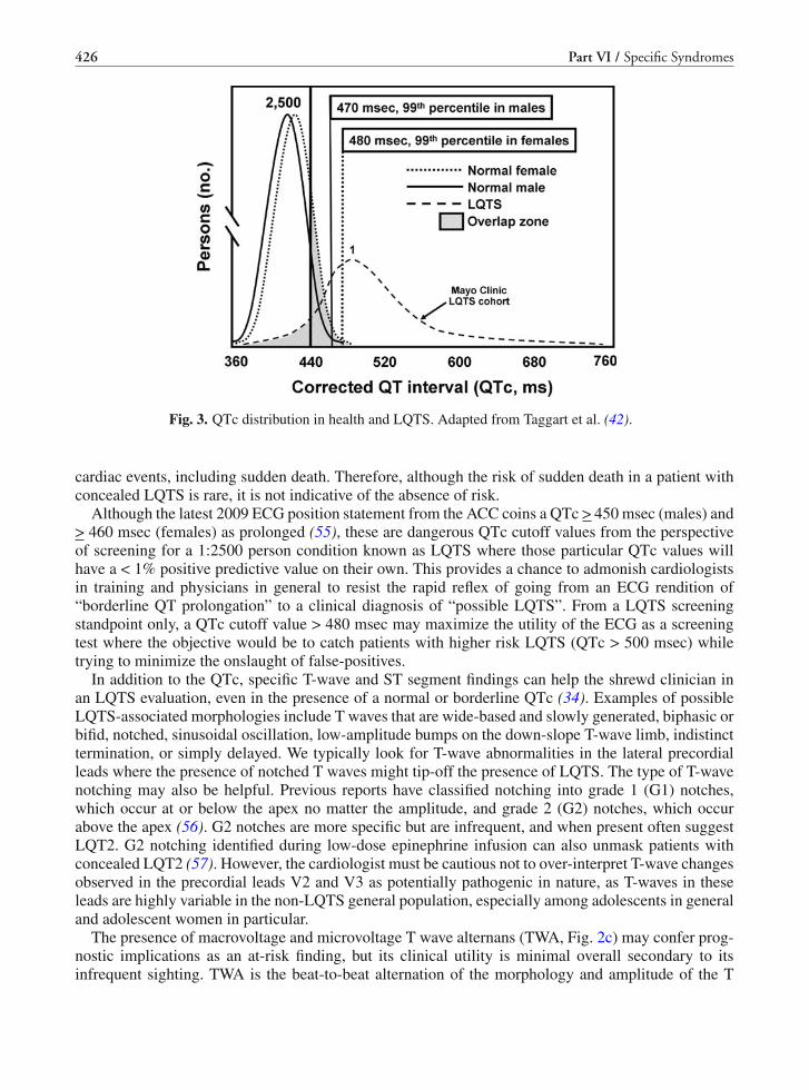
426 Part VI / Specific Syndromes
Fig. 3. QTc distribution in health and LQTS. Adapted from Taggart et al. (42).
cardiac events, including sudden death. Therefore, although the risk of sudden death in a patient withconcealed LQTS is rare, it is not indicative of the absence of risk.
Although the latest 2009 ECG position statement from the ACC coins a QTc > 450 msec (males) and> 460 msec (females) as prolonged (55), these are dangerous QTc cutoff values from the perspectiveof screening for a 1:2500 person condition known as LQTS where those particular QTc values willhave a < 1% positive predictive value on their own. This provides a chance to admonish cardiologistsin training and physicians in general to resist the rapid reflex of going from an ECG rendition of“borderline QT prolongation” to a clinical diagnosis of “possible LQTS”. From a LQTS screeningstandpoint only, a QTc cutoff value > 480 msec may maximize the utility of the ECG as a screeningtest where the objective would be to catch patients with higher risk LQTS (QTc > 500 msec) whiletrying to minimize the onslaught of false-positives.
In addition to the QTc, specific T-wave and ST segment findings can help the shrewd clinician inan LQTS evaluation, even in the presence of a normal or borderline QTc (34). Examples of possibleLQTS-associated morphologies include T waves that are wide-based and slowly generated, biphasic orbifid, notched, sinusoidal oscillation, low-amplitude bumps on the down-slope T-wave limb, indistincttermination, or simply delayed. We typically look for T-wave abnormalities in the lateral precordialleads where the presence of notched T waves might tip-off the presence of LQTS. The type of T-wavenotching may also be helpful. Previous reports have classified notching into grade 1 (G1) notches,which occur at or below the apex no matter the amplitude, and grade 2 (G2) notches, which occurabove the apex (56). G2 notches are more specific but are infrequent, and when present often suggestLQT2. G2 notching identified during low-dose epinephrine infusion can also unmask patients withconcealed LQT2 (57). However, the cardiologist must be cautious not to over-interpret T-wave changesobserved in the precordial leads V2 and V3 as potentially pathogenic in nature, as T-waves in theseleads are highly variable in the non-LQTS general population, especially among adolescents in generaland adolescent women in particular.
The presence of macrovoltage and microvoltage T wave alternans (TWA, Fig. 2c) may confer prog-nostic implications as an at-risk finding, but its clinical utility is minimal overall secondary to itsinfrequent sighting. TWA is the beat-to-beat alternation of the morphology and amplitude of the T

Chapter 19 / Long QT Syndrome 427
wave. It can be a marker of electrical instability and regional repolarization heterogeneity, and is asso-ciated with an increased risk of cardiac events (58, 59). Several investigators have reported the use ofQT dispersion (QTd), defined as the difference between the maximal and minimal QT intervals in the12 standard leads. It is thought to potentially represent spatial repolarization (60). QTd is reportedlyincreased in LQTS patients compared to normal patients, and there is a difference in QTd betweenmutation carriers and unaffected family members (61–63). However, the potential diagnostic utility ofQTd to make a distinction between LQTS and normal is unknown, and it is rarely used clinically.
Exercise TestingNormally, repolarization becomes more efficient during exercise, hence the expected shortening of
the QT and QTc with exercise stress testing. In contrast, patients with LQTS have been observed tohave “attenuated QTc shortening” with exercise, particularly patients with LQT1 (64). Additionally,the QT has been shown to lengthen abnormally during the recovery phase of exercise in patients withLQTS (65). While failure of appropriate shortening is relatively diagnostic in patients with LQT1,the presence of normal shortening of the QT interval during exercise does not rule out other LQTSgenotypes (66). While most research of exercise testing in LQTS patients has been performed onpatients with an obviously prolonged QTc at rest, provocative testing is most often utilized in thediagnosis of patients with concealed LQTS, where it can distinguish concealed LQTS from normalcy.
Other findings on exercise testing are uncommon in patients with LQTS. Exercise-induced ven-tricular ectopy is exceedingly rare. The presence of ventricular ectopy including premature ventric-ular contractions, in bigeminy or as couplets, or bidirectional ventricular tachycardia, is much moresuggestive of catecholaminergic polymorphic ventricular tachycardia than LQTS. The presence ofmacro- or microvoltage TWA at rest or during provocative testing can indicate increased risk in LQTS(67). Macrovoltage TWA is extremely rare, having been witnessed only twice in the over 1000 tread-mill/epinephrine QT stress tests that we have performed. Microvoltage TWA studies have not beenclinically helpful in our experience and are no longer performed at our LQTS clinic.
Epinephrine QT Stress TestLike exercise testing, the epinephrine QT stress test has been shown to be particularly helpful in
unmasking patients with concealed LQT1. Patients with LQT1 typically show a paradoxical length-ening of repolarization, measured by the QTc (Shimizu bolus protocol) or the absolute QT interval(Ackerman/Mayo continuous infusion protocol) during intravenous epinephrine infusion (67, 68). Thepresence of paradoxical lengthening (> 30 msec) of the QT interval is suggestive (75% positive predic-tive value) of concealed LQT1 with the Mayo protocol (69). The absence of paradoxical QT lengthen-ing is not effective in diagnosing other LQTS genotypes. It is critical to note that, as one might expect,the presence of concurrent beta-blockade will affect the outcome and interpretation of the epinephrineQT stress test.
We also use epinephrine QT stress testing in patients with novel LQT1-associated mutations. Thisallows us to physiologically challenge the patient’s potassium channel function, and provide an inde-pendent confirmation of the novel mutation’s pathogenicity, using the 96% negative predictive valueof the test. In other words, both treadmill and epinephrine stress testing represent in vivo functionalchallenges to assess the integrity of the IKs (Kv7.1) channel pathway where loss-of-function mutationscompromise the ability of this key channel to respond to sympathetic stimulation and accentuate itsphase 3 repolarizing force.
Molecular Genetic TestingAfter the sentinel discovery of LQTS as a cardiac channelopathy in 1995, LQTS genetic testing was
performed in several research laboratories for research and “pseudo-clinical” purposes for the next

428 Part VI / Specific Syndromes
decade. Then, in 2004, a commercially available genetic test called FAMILIONTM was introduced(Clinical Data, Inc, New Haven, CT). This clinical genetic test includes the comprehensive open read-ing frame analysis of the translated exons for the 3 canonical LQTS-susceptibility genes and severalof the minor genes. A second commercial genetic test has also been introduced recently (GeneDx, Inc,Gaithersburg, MD). A list of indications for LQTS genetic testing is summarized in Table 3.
Table 3Considerations for LQTS Genetic Testing
1. Unequivocal and unexplained QT prolongation (QTc > 500 msec)2. Clinically suspected LQTS regardless of (a) baseline QTc or (b) history of prior genetic testing, either
research-based or commercial/clinical.3. All first-degree relatives of a genotype-positive index case of LQTS regardless of their screening ECG,
with subsequent distantly-related family testing depending on results.4. Postmortem genetic testing for autopsy-negative sudden unexplained death (debatable)5. Patients with a history of drug-induced torsades (debatable)6. Postmortem genetic testing in victims of sudden infant death syndrome (debatable)
QTc Corrected QT Interval, LQTS Congenital Long QT Syndrome
The current generation LQTS clinical genetic tests will be positive about 75% of the time inpatients with clinically definite LQTS (70–72). This is clinically helpful, both in determination ofappropriate treatment as well as in guiding diagnosis for potentially affected family members. About5–10% of LQTS probands have spontaneous germline mutations. Conversely, approximately 5% ofgenotype-positive LQTS patients will have more than one mutation (70), and these patients typicallyhave a more severe arrhythmogenic phenotype (73). Therefore, it is critical that comprehensive genetictesting is performed, even after finding a single potentially causative mutation, as other mutations maybe present and drive therapy in a different direction.
Considering that 25% of patients with “no doubt about it” LQTS will have a negative LQTS genetictest, genetic testing, as a stand alone test, can not exclude LQTS. Secondary to the fear of “missing”this potentially fatal disease, many patients are given a clinical diagnosis of LQTS despite a low orintermediate diagnostic assessment. In our experience, 44% of patients who had LQTS genetic test-ing despite an intermediate probability “Schwartz score” had a positive genetic test, changing theirdiagnosis from clinical indeterminate to genetically possible/probable (72).
On the other hand, indiscriminate use of LQTS genetic testing (i.e., screening) rather than its properstory-driven indications would result in a tremendous number of false positives. Unlike nearly all ofthe 1500+ genetic tests that are clinically available, the background frequency of rare genetic variationthat could be viewed as “possible deleterious mutations” or “variants of uncertain significance” isknown for only a handful of disease genes including the breast cancer-associated genes and the majorLQTS-susceptibility genes (74, 75). This has been extended with an analysis of over 1300 reportedlyhealthy volunteers whereby approximately 4% of whites and 6–8% of non-whites hosted a rare geneticvariant that would have to be classified currently as a possible (maybe) disease-contributing mutation(76). Juxtaposed to the expected frequency of 75% for cases of robust LQTS, this implies that evenamong the purest of case mutation results, there is a 5–10% chance that the mutation elicited forthe patient with highest pre-test probability is nevertheless a “false positive”. Importantly, mutationtype and mutation location strongly inform the probability of pathogenicity for the current generationgenetic test (76). Like the probabilistic nature of virtually every clinical test that we utilize, genetictesting is slowly being grasped by the clinician community as a probabilistic, rather than deterministic,test that must be carefully scrutinized and interpreted.

Chapter 19 / Long QT Syndrome 429
MANAGEMENT
The key in understanding potential management strategies in LQTS rests with proper interpretationof clinical and genetic testing, and in appropriate risk stratification. For the entire population of LQTS,the annual mortality is < 1%/year, but may increase to 5–8%/year in the highest risk subsets of patients.
Risk StratificationDue to the inherent heterogeneity of LQTS, the phenotypic expression of LQTS may vary from a
purely asymptomatic life to premature sudden cardiac death (SCD) despite intervention. Any patientwho has a personal history of recurrent cardiac arrest carries an increased risk of subsequent SCD(77). Except for the subset that presents with aborted cardiac arrest as the sentinel event where thereis near universal agreement that an implantable cardioverter defibrillator (ICD) is indicated for sucha patient, primary prevention risk stratification attempts to determine which patients will experiencelife-threatening events in the future.
Factors associated with increased risk in LQTS-triggered SCD include a QTc >500 msec, a his-tory of LQTS-related symptoms (especially syncope and aborted cardiac arrest), the LQT2 or LQT3genotype, concurrent 2:1 AV block, and post-pubertal females (78). In particular, an LQTS-triggeredcardiac event such as syncope occurring in the first 5 years of life suggests a severe LQTS cardiacphenotype, and syncope in the first year of life portends a poor prognosis (79). The risk of cardiacevents is higher in males until they reach pubertal age, and is higher in females once they reach adultage (80). As such, asymptomatic males over the age of 20 years of life are at a particularly low riskof having cardiac events, especially among males with C-terminal localizing, haploinsufficient LQT1-associated mutations (80–82). Unlike males, asymptomatic females carry a continued risk for cardiacevents into adulthood, especially in the post-partum period (31). Patients with the autosomal recessiveJLNS are also a high risk subset.
Patients with LQT1 have the greatest number of LQTS-triggered events, with 60% of patients havinga history of syncope, aborted cardiac arrest, and sudden death (39). Around 40% of patients with LQT2have a history of these LQTS-related events, with LQT3 patients having the fewest cardiac events ofthe genotypes (18%) (32, 39, 83). Despite the different event frequencies, the mortality rate is similaramong the 3 genotypes. Hence, the so-called “lethality per event” rate is highest in patients with LQT3.
The correct distinction between vasovagal syncope and “torsadogenic syncope” is critical. Syncopewith a prodrome, occurring in common situations including overheating/dehydration, venipuncture,prolonged standing, or during micturition, is much more indicative of vasovagal syncope, althoughthere may be overlap. A history of syncope deemed to be “torsadogenic syncope” should be viewed asa strong sudden death warning sign prompting aggressive therapy though certainly not a self-sufficientindication for a prophylactic ICD (26). A family history of SCD, while important in the screeningof potential LQTS patients, does not appear to confer risk among the surviving genotype positive orclinically apparent LQTS family members (84). Similarly, a negative family history for SCD shouldnot invoke a conclusion towards a favorable outcome.
As discussed above, a QTc > 500 msec, particularly in the presence of LQTS-related symptoms,is associated with a relative risk ranging from 2 to 4 for a LQTS-triggered cardiac event of syncopeor worse (23–27, 81). There is a distinct direct relationship between clinical risk and degree of QTprolongation; the higher the QTc, the higher the clinical risk. Despite this however, there is essentiallyno QTc value at which there is absolutely zero risk.
T-wave notching is more common in symptomatic patients, and may be useful in prognosis (85).Macroscopic TWA (Fig. 2c) on the 12-lead ECG is a marker of severe electrical lability in patientswith LQTS, and may be of prognostic significance (58, 59). However, macroscopic TWA is rare, andthe relationship between degree of TWA and risk of SCD is unknown (79). There currently is no role

430 Part VI / Specific Syndromes
for invasive electrophysiology studies for the purposes of LQTS risk stratification, as the majority ofLQTS patients will not exhibit arrhythmias during programmed electrical stimulation (86).
TreatmentFirst and foremost, all patients with LQTS, either in a congenital or acquired form, should be coun-
seled to avoid all medications known to aggravate the QT interval as a drug-mediated side effect. A listof the most common offenders is shown in Table 4. The University of Arizona maintains an updatedlist of all known QT-prolonging medications at www.qtdrugs.org. If there is a clear medical necessityfor a listed medication, there should be a careful assessment of risks and benefits of administering themedication, as well as appropriate monitoring while on such medication(s).
Table 4Common Medications that Prolong the QT Interval
Class Medication
Anti-arrhythmicsAmiodaroneDofetilideProcainamideQuinidineSotalol
AntibioticsAzithromycinErythromycinGatifloxacinLevofloxacinMoxifloxacin
Motility agentsCisapride (withdrawn)Domperidone (withdrawn)
NarcoticsMethadone
PsychotropicsHaloperidolPhenothiazineThioridazineTricyclic antidepressants
The ultimate goal for the treatment of LQTS is to prevent SCD secondary to TdP with failure tospontaneously convert back to normal sinus rhythm. Given the risk of mortality in LQTS, all previ-ously symptomatic patients should be treated. However, the vast majority of even symptomatic LQTScan be treated pharmacologically. The full spectrum of therapeutic options includes pharmacologic,denervation, device, and preventative treatment measures. Indeed, recent years have shown a trendtowards increased use of the most aggressive but more definitive treatment option, the ICD. Given thepotential risk/benefit ratios of invasive treatments, particularly in children, it is the physician’s imper-ative to offer individualized treatment plans. In any case, it is a near universal recommendation thatany patient who survives LQTS-triggered aborted cardiac arrest should receive an ICD as secondaryprevention.
The treatment of asymptomatic LQTS patients is a matter of continued debate. It is true that in olderstudies, up to 9% of patients had a cardiac arrest as their first symptom/manifestation of LQTS (87),and that 4% of patients asymptomatic at the time of diagnosis later died suddenly. With these findings,

Chapter 19 / Long QT Syndrome 431
it became common for all patients with LQTS to be treated regardless of symptomatology, particularlyyoung patients (88). Once a patient has been diagnosed with LQTS, due to the >90% inherited natureof the disease, all first-degree relatives should be screened, with treatment offered on an individualbasis (89). Further, all patients should be seen by a physician well-versed in the intricacies of LQTS.
Medical therapy, particularly pharmacologic, should be considered for ALL asymptomatic patientswith manifest QT prolongation (QTc > 480 msec). Asymptomatic males over the age of 40 years withLQT1, especially those with lower risk LQT1-associated mutations, may not need any active interven-tion, only QT-drug avoidance and other simple preventative measures (23). This profile indicates the“near-zero” risk subpopulation of LQTS.
MedicationsIn LQT1 and LQT2, as well as most cases of genotype-negative LQTS, beta-blocker therapy has
been and should be the first-line therapy (77). Beta-blockers have been shown to reduce the frequencyof sudden death and ventricular arrhythmias, as well as reduce symptoms in patients with LQTS (87,90). In one study, mortality was significantly reduced to less than 2% in LQTS patients treated withbeta-blockers (77). At Mayo Clinic’s LQTS Clinic, infants with LQTS are generally started on liquidpropranolol, which is typically dosed three times a day (every 6 h has been advised by others butthis has not been shown to be necessary). Liquid nadolol can be used twice daily but has a relativelyshort (2–4 week) shelf-life. Generally, nadolol has been our drug of choice because of its long half-lifeand the ability to dose once daily (especially after age 10 or so), thereby contributing favorably tocompliance. However, a lateral move to propranolol or its long-acting version (Inderal) can be madewithout hesitation if there are unacceptable side effects to see if a different beta-blocker is bettertolerated.
Although atenolol continues to be one of the most frequent beta-blockers prescribed on the outside,it is not used in our program. Among the centers with the largest LQTS experience, nadolol andpropranolol/Inderal are the preferred beta-blockers (Table 5) and atenolol is viewed as potentiallyinferior (91). At this point in time, it is unclear whether atenolol is intrinsically a suboptimal beta-blocker or whether it has been pharmacokinetically misused. Patients still tend to arrive on atenololdosed once daily when its pharmacokinetic properties require twice daily dosing to provide 24-hourprotection. Regardless, each patient should have an individualized recommendation regarding whichbeta-blocker medication to use if indicated, taking into account the method of administration, dosingschedule, potential benefits, and side effects.
Table 5Preferred Medications and Suggested Dosages for the Management of LQTS
Class Medication Dosage Dose frequency
Beta-blockers Nadolol 1–2.0 mg/kg/day Once daily (Twice daily until around 10years)
Propranolol 3–4 mg/kg/day Three times daily or twice daily with longacting form (Inderal, LA)
Sodium-channelblockers
Mexiletine ∼10–15 mg/kg/day Three times dailyFlecainide 1–6 mg/kg/day Three times dailyRanolazine Up to 1000 mg/dose (adults) Twice daily
Overall, beta-blockers are the only pharmacotherapeutic agents that can be used alone for the twomost common subtypes of LQTS (LQT1 and LQT2) and for those patients with genotype-negative

432 Part VI / Specific Syndromes
LQTS. Other medications are not typically used as “stand alone” therapies. Calcium channel block-ers like verapamil or potassium channel openers like nicorandil may be used in patients with recurrentICD discharges despite beta-blocker therapy and surgical denervation. As will be discussed later, phar-macological targeting with late sodium current blockers may be a stand alone pharmacologic optionfor patients with LQT3.
PacingMany spontaneous arrhythmias in LQTS are pause-dependent, where the sentinel arrhythmia of TdP
is preceded by the sudden increase in cycle length (92). LQTS patients may have slower resting heartrates, and beta-blocker therapy can worsen bradycardia and potentiate the chance of a long-short-longtype of pause. Cardiac pacing can prevent pause-dependent TdP, especially in LQT2 patients (93). Itdoes not, however, reduce the risk of sudden death in high-risk patients (93). When considered, pacingshould seldom be stand alone therapy as it is usually used in conjunction with beta-blocker therapy(94). Potential indications for pacing in LQTS patients include those with preexisting AV block andthose with a history/Holter evidence of pause-dependent arrhythmias. Specific recommendations fordevice programming and pacing strategies are well summarized by Viskin et al. (94). In our clinicalpractice, if a pacemaker is considered due to a perceived high patient risk, we typically will placean implantable defibrillator/pacemaker instead of an isolated pacemaker. Over the past 10 years, noLQTS patient has received an isolated pacemaker in Mayo’s LQTS Clinic.
Implantable Cardioverter-Defibrillator (ICD)The ICD represents one of the most significant medical advances of our time, enabling a subset of
the population to survive potentially lethal arrhythmias while maintaining routine activities of dailylife. The current indications for ICD placement in LQTS patients are summarized in Fig. 4. Contraryto popular belief and the seeming trend in the United States towards universal ICD therapy in LQTS, <15% of the > 500 LQTS patients followed in our LQTS Clinic have an ICD. Indeed, the vast majority ofLQTS patients can and should be managed without an ICD. As mentioned previously, ICD therapy isalmost universally recommended as secondary prevention following an aborted cardiac arrest (95–97).
The use of ICD therapy as primary prevention is debatable and open to personal interpretation andpatient preference. ICD therapy as primary prevention is often recommended in cases where beta-blockade may significantly reduce, but not eradicate, the risk of sudden death (26, 98). Family history
Fig. 4. Current considerations for ICD therapy in LQTS. Adapted from Ackerman et al. (121).

Chapter 19 / Long QT Syndrome 433
of sudden death, despite current listing as a class IIb indication for ICD therapy, is not supported byevidence as an independent indication for ICD placement (84). As shown in Fig. 4, risk factors thatwarrant consideration of a prophylactic ICD include a LQTS-triggered cardiac event despite adequatebeta blocker therapy both in terms of drug and dose (26), intolerance of primary pharmacotherapy,severe QT prolongation (QTc > 550 msec) without the LQT1 genotype (97, 99), and infants with 2:1AV block (87). In addition, patients with JLNS, especially those with type 1 JLNS (100), probablywarrant a prophylactic ICD due to their severe cardiac phenotype.
Many patients with the LQT3 genotype have received ICDs , including nearly 50% of the LQT3patients at Mayo, due to LQT3’s comparatively higher lethality/event rate (39, 98, 101). This hasbeen called into question recently, and remains a controversial issue (102). If an ICD is needed,we typically place a single lead ICD system. If pacing is going to be used as an adjuvant therapy,then a dual chamber pacemaker/ICD should be implanted. No matter the scenario, it is key that allrisk-benefit ratios be weighed without bias, with patients and families being as fully informed aspossible.
Left Cardiac Sympathetic Denervation (LCSD)Left cardiac sympathetic denervation (LCSD) involves resection of the lower half of the left stellate
ganglion and the left-sided sympathetic chain at the level of T2, T3, and T4 (103, 104). Resection ofthe lower half of the stellate ganglion is critical; removal of T2-T4 only will not provide the sameantifibrillatory effect and should be considered an incomplete denervation (105). LCSD can be per-formed using open thoracotomy or via video-assisted thoracic surgery (VATS), depending on surgeonpreference. The rationale behind denervation therapy is based on evidence that the left-sided cardiacsympathetic nerves have a very high arrhythmogenic potential (106, 107), and may carry increasedactivity in patients with LQTS (107). There are now several well-published studies showing a signifi-cant decrease in events in LQTS patients after LCSD (104, 106, 108). Further, LCSD’s antifibrillatoryeffect appears to be relatively substrate independent with demonstrated efficacy seen in patients withboth genotype positive and genotype negative LQTS as well as the three major LQTS genotypes.Here, the predicted efficacy is LQT1 > LQT3 > LQT2, but this ranking awaits confirmation. Schwartzhas recommended that LCSD be considered in all LQTS patients who have syncope despite adequateβ-blocker therapy, as well as in patients with arrhythmia storms and recurrent ICD shocks (109).
Mortality associated with LCSD is near zero at most experienced centers and the complication ofHorner’s syndrome is seldom seen (< 5%). A list of current indications for videoscopic denervation
Fig. 5. Current considerations for LCSD in LQTS.

434 Part VI / Specific Syndromes
therapy/LCSD in our institution is shown in Fig. 5 (104). While initially reserved for patients with mul-tiple VF-terminating ICD therapies (secondary prevention denervation therapy), LCSD is now offeredas an alternative to ICD for patients with beta-blocker intolerance and as a “bridge to ICD” in patientssuch as infants and small children where ICD placement is associated with higher complication rates.In addition, LCSD may be used as a treatment strategy for patients where ICDs are either unavailableor unattainable.
Gene-Specific TherapyLQT1: Patients with LQT1 are significantly protected by the use of beta-blocker therapy, particularly since
many of their cardiac events are typically induced by adrenergic stimulation (99). LCSD can be used ifevents including syncope occur despite beta-blocker therapy (108). A prophylactic ICD should be leastfrequent among patients with LQT1.
LQT2: Patients with LQT2 are also protected well by beta-blockade, but tend to have more breakthroughevents compared to LQT1 patients (77). Medical augmentation of potassium channel function usingmedications including nicorandil is of unclear significance, and these are not typically used. LCSD canbe used in patients with breakthrough events. In high-risk patients (females, QTc > 500 msec), prophy-lactic ICD may be considered, typically with single lead ICD therapy. Potassium supplementation and/orpharmacological attempts to increase serum potassium (i.e., spironolactone) may enhance cardiac repo-larization and have been utilized (110). To date, however, spironolactone pharmacotherapy has not beenneeded at our institution.
LQT3: Fortunately LQT3 is the least common (5–10%) among the three canonical LQTS genotypes,as management of LQT3 continues to vex LQTS specialists. The combination of its highest lethal-ity per event label and concerns regarding insufficient protection afforded by beta-blocker therapyhas compelled a high use of prophylactic ICD. However, propranolol therapy with or without mexile-tine/flecainide/ranolazine may provide a cogent, gene-tailored treatment option for the low/moderate riskLQT3 patient. Propranolol is specifically advocated because in addition to its beta blocking properties, itis a weak blocker of late sodium current (111). This component of its mechanism of action may explainwhy clinically, pro-arrhythmias from beta blocker therapy have not been observed among LQT3 patientswhen the cellular in vitro data suggests this possibility of beta blocker-mediated LQT3-related cardiacevents (101). Historically, the largest LQTS programs treating the largest number of this subpopulationof LQT3 patients have utilized propranolol as the preferred beta blocker.
Regarding direct targeting of the late sodium current, this must be carefully monitored and individu-alized as not all LQT3-susceptibility mutations are created equal. For example, those LQT3 mutationsthat result in isolated accentuation of late sodium current could conceivably be “medically cured”by blocking the pathological current, thereby restoring repolarization to more normal like conditionswhich has been demonstrated for both dogs and humans (112, 113). As indicated previously, how-ever, although it makes sense as an effective anti-arrhythmic treatment strategy, there are no studiesto date demonstrating a survival benefit with this approach. In addition, given that some LQT3 muta-tions exhibit a hybrid, loss- and gain-of-function phenotype, it is conceivable that late sodium channelblocker therapy could unmask and accentuate the loss-of-function side of the equation and precip-itate a Brugada-like phase 2 re-entrant mechanism. This possibility serves as a clear reminder thatmanagement of all LQTS patients and LQT3 patients specifically must be carefully scrutinized andindividualized. This disease truly personifies the call for personalized/individualized medicine.
PREVENTION
As discussed above, it is critical for patients to consult their doctor before taking any medica-tions. Medications known to prolong the QT interval must be avoided. If a medication is required

Chapter 19 / Long QT Syndrome 435
that has known QT-aggravating potential, the physician and patient/family need to perform a carefulrisk/benefit analysis. If an alternative medication exists with similar efficacy but without the QT sideeffect, then that medication should be used. Common situations where this may arise includes the useof Albuterol in treating asthma and the use of Ritalin or other stimulant therapies in treating attentiondeficit disorder. Again, the physician and patient/family need to weight the risks/benefits carefully,recognizing the life-changing potential and risks of these medications. Over the past 10 years, we havenot seen a single albuterol-triggered or stimulant-triggered cardiac event among the subpopulation ofour patients with LQTS where jointly we concluded that continued use of such medications had afavorable benefit to risk ratio.
Physicians should inform patients and family members of several key preventative health measuresfor patients with LQTS, unrelated to the above pharmacotherapeutic and surgical therapies. Healthysleep and dietary habits, as in other disease processes, are important and should not be overlooked.Hypokalemia can precipitate an LQTS-related cardiac event, and thus a potassium-enriched diet maybe helpful. During gastrointestinal illnesses, hydration and electrolyte replacement is important toavoid hypokalemia and other electrolyte disturbances. Patients should be cautioned against drinkingexcessive amounts of grapefruit juice, especially purple grapefruit juice, as it contains a chemical thatinhibits the IKr potassium channel and can prolong the QT interval in a mechanism similar to thatof drug-induced QT prolongation (114). Over-the-counter supplements including cesium chloride oramphetamine-like agents should be avoided (115).
Patients with LQTS, especially those with LQT2, should avoid or at least minimize sources of loudauditory triggers in their home and workplace (i.e., telephones or alarm clocks), particularly duringsleep (109, 116, 117). This is often forgotten for patients in the hospital setting, when the recurrentalarms in patients’ rooms can trigger events. An identification card, necklace, or bracelet should becarried or worn at all times, to let emergency response personnel know of their diagnosis and allow forquicker activation of appropriate therapy. All families should consider having an automatic externaldefibrillator (AED) at home, and this should accompany the family whenever possible on vacations, cartrips, athletic outings, etc. Appropriate maintenance and checkups should be performed on the AEDto ensure that it will be ready whenever needed. Most schools now have AEDs in place as standardequipment, but this should be assessed by the family during the process of school enrollment. Familymembers should be trained in CPR, and all should be taught how to administer a precordial thumpprior to chest compressions. In LQTS, this “artificial” commotio cordis might restore sinus rhythm insome patients and prevent significant morbidity (118). Although we continue to recommend a personalAED as part of our overarching sudden death safety net strategy, there has not been an AED use amongany patient managed in our comprehensive LQTS Clinic at now > 5000 patient-years.
The issue of competitive athletic participation for the athlete with LQTS remains complex andcontroversial. The 36th Bethesda Conference guidelines were published in 2005 (119). Competitivesports disqualification was advised for any patient, regardless of underlying genotype or QTc, witha history of out of hospital cardiac arrest or LQTS-related syncopal episode. Asymptomatic patientswith a QTc over 470 in males or over 480 msec in females were recommended limitation to the classIA sports (billiards, bowling, cricket, curling, golf, riflery). Loosening of the restrictions was supported(from a guideline perspective) for asymptomatic LQT3 patients, as well as for asymptomatic genotype-positive LQTS patients with QTc values in the overlapping/borderline range, except for competitiveswimming in athletes with concealed LQT1 (119). The European Society of Cardiology (ESC) in 2006published a similar set of athletic activity guidelines (120). Disqualification was advised for LQTSathletes who were symptomatic, had a prolonged QT interval (over 440–470 msec in men, over 460–480 msec in women), or were carriers of a known genetic mutation. Patients considered at “low-risk”for cardiac events were allowed participation in light to moderate leisure activity. All sports involvingsudden bursts of activity or genotype-specific triggers were absolutely contraindicated. In contrast to

436 Part VI / Specific Syndromes
the Bethesda Conference guidelines, the ESC recommended disqualification even for patients withconcealed LQTS (i.e., positive gene test only) (120).
All guidelines withstanding, it is important to avoid the temptation to advise avoidance of activityaltogether. The risk of obesity and early-onset diabetes are higher in sedentary individuals, and wecertainly do not want to create a collection of new diseases by limiting exercise. We support continua-tion of most recreational activity for our LQTS patients, avoiding of course genotype-specific triggerssuch as swimming in patients with LQT1. Each patient and family should have a clear understand-ing of what is currently known about the risks and benefits of athletic activity in LQTS. In Mayo’sLQTS Clinic, we prefer the role of “informer/educator” rather than a mere “if in doubt, kick them out”approach to this very complex issue. Rather than mandating disqualification, we respect patient/familyautonomy whereby each patient and family should be fully involved in the decision-making processand be allowed to make an informed decision even if such a decision runs contrary to the publishedguidelines. This approach has guided our LQTS Clinic for the past decade and notably about half offamilies have chosen disqualification and half have chosen continued participation. To date, there havebeen no competitive sports precipitated cardiac events in the latter.
CONCLUSIONS
Over the past half century, LQTS has matriculated through several critical milestones including itssentinel clinical description, clinical diagnostic scorecard, pathogenetic discovery, decade of research-based genetic testing and genotype–phenotype correlations, and clinical availability of genetic testing.With all the diagnostic tools now available, the physician must be not only a wiser user but also awiser interpreter of these tests in order to accurately recognize the presence/absence of LQTS, and ifpresent, to tailor an informed and individualized treatment strategy.
CONFLICTS OF INTEREST
Dr. Ackerman is a consultant for PGxHealth with respect to the FAMILIONTM genetic tests forcardiac ion channel mutations and hypertrophic cardiomyopathy. Intellectual property derived fromMJA’s research program resulted in license agreements in 2004 between Mayo Clinic Health Solutions(formerly Mayo Medical Ventures) and PGxHealth (formerly Genaissance Pharmaceuticals).
REFERENCES
1. Jervell A, Lange-Nielsen F (1957) Congenital deaf-mutism, functional heart disease with prolongation of the QTinterval, and sudden death. Am Heart J 54
2. Romano C, Gemme G, Pongiglione R (1963) Aritmie cardiache rare dell’eta’pediatrica. II. Accessi sincopali perfibrillazione ventricolare parossistica. Clin Peditr (Bologna) 45:656–683
3. Ward OC (1964) A new famillial cardiac syndrome in children. J Irish Med Assoc 54:103–1064. Curran, ME, Splawski I, Timothy KW et al (1995) A molecular basis for cardiac arrhythmia: HERG mutations cause
long QT syndrome. Cell 80:795–8035. Wang Q, Shen J, Splawski I et al (1995) SCN5A mutations associated with an inherited cardiac arrhythmia, long QT
syndrome. Cell 80:805–8116. Wang Q, Curran ME, Splawski I et al (1996) Positional cloning of a novel potassium channel gene: KVLQT1 mutations
cause cardiac arrhythmias. Nat Genet 12:17–237. Zipes DP, Camm AJ, Borggrefe M et al (2006) ACC/AHA/ESC 2006 guidelines for management of patients with
ventricular arrhythmias and the prevention of sudden cardiac death: a report of the American college of cardiol-ogy/American heart association task force and the European society of cardiology committee for practice guidelines(writing committee to develop guidelines for management of patients with ventricular arrhythmias and the preventionof sudden cardiac death). J Am Coll Cardiol 48:1064–1108
8. Ackerman MJ (2004) Cardiac channelopathies: it’s in the genes. Nat Med 10:463–464

Chapter 19 / Long QT Syndrome 437
9. Tester DJ, Will ML, Haglund CM et al (2006) Effect of clinical phenotype on yield of long QT syndrome genetictesting. J Am Coll Cardiol 47:764–768
10. Chen L, Marquardt ML, Tester DJ et al (2007) Mutation of an A-kinase-anchoring protein causes long-QT syndrome.Proc Natl Acad Sci USA 104:20990–20995
11. Medeiros-Domingo, A, Kaku, T, Tester, DJ et al (2007) SCN4B-encoded sodium channel beta4 subunit in congenitallong-QT syndrome. Circulation 116:134–142
12. Ueda K, Valdivia C, Medeiros-Domingo A et al (2008) Syntrophin mutation associated with long QT syndrome throughactivation of the nNOS-SCN5A macromolecular complex. Proc Natl Acad Sci USA 105:9355–9360
13. Splawski I, Tristani-Firouzi M, Lehmann MH et al (1997) Mutations in the hminK gene cause long QT syndrome andsuppress IKs function. Nat Genet 17:338–340
14. Abbott GW, Sesti F, Splawski I et al (1999) MiRP1 forms IKr potassium channels with HERG and is associated withcardiac arrhythmia. Cell 97:175–187
15. Mohler PJ, Schott JJ, Gramolini AO et al (2003) Ankyrin-B mutation causes type 4 long-QT cardiac arrhythmia andsudden cardiac death. Nature 421:634–639
16. Vatta M, Ackerman MJ, Ye B et al (2006) Mutant caveolin-3 induces persistent late sodium current and is associatedwith long-QT syndrome. Circulation 114:2104–2112
17. Zhang L, Benson DW, Tristani-Firouzi M et al (2005) Electrocardiographic features in Andersen-Tawil syndromepatients with KCNJ2 mutations: characteristic T-U-wave patterns predict the KCNJ2 genotype. Circulation 111:2720–2726
18. Splawski I, Timothy KW, Sharpe LM et al (2004) Ca(V)1.2 calcium channel dysfunction causes a multisystem disorderincluding arrhythmia and autism. Cell 119:19–31
19. Schwartz PJ, Stramba-Badiale M, Crotti L et al (2009) Prevalence of the congenital long-QT syndrome. Circulation120:1761–1767
20. Tester DJ, Ackerman MJ (2007) Postmortem long QT syndrome genetic testing for sudden unexplained death in theyoung. J Am Coll Cardiol 49:240–246
21. Arnestad M, Crotti L, Rognum TO et al (2007) Prevalence of long-QT syndrome gene variants in sudden infant deathsyndrome. Circulation 115:361–367
22. Goldenberg I, Moss AJ, Zareba W et al (2006) Clinical course and risk stratification of patients affected with the Jervelland Lange-Nielsen syndrome. J Cardiovasc Electrophysiol 17:1161–1168
23. Goldenberg I, Moss AJ, Bradley J et al (2008) Long-QT syndrome after age 40. Circulation 117:2192–220124. Goldenberg I, Moss AJ, Peterson DR et al (2008) Risk factors for aborted cardiac arrest and sudden cardiac death in
children with the congenital long-QT syndrome. Circulation 117:2184–219125. Spazzolini C, Mullally J, Moss AJ et al (2009) Clinical implications for patients with long QT syndrome who experi-
ence a cardiac event during infancy. J Am Coll Cardiol 54:832–83726. Hobbs JB, Peterson DR, Moss AJ et al (2006) Risk of aborted cardiac arrest or sudden cardiac death during adolescence
in the long-QT syndrome. JAMA 296:1249–125427. Sauer AJ, Moss AJ, McNitt S et al (2007) Long QT syndrome in adults. J Am Coll Cardiol 49:329–33728. Moss AJ, Robinson JL (1992) Clinical aspects of the idiopathic long QT syndrome. Ann NY Acad Sci 644:103–11129. Vincent GM, Timothy K, Fox J et al (1999) The inherited long QT syndrome: from ion channel to bedside. Cardiol
Rev 7:44–5530. Moss AJ, Schwartz PJ, Crampton RS et al (1991) The long QT syndrome. Prospective longitudinal study of 328
families. Circulation 84:1136–114431. Rashba EJ, Zareba W, Moss AJ et al (1998) Influence of pregnancy on the risk for cardiac events in patients with
hereditary long QT syndrome. LQTS investigators. Circulation 97:451–45632. Schwartz PJ, Priori SG, Spazzolini C et al (2001) Genotype-phenotype correlation in the long-QT syndrome: gene-
specific triggers for life-threatening arrhythmias. Circulation 103:89–9533. Ackerman MJ, Tester DJ, Porter CJ (1999) Swimming, a gene-specific arrhythmogenic trigger for inherited long QT
syndrome. Mayo Clin Proc 74:1088–109434. Moss AJ, Zareba W, Benhorin J et al (1995) ECG T-wave patterns in genetically distinct forms of the hereditary long
QT syndrome. Circulation 92:2929–293435. Zhang L, Timothy KW, Vincent GM et al (2000) Spectrum of ST-T-wave patterns and repolarization parameters in
congenital long-QT syndrome: ECG findings identify genotypes. Circulation 102:2849–285536. Wilde AA, Jongbloed RJ, Doevendans PA et al (1999) Auditory stimuli as a trigger for arrhythmic events differentiate
HERG-related (LQTS2) patients from KVLQT1-related patients (LQTS1). J Am Coll Cardiol 33:327–33237. Khositseth A, Tester DJ, Will ML et al (2004) Identification of a common genetic substrate underlying postpartum
cardiac events in congenital long QT syndrome. Heart Rhythm 1:60–6438. Johnson JN, Hofman N, Haglund CM et al (2009) Identification of a possible pathogenic link between congenital long
QT syndrome and epilepsy. Neurology 72:224–231

438 Part VI / Specific Syndromes
39. Zareba W, Moss AJ, Schwartz PJ et al (1998) Influence of genotype on the clinical course of the long-QT syndrome.International long-QT syndrome registry research group. N Engl J Med 339:960–965
40. Schwartz PJ (1985) Idiopathic long QT syndrome: progress and questions. Am Heart J 109:399–41141. Schwartz PJ, Moss AJ, Vincent GM et al (1993) Diagnostic criteria for the long QT syndrome. An update. Circulation
88:782–78442. Taggart NW, Haglund CM, Tester DJ et al (2007) Diagnostic miscues in congenital long-QT syndrome. Circulation
115:2613–262043. Viskin S, Rosovski U, Sands AJ et al (2005) Inaccurate electrocardiographic interpretation of long QT: the majority of
physicians cannot recognize a long QT when they see one. Heart Rhythm 2:569–57444. Postema PG, De Jong JS, Van der Bilt IA et al (2008) Accurate electrocardiographic assessment of the QT interval:
teach the tangent. Heart Rhythm 5:1015–101845. Davey P (1999) A new physiological method for heart rate correction of the QT interval. Heart 82:183–18646. Sagie A, Larson MG, Goldberg RJ et al (1992) An improved method for adjusting the QT interval for heart rate (the
Framingham heart study). Am J Cardiol 70:797–80147. Fridericia L (1920) The duration of systole in the electrocardiogram of normal subjects and of patients with heart
disease. Acta Medica Scandinavica 469–48648. Bazett H (1920) An analysis of the time-relations of electrocardiograms. Heart 353–37049. Vincent GM, Richard J (2001) Calculation of the QTc interval during sinus arrhythmia in patients suspected to have
long QT syndrome. Circulation 104:II-690–II-69150. Martin AB, Perry JC, Robinson JL et al (1995) Calculation of QTc duration and variability in the presence of sinus
arrhythmia. Am J Cardiol 75:950–95251. Goldenberg I, Moss AJ, Zareba W (2006) QT interval: how to measure it and what is "normal". J Cardiovasc Electro-
physiol 17:333–33652. Basavarajaiah S, Wilson M, Whyte G et al (2007) Prevalence and significance of an isolated long QT interval in elite
athletes. Eur Heart J 28:2944–294953. Levine E, Rosero SZ, Budzikowski AS et al (2008) Congenital long QT syndrome: considerations for primary care
physicians. Cleve Clin J Med 75:591–60054. Merri M, Benhorin J, Alberti M et al (1989) Electrocardiographic quantitation of ventricular repolarization. Circulation
80:1301–130855. Rautaharju PM, Surawicz B, Gettes LS et al (2009) AHA/ACCF/HRS recommendations for the standardization and
interpretation of the electrocardiogram: part IV: the ST segment, T and U waves, and the QT interval: a scientificstatement from the American heart association electrocardiography and arrhythmias committee, council on clinicalcardiology; the American college of cardiology foundation; and the heart rhythm society. Endorsed by the internationalsociety for computerized electrocardiology. J Am Coll Cardiol 53:982–991
56. Lupoglazoff JM, Denjoy I, Berthet M et al (2001) Notched T waves on Holter recordings enhance detection of patientswith LQt2 (HERG) mutations. Circulation 103:1095–1101
57. Khositseth A, Hejlik J, Shen WK et al (2005) Epinephrine-induced T-wave notching in congenital long QT syndrome.Heart Rhythm 2:141–146
58. Schwartz PJ, Malliani A (1975) Electrical alternation of the T-wave: clinical and experimental evidence of its relation-ship with the sympathetic nervous system and with the long Q-T syndrome. Am Heart J 89:45–50
59. Zareba W, Moss AJ, le Cessie S et al (1994) T wave alternans in idiopathic long QT syndrome. J Am Coll Cardiol23:1541–1546
60. Napolitano C, Priori SG, Schwartz PJ (2000) Significance of QT dispersion in the long QT syndrome. Prog CardiovascDis 42:345–350
61. Day CP, McComb JM, Campbell RW (1990) QT dispersion: an indication of arrhythmia risk in patients with long QTintervals. Br Heart J 63:342–344
62. Priori SG, Napolitano C, Diehl L et al (1994) Dispersion of the QT interval. A marker of therapeutic efficacy in theidiopathic long QT syndrome. Circulation 89:1681–1689
63. Moennig G, Schulze-Bahr E, Wedekind H et al (2001) Clinical value of electrocardiographic parameters in genotypedindividuals with familial long QT syndrome. Pacing Clin Electrophysiol 24:406–415
64. Vincent GM, Jaiswal D, Timothy KW (1991) Effects of exercise on heart rate, QT, QTc and QT/QS2 in the Romano-Ward inherited long QT syndrome. Am J Cardiol 68:498–503
65. Swan H, Toivonen L, Viitasalo M (1998) Rate adaptation of QT intervals during and after exercise in children withcongenital long QT syndrome. Eur Heart J 19:508–513
66. Schwartz PJ, Priori SG, Locati EH et al (1995) Long QT syndrome patients with mutations of the SCN5A and HERGgenes have differential responses to Na+ channel blockade and to increases in heart rate. Implications for gene-specifictherapy. Circulation 92:3381–3386
67. Ackerman MJ, Khositseth A, Tester DJ, Hejlik J, Shen WK, Porter CJ (2002) Epinephrine-induced QT interval pro-longation: a gene-specific paradoxical response in congenital long QT syndrome. Mayo Clin Proc 77:413–421

Chapter 19 / Long QT Syndrome 439
68. ShimizuW, Noda T, Takaki H et al (2003) Epinephrine unmasks latent mutation carriers with LQT1 form of congenitallong-QT syndrome. J Am College Cardiol 41:633–642
69. Vyas H, Hejlik J, Ackerman MJ (2006) Epinephrine QT stress testing in the evaluation of congenital long-QT syn-drome: diagnostic accuracy of the paradoxical QT response. Circulation 113:1385–1392
70. TesterDJ, Will ML, Haglund CM et al (2005) Compendium of cardiac channel mutations in 541 consecutive unrelatedpatients referred for long QT syndrome genetic testing. Heart Rhythm 2:507–517
71. Napolitano C, Priori SG, Schwartz PJ et al (2005) Genetic testing in the long QT syndrome: development and validationof an efficient approach to genotyping in clinical practice [see comment]. JAMA 294:2975–2980
72. Tester DJ, Will ML, Haglund CM et al (2006) Effect of clinical phenotype on yield of long QT syndrome genetictesting. J Am College Cardiol 47:764–768
73. Westenskow P, Splawski I, Timothy KW et al (2004) Compound mutations: a common cause of severe long-QTsyndrome. Circulation 109:1834–1841
74. Ackerman MJ, Splawski I, Makielski JC et al (2004) Spectrum and prevalence of cardiac sodium channel variantsamong black, white, Asian, and Hispanic individuals: implications for arrhythmogenic susceptibility and Brugada/longQT syndrome genetic testing. Heart Rhythm 1:600–607
75. Ackerman MJ, Tester DJ, Jones GS et al (2003) Ethnic differences in cardiac potassium channel variants: implicationsfor genetic susceptibility to sudden cardiac death and genetic testing for congenital long QT syndrome. Mayo ClinProc 78:1479–1487
76. Kapa S, Tester DJ, Salisbury BA et al (2009) Genetic testing for long QT syndrome: distinuishing pathogenic mutationsfrom benign variants. Circulation 120:1752–1760
77. Moss AJ, Zareba W, Hall WJ et al (2000) Effectiveness and limitations of beta-blocker therapy in congenital long-QTsyndrome. Circulation 101:616–623
78. Priori SG, Schwartz PJ, Napolitano C et al (2003) Risk stratification in the long-QT syndrome. N Engl J Med 348:1866–1874
79. Priori SG, Aliot E, Blomstrom-Lundqvist C et al (2001) Task force on sudden cardiac death of the European societyof cardiology. Eur Heart J 22:1374–1450
80. Locati EH, Zareba W, Moss AJ et al (1998) Age- and sex-related differences in clinical manifestations in patients withcongenital long-QT syndrome: findings from the international LQTS registry. Circulation 97:2237–2244
81. Priori SG, Schwartz PJ, Napolitano C et al (2003) Risk stratification in the long-QT syndrome. N Engl J Med 348:1866–1874
82. Moss AJ, Shimizu W, Wilde AA et al (2007) Clinical aspects of type-1 long-QT syndrome by location, coding type,and biophysical function of mutations involving the KCNQ1 gene. Circulation 115:2481–2489
83. Zareba W, Moss AJ, Schwartz PJ et al (1998) Influence of genotype on the clinical course of the long-QT syndrome.International long-QT syndrome registry research group. N Engl J Med 339:960–965
84. Kaufman ES, McNitt S, Moss AJ et al (2008) Risk of death in the long QT syndrome when a sibling has died. HeartRhythm 5:831–836
85. Malfatto G, Beria G, Sala S et al (1994) Quantitative analysis of T wave abnormalities and their prognostic implicationsin the idiopathic long QT syndrome. J Am Coll Cardiol 23:296–301
86. Bhandari AK, Shapiro WA, Morady F et al (1985) Electrophysiologic testing in patients with the long QT syndrome.Circulation 71:63–71
87. Garson A Jr, Dick M 2nd, Fournier A et al (1993) The long QT syndrome in children. An international study of 287patients. Circulation 87:1866–1872
88. Priori SG, Maugeri FS, Schwartz PJ (1998) The risk of sudden death as first cardiac event in asymptomatic patientswith the long QT syndrome (abstract). Circulation 98(Suppl I):777
89. Ackerman MJ (1998) The long QT syndrome: ion channel diseases of the heart. Mayo Clin Proc 73:250–26990. Schwartz PJ (1997) The long QT syndrome. Curr Probl Cardiol 22:297–35191. Chatrath R, Bell CM, Ackerman MJ (2004) Beta-blocker therapy failures in symptomatic probands with genotyped
long-QT syndrome. Pediatr Cardiol 25:459–46592. Viskin S, Fish R, Zeltser D et al (2000) Arrhythmias in the congenital long QT syndrome: how often is torsade de
pointes pause dependent? Heart 83:661–66693. Dorostkar PC, Eldar M, Belhassen B et al (1999) Long-term follow-up of patients with long-QT syndrome treated with
beta-blockers and continuous pacing. Circulation 100:2431–243694. Viskin S (2000) Cardiac pacing in the long QT syndrome: review of available data and practical recommendations. J
Cardiovasc Electrophysiol 11:593–60095. Chatrath R, Porter CJ, Ackerman MJ (2002) Role of transvenous implantable cardioverter-defibrillators in preventing
sudden cardiac death in children, adolescents, and young adults. Mayo Clin Proc 77:226–23196. Zareba W, Moss AJ, Daubert JP et al (2003) Implantable cardioverter defibrillator in high-risk long QT syndrome
patients. J Cardiovasc Electrophysiol 14:337–341

440 Part VI / Specific Syndromes
97. Monnig G, Kobe J, Loher A et al (2005) Implantable cardioverter-defibrillator therapy in patients with congenitallong-QT syndrome: a long-term follow-up. Heart Rhythm 2:497–504
98. Moss AJ, Zareba W, Hall WJ et al (2000) Effectiveness and limitations of beta-blocker therapy in congenital long-QTsyndrome. Circulation 101:616–623
99. Villain E, Denjoy I, Lupoglazoff JM et al (2004) Low incidence of cardiac events with B-blocking therapy in childrenwith long QT syndrome. Eur Heart J 25:1405–1411
100. Schwartz PJ, Spazzolini C, Crotti L et al (2006) The Jervell and Lange-Nielsen syndrome: natural history, molecularbasis, and clinical outcome. Circulation 113:783–790
101. Shimizu W, Antzelevitch C (2000) Differential effects of beta-adrenergic agonists and antagonists in LQT1, LQT2 andLQT3 models of the long QT syndrome. J Am Coll Cardiol 35:778–786
102. Schwartz PJ, Spazzolini C, Crotti L (2009) All LQT3 patients need an ICD: true or false? Heart Rhythm 6:113–120103. Moss AJ, McDonald J (1971) Unilateral cervicothoracic sympathetic ganglionectomy for the treatment of long QT
interval syndrome. N Engl J Med 285:903–904104. Collura CA, Johnson JN, Moir C et al (2009) Left cardiac sympathetic denervation for the treatment of long QT
syndrome and catecholaminergic polymorphic ventricular tachycardia using video-assisted thoracic surgery. HeartRhythm 6:752–759
105. Schwartz PJ, Priori SG, Cerrone M et al (2004) Left cardiac sympathetic denervation in the management of high-riskpatients affected by the long-QT syndrome. Circulation 109:1826–1833
106. Moss AJ, McDonald J (1971) Unilateral cervicothoracic sympathetic ganglionectomy for the treatment of long QTinterval syndrome. N Engl J Med 285:903–904
107. Schwartz PJ, Locati E (1985) The idiopathic long QT syndrome: pathogenetic mechanisms and therapy. Eur Heart J6(Suppl D):103–114
108. Schwartz PJ, Priori SG, Cerrone M et al (2004) Left cardiac sympathetic denervation in the management of high-riskpatients affected by the long-QT syndrome. Circulation 109:1826–1833
109. Schwartz PJ (2006) The congenital long QT syndromes from genotype to phenotype: clinical implications. J Inter Med259:39–47
110. Etheridge SP, Compton SJ, Tristani-Firouzi M et al (2003) A new oral therapy for long QT syndrome: long-term oralpotassium improves repolarization in patients with HERG mutations. J Am Coll Cardiol 42:1777–1782
111. Bankston JR, Kass RS (2009) Molecular determinants of local anesthetic action of beta-blocking drugs: implicationsfor therapeutic management of long QT syndrome variant 3. J Mol Cell Cardiol 48:246–253
112. Schwartz PJ, Priori SG, Locati EH et al (1995) Long QT syndrome patients with mutations of the SCN5A and HERGgenes have differential responses to Na+ channel blockade and to increases in heart rate. Implications for gene-specifictherapy. Circulation 92:3381–3386
113. Shimizu W, Antzelevitch C (1997) Sodium channel block with mexiletine is effective in reducing dispersion of repo-larization and preventing torsade des pointes in LQT2 and LQT3 models of the long-QT syndrome. Circulation 96:2038–2047
114. Fitzgerald PT, Ackerman MJ (2005) Drug-induced torsades de pointes: the evolving role of pharmacogenetics. HeartRhythm 2:S30–S37
115. Vyas H, Johnson K, Houlihan R et al (2006) Acquired long QT syndrome secondary to cesium chloride supplement.Altern Complement Med 12:1011–1014
116. Moss AJ, Robinson JL, Gessman L et al (1999) Comparison of clinical and genetic variables of cardiac events associ-ated with loud noise versus swimming among subjects with the long QT syndrome. Am J Cardiol 84:876–879
117. Schwartz PJ, Priori SG, Spazzolini C et al (2001) Genotype-phenotype correlation in the long-QT syndrome: gene-specific triggers for life-threatening arrhythmias. Circulation 103:89–95
118. Caldwell G, Millar G, Quinn E et al (1985) Simple mechanical methods for cardioversion: defence of the precordialthump and cough version. Br Med J (Clin Res Ed) 291:627–630
119. Zipes DP, Ackerman MJ, Estes NA 3rd et al (2005) Task Force 7: arrhythmias. J Am Coll Cardiol 45:1354–1363120. Heidbuchel H, Corrado D, Biffi A et al (2006) Recommendations for participation in leisure-time physical activity
and competitive sports of patients with arrhythmias and potentially arrhythmogenic conditions. Part II: ventriculararrhythmias, channelopathies and implantable defibrillators. Eur J Cardiovasc Prev Rehabil 13:676–686
121. Ackerman et al (2008) Congenital long QT syndrome. In Electrical diseases of the heart; genetics, mechanisms, treat-ment, prevention. Springer, New York, NY, pp 462–482

20 Short QT Syndrome: Clinical Presentation,Molecular, Genetic, Cellular,and Ionic Basis
Chinmay Patel, Gan-Xin Yan,and Charles AntzelevitchCONTENTS
HISTORICAL BACKGROUND
HOW SHORT IS TOO SHORT?DEFINITION, DIAGNOSIS, AND DIFFERENTIAL DIAGNOSIS
MOLECULAR GENETICS
CLINICAL PRESENTATION
ELECTROPHYSIOLOGICAL STUDY
CELLULAR BASIS OF ARRHYTHMOGENESIS IN SQTSTREATMENT IN SQTSCONCLUSIONS
REFERENCES
Abstract
Short QT Syndrome is a recently recognized inherited channelopathy responsible for sudden cardiacdeath (SCD) in individuals with a structurally normal heart. It is characterized by abnormally short QTcinterval (<360 msec) on the electrocardiogram seen in conjunction with a family history of atrial and/or ven-tricular fibrillation. It is a genetically heterogeneous disease with mutations in five different genes encodingcardiac ion channels linked to familial or sporadic cases. Based on the chronology of discovery, gain-of-function mutations in KCNH2, KCNQ1, and KCNJ2 have been labeled SQT1, SQT2 and SQT3, respec-tively. In addition, loss-of-function mutations in two calcium channel genes CACNA1C and CACNB2Bhave been linked to a new clinical entity characterized by a SQTS and Brugada syndrome phenotype.These have been designated as SQT4 and SQT5, respectively. SCD is a common presenting symptom andhas been reported as early as the first year of life, suggesting that SQTS may be responsible for some casesof sudden infant death. Amplified transmural dispersion of repolarization along with abbreviation of therefractory period is thought to underlie the cellular basis for arrhythmogenesis in SQTS. Implantation of animplantable cardioverter defibrillator is recommended for both primary and secondary prevention of SCD.Data regarding a pharmacologic approach to therapy are limited, but quinidine has been identified as beingof benefit. This chapter provides an overview of the available literature.
Key Words: Short QT syndrome; sudden cardiac death; sudden infant death syndrome; atrial fibrilla-tion; idiopathic ventricular tachycardia; syncope; channelopathy; transmural dispersion of repolarization;
From: Contemporary Cardiology: Management of Cardiac ArrhythmiasEdited by: Gan-Xin Yan, Peter R. Kowey, DOI 10.1007/978-1-60761-161-5_20
C© Springer Science+Business Media, LLC 2011
441

442 Part VI / Specific Syndromes
QT interval; Tpeak–Tend/QT ratio; IKr; IKs; Ito; IK1; ICa; quinidine; sotalol; HERG; KCNQ1; KCNJ2;CACNA1C; CACNB2b.
Short QT Syndrome (SQTS) is a recently recognized inherited channelopathy responsible forsudden cardiac death (SCD) in individuals with a structurally normal heart. In contrast to long QTsyndrome, ion channel defects associated with SQTS lead to abnormal abbreviation of repolarization,predisposing affected individuals to a risk of atrial and ventricular arrhythmias. Since its first reportin 2000, significant progress has been achieved in defining the molecular, genetic, and cellular basisof SQTS and therapeutic approaches. SQTS is genetically heterogeneous disease. So far, mutation infive different genes encoding various cardiac ion channels has been causally linked to SQTS. Dataregarding genotype–phenotype correlation and genotype-specific treatment are promising but limited,primarily due to the paucity of clinical cases. The clinical presentation, diagnostic approach, and treat-ment modalities for SQTS are discussed in this chapter based on available data.
HISTORICAL BACKGROUND
Algra et al. in 1993 first proposed that shorter than normal QT intervals (<400 msec) are associatedwith 2.4-fold increased risk for SCD (1). Abnormally short QT interval observed before and after runsof VT/VF has been reported anecdotally (2, 3). Interestingly, certain species of kangaroo known tohave high incidence of SCD display abnormally short QT interval as a normal ECG feature (4, 5).SQTS was first described as a new clinical entity by Gussak et al. (6), who reported four patients withextremely short QT interval in association with paroxysmal atrial fibrillation and SCD. In 2003, Gaitaand his colleagues (7) provided further description of six patients of SQTS in two unrelated Europeanfamilies with a strong family history of sudden death in association with short QT interval on ECG.Since then nearly 100 cases of SQTS have been reported and existence of this novel channelopathyhas been validated.
HOW SHORT IS TOO SHORT?
The upper limit of normal QT interval is now fairly well defined, but the lower limit of the normalQT interval and the value below which it could be considered arrhythmogenic remains unclear. TheECGs of the first few SQTS patients described by Gussak and colleagues showed extremely short QTand QTc intervals of less than 300 msec. In coming years, patients with SQTS with QT interval longerthan 300 msec have been reported; however, in most cases the QT and QTc interval have been lessthan 360 msec.
To define the lower limit of the QT interval, many experts refer to a landmark study by Rautaharjuet al. (8), who investigated distribution of normal QT interval in 14,379 healthy individuals. He estab-lished the formula by which the QT interval can be predicted as QT predicted (QTp) = 656/(1+ heartrate/100). In his study, prevalence of QT interval shorter than 88% of QTp (QT/QTp <88%, whichis equivalent to two standard deviations below the mean) was 0.03%. Based on his observation, werecommend that QT interval less than 88% of QTp (two standard deviations below mean predictedvalue) at a particular heart rate may be considered as a short QT interval. For example, at heart rate of60 beats/min (bpm), per Rautaharju’s formula, QTp will be 410 msec; 88% of QTp (410 msec) wouldbe 360 msec. Thus a QTc of 360 msec or shorter may be considered a short QT interval. QT intervalvalues less than 80% of QTp may be considered extremely short (which is equivalent 320 msec atheart rate 60).

Chapter 20 / Short QT Syndrome 443
DEFINITION, DIAGNOSIS, AND DIFFERENTIAL DIAGNOSIS
SQTS can be best defined as congenital, inherited, primary electrical disorder of the heart character-ized by abnormally short QT interval on surface ECG (<360 msec) and increased proclivity to developatrial and/or ventricular tachyarrhythmia due to abbreviated refractoriness (9–12).
Before arriving at a diagnosis of SQTS, secondary causes of short QT interval like hyperkalemia,acidosis, hypercalcemia, hyperthermia, effect of drugs like digitalis, effect of acetylecholine or cate-cholamine, and abbreviation of QT interval related to activation of KATP current need to be ruled out(Table 1). A rare but interesting paradoxical ECG phenomenon called deceleration-dependent short-ening of QT interval (DDSQTI) should also be considered in the differential diagnosis of SQTS (13).A strong parasympathetic stimulation to the heart can lead to bradycardia and concurrent activation ofmyocardial KACh channels. In such cases, the QT interval abbreviates paradoxically with a decrease inheart rate instead of lengthening (Fig. 1). Such shortening of QT interval may be transient and shouldresolve as parasympathetic tone decreases.
Table 1Secondary Causes of SQT Interval on ECG
HyperkalemiaHypercalcemiaHyperthermiaAcidosisEffect of catecholamineActivation of KAchActivation of KATPEffects of drugs like digitalis
A comprehensive battery of tests as recommended in guidelines from the Joint Steering Commit-tees of UCARE and IVF-US (14), including and not limited to resting ECG, exercise stress testing,echocardiogram, 24-h Holter monitoring; cardiac MRI should be performed to rule out presence ofany organic heart disease. Paroxysmal atrial fibrillation is very common in patients with SQTS, hencediagnosis of SQTS should be considered in young individuals with lone atrial fibrillation with shorterthan normal QT intervals. A history of arrhythmic symptoms or family history of lone atrial fibrilla-tion, primary or resuscitated VF or SCD may provide additional clues. The isolated presence of shortQT interval without associated arrhythmogenic complication warrants further interrogation to rule outSQTS.
A typical ECG associated with SQTS is shown in Fig. 2a, and includes the following features(6, 7, 15):
1. Abnormally short QT interval, usually <360 msec with a range of 220–360 msec.2. Absence of ST segment.3. Tall and peaked T waves in the precordial leads which can be positive or negative, symmetrical or
asymmetrical.4. Poor rate adaptation of QT interval (diminished rate dependence) (16).5. Prolonged Tpeak–Tend interval and Tpeak–Tend/QT ratio (17, 18).
In cases of SQT4 and SQT5, short QT intervals may appear together with a Brugada-type ST seg-ment elevation in the right precordial leads V1–V3 at baseline or after administration of a potentsodium channel blocker (Fig. 2) (19).

444 Part VI / Specific Syndromes
Fig. 1. Deceleration-dependent shortening of QT interval. As the rhythm changes from sinus arrhythmia to atrialarrest, heart rate progressively decreases. This is accompanied by paradoxical progressive shortening of the QTinterval. Reproduced from Ref. (6), with permission.

Chapter 20 / Short QT Syndrome 445
Fig. 2. (a) A 12-lead ECG showing characteristic ECG features of SQTS. (b) A 12-lead ECG showing charac-teristic ECG features of new clinical entity with combined ECG phenotype of Brugada syndrome in additionto SQTS. Note that ECG shows Brugada-type ST elevation in V1 and V2 after administration of ajmaline inaddition to abbreviated QT intervals. (Modified from Refs. (15) and (19), with permission).
Fig. 3. (a) Reduced rate adaptation of QT interval. The QT–RR relationship is less linear and its slope is lesssteep in SQTS patient as compared to controls. Quinidine restores the relationship toward control values. QTpV3denotes the interval from the beginning of QRS complex to peak of T wave, measured in lead V3 (Reproducedfrom Ref. (16), with permission). (b) Holter monitoring showing impaired adjustment of QT interval with changein heart rate.
When the diagnosis of SQTS is suspected, a resting 12-lead ECG should be performed at a heart ratewithin normal limits, preferably less than 80 beats/min. In patients with SQTS, QT–RR relationship isless steep and less linear (Fig. 3) (16). In other words, the QT interval fails to lengthen appropriatelywith a decrease in the heart rate. As a consequence, QT correction using Bazzett’s or comparableformulas will overcorrect at initially fast rates, leading to a false negative diagnosis. Overnight Holtermonitoring or long-term ECG monitoring may prove helpful in such cases. Although T waves aregenerally positive and symmetric (SQT1 and 2), asymmetric T waves with less steep ascending limbfollowed by a rapid descending limb has been reported in SQT3 (20). In most cases, a distinct STsegment is short or absent with the T wave originating from the S wave.
MOLECULAR GENETICS
SQTS is a genetically heterogeneous disease. Mutations in five different genes (three gain of func-tion and two loss of function) encoding different cardiac ion channels have been identified and termedSQT1 to SQT5 based on the chronology of their discovery (Table 2). SQT1 and SQT3–5 have been

446 Part VI / Specific Syndromes
Table 2Genetic Basis of SQTS
Subtype Inheritance LocusIonchannel Gene
Electrophysiologiccharacteristic ofmutantcurrent/channel
Net effect ofmutation
SQT1 AD 7q35 IKr KCNH2,HERG
Shift of voltagedependence ofinactivation of IKrby +90 mV out ofthe range of theaction potential
Gain offunction ofIKr
SQT2 AD 11p15 IKs KCNQ1,KvLQT1
Shift of voltagedependence ofactivation of IKs by–20 mV andacceleration ofactivation kinetics
Gain offunction ofIKs
SQT3 AD 17q23.1–24.2 IK1 KCNJ2,Kir2.1
Increase in outwardIK1 current atpotentials between–75 and –45 mV
Gain offunction ofIK1
SQT4
SQT5
AD
AD
12p13.3
10p12.33
ICa
ICa
CACNA1C,Cav1.2
CACNB2b,Cavβ2b
Decrease in amplitudeof the inwardcalcium current
Loss offunction ofICa
reported in familial setting and SQT2 is reported only in a single patient in a sporadic setting. Mode oftransmission of SQTS traits is autosomal dominant. The genes involved in SQTS are the same as thoseresponsible for LQTS; however, the net effect of the mutations in SQTS is to increase repolarizingforces, an effect opposite to that encountered in LQTS.
CLINICAL PRESENTATION
The clinical presentation of SQTS is variable and many patients are asymptomatic. The initialpresentation and further clinical course vary between different families and even within differentmembers of the same family. In the largest available case series of SQTS, Giustetto et al. (15) describedthe clinical presentation of 29 patients with SQTS. Approximately 25% had a mutation in KCNH2(SQT1) and no mutation was found in the rest of the patients. Mutations in KCNQ1 and KCNJ2 werenot detected and CACNA1c and CACNB2b were not screened. In this case series, the first manifesta-tion of the disease was reported at an age as young as 1 month or as old as 80 years. About 62% ofthe patients were symptomatic. Cardiac arrest was the most frequently (34%) reported symptom andwas the presenting symptom in about 28% patients. Cardiac arrest had occurred in the first month oflife in two patients, suggesting that SQTS may be one of the causes of sudden infant death syndrome(SIDS). Palpitations were the second most frequently reported symptom (31%) followed by syncope(24%). AF was the first presenting symptom in about 17% of the patients. Many patients had frequentventricular extra systoles. About 38% patients were asymptomatic and were diagnosed based on strongfamily history of arrhythmic symptoms including SCD, a common finding in familial forms of SQTS.

Chapter 20 / Short QT Syndrome 447
The circumstances of onset of symptoms are highly variable and episodes of SCD have been reportedfollowing loud noise, at rest, during exercises, and during daily activities.
The only reported SQT2 patient is a 70-year-old male who was successfully resuscitated after anepisode of ventricular fibrillation (21). There are two reported cases of SQT3 – a 5-year-old femalechild who was asymptomatic and a 35-year-old father, who had frequent episodes of sudden awakeningat night with seizure-like activity followed by shortness of breath and palpitations (20).
SQT4 has thus far been reported in two different patients of two unrelated families (19): a 41-year-old male with a family history of SCD, who presented with AF and a QTc of 346 msec and a 44-yearmale with SQT4 with a family history of syncope and SCD, who was recently also diagnosed withfascioscapulohumeral muscular dystrophy. SQT5 has been described in seven patients belonging to afamily of European descent (19). The proband, a 25-year-old male presented with a QTc of 330 msecand had an episode of aborted SCD. His 23-year-old brother had frequent syncope as well. The rest ofthe family was asymptomatic.
Most patients with SQTS have QTc ≤ 340 msec with a range of 210–320 msec. However, in patientswith SQT4 and SQT5, a clinical entity with ECG phenotype of Brugada syndrome and SQTS, QTcintervals are little longer (340–360 msec). No correlation thus far has been identified between themagnitude of QT interval abbreviation and risk of arrhythmic events (15).
ELECTROPHYSIOLOGICAL STUDY
Upon invasive electrophysiological testing (EP), SQTS patients characteristically show extremelyshort atrial and ventricular effective refractory periods (ERP) regardless of genotype (7, 15, 19–21).The ventricular ERP measured at the right ventricular apex varies between 140 and 180 msec at cyclelength of 500–600 msec and 130 and 180 msec at pacing cycle length of 400–430 msec. Atrial ERPmeasured in the high lateral right atrium varies between 120 and 180 msec at cycle length of 600 msec.The programmed electrical stimulation with two to three premature stimuli up to the refractorinessinduced AF and VF in many patients. The inducibility of VF at EP study in SQTS patients is about60%. Moreover, in the case series presented by Giustetto et al. (15), VF was inducible at EP study inonly 3/6 patient with clinically documented VF, suggesting that sensitivity of EP study for inducibilityof VF may be as low as 50%.
CELLULAR BASIS OF ARRHYTHMOGENESIS IN SQTS
The QT interval is determined by the ventricular action potential duration, which in turn is dependenton delicate balance of currents active during the repolarization phases of the action potential. Anincrease in net outward current, due to either a reduction in inward depolarizing currents like INaand ICa or augmentation of outward repolarizing currents such as Ito, IK1, IK-ATP, IACh, IKr, and IKs orcombination of thereof, can shift the balance favoring early repolarization, thus leading to abbreviationof action potential duration, refractoriness, and QT interval. Available data suggest that abbreviationof the action potential in SQTS is heterogeneous with preferential abbreviation of either epicardial(22) or endocardial (23) cells as compared to sub-endocardial M cells, thereby causing an increasein transmural dispersion of repolarization (TDR). Dispersion of repolarization serves as substrate,and abbreviation of wavelength (product of refractory period and conduction velocity) promotes themaintenance of reentry under conditions of SQTS. The trigger responsible for generating the prematurebeat that precipitaties the rapid polymorphic VT in SQTS is not known but is believed to involve aphase 2 reentry or late phase 3 early afterdepolarization mechanisms. These mechanisms give rise toshort-coupled R-on-T extrasystoles (24).

448 Part VI / Specific Syndromes
Recent reports have shown that while electrical repolarization in SQTS patient is abbreviated,mechanical contraction is not, leading to electromechanical dissociation (25). A ventricular extrasys-tole in such milieu has been shown to induce incomplete tetanus in kangaroos (26, 27).
A gain-of-function mutation augments outward potassium current in SQT1–3 (20, 21, 28) and aloss-of-function mutation reduces ICa-L in SQT4–5 (19). Tpeak–Tend/QT ratio, an electrocardiographicindex of spatial dispersion of repoarlization, is significantly augmented in most cases of SQTS, sug-gesting an increase in TDR at the cellular level (17, 18). Interestingly, this ratio is more amplified inpatients who are symptomatic (29). The increase in TDR is known to predispose to phase 2 reentrymay be responsible for the closely coupled premature ventricular extrasystole that precedes the onsetof polymorphic VT in SQTS patients (Fig. 4) (30, 31).
Fig. 4. Holter monitoring showing self-terminating polymorphic VT in a patient with SQTS. The coupling inter-val of the first beat of pVT is very short. (Modified from Ref. (30), with permission).
TREATMENT IN SQTS
ICDSQTS patients are at a very high risk for SCD. ICD implantation is strongly recommended for both
primary and secondary prevention of SCD, unless absolutely contraindicated or refused by the patient(12). It should be kept in mind that because the sensitivity of inducibility of VF is only 50%, failureto induce VF during EP study does not rule out future risk of SCD (15, 31). Accordingly, a negativeEP study should not defer a clinician’s decision to implant an ICD. The decision to implant an ICDshould be based purely on clinical grounds rather than inducibility of VT/VF. The effectiveness of anICD in aborting episodes of VF has been reported in a primary prevention setting (31).
Oversensing of the T wave is a frequent clinical problem encountered in patients with SQTS whoreceive an ICD (32). The tall, peaked, and closely coupled T wave are often mistakenly sensed as Rwave, leading to inappropriate ICD shocks. Reprogramming the decay delay, the sensitivity, or bothgenerally prevents such inappropriate discharges. Caution must be exercised to avoid programmingmodifications that prevent the detection of lethal ventricular tachyarrhythmia.
Pharmacological TherapyPharmacological therapy may be used as an adjunct to ICD. It may be used as a primary treatment
modality in cases of patient’s refusal to receive ICD, absolute contraindication to ICD, or in veryyoung patients in whom ICD implantation is problematic.

Chapter 20 / Short QT Syndrome 449
Data regarding pharmacological therapy in SQTS is very limited and much of it pertains to patientswith SQT1. In six SQT1 patients, Gaita et al. (33) tested four different antiarrhythmic drugs: flecainide,sotalol, ibutilide, and hydroquinidine.
Surprisingly, only hydroquinidine normalized the QT, increased ventricular ERP, and rendered VFnoninducible while Class IC and III antiarrhythmic drugs failed to do so. In addition, quinidine restoredQT–RR relationship toward normal (16). In a 1-year follow-up, patient treated with hydroquinidineremained asymptomatic and no further episodes of ventricular arrhythmia were detected. Similarlydisopyramide has proved to be effective in prolonging the QT interval and restoring the ventriculareffective refractory period towards normal (34). Amiodarone has been shown to prolong the QT inter-val in two patients with SQTS of unknown genotype (30, 35).
The failure of Class IC and pure Class III antiarrhythmic drugs in SQT1 is due to the fact that N588Kmutation desensitizes the IKr channel to these agents. The inactivated state of the channel normallystabilizes the interaction of Class III agents such as sotalol and flecainide with the channel. The N588Kmissense mutation by eliminating the inactivated state of the channel reduces the affinity of the drugsfor the IKr channel (28). Consistent with clinical findings, heterologous expression systems have shownthat N588K mutation reduces the affinity of the mutant channel for E-4031 and D-sotalol by 11.5- and20-folds, respectively (28, 36–38). However, the affinity for quinidine is reduced by only by 3.5- to5.8-fold, affinity for disopyramide was reduced by 1.5-fold (36), and for amiodarone by 4.1-fold (39).
The efficacy of quinidine in SQT1 is due to its interaction with the activated state of the IKr channelas well as the fact that it blocks multiple K currents including Ito, IK1, IKr, and IKs. Its multi-ionchannel inhibition suggests that it should be effective in other forms of SQTS, particularly SQT4 andSQT5 where its Ito block may provide a therapeutic edge over other antiarrhythmic drugs by reducingthe substrate and trigger for Brugada syndrome. Prolongation of the QT interval by quinidine hasbeen reported in one patient with SQT4 (19). Class III antiarrhythmic drugs would be expected to beeffective in patients with SQT2 and SQT3; however, clinical data are not available as yet.
AF is another common clinical problem in SQTS. Some patients with SQTS exhibit only AF (40).Propafenone and quinidine have been shown to be therapeutically effective (11).
CONCLUSIONS
The short QT syndrome is a rare channelopathy associated with SCD in individuals with struc-turally normal heart. Timely diagnosis and treatment can significantly improve the overall prognosisof the patient and family members. In contrast to its mirror image disorder LQTS, there is scarcity ofdata regarding clinical presentation, diagnosis, genotype–phenotype correlation, risk stratification andtreatment of SQTS. However, considerable progress has been made in terms of defining the moleculargenetic and cellular basis of arrhythmogenesis since the first description of SQTS in 2000. ICD isrecommended as first-line treatment for both primary and secondary prevention of SCD. There is littleinformation about the long-term effectiveness of pharmacologic therapy and it is best reserved as anadjunct to ICD treatment. Quinidine has thus far proved to be the most effective pharmacotherapeuticagent.
REFERENCES
1. Algra A, Tijssen JGP, Roelandt JRTC, Pool J, Lubsen J (1993) QT interval variables from 24-hour electrocardiographyand the 2-year risk of sudden death. Br Heart J 70:43–48
2. Fei L, Camm AJ (1995) Shortening of the QT interval immediately preceding the onset of idiopathic spontaneousventricular tachycardia. Am Heart J 130(4):915–917
3. Kontny F, Dale J (1990) Self-terminating idiopathic ventricular fibrillation presenting as syncope: a 40-year follow-upreport. J Intern Med 227:211–213
4. Campbell TJ (1989) Characteristics of cardiac action potentials in marsupials. J Comp Physiol [B] 158(6):759–762

450 Part VI / Specific Syndromes
5. Rezakhani A, Webster JD, Atwell RB (1986) The electrocardiogram of the eastern grey kangaroo (Macropus giganteus).Aust Vet J 63(9):310–312
6. Gussak I, Brugada P, Brugada J et al (2000) Idiopathic short QT interval: a new clinical syndrome? Cardiology 94(2):99–102
7. Gaita F, Giustetto C, Bianchi F et al (2003) Short QT syndrome: a familial cause of sudden death. Circulation108(8):965–970
8. Rautaharju PM, Zhou SH, Wong S et al (1992) Sex differences in the evolution of the electrocardiographic QT intervalwith age. Can J Cardiol 8(7):690–695
9. Gussak I, Antzelevitch C, Goodman D, Bjerregaard P (2003) Short QT interval: ECG phenomenon and clinical syn-drome. In: Gussak I, Antzelevitch C (eds) Cardiac repolarization. bridging basic and clinical sciences. Humana, Totowa,NJ, pp 497–506
10. Gussak I, Bjerregaard P (2005) Short QT syndrome-5 years of progress. J Electrocardiol 38(4):375–37711. Bjerregaard P, Gussak I (2005) Short QT Syndrome. Ann Noninvasive Electrocardiol 10(4):436–44012. Bjerregaard P, Gussak I (2005) Short QT syndrome: mechanisms, diagnosis and treatment. Nat Clin Pract Cardiovasc
Med 2(2):84–8713. Gussak I, Liebl N, Nouri S, Bjerregaard P, Zimmerman F, Chaitman BR (1999) Deceleration-dependent shortening of
the QT interval: a new electrocardiographic phenomenon? Clin Cardiol 22(2):124–12614. Survivors of out-of-hospital cardiac arrest with apparently normal heart (1997) Need for definition and standardized
clinical evaluation. Consensus Statement of the joint steering committees of the unexplained cardiac arrest registry ofEurope and of the idiopathic ventricular fibrillation registry of the United States. Circulation 95(1):265–272
15. Giustetto C, Di MF, Wolpert C et al (2006) Short QT syndrome: clinical findings and diagnostic-therapeutic implications.Eur Heart J 27(20):2440–2447
16. Wolpert C, Schimpf R, Giustetto C et al (2005) Further insights into the effect of quinidine in short QT syndrome causedby a mutation in HERG. J Cardiovasc Electrophysiol 16(1):54–58
17. Anttonen O, Vaananen H, Junttila J, Huikuri HV, Viitasalo M (2008) Electrocardiographic transmural dispersion ofrepolarization in patients with inherited short QT syndrome. Ann Noninvasive Electrocardiol 13(3):295–300
18. Gupta P, Patel C, Patel H et al (2008) T(p-e)/QT ratio as an index of arrhythmogenesis. J Electrocardiol 41(6):567–57419. Antzelevitch C, Pollevick GD, Cordeiro JM et al (2007) Loss-of-function mutations in the cardiac calcium channel
underlie a new clinical entity characterized by ST-segment elevation, short QT intervals, and sudden cardiac death.Circulation 115(4):442–449
20. Priori SG, Pandit SV, Rivolta I et al (2005) A novel form of short QT syndrome (SQT3) is caused by a mutation in theKCNJ2 gene. Circ Res 96:800–807
21. Bellocq C, Van Ginneken AC, Bezzina CR et al (2004) Mutation in the KCNQ1 gene leading to the short QT-intervalsyndrome. Circulation 109(20):2394–2397
22. Patel C, Antzelevitch C (2008) Cellular basis for arrhythmogenesis in an experimental model of the SQT1 form of theshort QT syndrome. Heart Rhythm 5(4):585–590
23. Extramiana F, Antzelevitch C (2004) Amplified transmural dispersion of repolarization as the basis for arrhythmogenesisin a canine ventricular-wedge model of short QT syndrome. Circulation 110:3661–3666
24. Burashnikov A, Antzelevitch C (2006) Late-phase 3 EAD. A unique mechanism contributing to initiation of atrialfibrillation. PACE 29(3):290–295
25. Schimpf R, Antzelevitch C, Haghi D et al (2008) Electromechanical coupling in patients with the short QT syndrome:further insights into the mechanoelectrical hypothesis of the U wave. Heart Rhythm 5(2):241–245
26. O’Rourke MF, Avolio AP, Nichols WW (1996) The kangaroo as a model for the study of hypertrophic cardiomyopathyin man. Cardiovasc Res 20(6):398–402
27. Sugishita Y, Iida K, O’Rourke MF et al (1990) Echocardiographic and electrocardiographic study of the normal kanga-roo heart. Aust NZ J Med 20(2):160–165
28. Brugada R, Hong K, Dumaine R et al (2004) Sudden death associated with short-QT syndrome linked to mutations inHERG. Circulation 109(1):30–35
29. Anttonen O, Junttila MJ, Maury P et al (2009) Differences in twelve-lead electrocardiogram between symptomatic andasymptomatic subjects with short QT interval. Heart Rhythm 6(2):267–271
30. Lu LX, Zhou W, Zhang X, Cao Q, Yu K, Zhu C (2006) Short QT syndrome: a case report and review of literature.Resuscitation 71(1):115–121
31. Schimpf R, Bauersfeld U, Gaita F, Wolpert C (2005) Short QT syndrome: successful prevention of sudden cardiacdeath in an adolescent by implantable cardioverter-defibrillator treatment for primary prophylaxis. Heart Rhythm 2(4):416–417
32. Schimpf R, Wolpert C, Bianchi F et al (2003) Congenital short QT syndrome and implantable cardioverter defibrillatortreatment: inherent risk for inappropriate shock delivery. J Cardiovasc Electrophysiol 14(12):1273–1277

Chapter 20 / Short QT Syndrome 451
33. Gaita F, Giustetto C, Bianchi F et al (2004) Short QT syndrome: pharmacological treatment. J Am Coll Cardiol43(8):1494–1499
34. Schimpf R, Veltmann C, Giustetto C, Gaita F, Borggrefe M, Wolpert C (2007) In vivo effects of mutant HERG K+channel inhibition by disopyramide in patients with a short QT-1 syndrome: a pilot study. J Cardiovasc Electrophysiol18(11):1157–1160
35. Mizobuchi M, Enjoji Y, Yamamoto R et al (2008) Nifekalant and disopyramide in a patient with short QT syndrome:evaluation of pharmacological effects and electrophysiological properties. Pacing Clin Electrophysiol 31(9):1229–1232
36. McPate MJ, Duncan RS, Witchel HJ, Hancox JC (2006) Disopyramide is an effective inhibitor of mutant HERG K+channels involved in variant 1 short QT syndrome. J Mol Cell Cardiol 41(3):563–566
37. Cordeiro JM, Brugada R, Hong K, Antzelevitch C, Dumaine R (2004) Short QT syndrome mutation in HERG abolishesinactivation. Biophys J 86:134a
38. Cordeiro JM, Brugada R, Wu YS, Hong K, Dumaine R (2005) Modulation of IKr inactivation by mutation N588K inKCNH2: a link to arrhythmogenesis in short QT syndrome. Cardiovasc Res 67(3):498–509
39. McPate MJ, Duncan RS, Hancox JC, Witchel HJ (2008) Pharmacology of the short QT syndrome N588K-hERG K+
channel mutation: differential impact on selected class I and class III antiarrhythmic drugs. Br J Pharmacol 155(6):957–966
40. Hong K, Bjerregaard P, Gussak I, Brugada R (2005) Short QT syndrome and atrial fibrillation caused by mutation inKCNH2. J Cardiovasc Electrophysiol 16(4):394–396

21 J Wave Syndromes
Jianfang Lian, Peter R. Kowey, and Gan-Xin Yan
CONTENTS
HISTORICAL PERSPECTIVE OF J WAVE AND J WAVE SYNDROMES
IONIC AND CELLULAR MECHANISMS FOR THE J WAVE
AND ASSOCIATED ARRHYTHMOGENESIS
CLINICAL AND ECG FEATURES OF J WAVE SYNDROMES
THE BRUGADA SYNDROME
EARLY REPOLARIZATION
HYPOTHERMIA
VF IN THE ACUTE OF ST SEGMENT ELEVATION MYOCARDIAL
INFARCTION (STEMI)J WAVE SYNDROMES
REFERENCES
Abstract
J wave syndromes are a collection of clinical entities characterized by accentuation of the Ito-mediated Jwave, early repolarization and ST segment elevation. Although the risks of sudden cardiac death of thesesyndromes differ with respect to the magnitude and location of abnormal J wave manifestation and STsegment elevation, they share with a common ionic and cellular mechanism and their responses to heartrate and autonomic influences are similar. The congenital forms of J wave syndromes include early repolar-ization syndrome, which can be categorized into three subtypes according to J wave location and the riskof arrhythmogenesis, and the Brugada syndrome. J wave syndromes can be acquired in certain pathologi-cal conditions with augmentation of J-wave like hypothermia and acute ST segment elevation myocardialinfarction. In the chapter, we attempt to summarize current state of knowledge about J-wave syndromes,bridging basic and clinical aspects.
Key Words: J wave syndrome; early repolarization; Brugada syndrome; transient outward current;L-type calcium current; hypothermia; ST segment elevation myocardial infarction; polymorphic ventric-ular tachycardia; idiopathic ventricular fibrillation; ventricular fibrillation; sudden cardiac death; right bun-dle branch block; epicardium; endocardium; action potential spike and dome; transmural voltage gradi-ent; dispersion of repolarization; 4-aminopyridine; isoproterenol; procainamide; pilsicainide; propafenone;flecainide; sodium channel blockers; quinidine; amiodarone; β-blockers; ST-segment elevation; phase 2reentry; trigger; ECG; implantable cardioverter defibrillator; R-on-T extrasystoles.
The J wave is a deflection with a dome or hump morphology immediately following the QRS com-plex of the surface ECG. It is also referred to as the Osborn wave because of Osborn’s landmarkdescription in the early 1950s (1). The clinical and arrhythmogenic significance of J wave abnormal-
From: Contemporary Cardiology: Management of Cardiac ArrhythmiasEdited by: Gan-Xin Yan, Peter R. Kowey, DOI 10.1007/978-1-60761-161-5_21
C© Springer Science+Business Media, LLC 2011
453

454 Part VI / Specific Syndromes
ities was largely ignored until a report by Yan and Antzelevitch in 1996 elucidating the ionic andcellular basis of J wave, pointing out its potential role in life-threatening ventricular arrhythmias (2).An increasing amount of evidence has since been advanced, indicating that the early repolarization(ER) pattern and the Brugada syndrome (BrS) are mechanistically linked to abnormalities in the mani-festation of the transient outward current (Ito)-mediated J wave. In addition, J wave abnormalities maybe also involved in arrhythmogenesis associated with hypothermia and the acute phase of ST segmentelevation myocardial infarction (STEMI).
Although BrS and ER patterns differ with respect to the magnitude and location of abnormal J wavemanifestation, they can be considered to represent a variable spectrum of phenotypic expression thatwe and others have proposed be termed J wave syndromes (3–7). Here, we attempt to summarize thecurrent state of knowledge about J -wave syndromes, bridging basic and clinical aspects.
HISTORICAL PERSPECTIVE OF J WAVE AND J WAVE SYNDROMES
The J wave was first described by Kraus in animal experiments involving hypercalcemia in 1920 (8).Tomashewski et al. provided a description of the hypothermic J wave as a slowly inscribed deflectionbetween the QRS complex and the ST segment of the ECG in an accidentally frozen man in 1938 (9).In 1953, Osborn further characterized what he called a “current of injury” in dogs that were acidoticand hypothermic, and that went into ventricular fibrillation at rectal temperatures less than 25◦C (1).This so-called “current of injury” was later called the Osborn Wave (10).
In 1936, Shipley and Hallaran first described ER pattern related to J wave on the ECG. Theydescribed J deflection as slurring or notching of the terminal part of QRS complex and considered itand associated ER as a normal variant after evaluating the four-lead electrocardiogram of 200 healthyyoung men and women (11). This electrocardiographic phenomenon was ascribed to accelerated ven-tricular repolarization with a 2% prevalence in healthy adults. In 1961, Wasserburger and coworkersfurther defined ER as an elevated take-off of the ST segment at the J junction of the QRS complexvarying from 1 to 4 mm from the isoelectric line accompanied by downward concavity of the STsegment and symmetrically limbed T wave, often of large amplitude in mid to left precordial leads(12). In subsequent years, several investigators sought to characterize the clinical importance of theelectrocardiographic ER pattern and failed to find any immediate and long-term consequences (13).
Possible association of J wave and arrhythmias was first reported in 1984 when Otto and cowork-ers reported three survivors of sudden cardiac death that occurred during sleep. These were youngSoutheast Asian male refugees who appeared to have structurally normal hearts. The only ECG mani-festation in these patients that was potentially related to sudden cardiac death was a prominent J waveaccompanied by ST segment elevation although the authors failed to recognize this ECG marker (14).Ventricular fibrillation was readily induced in these three patients during electrophysiological study.But the authors were perplexed that ventricular fibrillation was inhibited by quinidine and not proca-nimide, even though both drugs are class Ia antiarrhythmic agents. This puzzle remained unresolveduntil 1990s when the ionic and cellular basis for J wave was defined (2, 15).
The phenomenon of death in males during sleep is well known in many Asian countries. In thePhilippine capital city Manila, a total of 722 apparently healthy young males died during sleep from1948 to 1982 from a disease then called “Bangungut” (to rise and moan during sleep) (16). The inci-dence is as high as 26 per 100,000 per year in Manila and some areas of Thailand (16, 17). Localsbelieve that widow ghosts might seduce and spirit the young and handsome males away during theirsleep, and many men therefore wear women’s clothes in their sleep to mislead the widow ghosts(Fig. 1).
In the 1980s, the US Center for Disease Control received approximately 120 case reports of suddencardiac death (SCD) in Southeast Asian refugees living in the United States (18). These victims who

Chapter 21 / J Wave Syndromes 455
Fig. 1. Sudden death of apparent young healthy males in Southeast Asia remained a mystery over many centuries.Local people believed that widow ghosts might seduce and spirit the males away during their sleep, and mentherefore wear women’s clothes in their sleep to mislead the widow ghosts.
had died during their sleep were young males without a history of any cardiac disease by autopsy.Several sudden cardiac death cases in which the ECGs exhibited prominent J waves in the inferiorleads were reported by Aizawa et al., although the authors considered J waves as intraventricularconduction delay at that time (19, 20).
In 1992, the Brugada brothers published a landmark study describing eight similar sudden cardiacdeath patients in whom the ECG revealed “right bundle branch block” and ST segment elevationlimited in the precordial leads V1 to V3 in the absence of ischemic or other structural disease (21).In 1996, we and others named this entity “Brugada syndrome” (BrS) (2, 22). The Brugada syndromehas attracted great interest because of its high incidence in many parts of the world and its associationwith high risk of sudden death, especially in males as they enter their third and fourth decades of life.In many cases of BrS the “right bundle branch block” appears without an S wave in the left precordialleads, suggesting that in these cases the RBBB is apparent and that the R’ represents accentuation ofthe J wave.
In 2000, Yan’s group reported recurrent ventricular fibrillation in a Vietnamese male whose ECGshowed prominent J wave with ST segment elevation in inferior leads without structural heart diseasesor myocardial ischemia, postulating that ER is mechanistically linked to BrS (23). Shortly afterwards,Takagi et al. provided further evidence in support of this hypothesis (24). More solid evidence wasprovided by two reports in the New England Journal of Medicine in 2008. Both Haissaguerre andcoworkers (25) and Nam and coworkers (6) demonstrated a definite association between J waves withER pattern and ventricular fibrillation. All of these clinical observations have suggested a critical rolefor the J wave in the pathogenesis of many different forms of idiopathic ventricular fibrillation (4, 23,26–29).
IONIC AND CELLULAR MECHANISMS FOR THE J WAVE AND ASSOCIATEDARRHYTHMOGENESIS
In the late 1980s, Antzelevitch et al. first proposed a difference in repolarization phases 1 and 2 ofthe action potential between the ventricular epicardium and the endocardium as the basis for the ECGJ wave (15, 30). The ventricular epicardium commonly displays action potentials with a relativelyprominent notch or spike and dome mediated by the 4-aminopyridine–sensitive transient outward cur-

456 Part VI / Specific Syndromes
rent (Ito). Ventricular endocardium, on the other hand, exhibits a much smaller Ito-mediated actionpotential spike and dome (2, 15). In 1996, Yan and Antzelevitch first provided direct evidence in sup-port of the hypothesis that the difference in Ito-mediated action potential spike and dome betweenventricular epicardium and endocardium is able to produce a transmural voltage gradient during theinitial repolarization phase that contributes to genesis of a J wave on the ECG (2). Therefore, factorsthat influence Ito kinetics or ventricular activation sequence can modify the manifestation of the J waveon the ECG. For example, because of its slow recovery from inactivation, Ito is reduced following anacceleration of heart rate, resulting in a decrease in the magnitude of the J wave (31).
The Ito-mediated epicardial action potential notch including spike and dome is sensitive to changesin net repolarizing current. An increase in net repolarizing current, due either to a decrease of inwardcurrents or augmentation of outward currents, accentuates the notch leading to augmentation of theJ wave. A further increase in net repolarizing current can result in partial or complete loss of theaction potential dome, leading to a transmural voltage gradient that manifests as ST segment elevation(32–34). This was the first compelling evidence that ST segment elevation can occur in the absence ofmyocardial ischemia or other structural heart diseases.
Because accentuation of the action potential notch and loss of the dome are due to an outward shiftof currents secondary to either a decrease in inward currents (INa and ICa) or an increase in outwardpotassium currents, sodium channel blockers (e.g., procainamide, pilsicainide, propafenone and fle-
Fig. 2. Cellular basis for the ER. (a) Surface ECG (lead V5) recorded from a 17-year-old healthy AfricanAmerican male. Note the presence of a small J wave and marked ST segment elevation. (b) Simultaneous record-ing of transmembrane action potentials from epicardial (Epi) and endocardial (Endo) regions and a transmuralECG in an isolated arterially perfused canine left ventricular wedge. A J wave in the transmural ECG is manifestdue to the presence of an action potential notch in epicardium but not endocardium. Pinacidil (2 μM), an ATP-sensitive potassium channel opener, causes depression of the action potential dome in epicardium, resulting inST segment elevation in the ECG resembling the early repolarization syndrome. Reprinted from Yan et al. (31),with permission.

Chapter 21 / J Wave Syndromes 457T
able
1J
wav
eSy
ndro
mes
:Sim
ilari
ties
and
Dif
fere
nces
Inhe
rite
dA
cqui
red
ER
inla
tera
lE
Rin
infe
rior
Glo
balE
RIs
chem
ia-
med
iate
dH
ypot
herm
ia-
med
iate
d
Type
1Ty
pe2
Type
3
Bru
gada
synd
rom
e(T
ype
4)V
FV
F
Ana
tom
iclo
catio
nre
spon
sibl
efo
rch
ief
EP
man
ifes
tatio
ns
Ant
erol
ater
alle
ftve
ntri
cle
Infe
rior
left
vent
ricl
eL
efta
ndri
ght
vent
ricl
esR
ight
vent
ricl
eL
efta
ndri
ght
vent
ricl
esL
efta
ndri
ght
vent
ricl
es
Lea
dsdi
spla
ying
Jpo
int/
Jw
ave
abno
rmal
ities
I,V
4-V
6II
,III
,aV
FG
loba
lV
1–V
3A
nyof
12le
ads
Any
ofth
e12
lead
s
Res
pons
eof
Jw
ave
ampl
itude
/ST
elev
atio
nto
:B
rady
card
iaN
a+ch
anne
lblo
cker
sIn
crea
seN
/AIn
crea
seIn
crea
seIn
crea
seN
/AIn
crea
seIn
crea
seN
AN
AN
AN
ASe
xdo
min
ance
Mal
eM
ale
Mal
eM
ale
Mal
eE
ither
gend
erV
FR
are
Yes
Yes
,VF
Stor
ms
Yes
Yes
Yes
Res
pons
eto
quin
idin
eN
orm
aliz
atio
nof
Jpo
int
elev
atio
nan
din
hibi
tion
ofV
T/V
F
Nor
mal
izat
ion
ofJ
poin
tel
evat
ion
and
inhi
bitio
nof
VT
/VF
Lim
ited
data
;N
orm
aliz
atio
nof
Jpo
int
elev
atio
nan
din
hibi
tion
ofV
T/V
F
Nor
mal
izat
ion
ofJ
poin
tel
evat
ion
and
inhi
bitio
nof
VT
/VF
Lim
ited
data
Inhi
bitio
nof
VT
/VF
Res
pons
eto
isop
rote
reno
lN
orm
aliz
atio
nof
Jpo
int
elev
atio
nan
din
hibi
tion
ofV
T/V
F
Nor
mal
izat
ion
ofJ
poin
tel
evat
ion
and
inhi
bitio
nof
VT
/VF
Lim
ited
data
;N
orm
aliz
atio
nof
Jpo
int
elev
atio
nan
din
hibi
tion
ofV
T/V
F
N/A
N/A
Gen
em
utat
ions
CA
CN
A1C
,C
AC
NB
2BK
CN
J8,
CA
CN
A1C
,C
AC
NB
2B
CA
CN
A1C
SCN
5A,
CA
CN
A1C
,C
AC
NB
2B,
GP
D1-
L,
SCN
1B,
KC
NE
3,SC
N3B
,KC
NJ8
SCN
5AN
/A
Mod
ified
from
Ant
zele
vitc
het
al.w
ithpe
rmis
sion
(7)

458 Part VI / Specific Syndromes
cainide), which can cause loss of the epicardial action potential dome under condition of a prominentIto, can be used to induce or unmask ST segment elevation in patients with concealed J wave syn-dromes (35, 36). Sodium channel blockers like quinidine that also inhibits Ito can restore the epicardialaction potential dome and, therefore, normalize ST segment elevation (32). In addition, sympatheticand parasympathetic tone influences ST-segment elevation via effects on inward and outward currentsactive in action potential phases 1 and 2 (22).
Since ST elevation in the presence of a prominent J wave is the consequence of loss of the Ito-mediated epicardial action potential dome (i.e., shortening and depression of the plateau phase) sec-ondary to an outward shift of the currents, early repolarization almost always co-exists. The degreeof accentuation of the action potential notch leading to loss of the dome depends on the magnitude ofIto (31, 32). When Ito is relatively small, as it is in the left ventricular epicardium, an outward shiftof current causes partial loss of the dome that is associated with only mild action potential shorteningand dispersion of repolarization (Fig. 2b). This is seen in patients with ER in lateral precordial leadsthat is considered benign (Fig. 2a, Table 1). When Ito is prominent, as it is in the right ventricularepicardium, an outward shift of current causes phase 1 of the action potential to progress to morenegative potentials at which the L-type calcium current (ICa,L) fails to activate, leading to an all-or-none repolarization and loss of the dome. Loss of the action potential dome is usually heterogeneous,resulting in marked abbreviation of the action potential at some sites but not others. The dome canthen propagate from regions where it is maintained to regions where it is lost, giving rise to a very
Fig. 3. J wave-mediated arrhythmogenesis. (a) Development of VF in a patient with prominent J waves in leadII (reprinted from Aizawa et al. (20), with permission). (b) VF initiated by phase 2 reentry in a canine right ven-tricular wedge in the presence of 2.5 μmol/L of pinacidil. Action potentials (AP) were simultaneously recordedfrom two epicardial sites (Epi1 and Epi2) and one Endo site. Loss of the AP dome in Epi1 but not in Epi2 led tophase 2 reentry capable of initiating VF (modified from Yan et al. (31) with permission).

Chapter 21 / J Wave Syndromes 459
closely coupled extrasystole via a mechanism that is termed phase 2 reentry (Fig. 3) (31, 32). Theextrasystole produced via phase 2 reentry occurs always on the preceding T wave resulting in an R-on-T phenomenon. This in turn can initiate polymorphic ventricular tachycardia (VT) or ventricularfibrillation (VF).
CLINICAL AND ECG FEATURES OF J WAVE SYNDROMES
J wave syndromes can be inherited, like BrS and ER syndrome, or acquired. The clinical and ECGfeatures of the J wave syndromes, including BrS, three subtypes of the ER, and acquired forms, aresummarized in Table 1.
It is important to point out that the clinical and ECG features of inherited J wave syndromes mayco-exist in an individual patient or among members of the same family. For example, in a familyreported by Takagi et al., one patient with recurrent syncope and inducible VF displayed ER in theinferior leads whereas his brother had a typical Brugada-like ECG pattern with ST segment elevationin the right precordial leads (37). SCN5A mutations have been associated with ST segment elevationin the right precordial leads as well as the inferior leads (38, 39). Not surprisingly, acceleration of heartrate during exercise is reported to result in a parallel reduction of J wave amplitude and ST segmentelevation in the inferior as well as left precordial leads (23). The common link among these clinicalentities is the Ito-mediated J wave and associated ST-segment elevation (3, 4, 31, 40, 41).
The fact that J wave syndromes represent a variable spectrum of phenotypic expression includ-ing the Brugada syndrome and ER is better demonstrated in a case reported by Qi et al. (Fig. 4)(29). The 12 lead ECG was of a 34-year-old Chinese man admitted following a cardiac arrestand recurrent VF. Myocardial infarction was ruled out and cardiac examination and laboratorytests, including echocardiogram and cardiac catheterization were normal. Prominent J waves andST segment elevation were observed in almost all ECG leads, displaying the ECG features ofall of inherited J wave syndromes: BrS and variants of ER. An ER pattern was apparent inleads I, II, aVL, aVF, and V4 to V6. A saddleback ST segment elevation suggestive of BrSin the right precordial leads V2 to V3 (thick arrows) was associated with R-on-T extrasystoles,likely due to phase 2 reentry (open arrows). The beat following the compensatory pause afterthe extrasystolic beat displayed a coved ST segment elevation characteristic of BrS (thin arrows).
Fig. 4. Body surface ECG obtained from a 34-year-old Chinese man who survived cardiac arrest displayed globalJ waves and ST segment elevation and ER. Prominent J waves and ST segment elevation are seen in almost allleads. Reprinted from Qi et al. (29), with permission.

460 Part VI / Specific Syndromes
THE BRUGADA SYNDROME
The BrS, a clinical entity described by Josep and Pedro Brugada in 1992, is associated with a highrisk of sudden cardiac arrest (21). The ECG features of the Brugada patient includes an accentuatedJ wave imitating right branch bundle block (RBBB) and ST segment elevation in the right precordialleads (V1–V3) (21, 22).
As one of the J wave syndromes, the ECG characteristics and clinical outcomes of the BrS aredictated by the ionic and cellular mechanism responsible for the manifestation of the epicardial actionpotential notch. This governs the appearance of the J wave. ST segment elevation in BrS patientsis significantly influenced by heart rate and autonomic tone (22) and its coved morphology, whichrepresents a highly accentuated epicardial action potential notch that can readily result in loss of theepicardial action potential dome. The syndrome is associated with a high incidence of sudden cardiacdeath.
Genetic Basis for BrSMutations in SCN5A (Nav1.5, BrS1) have been reported in approximately 15% of BrS probands.
CACNA1C(Cav1.2, BrS3) and CACNB2b(Cavβ2b, BrS4) mutations have been found in 6.7% and4.8%, respectively. Mutations in glycerol-3-phosphate dehydrogenase 1-like enzyme gene (GPD1L,BrS2), SCN1B (β1-subunit of sodium channel, BrS5), KCNE3 (MiRP2; BrS6), and SCN3B (β3-subunitof sodium channel, BrS7) are rare (42–47). These genetic defects lead to the development of BrS eitherdue to loss of function of inward currents (sodium (INa) or L-type calcium (Ica) current), or gain offunction of outward currents like transient outward current (Ito). However, approximately 60–70% ofBrS probands are genotype-negative (48).
Clinical Characteristics and EpidemiologyThe Brugada syndrome manifests clinically during adulthood. The average age at the time of initial
diagnosis or sudden death is 40 ± 15 year old (49). This syndrome appears to 8–10 times more fre-quently in males than in females, and is estimated to be responsible for about 20% of sudden deaths inpatients with structurally normal hearts. Because the Brugada ECG pattern is often concealed, it is dif-ficult to estimate its true prevalence in the general population. In a Japanese study, Brugada type 1 ECGwas observed in 12/10,000 inhabitants. Nondiagnostic type 2 and 3 Brugada waves (see Fig. 5) weremore prevalent (58/10,000 population) (50). VT/VF in the Brugada patients is bradycardia-dependent,and therefore sudden cardiac death often occurs during sleep.
Atrial fibrillation is reported in 10–20% of Brugada cases (51). Atrioventricular (AV) nodal reen-trant tachycardia and Wolff-Parkinson-White syndrome have also been described (52). Slow atrialconduction as well as atrial standstill have been reported in association with the syndrome (39, 53).Prolonged HV intervals may be seen during electrophysiology study.
Diagnostic Criteria and RecommendationsThree types of repolarization patterns in the right precordial leads V1 to V3 are considered as ECG
markers of the Brugada syndrome (Table 2 and Fig. 5) (54). Type 1 ST-segment elevation is diagnosticof the Brugada syndrome and is characterized by a coved ST-segment elevation ≥2 mm (0.2 mV)followed by a negative T wave. Type 2 ECG has a saddleback morphology with a high take-off ST-segment elevation of ≥2 mm. Type 3 pattern has either a saddleback or coved appearance with an ST-segment elevation of <1 mm. Dynamic changes from one to other types may be observed sequentiallyin the same patient or following the introduction of specific drugs. Type 2 and 3 ST-segment elevationshould not be considered diagnostic of the Brugada syndrome.

Chapter 21 / J Wave Syndromes 461
Fig. 5. Three Types of ST-segment elevation generally observed in patients with the Brugada syndrome. Shownare precordial leads recorded from a patient diagnosed with the Brugada syndrome. Note the dynamic ECGchanges occurring over a period of 2 days. The left panel shows a clear type 1 ECG that is characterized bycoved ST elevation and negative T waves. A saddleback ST segment elevation (type 2) is observed on February7, 1999. The ST segment is partially normalized on February 13, 1999, showing a type 3 ECG. Reprinted fromWilde et al. with permission (54).
Table 2Diagnostic Criteria for Brugada Syndrome ST-Segment Abnormality in Leads V1–V3
Type 1 Type 2 Type 3
J-points ≥2 mm ≥2 mm ≥2 mmT-wave Negative Positive or biphasic PositiveST–T configuration Coved type Saddleback SaddlebackST segment (terminal portion) Gradually descending Elevated ≥1 mm Elevated ≤1 mm
From Wilde et al. (54) with permission
The Brugada syndrome is definitively diagnosed when a spontaneous or drug-induced type 1 pat-tern is observed in more than one right precordial lead (V1 to V3) in conjunction with one of thefollowings: documented VF, polymorphic ventricular tachycardia, a family history of sudden cardiacdeath at <45 years old, coved-type ECGs in family members, inducibility of VT with programmedelectrical stimulation (PES), syncope, or nocturnal agonal respiration (41, 55). Diagnosis of the Bru-gada syndrome is also established when a type 2 or 3 ST-segment elevation in more than one rightprecordial leads under baseline conditions is converted to the diagnostic type 1 pattern upon exposureto a sodium channel blocker (ST-segment elevation should be ≥2 mm). One or more of the clinical

462 Part VI / Specific Syndromes
criteria described above should also be present. However, drug-induced conversion of a type 3 to type2 pattern is inconclusive for the diagnosis of the Brugada syndrome (55).
Drug challenge for diagnosis of the Brugada syndrome should be accompanied by continuous ECGrecording and should be terminated when the diagnostic type 1 Brugada ECG becomes obvious, whenR-on-T premature ventricular beats or other arrhythmias occur, or when the QRS widens to 130% ofbaseline (56). Intravenous infusion of sodium channel blockers may cause hypotension and arrhyth-mias. Therefore, the drug challenge test should be performed in a hospital setting equipped for resus-citation. The commonly used sodium channel blockers for the drug challenge test include procanimide(10 mg/kg over 10 min IV), pilsicainide (1 mg/kg over 10 min IV), or ajmalin (1 mg/kg over 5 min IV)(55). Placement of the right precordial leads up to the 2nd intercostal spaces increases the sensitivityof detecting the Brugada ECG waves in some patients, both in the presence or in the absence of adrug challenge (Fig. 6) (57, 58). However, an increase in the sensitivity may increase the chances of a
Fig. 6. Shift of right precordial leads to 2nd and 3rd intercostal space unmasks a Brugada type 1 ECG. (Top)Plot of 87 unipolar electrode sites (dots) and of six percordial electrocardiograms (ECG) (crosses). Eighty seven-lead points are arranged in a lattice-like pattern (13×7 matrix), except for four lead points on both mid-axillarylines and covered the entire thoracic surface. V1 and V2 leads of the ECG are located between D5 and E5,and between E5 and F5, respectively, whereas V4, V5, and V6 are coincident with G4, H4, and I4, repectively.(Bottom) Twelve-leads electrocardiograms (ECG) in a patient with Brugada syndrome. Type 2 saddleback STsegment elevation was obesrved in V1 and V2 of the standard 12-lead ECG (4th intercostal space), whereastypical Type 1 coved-type ST segment evelation was apparent in V1 and V2 recorded from the 2nd and 3rdintercostal space (arrows). Modified from Antzelevitch et al. (55) with permission.

Chapter 21 / J Wave Syndromes 463
false-positive result. ST elevation particularly when it is relatively minor may be nonspecific, and sothe diagnosis of the Brugada syndrome should be made with care. Some forms of ST segment eleva-tion in the right precordial leads may be simply the consequence of intraventricular conduction delay(31). Upslope instead of downslope from the J point to the onset of T wave suggests that ST elevationis unlikely unrelated to J wave and, therefore, represents the Brugada syndrome.
Risk Stratification and Current RecommendationsBrugada syndrome survivors of cardiac arrest are at a high risk for recurrence and should be
protected by an implantable cardioverter defibrillator (ICD). Patients with a history of syncope andspontaneous type 1 ECG, i.e., coved ST segment elevation and negative T waves in more than one ofright precordial leads, are at a higher risk as well. However, risk stratification of asymptomatic Bru-gada syndrome patients is debatable (59–63). Several invasive and noninvasive parameters have beenproposed for identification of patients at risk of sudden death, including induciblility of VT/VF withventricular programmed stimulation, observation for the spontaneous type 1 pattern, and testing forlate potentials (64–66).
Therapeutic Recommendations for Brugada SyndromeICD implantation is the only proven effective treatment for the Brugada syndrome. Current recom-
mendations for ICD implantation (55) are summarized in Table 3.
Table 3Indications for ICD Implantation in Patients with Brugada Syndrome
Cardiac events Type 1 Brugada wave Family history of SCD EPS
Class I Aborted SCDAborted SCD
Syncope/seizure/NAR∗
Occurs spontaneouslyInduced during drug
challenge testOccurs spontaneously
±±±
Not IndicatedNot Indicated
Not IndicatedClass IIa –
Syncope/seizure/NAR∗Occurs spontaneouslyInduced during drug
challenge test
Suspected due to BrS±
+Not indicated
∗Clear extracardiac causes must be ruled out; ± with or without; – negative; + positive; SCD sudden cardiac death; NARnocturnal agonal respiration; EPS electrophysiology study
Although ICD implantation is the mainstay of therapy for the Brugada syndrome, pharmacotherapymay be required for specific cases such as patients who are not suitable for ICD implantation and thosewith frequent ICD firings. Antiarrhythmic agents such as amiodarone and β-blockers have been shownto be ineffective. Quinidine may exert a therapeutic action via its Ito-blocking property (32). Clinicalefficacy of quinidine in normalizing ST-segment elevation in patients with the Brugada syndrome hasbeen reported (67–69). Quinidine has also been shown to be effective in suppressing arrhythmias in aninfant who was too young to receive an ICD (70). Relatively high doses of quinidine at 1200–1500 mgdaily are recommended (71).
EARLY REPOLARIZATION
The ER pattern is characterized by a distinct J wave or J point elevation, concave upward ST segmentelevation most commonly found in the inferior and left precordial leads V4–V6. The ER pattern haslong been considered to be a “benign” ECG manifestation that is more commonly seen in younghealthy men and athletes (12, 34, 72). However, there is accumulating evidence that the ER pattern

464 Part VI / Specific Syndromes
may be associated with a risk of VF, depending on the ER location, magnitude of J wave, and STelevation (6, 23–25).
The ST segment elevation responsible for the ER pattern is thought to be generated by depression ofthe left ventricular epicardial action potential dome, identical to that in the Brugada syndrome (31) butby itself not sufficient to give rise to repolarization heterogeneities necessary for the development ofreentrant tachycardia. The outward shift in the balance of current responsible for the ER pattern maynevertheless facilitate the development of phase 2 reentry and VF in the presence of other provocativeforces. A weaker Ito permits a partial loss of the epicardial action potential dome in the left ventricularepicardial lateral free wall that manifests as the ER in lateral precordial leads (31). Because only mildto moderate action potential shortening occurs under these conditions, phase 2 reentry is unlikely tooccur. However, in the presence of a more intense Ito as found in the right ventricle, or perhaps in theleft ventricular infero-posterior region, or with a greater outward shift of current in the left ventricle,all-or-none repolarization can occur, giving rise to phase 2 reentry and VT/VF (Figs. 2 and 3).
Genetic Basis for ERThe genetic basis for ER is not well defined. Consistent with the findings that IK-ATP activation can
generate an ER pattern in canine ventricular wedge preparations, a rare variant in KCNJ8, responsiblefor the pore forming subunit of the IK-ATP channel has recently been reported in a patient with ER(73). However, the functional study of the gene mutation is not available as yet. Preliminary reportsby Antzelevitch’s group have also identified loss-of-function mutations in the subunits of the cardiacL-type calcium channel in patients with ER (74).
EpidemiologyData in a most recent study showed that an ER pattern occurs in 5.8% of middle-aged subjects
(44±8 years), 3.5% in the inferiors leads, and 2.4% in the lateral leads (75). ER is more common inyoung men, perhaps especially those who are athletic. The incidence is lower in white and higher inyoung black males and Asians (76, 77). There is a decreasing incidence with advancing age. Althoughthe condition was considered benign, a number of cases previously labeled as idiopathic ventricularfibrillation, particularly in Asian males, have recently been associated with J wave and ER in theinferior leads (14, 19, 20, 23, 78).
There is accumulating evidence that the ER pattern may be associated with a risk of VF, dependingon the ER location, magnitude of J wave, and ST elevation (75). A recent multicenter study has shownthat ER in the inferolateral leads was more frequent in case subjects with idiopathic ventricular fib-rillation than in control subjects (31% vs. 5%, p<0.001) (25, 78). During a mean (±SD) follow-up of61±50 months, defibrillator monitoring showed a higher incidence of recurrent ventricular fibrillationin case subjects with a repolarization abnormality than in those without such an abnormality (haz-ard ratio, 2.1; 95% confidence interval, 1.2–3.5; p = 0.008) (25). The incidence of ER as a potentialcause of sudden cardiac death may be underestimated because of spontaneous fluctuation in the J waveinscription, and the rare opportunity to document arrhythmia initiation when repolarization changesare maximal.
Clinical CharacteristicsThe ER pattern can be divided into three subtypes (Table 1): type 1 is associated with an ER pattern
predominantly in the lateral precordial leads that is relatively benign; type 2 is more malignant withan ER pattern predominantly seen in inferior leads (23–25, 75); type 3 is a global ER pattern withaccentuation of J waves (6, 29). In fact, the Brugada syndrome can be considered as type 4.
The arrhythmic potential of a global ER pattern and ER in inferior leads was suggested in severalclinical reports (23, 29, 79), and highlighted in recent studies by Haissaguerre et al. (25), Nam et al.

Chapter 21 / J Wave Syndromes 465
(6, 80) and Tikkanen et al. (75). Haissaguerre and co-workers reviewed data from 206 case subjectswith IVF from 22 centers and compared them to 412 healthy control subjects (25). They reported amore prevalent ER pattern in subjects with idiopathic ventricular fibrillation as compared to controlsubjects. They found that patients with IVF who had an ER pattern on ECG were more likely to havea history of syncope and sudden cardiac arrest during sleep than those without the ER pattern. An ERand sudden cardiac death was therefore presented in this report by Haissaguerre and co-workers (25)as well as in an adjoining report by Nam and coworkers (6), and subsequent reports from Rosso et al.(78) and Tikkanen et al. (75).
These observations suggest that the substrate responsible for the development of malignant arrhyth-mias associated with ER is identical to those responsible for BrS. However, the ECG features, whilereminiscent of BrS, do not fully satisfy all of the diagnostic criteria for BrS (6). Salient diagnos-tic features of BrS such as provocation by sodium channel blockers were rarely observed in thesepatients (6, 80). This may be due to the fact that a relatively smaller Ito in the left ventricle com-pared with the right ventricle renders the left ventricular epicardium action potential more resistant tocomplete loss of the action potential dome (31). Although the ECG findings do not fulfill the classicpatterns of BrS, they closely match important ECG features of BrS in many aspects: (1) exercise, quini-dine and isoproterenol tend to normalize the ST segment elevation in ER; (2) accentuated J waves ispause-and bradycardia-dependent; and (3) a short coupled extrasystoles is able to initiate polymorphicVT/VF.
TherapyAt present, the most reliable treatment is ICD implantation for patients with documented ventric-
ular fibrillation or cardiac arrest. Unlike the Brugada syndrome, invasive and noninvasive parametersregarding the prognosis of the ER are not available in the asymptomatic patients. Although not a sen-sitive marker for SCD because of its high prevalence in the general population, ER, when observedin patients with syncope or a family history of sudden cardiac death, may be indicative of risk, sug-gesting that these patients should be followed closely. The available data suggest that transient J waveaugmentation followed by R-on-T extrasystoles, or a change in T wave, particularly a negative T wavein the inferior leads or globally, portends a high risk for VF in patients with ER (19, 20, 29).
Although an ICD is the only effective treatment, drug therapy may be necessary for preventingfrequent episodes of VF and reducing ICD shock burden in electrical storm. Isoproterenol is thoughtto be helpful by increasing ICa-Lcurrent to restore epicardial action potential dome, thus decreasing theelectrical gradient on the epicardial surface and suppressing phase 2 reentry (2, 31). Quinidine, a classIa antiarrhythmic drug with an Ito-blocking property, may be a better drug therapy for suppressing VFin ER patients with electrical storm by direct inhibition of Ito (32), although it has not been properlystudied for this purpose.
HYPOTHERMIA
The appearance of a prominent J wave in the ECG is considered pathognomonic of hypothermia.They may manifest diffusely in all leads or be confined to selected leads. Rarely, hypothermia caninduce ECG changes that mimic the BrS (81).
Profound hypothermia can lead to the development of VF. In dogs with profound hypothermia(reduction of body temperature by 5–10◦C), intravenous infusion of quinidine, which inhibits Ito, iseffective in preventing VF (82). However, it seems that there is no significant gender dominance in theappearance of hypothermia-induced VF (Table 1).

466 Part VI / Specific Syndromes
VF IN THE ACUTE OF ST SEGMENT ELEVATION MYOCARDIAL INFARCTION(STEMI)
Sudden cardiac death (SCD) accounts for more than 60% of all cardiac deaths, with annual deathsin excess of 400,000 in the United States (83). Most SCDs occur in the setting of coronary arterydisease. Interestingly, there are a number of similarities between the ECG and clinical features ofacute myocardial ischemia and those of ER and BrS (14, 23, 29, 32, 84–86). Prominent J waves on theECG has been reported to occur in association with acute myocardial ischemia (87, 88).
Clinical observations suggest an association between Ito density and the risk of primary VF duringthe acute phase of ST elevation myocardial infarction. For example, women with coronary heart dis-ease have only a quarter of the risk for sudden cardiac death as compared to men (89). This may bedue, in part, to a more prominent Ito in males versus females, which is thought to be responsible forthe predominance of J wave syndromes in men (Table 1) (90). Similarly, the incidence of primary VFis higher in patients with acute myocardial infarction who have right ventricular involvement (8.4%)than those without (2.7%), or with an anterior myocardial infarction (5.0%) (91). This may be due tothe fact that Ito is much more prominent in the right ventricle or in adjacent myocardial tissue like theleft ventricular inferoposterior region or left ventricular free wall epicardium (92, 93).
Taken together, these observations suggest that the fundamental mechanisms responsible for STsegment elevation and the initiation of VF are similar in the early phases of acute myocardial ischemiaand the inherited J wave syndromes in which phase 2 reentry functions as a trigger and enhanceddispersion of repolarization as substrate for the development of VF (3).
J WAVE SYNDROMES
Because of the similarity in ECG characteristics, clinical outcomes, and risk factors, and the fact thatthese entities share a common arrhythmic platform related to amplification of Ito-mediated J waves,which appear to be governed by similar ionic and cellular mechanisms, we and others consider itappropriate to group these congenital and acquired syndromes and entities under the heading of J wavesyndromes (Table 1) (3–7).
Our hypothesis is that an outward shift in net repolarizing current due to a decrease in sodium orcalcium channel currents, or an increase in Ito, IK–ATP, IK–ACh, or other outward currents, can give riseto J wave syndromes, which includes BrS, ER, hypothermia- and STEMI-induced VF (Table 1). Theparticular phenotype depends on what part of the heart is principally affected, the amplitude of theIto-mediated J wave, and what specific ion channels are involved.
We view the J wave syndromes as a spectrum of disorders that involve accentuation of the Ito-mediated epicardial action potential notch in different regions of heart, leading to the developmentof prominent J waves, ER, and ST segment elevation on the ECG (7). Phase 2 reentry as the trig-ger and enhanced dispersion of repolarization as the substrate, are the common mechanisms for thedevelopment of polymorphic VT/VF in J wave syndromes.
REFERENCES
1. Osborn JJ (1953) Experimental hypothermia: respiratory and blood pH changes in relation to cardiac function. AmJ Physiol 175:389–398
2. Yan GX, Antzelevitch C (1996) Cellular basis for the electrocardiographic J wave. Circulation 93(2):372–3793. Yan GX, Joshi A, Guo D, Hlaing T, Martin J, Xu X et al (2004) Phase 2 reentry as a trigger to initiate ventricular
fibrillation during early acute myocardial ischemia. Circulation 110(9):1036–10414. Shu J, Zhu T, Yang L, Cui C, Yan GX (2005) ST-segment elevation in the early repolarization syndrome, idiopathic
ventricular fibrillation, and the Brugada syndrome: cellular and clinical linkage. J Electrocardiol 38(4 Suppl):26–32

Chapter 21 / J Wave Syndromes 467
5. Hlaing T, DiMino T, Kowey PR, Yan GX (2005) ECG repolarization waves: their genesis and clinical implications. AnnNoninvasive Electrocardiol 10(2):211–223
6. Nam GB, Kim YH, Antzelevitch C (2008) Augmentation of J waves and electrical storms in patients with early repo-larization. N Engl J Med 358(19):2078–2079
7. Antzelevitch C, Yan GX (2010) J wave syndromes. Heart Rhythm 7(4): 549–5588. Kraus F (1920) Ueber die wirkung des kalziums auf den kreislauf. Dtsch Med Wochenschr 46:201–2039. Tomaszewski W (1938) Changement electrocardiographiques observes chez un homme mort de froid. Arch Mal Coeur
Vaiss 31:525–52810. Abbott JA, Cheitlin MD (1976) The nonspecific camel-hump sign. JAMA 235(4):413–41411. Shipley RA, Hallaran WR (1936) The four lead electrocardiogram in 200 normal men and women. Am Heart J 11:
325–34512. Wasserburger RH, Alt WJ (1961) The normal RS-T segment elevation variant. Am J Cardiol 8:184–19213. Kambara H, Phillips J (1976) Long-term evaluation of early repolarization syndrome (normal variant RS-T segment
elevation). Am J Cardiol 38(2):157–15614. Otto CM, Tauxe RV, Cobb LA, Greene HL, Gross BW, Werner JA et al (1984) Ventricular fibrillation causes sudden
death in Southeast Asian immigrants. Ann Intern Med 101(1):45–4715. Antzelevitch C, Sicouri S, Litovsky SH, Lukas A, Krishnan SC, Di Diego JM et al (1991) Heterogeneity within
the ventricular wall. Electrophysiology and pharmacology of epicardial, endocardial, and M cells. Circ Res 69(6):1427–1449
16. Munger RG, Booton EA (1998) Bangungut in Manila: sudden and unexplained death in sleep of adult Filipinos. IntJ Epidemiol 27(4):677–684
17. Nademanee K, Veerakul G, Nimmannit S, Chaowakul V, Bhuripanyo K, Likittanasombat K et al (1997) Arrhythmogenicmarker for the sudden unexplained death syndrome in Thai men. Circulation 96(8):2595–2600
18. Centers for Disease Control (CDC) (1981) Sudden, unexpected, nocturnal deaths among Southeast Asian refugees.MMWR Morb Mortal Wkly Rep 30:581–584
19. Aizawa Y, Tamura M, Chinushi M, Niwano S, Kusano Y, Naitoh N et al (1992) An attempt at electrical catheter ablationof the arrhythmogenic area in idiopathic ventricular fibrillation. Am Heart J 123(1):257–260
20. Aizawa Y, Tamura M, Chinushi M, Naitoh N, Uchiyama H, Kusano Y et al (1993) Idiopathic ventricular fibrillation andbradycardia-dependent intraventricular block. Am Heart J 126(6):1473–1474
21. Brugada P, Brugada J (1992) Right bundle branch block, persistent ST segment elevation and sudden cardiac death: adistinct clinical and electrocardiographic syndrome. A multicenter report. J Am Coll Cardiol 20(6):1391–1396
22. Miyazaki T, Mitamura H, Miyoshi S, Soejima K, Aizawa Y, Ogawa S (1996) Autonomic and antiarrhythmic drugmodulation of ST segment elevation in patients with Brugada syndrome. J Am Coll Cardiol 27(5):1061–1070
23. Kalla H, Yan GX, Marinchak R (2000) Ventricular fibrillation in a patient with prominent J (Osborn) waves and STsegment elevation in the inferior electrocardiographic leads: a Brugada syndrome variant? J Cardiovasc Electrophysiol11(1):95–98
24. Takagi M, Aihara N, Takaki H, Taguchi A, Shimizu W, Kurita T et al (2000) Clinical characteristics of patients withspontaneous or inducible ventricular fibrillation without apparent heart disease presenting with J wave and ST segmentelevation in inferior leads. J Cardiovasc Electrophysiol 11(8):844–848
25. Haissaguerre M, Derval N, Sacher F, Jesel L, Deisenhofer I, de Roy L et al (2008) Sudden cardiac arrest associated withearly repolarization. N Engl J Med 358(19):2016–2023
26. Geller JC, Reek S, Goette A, Klein HU (2001) Spontaneous episode of polymorphic ventricular tachycardia in a patientwith intermittent Brugada syndrome. J Cardiovasc Electrophysiol 12(9):1094
27. Komiya N, Imanishi R, Kawano H, Shibata R, Moriya M, Fukae S et al (2006) Ventricular fibrillation in a patient withprominent j wave in the inferior and lateral electrocardiographic leads after gastrostomy. Pacing Clin Electrophysiol29(9):1022–1024
28. Riera AR, Ferreira C, Schapachnik E, Sanches PC, Moffa PJ (2004) Brugada syndrome with atypical ECG: downslopingST-segment elevation in inferior leads. J Electrocardiol 37(2):101–104
29. Qi X, Sun F, An X (2004) A case of Brugada syndrome with ST segment elevation through entire precordial leads. ChinJ Cardiol 32:272–273
30. Litovsky SH, Antzelevitch C (1988) Transient outward current prominent in canine ventricular epicardium but notendocardium. Circ Res 62(1):116–126
31. Yan GX, Lankipalli RS, Burke JF, Musco S, Kowey PR (2003) Ventricular repolarization components on the electrocar-diogram: cellular basis and clinical significance. J Am Coll Cardiol 42(3):401–409
32. Yan GX, Antzelevitch C (1999) Cellular basis for the Brugada syndrome and other mechanisms of arrhythmogenesisassociated with ST-segment elevation. Circulation 100(15):1660–1666
33. Antzelevitch C, Yan GX (2000) Cellular and ionic mechanisms responsible for the Brugada syndrome. J Electrocardiol33(Suppl):33–39

468 Part VI / Specific Syndromes
34. Yan GX, Martin J (2003) Electrocardiographic T wave: a symbol of transmural dispersion of repolarization in theventricles. J Cardiovasc Electrophysiol 14(6):639–640
35. Brugada J, Brugada R, Brugada P (2000) Pharmacological and device approach to therapy of inherited cardiac diseasesassociated with cardiac arrhythmias and sudden death. Electrocardiol Suppl:41–47
36. Morita H, Morita ST, Nagase S, Banba K, Nishii N, Tani Y et al (2003) Ventricular arrhythmia induced by sodiumchannel blocker in patients with Brugada syndrome. J Am Coll Cardiol 42(9):1624–1631
37. Matsuo K, Shimizu W, Kurita T, Inagaki M, Aihara N, Kamakura S (1998) Dynamic changes of 12-lead electrocardio-grams in a patient with Brugada syndrome. J Cardiovasc Electrophysiol 9(5):508–512
38. Potet F, Mabo P, Le Coq G, Probst V, Schott JJ, Airaud F et al (2003) Novel brugada SCN5A mutation leading to STsegment elevation in the inferior or the right precordial leads. J Cardiovasc Electrophysiol 14(2):200–203
39. Takehara N, Makita N, Kawabe J (2004) A cardiac sodium channel mutation identified in Brugada syndrome associatedwith atrial standstill. J Intern Med 255:137–142
40. Viskin S, Antzelevitch C (2005) The cardiologists’ worst nightmare sudden death from "benign" ventricular arrhythmias.J Am Coll Cardiol 46(7):1295–1297
41. Antzelevitch C (2006) Brugada syndrome. Pacing Clin Electrophysiol 29(10):1130–115942. Chen Q, Kirsch GE, Zhang D, Brugada R, Brugada J, Brugada P et al (1998) Genetic basis and molecular mechanism
for idiopathic ventricular fibrillation. Nature 392(6673):293–29643. Schulze-Bahr E, Eckardt L, Breithardt G, Seidl K, Wichter T, Wolpert C et al (2003) Sodium channel gene (SCN5A)
mutations in 44 index patients with Brugada syndrome: different incidences in familial and sporadic disease. Hum Mutat21(6):651–652
44. London B, Michalec M, Mehdi H, Zhu X, Kerchner L, Sanyal S et al (2007) Mutation in glycerol-3-phosphatedehydrogenase 1 like gene (GPD1-L) decreases cardiac Na+ current and causes inherited arrhythmias. Circulation116(20):2260–2268
45. Antzelevitch C, Pollevick GD, Cordeiro JM, Casis O, Sanguinetti MC, Aizawa Y et al (2007) Loss-of-function mutationsin the cardiac calcium channel underlie a new clinical entity characterized by ST-segment elevation, short QT intervals,and sudden cardiac death. Circulation 115(4):442–449
46. Watanabe H, Koopmann TT, Le Scouarnec S, Yang T, Ingram CR, Schott JJ et al (2008) Sodium channel beta1 sub-unit mutations associated with Brugada syndrome and cardiac conduction disease in humans. J Clin Invest 118(6):2260–2268
47. Hu D, Barajas-Martinez H, Burashnikov E, Springer M, Wu Y, Varro A et al (2009) A mutation in the beta 3 subunit ofthe cardiac sodium channel associated with Brugada ECG phenotype. Circ Cardiovasc Genet 2(3):270–278
48. Boussy T, Paparella G, de Asmundis C, Sarkozy A, Chierchia GB, Brugada J et al (2008) Genetic basis of ventriculararrhythmias. Cardiol Clin 26(3):335–353
49. Antzelevitch C, Brugada P, Brugada J, Brugada R (2005) Brugada syndrome: from cell to bedside. Curr Probl Cardiol30(1):9–54
50. Miyasaka Y, Tsuji H, Yamada K, Tokunaga S, Saito D, Imuro Y et al (2001) Prevalence and mortality of the Brugada-type electrocardiogram in one city in Japan. J Am Coll Cardiol 38(3):771–774
51. Morita H, Kusano-Fukushima K, Nagase S, Fujimoto Y, Hisamatsu K, Fujio H et al (2002) Atrial fibrillation and atrialvulnerability in patients with Brugada syndrome. J Am Coll Cardiol 40(8):1437–1444
52. Eckardt L, Kirchhof P, Johna R, Haverkamp W, Breithardt G, Borggrefe M (2001) Wolff-Parkinson-White syndromeassociated with Brugada syndrome. Pacing Clin Electrophysiol 24(9 Pt 1):1423–1424
53. Morita H, Fukushima-Kusano K, Nagase S, Miyaji K, Hiramatsu S, Banba K et al (2004) Sinus node function in patientswith Brugada-type ECG. Circ J 68(5):473–476
54. Wilde AA, Antzelevitch C, Borggrefe M, Brugada J, Brugada R, Brugada P et al (2002) Proposed diagnostic criteria forthe Brugada syndrome: consensus report. Circulation 106(19):2514–2519
55. Antzelevitch C, Brugada P, Borggrefe M, Brugada J, Brugada R, Corrado D et al (2005) Brugada syndrome: report ofthe second consensus conference: endorsed by the Heart rhythm society and the European heart rhythm association.Circulation 111(5):659–670
56. Shimizu W, Antzelevitch C, SK, Kurita T, Taguchi A, Aihara N et al (2000) Effect of sodium channel blockers on STsegment, QRS duration, and corrected QT interval in patients with Brugada syndrome. J Cardiovasc Electrophysiol11:1320–1329
57. Shimizu W, Matsuo K, Takagi M, Tanabe Y, Aiba T, Taguchi A et al (2000) Body surface distribution and response todrugs of ST segment elevation in Brugada syndrome: clinical implication of eighty-seven-lead body surface potentialmapping and its application to twelve-lead electrocardiograms. J Cardiovasc Electrophysiol 11(4):396–404
58. Sangwatanaroj S, Prechawat S, Sunsaneewitayakul B, Sitthisook S, Tosukhowong P, Tungsanga K (2001) New elec-trocardiographic leads and the procainamide test for the detection of the Brugada sign in sudden unexplained deathsyndrome survivors and their relatives. Eur Heart J 22(24):2290–2296

Chapter 21 / J Wave Syndromes 469
59. Brugada J, Brugada R, Antzelevitch C, Towbin J, Nademanee K, Brugada P (2002) Long-term follow-up of individualswith the electrocardiographic pattern of right bundle-branch block and ST-segment elevation in precordial leads V1 toV3. Circulation 105(1):73–78
60. Priori SG, Napolitano C, Gasparini M, Pappone C, Della BP, Giordano U et al (2002) Natural history of Brugadasyndrome: insights for risk stratification and management. Circulation 105(11):1342–1347
61. Brugada P, Brugada R, Brugada J (2005) Should patients with an asymptomatic Brugada electrocardiogram undergopharmacological and electrophysiological testing? Circulation 112(2):279–292
62. Priori SG, Napolitano C (2005) Management of patients with Brugada syndrome should not be based on programmedelectrical stimulation. Circulation 112:285–291
63. Eckardt L, Probst V, Smits JP, Bahr ES, Wolpert C, Schimpf R et al (2005) Long-term prognosis of individuals withright precordial ST-segment-elevation Brugada syndrome. Circulation 111(3):257–263
64. Atarashi H, Ogawa S (2003) New ECG criteria for high-risk Brugada syndrome. Circ J 67(1):8–1065. Morita H, Takenaka-Morita S, Fukushima-Kusano K, Kobayashi M, Nagase S, Kakishita M et al (2003) Risk stratifica-
tion for asymptomatic patients with Brugada syndrome. Circ J 67(4):312–31666. Huang Z, Patel C, Li W, Xie Q, Wu R, Zhang L et al (2009) Role of signal-averaged electrocardiograms in arrhythmic
risk stratification of patients with Brugada syndrome: a prospective study. Heart Rhythm 6(8):1156–116267. Belhassen B, Viskin S, Antzelevitch C (2002) The Brugada syndrome: is an implantable cardioverter defibrillator the
only therapeutic option? Pacing Clin Electrophysiol 25(11):1634–164068. Alings M, Dekker L, Sadee A, Wilde A (2001) Quinidine induced electrocardiographic normalization in two patients
with Brugada syndrome. Pacing Clin Electrophysiol 24(9 Pt 1):1420–142269. Marquez MF, Rivera J, Hermosillo AG, Iturralde P, Colin L, Moragrega JL et al (2005) Arrhythmic storm responsive to
quinidine in a patient with Brugada syndrome and vasovagal syncope. Pacing Clin Electrophysiol 28(8):870–87370. Probst V, Evain S, Gournay V, Marie A, Schott JJ, Boisseau P et al (2006) Monomorphic ventricular tachycardia due
to Brugada syndrome successfully treated by hydroquinidine therapy in a 3-year-old child. J Cardiovasc Electrophysiol17(1):97–100
71. Hermida JS, Denjoy I, Clerc J, Extramiana F, Jarry G, Milliez P et al (2004) Hydroquinidine therapy in Brugada syn-drome. J Am Coll Cardiol 43(10):1853–1860
72. Mehta MC, Jain AC (1995) Early repolarization on scalar electrocardiogram. Am J Med Sci 309(6):305–31173. Haissaguerre M, Chatel S, Sacher F, Weerasooriya R, Probst V, Loussouarn G et al (2009) Ventricular fibrillation with
prominent early repolarization associated with a rare variant of KCNJ8/KATP channel. J Cardiovasc Electrophysiol20(1):93–98
74. Burashnikov E, Pfeifer R, Borggrefe M, Eldar M, Glikson M, Haissaguerre M et al (2009) Mutations in the cardiacL-type calcium channel associated with inherited sudden cardiac death syndromes. Circulation 120:S573
75. Tikkanen JT, Anttonen O, Junttila MJ, Aro AL, Kerola T, Rissanen HA et al (2009) Long-term outcome associated withearly repolarization on electrocardiography. N Engl J Med 361(26):2529–2537
76. Klatsky AL, Oehm R, Cooper RA, Udaltsova N, Armstrong MA (2003) The early repolarization normal variant elec-trocardiogram: correlates and consequences. Am J Med 115(3):171–177
77. Mehta M, Jain AC, Mehta A (1999) Early repolarization. Clin Cardiol 22(2):59–6578. Rosso R, Kogan E, Belhassen B, Rozovski U, Scheinman MM, Zeltser D et al (2008) J-point elevation in survivors
of primary ventricular fibrillation and matched control subjects: incidence and clinical significance. J Am Coll Cardiol52(15):1231–1238
79. Letsas KP, Efremidis M, Pappas LK, Gavrielatos G, Markou V, Sideris A et al (2007) Early repolarization syndrome: isit always benign? Int J Cardiol 114(3):390–392
80. Nam GB, Ko KH, Kim J, Park KM, Rhee KS, Choi KJ et al (2009) Mode of onset of ventricular fibrillation in patientswith early repolarization pattern vs. Brugada syndrome. Eur Heart J in press
81. Ansari E, Cook JR (2003) Profound hypothermia mimicking a Brugada type ECG. J Electrocardiol 36(3):257–26082. Johnson P, Lesage A, Floyd WL, Young WG Jr, Sealy WC (1960) Prevention of ventricular fibrillation during profound
hypothermia by quinidine. Ann Surg 151:490–49583. Zheng ZJ, Croft JB, Giles WH, Mensah GA (2001) Sudden cardiac death in the United States, 1989 to 1998. Circulation
104(18):2158–216384. Yan GX, Kowey PR (2000) ST segment elevation and sudden cardiac death: from the Brugada syndrome to acute
myocardial ischemia. J Cardiovasc Electrophysiol 11:1330–133285. Di Diego JM, Fish JM, Antzelevitch C (2005) Brugada syndrome and ischemia-induced ST-segment elevation. Similar-
ities and differences. J Electrocardiol 38(4 Suppl):14–1786. Haissaguerre M, Sacher F, Nogami A, Komiya N, Bernard A, Probst V et al (2009) Characteristics of recurrent
ventricular fibrillation associated with inferolateral early repolarization role of drug therapy. J Am Coll Cardiol53(7):612–619

470 Part VI / Specific Syndromes
87. Shinde R, Shinde S, Makhale C, Grant P, Sathe S, Durairaj M et al (2007) Occurrence of “J waves” in 12-lead ECG asa marker of acute ischemia and their cellular basis. Pacing Clin Electrophysiol 30(6):817–819
88. Jastrzebski M, Kukla P (2009) Ischemic J wave: novel risk marker for ventricular fibrillation? Heart Rhythm 6(6):829–835
89. Kannel WB, Wilson PW, D’Agostino RB, Cobb J (1998) Sudden coronary death in women. Am Heart J 136(2):205–21290. Di Diego JM, Cordeiro JM, Goodrow RJ, Fish JM, Zygmunt AC, Perez GJ et al (2002) Ionic and cellular basis for the
predominance of the Brugada syndrome phenotype in males. Circulation 106(15):2004–201191. Mehta SR, Eikelboom JW, Natarajan MK, Diaz R, Yi C, Gibbons RJ et al (2001) Impact of right ventricular involvement
on mortality and morbidity in patients with inferior myocardial infarction. J Am Coll Cardiol 37(1):37–4392. Peschar M, de Swart H, Michels KJ, Reneman RS, Prinzen FW (2003) Left ventricular septal and apex pacing for
optimal pump function in canine hearts. J Am Coll Cardiol 41(7):1218–122693. Di Diego JM, Sun ZQ, Antzelevitch C (1996) I(to) and action potential notch are smaller in left vs. right canine ventric-
ular epicardium. Am J Physiol 271(2 Pt 2):H548–H561

Subject Index
AAblation
atrial flutter, 197–198atrial fibrillation, 204–220in SVT, 141–160in VT from dilated cardiomyopathy without coronary artery
disease, 273in VT from prior myocardial infraction, 273in VT from repaired congenital heart diseases, 274in VT from RV cardiomyopathies, 273
Ablation for VT, 257–277ablation end points, 272ablation technologies, 268–269automaticity, 262electrophysiological basis for, 265–266endocardial mapping and, 269epicardial mapping and, 269idiopathic outflow tract (OT)-VTs, 261indications for, 266–267
arrhythmogenic right ventricular cardiomyopathy/dysplasia(ARVC/D), 266
dilated cardiomyopathy, 266patients with prior MI, 266patients without structural heart disease, 266–267
inducible VTs, 272mapping during VT, 271mechanisms and basis for, 261monomorphic VT in structural heart diseases, 269–270
Scar-related sustained monomorphic VT, 270QRS morphology as a guide to VT exit, 270
Purkinje system VT, 272–273radiofrequency (RF) lesions, 261scar-related reentry, 262–265substrate mapping, 271–272technical considerations, 267–268triggered activity, 262VT origin localization, mapping for, 268
Abnormal automaticity, 44Abnormal impulse formation, 43–49
abnormal automaticity, 44enhanced automaticity, 44hyperpolarization-activated inward current, 43normal automaticity, 43outward potassium current, decay of, 43pacemaking cells initiating, 43triggered activities, 44–45vagal-induced hyperpolarization, 43
Absolute mortality risk reduction (ARR), 287ACC/AHA/HRS guidelines, 313–317
See also under Sinus node dysfunction (SND)Accelerated AV junctional tachycardia, 42Accelerated idioventricular rhythm, 42Accessory pathways, 17–32Acetylcholine, 43Acquired atrioventricular block in adults
recommendations for pacemaker, 315–316Class I, 315Class IIA, 315Class IIB, 315–316Class III, 316
Acquired uncommon disorders, 299–300ICD indications in patients with, 299–300
infiltrative disorders, 299–300myocarditis, 299
Action potential spike and dome, 56, 456See also Phase 2 reentry
Activation mapping, 141–160atrial tachycardia (AT), 156
Acute termination, SVT, 145Adaptation, 340Adenosine, 145, 343–344, 380Adolescence, 299, 373, 386, 426Advanced Cardiac Life Support (ACLS), 251AFFIRM trial, 168, 175, 204–205Afterdepolarizations, 44, 141–160Albuterol, 435ALIVE trial, 250Alternans before Cardioverter Defibrillator (ABCD), 112Alternans in Patients with Heart Failure (ALPHA) study, 113Ambulatory electrocardiography in arrhythmia assessment,
102–103indications for, 102
Class I, 102Class IIa, 102Class IIb, 102Class III, 102
Amiodarone versus Implantable Cardioverter-defibrillator(AMIOVIRT), 12
Amiodarone, 8, 50, 141–160, 176, 181, 195–220, 245–254, 343,345, 379
intravenous, 250oral, 250side effects, 250
Andersen–Tawil syndrome (ATS), 65, 71, 420
From: Contemporary Cardiology: Management of Cardiac ArrhythmiasEdited by: Gan-Xin Yan, Peter R. Kowey, DOI 10.1007/978-1-60761-161-5
C© Springer Science+Business Media, LLC 2011
471

472 Subject Index
ANDROMEDA trial, 179Antiarrhythmic drug (AAD) therapy, 167, 245–254
amiodarone, 249–251beta blockers, 249bretylium, 249in children, 376–380disopyramide, 245–246during lactation, 342during pregnancy, 342electrophysiologically guided, 8flecainide, 248lidocaine, 246–247mexiletine, 247in NSVT, 233–234in patients with ICDS, 252procainamide, 245propafenone, 248quinidine, 246–247Sicilian Gambit classification of, 246sotalol, 251–252See also Class IA antiarrhythmic drugs
Antiarrhythmic surgery, 9Antiarrhythmic Versus Implantable Defibrillator (AVID), 10Anticoagulation, 30–31, 184–186Anticoagulation Study from Copenhagen, Denmark (AFASAK), 30Antitachycardia pacing, 9Aortic cusp VTs, 276Aortic stenosis, 364–365ARREST trial, 250Arrhythmias, 41, 395–415
characteristics, 42classification, 43diagnosis, EPS for, 131mechanisms, 41–42
enhanced automaticity, 42–44late-phase 3 EAD, 42–43, See also individual entrytirggered activity, 42, 44–45
reflection mechanism of, 54–56See also Reentrant arrhythmias
Arrhythmogenic right ventricular cardiomyopathy (ARVC), 80–81,123–136, 262, 266
cardiac ryanodine receptor (RYR2) in ARVC2, 80desmocollin-2 (DSC2), 80desmoglein-2 (DSG2), 80desmoplakin (DSP) in ARVC8, 80plakophilin-2 (PKP2) in ARVC9, 80
Arrhythmogenic right ventricular dysplasia (ARVD), 298, 405Aspirin (ASA), 185Atenolol, 174, 180, 343, 378ATHENA trial, 179Athletes, arrhythmias in, 323–335
atrial fibrillation, 328atrial flutter, 328atrial tachycardia, 328bradyarrhythmias, 324–325
second-degree Mobitz I AV block, 324second-degree Mobitz II AV block, 324sinus bradycardia, 324third-degree AV block, 324
concealed bypass tracts, 327ECG abnormalities, 332
evaluation of the athlete, 332heart block, 325‘manifest’ bypass tracts, 327pre-participation screening, 330–332
See also individual entrysupraventricular tachycardia (SVT), 326treatment, 333–335
Bethesda Guidelines, 334ventricular arrhythmia, 333
Wolfe-Parkinson-White syndrome (WPW), 327Atrial arrhythmias due to monogenic disorders, 69Atrial-esophageal fistula, 195–220Atrial extrastimuli (AES), 146Atrial fibrillation (AF), 29–32, 42, 165–187
AFFIRM trial, 168–169anticoagulation, 30, 184–186in athletes, 328
paroxysmal atrial fibrillation, 328catheter ablation of, 195–220
See also individual entrychoosing AADs for, clinical characteristics in, 182clinical implications of, 30–31epidemiology of, 30late-phase 3 EAD in initiation of, 47–49life and exercise tolerance, quality of, 169–170
thromboembolism prevention, 170ventricular and atrial structure and function, 170
mechanisms of, 30non-pharmacological therapy, 31–32
Catheter ablation, 205–220DC trans-thoracic defibrillation, 31MAZE procedure, 31
patterns of, 166–168classification, 166paroxysmal, 166–167permanent, 166–167persistent, 166–167
pharmacologic management, 165–187pharmacological therapy, 31population based drug selection, 182–186
Aspirin (ASA) therapy, 185CHADS2 risk score and associated stroke risk, 185coronary disease, 183heart failure, 183–186minimal disease, 183
RACE trial, 168thromboembolism, 30–31treatment goals, 168–170
mortality, 168–169rate control vs. rhythm control, 168
ventricular rate control in AF, specific drugs for, 180–182amiodarone, 181atenolol, 180bisoprolol, 180carvedilol, 180Class I AAD, 180Class II AAD, 180Class III AAD, 181Class IV AAD, 181digoxin, 181–182diltiazem, 181

Subject Index 473
diltiazem, 181dronedarone, 181esmolol, 180metoprolol, 180nadolol, 180propranolol, 180sotalol, 181verapamil, 181
See also Sinus rhythm in AF, specific drugs forAtrial fibrosis, 195–220Atrial flutter (AFL), 25–29, 42, 196–204
in athletes, 328cavotricuspid isthmus-dependent AFL, 27–28catheter ablation of, 196–204
See also individual entryhistorical background, 196historical studies of, 25–26non-CTI-dependent AFL, 28–29
See also individual entrypharmacologic management, 186–187
catheter ablation of, 195–220, See also individual entryterminology of, 26–27typical atrial flutter pathway, 196
contemporary model, 196Atrial pacing, 307Atrial premature contractions (APCs), in athletes, 326Atrial septal defect (ASD), 363Atrial tachycardia (AT), 42, 141–160
activation mapping, 156three-dimensional, 156
in athletes, 328catheter ablation, 158–159in children, 366–369
intraatrial reentry tachycardia (IART), 366–367diagnosis and ablation, 156entrainment mapping, 157–158mechanisms, 155
enhanced or abnormal automaticity, 155reentry, 155triggered activity, 155
pace mapping, 157Atrio-His (AH) interval, 125Atrioventricular (AV) conduction, 195–220, 404, 413–414Atrioventricular (AV) nodal reentry, 17–21
anatomy of AV nodal and AV junction, 18–19‘classical’ model of, 18human electrophysiologic studies, 20posterior nodal extension (PNE), 19See also AV nodal reentrant tachycardia (AVNRT);
Wolff-Parkinson-White (WPW) syndromeAtrio-ventricular nodal reentrant tachycardia (AVNRT), 141–160
in athletes, 326–327catheter ablation of, 154–155
ablation success rate, 154–155AV node modification using radiofrequency energy, 154
electrophysiologic diagnostic techniques, 150–152Atrioventricular node (AVN), 356Atrioventricular reentrant tachycardia (AVRT), 141–160, 372–374
in athletes, 326–327concealed bypass tracts, 327‘manifest’ pre-excitation, 327
catheter ablation of, 159–160electrophysiologic diagnostic techniques, 150–152
See also AV reentrant tachycardia (AVRT);Wolff-Parkinson-White (WPW) syndrome
Atrioventricular septal defect (AVSD), 361Atypical atrial flutter, 17–32Atypical forms, AFL, 200–201
ablation technique, 201–203forms of, 201
electroanatomical map of, 202left atrial origin, 200right atrial origin, 200
Automatic external defibrillator (AED), 435Automaticity, 43, 262
abnormal, 44enhanced, 44normal, 43SVT, 144
AVID trial, 286AV junction ablation, 195–220AV nodal reentrant tachycardia (AVNRT), 20–21, 42
catheter ablation of, 21left-sided inputs to, 21surgical ablation of, 20–21
AV node dysfunction, 308–312cause of
myocardial ischemia, 311collagen vascular disorders, 311endocarditis, 311endocrinopathies, 311first-degree AVB, 309high-grade AVB, 310implanted pacemaker, indications for, 314infiltrative diseases, 311metabolic disorders, 311pathophysiology, 310–312Type I 2nd-degree AVB, 309Type II 2nd-degree AVB, 309–310
AV node function, EPS for, 129AV reentrant tachycardia (WPW or concealed accessory AV
connection), 42AV Wenckebach (AVWB), 129Azimilide, 179
BBardycardia, 323–335Baroreceptor sensitivity (BRS), 289Baroreflex sensitivity, 114–115Bazett formula, 425Beta-adrenergic blockers, 412Beta Blocker Heart Attack Study (BHAT), 4Beta blockers, 249Bethesda Guidelines, 334Bidirectional ventricular tachycardia (BVT), 46Bisoprolol, 174, 180Boston Area Anticoagulation Trial in Atrial Fibrillation
(BAATAF), 30Bradyarrhythmias, 305–317
in athletes, 324–325causes of bradycardia, 306diagnostic evaluation, 312–313

474 Subject Index
electrocardiographic clues, 312electrophysiologic (EP) testing, 312
EP study for, 128–130AV node function, 129corrected sinus node recovery time (CSRT), 128Sinoatrial conduction time (SACT), 128sinus node function, 128
sinus node dysfunction, 307–308, 310–312See also AV node dysfunction; Device-based therapy
Bradycardia, 395–415causes, 306
Bretylium, 249Brugada syndrome (BrS), 77–79, 296, 298, 455, 460–463
clinical characteristics, 460current recommendations, 463diagnostic criteria, 460–463epidemiology, 460genetic basis for, 78, 460
CACNA1C gene, mutations in, 78CACNB2b gene, mutations in, 78SCN5A gene alterations in, 78
J wave-mediated arrhythmogenesis, 458recommendations, 460–463risk stratification, 463therapeutic recommendations for, 463
Bundle branch reentrant tachycardia, 42
CCACNA1C gene, mutations in, 78CACNB2b gene, mutations in, 78Calcium channel blockers, 31, 131, 146, 178, 181, 262,
328–329, 346Canadian Atrial Fibrillation Anticoagulation (CAFA) study, 30Canadian Myocardial Infarction Amiodarone Trial (CAMIAT), 234Cardiac Arrhythmia Suppression Trial (CAST), 5, 233Cardiac Arrhythmia Survival Trial (CAST) study, 248Cardiac arrhythmias as primary cause of syncope, 413–415
atrioventricular (AV) conduction disorders, 413–414left ventricular outflow tract (LVOT), 414right ventricular outflow tract (RVOT), 414sinus node dysfunction (SND), 413structural cardiovascular or cardiopulmonary disease, 414–415supraventricular tachycardia, 414ventricular tachyarrhythmias, 414
Cardiac channelopathies, 67Cardiac electrosurgery, 17–32Cardiac output, 340, 399Cardiac ryanodine receptor (RYR2) in ARVC2, 80Cardiomyopathies, RV, 273Cardiopulmonary disease as syncope cause, 405, 414–415Cardiovascular disease as syncope cause, 405Cardioversion, 145, 167, 170, 174, 260, 346, 367Carotid sinus hypersensitivity (CSH), 402Carotid sinus massage (CSM), 407Carotid sinus syndrome (CSS), 401–403, 412Carvedilol, 174, 180CASH trial, 286CASQ2 mutations, 79Catecholaminergic Polymorphic Ventricular Tachycardia (CPVT),
46–47, 79–80, 299, 422
CASQ2 mutations, 79ryanodine receptor in, 79
Catheter ablation, 9, 24–25, 225–235, 381–384of atrial tachycardia (AT), 158–159of AVNRT, 21, 154–155of AVRT, 159–160electrophysiological basis for, 265–266See also under Pediatric arrhythmias, therapies for
Catheter ablation of AF, 204–220circumferential approach, 208–210
electroanatomic mapping, 208questions concerning, 210–213
clinical background, 204–205contemporary approach, 213–214
electroanatomical mapping, 213focal approach, 207future approaches to, 219–220historical background, 205–206HRS consensus document, 217–219hybrid approaches to, 210–213
CFAE ablation, 212stepwise approach, 212targeting left atrial ganglionated plexi, 212
linear approach, 206Maze-III procedure, 205–206randomized trials and multicenter registries, 218risks of, 214–216
diaphragmatic paralysis, 216ischemic attack, 216left atrial-esophageal fistula, 216pericardial tamponade, 214pulmonary vein stenosis, 216stroke, 216
segmental ostial approach, 207210segmental PV ablation, clinical studies of, 209
Catheter ablation of AFL, 196–204ablation, 198–199
evidence-based guidelines, 200peri-mitral left atrial flutter occurring after, 203risks, 199–200
contemporary technique, 197–198limitation to, 199technique evolution, 197See also Atypical forms, AFL
Catheter endocardial mapping, 9Catheter placement in EPS, 124Cavotricuspid isthmus (CTI), 195–220, 367Cavotricuspid isthmus-dependent AFL, 27–28
counterclockwise or clockwise (CW) AFL, 27double-wave reentry (DWR), 27intra-isthmus reentry (IIR), 27lower loop reentry (LLR), 27post-pacing interval (PPI), 27
Cell-to-cell junction proteins, 68Cerebral hypoperfusion, 395–415Cerebrovascular disease as syncope cause, 405–406CHADS2 risk score and associated stroke risk, 185Chagas disease, 305–317Channelopathy, 60–70, 297, 299, 422, 449Chaotic atrial tachycardia, 374CHF-STAT study, 233
Subject Index 475
Children, arrhythmias in, 355–387See also Congenital abnormalities in children; Pediatric
arrhythmias, therapies forChinese Hamster Ovary (CHO) cells, 75Chronic bifascicular block
permanent pacing in, recommendations for, 316Class I, 316Class IIa, 316Class IIb, 316Class III, 316
Chronotropic incompetence, 116, 312, 325, 407Chronotropy, 305–317CIDS trial, 286Circumferential ablation, 208–210Circus movement reentry, 49–54
figure of eight model, 49leading circle model, 49ring models, 49–51spiral wave model, 49
Circus-movement tachycardia, historical evolution, 23Class IA antiarrhythmic drugs, 171–172, 245–248
Disopyramide, 172, 245–246Procainamide, 245Quinidine, 171, 246–248
Clockwise atrial flutter, 195–220Complex fractionated atrial electrogram (CFAE),
195–220Concealed bypass tracts, 327Congenital abnormalities in children
aortic stenosis, 364–365atrial septal defect (ASD), 363atrial tachycardias, 366–369chaotic atrial tachycardia, 374congenital atrioventricular block (CAVB), 371–372congenital junctional ectopic tachycardia, 374postoperative AV block, 370–371preexcitation, 372–374pulmonic stenosis, 363–364sinoatrial node dysfunction (SAND), 365–366specialized conduction system, 356–363
Ebstein anomaly of tricuspid valve, 359–361heterotaxy, 361–362levotransposition of the great arteries (L-TGA), 357–359tricuspid atresia, 359
supraventricular tachycardia (SVT), 372–374ventricular septal defect (VSD), 363–364ventricular tachyarrhythmias, 369–370
Congenital atrioventricular block (CAVB), 371–372Congenital heart disease, 311Congenital junctional ectopic tachycardia, 374Congenital long QT syndrome, 70–72
ANK2 mutations in, 71beta-blockers for, 72KCNE1 gene in, 71KCNE2 gene in, 71KCNH2 gene in, 71KCNQ1 gene in, 71SCN5A gene in, 71
Congenitally corrected transposition of the great arteries, 358Congestive heart failure (CHF), 287Contractile sarcomeric proteins, 68
Coronary artery disease, 183, 230ICD in, 234–235
Corrected sinus node recovery time (CSRT), 128Counter pressure maneuvers (CPMs), 410–411Counterclockwise atrial flutter, 195–220Counterclockwise or clockwise (CW) AFL, 27Cryoablation, 195–220, 381CTAF trial, 175Cytoskeletal proteins, 68
DDeath in heart failure trial (SCD-HeFT), 245–254Deceleration-dependent shorteningof QT interval (DDSQTI),
443–444Defibrillation therapy, 284–285
See also Implantable cardioverter defibrillators (ICD) therapyDefibrillator activity, transtelephonic monitoring of, 105–106Defibrillator in Acute Myocardial Infarction Trial (DINAMIT), 290Defibrillators in Non-Ischemic Cardiomyopathy Treatment
Evaluation (DEFINITE) trial, 12, 290Delayed afterdepolarization (DAD) induced triggered activity,
44–47in catecholaminergic polymorphic ventricular tachycardia
(CPVT), 46–47digitalis-induced, 48idiopathic ventricular tachyarrhythmias, 46idioventricular rhythms, 46in M cells, 58
Denervation therapy, 433–434Desmocollin-2 (DSC2) gene in ARVC, 80Desmoglein-2 (DSG2) gene in ARVC, 80Desmoplakin (DSP) in ARVC8, 80Device-based therapy, 313–317
ACC/AHA/HRS guidelines for, 313–317See also under Sinus node dysfunction (SND)
acquired atrioventricular block in adults, 315–316chronic bifascicular block, 316myocardial infarction, 316–317
DIAMOND trial, 177Diaphragmatic paralysis, 216Digoxin, 181–182, 245–254, 343–344, 347, 380Dilated cardiomyopathy (DCM), 68, 273Diltiazem, 181, 343Disopyramide, 172, 245–246, 342, 344, 376Dispersion of repolarization, 56–59Dofetilide, 50, 141–160, 174, 177
DIAMOND trial, 177EMERALD trial, 177SAFIRE-D trial, 177
Double-wave reentry (DWR), 27Dromotropy, 305–317Dronedarone, 178–179, 181D-transposition of the great arteries, 365, 369, 383Dual AV nodal pathways, 147
EEarly afterdepolarization (EAD) induced triggered activities, 44–45
in M cells, 58See also Late-phase 3 EAD mechanism
Early repolarization (ER), 463–465See also under J wave/J wave syndromes

476 Subject Index
Early Repolarization Syndrome (ERS), 299Ebstein anomaly of tricuspid valve, 359–361
atrioventricular septal defect (AVSD), 361right bundle branch block (RBBB), 359Wolff–Parkinson–White pattern, 359
Ectopy, 102Effective refractory period (ERP), 148Ejection fraction, 114, 133–134, 167, 170, 231, 292–293Electroanatomic mapping, 208Electrocardiography, 91, 96, 407–408
NSVT, 226–227Electrophysiologic (EP) testing, 312Electrophysiologic diagnostic techniques, SVT, 146–153
atrial activation (AV block), 150AVNRT, 150–152AVRT, 150–152His bundle refractory ventricular extrastimuli, 150–152overdrive ventricular pacing, 151ventricular activation (VA block), 150ventricular extrastimuli, 150
EP study guided therapy, 6–8limitations, 8
Electrophysiological basis for catheter ablation, 265–266Electrophysiology study (EPS), 3–15, 123–136
arrhythmia diagnosis, 131basic intracardiac intervals, 125–127basic studies, 124–125catheter placement, 124–125complications of, 127Electrophysiologic Study versus Electrocardiographic
Monitoring (ESVEM) Study, 8, 245limitations, 8
His-Purkinje function, 129–130human electrophysiologic studies, 20indications for, 124, 127MUSTT, 132–134in non-ischemic cardiomyopathies, 134–135NSVT, 230–231for risk stratification, 135–136in risk stratification for sudden cardiac death, 132–133supraventricular arrhythmias, 130syncope evaluation using, 127, 408tachyarrhythmias, 130ventricular arrhythmias, 130Wolff-Parkinson-White (WPW) syndrome, 131–132See also Bradyarrhythmias, EPS for
EMERALD trial, 177Emergency Medical Systems (EMS), 283Encainide, 172–174, 233End points, VT ablation, 272Endocardial cells, 56–59Endocardial mapping and ablation, 269Endocarditis, 311Enhanced automaticity, 44Entrainment mapping, 12–13, 195–220, 257–277
AT, 157–158Epicardial cells, 56–59Epicardial mapping and ablation, 269Epicardial outflow tract VTs, 276Epinephrine QT stress test, 427Episodic ‘skipped’ beats, 95
ERAFT studies, 172–173Esmolol, 180, 378Esophagus, 195–220European Myocardial Infarction Amiodarone Trial (EMIAT), 234Event recorder, 92–93, 103–105
in asymptomatic patient, 104drawbacks, 104looping monitors, 103non-looping recorders, 104
Exercise testing, 92, 106–108prognosis determination, 107in rhythm disorders assessment, 107
Class I, 107Class IIa, 107Class IIb, 107Class III, 107
treadmill testing, 107–108Exercise, 323–335
See also Athletes, arrhythmias in
FFainting, 395–415Familial atrial fibrillation (AF), 73–77
ion channel mutations associated with, 73–74KCNA5 mutation, 73–75KCNQ1 mutation, 73, 76
monogenic, 73non-ion channel mutations associated with, 74
NPPA mutation, 74, 76FAMILIONTM, 428Fetal arrhythmia, 344, 346Figure of eight model reentry, 51–52First degree AV block (f), 305–317Fixed cycle length pacing, 126Flecainide, 141–160, 172–173, 248, 342, 344, 377Fludrocortisone, 412Flunarizine, 46Fluoroscopy, 198, 206, 346–347Focal approach, catheter ablation of AF, 207Focal atrial tachycardia (FAT), 369Fontan operations, 365–366Four ring theory, 356Frameshift mutations, 67Functional reentry, 51–54
figure of eight model, 51–52leading circle model, 51quatrefoil-shaped reentry, 52spiral waves and rotors, 52–53
Functional status, 169, 295
GGanglionated plexi, 195–220Gene-specific therapy, LQTS, 434
LQT1, 434LQT2, 434LQT3, 434
Genetic arrhythmia syndromes associated with ventriculararrhythmias, 69–70
Genetic basis of arrhythmias, 65–82basics, 66–67

Subject Index 477
frameshift mutations, 67missense mutations, 67nonsense mutations, 67
genome-wide association studies (GWAS), 67Genetic heterogeneity, 68Glycinexylidide (GX), 246Guidelines, 195–220
HHeart block, in athletes, 325Heart failure, 183–186Heart rate turbulence (HRT), 115Heart rate variability (HRV), 114, 289Heart Rhythm Society (HRS) consensus document, 195–220
ablation strategies, 219ablation, 217linear lesions, 219surgical ablation, 217
Head-up sleeping, 411Head Up Tilt Table (HUT) testing, 408Head-up tilt-table testing (HUT), 402Hemodynamically unstable VT, 258HERG, 73, 442, 446Heterotaxy, 361–362High-grade AV block, 305–317High right atrium (HRA), 124His bundle, 123–136His–Purkinje function, 356
EPS for, 129–130His-ventricular (HV) interval, 126Holter monitoring, 4, 91, 100–103, 231
ectopy, 102limitations, 4–5, 100premature ventricular contractions (PVCs), 101tachycardia, 101
Holt–Oram syndrome, 371Homeodomain families, 356HOT-CAFÉ, 168HRS expert consensus statement, 195Human electrophysiologic studies, 20HV interval, 126, 313Hydrops fetalis, 371Hyperpolarization-activated inward current, 43Hypertrophic cardiomyopathy (HCM), 68, 229–230, 235,
297–298, 348Hypertrophic obstructive cardiomyopathy (HOCM), 405, 414Hypoperfusion, 397–398Hypotension, 395–415Hypothermia, 465Hypothyroidism, 311
IIbutilide, 141–160, 174, 178, 343, 345Idiopathic monomorphic VTs, 274–276
aortic cusp VTs, 276epicardial outflow tract VTs, 276LV fascicular idiopathic VT, 276LV outflow tract VTs, 275mitral annulus VT, 276outflow-type idiopathic VT, 274–275
pulmonary artery VTs, 275RVOT tachycardia, 275
Idiopathic outflow tract (OT)-VTs, 261Idiopathic ventricular fibrillation (VF), 65Idiopathic ventricular tachyarrhythmias, 46Idiopathic VT, 260Idioventricular rhythm, 46
definition, 258Immediate hypotension, 403Implantable cardioverter defibrillators (ICD) therapy, 10–11,
234–235, 251, 283–300, 432–433antiarrhythmic drugs in patients with, 252in children, 385–387
indications for, 386in coronary artery disease, 234–235defibrillation therapy, 284–285in LQTS, 432–433indications for, 283–300
classification, 285levels of evidence, 285in patients with acquired uncommon disorders, 299–300,
See also under Acquired uncommon disordersin patients with well-identified inherited arrhythmogenic
syndromes, 285, 295–299, See also under Inheritedarrhythmogenic syndromes
primary prevention in high-risk populations, 285, 288–292,See also under Sudden cardiac death (SCD)
secondary prevention indications, 285–288, See alsoindividual entry
nonischemic dilated cardiomyopathy, 235stratifying tools for, 292–295
LVEF, 292–293microvolt T wave alternans (TWA), 294programmed electrical stimulation studies, 293–294risk stratification, 293–294signal averaged electrocardiography, 294–295
Implantable defibrillator, 10Implantable loop recorders (ILRs), 92, 105
minimally invasive, 105Implanted pacemakers, 307Incessant VT, 258Inducible VTs, 123–136, 272Infancy, 379, 421Infiltrative disorders, 299–300, 305–317Inherited arrhythmogenic syndromes, 295–299
ICD indications in patients with, 295–299ARVD/RVCM, 296, 298Brugada syndrome, 296, 298, 463congenital LQT, 296, 432CPVT/F, 296, 299early repolarization syndrome (ERS), 299familial SQT, 296genetic disorders associated with SCD risk, 296HCM, 296hypertrophic cardiomyopathy (HCM), 296–298long QT syndrome (LQTS), 297, 432
In-hospital telemetry, 91, 96–100disclosure recordings, 983-electrode bipolar lead system, 965-electrode approach, 97indications for, 99

478 Subject Index
Class I, 99Class II, 99Class III, 99
Intraatrial reentry tachycardia (IART), 366–367catheter ablation of, 367Mustard patients, 366–367Senning patients, 366–367surgical approach to management, 367
Intracardiac intervals, 125–127, 313atrio-His (AH) interval, 125, 313fixed cycle length pacing, 126His-ventricular (HV) interval, 126, 313normal AH interval, 125–126
Intra-isthmus reentry (IIR), 27Invasive electrophysiological testing, 408Ionic and cellular basis for arrhythmogenesis, 41–59
See also Abnormal impulse formationIschemic attack, 216Isoproterenol, 141–160Isorhythmic AV dissociation; congenital heart block, 305–317
JJervell and Lange-Nielsen syndrome (JLNS), 70, 420Junctional ectopic tachycardia (JET), 374
congenital, 374postoperative, 374–375
Junctional rhythm, 305–317J wave/J wave syndromes, 41–59, 453–466
associated arrhythmogenesis, 455–459cellular mechanisms for, 455–459clinical features of, 459differences, 457early repolarization, 463–465
clinical characteristics, 463–465epidemiology, 463genetic basis, 463therapy, 465
ECG features of, 459hypothermia, 465ionic mechanisms for, 455–459J wave-mediated arrhythmogenesis, 458similarities, 457ST segment elevation myocardial infarction (STEMI), 466See also Brugada syndrome (BrS)
KKoch’s triangle, 20
LLabor and delivery, 349
See also Pregnancy-related arrhythmiasLactation, antiarrhythmic drugs during, 342Late Assessment of Thrombolytic Efficiency (LATE), 110Late sodium current (INa,L), 41–59Late-phase 3 EAD mechanism, 42–43, 47–49
in AF initiation, 47–49development mechanism, 48
12-Lead electrocardiogram (ECG), 423–427‘borderline’ LQTS, 425epinephrine QT stress test, 427
exercise testing, 427QT interval, 424‘teach-the-tangent’ method, 424U-wave, 424
Leading circle model reentry, 51Left atrial flutter circuits, 28–29Left atrial-esophageal fistula, 216Left cardiac sympathetic denervation (LCSD), 433–434Left ventricular ejection fraction (LVEF), 292–293Left ventricular outflow tract (LVOT), 414Lenègre’s disease, 305–317‘Lethality per event’ rate, 429Levotransposition of the great arteries (L-TGA), 357–359Lev’s disease, 305–317Lidocaine, 246–247, 342, 344, 377Linear catheter ablation, AF, 206Long QT syndrome (LQTS), 65–82, 297, 348,
419–436acquired by drugs, 430
anti-arrhythmics, 430antibiotics, 430motility agents, 430narcotics, 430psychotropics, 430
Andersen-Tawil syndrome (ATS), 420clinical presentation, 421–423diagnosis, 423–428epidemiology, 421gene-specific therapy, 434ICD for, 432–43312-lead electrocardiogram (ECG), 423–427
See also individual entryleft cardiac sympathetic denervation (LCSD), 433–434management, 429–434
risk stratification, 429–430medications, 431–432
beta-blockers, 431pacing, 432sodium-channel blockers, 431
molecular basis of, 420molecular genetic testing, 427–428prevention, 434–436
Albuterol, 435Ritalin, 435
Timothy syndrome (TS), 420treatment, 430–431Type 1 LQTS (LQT1), 422Type 2 LQTS (LQT2), 422–423Type 3 LQTS (LQT3), 423See also Congenital long QT syndrome
Loop-recorders, 395–415Lower loop reentry (LLR), 27L-type calcium current (ICa,L), 41–59LV fascicular idiopathic VT, 257–277Lyme disease, 305–317
MMacroreentry, 141–160, 260‘Manifest’ bypass tracts, 327Map-guided endocardial resections, 9–10
Subject Index 479
Mapping during VT, 270–271Marfan syndrome, 349MAZE procedure, 31Maze-III procedure, 205–206M cells, 56–59
delayed afterdepolarizations (DAD) in, 58distribution within ventricular walls, 58early afterdepolarizations (EAD) in, 58repolarization of, 58
Meta-analysis, 195–220Metoprolol, 145, 180, 343Mexiletine, 247, 342, 377Microreentry, 260Microvolt T wave alternans (TWA),
113, 294Midodrine, 411Migraines, 406Mimics, syncope, 406Minimal disease, 183Minimally invasive implantable loop recorders (ILRs),
92, 105Minimally invasive monitor, 92Missense mutations, 67Mitral annular (MA) AFL, 29Mitral annulus VT, 276Mitral isthmus, 195–220Mobile Cardiac Outpatient Telemetry
(MCOT), 407Molecular basis of arrhythmias, 67–70
atrial arrhythmias due to monogenic disorders, 69due to abnormalities in
cell-to-cell junction proteins, 68contractile sarcomeric proteins, 68cytoskeletal proteins, 68ion channels, exchangers, and their modulators (primary
electrical disease), 68ventricular arrhythmias, 69–70
Molecular genetic testing, LQTS, 427–428Monoethylglycinexylidide (MEG), 246Monogenic disorders, atrial arrhythmias due to, 69Monogenic familial AF, 73Monomorphic ventricular tachycardia, 41–59Monomorphic VT, 258
in structural heart diseases, ablation of, 269–270Moricizine, 376Mortality in AF, 168–169Multicenter Automatic Defibrillator Implantation Trial (MADIT),
11–12, 123–136, 289, 291MADIT-II, 113
Multicenter Unsustained Tachycardia Trial (MUSTT), 11, 110,132–134, 231, 234–235, 290, 291
Multiple monomorphic VT, 258Multiple wavelet hypothesis, 195–220Mustard operation, 365, 369, 384Myocardial infarction
permanent pacing after acute phase of, recommendations for,316–317
Class I, 316Class IIA, 317Class III, 317
Myocarditis, 299
NNadolol, 180, 378Neural reflex syncope, 395–415Neurally mediated reflex syncope (NMS) syndromes,
401–403carotid sinus syndrome (CSS), 401–403situational syncope, 401, 403–404
cardiac arrhythmias as primary cause ofsyncope, 404
orthostatic syncope, 403vasovagal syncope (common faint), 401–402
Neurological studies, syncope, 408Neuromuscular disorders, 305–317New York Heart Association (NYHA) Functional Class II–III,
108–109Non-antiarrhythmic drugs, for maintaining of sinus
rhythm, 179Non-CTI-dependent AFL, 28–29
left atrial flutter circuits, 28–29mitral annular (MA) AFL, 29pulmonary vein(s) (PVs) with or without LA scar circuits, 29right atrial flutter circuits, 28
scar-related RA macroreentrant tachycardia, 28upper loop reentry (ULR), 28
Non-invasive evaluation, 89–116for SVT, 142–143, 145
Non-invasive tools for arrhythmias diagnosis, 89–116advantages, 91–92baroreflex sensitivity, 114–115disadvantages, 91–92drawbacks, 93electrocardiography, 91, 96event monitor, 92–93, 103–105
See also individual entryexercise testing, 92, 106–108future, 115–116heart rate turbulence (HRT), 115heart rate variability, 114Holter monitoring, 91, 100–103
See also individual entryinitial arrhythmia assessment, 95
episodic ‘skipped’ beats, 95history, 95physical examination, 95
minimally invasive implantable loop recorder, 92, 105prognosis, 108–110risk stratification, 109telemetry, 91, 96–100
See also In-hospital telemetrytranstelephonic monitoring, 92, 105–106
Non-ischemic cardiomyopathies, 261endocardial mapping, 261epicardial mapping, 261EPS in, 134–135Non-invasive diagnostic tools in, 110–113
Nonischemic dilated cardiomyopathy, 229, 235Nonpharmacologic therapy
for AF, 31–32, 204–220for pregnancy-related arrhythmias, 346–349
cardiopulmonary resuscitation, 346hypertrophic cardiomyopathy, 348

480 Subject Index
long QT syndrome, 348management, 347Marfan syndrome, 349radiofrequency ablation, 347structural heart disease, 348syncope, 349Wolff-Parkinson-White syndrome, 348
for VT/VF, 9–15, 257–278, 283–300Amiodarone Versus Implantable Cardioverter-defibrillator
(AMIOVIRT), 12antiarrhythmic surgery, 9antitachycardia pacing, 9catheter ablation, 9, 257–278catheter endocardial mapping, 9, 257–278Defibrillators in Nonischemic Cardiomyopathy Treatment
Evaluation (DEFINITE), 12entrainment mapping, 12–13ICD therapy, 10–11implantable cardioverter defibrillators, 9, 283–300implantable defibrillator, 10map-guided endocardial resections, 10Multicenter Automatic Defibrillator Trial (MADIT), 11Multicenter Unsustained Tachycardia Trial (MUSTT),
11–12non-map-guided therapies, 10subendocardial resection technique, 10voltage mapping using electroanatomic mapping, 14, 131,
271–272Nonsense mutations, 67Nonsustained ventricular tachycardia (NSVT), 225–235, 258
CAMIAT, 234CAST, 233CHF-STAT study, 233clinical manifestations, 228clinical scenarios, 229–230
coronary artery disease, 230hypertrophic cardiomyopathy, 229nonischemic dilated cardiomyopathy, 229structurally normal heart, 229
electrophysiologic study, 230–231MUSTT, 231programmed electrical stimulation, 231
EMIAT, 234GESICA trial, 233hypertrophic cardiomyopathy, 235implantable cardioverter defibrillators role, 234–235mechanisms, 228–229MUSTT, 234–235presentations, 226–228
electrocardiographic, 226–228prevalence, 226risk stratification of patients with, 230–232signal-averaged electrocardiography, 231–232symptoms, 228treatment, 232–235
antiarrhythmic drugs role, 233–234structurally abnormal heart, 233structurally normal heart, 232–233
Normal automaticity, 43Normal heart, structurally, 229, 232–233
OObstructive sleep apnea, 305–317Orthodromic atrio-ventricular reentrant tachycardia, 143, 159Orthostatic intolerance, pharmacological therapy for, 411–412
beta-adrenergic blockers, 412fludrocortisone, 412Midodrine, 411serotonin re-uptake inhibitors, 412
Orthostatic syncope, 403, 413delayed form, 403immediate hypotension, 403
Orthostatic tolerance, physical techniques to improve, 409–411Osborn Wave phenomenon, 454Outflow tract ventricular tachycardia, 257–277Outflow-type idiopathic VT, 274–275Outward potassium current, decay of, 43Overdrive suppression, 43
PPace mapping, AT, 157Pacemaker implantation
in adolescents, indications for, 373in children, indications for, 373congenital heart disease patients, 373
Pacemaker/Pacemaker therapy, 305–317after the acute phase of MI, 316–317in bradyarrythmias
sinus node dysfunction, 307–308, 313–314AV block, 308–310, 315–316chronic bifascicular block, 316
in children and adolescents, 373, 384–385in congenital heart disease, 384–385indications for, 313transtelephonic monitoring of, 105–106
Pacemaking cells initiating impulses, 43Pacing, in LQTS, 432PAFAC, 171Palpitations, 225–235Para-Hisian pacing, 148–149Paroxysmal AF, 166–167Paroxysmal atrial fibrillation, 195–220, 328Paroxysmal supraventricular tachycardia (PSVT), 359Pediatric arrhythmias, therapies for, 375–387
catheter ablation, 381–384anesthesia use, 3832004 Expert Consensus Conference Report, 382
implantable cardioverter/defibrillators (ICDs), 385–387pacemakers in CHD, 384–385
ventricular resynchronization pacing, 384pharmacologic therapies, 375–381
Adenosine, 380Amiodarone, 379antiarrhythmic drug use, 376–380Atenolol, 378Digoxin, 380Disopyramide, 376Esmolol, 378Flecainide, 377Lidocaine, 377Mexiletine, 377
Subject Index 481
Moricizine, 376Nadolol, 378Phenytoin, 377Procainamide, 376Propafenone, 378Propranolol, 379Quinidine, 376Sotalol, 379Tocainide, 377Verapamil, 379
Pericardial tamponade, 24, 199, 210, 214, 268, 273Perimitral flutter, 202Permanent AF, 166–167Permanent pacemakers
for syncope, 412–413carotid sinus syndrome (CSS), 412orthostatic syncope, 413situational syncope, 412See also Pacemaker/Pacemaker therapy
Persistent AF, 166–167, 204–220Persistent atrial fibrillation, 195–220Pharmacologic therapy for SVT, 145
acute termination, 145adenosine, 145intravenous verapamil, 145maintenance pharmacotherapy, 146maneuvers, 145metoprolol, 145procainamide, 145propranolol, 145valsalva maneuver, 145
Pharmacological therapy, AF, 31Pharmacotherapy, 141–160Phase 2 reentry, 56, 458–460, 464–466Phenytoin, 377Phrenic nerve injury, 203, 215, 219Physical counter pressure maneuvers (CPMs), 410–411‘Pill-in-pocket’ approach, 144, 167, 173Pilsicainide, 456, 462Pinacidil, 46, 456Plakophilin-2 (PKP2) in ARVC9, 80Pleomorphic VT, 258Polymorphic VT (PVT), 46, 258
ablation for, 276–277Population based drug selection for AF, 182–183Population-based arrhythmias, 339–349
See also Pregnancy-related arrhythmiasPosterior nodal extension (PNE), 19Postoperative AV block in children, 370–371Postoperative junctional ectopic tachycardia (JET), 374–375Post-pacing interval (PPI), 27Postural orthostatic tachycardia syndrome (POTS), 403, 413Potassium channel, 73–74, 420–421Preexcitation, 372–374Pregnancy-related arrhythmias, 339–342
antiarrhythmic drugs during, 342Adenosine, 343–344Amiodarone, 343Atenolol, 343Class I agents, 344–345Class II agents, 345
Class III agents, 345–346Class IV agents, 346Digoxin, 343–344Diltiazem, 343Disopyramide, 342, 344Flecainide, 342, 344Ibutilide, 343, 345Lidocaine, 342, 344Metoprolol, 343Mexiletine, 342Procainamide, 342, 344Propafenone, 343Propranolol, 343, 345Quinidine, 342, 344Sotalol, 343, 345Verapamil, 343, 346See also under Nonpharmacologic therapy
antiarrhythmic drugs in, Vaughn Williams classification,342–343
epidemiology, 341labor and delivery, 349lactation, 349pharmacologic changes, 341–342physiologic adaptations of pregnancy, 340postpartum, 349therapy of, 341
Premature ventricular complexes (PVCs), 3–4, 245Pre-participation screening, in athletes, 330–332
SCD, 330Primary prevention indications, ICDs, 288–292
2010 ACC/AHA/HRS Guidelines, 295Class I, 295Class IIa, 295Class IIb, 295Class III, 295
in high-risk populations, 288–292CABG-Patch, 291DEFINITE trial, 290–291DINAMIT, 290–291MADIT, 289, 291MUSTT, 290SCD-HeFT, 290–291See also under Sudden cardiac death (SCD)
Prior MI, ablation in, 273Proarrhythmia, 141–160, 195–220Procainamide, 23, 145, 171–172, 245, 342,
344, 376Prognosis, 89–116Programmed electrical stimulation studies, 293–294Progressive cardiac conduction disease, 65Propafenone, 145, 172–173, 248, 343, 378‘Prophylactic’ catheter-based ablation, 144Propranolol, 180, 343, 345, 379Prospective Assessment of Pediatric Catheter Ablation
(PAPCA), 381Pseudosyncope, 406Pulmonary artery VTs, 275Pulmonary vein stenosis, 195–220Pulmonary vein(s) (PVs) with or without LA scar circuits, 29Pulmonic stenosis, 363–364Pulseless electrical activity (PEA), 284

482 Subject Index
Purkinje systemreentry in, 42, 50, 55, 265–266VT, 272–273
P wave location, SVT diagnosis by, 143
QQT interval, 424Quality of life, 169–170Quinidine, 171, 245–247, 342, 344, 376
RRACE trials, 168, 204RAFT, 172–173Radiofrequency ablation, 257–277, 347Radiofrequency catheter ablation (RFA), 141–160, 195–220,
257–277, 347, 381Radiofrequency energy, AV node modification using, 154Randomized trial, 195–220Ranolazine, 46Rate control in AF, 168Reciprocal rhythms, 23Reduced repolarization reserve, 72Reentrant arrhythmias, 49–59
Phase 2 reentry, 56, 458–460, 464–466reentry due to spatial dispersion of repolarization, 56–59
endocardial cells, 56–59epicardial cells, 56–59M cells, 56–59
See also Circus movement reentry; Functional reentryReentry in Purkinje system, 42, 50, 55, 265–266Reentry mechanism of SVT, 50, 144Reflection mechanism of arrhythmogenesis, 54–56Remodeling, 72, 80, 170, 178, 205, 263Remote navigation, 219Repaired congenital heart disease, 274, 311Repetitive monomorphic VT, 258Reticular activating system (RAS), 405Rhythm control in AF, 168, 171Right and left bundle branch block-like VT configurations, 258Right atrial flutter circuits, 28Right bundle branch block (RBBB), 359, 370Right ventricular outflow tract (RVOT) tachycardias, 262, 414Right ventricular outflow tract, 42Ring models of reentry (anatomical reentry), 49–51
Purkinje bundle, theoretical model consisting of, 49Risk stratification in ICD indications, 283–300
EP study in, 135–136See also Implantable cardioverter defibrillators (ICD) therapy
Ritalin, 435Romano-Ward syndrome (RWS), 71, 420
SSAFE-T trial, 175–176SAFIRE-D trial, 177Scar-related RA macroreentrant tachycardia, 28Scar-related reentry, 260, 262–265
conduction block, 263spiral wave reentry, 264substrate supporting, 262
Scar-related sustained monomorphic VT, 270
SCD-HeFT, 3–15, 113, 235. 251, 290–291, 293, 385Schwartz score in congenital LQTS, 423–424, 428SCN5A mutations, 60–70, 78, 420, 457Secondary prevention indications, ICDs, 285–288
2010 ACC/AHA/HRS Guidelines, 288Class I, 288Class IIa, 288Class IIb, 288Class III, 288
AVID trial, 286CASH trial, 286CIDS trial, 286
Second-degree AV blocktype I, 309–310type II, 309–310
Segmental ostial approach, catheter ablation of AF, 207–208Senning operation, 366–367Serotonin re-uptake inhibitors, 412Short QT Syndrome (SQTS), 65, 298–299, 441–449
arrhythmogenesis in, cellular basis of, 447cellular, 441–449clinical presentation, 441–449clinical presentation, 446definition, 443–445diagnosis, 443–445differential diagnosis, 443–445electrophysiological study, 447genetic basis of, 446genetic, 441–449historical background, 442ionic basis, 441–449molecular genetics, 445–446molecular, 441–449normal low limit of QT interval, 442normal upper limit of QT interval, 442secondary causes of, 443treatment in, 448–449
ICD, 448pharmacological therapy, 448–449
Sicilian Gambit classification of antiarrhythmic drugs, 246Sick sinus syndrome, 99, 105, 307, 310, 348
See also sinus node dysfunctionSignal-averaged electrocardiogram (SAECG), 110–112, 231–232,
294–295Late Assessment of Thrombolytic Efficiency (LATE), 110Multicenter Unsustained Tachycardia Trial (MUSTT), 110
Sinoatrial conduction time (SACT), 128Sinoatrial node (SAN), 305–317, 356Sinoatrial node dysfunction (SAND), 365–366
See also sinus node dysfunctionSinus bradycardia, 324Sinus node dysfunction (SND), 99, 105, 307–308, 310–315, 348,
404, 413implanted pacemaker, indications for, 314pathophysiology, 310–312permanent pacing in, recommendations for, 313–315
Class I, 313–314Class IIA, 314Class IIB, 314Class III, 314
Sinus node function, 128

Subject Index 483
Sinus node recovery time, 124–136Sinus node reentry, 42Sinus rhythm in AF, antiarrhythmic drugs for, 171–179
Azimilide, 179Class Ia, 171
Disopyrimide, 172Procainamide, 171–172Quinidine, 171
Class Ib, 172Class Ic, 172
Flecainide, 172–173Propafenone, 172–173
Class II, 173Atenolol, 174Bisoprolol, 174Carvedilol, 174β (beta)1-adrenergic receptors, 173β (beta)2-adrenergic receptors, 173–174
Class III, 174–175AFFIRM trial, 175Amiodarone, 176CTAF trial, 175Dofetilide, 174, 177Ibutilide, 174, 178SAFE-T trial, 175–176Sotalol, 176–177
Class IV, 178Dronedarone, 178–179
ANDROMEDA trial, 179ATHENA trial, 179
ERAFT studies, 172–173non-antiarrhythmic drugs, 179RAFT studies, 172–173Vaughan-Williams antiarrhythmic drug classification, 171Vernakalant, 179
Situational syncope, 401, 403, 412Sodium channel, 44–47, 71–72, 74, 78–80,
246–250, 420Sodium channel blockers, 4–5, 8, 44, 47, 50, 72, 171–172, 201,
234, 246–250, 298, 344–345, 431, 434, 443Sotalol, 8, 50, 141–160, 176–177, 181, 251–252, 342–343,
345, 379oral sotalol, 176
Spatial dispersion of repolarization, reentry due to, 56–59Specialized conduction system, 356–363
development, 357Sphrenic nerve injury, 195–220Spiral wave and rotor, 52–54Spiral wave reentry, 264ST segment elevation myocardial infarction (STEMI), 453, 466STAF study, 168Stepwise approach (to AF ablation), 195–219Stimulation protocols, in ventricular arrhythmias management, 6Stroke, 216
Stroke Prevention in Atrial Fibrillation (SPAF study), 30stroke Prevention in AF, 184–186
Structural cardiovascular disease, 348, 414–415Structural heart disease, 348Subclavian steal, 406Subendocardial resection technique for VT, 10Substrate mapping (for VT ablation), 271–272
Sudden cardiac death (SCD), 283–300, 421in athletes, 330
cardiovascular conditions associated with, 330EP study in risk stratification for, 132–133patients at risk of, identification, 288–292
genetic markers, 289markers of abnormal autonomic balance, 289markers of abnormal repolarization or electrical
instability, 289markers of abnormal substrate or structural heart
disease, 289Sudden Cardiac Death in Heart Failure Trial (SCD-HeFT), 3–15,
113, 235. 251, 290–291, 293, 385Sudden infant death syndrome (SIDS), 421Supraventricular arrhythmias, EPS for, 130Supraventricular tachycardia (SVT), 17–32, 142–143, 372–374,
404–405, 414in athletes, 326
APCs, 326atrial fibrillation, 326Atrial flutter, 326AVNRT, 326–327AVRT, 326–327
electrophysiologic diagnostic techniques, 149–153See also individual entry
history of, 17–32initiation, 143management of, 144
‘prophylactic’ catheter-based ablation, 144mechanisms of, 144
automaticity, 144reentry, 144triggered activity, 144
noninvasive diagnosis of, 142–143ECG features, 142–143history, 142by P wave location, 143
non-invasive therapies for, 145pharmacologic therapies for, 145
acute termination, 145adenosine, 145intravenous verapamil, 145maneuvers, 145metoprolol, 145procainamide, 145propranolol, 145valsalva maneuver, 145
tachycardia ablation, 146–148atrial extrastimuli (AES), 146atrial tachycardias, 149dual AV nodal pathways, 147induction of, 148–149Para-Hisian pacing, 148–149ventricular extrastimuli (VES), 147Wolff-Parkinson-White (WPW) syndrome, 149
See also Atrial fibrillation (AF); Atrial flutter (AFL); Atrialtachycardia (AT); Atrio-ventricular nodal reentranttachycardia (AVNRT)
Surgical ablation of AVNRT, 20–21Surveillance, in non-invasive diagnostic tools, 89–93, 104–106Symptomatic sustained monomorphic VT (SMVT), 266

484 Subject Index
Syncope evaluation using EP study, 127, 408ACC/AHA guidelines, 127interpretations of, 128
Syncope, 349, 395–415cardiac arrhythmias as primary cause of, 404cardiopulmonary disease as cause of, 405cardiovascular disease as cause of, 405causes of, classification, 401–406cerebrovascular disease as cause of, 405–406classification, 396diagnostic testing procedures, 406–408
carotid sinus massage (CSM), 407ECG documentation, 407echocardiography, 408Head Up Tilt Table (HUT) testing, 408invasive electrophysiological testing, 408Mobile Cardiac Outpatient Telemetry (MCOT), 407neurological studies, 408
essential features of, 397–398loss of consciousness, 397relatively rapid onset, 397spontaneous, complete, and usually prompt recovery, 397transient global cerebral hypoperfusion, 397
initial evaluation, 398–399pre-prepared patient questionnaire, 398
management, hospital admission, question of, 399–401high risk patients, 400intermediate risk patients, 400low risk patients, 400–401
mimics, 406occurrence, 396pseudosyncope, 406syncope management units (SMUs), 401treatment, 409–413
head-up sleeping, 411orthostatic tolerance, physical techniques to improve,
409–411permanent pacemakers, 412–413physical counter pressure maneuvers (CPMs), 410–411tilt training (standing training), 409–410vasovagal syncope, 409
See also Neurally mediated reflex syncope (NMS) syndromes
TTachyarrhythmias, EP study for, 130‘Tachy-brady’ syndrome, 128, 307
See also sinus node dysfunctionTachycardia, 395–415
ablation, SVT, 146–148T-box (in specified conduction system development), 356‘Teach-the-tangent’ method, 424Teenagers, arrhythmias in, 371–375
congenital atrioventricular block (CAVB), 371–372Telemetry, 91Third-degree AV block, 99, 200, 308, 359, 371, 373, 385, 404Three-dimensional mapping of a focal atrial tachycardia, 156Thromboembolism in AF, 30, 170, 184–186
prevention, 170, 184–186Tilt training (standing training), 409–410Timothy syndrome (TS), 71, 420
Tocainide, 377Torsades de Pointes (TdP), 42, 225–236, 421Torsadogenic syncope, 429Transient ischemic attacks (TIAs), 405Transient loss of consciousness (TLOC), 395–415Transient outward current (Ito), 41–59Transmural dispersion of repolarization (TDR), 41–59, 447Transseptal puncture, 141–160, 195–220Transtelephonic monitoring, 92, 105–106
defibrillator activity, 105–106of pacemaker, 105–106
Treadmill testing, 107–108Treatment/response to therapy, 89–116Tricuspid atresia, 359Triggered activities, 225–236, 262
in abnormal impulse formation, 44–45afterdepolarizations, 44
delayed afterdepolarizations (DAD), 44–47early afterdepolarizations (EAD), 44–45
SVT, 144T wave alternans (TWA), 89–116, 112–113, 426
Alternans before Cardioverter Defibrillator (ABCD), 112microvolt T wave alternans, 113Sudden Cardiac Death in Heart Failure Trial (SCD-HeFT), 113T-wave Alternans in Patients with Heart Failure (ALPHA)
study, 113ventricular arrhythmias, 112
T-wave notching, 429
UUnmappable VT, 258Unrepaired congenital heart disease, 311Upper loop reentry (ULR), 28U-wave, 424
VVagal denervation in AF ablation, 212Vagal-induced hyperpolarization, 43Vasodilatation in syncope, 402, 407Vasovagal syncope (common faint), 401–402, 429
treatment, 409Vasovagal syncope, 429Vaughan-Williams antiarrhythmic drug classification, 171Ventricular arrhythmias
in athlete, 333EP study for, 130genetic arrhythmia syndromes associated with, 69–70
Ventricular arrhythmias management, 3–15Beta Blocker Heart Attack Study (BHAT), 4Cardiac Arrhythmia Suppression Trial (CAST), 5electrophysiology, 6
stimulation protocols, 6–7historical perspective, 3–15premature ventricular complexes (PVCs) hypothesis, 3–4See also Holter monitoring
Ventricular extra stimuli (VES), 135, 147Ventricular fibrillation (VF), 3–15, 225–235, 245–254, 466
ablation for, 276–277electrophysiologic testing of, 6nonpharmacologic therapy for, 9–15

Subject Index 485
See also individual entrytreatment strategies, 4
non-phamacologic, 4pharmacologic, 4
treatment, pharmacologic therapy for, 5advantages, 5disadvantages, 5
Ventricular flutter, definition, 258Ventricular pre-excitation, historical evolution, 23Ventricular rate control in AF, specific drugs for, 180–182
See also under Atrial fibrillation (AF)Ventricular resynchronization pacing, 384Ventricular rhythm, 305–317Ventricular septal defect (VSD), 357, 363–364Ventricular stimulation, 123–136Ventricular tachyarrhythmias, 369–370Ventricular tachycardia (VT), 3–15, 42, 225–236, 245–254,
404–405, 414ablation for, see Ablation for VTdefinition, 258electrophysiologic testing of, 6nonpharmacologic therapy for, 9–15
See also individual entrytreatment strategies, 4
non-phamacologic, 4pharmacologic, 4
treatment, pharmacologic therapy for, 5
advantages, 5disadvantages, 5
Verapamil, 145, 181, 343, 346, 379Vernakalant, 179Vertebrobasilar TIAs, 406Veterans Affairs Stroke Prevention in Nonrheumatic Atrial
Fibrillation (SPINAF) study), 30–31Video-assisted thoracic surgery (VATS), 433Voltage mapping using electroanatomic mapping, 14, 271–272
See also substrate mapping
WWide complex tachycardia (WCT), 131Wolff-Parkinson-White (WPW) syndrome, 21–25, 149, 348
in athletes, 327atrioventricular reentrant tachycardia in, 25AV connections, anatomic studies, 22–23cardiac-surgical contribution, 23–24catheter ablation, 24–25, 149circus-movement tachycardia, historical evolution, 23clinical electrophysiologic study, 23EP study for, 131–132
in asymptomatic patients, 132story of, 21–22ventricular pre-excitation, historical evolution, 23
Worldwide Survey on AF ablation, 216–217