lordoplasty: an alternative technique for the treatment of ... · lordoplasty: an alternative...
TRANSCRIPT
Lordoplasty: An Alternative Technique for the Treatment of Osteoporotic Compression Fracture
Teak-Soo Jeon, MD, Sang-Bum Kim, MD*, Won-Ki Park, MD*
Department of Orthopaedic Surgery, Inje University Haeundae Paik Hospital, Busan, *Department of Orthopedic Surgery, Konyang University Hospital, Daejeon, Korea
Case Report Clinics in Orthopedic Surgery 2011;3:161-166 • doi:10.4055/cios.2011.3.2.161
Copyright © 2011 by The Korean Orthopaedic AssociationThis is an Open Access article distributed under the terms of the Creative Commons Attribution Non-Commercial License (http://creativecommons.org/licenses/by-nc/3.0)
which permits unrestricted non-commercial use, distribution, and reproduction in any medium, provided the original work is properly cited.Clinics in Orthopedic Surgery • pISSN 2005-291X eISSN 2005-4408
Received April 25, 2009; Accepted June 9, 2009Correspondence to: Sang-Bum Kim, MDDepartment of Orthopedic Surgery, Konyang University Hospital, 685 Gasoowon-dong, Seo-gu, Daejon 302-718, KoreaTel: +82-42-600-9120, Fax: +82-42-545-2373E-mail: [email protected]
We report here on a new technique using polymethylmethacrylate to manage vertebral osteoporotic compression fractures in three patients. These patients presented with acute back pain that manifested itself after minor trauma. Osteoporotic compres-sion fractures were diagnosed via plain X-ray and magnetic resonance imaging studies. The patients were treated with absolute bed rest and non-steroidal anti-inflammatory drugs. Despite of the conservative treatment, the patients experienced severe, recal-citrant and progressive pain. The vertebrae were collapsed over 50% or kyphotic deformity was seen on the radiologic materials. We performed a new technique called lordoplasty, which is derived from percutaneous vertebroplasty. The patients experienced a reduction in pain after the procedure. The wedge and kyphotic angles of the fractured vertebrae were significantly restored.Keywords: Compression fracture, Osteoporosis, Vertebroplasty, Lordoplasty
The use of percutaneous vertebroplasty (PVP) is increas-ing as a treatment for painful vertebral compression frac-ture. When performed by experienced practitioners for carefully selected patients, PVP has been found to be a safe, inexpensive and highly effective procedure. Despite the high success of PVP, the procedure does not address the associated spinal malalignment because of kyphotic deformity. This kyphotic deformity leads to the relapse of pain, cosmetic problems and consequently pulmonary and gastrointestinal problems.1) There is also an increased risk of further osteoporotic fractures of the adjacent ver-tebrae.2) It is well known that a loss of lordosis encourages further vertebral compression fractures due to overloading the anterior column of the spine and that the height of the vertebral body and the segmental lordotic curve cannot be sufficiently restored using PVP.3) One way to restore the height and lordosis is performing kyphoplasty (KP). However, the restoration of the kyphotic angle is limited
up to 6 to 9 degrees due to collapse of height after deflating bone temps.4) A more effective method has recently been introduced by Heini et al. and it is known as lordoplasty (LP). We present here three cases of patients who were treated by this new technique and the radiologic factors were measured to evaluate the effectiveness of the proce-dure. The patients were informed that their data would be submitted for publication, and they gave us their consent.
CASE REPORTS
With the patient under spinal anesthesia, we placed the patient in hyperextension in order to support a restoration operation. When a high level anesthesia was needed, ad-vice and suggestions were provided by the anesthetist. The patient’s abdomen was allowed to hang freely with a pad supporting the pelvis and sternum. A stab incision was made on the pedicle level of the skin. The correct incision site was indentified using the anteroposterior of an image intensifier. A guide wire was placed via a stab incision, and the position of the tip of the wire should be cranial and lat-eral of the pedicle projection. The wire was led by means of long forceps to keep the operator’s hand out of the X-ray path. Then the guide wire was penetrated further with
162
Jeon et al. Lordoplasty for Osteoporotic Compression FractureClinics in Orthopedic Surgery • Vol. 3, No. 2, 2011 • www.ecios.org
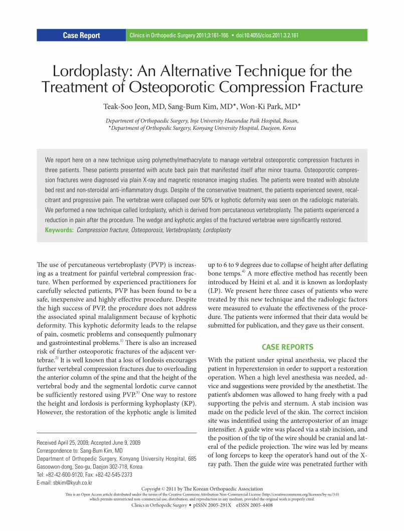
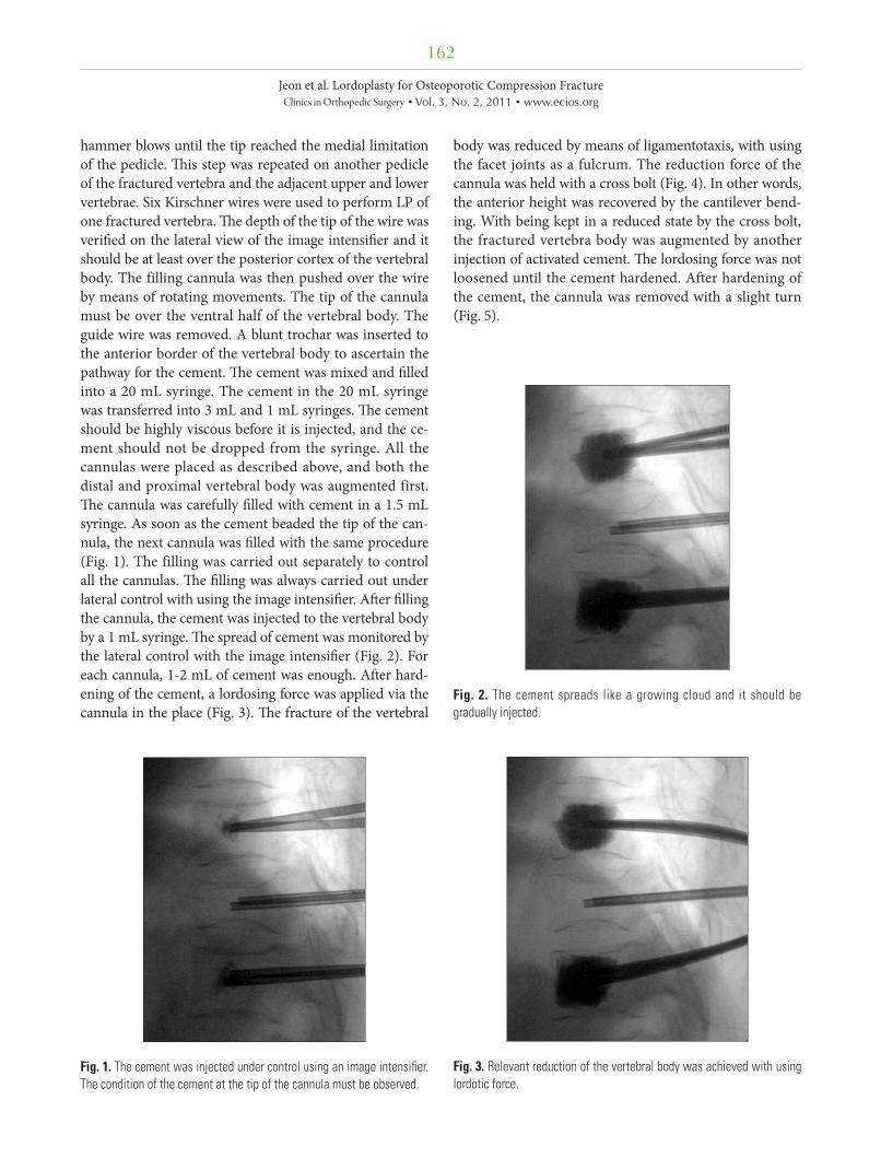
hammer blows until the tip reached the medial limitation of the pedicle. This step was repeated on another pedicle of the fractured vertebra and the adjacent upper and lower vertebrae. Six Kirschner wires were used to perform LP of one fractured vertebra. The depth of the tip of the wire was verified on the lateral view of the image intensifier and it should be at least over the posterior cortex of the vertebral body. The filling cannula was then pushed over the wire by means of rotating movements. The tip of the cannula must be over the ventral half of the vertebral body. The guide wire was removed. A blunt trochar was inserted to the anterior border of the vertebral body to ascertain the pathway for the cement. The cement was mixed and filled into a 20 mL syringe. The cement in the 20 mL syringe was transferred into 3 mL and 1 mL syringes. The cement should be highly viscous before it is injected, and the ce-ment should not be dropped from the syringe. All the cannulas were placed as described above, and both the distal and proximal vertebral body was augmented first. The cannula was carefully filled with cement in a 1.5 mL syringe. As soon as the cement beaded the tip of the can-nula, the next cannula was filled with the same procedure (Fig. 1). The filling was carried out separately to control all the cannulas. The filling was always carried out under lateral control with using the image intensifier. After filling the cannula, the cement was injected to the vertebral body by a 1 mL syringe. The spread of cement was monitored by the lateral control with the image intensifier (Fig. 2). For each cannula, 1-2 mL of cement was enough. After hard-ening of the cement, a lordosing force was applied via the cannula in the place (Fig. 3). The fracture of the vertebral
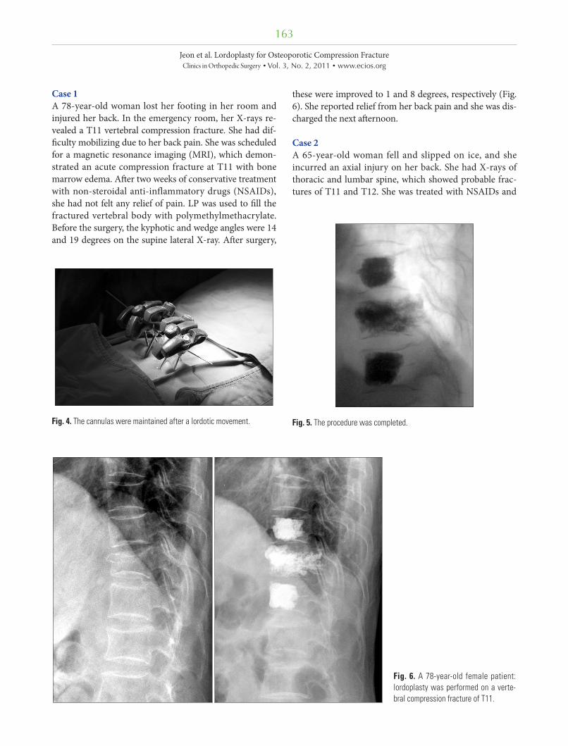
body was reduced by means of ligamentotaxis, with using the facet joints as a fulcrum. The reduction force of the cannula was held with a cross bolt (Fig. 4). In other words, the anterior height was recovered by the cantilever bend-ing. With being kept in a reduced state by the cross bolt, the fractured vertebra body was augmented by another injection of activated cement. The lordosing force was not loosened until the cement hardened. After hardening of the cement, the cannula was removed with a slight turn (Fig. 5).
Fig. 1. The cement was injected under control using an image intensifier. The condition of the cement at the tip of the cannula must be observed.
Fig. 2. The cement spreads like a growing cloud and it should be gradually injected.
Fig. 3. Relevant reduction of the vertebral body was achieved with using lordotic force.
163
Jeon et al. Lordoplasty for Osteoporotic Compression FractureClinics in Orthopedic Surgery • Vol. 3, No. 2, 2011 • www.ecios.org
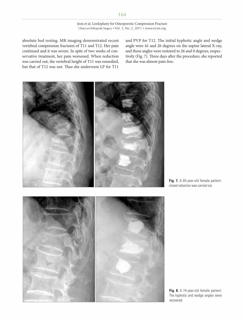
Case 1A 78-year-old woman lost her footing in her room and injured her back. In the emergency room, her X-rays re-vealed a T11 vertebral compression fracture. She had dif-ficulty mobilizing due to her back pain. She was scheduled for a magnetic resonance imaging (MRI), which demon-strated an acute compression fracture at T11 with bone marrow edema. After two weeks of conservative treatment with non-steroidal anti-inflammatory drugs (NSAIDs), she had not felt any relief of pain. LP was used to fill the fractured vertebral body with polymethylmethacrylate. Before the surgery, the kyphotic and wedge angles were 14 and 19 degrees on the supine lateral X-ray. After surgery,
these were improved to 1 and 8 degrees, respectively (Fig. 6). She reported relief from her back pain and she was dis-charged the next afternoon.
Case 2A 65-year-old woman fell and slipped on ice, and she incurred an axial injury on her back. She had X-rays of thoracic and lumbar spine, which showed probable frac-tures of T11 and T12. She was treated with NSAIDs and
Fig. 4. The cannulas were maintained after a lordotic movement. Fig. 5. The procedure was completed.
Fig. 6. A 78-year-old female patient: lordoplasty was performed on a verte-bral compression fracture of T11.
164
Jeon et al. Lordoplasty for Osteoporotic Compression FractureClinics in Orthopedic Surgery • Vol. 3, No. 2, 2011 • www.ecios.org
absolute bed resting. MR imaging demonstrated recent vertebral compression fractures of T11 and T12. Her pain continued and it was severe. In spite of two weeks of con-servative treatment, her pain worsened. When reduction was carried out, the vertebral height of T11 was remedied, but that of T12 was not. Thus she underwent LP for T11
and PVP for T12. The initial kyphotic angle and wedge angle were 41 and 26 degrees on the supine lateral X-ray, and these angles were restored to 26 and 0 degrees, respec-tively (Fig. 7). Three days after the procedure, she reported that she was almost pain free.
Fig. 7. A 65-year-old female patient: closed reduction was carried out.
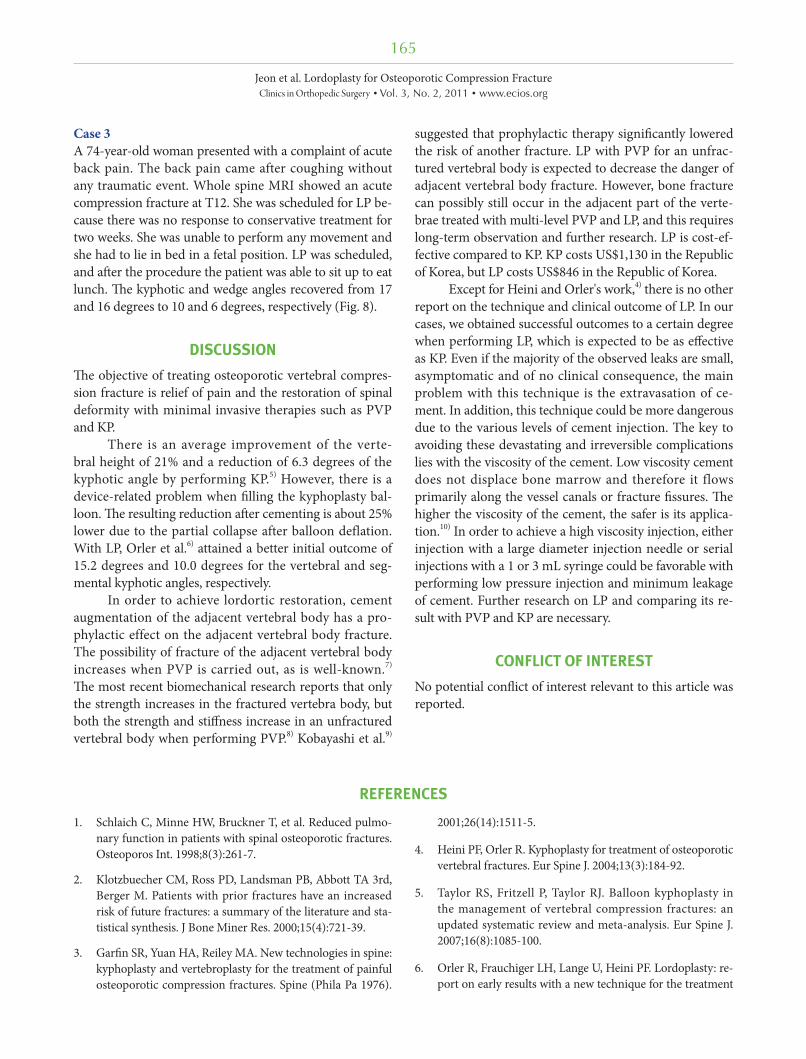
Fig. 8. A 74-year-old female patient: The kyphotic and wedge angles were recovered.
165
Jeon et al. Lordoplasty for Osteoporotic Compression FractureClinics in Orthopedic Surgery • Vol. 3, No. 2, 2011 • www.ecios.org
Case 3 A 74-year-old woman presented with a complaint of acute back pain. The back pain came after coughing without any traumatic event. Whole spine MRI showed an acute compression fracture at T12. She was scheduled for LP be-cause there was no response to conservative treatment for two weeks. She was unable to perform any movement and she had to lie in bed in a fetal position. LP was scheduled, and after the procedure the patient was able to sit up to eat lunch. The kyphotic and wedge angles recovered from 17 and 16 degrees to 10 and 6 degrees, respectively (Fig. 8).
DISCUSSION
The objective of treating osteoporotic vertebral compres-sion fracture is relief of pain and the restoration of spinal deformity with minimal invasive therapies such as PVP and KP.
There is an average improvement of the verte-bral height of 21% and a reduction of 6.3 degrees of the kyphotic angle by performing KP.5) However, there is a device-related problem when filling the kyphoplasty bal-loon. The resulting reduction after cementing is about 25% lower due to the partial collapse after balloon deflation. With LP, Orler et al.6) attained a better initial outcome of 15.2 degrees and 10.0 degrees for the vertebral and seg-mental kyphotic angles, respectively.
In order to achieve lordortic restoration, cement augmentation of the adjacent vertebral body has a pro-phylactic effect on the adjacent vertebral body fracture. The possibility of fracture of the adjacent vertebral body increases when PVP is carried out, as is well-known.7) The most recent biomechanical research reports that only the strength increases in the fractured vertebra body, but both the strength and stiffness increase in an unfractured vertebral body when performing PVP.8) Kobayashi et al.9)
suggested that prophylactic therapy significantly lowered the risk of another fracture. LP with PVP for an unfrac-tured vertebral body is expected to decrease the danger of adjacent vertebral body fracture. However, bone fracture can possibly still occur in the adjacent part of the verte-brae treated with multi-level PVP and LP, and this requires long-term observation and further research. LP is cost-ef-fective compared to KP. KP costs US$1,130 in the Republic of Korea, but LP costs US$846 in the Republic of Korea.
Except for Heini and Orler's work,4) there is no other report on the technique and clinical outcome of LP. In our cases, we obtained successful outcomes to a certain degree when performing LP, which is expected to be as effective as KP. Even if the majority of the observed leaks are small, asymptomatic and of no clinical consequence, the main problem with this technique is the extravasation of ce-ment. In addition, this technique could be more dangerous due to the various levels of cement injection. The key to avoiding these devastating and irreversible complications lies with the viscosity of the cement. Low viscosity cement does not displace bone marrow and therefore it flows primarily along the vessel canals or fracture fissures. The higher the viscosity of the cement, the safer is its applica-tion.10) In order to achieve a high viscosity injection, either injection with a large diameter injection needle or serial injections with a 1 or 3 mL syringe could be favorable with performing low pressure injection and minimum leakage of cement. Further research on LP and comparing its re-sult with PVP and KP are necessary.
CONFLICT OF INTEREST
No potential conflict of interest relevant to this article was reported.
REFERENCES
1. Schlaich C, Minne HW, Bruckner T, et al. Reduced pulmo-nary function in patients with spinal osteoporotic fractures. Osteoporos Int. 1998;8(3):261-7.
2. Klotzbuecher CM, Ross PD, Landsman PB, Abbott TA 3rd, Berger M. Patients with prior fractures have an increased risk of future fractures: a summary of the literature and sta-tistical synthesis. J Bone Miner Res. 2000;15(4):721-39.
3. Garfin SR, Yuan HA, Reiley MA. New technologies in spine: kyphoplasty and vertebroplasty for the treatment of painful osteoporotic compression fractures. Spine (Phila Pa 1976).
2001;26(14):1511-5.
4. Heini PF, Orler R. Kyphoplasty for treatment of osteoporotic vertebral fractures. Eur Spine J. 2004;13(3):184-92.
5. Taylor RS, Fritzell P, Taylor RJ. Balloon kyphoplasty in the management of vertebral compression fractures: an updated systematic review and meta-analysis. Eur Spine J. 2007;16(8):1085-100.
6. Orler R, Frauchiger LH, Lange U, Heini PF. Lordoplasty: re-port on early results with a new technique for the treatment
166
Jeon et al. Lordoplasty for Osteoporotic Compression FractureClinics in Orthopedic Surgery • Vol. 3, No. 2, 2011 • www.ecios.org
of vertebral compression fractures to restore the lordosis. Eur Spine J. 2006;15(12):1769-75.
7. Uppin AA, Hirsch JA, Centenera LV, Pfiefer BA, Pazianos AG, Choi IS. Occurrence of new vertebral body fracture after percutaneous vertebroplasty in patients with osteopo-rosis. Radiology. 2003;226(1):119-24.
8. Furtado N, Oakland RJ, Wilcox RK, Hall RM. A biome-chanical investigation of vertebroplasty in osteoporotic compression fractures and in prophylactic vertebral rein-
forcement. Spine (Phila Pa 1976). 2007;32(17):E480-7.
9. Kobayashi N, Numaguchi Y, Fuwa S, et al. Prophylactic ver-tebroplasty: cement injection into non-fractured vertebral bodies during percutaneous vertebroplasty. Acad Radiol. 2009;16(2):136-43.
10. Bohner M, Gasser B, Baroud G, Heini P. Theoretical and ex-perimental model to describe the injection of a polymethyl-methacrylate cement into a porous structure. Biomaterials. 2003;24(16):2721-30.