local spread of cervical cancer revisited: a clinical and pathological
TRANSCRIPT
Gynecologic Oncology 117 (2010) 401–408
Contents lists available at ScienceDirect
Gynecologic Oncology
j ourna l homepage: www.e lsev ie r.com/ locate /ygyno
Local spread of cervical cancer revisited: A clinical and pathological pattern analysis
Michael Höckel a,⁎, Thomas Kahn b, Jens Einenkel a, Norma Manthey a, Ulf-Dietrich Braumann c,Guido Hildebrandt d, Cornelia Leo e, Bettina Hentschel f, Peter Vaupel g, Lars-Christian Horn h
a Department of Obstetrics & Gynecology, University of Leipzig, Leipzig, Germanyb Department of Diagnostic and Interventional Radiology, University of Leipzig, Leipzig, Germanyc Interdisciplinary Center for Bioinformatics, University of Leipzig, Leipzig, Germanyd Department of Radiotherapy, University of Rostock, Rostock, Germanye Department of Gynecology, University Hospital of Zurich, Zurich, Switzerlandf Institute for Medical Informatics, Statistics and Epidemiology, University of Leipzig, Leipzig, Germanyg Department of Radiotherapy & Radiooncology, Klinikum rechts der Isar, Technical University, Munich, Germanyh Division of Breast, Gynecologic & Perinatal Pathology, Institute of Pathology, University of Leipzig, Leipzig, Germany
⁎ Corresponding author. Department of Obstetrics aChildren's Center, University of Leipzig, LiebigstrasseFax: +49 341 9723409.
E-mail address: [email protected]
0090-8258/$ – see front matter © 2010 Elsevier Inc. Adoi:10.1016/j.ygyno.2010.02.014
a b s t r a c t
a r t i c l e i n f oArticle history:
Received 6 January 2010Available online 16 March 2010Keywords:Cervical cancerLocal tumor spreadEmbryonic developmentRadical tumor resectionOncologic surgery
Background. Local tumor spread of cervical cancer is currently considered as radial progressive intra- andextracervical permeation. For radical tumor resection or radiation the inclusion of a wide envelope of tumor-free tissue is demanded. However, this concept may lead to considerable treatment-related morbidity anddoes not prevent local relapse. We propose an alternative model of local tumor propagation involvingpermissive compartments related to embryonic development.
Methods. We analyzed local tumor spread macroscopically and microscopically in consecutive patientswith advanced cervical cancer and post-irradiation recurrences.
Results. Macroscopically, all 33 stage I B (N2 cm) tumors, 40 of 42 stage II tumors and 32 of 44 stage III Btumors were confined to the embryologically defined uterovaginal (Müllerian) compartment. Local tumor
permeation deformed the uterovaginal compartment mirroring the mesenchyme distribution of theMüllerian anlage at the corresponding pelvic level in cases of symmetrical tumor growth. Tumortransgression into adjacent compartments mainly involved the embryologically related lower urinary tract.Compartmental transgression was associated with larger tumor size, paradox improvement in oxygenationand an increase in microvessel density. Post-irradiation pelvic relapse landscapes were congruent with theinflated Müllerian compartment. Microscopically, all locally advanced primary cancers and post-irradiationrecurrences were confined to the uterovaginal and lower urinary tract compartments.Conclusion. Cervical cancer spreads locally within the uterovaginal compartment derived from theMüllerian anlage. Compartment transgression is a relatively late event in the natural disease courseassociated with distinct phenotypic changes of the tumor. Compartmental tumor permeation suggests a newdefinition of local treatment radicality.
© 2010 Elsevier Inc. All rights reserved.
Introduction
Standard treatment of surgical cancer is surgery for early diseaseand chemoradiation for more advanced tumors [1]. The basis for thelocal treatment of cervical cancer, with surgery as well as withradiation, is a conception of local tumor spread assuming radialprogressive intra- and extracervical permeation favoring planes oflow mechanical resistance with microscopic or occult diseasepreceding the macroscopic tumor front. The lateral paracervical tissueis assumed to represent the dominant route for extracervical local
nd Gynecology, Women's and20a, 04103 Leipzig, Germany.
e (M. Höckel).
ll rights reserved.
tumor propagation. Consequently, radical hysterectomy, the currentsurgical treatment standard aims to resect the cervical cancer with atumor adapted margin of uninvolved paracervical tissue with specialreference to the lateral parametrium [2]. However, despite adjuvantpelvic radiation in about every second operated case with early stagedisease, local recurrence rates of 15% occur [1]. Extension of theparametrectomy does not improve the local control rate; it onlyincreases treatment-related morbidity which is represented by a 20–40% rate of moderate and severe complications [3].
We argue that these relatively unfavorable clinical results andinconsistencies may challenge the current treatment principles. Wehave suggested an alternative concept of local tumor spread providingthe basis for a different definition of local oncologic treatmentradicality. Malignant solid tumors are confined for a relatively longphase during their natural course to a permissive compartment,

402 M. Höckel et al. / Gynecologic Oncology 117 (2010) 401–408
which can be deduced from embryonic development as the finaldifferentiation product of the corresponding anlage [4,5]. Compart-ment borders are primarily tumor suppressive. For transgression intoadjacent compartments of different embryonic origin, phenotypicalchanges are necessary, which evolve relatively late during malignantprogression. Local relapses arise from remnants of the compartmentthat remain in situ after treatment harboring or recruiting residualtumor (stem) cells.
We have identified the uterovaginal (Müllerian) compartment asthe differentiation product of the Müllerian anlage, the parameso-nephric–mesonephric complex connected to the deep urogenitalsinus, in the adult female and reported its visualization by pelvic MRIand surgical exposition [4,5].
Indirect proof for the theory of compartmental tumor spread hasbeen provided in early stage (IB – IIB) cervical cancer
1. by demonstrating that the removal of the uterovaginal compart-mentwith intact borderswith total mesometrial resection (TMMR)leads to excellent local tumor control without adjuvant radiationand irrespective of the metrical extension of the tumor-freemargins [4,5];
2. by showing that local recurrences after standard radical hysterec-tomy arise and propagate within retained remnants of theMüllerian compartment [5].
Other investigators have meanwhile confirmed anatomical andconceptual aspects of our proposals by different methodologicalapproaches [6,7]. Here, we add direct evidence for compartmentaltumor spread by analyzing macroscopically and microscopically localgrowth patterns of advanced primary cervical cancer and post-irradiation local recurrences within anatomically intact pelves.
Patients and methods
During 2001–2006 consecutive patients with histologically provencancer of the uterine cervix and clinically estimated tumor size N2 cm(defined as locally advanced disease) admitted to our center wereinvited to participate in a prospective study of tumor oxygenation. Thetrialwas approved by the local ethics committee and informed consentwas obtained from all patients. Patients were examined by gross andspeculum inspection, vaginal and rectovaginal palpation, cysto- andrectoscopy under anesthesia. Core biopsies were taken under clinicalguidance. The carcinomas were staged according to the FIGO rules.Tumor size was estimated clinically. In addition, the patients receivedpelvic MRI. MR imaging was performed with 1.5 T magnets (SiemensMedical Systems, Erlangen, Germany). Using surface coils and parallelimaging, sagittal and axial T2-weighted fast spin echo images, axial T1-weighted images pre and post gadolinium-diethylenetriamine penta-acetic acid, Gd-DTPA (0.1 mmol/kg body weight), T1-weighted axialfat-suppressed images after Gd-DTPA, and sagittal T1-weightedimages after Gd-DTPA were acquired. Tumor oxygenation wasmeasured with the Eppendorf histography system (Eppendorf,Hamburg, Germany) adhering to the standard procedure as developedand validated earlier [8]. This patient cohort was retrospectivelyanalyzed for the macroscopic pattern of local tumor spread.
To develop pelvic relapse maps of cervical cancer recurring aftercurative chemoradiation we analyzed all consecutive patients withhistologically proven post-irradiation pelvic relapses examined forpotential surgical salvage treatment from October 2001 to July 2003.The group consisted of 9 patients with a median age of 51 years(range: 32–74 years). Eight patients had squamous cell carcinomaand one had adenocarcinoma. Stage distribution of the primarydisease was II B n=2, III B n=7. Median time to relapse was15 months (range: 12–448 months).
T2-weighted transverse pelvic MRI scans were used to delineatethe recurrences and to determine the tumor volumes. Co-registrationof the relapse volume data within the “VisibleWoman” pelvic data set
(“The Visible Human Project®”, National Library of Medicine,Bethesda, Maryland, USA; www.ulm.nih.gov/research/visible_hu-man.html) was done as described [5].
Since 2001 selected patients with locally advanced primarycervical cancer who were not candidates or refuted chemoradiationand patients with post-irradiation local recurrences were offeredsurgical treatment with laterally extended endopelvic resection(LEER) [9]. The surgical specimens of this patient cohort wereinvestigated histopathologically for local tumor spread. Histopatho-logical work-up of the LEER specimens was done in accordance withthe general recommendations of the Cancer Committee of AmericanPathologists [10]. Histologic type and grade were determinedaccording to the WHO classification. Tumor size was evaluated in all3 dimensions. Neoplastic infiltration of the uterovaginal and adjacenttissues, the distance of the microscopic tumor front to all exposedresection margins, lymphatic space and venous vessel involvement,and lymph nodemetastases were assessed by two pathologists in H&Estained sections. Intratumoral microvascular density determinationby CD34 immunohistochemistry was conducted as developed byWeidner et al. [11]. Microvessels were counted at a total magnifica-tion of ×200 within tumor “hot spots” located in the endocervicalstroma and in the lamina muscularis of the urinary bladder. All slideswere evaluated blindly by two observers using a multiheadmicroscope.
Statistical analysis was performedwith the SPSS 15.0 software. TheMann–Whitney U-test was used to compare two unpaired groups. Forthe comparison of two paired groups the Wilcoxon signed-rank testwas applied. More than two unpaired groups were analyzed with theKruskal–Wallis test. P-values less than 0.05 were considered toindicate statistical significance.
Results
148 consecutive patients with histologically proven advancedcarcinoma of the uterine cervix defined by clinical tumor size N2 cmwere prospectively studied regarding FIGO stage, tumor size, andoxygenation. One patient with simultaneous bladder cancer wasexcluded. The patient and tumor data are compiled in the Supple-mentary Table 1. This cohort of patients was analyzed for macroscopiclocal tumor spread using their MRI scans and cysto- and rectoscopyfindings. In 3 patients MRI had to be substituted by CT. All 33 stage I B,40 of the 42 stage II A, B tumors and 33 of the 44 stage III B tumorswere confined to the uterovaginal compartment which is schemat-ically shown in Fig. 1. Generally, local tumor spread of advanced stagecervical cancer as seen with MRI deformed the embryologicallydefined uterovaginal compartment by “inflation” or destruction(Fig. 2 a, b). Cases of symmetrical tumor propagation resembled themesenchyme distribution of the corresponding section of theuterovaginal anlage. Bilateral transvaginal tumor spread was alwaysdirected dorsally towards the anterior mesorectum, transcervicaltumor propagation could be traced dorsolaterally tangential to thelateral mesorectum. Only transcorporal tumor permeation showed atransverse direction approaching the pelvic walls. Fig. 3 comparestypical tumor growth patterns with the corresponding mesenchymedistribution of the organ anlage at three transverse sectional levels:midvagina, cervix, and corpus. Transverse lateral tumor permeationfrom the cervix towards the pelvic wall, currently assumed as themain route of local tumor propagation, was never observed. The MRIcorrelation of cervical cancer clinically infiltrating the pelvic side wallwas a fixation of the intracompartmental tumor to the parietalendopelvic fascia at the level of the levator ani muscle or at the sciaticforamen in all but one of 44 stage III B tumors (Figs. 2 c, d). Tumortransgression into adjacent non-Müllerian compartments involvedpredominantly the lower urinary tract and represented a late event inthe disease course. No case of cervical cancer infiltrating the rectalmucosa could be demonstrated by rectoscopy. MRI found signs of

Fig. 1. Graphical representation of the uterovaginal compartment established as thedifferentiation product of the Müllerian anlage in the female pelvis. In this drawing thepelvic peritoneum, the right distal adnexal structures and all fatty and lymphatic tissuehave been omitted to clarify the topographic anatomy. The uterovaginal compartmentis highlighted in green.
403M. Höckel et al. / Gynecologic Oncology 117 (2010) 401–408
cervical cancer transgression into the rectal, lateral parietal, andperitoneal compartments in 6, 5, and 3 patients (Fig. 4). However,cystoscopy unequivocally identified 34 tumors transgressing into thebladder compartment, 17 cases showed gross tumor infiltration of the
Fig. 2. Pelvic MRI scans demonstrating local permeation of cervical cancer by inflation (a) oindicated by an arrow, the tissue defect by an arrowbar. c, d: MRI correlations of cervical canconfined to the uterovaginal compartment is fixed to the endopelvic fascia (stars) at the le
bladder mucosa and 17 cases demonstrated bullous edema withpathological microvessels indicative for tumor infiltration of thebladder muscle. No carcinoma classified as stage I B and only twostage II B carcinomas had transgressed into the lower urinary tract.Seven of the 44 stage III cases and 23 of the 28 stage IV cancersinvolved the bladder compartment. All 19 cases of IV A tumorstransgressed into the lower urinary tract.
Median tumor pO2 values decreasedwith increasing stages I B to IIIB along with increasing tumor sizes. Unexpectedly, in stage IV Acarcinomas the median pO2 value increased although tumor sizeswere maximal (Figs. 5 a, b). We investigated this paradox “re-oxygenation phenomenon” by comparing the oxygenation profiles ofall locally advanced tumors stages III and IV with and without bladderinvolvement (i.e., infiltration of the bladder mucosa and muscle). The30 tumors with signs of transgression into the bladder compartmenthad significantly higher median pO2 values than the 42 tumorswithout transgression: 5.5 mm Hg (range: 0–36) vs. 4 mm Hg (range:0–27) p=0.026 (Figs. 5 c, d). Selecting only squamous cell carcinomasfor that comparison raised the level of significance: 7 mm Hg (range:2–36) vs. 3.8 mm Hg (range: 0–27) p=0.003 (Figs. 5 e, f). Theimprovement in oxygenation in tumors transgressing into the bladdercompartment despite an increasing tumor mass indicates a pheno-type of locally advanced cervical cancer different from tumorsconfined to the uterovaginal compartment.
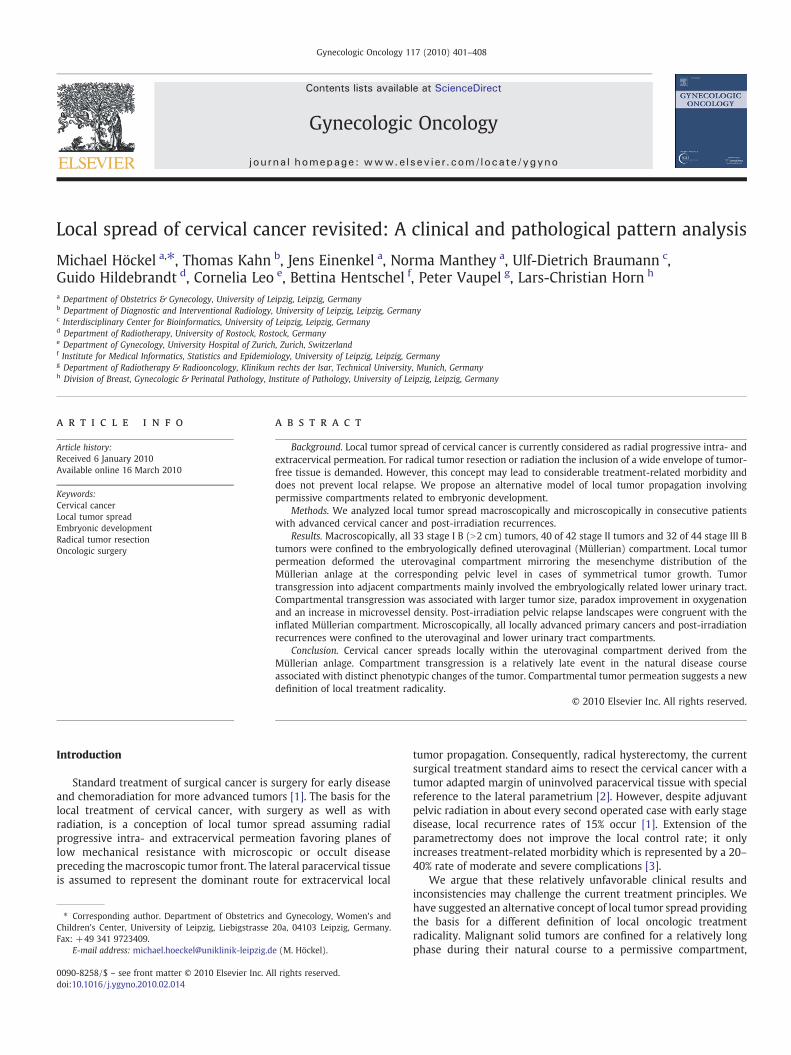
We also studied the pattern of pelvic recurrences of cervicalcarcinoma after curative (chemo-)radiotherapy. Frequency-weightedpelvic relapse maps were generated from a cohort of 9 unselectedpatients with histologically proven post-radiation recurrences. Medi-an tumor volumewas 15 cm3, range 1–117 cm3. The co-registration ofrecurrences from multiple patients within the same anatomicalreference provides clues for the tumor permeation of cervical cancerrecurring after primary chemoradiation in an anatomically intactpelvis. Fig. 6 shows that post-radiation recurrent cervical cancer
r destruction (b) of the uterovaginal compartment. The neoplastic tissue expansion iscer with clinical pelvic wall involvement. The subperitoneal tumor macroscopically stillvel of the levator ani muscle (c) or at the level of the sciatic foramen (d).

Fig. 3. Comparison of symmetrical neoplastic expansion of the uterovaginal compartment shown in pelvic MRI scans of patients with cervical cancer at three transverse levels (a, c, e)with histological sections of the Müllerian anlage at corresponding pelvic levels in a female 9 weeks old embryo (b, d, f). Transverse vaginal (a, b), cervical (c, d), and corporal (e, f)planes. The histologic work up of the embryonic tissue has been described earlier [5]. 5 μm thick transverse pelvic sections were stained with azocarmine, aniline blue, orange 6 andphotographed at ×100 magnification.
404 M. Höckel et al. / Gynecologic Oncology 117 (2010) 401–408
propagates macroscopically within the Müllerian compartment.Macroscopic transgression into adjacent compartments was detectedin 2 of the 9 recurrent tumors. Two transgressing tumors involved thebladder compartment, in one of them additional infiltration of therectum muscularis was diagnosed.
Since 2001, 21 consecutive patients underwent LEER for previ-ously untreated locally advanced primary and post-irradiationrecurrent cervical cancer in anatomically intact pelves. The patientand tumor characteristics of 11 patients with primary and 10 patientswith recurrent disease are listed in Tables 2 and 3 of the supplement.Histopathological assessment showed that all 21 tumors were
resected with microscopically tumor-free margins (R0). The tumorsinvolved the uterovaginal compartment in all cases and adjacentregions of the lower urinary tract in 90% (19/21) of the cases. Sixtumors infiltrated the mucosa of the bladder. The perirectal fattytissue was infiltrated in only one locally advanced primary and in tworecurrent carcinomas, one relapse infiltrated the muscularis of therectum. There was no microscopic involvement of the rectal mucosa,peritoneum or any lateral structure such as the endopelvic fascia orinternal iliac vessel system. Urinary tract infiltration was multifocaland associated with an apparent increase of intratumoral micro-vessels (Fig. S1). In 7 of the 9 previously untreated primary squamous

Fig. 4. MRI demonstration of advanced cervical cancer transgressing into the bladder (a), mesorectum (b), pelvic wall (c), and peritoneum (d). Arrows indicate sites ofextracompartmental local tumor propagation.
405M. Höckel et al. / Gynecologic Oncology 117 (2010) 401–408
cell carcinomas of the uterine cervix transgressing into the bladder,tumor microvessel density (TMVD) was higher within the vesicalextension as compared to their uterovaginal site of propagation.Median TMVD was 63 (range 39–123) within the bladder and 49(range 37–156) within the uterovaginal compartment.
Discussion
Essential results of this study challenge the current view of localspread of cervical cancer which assumes radial progressive intra- andextracervical tumor permeation. This model of local tumor spread canneither explain the dominance of bladder involvement as comparedto the much less frequent infiltration of the rectum, peritoneum, andparietal endopelvic fascia nor the “re-oxygenation phenomenon”associated with clinical signs of bladder infiltration. Likewise,subperitoneal transverse parametrial tumor propagation to the lateralpelvic wall at the level of the cervixwas never observedwithMRI. Thisroute of local tumor spread could only be detected in the few cases oftranscorporal propagation of cervical cancer within the broadligament in the peritoneal part of the female pelvis. Our results,however, support a concept of tumor permeation within theembryologically deduced uterovaginal (Müllerian) compartment.Previously, we have demonstrated the representation of theMülleriancompartment both in high resolution MRI and by surgical exposition[4,5]. We now show that pelvic MRI of locally advanced primarycervical carcinomas always demonstrated the major tumor masswithin the uterovaginal compartment which was inflated or partiallydestroyed by the neoplasm. 98% of stage III B tumors clinicallydiagnosed as involving the pelvic side wall by parametrial tumorpermeation were laterally restricted to the Müllerian compartmentwithout evidence of infiltration of the parietal endopelvic fascia. Caseswith symmetrical tumor involvement mirrored the mesenchymedistribution of the Müllerian anlage at the corresponding level within
the pelvis. This congruence is confirmed by the pelvic relapselandscapes we generated from cervical cancer recurring after curativechemoradiation. In post-irradiation pelves the topographic pelvicanatomy is retained. Due to the radiation-induced tumor bed effect[12], neoplastic volumes are diminished compared to the primarydisease of advanced malignant progression. The relatively smalltumor masses facilitated their delineation and co-registration withinthe unit pelvis to demonstrate the spatial propagation of cervicalcancer. As with primary disease, the spatial distribution of the post-irradiation recurrent tumors is not in line with a radial progressivemodel of tumor permeation. Neither is the idea of transverse tumorpropagation substantiated by our results.
This study confirms that the infiltration of adjacent non-Mülleriantissues by cervical cancer is a relatively late event in the natural courseand compartmental transgression is associated with phenotypicchanges of the tumor. Compartment transgression was observed in5% of stage II and in 27% of stage III tumors. Transgressing tumorsexhibited the largest sizes. A phenotypic change detectedwith tumorsinfiltrating the (non-Müllerian) bladder compartment is representedby the paradox “re-oxygenation phenomenon”. The fact thattransgressing primary and post-irradiation recurrent cervical cancerinfiltrated mainly the bladder may be explained by the embryologicalkinship of the Müllerian system with the lower urinary tract. Themesonephric and paramesonephric tissues are both derived fromintermediate mesoderm. The mesonephric system participates bothin the development of the urinary and the genital tracts.
The histopathological investigation of the patients treated withLEER for locally advanced primary and post-irradiation recurrentcervical cancer supports the macroscopic findings of local tumorpermeation. Although tumor fixation to or involvement of parietaltissues at the site of the sciatic foramen has been considered ascontraindication for treatment with LEER, the majority of tumorsoperated on were fixed to the pelvic side wall at the level of the

Fig. 5. Box plots showing median pO2 values and tumor sizes of: a, b: cervical carcinomas related to FIGO stage; c, d: stage≥ III B tumors with (≥ III B+) or without (≥ III B−)transgression into the bladder; e, f: stage≥ III B squamous cell carcinomaswith (SCC≥ III B+) orwithout (SCC≥ III B−) transgression into the bladder. Transgression into the bladder isassociated with an increase in oxygenation despite larger size, whereas intracompartmental growth is associated with a decrease in oxygenation.
406 M. Höckel et al. / Gynecologic Oncology 117 (2010) 401–408
levator ani muscles. The selection process cannot explain thepreference for tumor transgression into the lower urinary tractcompartment. Likewise, the lateral confinement of the tumors to theuterovaginal and lower urinary tract compartments was substantiat-ed by the fact that involvement of the endopelvic fascia or adjacentstriated pelvic wall and floor muscles was never detected.
The apparent increase of microvessel density of advancedsquamous cell carcinoma of the uterine cervix at the sites of bladderinfiltration may be causatively involved in the “re-oxygenationphenomenon”. One can speculate that compartment transgressionenables the tumor to tap another vascular territory in addition to theone within the permissive compartment. However, our study was not
Fig. 6. 3-D pelvic relapse map (“landscape”) obtained from 9 unselected consecutive patients with cervical cancer recurring after curative chemoradiation. Major tumor mass isconfined to the embryologically deduced uterovaginal compartment at the cervical level. Representative transversal (a), sagittal (b), and coronal (c) planes. Relapse occurrencefrequency is color-coded from blue indicating minimal frequency with at least one case via purple and red to bright yellow highlighting the maximum number of overlappingoccurrences. Minimum frequency is one, maximum frequency is five for this map.
407M. Höckel et al. / Gynecologic Oncology 117 (2010) 401–408
designed to investigate the pathomechanisms of tumor oxygenationand perfusion.
Limitations of our study are represented by the methodologicaluncertainties of the diagnostic procedures applied for macroscopicinvestigation and the relatively low number of patientswith advanceddisease available for microscopic assessment. However, diagnosticinaccuracy to be expected for MRI and endoscopy does not appear tobias the main insights obtained from our investigation. The fact thatboth the macroscopic and the microscopic evaluation led to the sameresults in terms of extracompartmental tumor involvement ofadvanced disease fosters the theory of embryologically definedcompartmental tumor permeation.
The results of this study may have significant clinical implicationswith regard to cervical cancer treatment. They confirm the call forresection of the embryologically defined uterovaginal compartmentfor early cervical cancer instead of applying the treatment principle ofwide radial paracervical margins [2]. In the surgical treatment ofpatients with locally advanced primary cancer that are not candidatesfor chemoradiation and for the salvage of post-irradiation persistenceor recurrent disease, resection of both uterovaginal and lower urinarytract compartments is usually necessary, but the rectum may beretained if the intact compartment border can be exposed. Tumorfixation at the pelvic wall can no longer be regarded as a generalcontraindication for surgical treatment as these clinical features mayrepresent fibrotic adherence of the tumor still confined to theuterovaginal compartment with the parietal endopelvic fascia at thelevator ani level. Laterally extended endopelvic resection (LEER)including the en bloc removal of the pelvic floor and side wall musclesto assure the complete multicompartmental extirpation has a highpotential of local tumor control in these cases. The design of radiation
target volumes with regard to the topography of the uterovaginalcompartment instead of adding a metrically defined tissue envelopeto the gross tumor volumemay further improve the therapeutic indexin the radiotherapy of cervical cancer.
Conflict of interest statementNone of the authors has any conflicts of interest.
Acknowledgments
Katja Schmidt and Carola Koschka from the Department ofObstetrics & Gynecology, University of Leipzig, contributed withclinical data management.
Fig. 1 was drawn by Nikolaus Lechenbauer, Ehenbichl, Austria.Angela Steller, University of Leipzig Medical Center, helped with
the production of the figures. The study was supported by DeutscheKrebshilfe (grant 106795).
Appendix A. Supplementary data
Supplementary data associated with this article can be found, inthe online version, at doi:10.1016/j.ygyno.2010.02.014.
References
[1] Waggoner SE. Cervical cancer. Lancet 2003;361:2217–25.[2] Querleu D, Morrow CP. Classification of radical hysterectomy. Lancet Oncol
2008;9:297–303.[3] Landoni F, Maneo A, Cormio G, Perego P, Milani R, Caruso O, et al. Class II versus
class III radical hysterectomy in stage IB–IIA cervical cancer: a prospectiverandomized study. Gynecol Oncol 2001;80:3–12.

408 M. Höckel et al. / Gynecologic Oncology 117 (2010) 401–408
[4] Höckel M, Horn LC, Fritsch H. Association between the mesenchymal compart-ment of uterovaginal organogenesis and local tumor spread in stage IB–IIB cervicalcarcinoma: a prospective study. Lancet Oncol 2005;6:751–6.
[5] Höckel M, Horn LC, Manthey N, Braumann U-D, Wolf U, Teichmann G, et al.Resection of the embryologically defined uterovaginal (Müllerian) compartmentand pelvic control in patients with cervical cancer: a prospective analysis. LancetOncol 2009;10:683–92.
[6] Touboul C, Fauconnier A, Zareski E, Bouhanna P, Darai E. The lateral infraureteralparametrium: myth or reality? Am J Obstet Gynecol 2008;199:242–3.
[7] Van den Tillaart SAHM, Kenter GG, Peters AAW, Dekker FW, Gaarenstroom KN,Fleuren GJ, et al. Nerve-sparing radical hysterectomy. Local recurrence rate,feasibility, and safety in cervical cancer patients stage IA to IIA. Int J Gynecol Cancer2009;19:39–45.
[8] Höckel M, Schlenger K, Knoop C, Vaupel P. Oxygenation of carcinomas of theuterine cervix: evaluation by computerized O2 tension measurements. Cancer Res1991;51:6098–102.
[9] Höckel M, Dornhöfer N. Pelvic exenteration for gynaecological tumours:achievements and unanswered questions. Lancet Oncol 2006;7:837–47.
[10] Kurman RJ, Amin MB, Cancer Committee College of American Pathologists.Protocol for the examination of the specimens from patients with carcinomas ofthe cervix. A basis for checklists. Arch Pathol Lab Med 1999;123:55–61.
[11] Weidner N, Semple JP, Welch WR, Folkman J. Tumor angiogenesis and metastasis— correlation in invasive breast carcinoma. N Engl J Med 1991;324:1–8.
[12] Clifton KH, Jirtle R. Mammary carcinoma cell population growth in preirradiatedand unirradiated transplant sites. Viable tumor growth, vascularity, and thetumor-bed effect. Radiology 1975;117:459–65.