lkb1 and ampk differentially regulate pancreatic b-cell ... j... · lkb1 and ampk differentially...
TRANSCRIPT

The FASEB Journal • Research Communication
LKB1 and AMPK differentially regulate pancreaticb-cell identity
Marina Kone,*,1 Timothy J. Pullen,*,1 Gao Sun,*,1 Mark Ibberson,§ Aida Martinez-Sanchez,*Sophie Sayers,* Marie-Sophie Nguyen-Tu,* Chase Kantor,* Avital Swisa,{ Yuval Dor,{
Tracy Gorman,# Jorge Ferrer,† Bernard Thorens,k Frank Reimann,** Fiona Gribble,**James A. McGinty,‡ Lingling Chen,‡ Paul M. French,‡ Fabian Birzele,†† Tobias Hildebrandt,††
Ingo Uphues,†† and Guy A. Rutter*,2
*Section of Cell Biology and †Section of b-Cell Development, Division of Diabetes, Endocrinology, andMetabolism, Department of Medicine, and ‡Photonics Group, Department of Physics, Imperial CollegeLondon, London, UK; §Swiss Institute of Bioinformatics and kCenter for Integrative Genomics,University of Lausanne, Lausanne, Switzerland; {Department of Developmental Biology and CancerResearch, Institute for Medical Research Israel–Canada, Hebrew University–Hadassah Medical School,Jerusalem, Israel; #AstraZeneca Diabetes and Obesity Drug Discovery, Alderley Edge, UK; **MetabolicResearch Laboratories, University of Cambridge, Cambridge, UK and ††Boehringer Ingelheim Pharma,Ingelheim, Germany
ABSTRACT Fully differentiated pancreatic b cellsare essential for normal glucose homeostasis in mam-mals. Dedifferentiation of these cells has been sug-gested to occur in type 2 diabetes, impairing insulinproduction. Since chronic fuel excess (“glucotoxicity”)is implicated in this process, we sought here to identifythe potential roles in b-cell identity of the tumor sup-pressor liver kinase B1 (LKB1/STK11) and the down-stream fuel-sensitive kinase, AMP-activated proteinkinase (AMPK). Highly b-cell-restricted deletion ofeach kinase in mice, using an Ins1-controlled Cre, wastherefore followed by physiological, morphometric,and massive parallel sequencing analysis. Loss of LKB1strikingly (2.0–12-fold, E<0.01) increased the expres-sion of subsets of hepatic (Alb, Iyd, Elovl2) and neuronal(Nptx2, Dlgap2, Cartpt, Pdyn) genes, enhancing glutamatesignaling. These changes were partially recapitulatedby the loss of AMPK, which also up-regulated b-cell“disallowed” genes (Slc16a1, Ldha, Mgst1, Pdgfra) 1.8- to3.4-fold (E<0.01). Correspondingly, targeted promoterswere enriched for neuronal (Zfp206; P51.3310233)and hypoxia-regulated (HIF1; P52.5310216) transcrip-tion factors. In summary, LKB1 and AMPK, through onlypartly overlapping mechanisms, maintain b-cell identity
by suppressing alternate pathways leading to neuronal,hepatic, and other characteristics. Selective targetingof these enzymes may provide a new approach tomaintaining b-cell function in some forms of dia-betes.—Kone, M., Pullen, T. J., Sun, G., Ibberson, M.,Martinez-Sanchez, A., Sayers, S., Nguyen-Tu, M.-S.,Kantor, C., Swisa, A., Dor, Y., Gorman, T., Ferrer, J.,Thorens, B., Reimann, F., Gribble, F., McGinty, J. A.,Chen, L., French, P. M., Birzele, F., Hildebrandt, T.,Uphues, I., Rutter, G. A. LKB1 and AMPK differen-tially regulate pancreatic b-cell identity. FASEB J.28, 4972–4985 (2014). www.fasebj.org
Key Words: islet • diabetes • insulin secretion • RNASeq
DIABETES MELLITUS IS a socioeconomically costly diseaseaffecting ;8% of the adult population worldwide (1).The most prevalent form, type 2 diabetes (T2D),involves a decline in the number of normally func-tioning b cells through incompletely defined mecha-nisms (2).
Glucose-induced insulin secretion from the healthypancreatic b cell requires intracellular metabolism ofthe sugar, increased intracellular ATP/ADP levels, and
Abbreviations: AMPA, a-amino-3-hydroxy-5-methyl-4-isoxa-zolepropionic; AMPK, 59AMP-activated protein kinase;AMPKRK, AMPK-related kinase; Dlgap2, discs, large (Drosophila)homologue-associated protein 2; dKO, double knockout;FACS, fluorescence-activated cell sorting; FDR, false discov-ery rate; GCG, glucagon; GCGR, glucagon receptor; GIP,glucose-dependent insulinotropic polypeptide; GLP-1,glucagon-like peptide-1; GSEA, gene set enrichment analysis;GWAS, genome-wide association study; HIF1, hypoxia-inducible factor 1; KO, knockout; LKB1, liver kinase B1;OPT, optical projection tomography; REST, repressor ele-ment 1 silencing transcription; RNASeq, massive parallelRNA sequencing; TFBS, transcription factor binding site;T2D, type 2 diabetes; WT, wild type
1 These authors contributed equally to this work.2 Correspondence: Imperial College London, 3rd floor,
ICTEM, Hammersmith Hospital, du Cane Rd., London W12ONN, UK. E-mail: [email protected] is an Open Access article distributed under the terms of
the Creative Commons Attribution 4.0 International (CC BY 4.0)(http://creativecommons.org/licenses/by/4.0/) which permitsunrestricted use, distribution, and reproduction in any medium,provided the original work is properly cited.doi: 10.1096/fj.14-257667This article includes supplemental data. Please visit http://
www.fasebj.org to obtain this information.
4972 0892-6638/14/0028-4972 © The Author(s) Vol.28, No.11 , pp:4972-4985, August, 2016The FASEB Journal. 155.198.12.147 to IP www.fasebj.orgDownloaded from
elevated free Ca2+ (3). Although T2D progression inhumans is characterized by a limited decrease in overallb-cell mass (4, 5) it is increasingly apparent that a loss ofthe normal differentiated state of the remaining cells,and their consequent “glucose blindness,” also playsa role (6–9). As well as decreased expression of signa-ture genes, such as the glucose transporter Glut2/Slc2a2and glucokinase (Gck) (6), b-cell dedifferentiation ischaracterized by the increased expression of normallyrepressed (“disallowed”) genes, such as LDHA and thelactate transporter MCT-1/Slc16a1 (10), leading to ab-errant fuel sensing (11).
Liver kinase B1 (LKB1), also called STK11, is a mam-malian Ser/Thr kinase and tumor suppressor whoseinvertebrate homologue (Par-4; ref. 12) controls em-bryo polarity. LKB1 was shown [alongside calmodulinkinase kinase 2 and transforming growth factor (TGF)-b-activated kinase (TAK); ref. 13] to be one of 3 physi-ologically relevant upstream kinases for AMP-activatedprotein kinase (AMPK; refs. 14, 15), and othermembersof the AMPK-related kinase (AMPKRK) family (16),previously implicated in b-cell glucose sensing (17, 18).Demonstrating the role of LKB1 in restricting cellgrowth in humans, mutations in the human LKB1 genelead to Peutz-Jegers syndrome (19), an autosomaldominant disorder characterized by the development ofintestinal polyps.
We (20, 21) and others (22) have previously demon-strated that inactivation of either LKB1 or AMPK (23, 24)selectively inpancreaticb cells anda smallnumberofothercell types exerts dramatic effects on insulin secretion invivo. Thus, loss of LKB1 causes b-cell hyperplasia and anincrease in overall b-cell mass, associated with dramaticchanges in cell polarity. In marked contrast, deletion ofboth AMPK catalytic subunits (a1, global; and a2, bcells, brain) had no effect on b-cell size and mass butstrongly inhibited insulin secretion in vivo (23, 24). Themolecular underpinnings of these changes remain, how-ever, unexplored.
To examine in detail the cell autonomous roles ofLKB1 and AMPK in the b cell, we have therefore de-veloped new models using recombination based on Creexpression under Ins1 promoter control, avoidingdeletion in the brain (25, 26). Metabolic analysis andmassive parallel sequencing of islets from each modelreveal both overlapping and distinct roles for LKB1 andAMPK inb cells.We show that these enzymes are essentialto avoid the misexpression of a subset of genes normallyexpressed at relatively low levels in b cells, includingthose involved in glutamate signaling and in allowingalternative metabolic fates for glucose.
MATERIALS AND METHODS
Generation of mutant mice lacking LKB1 selectively inpancreatic b cells
Mice heterozygous for floxed alleles of the Lkb1/Stk11 gene(mixed FVB/129S6 and C57BL/6 background; ref. 27) wereobtained from the Mouse Models of Human Cancer Con-sortium [U.S. National Institutes of Health (NIH), Bethesda,MD, USA; http://www.nih.gov/science/models/mouse/
resources/hcc.html] and backcrossed with C57BL/6 mice4 times. Mice were then crossed withmice expressing Cre underthe mouse Ins1 promoter (Ins1.Cre), and the resultingheterozygous mice were intercrossed with siblings to generateIns1LKB1-knockout (Ins1LKB1KO) mice (Lkb1fl/fl, Cre+).Ins1LKB1KO mice were further bred with Lkb1fl/fl mice togenerate littermate controls (Lkb1fl/fl).
Generation of mutant mice selectively lacking AMPK a1 anda2 in pancreatic b cells
Mice homozygous for Ampka1fl/fl (Dr. Benoit Viollet, InstitutNational de la Sante et de la Recherche Medicale, U1016, Paris,France) were crossed to mice heterozygous for floxed alleles ofAMPKa2 (Ampka2fl/+; ref. 23). The resulting double hetero-zygotes (Ampka1fl/+,a2fl/+) were crossedwith Ins1Cre-expressinganimals to generate triple heterozygous mice (Ampka1fl/+, a2fl/+,Cre+). The latter were then bred with mice homozygous for bothfloxed Ampka1 and a2 alleles (Ampka1fl/fl, a2fl/fl) to produceIns1AMPKdouble-KO(Ins1AMPKdKO)mice (Ampka1fl/fl,a2fl/fl,Cre+). Ins1AMPKdKOmice were further crossed with Ampka1fl/fl,a2fl/flmice to generate littermate controls (Ampka1fl/fl,a2fl/fl). Allmice were maintained on a C57BL/6 background.
Mouse maintenance and diet
Animals were housed 2 to 5 per individually ventilated cage ina pathogen-free facility with 12-h light-dark cycle and had freeaccess to standard mouse chow diet. All in vivo procedures de-scribed were performed at the Imperial College Central Bio-medical Service and approved by the UK Home Office AnimalsScientific Procedures Act, 1986 (HO License PPL 70/7349).
Isolation of mouse islets and b cells
Islets were isolated by pancreatic distension and digestion withcollagenase as described previously (28). b Cells were purified byfluorescence-activatedcell sorting(FACS)asdescribedpreviously(29) and directly collected in Trizol (Life Techonologies, GrandIsland, NY, USA).
RNA extraction and massive parallel RNAsequencing (RNAseq)
Islets (50–100) extracted from Ins1LKB1KO or Ins1AMPKdKOmice and their wild-type (WT) controls, age 12–14 wk, were in-cubated in RPMI medium containing 11 mM glucose, 10% FCS,100 IU/ml penicillin, and 100 mg/ml streptomycin, at 5% CO2and 37°C for 24 h prior to being lyzed in RNA lysis buffer usingthe RNAeasy kit according to the manufacturer’s instructions(Qiagen, Valencia, CA, USA).
Library preparation and sequencing
All libraries were prepared using the TruSeq RNA SamplePreparation Kit v2 (Illumina, San Diego, CA, USA) accordingto the manufacturer’s instructions. In brief, magnetic beadscontaining polydT molecules were first used to purify mRNAfrom 250 ng of total RNA. Second, samples were chemicallyfragmented and reverse transcribed into cDNA. Finally,end repair and A-base tailing was performed beforeIllumina adapters were ligated to the cDNA fragments. Purifiedsamples were amplified by 15-cycle PCR. Amplified material wasvalidated and quantified using an Agilent 2100 bioanalyzer
LKB1 AND AMPK IN b-CELL DEVELOPMENT 4973 Vol.28, No.11 , pp:4972-4985, August, 2016The FASEB Journal. 155.198.12.147 to IP www.fasebj.orgDownloaded from
and the DNA 1000 Nano Chip Kit (Agilent, Technologies,Santa Clara, CA, USA).
Libraries were loaded onto the channels of the flowcell at9 pM concentration. Sequencing was carried out on the Hiseq2000 (Illumina) by using Illumina’s Trueseq Single ReadCluster Generation Kit v3 CBot Hs and running 50 cycles withthe Cycle Sequencing Kit according to the manufacturer’sinstructions.
Transcriptomic data analysis and identification of putativetranscription factor binding sites
Datasets from RIPCre strains had reads mapped to the mousegenome (Ensembl56) using the Genomatix Mapping station al-gorithm(allowing for up to 3mismatches, no indels; Genomatix,Munich, Germany). Reads were additionally mapped to a set of(artificial) splice junctions of all known exons in the mousepreserving exon order within a gene. Reads from Ins1Cre strainswere mapped to the mouse genome (Ensembl66) using theBowtie2-Tophat2 spliced read mapper (30). Differential ex-pression was analyzed using SageBetaBin (31), and expressionvalues [reads per kilobase of transcript per million reads read(RPKM)] were calculated according to Mortazavi et al. (32).Transcription factor binding sites (TFBSs) enriched in the pro-moters ofdifferentially expressedgeneswere identifiedusing theWhole Genome rVista tool (33).
Kyoto Encyclopedia of Genes and Genomes (KEGG) pathwayanalysis (Kanehisa Laboratories, Kyoto, Japan; http://www.genome.jp/kegg/) was performed using the Database for Anno-tation,Visualizationand IntegratedDiscovery (DAVID) functionalanalysis tool (U.S. National Institute for Allergy and InfectiousDiseases,NIH,Bethesda,MD,USA;http://david.abcc.ncifcrf.gov).Expression patterns in mouse tissues and the likely functions ofidentified genes were assessed by reference to BioGPS (ScrippsResearch Institute, La Jolla, CA, USA; http://biogps.org/).
Gene set enrichment analysis (GSEA)
GSEA(34)wasperformedbyfirst rankingdifferentially expressedgenes according to fold change (high to low; either absolute ortaking direction into account) and testing for enrichment againstMSigDB V4 gene sets, or a gene set containing disallowed genes.Empirical P values were calculated by performing a bootstrapwhere gene labels were shuffled10,000 times and theenrichmentwas recalculated.Anestimateof the falsediscovery rate (FDR)wascalculated using the Benjamini-Hochberg procedure using theBioconductor multtest package in R (35). An FDR of 30% wasconsidered as indicating significant enrichment.
RNA-Seq accession codes
Raw sequence data for RNA-Seq are available at the EuropeanMolecular Biology Laboratory–European Bioinformatics In-stitute (EMBL-EBI) ArrayExpress website (accession numberE-MTAB-2791; https://www.ebi.ac.uk/arrayexpress/).
Imaging of pancreatic slices and optical projectiontomography (OPT)
Permeabilized slices were prepared as described previously(20) and blotted with primary antibodies, anti-guinea piginsulin (1:200; Dako, Ely, UK), anti-rabbit glucagon (GCG;1:100; Santa Cruz Biotechnology, Santa Cruz, CA, USA), andanti-rabbit NPTX2 (5 mg/ml; Abcam, Cambridge, UK). Slices
were visualized with Alexa Fluor 488 goat anti-guinea pig IgGand with Alexa Fluor 568 donkey anti-rabbit IgG (Invitrogen,Paisley, UK) using an Axiovert 200 M microscope (Zeiss,WelwynGardenCity, UK). ImageJ software (Wayne Rasband,NIH, Bethesda, MD, USA) was used to calculate b- and a-cellmass. OPT of whole pancreata stained with anti-insulinantibodies was performed as described previously (20).
Islet isolation for calcium imaging
Pancreatic islets were isolated as described above, dispersedinto single b cells, and plated on glass coverslips (36). Imagingexperiments were performed essentially as described pre-viously (37) on an Olympus IX-71 microscope with 340 objec-tive, using an Andor CMOS Zyla camera (Andor Technology,Belfast, UK) and MT-20 excitation system equipped witha Hg/Xe arc lamp controlled by Micro-Manager software(38). Images were acquired at a frequency of 0.5 Hz withtypical excitation times of 50 ms.
Statistical analysis
Data are presented as means 6 SEM. Significance was assessedby Student’s t test with appropriate Bonferroni correction, orANOVA using Prism (GraphPad, San Diego, CA, USA).
RESULTS
Effect on glucose homeostasis and insulin secretion ofdeleting LKB1 or AMPK highly selectively in the b cellwith Ins1Cre
To avoid the complications associated with deletion ofLKB1 (39) or AMPK (23, 24) in the brain after Ins2Cre-mediated recombination (40) we used a knock-inmouse in which Cre recombinase was introduced at theIns1 locus (refs. 25, 26 and unpublished results). Incontrast to RIP2LKB1KO mice, postnatal body weightgain was normal in Ins1LKB1KO animals; for malesaged 10–14 wk: WT, 26.9 6 0.4 g (n54 mice), and forIns1LKB1KO, 27.4 6 0.9 g (n58, P.0.05). Further-more, Ins1LKB1KO animals showed no signs of paral-ysis or premature mortality up to 12 mo of age (resultsnot shown).
Examined at ages up to 12 wk, Ins1LKB1KO micedisplayed improved glucose tolerance (Fig. 1A), un-altered insulin sensitivity (Fig. 1B), and sharply im-proved in vivo insulin secretion (Fig. 1C) compared toWT littermates. An increase in b-cell size (Fig. 2A–J),and the appearance of “rosette-like” arrangements ofb cells around a central organizing center (Fig. 2G,H), were also observed, as described previously afterRIP2Cre-mediated (20) or Pdx1CreER-mediated(21) LKB1 deletion. Pancreatic b-cell mass was alsosignificantly increased (Fig. 2K, L, P) as well as b-cell size(Fig. 2M, N) and b:a cell ratio, consistent with earlierfindings using alternative Cres (20, 21). Examined in vitro,glucose-stimulated insulin secretion from Ins1LKB1KOislets was largely unchangedwith respect to controlmouseislets (Supplemental Fig. S1A and Fig. 6C).
We next generated an analogous mouse line in whichAMPK activity was eliminated selectively in the b cell by
4974 Vol. 28 November 2014 KONE ET AL.The FASEB Journal x www.fasebj.org Vol.28, No.11 , pp:4972-4985, August, 2016The FASEB Journal. 155.198.12.147 to IP www.fasebj.orgDownloaded from
deleting both catalytic subunits of AMPK (a1 and a2).Body weight gain was not different in Ins1AMPKdKO(Cre+, a1f/f, a2f/f) mice with respect to Cre2 controls (at10 wk, means6SEM,: WT, 27.0760.42 g; AMPKdKO,27.7560.20 g).
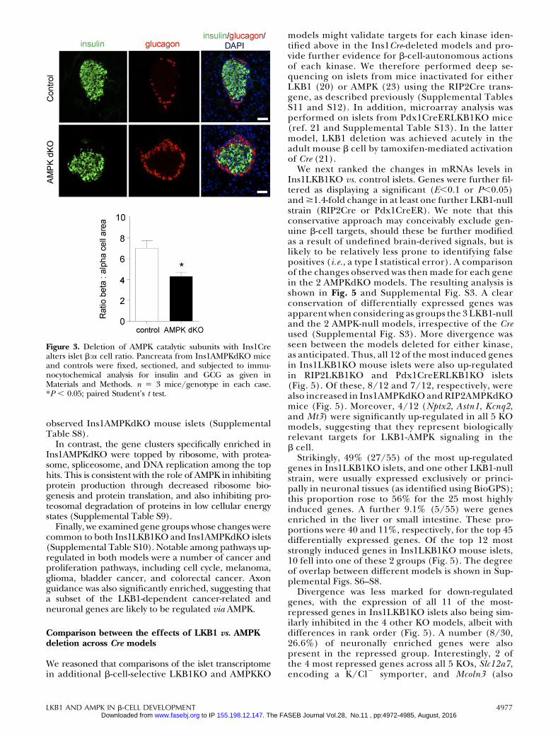
Ins1AMPKdKO mice displayed a more minor pheno-type compared to that observed in the RIP2AMPKdKOmodel (23, 24). Thus, glucose tolerance was onlyslightly impaired in Ins1AMPKdKO animals (Fig. 1D),with blood glucose significantly elevated only at 30 minduring intraperitoneal glucose tests (IPGTTs). More-over, whereas insulin sensitivity was markedly im-proved in RIP2AMPKdKO mice vs. heterozygouscontrols (23), no alterations in this parameter wereseen in Ins1AMPKdKO animals (Fig. 1E). Finally,whereas the first phase of glucose-stimulated in-sulin release in vivo was completely abolished inRIP2AMPKdKO mice (23), release of the hormonewas diminished by only ;50% in Ins1AMPKdKO micevs. controls (Fig. 1F). No evident changes in b-cellmass were observed in Ins1AMPKdKO mice vs. litter-mate controls (pancreatic b-cell area: 0.3160.06 vs.0.3960.5%, respectively, n55–6 mice/genotype),though the ratio of b:a cells was significantly re-duced in these mice (Fig. 3). In common withRIP2AMPKdKO mice (23), a dramatic improvementin glucose-stimulated insulin secretion was observedin vitro with islets isolated from Ins1AMPKdKO mice(Supplemental Fig. S1B).
Deep sequencing identifies gene modulesdifferentially affected in Ins1LKB1KO andIns1AMPKdKO mouse islets
We next explored whether the phenotypic differencesbetween Ins1LKB1KO and Ins1AMPKdKO mouse b cellsmight be reflected at the transcriptome level using RNA-Seq (Supplemental Tables S1 and S2).
Ins1LKB1KO mouse islets displayed no significantchanges in b-cell markers, including Slc2a2 (Glut2),Pdx1, MafA, NeuroD, and Nkx6.2, while Pcsk1 mRNAwas up-regulated 1.75-fold, consistent with an in-crease in b-cell volume within the islet. Conversely,expression of the a-cell-enriched factor Arx (41) wassignificantly (0.65-fold, E50.05) lowered, and GcgmRNA levels tended (0.84-fold, E50.19) also to bedecreased, consistent with the increase in b:a cellratio (Fig. 2O). Somatostatin (Sst) mRNA levels alsoshowed a minor tendency to decrease (0.74-fold,P50.31) in Ins1LKB1KO islets. Significant increaseswere also observed in the expression of genes usuallyrestricted to a cells (42) including Pcsk2 [encodingprohormone convertase 2 (PC2), 1.61-fold, E,0.01]as well asDpp4 (1.51-fold, E50.03). The latter findingsmay suggest that LKB1 ablation loosens the re-pression in the b cell of a subset of usually a-cell-restricted genes.
Conversely, in Ins1AMPKdKO mouse islets, Slc2a2(Glut2; 0.59-fold, E50.04; ref. 43), mRNA levels were
Figure 1. Glucose homeostasis and insulin secretion in Ins1LKB1KO and Ins1AMPKdKO mice. A–C) Glucose (1 g/kgintraperitoneal) tolerance (A), insulin (0.75 U/kg) tolerance (B), and glucose (3 g/kg)-induced insulin secretion (C) in WT orIns1LKB1KO mice (10- to 12-wk-old males, n53 WT, 5 KO). D–F) As in A–C but comparing WT (AMPKa1f/f:AMPKa2f/f) andIns1AMPKdKO mice (n55–10 mice/genotype). *P , 0.05, **P , 0.01 for effects of genotype.
LKB1 AND AMPK IN b-CELL DEVELOPMENT 4975 Vol.28, No.11 , pp:4972-4985, August, 2016The FASEB Journal. 155.198.12.147 to IP www.fasebj.orgDownloaded from
significantly reduced compared to WT controls, andother b-cell markers, including Ins2 and Slc30a8, alsotended to be more weakly expressed in the absence ofAMPK. On the other hand, Gcg and Arx mRNA levelstended to increase, while Pcsk2 was significantly up-regulated (1.21-fold, E,0.04). Thus, AMPK signalingis required to maintain normal islet b-cell mass and isnot rescued by other AMPK-related kinases.
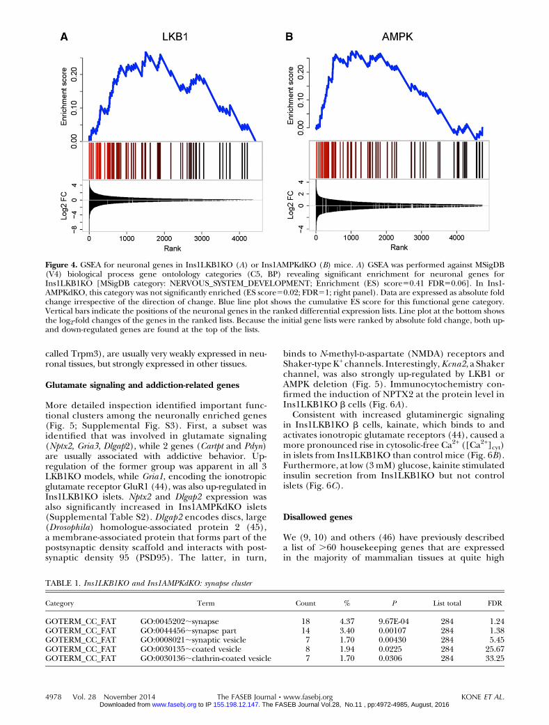
To identify gene clusters affected by either kinase, wefirst used GSEA (ref. 33; see Materials and Methods andSupplemental Tables S3 and S4). Whereas genes in-volved in neuronal function (synapse organization,synaptic transmission, synaptogenesis, etc.) were stronglyand significantly enriched in Ins1LKB1KO islets (Fig.4A, Table 1, and Supplemental Fig. S2A), the most
strongly enriched gene groups in Ins1AMPKdKO isletswere involved in second messenger signaling andmetal ion transport (Supplemental Table S3). None-theless, a tendency was observed for enrichment of neuro-nal genes in Ins1AMPKdKOislets, though thisdidnot reachstatistical significance (Fig. 4B and Supplemental Fig. S2B).
Wenext compared genes whichwere changed in bothor in a single Ins1Cre-deleted model (SupplementalTables S5–S7) and identified functional pathways usingKEGG analysis (see Materials and Methods). Pathwaysin cancer was the top hit for genes regulated specificallyin the Ins1LKB1KO model, with MAPK signaling path-way, apoptosis, and pancreatic cancer clusters all havingFDR ,30%. These changes are thus consistent with in-creasedoverallb-cellmassobserved in Ins1LKB1KObutnot
Figure 2. LKB1 deletion increases b-cellsize and mass in Ins1LKB1KO mice.A–J) Consecutive pancreatic sectionsfrom WT (A–I) or Ins1LKB1KO (B–J)mice were stained for DAPI (A, B), E-cadherin (C, D), insulin (E, F), insulinplus E-cadhrin (G, H), or insulin plusGCG (I, J). Scale bars 5 20 mm. K, L)Representative optical tomography pro-jections for whole pancreata from WT(K) and Ins1LKB1KO (L) mice. Scalebar 5 400 mm. M, N) Distribution ofb-cell sizes (M) and average b-cell size(N) was calculated from data as shownin C and D. O) Quantitation of the datain I and J. P) Quantification of OPTdata from K and L. n 5 3–6 mice/genotype. *P , 0.05; Student’s t test.
4976 Vol. 28 November 2014 KONE ET AL.The FASEB Journal x www.fasebj.org Vol.28, No.11 , pp:4972-4985, August, 2016The FASEB Journal. 155.198.12.147 to IP www.fasebj.orgDownloaded from
observed Ins1AMPKdKO mouse islets (SupplementalTable S8).
In contrast, the gene clusters specifically enriched inIns1AMPKdKO were topped by ribosome, with protea-some, spliceosome, and DNA replication among the tophits. This is consistent with the role of AMPK in inhibitingprotein production through decreased ribosome bio-genesis and protein translation, and also inhibiting pro-teosomal degradation of proteins in low cellular energystates (Supplemental Table S9).
Finally, we examined gene groups whose changes werecommon to both Ins1LKB1KO and Ins1AMPKdKO islets(Supplemental Table S10). Notable among pathways up-regulated in both models were a number of cancer andproliferation pathways, including cell cycle, melanoma,glioma, bladder cancer, and colorectal cancer. Axonguidance was also significantly enriched, suggesting thata subset of the LKB1-dependent cancer-related andneuronal genes are likely to be regulated via AMPK.
Comparison between the effects of LKB1 vs. AMPKdeletion across Cre models
We reasoned that comparisons of the islet transcriptomein additional b-cell-selective LKB1KO and AMPKKO
models might validate targets for each kinase iden-tified above in the Ins1Cre-deleted models and pro-vide further evidence for b-cell-autonomous actionsof each kinase. We therefore performed deep se-quencing on islets from mice inactivated for eitherLKB1 (20) or AMPK (23) using the RIP2Cre trans-gene, as described previously (Supplemental TablesS11 and S12). In addition, microarray analysis wasperformed on islets from Pdx1CreERLKB1KO mice(ref. 21 and Supplemental Table S13). In the lattermodel, LKB1 deletion was achieved acutely in theadult mouse b cell by tamoxifen-mediated activationof Cre (21).
We next ranked the changes in mRNAs levels inIns1LKB1KO vs. control islets. Genes were further fil-tered as displaying a significant (E,0.1 or P,0.05)and$1.4-fold change in at least one further LKB1-nullstrain (RIP2Cre or Pdx1CreER). We note that thisconservative approach may conceivably exclude gen-uine b-cell targets, should these be further modifiedas a result of undefined brain-derived signals, but islikely to be relatively less prone to identifying falsepositives (i.e., a type I statistical error). A comparisonof the changes observed was then made for each genein the 2 AMPKdKO models. The resulting analysis isshown in Fig. 5 and Supplemental Fig. S3. A clearconservation of differentially expressed genes wasapparent when considering as groups the 3 LKB1-nulland the 2 AMPK-null models, irrespective of the Creused (Supplemental Fig. S3). More divergence wasseen between the models deleted for either kinase,as anticipated. Thus, all 12 of the most induced genesin Ins1LKB1KO mouse islets were also up-regulatedin RIP2LKB1KO and Pdx1CreERLKB1KO islets(Fig. 5). Of these, 8/12 and 7/12, respectively, werealso increased in Ins1AMPKdKO and RIP2AMPKdKOmice (Fig. 5). Moreover, 4/12 (Nptx2, Astn1, Kcnq2,and Mt3) were significantly up-regulated in all 5 KOmodels, suggesting that they represent biologicallyrelevant targets for LKB1-AMPK signaling in theb cell.
Strikingly, 49% (27/55) of the most up-regulatedgenes in Ins1LKB1KO islets, and one other LKB1-nullstrain, were usually expressed exclusively or princi-pally in neuronal tissues (as identified using BioGPS);this proportion rose to 56% for the 25 most highlyinduced genes. A further 9.1% (5/55) were genesenriched in the liver or small intestine. These pro-portions were 40 and 11%, respectively, for the top 45differentially expressed genes. Of the top 12 moststrongly induced genes in Ins1LKB1KO mouse islets,10 fell into one of these 2 groups (Fig. 5). The degreeof overlap between different models is shown in Sup-plemental Figs. S6–S8.
Divergence was less marked for down-regulatedgenes, with the expression of all 11 of the most-repressed genes in Ins1LKB1KO islets also being sim-ilarly inhibited in the 4 other KO models, albeit withdifferences in rank order (Fig. 5). A number (8/30,26.6%) of neuronally enriched genes were alsopresent in the repressed group. Interestingly, 2 ofthe 4 most repressed genes across all 5 KOs, Slc12a7,encoding a K/Cl2 symporter, and Mcoln3 (also
Figure 3. Deletion of AMPK catalytic subunits with Ins1Crealters islet b:a cell ratio. Pancreata from Ins1AMPKdKO miceand controls were fixed, sectioned, and subjected to immu-nocytochemical analysis for insulin and GCG as given inMaterials and Methods. n 5 3 mice/genotype in each case.*P , 0.05; paired Student’s t test.
LKB1 AND AMPK IN b-CELL DEVELOPMENT 4977 Vol.28, No.11 , pp:4972-4985, August, 2016The FASEB Journal. 155.198.12.147 to IP www.fasebj.orgDownloaded from
called Trpm3), are usually very weakly expressed in neu-ronal tissues, but strongly expressed in other tissues.
Glutamate signaling and addiction-related genes
More detailed inspection identified important func-tional clusters among the neuronally enriched genes(Fig. 5; Supplemental Fig. S3). First, a subset wasidentified that was involved in glutamate signaling(Nptx2, Gria3, Dlgap2), while 2 genes (Cartpt and Pdyn)are usually associated with addictive behavior. Up-regulation of the former group was apparent in all 3LKB1KO models, while Gria1, encoding the ionotropicglutamate receptor GluR1 (44), was also up-regulated inIns1LKB1KO islets. Nptx2 and Dlgap2 expression wasalso significantly increased in Ins1AMPKdKO islets(Supplemental Table S2). Dlgap2 encodes discs, large(Drosophila) homologue-associated protein 2 (45),a membrane-associated protein that forms part of thepostsynaptic density scaffold and interacts with post-synaptic density 95 (PSD95). The latter, in turn,
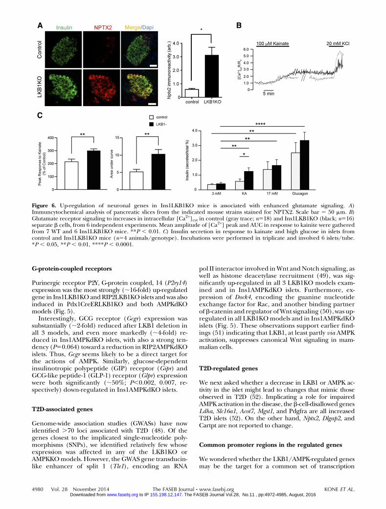
binds to N-methyl-D-aspartate (NMDA) receptors andShaker-type K+ channels. Interestingly,Kcna2, a Shakerchannel, was also strongly up-regulated by LKB1 orAMPK deletion (Fig. 5). Immunocytochemistry con-firmed the induction of NPTX2 at the protein level inIns1LKB1KO b cells (Fig. 6A).
Consistent with increased glutaminergic signalingin Ins1LKB1KO b cells, kainate, which binds to andactivates ionotropic glutamate receptors (44), caused amore pronounced rise in cytosolic-free Ca2+ ([Ca2+]cyt)in islets from Ins1LKB1KO than control mice (Fig. 6B).Furthermore, at low (3 mM) glucose, kainite stimulatedinsulin secretion from Ins1LKB1KO but not controlislets (Fig. 6C).
Disallowed genes
We (9, 10) and others (46) have previously describeda list of .60 housekeeping genes that are expressedin the majority of mammalian tissues at quite high
Figure 4. GSEA for neuronal genes in Ins1LKB1KO (A) or Ins1AMPKdKO (B) mice. A) GSEA was performed against MSigDB(V4) biological process gene ontolology categories (C5, BP) revealing significant enrichment for neuronal genes forIns1LKB1KO [MSigDB category: NERVOUS_SYSTEM_DEVELOPMENT; Enrichment (ES) score50.41 FDR50.06]. In Ins1-AMPKdKO, this category was not significantly enriched (ES score50.02; FDR51; right panel). Data are expressed as absolute foldchange irrespective of the direction of change. Blue line plot shows the cumulative ES score for this functional gene category.Vertical bars indicate the positions of the neuronal genes in the ranked differential expression lists. Line plot at the bottom showsthe log2-fold changes of the genes in the ranked lists. Because the initial gene lists were ranked by absolute fold change, both up-and down-regulated genes are found at the top of the lists.
TABLE 1. Ins1LKB1KO and Ins1AMPKdKO: synapse cluster
Category Term Count % P List total FDR
GOTERM_CC_FAT GO:0045202;synapse 18 4.37 9.67E-04 284 1.24GOTERM_CC_FAT GO:0044456;synapse part 14 3.40 0.00107 284 1.38GOTERM_CC_FAT GO:0008021;synaptic vesicle 7 1.70 0.00430 284 5.45GOTERM_CC_FAT GO:0030135;coated vesicle 8 1.94 0.0225 284 25.67GOTERM_CC_FAT GO:0030136;clathrin-coated vesicle 7 1.70 0.0306 284 33.25
4978 Vol. 28 November 2014 KONE ET AL.The FASEB Journal x www.fasebj.org Vol.28, No.11 , pp:4972-4985, August, 2016The FASEB Journal. 155.198.12.147 to IP www.fasebj.orgDownloaded from

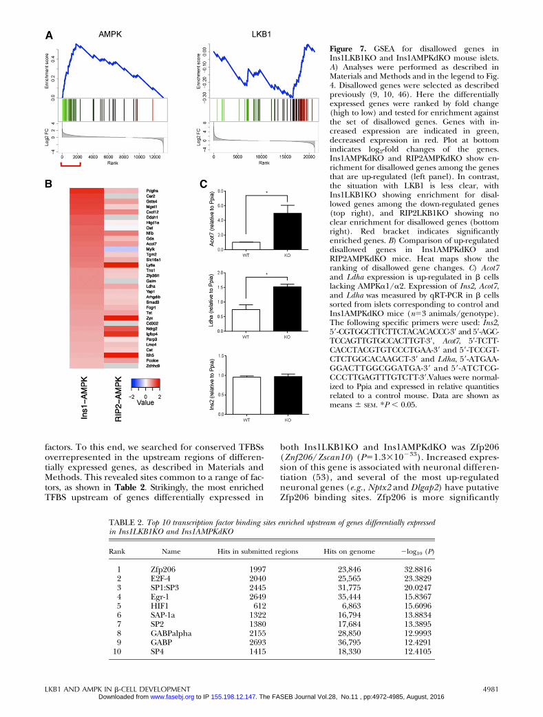
levels, but at much lower levels in islets and b cells; ofthese, 11 genes were reported in both of the previousstudies (10). We noticed that several of the genes inboth lists were up-regulated in RIP2AMPKdKO andIns1AMPKdKO islets. GSEA analysis (Fig. 7A) revealedthat 16 members of this family were specifically up-regulated in Ins1AMPKdKO islet models. By contrast,Ins1CreLKB1KO islets showed a negative enrichmentfor this class (Fig. 7A).
The most up-regulated genes in this group are com-pared in the Ins1AMPKdKOandRIP2AMPKdKOmodelsin Fig. 7B. Suggesting that these increases are unlikely toreflect an enrichment of AMPKdKO islets with nonisletmaterial, levels of the pancreatic acinar cell marker am-ylase were not different between Ins1- or RIP2AMPKdKOvs. control islets. Moreover, examined in FACS-sortedmouse islet cells (47), with the exception of Oat andIgfbp4, similar levels of expression of each gene weredetected in WT b and a cells (Supplemental Table 14).Finally, and providing direct confirmation of the above
conclusion, expression of both Ldha and Acot7 were sig-nificantly increased in FACS-purified b cells fromIns1AMPKdKO vs. control mice (Fig. 7C).
Hepatic genes
Striking changes were observed across all 3 LKB1KO plusIns1AMPKdKO models in the expression of 3 geneslargely restricted to the liver, namely, albumin (Alb),iodotyrosine deiodinase (Iyd), and elongation of very longchain fatty acids 2 (Elovl2). Furthermore, the liver/kidney-enriched gluconeogenic gene, fructose 1,6-bisphoshatase-1 (Fbp1), was significantly up-regulated in 2 of the 3LKB1KO models (RIP2- and Pdx1CreER), and tended(P50.12) to be activated in Ins1LKB1KO mouse islets.Fructose 1,6-biosphosphatase-2 (Fbp2), usually restrictedto stomach, small intestine, and skeletal muscle, was sig-nificantly up-regulated in each of the LKB1models and inRIP2AMPKdKO mice (Fig. 5), suggesting increased po-tential for gluconeogenesis.
Figure 5. Differentially regulated genes afterLKB1 or AMPK deletion in b cells. Heat mapshowing genes significantly up-regulated (redhues) or down-regulated (blue hues) by .1.4-fold in islets after deletion of the indicatedkinases. Gray indicates genes absent from 1 ormore samples (see Supplemental Tables S1 andS2). All genes shown were up-regulated inIns1CreLKB1KO vs. control islets and $1 otherLKB1KO model. Color bars at left indicateneuronal genes (brown) and liver-enrichedgenes (purple). Arrows indicate messages selec-tively enriched in LKB1 vs. AMPKdKO models.
LKB1 AND AMPK IN b-CELL DEVELOPMENT 4979 Vol.28, No.11 , pp:4972-4985, August, 2016The FASEB Journal. 155.198.12.147 to IP www.fasebj.orgDownloaded from
G-protein-coupled receptors
Purinergic receptor P2Y, G-protein coupled, 14 (P2ry14)expression was the most strongly (;16-fold) up-regulatedgene in Ins1LKB1KOandRIP2LKB1KO islets andwas alsoinduced in Pdx1CreERLKB1KO and both AMPKdKOmodels (Fig. 5).
Interestingly, GCG receptor (Gcgr) expression wassubstantially (;2-fold) reduced after LKB1 deletion inall 3 models, and even more markedly (;4-fold) re-duced in Ins1AMPKdKO islets, with also a strong ten-dency (P50.064) toward a reduction in RIP2AMPKdKOislets. Thus, Gcgr seems likely to be a direct target forthe actions of AMPK. Similarly, glucose-dependentinsulinotropic polypeptide (GIP) receptor (Gipr) andGCG-like peptide-1 (GLP-1) receptor (Glpr) expressionwere both significantly (;50%; P,0.002, 0.007, re-spectively) down-regulated in Ins1AMPKdKO islets.
T2D-associated genes
Genome-wide association studies (GWASs) have nowidentified .70 loci associated with T2D (48). Of thegenes closest to the implicated single-nucleotide poly-morphisms (SNPs), we identified relatively few whoseexpression was affected in any of the LKB1KO orAMPKKOmodels. However, the GWAS gene transducin-like enhancer of split 1 (Tle1), encoding an RNA
pol II interactor involved inWnt andNotch signaling, aswell as histone deacetylase recruitment (49), was sig-nificantly up-regulated in all 3 LKB1KO models exam-ined and in Ins1AMPKdKO islets. Furthermore, ex-pression of Dock4, encoding the guanine nucleotideexchange factor for Rac, and another binding partnerofb-catenin and regulator ofWnt signaling (50), was up-regulated in all LKB1KOmodels and in Ins1AMPKdKOislets (Fig. 5). These observations support earlier find-ings (51) indicating that LKB1, at least partly via AMPKactivation, suppresses canonical Wnt signaling in mam-malian cells.
T2D-regulated genes
We next asked whether a decrease in LKB1 or AMPK ac-tivity in the islet might lead to changes that mimic thoseobserved in T2D (52). Implicating a role for impairedAMPK activation in the disease, theb-cell-disallowed genesLdha, Slc16a1, Acot7, Mgst1, and Pdgfra are all increasedT2D islets (52). On the other hand, Nptx2, Dlgap2, andCartpt are not reported to change.
Common promoter regions in the regulated genes
We wondered whether the LKB1/AMPK-regulated genesmay be the target for a common set of transcription
Figure 6. Up-regulation of neuronal genes in Ins1LKB1KO mice is associated with enhanced glutamate signaling. A)Immuncytochemical analysis of pancreatic slices from the indicated mouse strains stained for NPTX2. Scale bar 5 50 mm. B)Glutamate receptor signaling to increases in intracellular [Ca2+]cyt in control (gray trace; n518) and Ins1LKB1KO (black; n516)separate b cells, from 6 independent experiments. Mean amplitude of [Ca2+] peak and AUC in response to kainite were gatheredfrom 7 WT and 6 Ins1LKB1KO mice. **P , 0.01. C) Insulin secretion in response to kainate and high glucose in islets fromcontrol and Ins1LKB1KO mice (n54 animals/genotype). Incubations were performed in triplicate and involved 6 islets/tube.*P , 0.05, **P , 0.01, ****P , 0.0001.
4980 Vol. 28 November 2014 KONE ET AL.The FASEB Journal x www.fasebj.org Vol.28, No.11 , pp:4972-4985, August, 2016The FASEB Journal. 155.198.12.147 to IP www.fasebj.orgDownloaded from
factors. To this end, we searched for conserved TFBSsoverrepresented in the upstream regions of differen-tially expressed genes, as described in Materials andMethods. This revealed sites common to a range of fac-tors, as shown in Table 2. Strikingly, the most enrichedTFBS upstream of genes differentially expressed in
both Ins1LKB1KO and Ins1AMPKdKO was Zfp206(Znf206/Zscan10) (P51.3310233). Increased expres-sion of this gene is associated with neuronal differen-tiation (53), and several of the most up-regulatedneuronal genes (e.g., Nptx2 and Dlgap2) have putativeZfp206 binding sites. Zfp206 is more significantly
Figure 7. GSEA for disallowed genes inIns1LKB1KO and Ins1AMPKdKO mouse islets.A) Analyses were performed as described inMaterials and Methods and in the legend to Fig.4. Disallowed genes were selected as describedpreviously (9, 10, 46). Here the differentiallyexpressed genes were ranked by fold change(high to low) and tested for enrichment againstthe set of disallowed genes. Genes with in-creased expression are indicated in green,decreased expression in red. Plot at bottomindicates log2-fold changes of the genes.Ins1AMPKdKO and RIP2AMPKdKO show en-richment for disallowed genes among the genesthat are up-regulated (left panel). In contrast,the situation with LKB1 is less clear, withIns1LKB1KO showing enrichment for disal-lowed genes among the down-regulated genes(top right), and RIP2LKB1KO showing noclear enrichment for disallowed genes (bottomright). Red bracket indicates significantlyenriched genes. B) Comparison of up-regulateddisallowed genes in Ins1AMPKdKO andRIP2AMPKdKO mice. Heat maps show theranking of disallowed gene changes. C) Acot7and Ldha expression is up-regulated in b cellslacking AMPKa1/a2. Expression of Ins2, Acot7,and Ldha was measured by qRT-PCR in b cellssorted from islets corresponding to control andIns1AMPKdKO mice (n53 animals/genotype).The following specific primers were used: Ins2,59-CGTGGCTTCTTCTACACACCC-39 and 59-AGC-TCCAGTTGTGCCACTTGT-39, Acot7, 59-TCTT-CACCTACGTGTCCCTGAA-39 and 59-TCCGT-CTCTGGCACAAGCT-39 and Ldha, 59-ATGAA-GGACTTGGCGGATGA-39 and 59-ATCTCG-CCCTTGAGTTTGTCTT-39.Values were normal-ized to Ppia and expressed in relative quantitiesrelated to a control mouse. Data are shown asmeans 6 SEM. *P , 0.05.
TABLE 2. Top 10 transcription factor binding sites enriched upstream of genes differentially expressedin Ins1LKB1KO and Ins1AMPKdKO
Rank Name Hits in submitted regions Hits on genome 2log10 (P)
1 Zfp206 1997 23,846 32.88162 E2F-4 2040 25,565 23.38293 SP1:SP3 2445 31,775 20.02474 Egr-1 2649 35,444 15.83675 HIF1 612 6,863 15.60966 SAP-1a 1322 16,794 13.88347 SP2 1380 17,684 13.38958 GABPalpha 2155 28,850 12.99939 GABP 2693 36,795 12.4291
10 SP4 1415 18,330 12.4105
LKB1 AND AMPK IN b-CELL DEVELOPMENT 4981 Vol.28, No.11 , pp:4972-4985, August, 2016The FASEB Journal. 155.198.12.147 to IP www.fasebj.orgDownloaded from

enriched in genes only differentially expressed inthe Ins1LKB1KO model (P59.1310296), consistentwith the more significant degree of up-regulationof neuronal genes observed in this model vs.Ins1AMPKdKO (Fig. 4 and Supplemental Figs. S3and S4).
Hypoxia-inducible factor 1 (HIF1) binding siteswere also enriched in genes differentially expressed inboth models (P52.5310216). More detailed exami-nation, taking the direction of differential expres-sion into account, revealed that HIF1 sites wereenriched in genes significantly up-regulated in justthe Ins1AMPKdKOmodel (P55.7310213) and also ingenes down-regulated in just the Ins1LKB1KO model(P52.2310226). LKB1 and AMPK therefore bothappear to have nonoverlapping roles in HIF1 regula-tion. Hypoxia is known to up-regulate a number ofb-cell disallowed genes (e.g., Ldha and Slc16a1; ref.54), and the enrichment of HIF1 binding sites in genesselectively up-regulated in Ins1AMPKdKO supportsa role for this transcription factor in disallowed genesuppression.
DISCUSSION
The present study provides the first detailed tran-scriptomic analysis exploring the roles of LKB1 andAMPK in the endocrine pancreas. First, we confirmseveral previous findings (20–23) based on the de-letion of these kinases in multiple tissues, showingthat when restricted to the b cell, loss of LKB1 exertsmarkedly different effects on insulin secretion andglucose metabolism compared to the loss of AMPK(Figs. 1–3). We now demonstrate that these func-tional differences are mirrored by complex alter-ations at the level of gene expression (Figs. 4 and 5,Supplemental Figs. S1–S3, and Supplemental TablesS1–S13).
Differences in gene expression between Ins1LKB1KOand Ins1AMPKdKO islets are likely to reflect signal-ing by members of the AMPKRK family (16) in theformer case. However, the identity of the kinases in-volved is unclear. Thus, deletion of synapses ofamphids defective kinase-A (SAD-A) in the pancreascauses defective glucose signaling and the appearanceof small islets (55), suggesting that this enzyme ratheropposes the actions of LKB1, mediating insteadthe effects of mammalian target of rapamycin-1(mTORC1). Likewise, deletion of salt-inducible kinase2 (SIK2; ref. 56) impairs glucose tolerance and insulinsecretion.
An interesting observation, replicating findingsin RIP2AMPKdKO islets (23), is that while glucose-stimulated insulin secretion was strongly impairedin vivo in Ins1AMPKdKO mice (Fig. 1C), it wasenhanced from isolated islets (Supplemental Fig.2B). There may be several explanations for thisdifference. First, it may reflect the loss in vitro ofan action of circulating factors that normally regu-late insulin secretion in vivo. Obvious candi-dates are the incretins GIP and GLP-1, as well as
GCG, given the lowered expression of the corre-sponding receptors in Ins1AMPKdKO mice. In ad-dition, we noted a significant (P,0.01) ;2-foldup-regulation in the expression of both the somato-statin receptor gene Sstr1, and the adrenoreceptorAdrb1 in Ins1AMPKdKO islets, expected to potenti-ate the inhibitory actions of the cognate hormoneson insulin secretion; neither gene was affected inIns1LKB1KO islets.
Identification of genes differentially regulated byLKB1 and AMPK
It seems reasonable to speculate that genes affected bythe loss of LKB1, but not AMPK, may contribute toenhanced insulin secretion in the former. Of thegenes most strongly up-regulated in all 3 LKB1KOmodels, 4 were unaffected in both AMPKdKOmodels:Baiap2l2, Cabp1, Rspo4, and Bcl6b (Fig. 5). Bcl6b enc-odes a transcription factor and downstream target ofFGF2 capable of promoting germ cell tumors (57).Likewise, RSPO4 is a member of the R-spondin familyof secreted agonists of canonical Wnt/b-catenin sig-naling that binds to Lgr receptors to enhance cellgrowth (58). BAIAP2L2 is a member of the I-BAR do-main family of proteins involved in the control of actindynamics and signaling at glutaminergic synapses (59)and filopodia extension (60), and may thus alsobe involved in Wnt or Hedgehog signaling. Finally,CaBP1 is a neuronal calcium binding protein thatregulates several Ca2+ channels, including transientreceptor potential 5 (Trp5; ref. 61).
Regulation of neuronal genes in the b cell by LKB1and AMPK
A striking finding of the present study is that both LKB1and AMPK control the expression of a large number ofneuronal genes in the b cell. LKB1 deletion had themost dramatic effect on this group of genes (Fig. 4A),and this effect was also observed after short-term (1 wk)loss of LKB1 in the Pdx1CreER-deleted model. Severalof the most strongly affected genes, notably Nptx2 andDlgap2, were also subject to control by AMPK (Fig. 5).Thus, both AMPK and AMPKRKs may mediate theeffects of LKB1 on this gene group. Analysis of thepromoter regions of genes regulated by either kinaserevealed several potential downstream regulators(Table 2), of which Zfp206, a neuron-enriched tran-scription factor, is an attractive candidate for mediat-ing these effects.
We also considered the possibility that some ofthe changes may be the result of altered expression ofrepressor element 1 silencing transcription factor(REST), which represses neuronal genes in non-neuronal cells and is also relatively weakly expressed inunmodified adult b cells (62). However, while RESTexpression was reduced in islets from Ins1LKB1KOislets, its expression was slightly increased, or barelyaffected, in RIP2LKB1KO and Pdx1CreERLKB1KOislets, respectively, while REST expression was slightly
4982 Vol. 28 November 2014 KONE ET AL.The FASEB Journal x www.fasebj.org Vol.28, No.11 , pp:4972-4985, August, 2016The FASEB Journal. 155.198.12.147 to IP www.fasebj.orgDownloaded from

increased in both AMPKdKO model islets (Fig. 5).Nonetheless, differences in REST expression betweenIns1LKB1 and the AMPKdKOs may contribute to thedifferential enrichment for neuronal genes (Fig. 4).Thus, several well-defined targets for REST, includingSnap25 (P50.05) and the synaptotagmin family mem-ber Syt4 (P,0.01), were up-regulated in Ins1LKB1KOislets.
Regulation in the b cell of genes involved in glutamatesignaling and addictive behavior
Gria3, encoding the GluR3 glutamate receptor, wasup-regulated in all 5 KO models, while Gria1 (GluR1)expression was also increased in Ins1LKB1KO islets,as were Dlgap2 and Nptx2. Nptx2 encodes neuronalpentraxin 2 (also known as Narp in the rat), whichbinds to the extracellular domain of a-amino-3-hydroxy-5-methyl-4-isoxazolepropionic (AMPA)-activatedsubclass of glutamate receptors, causing their clustering.Thus, pentraxins are secreted from presynaptic neurons(63), and recruit GluR4 AMPA receptors to synapses.Pentraxins are also implicated in neuronal plasticity andthe behavioral responses to drug abuse (64).
We show that inductionofNptx2 (alongside that of othergenes involved in glutamate signaling; see above) was ac-companied functionally by enhancedkainite-inducedCa2+
signals, and insulin secretion (Fig. 6B,C).This suggests thatLKB1, and possibly AMPK, are necessary to suppresspathways that may lead to the dysregulation of secretionin b cells.
Regulation of disallowed genes by AMPK
Ourfindinghere that loss ofAMPK(butnotLKB1) frombcells up-regulates the b-cell-disallowed gene family (10)raises the interesting possibility that this enzyme is a keydeterminant in maintaining normal glucose respon-siveness. Thus, elevated AMPK activity in the b cell be-tween meals is likely to be required to maintain low levelsof these enzymes. Indeed, AMPK activators such as met-formin, used clinically as oral antihyperglycemics, may actin part in this way (18).
We have considered the possibility that an increasein postprandial glycemia in the AMPKdKO mouse vs.controls may contribute to the up-regulation of thesegenes (6, 65). However, and against this view, neitherfasting (Fig. 1D) nor fed (10.960.26 and 10.860.2 mM, for WT and Ins1AMPKdKO mice, respectively;n54, P.0.05) glycemia differed between genotypes;and mRNA changes in Ins1AMPKdKO islets werepreserved after culture at 11 mM glucose for 24 hpostisolation.
CONCLUSIONS
LKB1 and AMPK are shown here to be powerful reg-ulators of b-cell differentiation, acting partly in con-cert and partly independently of one another.Although cell fate switching is implied by the effects of
loss-of-function mutations in LKB1 in Peutz-Jegherssyndrome (19), to our knowledge this report providesthe first example of developmental fate being affectedby the fuel-sensitive protein kinase AMPK. Interest-ingly, these changes occurred without apparent re-version to a more progenitor-like state, as indicated byunchanged expression of Ngn3, Oct4, or Nanog, in con-trast to recently described models (8).
Further dissection of the mechanisms acting down-stream of LKB1 in b cells may thus provide the basis ofstrategies to maintain cellular differentiation in someforms of diabetes.
G.A.R. thanks the Medical Research Council (UK) forprogramme grant MR/J0003042/1, the Biotechnology andBiological Sciences Research Council (UK) for project grantBB/J015873/1, the Royal Society for a Wolfson ResearchMerit Award, and the Wellcome Trust for Senior InvestigatorAward WT098424AIA. G.A.R. and P.F. received an ImperialCollege/Wellcome Trust Institutional Strategic SupportFunds (ISSF) grant. T.J.P. was supported by a DiabetesResearch and Wellness (DRWF) nonclinical fellowship, andM.K. by an Imperial College Ph.D. studentship. The workleading to this publication has received support from theInnovative Medicines Initiative (IMIDIA) Joint Undertakingunder grant agreement no. 155005, resources of which arecomposed of a financial contribution from the EuropeanUnion’s Seventh Framework Programme (FP7/2007–2013)and European Federation of Pharmaceutical Industries andAssociations (EFPIA) member companies’ in kind contribu-tion (G.A.R., B.T.). The authors thank Dr. Gabriela da SilvaXavier for help with pancreatic morphology, Jane Srivastavafor assistance with FACS analysis, and Dr. Isabelle Leclerc(Imperial College London) for useful discussions.
REFERENCES
1. Scully, T. (2012) Diabetes in numbers. Nature 485, S2–S32. Rutter, G. A., and Parton, L. E. (2008) The beta-cell in type 2
diabetes and in obesity. Front. Horm. Res. 36, 118–1343. Rutter, G. A. (2001) Nutrient-secretion coupling in the pancre-
atic islet b-cell: recent advances. Mol. Aspects Med. 22, 247–2844. Butler, A. E., Janson, J., Bonner-Weir, S., Ritzel, R., Rizza,
R. A., and Butler, P. C. (2003) Beta-cell deficit and increasedbeta-cell apoptosis in humans with type 2 diabetes. Diabetes52, 102–110
5. Rahier, J., Guiot, Y., Goebbels, R. M., Sempoux, C., and Henquin,J. C. (2008) Pancreatic beta-cell mass in European subjects withtype 2 diabetes. Diabetes Obes. Metab. 10, 32–42
6. Jonas, J. C., Sharma, A., Hasenkamp, W., Ilkova, H., Patane,G., Laybutt, R., Bonner-Weir, S., and Weir, G. C. (1999)Chronic hyperglycemia triggers loss of pancreatic beta celldifferentiation in an animal model of diabetes. J. Biol. Chem.274, 14112–14121
7. Del Guerra, S., Lupi, R., Marselli, L., Masini, M., Bugliani, M.,Sbrana, S., Torri, S., Pollera, M., Boggi, U., Mosca, F., Del Prato,S., and Marchetti, P. (2005) Functional and molecular defects ofpancreatic islets in human type 2 diabetes. Diabetes 54, 727–735
8. Talchai, C., Xuan, S., Lin, H. V., Sussel, L., and Accili, D. (2012)Pancreatic beta cell dedifferentiation as a mechanism of diabeticbeta cell failure. Cell 150, 1223–1234
9. Pullen, T. J., Khan, A. M., Barton, G., Butcher, S. A., Sun, G., andRutter, G. A. (2010) Identification of genes selectively disallowedin the pancreatic islet. Islets 2, 89–95
10. Pullen, T. J., and Rutter, G. A. (2013) When less is more: theforbidden fruits of gene repression in the adult beta-cell. DiabetesObes. Metab. 15, 503–512
11. Pullen, T. J., Sylow, L., Sun, G., Halestrap, A. P., Richter, E. A.,and Rutter, G. A. (2012) Over-expression of Monocarboxylate
LKB1 AND AMPK IN b-CELL DEVELOPMENT 4983 Vol.28, No.11 , pp:4972-4985, August, 2016The FASEB Journal. 155.198.12.147 to IP www.fasebj.orgDownloaded from
Transporter-1 (Slc16a1) in the pancreatic a-cell leads to relativehyperinsulinism during exercise. Diabetes 61, 1725
12. Partanen, J. I., Tervonen, T. A., and Klefstrom, J. (2013)Breaking the epithelial polarity barrier in cancer: the strangecase of LKB1/PAR-4. Philos. Trans. R. Soc. Lond. B Biol. Sci. 368,20130111
13. Momcilovic, M., Hong, S. P., and Carlson, M. (2006) MammalianTAK1 activates Snf1 protein kinase in yeast and phosphorylatesAMP-activated protein kinase in vitro. J. Biol. Chem. 281, 25336–25343
14. Hawley, S. A., Boudeau, J., Reid, J. L., Mustard, K. J., Udd, L.,Makela, T. P., Alessi, D. R., and Hardie, D. G. (2003) Complexesbetween the LKB1 tumor suppressor, STRADalpha/beta andMO25alpha/beta are upstream kinases in the AMP-activatedprotein kinase cascade. J. Biol. 2, 28
15. Woods, A., Johnstone, S. R., Dickerson, K., Leiper, F. C., Fryer,L. G., Neumann, D., Schlattner, U., Wallimann, T., Carlson, M.,and Carling, D. (2003) LKB1 is the upstream kinase in the AMP-activated protein kinase cascade. Curr. Biol. 13, 2004–2008
16. Lizcano, J. M., Goransson, O., Toth, R., Deak, M., Morrice, N. A.,Boudeau, J., Hawley, S. A., Udd, L., Makela, T. P., Hardie, D. G.,and Alessi, D. R. (2004) LKB1 is a master kinase that activates 13kinases of the AMPK subfamily, including MARK/PAR-1. EMBOJ. 23, 833–843
17. Rutter, G. A., daSilvaXavier, G., and Leclerc, I. (2003) Roles of59-AMP-activated protein kinase (AMPK) in mammalian glucosehomoeostasis. Biochem. J. 375, 1–16
18. Rutter, G. A., and Leclerc, I. (2009) The AMP-regulated kinasefamily: enigmatic targets for diabetes therapy. Mol. Cell. Endo-crinol. 297, 41–49
19. Jenne, D. E., Reimann, H., Nezu, J., Friedel, W., Loff, S., Jeschke,R., Muller, O., Back, W., and Zimmer, M. (1998) Peutz-Jegherssyndrome is caused by mutations in a novel serine threoninekinase. Nat. Genet. 18, 38–43
20. Sun, G., Tarasov, A. I., McGinty, J. A., French, P. M., McDonald,A., Leclerc, I., and Rutter, G. A. (2010) LKB1 deletion with theRIP. Cre-transgene modifies pancreatic b-cell morphology andenhances insulin secretion in vivo. Am. J. Physiol. Endocrinol.Metab. 298, E1261–E1273
21. Granot, Z., Swisa, A., Magenheim, J., Stolovich-Rain, M.,Fujimoto, W., Manduchi, E., Miki, T., Lennerz, J. K., Stoeckert,C. J.Jr., Meyuhas, O., Seino, S., Permutt, M. A., Piwnica-Worms,H., Bardeesy, N., and Dor, Y. (2009) LKB1 regulates pancreaticbeta cell size, polarity, and function. Cell Metab. 10, 296–308
22. Fu, A., Ng, A. C., Depatie, C., Wijesekara, N., He, Y., Wang, G. S.,Bardeesy, N., Scott, F. W., Touyz, R. M., Wheeler, M. B., andScreaton, R. A. (2009) Loss of Lkb1 in adult beta cells increasesbeta cell mass and enhances glucose tolerance in mice. CellMetab. 10, 285–295
23. Sun, G., Tarasov, A. I., McGinty, J., McDonald, A., DaSilva Xavier,G., Gorman, T., Marley, A., French, P. M., Parker, H., Gribble, F.,Reimann, F., Prendiville, O., Carzaniga, R., Viollet, B., Leclerc, I.,and Rutter, G. A. (2010) Ablation of AMP-activated protein kinasealpha1 and alpha2 from pancreatic beta-cells and RIP. Cre neu-rons suppresses insulin release in vivo. Diabetologia 53, 924–936
24. Beall, C., Piipari, K., Al-Qassab, H., Smith, M. A., Parker, N.,Carling, D., Viollet, B., Withers, D. J., and Ashford, M. L. (2010)Loss of AMP-activated protein kinase alpha2 subunit in mousebeta-cells impairs glucose-stimulated insulin secretion andinhibits their sensitivity to hypoglycaemia. Biochem. J. 429, 323–333
25. Wicksteed, B., Brissova, M., Yan, W., Opland, D. M., Plank, J. L.,Reinert, R. B., Dickson, L. M., Tamarina, N. A., Philipson, L. H.,Shostak, A., Bernal-Mizrachi, E., Elghazi, L., Roe, M. W., Labosky,P. A., Myers, M. M.Jr., Gannon, M., Powers, A. C., and Dempsey,P. J. (2010) Conditional gene targeting in mouse pancreatic{beta}-cells: analysis of ectopic Cre transgene expression in thebrain. Diabetes 59, 3090–3098
26. Tamarina, N. A., Roe, M. W., and Philipson, L. (2014)Characterization of mice expressing Ins1 gene promoter drivenCreERT recombinase for conditional gene deletion in pancreaticbeta-cells. [E-pub ahead of print] Islets 6, doi: 10.4161/isl.27685
27. Bardeesy, N., Sinha, M., Hezel, A. F., Signoretti, S., Hathaway,N. A., Sharpless, N. E., Loda, M., Carrasco, D. R., and DePinho,R. A. (2002) Loss of the Lkb1 tumour suppressor provokesintestinal polyposis but resistance to transformation. Nature 419,162–167
28. Ravier, M. A., and Rutter, G. A. (2010) Isolation and culture ofmouse pancreatic islets for ex vivo imaging studies with trappableor recombinant fluorescent probes. Methods Mol. Biol. 633,171–184
29. Stange, G., Van De Casteele, M., and Heimberg, H. (2003)Purification of rat pancreatic B-cells by fluorescence-activatedcell sorting. Methods Mol. Med. 83, 15–22
30. Kim, D., Pertea, G., Trapnell, C., Pimentel, H., Kelley, R., andSalzberg, S. L. (2013) TopHat2: accurate alignment oftranscriptomes in the presence of insertions, deletions andgene fusions. Genome Biol. 14, R36
31. Vencio, R. Z., Brentani, H., Patrao, D. F., and Pereira, C. A.(2004) Bayesian model accounting for within-class biologicalvariability in Serial Analysis of Gene Expression (SAGE). BMCBioinformatics 5, 119
32. Mortazavi, A., Williams, B. A., McCue, K., Schaeffer, L., andWold, B. (2008) Mapping and quantifying mammaliantranscriptomes by RNA-seq. Nat. Methods. 5, 621–628
33. Zambon, A. C., Zhang, L., Minovitsky, S., Kanter, J. R., Prabhakar,S., Salomonis, N., Vranizan, K., Dubchak, I., Conklin, B. R., andInsel, P. A. (2005) Gene expression patterns define keytranscriptional events in cell-cycle regulation by cAMP and pro-tein kinase A. Proc. Natl. Acad. Sci. U. S. A. 102, 8561–8566
34. Subramanian, A., Tamayo, P., Mootha, V. K., Mukherjee, S.,Ebert, B. L., Gillette, M. A., Paulovich, A., Pomeroy, S. L., Golub,T. R., Lander, E. S., and Mesirov, J. P. (2005) Gene setenrichment analysis: a knowledge-based approach for interpret-ing genome-wide expression profiles. Proc. Natl. Acad. Sci. U. S. A.102, 15545–15550
35. Gentleman, R. C., Carey, V. J., Bates, D. M., Bolstad, B., Dettling,M., Dudoit, S., Ellis, B., Gautier, L., Ge, Y., Gentry, J., Hornik, K.,Hothorn, T., Huber, W., Iacus, S., Irizarry, R., Leisch, F., Li, C.,Maechler, M., Rossini, A. J., Sawitzki, G., Smith, C., Smyth, G.,Tierney, L., Yang, J. Y., and Zhang, J. (2004) Bioconductor: opensoftware development for computational biology and bioin-formatics. Genome Biol. 5, R80
36. Ravier, M. A., and Rutter, G. A. (2005) Glucose or insulin, butnot zinc ions, inhibit glucagon secretion from mouse pancreatica-cells. Diabetes 54, 1789–1797
37. Tarasov, A. I., Ravier, M. A., Semplici, F., Bellomo, E. A.,Pullen, T. J., Gilon, P., Sekler, I., Rizzuto, R., and Rutter, G. A.(2012) The mitochondrial Ca2+ uniporter MCU is essential forglucose-induced ATP increases in pancreatic b-cells. PLoS ONE7, e39722
38. Edelstein, A., Amodaj, N., Hoover, K., Vale, R., and Stuurman, N.(2010) Computer control of microscopes using mManager. Curr.Protoc. Mol. Biol. 92, 14.20.1–14.20.17
39. Sun, G., Reynolds, R., Leclerc, I., and Rutter, G. A. (2010) RIP2-mediated LKB1 deletion causes axon degeneration in the spinalcord and hind-limb paralysis. Dis. Models Mech. 4, 193–202
40. Rother, E., Belgardt, B. F., Tsaousidou, E., Hampel, B., Waisman,A., Myers, M. G.Jr., and Bruning, J. C. (2012) Acute selectiveablation of rat insulin promoter-expressing (RIPHER) neuronsdefines their orexigenic nature. Proc. Natl. Acad. Sci. U.S.A. 109,18132–18137
41. Courtney, M., Gjernes, E., Druelle, N., Ravaud, C., Vieira, A.,Ben-Othman, N., Pfeifer, A., Avolio, F., Leuckx, G., Lacas-Gervais,S., Burel-Vandenbos, F., Ambrosetti, D., Hecksher-Sorensen, J.,Ravassard, P., Heimberg, H., Mansouri, A., and Collombat, P.(2013) The inactivation of Arx in pancreatic alpha-cells triggerstheir neogenesis and conversion into functional beta-like cells.PLoS Genet. 9, e1003934
42. Bramswig, N. C., Everett, L. J., Schug, J., Dorrell, C., Liu, C., Luo,Y., Streeter, P. R., Naji, A., Grompe, M., and Kaestner, K. H.(2013) Epigenomic plasticity enables human pancreatic alpha tobeta cell reprogramming. J. Clin. Invest. 123, 1275–1284
43. Thorens, B., Sarkar, H. K., Kaback, H. R., and Lodish, H. F.(1988) Cloning and functional expression in bacteria of a novelglucose transporter present in liver intestine kidney and B-pancreatic islet cells. Cell 55, 281–290
44. Palmer, C. L., Cotton, L., and Henley, J. M. (2005) Themolecular pharmacology and cell biology of alpha-amino-3-hydroxy-5-methyl-4-isoxazolepropionic acid receptors. Pharmacol.Rev. 57, 253–277
45. Takeuchi, M., Hata, Y., Hirao, K., Toyoda, A., Irie, M., andTakai, Y. (1997) SAPAPs. A family of PSD-95/SAP90-associated
4984 Vol. 28 November 2014 KONE ET AL.The FASEB Journal x www.fasebj.org Vol.28, No.11 , pp:4972-4985, August, 2016The FASEB Journal. 155.198.12.147 to IP www.fasebj.orgDownloaded from
proteins localized at postsynaptic density. J. Biol. Chem. 272,11943–11951
46. Thorrez, L., Laudadio, I., Van, D. K., Quintens, R., Hendrickx,N., Granvik, M., Lemaire, K., Schraenen, A., Van, L. L., Lehnert,S., Guayo-Mazzucato, C., Cheng-Xue, R., Gilon, P., Van, M. I.,Bonner-Weir, S., Lemaigre, F., and Schuit, F. (2011) Tissue-specific disallowance of housekeeping genes: the other face ofcell differentiation. Genome Res. 21, 95–105
47. Reimann, F., Habib, A. M., Tolhurst, G., Parker, H. E., Rogers,G. J., and Gribble, F. M. (2008) Glucose-sensing in L-cells: a pri-mary cell study. Cell Metab. 8, 532–539
48. Da Silva Xavier, G., Bellomo, E. A., McGinty, J. A., French, P. M.,and Rutter, G. A. (2013) Animal models of GWAS-identified type2 diabetes genes. J. Diabetes Res. 2013, 906590
49. Chen, G., and Courey, A. J. (2000) Groucho/TLE family proteinsand transcriptional repression. Gene 249, 1–16
50. Upadhyay, G., Goessling, W., North, T. E., Xavier, R., Zon, L. I.,and Yajnik, V. (2008) Molecular association between beta-catenindegradation complex and Rac guanine exchange factor DOCK4 isessential for Wnt/beta-catenin signaling. Oncogene 27, 5845–5855
51. Jacob, L. S., Wu, X., Dodge, M. E., Fan, C. W., Kulak, O., Chen,B., Tang, W., Wang, B., Amatruda, J. F., and Lum, L. (2011)Genome-wide RNAi screen reveals disease-associated genes thatare common to Hedgehog and Wnt signaling. Sci. Signal. 4, ra4
52. Marselli, L., Thorne, J., Dahiya, S., Sgroi, D. C., Sharma, A.,Bonner-Weir, S., Marchetti, P., and Weir, G. C. (2010) Geneexpression profiles of beta-cell enriched tissue obtained by lasercapture microdissection from subjects with type 2 diabetes. PLoSONE 5, e11499
53. Kawashima, H., Sugito, K., Yoshizawa, S., Uekusa, S., Furuya, T.,Ikeda, T., Koshinaga, T., Shinojima, Y., Hasegawa, R., Mishra, R.,Igarashi, J., Kimura, M., Wang, X., Fujiwara, K., Gosh, S., andNagase, H. (2012) DNA hypomethylation at the ZNF206-exon 5CpG island associated with neuronal differentiation in mice anddevelopment of neuroblastoma in humans. Int. J. Oncol. 40, 31–39
54. De Saedeleer, C. J., Porporato, P. E., Copetti, T., Perez-Escuredo,J., Payen, V. L., Brisson, L., Feron, O., and Sonveaux, P. (2013)Glucose deprivation increases monocarboxylate transporter 1(MCT1) expression and MCT1-dependent tumor cell migration.[E-pub ahead of print] Oncogene doi: 10.1038/onc.2013.454
55. Nie, J., Liu, X., Lilley, B. N., Zhang, H., Pan, Y. A., Kimball, S. R.,Zhang, J., Zhang, W., Wang, L., Jefferson, L. S., Sanes, J. R., Han,X., and Shi, Y. (2013) SAD-A kinase controls islet beta-cell sizeand function as a mediator of mTORC1 signaling. Proc. Natl.Acad. Sci. U. S. A. 110, 13857–13862
56. Sakamaki, J. I., Fu, A., Reeks, C., Baird, S., Depatie, C., Al, A. M.,Bardeesy, N., Gingras, A. C., Yee, S. P., and Screaton, R. A. (2014)Role of the SIK2-p35-PJA2 complex in pancreatic beta-cellfunctional compensation. Nat. Cell Biol. 16, 234–244
57. Ishii, K., Kanatsu-Shinohara, M., Toyokuni, S., and Shinohara, T.(2012) FGF2 mediates mouse spermatogonial stem cell self-renewal via upregulation of Etv5 and Bcl6b through MAP2K1activation. Development 139, 1734–1743
58. de Lau, W. B., Snel, B., and Clevers, H. C. (2012) The R-spondinprotein family. Genome Biol. 13, 242–213
59. Fromer, M., Pocklington, A. J., Kavanagh, D. H., Williams, H. J.,Dwyer, S., Gormley, P., Georgieva, L., Rees, E., Palta, P., Ruderfer,D. M., Carrera, N., Humphreys, I., Johnson, J. S., Roussos, P.,Barker, D. D., Banks, E., Milanova, V., Grant, S. G., Hannon, E.,Rose, S. A., Chambert, K., Mahajan, M., Scolnick, E. M., Moran,J. L., Kirov, G., Palotie, A., McCarroll, S. A., Holmans, P., Sklar, P.,Owen, M. J., Purcell, S. M., and O’Donovan, M. C. (2014) De novomutations in schizophrenia implicate synaptic networks. Nature506, 179–184
60. Ahmed, S., Goh, W. I., and Bu, W. (2010) I-BAR domains, IRSp53and filopodium formation. Semin. Cell. Dev. Biol. 21, 350–356
61. Zhu, M. X. (2005) Multiple roles of calmodulin and other Ca(2+)-binding proteins in the functional regulation of TRP channels.Pflugers Arch. 451, 105–115
62. Martin, D., Allagnat, F., Chaffard, G., Caille, D., Fukuda, M.,Regazzi, R., Abderrahmani, A., Waeber, G., Meda, P., Maechler,P., and Haefliger, J. A. (2008) Functional significance ofrepressor element 1 silencing transcription factor (REST)target genes in pancreatic beta cells. Diabetologia 51, 1429–1439
63. Sia, G. M., Beique, J. C., Rumbaugh, G., Cho, R., Worley, P. F., andHuganir, R. L. (2007) Interaction of the N-terminal domain of theAMPA receptor GluR4 subunit with the neuronal pentraxin NP1mediates GluR4 synaptic recruitment. Neuron 55, 87–102
64. Crombag, H. S., Dickson, M., Dinenna, M., Johnson, A. W., Perin,M. S., Holland, P. C., Baraban, J. M., and Reti, I. M. (2009) Narpdeletion blocks extinction of morphine place preferenceconditioning. Neuropsychopharmacology 34, 857–866
65. Laybutt, D. R., Glandt, M., Xu, G., Ahn, Y. B., Trivedi, N.,Bonner-Weir, S., and Weir, G. C. (2003) Critical reduction inbeta-cell mass results in two distinct outcomes over time. Adap-tation with impaired glucose tolerance or decompensated di-abetes. J. Biol. Chem. 278, 2997–3005
Received for publication June 3, 2014.Accepted for publication July 14, 2014.
LKB1 AND AMPK IN b-CELL DEVELOPMENT 4985 Vol.28, No.11 , pp:4972-4985, August, 2016The FASEB Journal. 155.198.12.147 to IP www.fasebj.orgDownloaded from

10.1096/fj.14-257667Access the most recent version at doi:2014 28: 4972-4985 originally published online July 28, 2014FASEB J
Marina Kone, Timothy J. Pullen, Gao Sun, et al.
-cell identityβLKB1 and AMPK differentially regulate pancreatic
Material
Supplemental
http://www.fasebj.org/content/suppl/2014/07/17/fj.14-257667.DC1.html
References
http://www.fasebj.org/content/28/11/4972.full.html#ref-list-1
This article cites 65 articles, 20 of which can be accessed free at:
Subscriptions
http://www.faseb.org/The-FASEB-Journal/Librarian-s-Resources.aspx
is online at The FASEB JournalInformation about subscribing to
Permissions
http://www.fasebj.org/site/misc/copyright.xhtmlSubmit copyright permission requests at:
Email Alerts
http://www.fasebj.org/cgi/alertsReceive free email alerts when new an article cites this article - sign up at
© The Author(s)
Vol.28, No.11 , pp:4972-4985, August, 2016The FASEB Journal. 155.198.12.147 to IP www.fasebj.orgDownloaded from