lipopolysaccharide activates calcineurin in ventricular ... · ischemia-reperfusion. calcineurin...
TRANSCRIPT
Chc
tso
FSDDGJwSR
2
Journal of the American College of Cardiology Vol. 49, No. 4, 2007© 2007 by the American College of Cardiology Foundation ISSN 0735-1097/07/$32.00P
PRECLINICAL RESEARCH
Lipopolysaccharide ActivatesCalcineurin in Ventricular Myocytes
Jun Suzuki, MD, PHD,*† Evelyn Bayna, PHD,* Hai Ling Li, PHD,* Erminia Dalle Molle, BS,*†Wilbur Y. W. Lew, MD, FACC*†
San Diego, California
Objectives We investigated whether lipopolysaccharide (LPS), a proximate cause of inflammation, activates calcineurin incardiac myocytes and if calcineurin regulates apoptosis in this setting.
Background Calcineurin regulates myocardial growth and hypertrophy, but its role in inflammation is unknown. Calcineurinhas proapoptotic or antiapoptotic effects depending on the stimuli.
Methods Calcineurin activity was measured in left ventricular myocytes from adult Sprague Dawley rats. Cardiac apopto-sis was measured by terminal deoxy-nucleotidyl transferase-mediated dUTP nick end-labeling staining andcaspase-3 activity after in vitro and in vivo exposure to LPS.
Results Lipopolysaccharide increased calcineurin activity in myocytes over 1 to 24 h (t 1/2 � 4.8 h) with an EC50 of0.80 ng/ml LPS (p � 0.05, n � 4). The LPS (10 ng/ml) effects were mimicked by angiotensin II (Ang II) (100nmol/l); both increased calcineurin activity and induced apoptosis without additive effects (p � 0.05, n � 5 to 9).Lipopolysaccharide and/or Ang II effects were prevented by 1 h pre-treatment with an Ang II type 1 receptorblocker (losartan, 1 �mol/l), calcineurin inhibitor (cyclosporin A, 0.5 �mol/l), calcium chelator (1,2-Bis(2-amino-5-fluorophenoxy)ethane-N,N,N=,N=-tetraacetic acid tetrakis(acetoxymethyl) ester, 0.1 �mol/l), or by inhibitingsarcoplasmic reticulum (SR) calcium (Ca)-ATPase (thapsigargin, 1 �mol/l) or SR calcium release channel (ryano-dine, 1 �mol/l). Left ventricular apoptosis increased from 4 to 24 h after LPS (1 mg/kg intravenously) in vivo,but not in rats pre-treated with cyclosporin A (20 mg/kg/day subcutaneously) for 3 days (p � 0.05, n � 5).
Conclusions In cardiac myocytes, LPS activates calcineurin in association with apoptosis by Ang II and SR calcium-dependentmechanisms. This expands the paradigm for cardiac calcineurin to be activated by low levels of LPS in inflam-mation and chronic conditions (e.g., infections, smoking, and heart failure). (J Am Coll Cardiol 2007;49:491–9)© 2007 by the American College of Cardiology Foundation
ublished by Elsevier Inc. doi:10.1016/j.jacc.2006.10.043
taim
itLattaecldar
alcineurin is an important mediator of cardiac growth andypertrophy (1,2). The stress signals that activate cardiacalcineurin in physiological and pathophysiological condi-
See page 500
ions, such as pressure overload and ischemia, have beentudied extensively. However, little is known about the rolef calcineurin in inflammation. Inflammation contributes to
rom the *Cardiology Section, Department of Medicine, V.A. San Diego Healthcareystem, San Diego, California; and the †University of California, San Diego, Saniego, California. This research was supported by the Medical Research Service,epartment of Veterans Affairs, and by Tobacco-Related Disease Research Programrant TRDRP 9RT-0166 from the University of California, Office of the President.
oel S. Karliner acted as guest editor for this article. Dr. Suzuki is currently affiliatedith the Department of Cardiovascular Medicine, Tohoku University Graduatechool of Medicine, Sendai, Japan; and Dr. Li is currently affiliated with Discoveryesearch, Schering Plough Biopharma, Palo Alto, California.
LManuscript received November 1, 2005; revised manuscript received August 31,
006, accepted September 1, 2006.
he pathogenesis of atherosclerosis and vascular events (3)nd may contribute to the progression of heart failure (4). Its unknown if inflammation activates calcineurin in cardiac
yocytes.Lipopolysaccharide (LPS) from gram negative bacteria
s one of the most common causes of inflammation andhe best characterized activator of innate immunity (5,6).ow levels of LPS may mediate vascular inflammation intherosclerosis (7). Cells sense minute amounts of LPShrough Toll-like receptor-4 (TLR-4), an LPS receptorhat recognizes pathogen-associated molecular patternsnd is required for cell signaling. Cardiac myocytesxpress TLR-4 (8), although they are not professionalells involved in innate immunity. Studies from thisaboratory demonstrated that LPS directly activates car-iac myocytes to depress contractility (9) and inducepoptosis (10), independently of secondary mediatorseleased by non-myocytes. This occurs with low levels of
PS comparable to those found circulating in subacute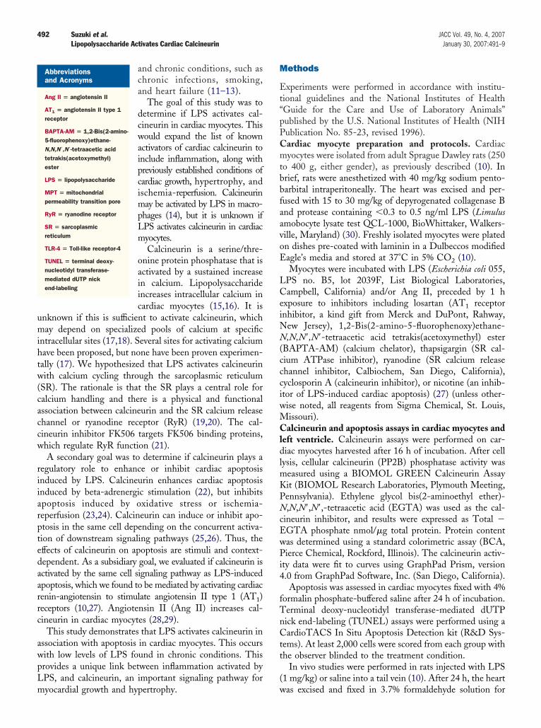
umihtw(caccw
riiarptedaarrc
awpLm
M
Et“pPCmtbbfaavoE
LCeiNN(ccciwMCldlmKPNcEwPi4
fTnCtt
(
492 Suzuki et al. JACC Vol. 49, No. 4, 2007Lipopolysaccharide Activates Cardiac Calcineurin January 30, 2007:491–9
and chronic conditions, such aschronic infections, smoking,and heart failure (11–13).
The goal of this study was todetermine if LPS activates cal-cineurin in cardiac myocytes. Thiswould expand the list of knownactivators of cardiac calcineurin toinclude inflammation, along withpreviously established conditions ofcardiac growth, hypertrophy, andischemia-reperfusion. Calcineurinmay be activated by LPS in macro-phages (14), but it is unknown ifLPS activates calcineurin in cardiacmyocytes.
Calcineurin is a serine/thre-onine protein phosphatase that isactivated by a sustained increasein calcium. Lipopolysaccharideincreases intracellular calcium incardiac myocytes (15,16). It is
nknown if this is sufficient to activate calcineurin, whichay depend on specialized pools of calcium at specific
ntracellular sites (17,18). Several sites for activating calciumave been proposed, but none have been proven experimen-ally (17). We hypothesized that LPS activates calcineurinith calcium cycling through the sarcoplasmic reticulum
SR). The rationale is that the SR plays a central role foralcium handling and there is a physical and functionalssociation between calcineurin and the SR calcium releasehannel or ryanodine receptor (RyR) (19,20). The cal-ineurin inhibitor FK506 targets FK506 binding proteins,hich regulate RyR function (21).A secondary goal was to determine if calcineurin plays a
egulatory role to enhance or inhibit cardiac apoptosisnduced by LPS. Calcineurin enhances cardiac apoptosisnduced by beta-adrenergic stimulation (22), but inhibitspoptosis induced by oxidative stress or ischemia-eperfusion (23,24). Calcineurin can induce or inhibit apo-tosis in the same cell depending on the concurrent activa-ion of downstream signaling pathways (25,26). Thus, theffects of calcineurin on apoptosis are stimuli and context-ependent. As a subsidiary goal, we evaluated if calcineurin isctivated by the same cell signaling pathway as LPS-inducedpoptosis, which we found to be mediated by activating cardiacenin-angiotensin to stimulate angiotensin II type 1 (AT1)eceptors (10,27). Angiotensin II (Ang II) increases cal-ineurin in cardiac myocytes (28,29).
This study demonstrates that LPS activates calcineurin inssociation with apoptosis in cardiac myocytes. This occursith low levels of LPS found in chronic conditions. Thisrovides a unique link between inflammation activated byPS, and calcineurin, an important signaling pathway for
Abbreviationsand Acronyms
Ang II � angiotensin II
AT1 � angiotensin II type 1receptor
BAPTA-AM � 1,2-Bis(2-amino-5-fluorophenoxy)ethane-N,N,N=,N=-tetraacetic acidtetrakis(acetoxymethyl)ester
LPS � lipopolysaccharide
MPT � mitochondrialpermeability transition pore
RyR � ryanodine receptor
SR � sarcoplasmicreticulum
TLR-4 � Toll-like receptor-4
TUNEL � terminal deoxy-nucleotidyl transferase-mediated dUTP nickend-labeling
yocardial growth and hypertrophy. w
ethods
xperiments were performed in accordance with institu-ional guidelines and the National Institutes of HealthGuide for the Care and Use of Laboratory Animals”ublished by the U.S. National Institutes of Health (NIHublication No. 85-23, revised 1996).ardiac myocyte preparation and protocols. Cardiacyocytes were isolated from adult Sprague Dawley rats (250
o 400 g, either gender), as previously described (10). Inrief, rats were anesthetized with 40 mg/kg sodium pento-arbital intraperitoneally. The heart was excised and per-used with 15 to 30 mg/kg of depyrogenated collagenase Bnd protease containing �0.3 to 0.5 ng/ml LPS (Limulusmobocyte lysate test QCL-1000, BioWhittaker, Walkers-ille, Maryland) (30). Freshly isolated myocytes were platedn dishes pre-coated with laminin in a Dulbeccos modifiedagle’s media and stored at 37°C in 5% CO2 (10).Myocytes were incubated with LPS (Escherichia coli 055,
PS no. B5, lot 2039F, List Biological Laboratories,ampbell, California) and/or Ang II, preceded by 1 h
xposure to inhibitors including losartan (AT1 receptornhibitor, a kind gift from Merck and DuPont, Rahway,
ew Jersey), 1,2-Bis(2-amino-5-fluorophenoxy)ethane-,N,N=,N=-tetraacetic acid tetrakis(acetoxymethyl) ester
BAPTA-AM) (calcium chelator), thapsigargin (SR cal-ium ATPase inhibitor), ryanodine (SR calcium releasehannel inhibitor, Calbiochem, San Diego, California),yclosporin A (calcineurin inhibitor), or nicotine (an inhib-tor of LPS-induced cardiac apoptosis) (27) (unless other-ise noted, all reagents from Sigma Chemical, St. Louis,issouri).
alcineurin and apoptosis assays in cardiac myocytes andeft ventricle. Calcineurin assays were performed on car-iac myocytes harvested after 16 h of incubation. After cell
ysis, cellular calcineurin (PP2B) phosphatase activity waseasured using a BIOMOL GREEN Calcineurin Assayit (BIOMOL Research Laboratories, Plymouth Meeting,ennsylvania). Ethylene glycol bis(2-aminoethyl ether)-,N,N=,N=,-tetraacetic acid (EGTA) was used as the cal-
ineurin inhibitor, and results were expressed as Total �GTA phosphate nmol/�g total protein. Protein contentas determined using a standard colorimetric assay (BCA,ierce Chemical, Rockford, Illinois). The calcineurin activ-
ty data were fit to curves using GraphPad Prism, version.0 from GraphPad Software, Inc. (San Diego, California).Apoptosis was assessed in cardiac myocytes fixed with 4%
ormalin phosphate-buffered saline after 24 h of incubation.erminal deoxy-nucleotidyl transferase-mediated dUTPick end-labeling (TUNEL) assays were performed using aardioTACS In Situ Apoptosis Detection kit (R&D Sys-
ems). At least 2,000 cells were scored from each group withhe observer blinded to the treatment condition.
In vivo studies were performed in rats injected with LPS1 mg/kg) or saline into a tail vein (10). After 24 h, the heart
as excised and fixed in 3.7% formaldehyde solution for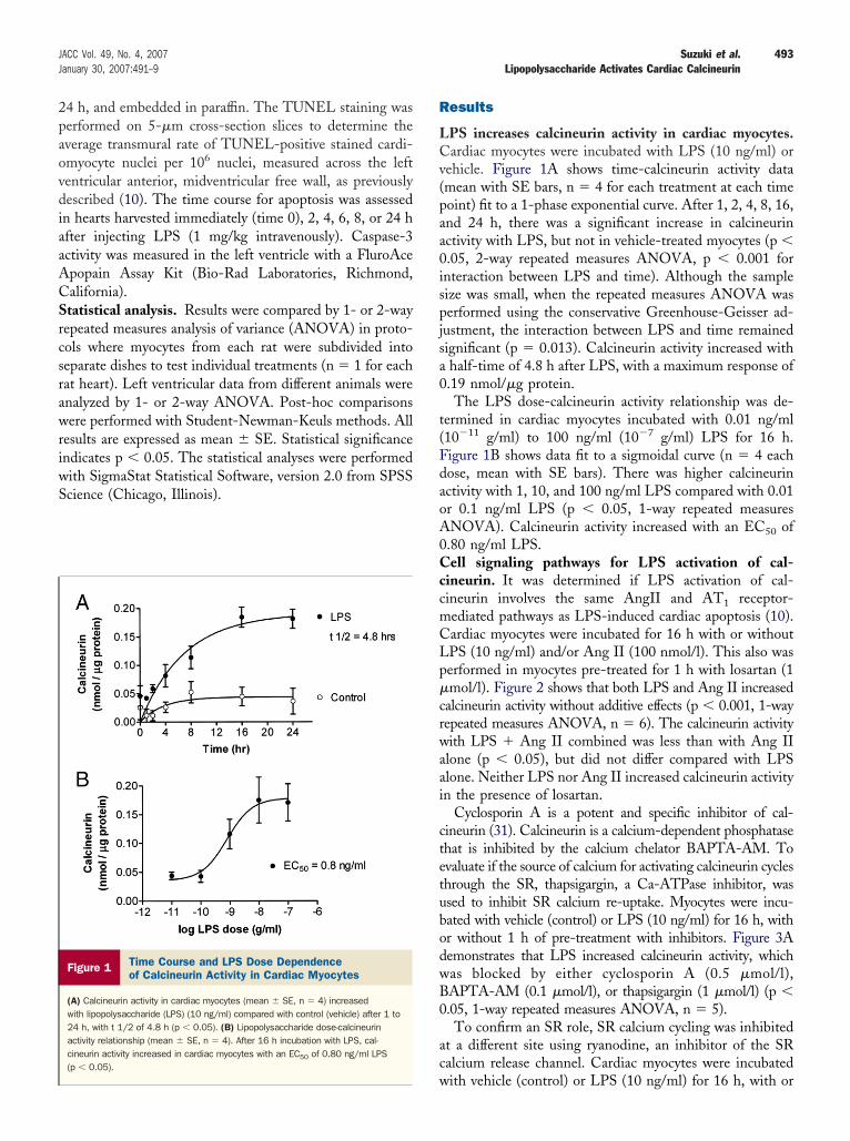
2paovdiaaACSrcsrawriwS
R
LCv(paa0ispjsa0
t(FdaoA0CccmCLp�crwaai
ctetubodwB0
ac
493JACC Vol. 49, No. 4, 2007 Suzuki et al.January 30, 2007:491–9 Lipopolysaccharide Activates Cardiac Calcineurin
4 h, and embedded in paraffin. The TUNEL staining waserformed on 5-�m cross-section slices to determine theverage transmural rate of TUNEL-positive stained cardi-myocyte nuclei per 106 nuclei, measured across the leftentricular anterior, midventricular free wall, as previouslyescribed (10). The time course for apoptosis was assessed
n hearts harvested immediately (time 0), 2, 4, 6, 8, or 24 hfter injecting LPS (1 mg/kg intravenously). Caspase-3ctivity was measured in the left ventricle with a FluroAcepopain Assay Kit (Bio-Rad Laboratories, Richmond,alifornia).tatistical analysis. Results were compared by 1- or 2-wayepeated measures analysis of variance (ANOVA) in proto-ols where myocytes from each rat were subdivided intoeparate dishes to test individual treatments (n � 1 for eachat heart). Left ventricular data from different animals werenalyzed by 1- or 2-way ANOVA. Post-hoc comparisonsere performed with Student-Newman-Keuls methods. All
esults are expressed as mean � SE. Statistical significancendicates p � 0.05. The statistical analyses were performedith SigmaStat Statistical Software, version 2.0 from SPSScience (Chicago, Illinois).
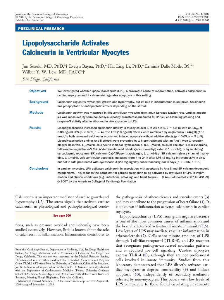
Figure 1 Time Course and LPS Dose Dependenceof Calcineurin Activity in Cardiac Myocytes
(A) Calcineurin activity in cardiac myocytes (mean � SE, n � 4) increasedwith lipopolysaccharide (LPS) (10 ng/ml) compared with control (vehicle) after 1 to24 h, with t 1/2 of 4.8 h (p � 0.05). (B) Lipopolysaccharide dose-calcineurinactivity relationship (mean � SE, n � 4). After 16 h incubation with LPS, cal-cineurin activity increased in cardiac myocytes with an EC50 of 0.80 ng/ml LPS(p � 0.05).
w
esults
PS increases calcineurin activity in cardiac myocytes.ardiac myocytes were incubated with LPS (10 ng/ml) or
ehicle. Figure 1A shows time-calcineurin activity datamean with SE bars, n � 4 for each treatment at each timeoint) fit to a 1-phase exponential curve. After 1, 2, 4, 8, 16,nd 24 h, there was a significant increase in calcineurinctivity with LPS, but not in vehicle-treated myocytes (p �.05, 2-way repeated measures ANOVA, p � 0.001 fornteraction between LPS and time). Although the sampleize was small, when the repeated measures ANOVA waserformed using the conservative Greenhouse-Geisser ad-
ustment, the interaction between LPS and time remainedignificant (p � 0.013). Calcineurin activity increased withhalf-time of 4.8 h after LPS, with a maximum response of.19 nmol/�g protein.The LPS dose-calcineurin activity relationship was de-
ermined in cardiac myocytes incubated with 0.01 ng/ml10�11 g/ml) to 100 ng/ml (10�7 g/ml) LPS for 16 h.igure 1B shows data fit to a sigmoidal curve (n � 4 eachose, mean with SE bars). There was higher calcineurinctivity with 1, 10, and 100 ng/ml LPS compared with 0.01r 0.1 ng/ml LPS (p � 0.05, 1-way repeated measuresNOVA). Calcineurin activity increased with an EC50 of.80 ng/ml LPS.ell signaling pathways for LPS activation of cal-
ineurin. It was determined if LPS activation of cal-ineurin involves the same AngII and AT1 receptor-ediated pathways as LPS-induced cardiac apoptosis (10).ardiac myocytes were incubated for 16 h with or withoutPS (10 ng/ml) and/or Ang II (100 nmol/l). This also waserformed in myocytes pre-treated for 1 h with losartan (1mol/l). Figure 2 shows that both LPS and Ang II increased
alcineurin activity without additive effects (p � 0.001, 1-wayepeated measures ANOVA, n � 6). The calcineurin activityith LPS � Ang II combined was less than with Ang IIlone (p � 0.05), but did not differ compared with LPSlone. Neither LPS nor Ang II increased calcineurin activityn the presence of losartan.
Cyclosporin A is a potent and specific inhibitor of cal-ineurin (31). Calcineurin is a calcium-dependent phosphatasehat is inhibited by the calcium chelator BAPTA-AM. Tovaluate if the source of calcium for activating calcineurin cycleshrough the SR, thapsigargin, a Ca-ATPase inhibitor, wassed to inhibit SR calcium re-uptake. Myocytes were incu-ated with vehicle (control) or LPS (10 ng/ml) for 16 h, withr without 1 h of pre-treatment with inhibitors. Figure 3Aemonstrates that LPS increased calcineurin activity, whichas blocked by either cyclosporin A (0.5 �mol/l),APTA-AM (0.1 �mol/l), or thapsigargin (1 �mol/l) (p �.05, 1-way repeated measures ANOVA, n � 5).
To confirm an SR role, SR calcium cycling was inhibitedt a different site using ryanodine, an inhibitor of the SRalcium release channel. Cardiac myocytes were incubated
ith vehicle (control) or LPS (10 ng/ml) for 16 h, with or
wbai1
niaLTa2ipodimpo�A0
LiIBFsmm
ntATrtiaaaLcFS1i
494 Suzuki et al. JACC Vol. 49, No. 4, 2007Lipopolysaccharide Activates Cardiac Calcineurin January 30, 2007:491–9
ithout 1-h pre-treatment with ryanodine (1 �mol/l, 1 hefore LPS), which locks the SR calcium release channel inn open state (32). Figure 3B shows that ryanodine inhib-ted LPS-induced increase in calcineurin activity (p � 0.05,-way repeated measures ANOVA, n � 6).Figure 3B shows that pre-treatment of myocytes with
icotine (15 ng/ml, 4 h before LPS), which inhibits LPS-nduced apoptosis (27), prevented increased calcineurinctivity with LPS (p � 0.005, n � 6).PS activation of calcineurin is associated with apoptosis.o determine if calcineurin induces or inhibits cardiac
poptosis with LPS, cardiac myocytes were incubated for4 h with or without LPS (10 ng/ml) and the calcineurinnhibitor cyclosporin A (0.5 �mol/l, 1 h before LPS). Wereviously observed peak changes in TUNEL staining toccur 24 h after LPS in this model (10). Figure 4Aemonstrates that cyclosporin A blocked an LPS-induced
ncrease in TUNEL staining (p � 0.05, 2-way repeatedeasures ANOVA, n � 7). For comparison, a similar
rotocol was performed in myocytes incubated for 24 h withr without Ang II (100 nmol/l) and cyclosporin A (0.5mol/l, 1 h before Ang II). Figure 4B shows that cyclosporinalso blocked increased TUNEL staining with Ang II (p �
.05, n � 9).Similar results were obtained with other inhibitors of
PS-induced calcineurin activation. Cardiac myocytes werencubated for 24 h with or without LPS (10 ng/ml) or AngI (100 nmol/l), with or without pre-treatment withAPTA-AM (0.1 �mol/l, 1 h before LPS or Ang II). Inigure 5A, BAPTA-AM prevented increased TUNELtaining with LPS or Ang II (p � 0.05, 1-way repeatedeasures ANOVA, n � 5). In a separate protocol, cardiac
Figure 2 Calcineurin Activity in Cardiac MyocytesWith LPS and/or Ang II Inhibited by Losartan
Calcineurin activity in cardiac myocytes (mean � SE, n � 6) increased after16-h incubation with lipopolysaccharide (LPS) (10 ng/ml) and/or angiotensin II(Ang II) (100 nmol/l) without cumulative effects (p � 0.05). This was blocked bypre-treating myocytes with losartan (1 �mol/l, Ang II type 1 receptor inhibitor) 1 hbefore LPS or Ang II, while losartan alone had no effect.
yocytes were incubated for 24 h with or without LPS (10
g/ml) or Ang II (100 nmol/l), with or without pre-reatment with thapsigargin (1 �mol/l, 1 h before LPS orng II). In Figure 5B, thapsigargin blocked increasedUNEL staining with LPS or Ang II (p � 0.05, 1-way
epeated measures ANOVA, n � 9). Thus, interventionshat inhibited LPS-induced calcineurin activation, includ-ng cyclosporin A, BAPTA-AM, thapsigargin, losartan,nd nicotine, also prevented LPS and Ang II inducedpoptosis. This supports a proapoptotic effect of calcineurinctivated by LPS.PS-induced left ventricular apoptosis in vivo. The time
ourse for LPS-induced apoptosis was measured in vivo.igure 6 shows caspase-3 activity in the left ventricle (mean �E) immediately (time � 0, n � 18), 2 h (n � 11), 4 h (n �1), 6 h (n � 7), 8 h (n � 11), and 24 h (n � 19) afternjecting LPS (1 mg/kg intravenously) in vivo. Caspase-3
Figure 3 Cyclosporin A, BAPTA-AM, TG, Ryanodine, orNicotine Inhibit LPS-Induced Calcineurin Activity
Calcineurin activity in cardiac myocytes (mean � SE) increased after 16-h incuba-tion with lipopolysaccharide (LPS) (10 ng/ml) compared with control (vehicle) (p �
0.05). (A) This was blocked by pre-treating myocytes (1 h before LPS) witheither cyclosporin A (0.5 �mol/l), 1,2-Bis(2-amino-5-fluorophenoxy)ethane-N,N,N=,N=-tetraacetic acid tetrakis(acetoxymethyl) ester (BAPTA-AM) (0.1 �mol/l), or thapsigargin (TG) (1 �mol/l) (n � 5). (B) Lipopolysaccharide effects wereblocked by pre-treating myocytes with ryanodine (1 �mol/l, 1 h before LPS), ornicotine (15 ng/ml, 4 h before LPS) (n � 6).
atcm
r3ddT(swp
Ic0
D
Tcphtwcv
495JACC Vol. 49, No. 4, 2007 Suzuki et al.January 30, 2007:491–9 Lipopolysaccharide Activates Cardiac Calcineurin
ctivity increased from 4 to 24 h after LPS (p � 0.05 vs.ime 0, 1-way ANOVA). This is consistent with the timeourse for LPS to increase calcineurin activity in cardiacyocytes in vitro (Fig. 1A).The role of calcineurin in vivo was examined by treating
ats with cyclosporin A (20 mg/kg/day subcutaneously) fordays before injecting LPS (1 mg/kg) by tail vein. This
ose of LPS induces cardiac apoptosis without causingistress or affecting blood pressure (10,27). Figure 7 showsUNEL staining in the left ventricle in 4 groups of rats
n � 5 per group) 24 h after intravenous injections of: 1)aline (control); 2) LPS; 3) saline after 3 days pre-treatmentith cyclosporin A (cyclosporin A); or 4) LPS after 3 days
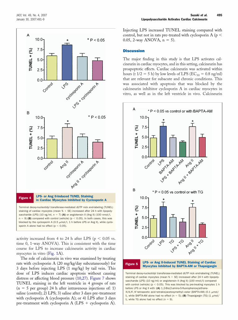
Figure 4 LPS- or Ang II-Induced TUNEL Stainingin Cardiac Myocytes Inhibited by Cyclosporin A
Terminal deoxy-nucleotidyl transferase-mediated dUTP nick end-labeling (TUNEL)staining of cardiac myocytes (mean % � SE) increased after 24 h with lipopoly-saccharide (LPS) (10 ng/ml, n � 7) (A) or angiotensin II (Ang II) (100 nmol/l,n � 9) (B) compared with control (vehicle) (p � 0.05). In both cases, this wasblocked by the cyclosporin A (0.5 �mol/l, 1 h before LPS or Ang II), while cyclo-sporin A alone had no effect (p � 0.05).
re-treatment with cyclosporin A (LPS � cyclosporin A).
njecting LPS increased TUNEL staining compared withontrol, but not in rats pre-treated with cyclosporin A (p �.05, 2-way ANOVA, n � 5).
iscussion
he major finding in this study is that LPS activates cal-ineurin in cardiac myocytes, and in this setting, calcineurin hasroapoptotic effects. Cardiac calcineurin was activated withinours (t 1/2 � 5 h) by low levels of LPS (EC50 � 0.8 ng/ml)hat are relevant for subacute and chronic conditions. Thisas associated with apoptosis that was blocked by the
alcineurin inhibitor cyclosporin A in cardiac myocytes initro, as well as in the left ventricle in vivo. Calcineurin
Figure 5 LPS- or Ang II-Induced TUNEL Staining of CardiacMyocytes Inhibited by BAPTA-AM or Thapsigargin
Terminal deoxy-nucleotidyl transferase-mediated dUTP nick end-labeling (TUNEL)staining of cardiac myocytes (mean % � SE) increased after 24 h with lipopoly-saccharide (LPS) (10 ng/ml) or angiotensin II (Ang II) (100 nmol/l) comparedwith control (vehicle) (p � 0.05). This was blocked by pre-treating myocytes 1 hbefore LPS or Ang II with: (A) 1,2-Bis(2-amino-5-fluorophenoxy)ethane-N,N,N=,N=-tetraacetic acid tetrakis(acetoxymethyl) ester (BAPTA-AM) (0.1 �mol/l), while BAPTA-AM alone had no effect (n � 5); (B) Thapsigargin (TG) (1 �mol/l), while TG alone had no effect (n � 9).
aAifmbt(wracnt
(taLdchtaesar
thcctc
dwcstict
um4A((acBnaimcp�cacmdL
496 Suzuki et al. JACC Vol. 49, No. 4, 2007Lipopolysaccharide Activates Cardiac Calcineurin January 30, 2007:491–9
ctivation was inhibited by the calcium chelator BAPTA-M, SR Ca-ATPase inhibitor thapsigargin and SR RyR
nhibitor ryanodine, indicating that the activating calciumor calcineurin is SR-dependent. Calcineurin activation wasimicked by Ang II without cumulative effects, and blocked
y losartan (AT1 receptor inhibitor), similar to the pathwayhat we demonstrated to mediate LPS-induced apoptosis10). Calcineurin activation was inhibited by nicotine,hich inhibits LPS-induced apoptosis proximal to AT1
eceptor activation (27). The simultaneous inhibition ofpoptosis with multiple inhibitors of calcineurin, includingyclosporin A, BAPTA-AM, thapsigargin, losartan, andicotine indicates that calcineurin has proapoptotic effects inhe context of LPS.
Low levels of LPS have direct effects on cardiac myocytes9,10) mediated through TLR4 (8) that are distinct fromhe cardiotoxic effects that occur when high levels of LPSctivate a cascade of mediators in sepsis (33). CirculatingPS levels in the pg/ml to low ng/ml range occur inecompensated heart failure (12), periodontitis (34),hronic infections (e.g., lung, urinary tract), smoking (13),emodialysis, cirrhosis, pancreatitis, abdominal and cardio-horacic surgery (11). Thus, LPS may activate calcineurin invariety of clinical scenarios. Calcineurin has proapoptotic
ffects with LPS, similar to its role with beta-adrenergictimulation (22), whereas calcineurin has primarily anti-poptotic effects in hypertrophy (2) and ischemia-eperfusion (23,24).
Chronic activation of calcineurin by LPS may contributeo the progression of pre-existing heart disease since theeart has a limited capacity to compensate for the loss ofardiac myocytes by apoptosis. In support of this postulate,hronic low levels of apoptosis induce heart failure (35), andransgenic mice that overexpress calcineurin have depressed
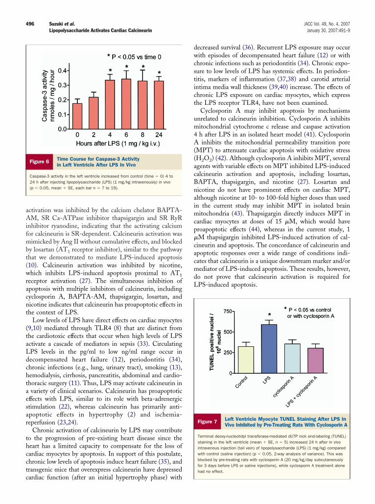
Figure 6 Time Course for Caspase-3 Activityin Left Ventricle After LPS In Vivo
Caspase-3 activity in the left ventricle increased from control (time � 0) 4 to24 h after injecting lipopolysaccharide (LPS) (1 mg/kg intravenously) in vivo(p � 0.05, mean � SE, each bar n � 7 to 19).
ardiac function (after an initial hypertrophy phase) with
ecreased survival (36). Recurrent LPS exposure may occurith episodes of decompensated heart failure (12) or with
hronic infections such as periodontitis (34). Chronic expo-ure to low levels of LPS has systemic effects. In periodon-itis, markers of inflammation (37,38) and carotid arterialntima media wall thickness (39,40) increase. The effects ofhronic LPS exposure on cardiac myocytes, which expresshe LPS receptor TLR4, have not been examined.
Cyclosporin A may inhibit apoptosis by mechanismsnrelated to calcineurin inhibition. Cyclosporin A inhibitsitochondrial cytochrome c release and caspase activationh after LPS in an isolated heart model (41). Cyclosporininhibits the mitochondrial permeability transition pore
MPT) to attenuate cardiac apoptosis with oxidative stressH2O2) (42). Although cyclosporin A inhibits MPT, severalgents with variable effects on MPT inhibited LPS-inducedalcineurin activation and apoptosis, including losartan,APTA, thapsigargin, and nicotine (27). Losartan andicotine do not have prominent effects on cardiac MPT,lthough nicotine at 10- to 100-fold higher doses than usedn the current study may inhibit MPT in isolated brain
itochondria (43). Thapsigargin directly induces MPT inardiac myocytes at doses of 15 �M, which would haveroapoptotic effects (44), whereas in the current study, 1M thapsigargin inhibited LPS-induced activation of cal-
ineurin and apoptosis. The concordance of calcineurin andpoptotic responses over a wide range of conditions indi-ates that calcineurin is a unique downstream marker and/orediator of LPS-induced apoptosis. These results, however,
o not prove that calcineurin activation is required forPS-induced apoptosis.
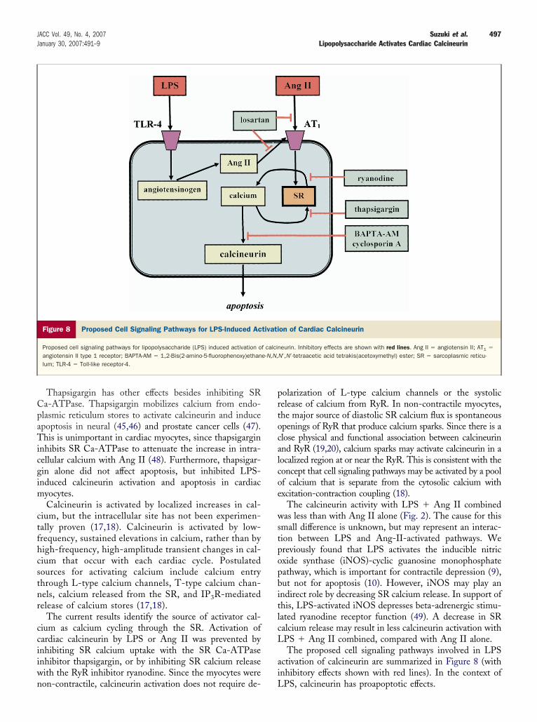
Figure 7 Left Ventricle Myocyte TUNEL Staining After LPS InVivo Inhibited by Pre-Treating Rats With Cyclosporin A
Terminal deoxy-nucleotidyl transferase-mediated dUTP nick end-labeling (TUNEL)staining in the left ventricle (mean � SE, n � 5) increased 24 h after in vivointravenous injection (tail vein) of lipopolysaccharide (LPS) (1 mg/kg) comparedwith control (saline injection) (p � 0.05, 2-way analysis of variance). This wasblocked by pre-treating rats with cyclosporin A (20 mg/kg/day subcutaneouslyfor 3 days before LPS or saline injections), while cyclosporin A treatment alonehad no effect.
CpaTicgim
ctfhcstnr
cciiwn
prtocalcoe
wstpopbitlcL
ai
497JACC Vol. 49, No. 4, 2007 Suzuki et al.January 30, 2007:491–9 Lipopolysaccharide Activates Cardiac Calcineurin
Thapsigargin has other effects besides inhibiting SRa-ATPase. Thapsigargin mobilizes calcium from endo-lasmic reticulum stores to activate calcineurin and inducepoptosis in neural (45,46) and prostate cancer cells (47).his is unimportant in cardiac myocytes, since thapsigargin
nhibits SR Ca-ATPase to attenuate the increase in intra-ellular calcium with Ang II (48). Furthermore, thapsigar-in alone did not affect apoptosis, but inhibited LPS-nduced calcineurin activation and apoptosis in cardiac
yocytes.Calcineurin is activated by localized increases in cal-
ium, but the intracellular site has not been experimen-ally proven (17,18). Calcineurin is activated by low-requency, sustained elevations in calcium, rather than byigh-frequency, high-amplitude transient changes in cal-ium that occur with each cardiac cycle. Postulatedources for activating calcium include calcium entryhrough L-type calcium channels, T-type calcium chan-els, calcium released from the SR, and IP3R-mediatedelease of calcium stores (17,18).
The current results identify the source of activator cal-ium as calcium cycling through the SR. Activation ofardiac calcineurin by LPS or Ang II was prevented bynhibiting SR calcium uptake with the SR Ca-ATPasenhibitor thapsigargin, or by inhibiting SR calcium releaseith the RyR inhibitor ryanodine. Since the myocytes were
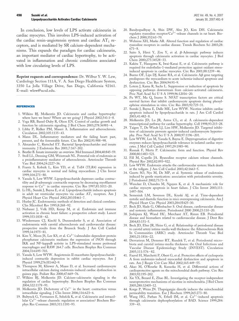
Figure 8 Proposed Cell Signaling Pathways for LPS-Induced Ac
Proposed cell signaling pathways for lipopolysaccharide (LPS) induced activation oangiotensin II type 1 receptor; BAPTA-AM � 1,2-Bis(2-amino-5-fluorophenoxy)ethanlum; TLR-4 � Toll-like receptor-4.
on-contractile, calcineurin activation does not require de- L
olarization of L-type calcium channels or the systolicelease of calcium from RyR. In non-contractile myocytes,he major source of diastolic SR calcium flux is spontaneouspenings of RyR that produce calcium sparks. Since there is alose physical and functional association between calcineurinnd RyR (19,20), calcium sparks may activate calcineurin in aocalized region at or near the RyR. This is consistent with theoncept that cell signaling pathways may be activated by a poolf calcium that is separate from the cytosolic calcium withxcitation-contraction coupling (18).
The calcineurin activity with LPS � Ang II combinedas less than with Ang II alone (Fig. 2). The cause for this
mall difference is unknown, but may represent an interac-ion between LPS and Ang-II-activated pathways. Wereviously found that LPS activates the inducible nitricxide synthase (iNOS)-cyclic guanosine monophosphateathway, which is important for contractile depression (9),ut not for apoptosis (10). However, iNOS may play anndirect role by decreasing SR calcium release. In support ofhis, LPS-activated iNOS depresses beta-adrenergic stimu-ated ryanodine receptor function (49). A decrease in SRalcium release may result in less calcineurin activation withPS � Ang II combined, compared with Ang II alone.The proposed cell signaling pathways involved in LPS
ctivation of calcineurin are summarized in Figure 8 (withnhibitory effects shown with red lines). In the context of
on of Cardiac Calcineurin
eurin. Inhibitory effects are shown with red lines. Ang II � angiotensin II; AT1 �
N=,N=-tetraacetic acid tetrakis(acetoxymethyl) ester; SR � sarcoplasmic reticu-
tivati
f calcine-N,N,
PS, calcineurin has proapoptotic effects.
ctcnavw
RC3E
R
1
1
1
1
1
1
1
1
1
1
2
2
2
2
2
2
2
2
2
2
3
3
3
3
3
3
3
3
3
3
4
4
4
4
4
4
498 Suzuki et al. JACC Vol. 49, No. 4, 2007Lipopolysaccharide Activates Cardiac Calcineurin January 30, 2007:491–9
In conclusion, low levels of LPS activate calcineurin inardiac myocytes. This involves LPS-induced activation ofhe cardiac renin-angiotensin system and cardiac AT1 re-eptors, and is mediated by SR calcium-dependent mecha-isms. This expands the paradigm for cardiac calcineurin,n important mediator of cardiac hypertrophy, to be acti-ated in inflammation and chronic conditions associatedith low circulating levels of LPS.
eprint requests and correspondence: Dr. Wilbur Y. W. Lew,ardiology Section 111A, V. A. San Diego Healthcare System,350 La Jolla Village Drive, San Diego, California 92161.-mail: [email protected].
EFERENCES
1. Wilkins BJ, Molkentin JD. Calcineurin and cardiac hypertrophy:where have we been? Where are we going? J Physiol 2002;541:1–8.
2. Vega RB, Bassel-Duby R, Olson EN. Control of cardiac growth andfunction by calcineurin signaling. J Biol Chem 2003;278:36981–4.
3. Libby P, Ridker PM, Maseri A. Inflammation and atherosclerosis.Circulation 2002;105:1135–43.
4. Mann DL. Inflammatory mediators and the failing heart: past,present, and the foreseeable future. Circ Res 2002;91:988–98.
5. Alexander C, Rietschel ET. Bacterial lipopolysaccharides and innateimmunity. J Endotoxin Res 2001;7:167–202.
6. Beutler B. Innate immunity: an overview. Mol Immunol 2004;40:845–59.7. Stoll LL, Denning GM, Weintraub NL. Potential role of endotoxin as
a proinflammatory mediator of atherosclerosis. Arterioscler ThrombVasc Biol 2004;24:2227–36.
8. Frantz S, Kobzik L, Kim YD, et al. Toll4 (TLR4) expression incardiac myocytes in normal and failing myocardium. J Clin Invest1999;104:271–80.
9. Yasuda S, Lew WYW. Lipopolysaccharide depresses cardiac contrac-tility and �-adrenergic contractile response by decreasing myofilamentresponse to Ca2� in cardiac myocytes. Circ Res 1997;81:1011–20.
0. Li HL, Suzuki J, Bayna E, et al. Lipopolysaccharide induces apoptosisin adult rat ventricular myocytes via cardiac AT1 receptors. Am JPhysiol Heart Circ Physiol 2002;283:H461–7.
1. Hurley JC. Endotoxemia: methods of detection and clinical correlates.Clin Microbiol Rev 1995;8:268–92.
2. Niebauer J, Volk HD, Kemp M, et al. Endotoxin and immuneactivation in chronic heart failure: a prospective cohort study. Lancet1999;353:1838–42.
3. Wiedermann CJ, Kiechl S, Dunzendorfer S, et al. Association ofendotoxemia with carotid atherosclerosis and cardiovascular disease:prospective results from the Bruneck Study. J Am Coll Cardiol1999;34:1975–81.
4. Kim Y, Moon JS, Lee KS, et al. Ca2�/calmodulin-dependent proteinphosphatase calcineurin mediates the expression of iNOS throughIKK and NF-kappaB activity in LPS-stimulated mouse peritonealmacrophages and RAW 264.7 cells. Biochem Biophys Res Commun2004;314:695–703.
5. Yasuda S, Lew WYW. Angiotensin II exacerbates lipopolysaccharide-induced contractile depression in rabbit cardiac myocytes. Am JPhysiol 1999;276:H1442–9.
6. Thompson M, Kliewer A, Maass D, et al. Increased cardiomyocyteintracellular calcium during endotoxin-induced cardiac dysfunction inguinea pigs. Pediatr Res 2000;47:669–76.
7. Wilkins BJ, Molkentin JD. Calcium-calcineurin signaling in theregulation of cardiac hypertrophy. Biochem Biophys Res Commun2004;322:1178–91.
8. Molkentin JD. Dichotomy of Ca2� in the heart: contraction versusintracellular signaling. J Clin Invest 2006;116:623–6.
9. Bultynck G, Vermassen E, Szlufcik K, et al. Calcineurin and intracel-lular Ca2�-release channels: regulation or association? Biochem Bio-
phys Res Commun 2003;311:1181–93.0. Bandyopadhyay A, Shin DW, Ahn JO, Kim DH. Calcineurinregulates ryanodine receptor/Ca2�-release channels in rat heart. Bio-chem J 2000;352:61–70.
1. Wehrens XH, Marks AR. Altered function and regulation of cardiacryanodine receptors in cardiac disease. Trends Biochem Sci 2003;28:671–8.
2. Saito S, Hiroi Y, Zou Y, et al. �-Adrenergic pathway inducesapoptosis through calcineurin activation in cardiac myocytes. J BiolChem 2000;275:34528–33.
3. Kakita T, Hasegawa K, Iwai-Kanai E, et al. Calcineurin pathway isrequired for endothelin-1-mediated protection against oxidant stress-induced apoptosis in cardiac myocytes. Circ Res 2001;88:1239–46.
4. Bueno OF, Lips DJ, Kaiser RA, et al. Calcineurin A� gene targetingpredisposes the myocardium to acute ischemia-induced apoptosis anddysfunction. Circ Res 2004;94:91–9.
5. Lotem J, Kama R, Sachs L. Suppression or induction of apoptosis byopposing pathways downstream from calcium-activated calcineurin.Proc Natl Acad Sci U S A 1999;96:12016–20.
6. Pu WT, Ma Q, Izumo S. NFAT transcription factors are criticalsurvival factors that inhibit cardiomyocyte apoptosis during phenyl-ephrine stimulation in vitro. Circ Res 2003;92:725–31.
7. Suzuki J, Bayna E, Dalle ME, Lew WYW. Nicotine inhibits cardiacapoptosis induced by lipopolysaccharide in rats. J Am Coll Cardiol2003;41:482–8.
8. Molkentin JD, Lu JR, Antos CL, et al. A calcineurin-dependenttranscriptional pathway for cardiac hypertrophy. Cell 1998;93:215–28.
9. Taigen T, De Windt LJ, Lim HW, Molkentin JD. Targeted inhibi-tion of calcineurin prevents agonist-induced cardiomyocyte hypertro-phy. Proc Natl Acad Sci U S A 2000;97:1196–201.
0. Lew WYW, Lee M, Yasuda S, Bayna E. Depyrogenation of digestiveenzymes reduces lipopolysaccharide tolerance in isolated cardiac myo-cytes. J Mol Cell Cardiol 1997;29:1985–90.
1. Rusnak F, Mertz P. Calcineurin: form and function. Physiol Rev2000;80:1483–521.
2. Fill M, Copello JA. Ryanodine receptor calcium release channels.Physiol Rev 2002;82:893–922.
3. Lew WYW. Endotoxin attacks the cardiovascular system: black deathat the tollgate. J Am Coll Cardiol 2003;42:1663–5.
4. Geerts SO, Nys M, De MP, et al. Systemic release of endotoxinsinduced by gentle mastication: association with periodontitis severity.J Periodontol 2002;73:73–8.
5. Wencker D, Chandra M, Nguyen K, et al. A mechanistic role forcardiac myocyte apoptosis in heart failure. J Clin Invest 2003;111:1497–504.
6. Semeniuk LM, Severson DL, Kryski AJ, et al. Time-dependentsystolic and diastolic function in mice overexpressing calcineurin. Am JPhysiol Heart Circ Physiol 2003;284:H425–30.
7. Beck JD, Slade G, Offenbacher S. Oral disease, cardiovascular diseaseand systemic inflammation. Periodontol 2000 2000;23:110–20.
8. Joshipura KJ, Wand HC, Merchant AT, Rimm EB. Periodontaldisease and biomarkers related to cardiovascular disease. J Dent Res2004;83:151–5.
9. Beck JD, Elter JR, Heiss G, et al. Relationship of periodontal diseaseto carotid artery intima-media wall thickness: the Atherosclerosis RiskIn Communities (ARIC) study. Arterioscler Thromb Vasc Biol2001;21:1816–22.
0. Desvarieux M, Demmer RT, Rundek T, et al. Periodontal micro-biota and carotid intima-media thickness: the Oral Infections andVascular Disease Epidemiology Study (INVEST). Circulation2005;111:576 – 82.
1. Fauvel H, Marchetti P, Obert G, et al. Protective effects of cyclosporinA from endotoxin-induced myocardial dysfunction and apoptosis inrats. Am J Respir Crit Care Med 2002;165:449–55.
2. Akao M, O’Rourke B, Kusuoka H, et al. Differential actions ofcardioprotective agents on the mitochondrial death pathway. Circ Res2003;92:195–202.
3. Xie YX, Bezard E, Zhao BL. Investigating the receptor-independentneuroprotective mechanisms of nicotine in mitochondria. J Biol Chem2005;280:32405–12.
4. Korge P, Weiss JN. Thapsigargin directly induces the mitochondrialpermeability transition. Eur J Biochem 1999;265:273–80.
5. Wang HG, Pathan N, Ethell IM, et al. Ca2�-induced apoptosis
through calcineurin dephosphorylation of BAD. Science 1999;284:339–43.
4
4
4
4
499JACC Vol. 49, No. 4, 2007 Suzuki et al.January 30, 2007:491–9 Lipopolysaccharide Activates Cardiac Calcineurin
6. Mukerjee N, McGinnis KM, Park YH, et al. Caspase-mediatedproteolytic activation of calcineurin in thapsigargin-mediated apopto-sis in SH-SY5Y neuroblastoma cells. Arch Biochem Biophys 2000;379:337–43.
7. Tombal B, Weeraratna AT, Denmeade SR, Isaacs JT. Thapsigar-gin induces a calmodulin/calcineurin-dependent apoptotic cascaderesponsible for the death of prostatic cancer cells. Prostate 2000;
43:303–17.8. Fukuta Y, Yoshizumi M, Kitagawa T, et al. Effect of angiotensin II onCa2� efflux from freshly isolated adult rat cardiomyocytes: possibleinvolvement of Na�/Ca2� exchanger. Biochem Pharmacol 1998;55:481–7.
9. Ziolo MT, Katoh H, Bers DM. Expression of inducible nitric oxidesynthase depresses beta-adrenergic-stimulated calcium release fromthe sarcoplasmic reticulum in intact ventricular myocytes. Circulation
2001;104:2961–6.