lipin-1 integrates lipid synthesis with proinflammatory ...balsinde.org/papers/p105-2014-ji.pdf ·...
TRANSCRIPT
The Journal of Immunology
Lipin-1 Integrates Lipid Synthesis with ProinflammatoryResponses during TLR Activation in Macrophages
Clara Meana,*,† Lucıa Pena,*,† Gema Lorden,*,† Esperanza Esquinas,* Carlos Guijas,*,†
Martın Valdearcos,* Jesus Balsinde,*,† and Marıa A. Balboa*,†
Lipin-1 is a Mg2+-dependent phosphatidic acid phosphatase involved in the de novo synthesis of phospholipids and triglycerides.
Using macrophages from lipin-1–deficient animals and human macrophages deficient in the enzyme, we show in this work that this
phosphatase acts as a proinflammatory mediator during TLR signaling and during the development of in vivo inflammatory
processes. After TLR4 stimulation lipin-1–deficient macrophages showed a decreased production of diacylglycerol and activation
of MAPKs and AP-1. Consequently, the generation of proinflammatory cytokines like IL-6, IL-12, IL-23, or enzymes like
inducible NO synthase and cyclooxygenase 2, was reduced. In addition, animals lacking lipin-1 had a faster recovery from
endotoxin administration concomitant with a reduced production of harmful molecules in spleen and liver. These findings
demonstrate an unanticipated role for lipin-1 as a mediator of macrophage proinflammatory activation and support a critical
link between lipid biosynthesis and systemic inflammatory responses. The Journal of Immunology, 2014, 193: 4614–4622.
Macrophages are highly plastic phagocytic cells respon-sible for the maintenance of physiological homeostasisowing to their ability to clear pathogens and influence
the behavior of other immune cells (1). They are activated bypathogen-associated molecular patterns through specific receptorsto generate an inflammatory response characterized by a cascadeof cytokines and molecules that limit ongoing infection or tissuedamage. Excessive macrophage activation responses, however,can lead to acute pathological diseases, best exemplified by sepsis,or chronic disorders such as arthritis, asthma, atherosclerosis, ordiabetes type 2 (1).Engagement of TLRs, specifically TLR4, by molecular patterns
like LPS triggers a cascade of signaling events conducted by
kinases and adaptor proteins that culminates in the phosphorylationand activation of the MAPK family of proteins and the family ofkinases for the NF-kB transcription factor inhibitors IkB (2–4).MAPKs are responsible for the phosphorylation and activation ofproteins that are part of the transcription factor AP-1, whereas thephosphorylation of IkB inhibitors promotes their proteasomal deg-radation, thereby releasing active NF-kB proteins. AP-1 and NF-kBare involved in the transcription of multiple proinflammatory genes,including IL-6, IL-12, IL-23, and TNF-a (5).LPS-stimulated macrophages accumulate triacylglycerol (TAG)
molecules, which are used to meet the increased demands ofenergy of these highly active cells (6, 7). TAG is formed by acy-lation of part of the diacylglycerol (DAG) generated via the Kennedypathway of phospholipid synthesis (8). The enzymes that directlygenerate DAG, in the Kennedy pathway, display phosphatidic acidphosphatase activity and are a family known as the lipins (9, 10).We have previously shown that a member of this family ofenzymes, lipin-1, is expressed in human macrophages and reg-ulates eicosanoid production and size and number of TAG-loadedlipid droplets (11). However, little is known about the role of lipin-1 during macrophage activation through TLRs. Questions on thepossible involvement of lipin-1 in the production of DAG duringTLR stimulation, the role of lipin-1 during TLR signaling, whetherlipin-1 activity modulates proinflammatory gene upregulation, andwhether lipin-1 influences inflammatory conditions in vivo remainunanswered. In this study, we have used genetic and omicsapproaches to determine the role of lipin-1 during TLR activationof macrophages. We report that lipin-1 contributes positively tomacrophage stimulation through TLR4, and other TLRs, by af-fecting MAPKs and AP-1 activation and, as a consequence, thegeneration of proinflammatory factors during in vitro and in vivomodels of inflammation. Thus, lipin-1 connects lipid synthesiswith macrophage proinflammatory activation.
Materials and MethodsAnimals
BALB/cByJ-Lpin1fld/J mice carrying a spontaneous mutation in the Lpin1gene (fatty liver dystrophy, fld) (9, 12) were purchased from The JacksonLaboratory and bred in the Service of Animal Research and Welfare of theUniversity of Valladolid. Males and females of Lpinfld/+ genotype were
*Instituto de Biologıa y Genetica Molecular, Consejo Superior de InvestigacionesCientıficas, Universidad de Valladolid, 47003 Valladolid, Spain; and †Centro deInvestigacion Biomedica en Red de Diabetes y Enfermedades Metabolicas Asocia-das, 28029 Madrid, Spain
Received for publication January 27, 2014. Accepted for publication August 26,2014.
This work was supported by the Spanish Ministry of Science and Innovation (GrantsSAF2007-60055, SAF2010-18831, and BFU2010-18826) and the Regional Govern-ment of Castile and Leon (Grants BIO39/VA04/10 and CSI168A12-1). L.P. and G.L.were supported by predoctoral fellowships from the Spanish Ministry of Science andInnovation (Plan de Formacion de Personal Investigador and Plan de Formacion deProfesorado Universitario programs). M.V. was supported by a predoctoral fellow-ship from the Regional Government of Castile and Leon. E.E. was supported bya predoctoral fellowship from the Spanish National Research Council (Junta deAmpliacion de Estudios Program). C.G. was supported by a predoctoral fellowshipfrom the University of Valladolid.
The sequences presented in this article have been submitted to the Gene ExpressionOmnibus database (www.ncbi.nlm.nih.gov/geo/query/acc.cgi?acc=GSE54155) underaccession number GSE54155.
Address correspondence and reprint requests to Prof. Jesus Balsinde and Prof. MarıaA. Balboa, University of Valladolid School of Medicine, Calle Sanz y Fores 3, 47010Valladolid, Spain. E-mail addresses: [email protected] (J.B.) and [email protected] (M.A.B.)
The online version of this article contains supplemental material.
Abbreviations used in this article: ALT, alanine aminotransferase; AST, aspartateaminotransferase; BMDM, bone marrow–derived macrophage; BUN, blood ureanitrogen; COX-2, cyclooxygenase-2; DAG, diacylglerol; iNOS, inducible NO syn-thase; PKC, protein kinase C; poly(I:C), polyinosinic-polycytidylic acid; siRNA,small interfering RNA; TAG, triacylglycerol; wt, wild-type.
Copyright� 2014 by TheAmericanAssociation of Immunologists, Inc. 0022-1767/14/$16.00
www.jimmunol.org/cgi/doi/10.4049/jimmunol.1400238
by Jesus Balsinde on O
ctober 17, 2014http://w
ww
.jimm
unol.org/D
ownloaded from
bred to generate Lpinfld/fld (hereafter fld), Lpinfld/+, and wild-type (wt)sibling animals. Mice were housed in filter-top cages and were providedwith sterile water and food ad libitum (Global diet 2014, Harlan). Sex-matched 12-week-old animals were used for experimentation. All theprotocols and procedures were approved by the Institutional Animal Careand Usage Committee and are in accordance with the Spanish and Euro-pean Union guidelines for the use of experimental animals.
Reagents
LPS from Escherichia coli 0111:B4, 1,2-diheptadecanoyl-sn-glycerol, andthe Ab against b-actin were obtained from Sigma-Aldrich. Abs againstIkBa, IkBb, phospho-p38 MAPK (Thr180/Tyr182), p38 MAPK, phospho-p44/42 MAPK (Thr202/Tyr204), p44/42 MAPK, phospho-SAPK/JNK (Thr183/Tyr185), SAPK/JNK, and phospho–c-Jun were purchased from Cell Sig-naling. Abs against the nuclear protein p84 were obtained from Abcam.Abs against inducible NO synthase (iNOS) were obtained from BD Bio-sciences and anti–cyclooxygenase-2 (COX-2) from Cayman. The rabbitantiserum against lipin-1 was previously described (11). PE-conjugated Absagainst CD11b, APC-conjugated Abs against F4/80, FITC-conjugated Absagainst Gr1, and 7-amino-actinomycin D were purchased from eBioscience.Specific ON-TARGETplus small interfering RNAs (siRNAs) against murinemRNAs were obtained from Dharmacon (Thermo Scientific). Silencer SelectsiRNAs specific to decrease the expression of human lipin-1 mRNA andnegative controls were purchased from Ambion.
Cells
To obtain peritoneal macrophages, the peritoneal cavity was flushed twicewith 5 ml ice-cold PBS. Resident cells were centrifuged for 10 min at 3003g and allowed to adhere to plastic for 18 h in RPMI 1640 medium con-taining 10% FBS. Nonadherent cells were washed away, and attached cellswere maintained in culture until use.
To obtain bone marrow–derived macrophages (BMDMs), intact femursand tibias were aseptically dislocated from the hind legs of the mice. Themarrow was flushed with 5 ml PBS using a 25-gauge sterile needle. Afterfiltration, bone marrow cells were centrifuged for 10 min at 300 3 g, cul-tured and differentiated according to the procedure described by Johnsonet al. (13). Briefly, the cells were cultured in growth medium supplementedwith 20% supernatant of the mouse L929 cell (conditioned medium) andcultured for 7 d. The medium was changed at day 4, washing out the non-adherent cells, and differentiation was continued for a total of 7 d.
Neutrophils were isolated from bone marrow using a Percoll gradient asdescribed (14). T and B lymphocyte populations were isolated from spleenusing nylon wool columns (15). Briefly, splenocytes were depleted oferythrocytes by ammonium chloride–mediated lysis. Cells were then in-cubated in plastic plates at 37˚C and 5% CO2 for 2 h. Nonadherent cellscontaining mainly B and T cells were further subjected to nylon wool(Polysciences) purification, allowing them to adhere to the column at 37˚Cand 5% CO2 in RPMI 1640 for 1 h. Nonadherent T cells were eluted withwarm RPMI 1640, and B cells were also collected afterward by addingcold media, knocking the column to dislodge binding cells. The cell sur-face markers of the resulting populations were routinely monitored.
Human macrophages were obtained from blood monocytes and trans-fected using the nucleofection technique (Amaxa), as previously described(11, 16–18).
Measurement of DAG mass
A cell extract corresponding to 1 mg protein was used, and before theextraction and separation of lipid classes, 1,2-diheptadecanoyl-sn-glycerolwas added as an internal standard. Total lipids were extracted according toBligh and Dyer (19), and the resulting lipid extract was separated by thin-layer chromatography using n-hexane/diethyl ether/acetic acid (70:30:1,by vol.) as the mobile phase. Spots corresponding to the various lipidclasses were scraped, and DAG was extracted from the silica with 1 mlchloroform/methanol (1:1, v/v) followed by 1 ml chloroform/methanol(2:1, v/v). The DAG fraction was transmethylated with 500 ml 0.5 MKOH in methanol for 45 min at 37˚C, and a volume of 0.5 M HCl wasadded before extracting twice with 1 ml n-hexane. Analysis of fatty acidmethyl esters was carried out in a Agilent 7890A gas chromatograph coupledto an Agilent 5975C mass-selective detector operated in electron impactmode (70 eV) equipped with an Agilent 7693 autosampler and an AgilentDB23 column (60-m length 3 250-mm internal diameter 3 0.15-mm filmthickness) under the conditions established previously (20–22). The amountof DAG mass in each sample was calculated by adding the molar masses ofall fatty acids measured and dividing by 2. Data analysis was carried out withthe Agilent G1701EA MSD Productivity Chemstation software, revisionE.02.00.
Real-time PCR
Total RNA from mouse tissues and cells was extracted using the RNeasy MiniKit (QIAGEN) and TRIzol reagent (Ambion), respectively. The cDNA tem-plates were synthesized using M-MLW Reverse Transcriptase (Ambion) fol-lowing the manufacturer’s instructions. Quantitative real time RT-PCR analysiswas performed in a LightCycler 480 (Roche) as previously described (18),using specific primers obtained from the Primer Bank database (23).
Cytokine determination and serum analysis
Supernatants from activated cells or serum from LPS-treated animals wereused for quantification of IL-6, IL12p40, IL2p70, and IL23p19 by specificELISA kits (eBioscience) following the manufacturer’s instructions. Urea,blood urea nitrogen (BUN), creatinine, aspartate aminotransferase (AST),and alanine aminotransferase (ALT) measurements in serum were per-formed by Laboratorios Echevarne (Barcelona, Spain).
LPS treatment of animals
Mice were i.p. injected with LPS at a lethal dose of 25 mg/kg or a sublethaldose of 1.5 mg/kg and monitored for 10 d. For the analysis of proin-flammatory factors, some animals were sacrificed by ketamine (100mg/kg):xylacine (10 mg/kg) administration and cervical dislocation 6 h after LPStreatment. Blood was collected through cardiac puncture or from the facialvein. Livers and spleens were collected in RNAlater (Ambion) for furtheranalysis by real-time PCR.
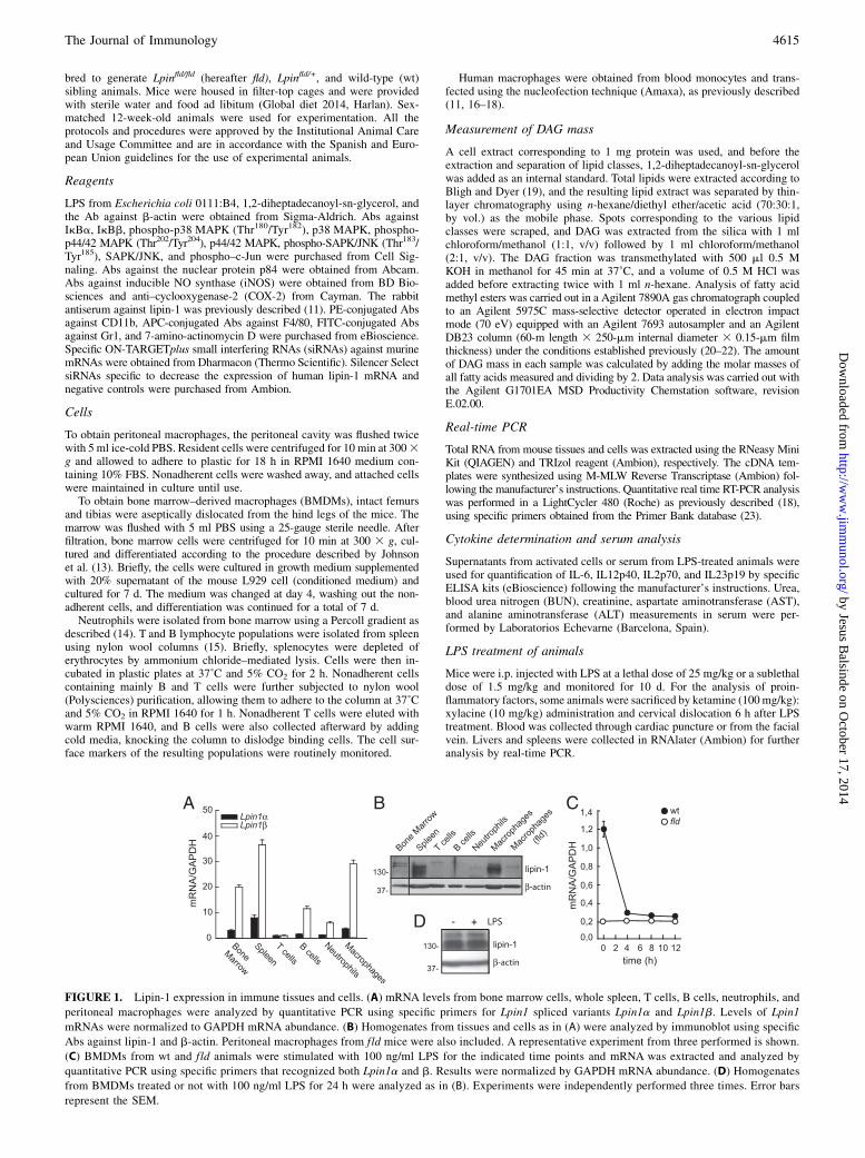
FIGURE 1. Lipin-1 expression in immune tissues and cells. (A) mRNA levels from bone marrow cells, whole spleen, T cells, B cells, neutrophils, and
peritoneal macrophages were analyzed by quantitative PCR using specific primers for Lpin1 spliced variants Lpin1a and Lpin1b. Levels of Lpin1
mRNAs were normalized to GAPDH mRNA abundance. (B) Homogenates from tissues and cells as in (A) were analyzed by immunoblot using specific
Abs against lipin-1 and b-actin. Peritoneal macrophages from fld mice were also included. A representative experiment from three performed is shown.
(C) BMDMs from wt and fld animals were stimulated with 100 ng/ml LPS for the indicated time points and mRNA was extracted and analyzed by
quantitative PCR using specific primers that recognized both Lpin1a and b. Results were normalized by GAPDH mRNA abundance. (D) Homogenates
from BMDMs treated or not with 100 ng/ml LPS for 24 h were analyzed as in (B). Experiments were independently performed three times. Error bars
represent the SEM.
The Journal of Immunology 4615
by Jesus Balsinde on O
ctober 17, 2014http://w
ww
.jimm
unol.org/D
ownloaded from
Microarray gene expression
Peritoneal macrophages from four different control (Lpinfld/+) and fld(Lpinfld/ fld) male animals were separately stimulated with 100 ng/ml LPSfor 5 h, and RNA was isolated using TRIzol reagent (Ambion). LabeledRNA was hybridized overnight (17 h, 65˚C) to Agilent Whole Mouse Ge-nome Oligo Microarrays 43 44K, using the Agilent-recommended protocol.After extensive washing, fluorescence signals were detected using AgilentMicroarray Scanner System (Agilent Technologies). The Agilent FeatureExtraction Software was used to read out and process the microarray imagefiles, and the Rosetta Resolver gene expression data analysis system (RosettaBiosoftware) was used for further analysis. The ratios represent comparisonsto a common artificial reference in which all untreated samples are included(control and fld). For selection, genes were required to be $1.7-fold changeup- or downregulated with an associated p value of 0.01 in relation to thereference. All microarray data have been deposited into the Gene ExpressionOmnibus database (accession number GSE54155; http://www.ncbi.nlm.nih.gov/geo/query/acc.cgi?acc=GSE54155).
Flow cytometry
Cells from whole-spleen or peritoneal lavage were incubated with Absagainst CD16/CD32 (eBioscience) to block nonspecific Ab binding to Fcreceptors. Cells were then stained with PE-conjugated rat anti-mouseCD11b IgG2b, APC-conjugated rat anti-mouse F4/80 IgG2a, and FITC-conjugated rat anti-mouse Gr1 IgG2b (eBioscience). Isotype control Abswere used to subtract background staining. Staining with 7-amino-actinomycin D was also performed to exclude nonviable cells during theanalysis. Data collection was performed in a Beckman Coulter Gallios flowcytometer, and data analyses were performed using Kaluza software.
DNA binding assays
The DNA binding activity of nuclear c-Jun was assayed by a commercial kit(Active Motif) following the manufacturer’s instructions.
Constructs and transfections
Lipin1b-EGFP plasmid was constructed by introducing the cDNA se-quence of the mouse Lpin1b (clone 4211202; Thermo Scientific) in theEGFP expression vector pEGFP-N3 (Clontech), using EcoRI and SalIrestriction enzymes. The primers used were as follows: 59-CACACA-GAATTCAATGAATTACGTGGGGCAGC-39 and 59-CACACAGTCGA-CAGCTGAGGCTGAATGCATGT-3. Confirmation of the correct insertionof the cDNA was performed by sequencing. Plasmids (EGFP or lipin1-EGFP) were transfected into BMDMs using the nucleofection method, andthe kit specifications for murine macrophages were followed. A total of3 mg plasmid was used, and the program was Y-001.
Statistical analysis
Data are represented as the mean 6 SEM. Statistical significance wasdetermined by the Student t test. A p value , 0.05 was considered sta-tistically significant.
ResultsLipin-1–deficient macrophages have a decreased inflammatorygene expression after TLR4 stimulation
We began this study by analyzing the expression levels of lipin-1in immune cells and tissues. The highest levels of lipin-1 were pre-sent in peritoneal macrophages and spleen, at both the mRNA and theprotein levels (Fig. 1A, 1B). The absence of lipin-1 altered neitherthe overall percentage of macrophages in peritoneal cells and spleennor the percentage of cells in the blood of fld animals (SupplementalFig. 1). Next, we evaluated whether the expression of lipin-1 wasaffected by TLR activation in macrophages. Fig. 1C shows that al-though the mRNA levels rapidly decreased within the first 4 h afterTLR4 activation by LPS, lipin-1 protein levels were still maintainedafter 24 h of treatment. Protein levels did not change between 0 and12 h, nor did mRNA levels change between 12 and 24 h.To assess whether lipin-1 is involved in macrophage responses to
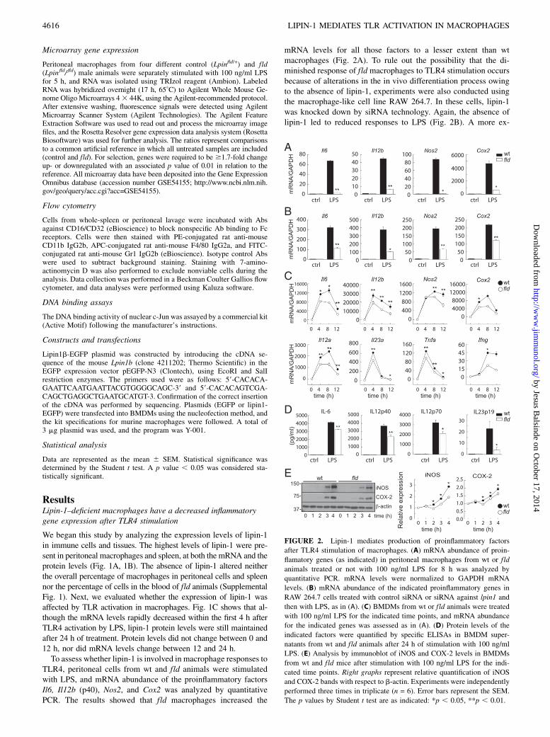
TLR4, peritoneal cells from wt and fld animals were stimulatedwith LPS, and mRNA abundance of the proinflammatory factorsIl6, Il12b (p40), Nos2, and Cox2 was analyzed by quantitativePCR. The results showed that fld macrophages increased the
mRNA levels for all those factors to a lesser extent than wtmacrophages (Fig. 2A). To rule out the possibility that the di-minished response of fld macrophages to TLR4 stimulation occursbecause of alterations in the in vivo differentiation process owingto the absence of lipin-1, experiments were also conducted usingthe macrophage-like cell line RAW 264.7. In these cells, lipin-1was knocked down by siRNA technology. Again, the absence oflipin-1 led to reduced responses to LPS (Fig. 2B). A more ex-
FIGURE 2. Lipin-1 mediates production of proinflammatory factors
after TLR4 stimulation of macrophages. (A) mRNA abundance of proin-
flamatory genes (as indicated) in peritoneal macrophages from wt or fld
animals treated or not with 100 ng/ml LPS for 8 h was analyzed by
quantitative PCR. mRNA levels were normalized to GAPDH mRNA
levels. (B) mRNA abundance of the indicated proinflammatory genes in
RAW 264.7 cells treated with control siRNA or siRNA against lpin1 and
then with LPS, as in (A). (C) BMDMs from wt or fld animals were treated
with 100 ng/ml LPS for the indicated time points, and mRNA abundance
for the indicated genes was assessed as in (A). (D) Protein levels of the
indicated factors were quantified by specific ELISAs in BMDM super-
natants from wt and fld animals after 24 h of stimulation with 100 ng/ml
LPS. (E) Analysis by immunoblot of iNOS and COX-2 levels in BMDMs
from wt and fld mice after stimulation with 100 ng/ml LPS for the indi-
cated time points. Right graphs represent relative quantification of iNOS
and COX-2 bands with respect to b-actin. Experiments were independently
performed three times in triplicate (n = 6). Error bars represent the SEM.
The p values by Student t test are as indicated: *p , 0.05, **p , 0.01.
4616 LIPIN-1 MEDIATES TLR ACTIVATION IN MACROPHAGES
by Jesus Balsinde on O
ctober 17, 2014http://w
ww
.jimm
unol.org/D
ownloaded from
haustive analysis in BMDMs indicated that many inflammation-related genes were affected in fld-derived cells, including Il12a(p35), Il23a (p19), Tnfa, and Ifng (Fig. 2C). These results werealso corroborated by analysis of protein levels in the supernatantsor cell homogenates of TLR4-activated cells (Fig. 2D, 2E). Theobserved responses were not due to a lower expression of TLR4 inthe fld macrophages, as the latter cells expressed TLR4 at levelsidentical to those in wt cells (data not shown). In addition, nodifferences in macrophage cell surface markers were found be-tween wt and fld BMDMs, suggesting no differences in their levelof differentiation (Supplemental Fig. 2). Collectively, these resultssuggest that lipin-1 is centrally involved in the inflammatory re-sponse to TLR4 occupancy in macrophages.
Lipin-1–deficient macrophages show a distinctive geneexpression pattern after TLR4 stimulation
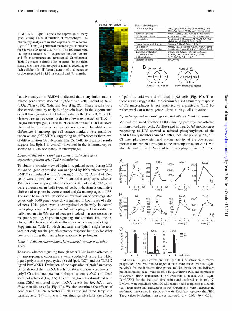
To obtain a broader view of lipin-1–regulated genes during LPSactivation, gene expression was analyzed by RNA microarrays inBMDMs stimulated with LPS during 5 h (Fig. 3). A total of 1640genes were upregulated by LPS in control macrophages, whereas1519 genes were upregulated in fld cells. Of note, only 943 geneswere upregulated in both types of cells, indicating a qualitativedifferential response between control and fld macrophages to LPS.The same behavior was observed on examination of downregulatedgenes; only 1009 genes were downregulated in both types of cells,whereas 1044 genes were downregulated exclusively in controlmacrophages and 780 genes in fld macrophages. Genes differen-tially regulated in fldmacrophages are involved in processes such asreceptor signaling, G-protein signaling, transcription, lipid metab-olism, cell adhesion, and extracellular matrix, among others (Fig. 3,Supplemental Table I), which indicates that lipin-1 might be rele-vant not only for the proinflammatory response but also for otherprocesses during the macrophage response to pathogens.
Lipin-1–deficient macrophages have altered responses to otherTLRs
To assess whether signaling through other TLRs is also affected infld macrophages, experiments were conducted using the TLR3ligand polyinosinic-polycytidylic acid [poly(I:C)] and the TLR1/2ligand Pam3CSK4. Evaluation of the expression of proinflammatorygenes showed that mRNA levels for Il6 and Il13a were lower inpoly(I:C)-stimulated fld macrophages, whereas Nos2 and Cox2were not affected (Fig. 4A). In addition, fld cells stimulated withPam3CSK4 exhibited lower mRNA levels for Il6, Il23a, andNos2 than did wt cells (Fig. 4B). We also examined the effects ofnonclassical TLR4 activators such as the saturated fatty acidpalmitic acid (24). In line with our findings with LPS, the effects
of palmitic acid were diminished in fld cells (Fig. 4C). Thus,these results suggest that the diminished inflammatory responseof fld macrophages is not restricted to a particular TLR butrather works at a more general level during cell activation.
Lipin-1–deficient macrophages exhibit altered TLR4 signaling
We next evaluated whether TLR4 signaling pathways are affectedin lipin-1–deficient cells. As illustrated in Fig. 5, fld macrophagesresponding to LPS showed a reduced phosphorylation of theMAPK family members p44/p42 ERKs, JNK, and p38 (Fig. 5A, 5B).Of note, phosphorylation and nuclear activity of the downstreamprotein c-Jun, which forms part of the transcription factor AP-1, wasalso diminished in LPS-stimulated macrophages from fld mice
FIGURE 3. Lipin-1 affects the expression of many
genes during TLR4 stimulation of macrophages. (A)
Microarray analysis of mRNA expression from control
(Lpin1fld/+) and fld peritoneal macrophages stimulated
for 5 h with 100 ng/ml LPS (n = 4). The 100 genes with
the highest difference in expression between control
and fld macrophages are represented. Supplemental
Table I contains a detailed list of genes. To the right,
some genes have been grouped in families according to
their cellular role. (B) Venn diagrams of total genes up
or downregulated by LPS in control and fld animals.
FIGURE 4. Lipin-1 effects on TLR3 and TLR1/2 activation in macro-
phages. (A) BMDMs from wt or fld animals were treated with 50 mg/ml
poly(I:C) for the indicated time points. mRNA levels for the indicated
proinflammatory genes were assessed by quantitative PCR and normalized
to GAPDH mRNA abundance. (B) BMDMs were stimulated with 1 mg/ml
Pam3CSK4 for the indicated time points and analyzed as in (A). (C)
BMDMs were stimulated with 300 mM palmitic acid complexed to albumin
(2:1 molar ratio) and analyzed as in (A). Experiments were independently
performed three times in triplicate (n = 6). Error bars represent the SEM.
The p values by Student t test are as indicated: *p , 0.05, **p , 0.01.
The Journal of Immunology 4617
by Jesus Balsinde on O
ctober 17, 2014http://w
ww
.jimm
unol.org/D
ownloaded from
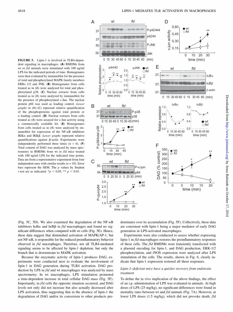
(Fig. 5C, 5D). We also examined the degradation of the NF-kBinhibitors IkBa and IkBb in fld macrophages and found no sig-nificant differences when compared with wt cells (Fig. 5E). Hence,these data suggest that diminished activation of MAPK/AP-1, butnot NF-kB, is responsible for the reduced proinflammatory behaviorobserved in fld macrophages. Therefore, not all TLR4-mediatedsignaling seems to be affected by lipin-1 depletion, but only thebranch that is downstream to MAPK activation.Because the enzymatic activity of lipin-1 produces DAG, ex-
periments were conducted next to evaluate the involvement oflipin-1 in DAG generation during TLR4 activation. DAG pro-duction by LPS in fld and wt macrophages was analyzed by massspectrometry. In wt macrophages, LPS stimulation promoteda time-dependent increase in total cellular DAG mass (Fig. 5F).Importantly, in fld cells the opposite situation occurred, and DAGlevels not only did not increase but also actually decreased afterLPS activation, thus suggesting that in the absence of lipin-1 thedegradation of DAG and/or its conversion to other products pre-
dominates over its accumulation (Fig. 5F). Collectively, these dataare consistent with lipin-1 being a major mediator of early DAGgeneration in LPS-activated macrophages.Experiments were also conducted to assess whether expressing
lipin-1 in fld macrophages restores the proinflammatory responsesof these cells. The fld BMDMs were transiently transfected witha plasmid encoding for lipin-1, and DAG production, ERK-1/2phosphorylation, and iNOS expression were analyzed after LPSstimulation of the cells. The results, shown in Fig. 6, clearly in-dicate that lipin-1 expression restored all these responses.
Lipin-1–deficient mice have a quicker recovery from endotoxintreatment
To define the in vivo implication of the above findings, the effectof an i.p. administration of LPS was evaluated in animals. At highdoses of LPS (25 mg/kg), no significant differences were found inmortality rates between wt and fld animals (Fig. 7A). However, atlower LPS doses (1.5 mg/kg), which did not provoke death, fld
FIGURE 5. Lipin-1 is involved in TLR4-depen-
dent signaling in macrophages. (A) BMDMs from
wt or fld animals were stimulated with 100 ng/ml
LPS for the indicated periods of time. Homogenates
were then evaluated by immunoblot for the presence
of total and phosphorylated MAPK family members
ERKs 1/2 and JNK. (B) Homogenates from cells
treated as in (A) were analyzed for total and phos-
phorylated p38. (C) Nuclear extracts from cells
treated as in (A) were analyzed by immunoblot for
the presence of phosphorylated c-Jun. The nuclear
protein p84 was used as loading control. Lower
graphs in (A)–(C) represent relative quantification
of the phosphoproteins against total protein or
a loading control. (D) Nuclear extracts from cells
treated as (A) were assayed for c-Jun activity using
a commercially available kit. (E) Homogenates
from cells treated as in (A) were analyzed by im-
munoblot for expression of the NF-kB inhibitors
IKKa and IKKb. Lower graphs represent relative
quantifications against b-actin. Experiments were
independently performed three times (n = 6). (F)
Total content of DAG was analyzed by mass spec-
trometry in BMDMs from wt or fld mice treated
with 100 ng/ml LPS for the indicated time points.
Data are from a representative experiment from four
independent ones with similar results (n = 10). Error
bars represent the SEM. The p values by Student
t test are as indicated: *p , 0.05, ** p , 0.01.
4618 LIPIN-1 MEDIATES TLR ACTIVATION IN MACROPHAGES
by Jesus Balsinde on O
ctober 17, 2014http://w
ww
.jimm
unol.org/D
ownloaded from
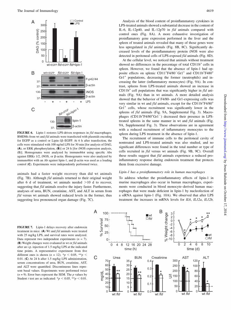
animals had a faster weight recovery than did wt animals(Fig. 7B). Although fld animals returned to their original weightafter 6 d of treatment, wt animals needed .10 d to recover,suggesting that fld animals resolve the injury faster. Furthermore,analyses of urea, BUN, creatinine, AST, and ALT in serum fromfld versus wt animals showed reduced levels in the former, thussuggesting less pronounced organ damage (Fig. 7C).
Analysis of the blood content of proinflammatory cytokines inLPS-treated animals showed a substantial decrease in the content ofIL-6, IL-12p40, and IL-12p70 in fld animals compared withcontrol ones (Fig. 8A). A more exhaustive investigation ofproinflamatory gene expression performed in the liver and thespleen of treated animals revealed that many of those genes wereless upregulated in fld animals (Fig. 8B, 8C). Significantly de-creased levels of the proinflammatory protein iNOS were alsodetected in peritoneal cells of LPS-exposed fld animals (Fig. 8D).At the cellular level, we noticed that animals without treatment
showed no differences in the percentage of total CD11b+ cells inspleen. However, we found that the absence of lipin-1 had op-posite effects on splenic CD11+F4/802Gr1+ and CD11b+F480+
Gr1+ populations, decreasing the former (neutrophils) and in-creasing the latter (inflammatory monocytes) (Fig. 9A). In con-trast, spleens from LPS-treated animals showed an increase inCD11b+ cell populations that was significantly higher in fld ani-mals (Fig. 9A) than in wt animals. A more detailed analysisshowed that the behavior of F4/80- and Gr1-expressing cells wasvery similar in wt and fld animals, except for the CD11b+F4/80+
Gr1+ cells, whose recruitment was significantly lower in thespleens of fld animals (Fig. 9A, Supplemental Fig. 3). Macro-phages (CD11b+F4/80+Gr12) decreased their presence in LPS-treated spleens in the same manner in wt and fld animals (Fig.9A, Supplemental Fig. 3). These observations are in agreementwith a reduced recruitment of inflammatory monocytes to thespleen during LPS treatment in the absence of lipin-1.The recruitment of immune cells to the peritoneal cavity of
nontreated and LPS-treated animals was also studied, and nosignificant differences were found in the total number or type ofcells recruited in fld versus wt animals (Fig. 9B, 9C). Overallthese results suggest that fld animals experience a reduced pro-inflammatory response during endotoxin treatment that protectsthem from excessive damage.
Lipin-1 has a proinflammatory role in human macrophages
To address whether the proinflammatory effects of lipin-1 inmurine macrophages also occur in human macrophages, experi-ments were conducted in blood monocyte–derived human mac-rophages that were made deficient in lipin-1 by nucleofection ofa siRNA against lipin-1 (Fig. 10A). We observed that after LPStreatment the increases in mRNA levels for IL6, IL12a, IL12b,
FIGURE 6. Lipin-1 restores LPS-driven responses in fld macrophages.
BMDMs from wt and fld animals were transfected with plasmids encoding
for EGFP as a control or Lipin-1b–EGFP. At 6 h after transfection, the
cells were stimulated with 100 ng/ml LPS for 30 min [for analysis of DAG,
(A), or ERK phosphorylation, (B)] or 24 h [for iNOS expression analysis,
(B)]. Homogenates were analyzed by immunoblot using specific Abs
against ERKs 1/2, iNOS, or b-actin. Homogenates were also analyzed by
immunoblot with an Ab against lipin-1, and b-actin was used as a loading
control (C). Experiments were independently performed twice.
FIGURE 7. Lipin-1 delays recovery after endotoxin
treatment in mice. (A) Wt and fld animals were treated
with 25 mg/kg LPS, and survival rates were analyzed.
Data represent two independent experiments (n = 7).
(B) Weight changes were evaluated in wt or fld animals
after an i.p. injection of 1.5 mg/kg LPS at the indicated
time points. A representative experiment from five
different ones is shown (n = 12). *p , 0.05, **p ,0.01. (C) At 24 h after 1.5 mg/kg LPS administration,
serum concentrations of urea, BUN, creatinine, AST,
and ALT were quantified. Discontinuous lines repre-
sent basal values. Experiments were performed twice
(n = 9). Error bars represent the SEM. The p values by
Student t test are as indicated: *p , 0.05, **p , 0.01.
The Journal of Immunology 4619
by Jesus Balsinde on O
ctober 17, 2014http://w
ww
.jimm
unol.org/D
ownloaded from

and IL23a in control cells were less prominent in the lipin-1–deficient macrophages. These data are in accordance with ourprevious results in mice.
DiscussionThe execution of immune responses by macrophages requires anexquisite balance between effector and regulatory pathways, andperturbation of this network can result in chronic inflammation orpersistent infection. Thus it is important to define the effectors thatpositively and negatively modulate these responses to open newavenues for the control of inflammation-related conditions. Our
studies demonstrate that lipin-1 plays a key regulatory role in thegeneration of proinflammatory factors by mediating the activationof downstream pathways during TLR activation by microbialcomponents. This conclusion is supported by in vitro data usingprimary human and murine macrophages and cell lines, as well asby in vivo observations obtained from LPS-treated mouse models.Our key findings can be summarized as follows: 1) in macrophages,lipin-1 mediates responses to LPS treatment, regulates cellular DAGlevels, and mediates activation of MAPKs and proteins of thetranscription factor AP-1 that ultimately coordinate the expression ofproinflammatory genes; 2) mRNA expression microarray analysessuggest that lipin-1 also affects other processes like G-protein sig-naling, transcription, cell adhesion, and so forth, in TLR4-stimulatedmacrophages; 3) lipin-1 delays the recovery of animals to endotoxintreatment by limiting the expression of detrimental mediators; 4) theexpression levels of lipin-1 also affect other TLRs in addition toTLR4, namely, TLR3 and TLR1/2; and 5) the proinflammatory roleof lipin-1 is also detected in human macrophages.During LPS stimulation of macrophages, there is an increase in
intracellular DAG content that does not occur in cells lackinglipin-1, suggesting that lipin-1 is a major enzyme involved in theshort-term generation of DAG in LPS-treated cells. A recentexhaustive state-of-the-art lipidomic study performed in RAW264.7 macrophages has also demonstrated increased DAG levelsduring TLR4 stimulation (6). DAG may serve different roles incells. From a lipid viewpoint, its classical role is to serve as a bio-synthetic precursor of different species of phospholipids, whichare important for membrane organization and signaling, and alsoof TAG, the main energy storage of cells. LPS-treated cells dis-play a very robust membrane rearrangement (25, 26); hence theyrequire a high energy supply to accomplish all the remodeling thatactivation encompasses, including the upregulation of many genesand proteins. In fact, TAG production is increased in activatedmacrophages (6, 27–29), and its hydrolysis is an absolute re-quirement for efficient ATP supply and macrophage functioning(30). Thus, the possibility exists that in the absence of lipin-1macrophages may not be able to meet the necessary energy lev-els to fulfill all their activation requirements, resulting in alterationof the whole cell reprograming. In such a scenario, a reduction ofthe whole activated transcriptional program would be expected.However, the wide analysis of gene expression performed in fldmacrophages indicates that in these macrophages the gene tran-scription footprint is quite different from that in wt cells and is notthe result of a mere reduction of transcriptional events. In contrast,the activation of transcriptional effectors such as NF-kB does notseem to be altered in the absence of lipin-1, and this is a processthat also needs ATP for the phosphorylation and degradation ofthe NF-kB inhibitor, IkB. Thus it could be deduced from thesedata that the effects observed for lipin-1 may not necessarily berelated to reduced energy availability, at least during the earlyphases of activation.From an intracellular signaling viewpoint, DAG may bind and
change the activation/localization state of many enzymes in thecell (31). Several DAG-activated enzymes are known to be re-quired for full downstream responses during LPS activation. Forexample, enzymes from the protein kinase C (PKC) family, suchas PKCε and PKCd, can associate with different adaptor proteinsthat are recruited to TLR4 (32, 33). Activation of PKCs requiresphosphorylation and enhanced levels of DAG, and the action ofthese kinases affects TLR4 downstream pathways such as MAPKactivation, which ensures full production of inflammatory factors(34). Our studies support a scenario whereby lipin-1, by regulatingDAG levels and the activation of upstream effectors like PKC,regulates TLR4 downstream signaling. In this regard, we have
FIGURE 8. Lipin-1 contributes to the proinflammatory response that
follows endotoxin treatment in mice. (A) Analysis by specific ELISAs of
proinflamatory factors in the blood of wt or fld mice after an i.p. injection
of 1.5 mg/kg LPS for 6 h. (B and C) mRNA levels of different proin-
flammatory genes analyzed by quantitative PCR in livers (B) or spleens (C)
from wt or fld mice treated as in (A). mRNA levels were normalized to
GAPDH. Experiments were independently performed twice (n = 4). (D)
Peritoneal cell homogenates from animals treated with 1.5 mg/ml for 24 h
were analyzed by immunoblot using Abs against iNOS and b-actin. The
graph on the right shows the relative quantification of iNOS against
b-actin. Responses from three different animals are shown. Experiments
were repeated three times. Error bars represent the SEM. The p values by
Student t test are as indicated: *p , 0.05, ** p , 0.01.
4620 LIPIN-1 MEDIATES TLR ACTIVATION IN MACROPHAGES
by Jesus Balsinde on O
ctober 17, 2014http://w
ww
.jimm
unol.org/D
ownloaded from

previously shown that a Mg2+-dependent phosphatidate phospha-tase activity is involved in arachidonic acid mobilization, COXexpression, and eicosanoid formation when WISH cells are acti-vated through PKC (35, 36) and when macrophages are stimulatedwith LPS (37). Although the mechanisms for these actions are notknown, these results highlight a role for lipin-1 in cell signalingthrough modulating phosphatidic acid and DAG levels. Studies arebeing carried out in our laboratory to further explore these pos-sibilities.Excessive activation of LPS-promoted responses results in
sepsis and septic shock. These are systemic inflammatory con-ditions that constitute a major cause of morbidity and mortality inhospitalized patients. It is clear that alleviation of the exacerbatedproinflammatory response would be a good strategy for theirtreatment. The studies presented in this article suggest that lipin-1participates in the development of these acute conditions. Theabsence of lipin-1 clearly promotes an earlier recovery of animals
treated with a low dose of LPS. The effect seems to be related toa lower expression of harmful proinflammatory mediators, whichmay favor reduced tissue damage and shortens healing time.Furthermore, upregulation of enzymes that generate key factors forthe development of sepsis such as iNOS and COX-2 are alsodecreased in fld animals. In this sense, we have previously re-ported that in human macrophages decreased expression of lipin-1reduces the activation of group IVA cytosolic phospholipase A2,the enzyme that controls the release of arachidonic acid fromphospholipids (11). Availability of free arachidonic acid is welldescribed to constitute a limiting factor for eicosanoid productionvia various pathways, including COX-2 (38, 39). Collectively,these results suggest that lipin-1 may affect sepsis not only byregulating the expression of enzymes such as COX-2 but also byaffecting the levels of their substrates.Owing to the diminished inflammatory response of fld animals,
a lower death rate during treatment with high LPS doses would be
FIGURE 9. Lipin-1 effects on CD11+ cell populations during endotoxin treatment. (A) Splenocytes from wt or fld animals treated or not with 1.5 mg/kg
LPS for 24 h (i.p. injection) were characterized by flow cytometry using Abs against CD11, F4/80, and Gr-1. Gated CD11+ cells were analyzed for F4/80
and Gr-1 expression. (B) Total peritoneal cells from mice treated as in (A) were counted and (C) analyzed by flow cytometry. Percentage of gated CD11+
cells expressing F4/80 or Gr-1 is shown. Experiments were independently performed three times (n = 9). Error bars represent the SEM. The p values by
Student t test are as indicated: *p , 0.05, **p , 0.01.
FIGURE 10. Lipin-1 mediates proinflammatory ac-
tivation in human macrophages. Blood monocyte–de-
rived macrophages were nucleofected with 20 nM
control siRNA or siRNA against Lpin1 and stimulated
with 100 ng/ml LPS for 8 h. (A) Cellular homogenates
were analyzed by immunoblot using Abs against lipin-
1 and b-actin. (B) mRNA levels were analyzed by
quantitative PCR using cyclophilin for normalization.
Experiments were independently performed three
times in triplicate. Error bars represent the SEM. The p
values by Student t test are as indicated: *p , 0.05,
**p , 0.01.
The Journal of Immunology 4621
by Jesus Balsinde on O
ctober 17, 2014http://w
ww
.jimm
unol.org/D
ownloaded from
expected. However, animals die within 24 h of treatment, and nodifferences between wt or fld groups are appreciated. It should benoted in this regard that recent work has shown that fld miceexhibit cardiac dysfunction in vivo (40). Such a defect, togetherwith the well-described cardiac dysfunction induced by high levelsof LPS, could explain why fld mice are not protected against highLPS doses (40, 41).To conclude, in this work we have unveiled a hitherto unrec-
ognized role for lipin-1, an enzyme of lipid metabolism, in mac-rophage signaling and animal responses to bacterial components.The data presented in this article support the idea that reducinglipin-1 levels would limit the inflammatory response and thedamage that exacerbated responses during TLR activation couldproduce. Whether targeted modulation of lipin-1 can providetherapeutic benefits for the control of inflammatory-related con-ditions should be the focus of future research.
AcknowledgmentsWe thank Fernando Martınez, Montserrat Duque, Yolanda Noriega, and
Alvaro Martın for excellent technical assistance. Centro de Investigacion
Biomedica en Red de Diabetes y Enfermedades Metabolicas Asociadas is
an initiative of Instituto de Salud Carlos III.
DisclosuresThe authors have no financial conflicts of interest.
References1. Wynn, T. A., A. Chawla, and J. W. Pollard. 2013. Macrophage biology in de-
velopment, homeostasis and disease. Nature 496: 445–455.2. Medzhitov, R., P. Preston-Hurlburt, and C. A. Janeway, Jr. 1997. A human ho-
mologue of the Drosophila Toll protein signals activation of adaptive immunity.Nature 388: 394–397.
3. Dong, C., R. J. Davis, and R. A. Flavell. 2002. MAP kinases in the immuneresponse. Annu. Rev. Immunol. 20: 55–72.
4. Kawai, T., and S. Akira. 2006. TLR signaling. Cell Death Differ. 13: 816–825.5. Mackman, N., K. Brand, and T. S. Edgington. 1991. Lipopolysaccharide-
mediated transcriptional activation of the human tissue factor gene in THP-1monocytic cells requires both activator protein 1 and nuclear factor kappa Bbinding sites. J. Exp. Med. 174: 1517–1526.
6. Dennis, E. A., R. A. Deems, R. Harkewicz, O. Quehenberger, H. A. Brown,S. B. Milne, D. S. Myers, C. K. Glass, G. Hardiman, D. Reichart, et al. 2010. Amouse macrophage lipidome. J. Biol. Chem. 285: 39976–39985.
7. Huang, Y. L., J. Morales-Rosado, J. Ray, T. G. Myers, T. Kho, M. Lu, andR. S. Munford. 2014. Toll-like receptor agonists promote prolonged triglyceridestorage in macrophages. J. Biol. Chem. 289: 3001–3012.
8. Kennedy, E. P. 1961. Biosynthesis of complex lipids. Fed. Proc. 20: 934–940.9. Peterfy, M., J. Phan, P. Xu, and K. Reue. 2001. Lipodystrophy in the fld mouse
results from mutation of a new gene encoding a nuclear protein, lipin. Nat.Genet. 27: 121–124.
10. Kok, B. P., G. Venkatraman, D. Capatos, and D. N. Brindley. 2012. Unlike twopeas in a pod: lipid phosphate phosphatases and phosphatidate phosphatases.Chem. Rev. 112: 5121–5146.
11. Valdearcos, M., E. Esquinas, C. Meana, L. Gil-de-Gomez, C. Guijas, J. Balsinde,and M. A. Balboa. 2011. Subcellular localization and role of lipin-1 in humanmacrophages. J. Immunol. 186: 6004–6013.
12. Langner, C. A., E. H. Birkenmeier, O. Ben-Zeev, M. C. Schotz, H. O. Sweet,M. T. Davisson, and J. I. Gordon. 1989. The fatty liver dystrophy (fld) mutation.A new mutant mouse with a developmental abnormality in triglyceride metab-olism and associated tissue-specific defects in lipoprotein lipase and hepatic li-pase activities. J. Biol. Chem. 264: 7994–8003.
13. Johnson, C. R., D. Kitz, and J. R. Little. 1983. A method for the derivation andcontinuous propagation of cloned murine bone marrow macrophages. J. Immu-nol. Methods 65: 319–332.
14. Boxio, R., C. Bossenmeyer-Pourie, N. Steinckwich, C. Dournon, and O. N€usse.2004. Mouse bone marrow contains large numbers of functionally competentneutrophils. J. Leukoc. Biol. 75: 604–611.
15. Julius, M. H., E. Simpson, and L. A. Herzenberg. 1973. A rapid method for the iso-lation of functional thymus-derived murine lymphocytes. Eur. J. Immunol. 3: 645–649.
16. Casas, J., C. Meana, E. Esquinas, M. Valdearcos, J. Pindado, J. Balsinde, andM. A. Balboa. 2009. Requirement of JNK-mediated phosphorylation for trans-location of group IVA phospholipase A2 to phagosomes in human macrophages.J. Immunol. 183: 2767–2774.
17. Casas, J., M. Valdearcos, J. Pindado, J. Balsinde, and M. A. Balboa. 2010. Thecationic cluster of group IVA phospholipase A2 (Lys488/Lys541/Lys543/Lys544)
is involved in translocation of the enzyme to phagosomes in human macro-phages. J. Lipid Res. 51: 388–399.
18. Valdearcos, M., E. Esquinas, C. Meana, L. Pena, L. Gil-de-Gomez, J. Balsinde,and M. A. Balboa. 2012. Lipin-2 reduces proinflammatory signaling induced bysaturated fatty acids in macrophages. J. Biol. Chem. 287: 10894–10904.
19. Bligh, E. G., and W. J. Dyer. 1959. A rapid method of total lipid extraction andpurification. Can. J. Biochem. Physiol. 37: 911–917.
20. Astudillo, A. M., G. Perez-Chacon, D. Balgoma, L. Gil-de-Gomez, V. Ruiperez,C. Guijas, M. A. Balboa, and J. Balsinde. 2011. Influence of cellular arachidonicacid levels on phospholipid remodeling and CoA-independent transacylase ac-tivity in human monocytes and U937 cells. Biochim. Biophys. Acta 1811: 97–103.
21. Astudillo, A. M., G. Perez-Chacon, C. Meana, D. Balgoma, A. Pol, M. A. DelPozo, M. A. Balboa, and J. Balsinde. 2011. Altered arachidonate distribution inmacrophages from caveolin-1 null mice leading to reduced eicosanoid synthesis.J. Biol. Chem. 286: 35299–35307.
22. Guijas, C., G. Perez-Chacon, A. M. Astudillo, J. M. Rubio, L. Gil-de-Gomez,M. A. Balboa, and J. Balsinde. 2012. Simultaneous activation of p38 and JNK byarachidonic acid stimulates the cytosolic phospholipase A2-dependent synthesisof lipid droplets in human monocytes. J. Lipid Res. 53: 2343–2354.
23. Wang, X., A. Spandidos, H. Wang, and B. Seed. 2012. PrimerBank: a PCRprimer database for quantitative gene expression analysis, 2012 update. NucleicAcids Res. 40: D1144–D1149.
24. Lee, J. Y., K. H. Sohn, S. H. Rhee, and D. Hwang. 2001. Saturated fatty acids,but not unsaturated fatty acids, induce the expression of cyclooxygenase-2mediated through Toll-like receptor 4. J. Biol. Chem. 276: 16683–16689.
25. Peppelenbosch, M. P., M. DeSmedt, T. ten Hove, S. J. van Deventer, andJ. Grooten. 1999. Lipopolysaccharide regulates macrophage fluid phase pino-cytosis via CD14-dependent and CD14-independent pathways. Blood 93: 4011–4018.
26. Jain, V., A. Halle, K. A. Halmen, E. Lien, M. Charrel-Dennis, S. Ram,D. T. Golenbock, and A. Visintin. 2008. Phagocytosis and intracellular killing ofMD-2 opsonized gram-negative bacteria depend on TLR4 signaling. Blood 111:4637–4645.
27. Kazemi, M. R., C. M. McDonald, J. K. Shigenaga, C. Grunfeld, andK. R. Feingold. 2005. Adipocyte fatty acid-binding protein expression and lipidaccumulation are increased during activation of murine macrophages by toll-likereceptor agonists. Arterioscler. Thromb. Vasc. Biol. 25: 1220–1224.
28. Posokhova, E. N., O. M. Khoshchenko, M. I. Chasovskikh, E. N. Pivovarova,and M. I. Dushkin. 2008. Lipid synthesis in macrophages during inflam-mation in vivo: effect of agonists of peroxisome proliferator activatedreceptors alpha and gamma and of retinoid X receptors. Biochemistry(Mosc.) 73: 296–304.
29. Tannahill, G. M., A. M. Curtis, J. Adamik, E. M. Palsson-McDermott,A. F. McGettrick, G. Goel, C. Frezza, N. J. Bernard, B. Kelly, N. H. Foley, et al.2013. Succinate is an inflammatory signal that induces IL-1b through HIF-1a.Nature 496: 238–242.
30. Chandak, P. G., B. Radovic, E. Aflaki, D. Kolb, M. Buchebner, E. Frohlich,C. Magnes, F. Sinner, G. Haemmerle, R. Zechner, et al. 2010. Efficient phago-cytosis requires triacylglycerol hydrolysis by adipose triglyceride lipase. J. Biol.Chem. 285: 20192–20201.
31. Colon-Gonzalez, F., and M. G. Kazanietz. 2006. C1 domains exposed: fromdiacylglycerol binding to protein-protein interactions. Biochim. Biophys. Acta1761: 827–837.
32. Kubo-Murai, M., K. Hazeki, N. Sukenobu, K. Yoshikawa, K. Nigorikawa,K. Inoue, T. Yamamoto, M. Matsumoto, T. Seya, N. Inoue, and O. Hazeki. 2007.Protein kinase Cdelta binds TIRAP/Mal to participate in TLR signaling. Mol.Immunol. 44: 2257–2264.
33. Faisal, A., A. Saurin, B. Gregory, B. Foxwell, and P. J. Parker. 2008. The scaffoldMyD88 acts to couple protein kinase Cepsilon to Toll-like receptors. J. Biol.Chem. 283: 18591–18600.
34. Loegering, D. J., and M. R. Lennartz. 2011. Protein kinase C and toll-like re-ceptor signaling. Enzyme Res. 2011: 537821.
35. Balboa, M. A., J. Balsinde, and E. A. Dennis. 1998. Involvement of phospha-tidate phosphohydrolase in arachidonic acid mobilization in human amnionicWISH cells. J. Biol. Chem. 273: 7684–7690.
36. Johnson, C. A., M. A. Balboa, J. Balsinde, and E. A. Dennis. 1999. Regulation ofcyclooxygenase-2 expression by phosphatidate phosphohydrolase in humanamnionic WISH cells. J. Biol. Chem. 274: 27689–27693.
37. Grkovich, A., C. A. Johnson, M. W. Buczynski, and E. A. Dennis. 2006.Lipopolysaccharide-induced cyclooxygenase-2 expression in human U937macrophages is phosphatidic acid phosphohydrolase-1-dependent. J. Biol. Chem.281: 32978–32987.
38. Perez-Chacon, G., A. M. Astudillo, D. Balgoma, M. A. Balboa, and J. Balsinde.2009. Control of free arachidonic acid levels by phospholipases A2 and lyso-phospholipid acyltransferases. Biochim. Biophys. Acta 1791: 1103–1113.
39. Astudillo, A. M., D. Balgoma, M. A. Balboa, and J. Balsinde. 2012. Dynamics ofarachidonic acid mobilization by inflammatory cells. Biochim. Biophys. Acta1821: 249–256.
40. Kok, B. P., P. C. Kienesberger, J. R. Dyck, and D. N. Brindley. 2012. Rela-tionship of glucose and oleate metabolism to cardiac function in lipin-1 deficient(fld) mice. J. Lipid Res. 53: 105–118.
41. Tavener, S. A., E. M. Long, S. M. Robbins, K. M. McRae, H. Van Remmen, andP. Kubes. 2004. Immune cell Toll-like receptor 4 is required for cardiac myocyteimpairment during endotoxemia. Circ. Res. 95: 700–707.
4622 LIPIN-1 MEDIATES TLR ACTIVATION IN MACROPHAGES
by Jesus Balsinde on O
ctober 17, 2014http://w
ww
.jimm
unol.org/D
ownloaded from