linking bronchopulmonary dysplasia to adult chronic lung ... · linking bronchopulmonary dysplasia...
TRANSCRIPT
University of Groningen
Linking bronchopulmonary dysplasia to adult chronic lung diseasesOta, Chiharu; Baarsma, Hoeke A; Wagner, Darcy E; Hilgendorff, Anne; Königshoff, Melanie
Published in:Molecular and cellular pediatrics
DOI:10.1186/s40348-016-0062-6
IMPORTANT NOTE: You are advised to consult the publisher's version (publisher's PDF) if you wish to cite fromit. Please check the document version below.
Document VersionPublisher's PDF, also known as Version of record
Publication date:2016
Link to publication in University of Groningen/UMCG research database
Citation for published version (APA):Ota, C., Baarsma, H. A., Wagner, D. E., Hilgendorff, A., & Königshoff, M. (2016). Linkingbronchopulmonary dysplasia to adult chronic lung diseases: role of WNT signaling. Molecular and cellularpediatrics, 3(1), [34]. https://doi.org/10.1186/s40348-016-0062-6
CopyrightOther than for strictly personal use, it is not permitted to download or to forward/distribute the text or part of it without the consent of theauthor(s) and/or copyright holder(s), unless the work is under an open content license (like Creative Commons).
Take-down policyIf you believe that this document breaches copyright please contact us providing details, and we will remove access to the work immediatelyand investigate your claim.
Downloaded from the University of Groningen/UMCG research database (Pure): http://www.rug.nl/research/portal. For technical reasons thenumber of authors shown on this cover page is limited to 10 maximum.
Download date: 03-03-2019
REVIEW Open Access
Linking bronchopulmonary dysplasia toadult chronic lung diseases: role of WNTsignalingChiharu Ota1*, Hoeke A. Baarsma1, Darcy E. Wagner1, Anne Hilgendorff1,2 and Melanie Königshoff1
Abstract
Bronchopulmonary dysplasia (BPD) is one of the most common chronic lung diseases in infants caused by pre-and/or postnatal lung injury. BPD is characterized by arrested alveolarization and vascularization due to extracellularmatrix remodeling, inflammation, and impaired growth factor signaling. WNT signaling is a critical pathway fornormal lung development, and its altered signaling has been shown to be involved in the onset and progression ofincurable chronic lung diseases in adulthood, such as chronic obstructive pulmonary disease (COPD) or idiopathicpulmonary fibrosis (IPF). In this review, we summarize the impact of WNT signaling on different stages of lungdevelopment and its potential contribution to developmental lung diseases, especially BPD, and chronic lungdiseases in adulthood.
Keywords: Bronchopulmonary dysplasia (BPD), WNT signaling, Lung development, Adult chronic lung diseases
IntroductionBronchopulmonary dysplasia (BPD) is one of the mostcommon chronic lung diseases in infants. “Old” or “clas-sical” BPD was first defined by Northway et al. in 1967as structural lung damage and subsequent appearance ofparenchymal fibrosis caused by prolonged hyperoxia andventilator-associated lung injury during the saccular toalveolar stage of lung development [1]. Improvement ofclinical neonatal intensive care practices, includingprenatal steroid therapy, exogenous surfactant adminis-tration, protective lung ventilation strategies, and thecareful monitoring of oxygen supplementation, has ledto a significant reduction in perinatal respiratory-associated death. With the current clinical practices,newborns as early as 23 to 26 weeks of gestation are ableto survive; however, these newborns present with adistinct form of “new” BPD. The prominent new BPDcomprises arrested alveolarization and vascularization,due to the impact of different risk factors on the func-tionally and structurally immature lung during the early
canalicular and saccular periods of lung development[2]. Risk factors include hyperoxia-induced oxygen tox-icity, mechanical ventilation-induced lung injury, and in-fection/inflammation of the lungs, which results inaberrant lung development due to extensive remodelingof the extracellular matrix (ECM), perturbations of in-flammatory response, and impaired growth factor signal-ing [3]. Newborns which have survived and developednew BPD are approaching adolescence and adulthood.Several longitudinal studies following patients with newBPD have demonstrated a decline of forced expiratoryvolume in 1 second (FEV1) compared with term-borncontrols, reflecting the development of airflow obstruc-tion in new BPD survivors over time [2, 4–6]. Thesedata indicate that impairment of alveolarization/vascularization during childhood, which is a feature ofthe new BPD, might contribute to deranged lung alveo-lar injury/repair processes in adulthood.Environmental insults, such as smoking, infection, or
hyperoxia, are known to cause aberrant alveolar repairprocesses in deranged lung development and also theadult lung [7, 8]. These environmental insults contributenot only BPD but also other childhood respiratory dis-eases including bronchial asthma. A better understanding
* Correspondence: [email protected] Pneumology Center, Helmholtz Center Munich,Ludwig-Maximilians-University, University Hospital Grosshadern, GermanCenter of Lung Research (DZL), Munich, GermanyFull list of author information is available at the end of the article
Molecular and CellularPediatrics
© 2016 The Author(s). Open Access This article is distributed under the terms of the Creative Commons Attribution 4.0International License (http://creativecommons.org/licenses/by/4.0/), which permits unrestricted use, distribution, andreproduction in any medium, provided you give appropriate credit to the original author(s) and the source, provide a link tothe Creative Commons license, and indicate if changes were made.
Ota et al. Molecular and Cellular Pediatrics (2016) 3:34 DOI 10.1186/s40348-016-0062-6
of the processes and signaling pathways altered by theseinsults are clearly needed.Impaired signaling of essential lung development path-
ways, such as fibroblast growth factor (FGF) [9, 10],Wingless/integrase-1 (WNT) signaling [11], or bone mor-phogenetic proteins (BMPs) [12], have been reported tocontribute to the pathogenesis of adult chronic lung dis-eases, such as chronic obstructive pulmonary disease(COPD) or idiopathic pulmonary fibrosis (IPF) [13]. Of par-ticular interest, WNT signaling has been linked to aberrantalveolar epithelial injury and repair processes [11, 14–17].Because these pathways are mostly attributed to lung devel-opment and are normally thought to be quiescent in theadult lung, this raises the question of why these pathwaysbecome aberrant in the adult and whether pre- or postnatalinsults impact developmental signal activity early on, thuscontributing to an increased susceptibility for chronic lungdiseases later in life [7, 18]. In this review, we focus on the
potential role of WNT signaling in lung development andperinatal lung disease, with a focus on BPD, as a disease ofimpaired alveolarization/vascularization, and discuss thepotential link between perinatal and adult chronic lung dis-eases, such as COPD and IPF.
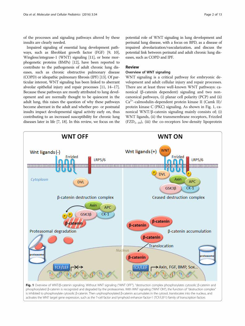
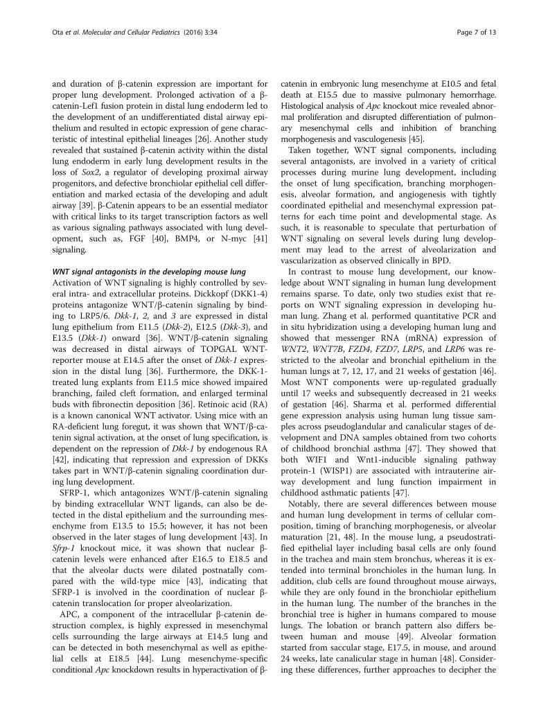
ReviewOverview of WNT signalingWNT signaling is a critical pathway for embryonic de-velopment and adult cellular injury and repair processes.There are at least three well-known WNT pathways: ca-nonical (β-catenin dependent) signaling and two non-canonical pathways, (i) planar cell polarity (PCP) and (ii)Ca2+-calmodulin-dependent protein kinase II (Camk II)/protein kinase C (PKC) signaling. As shown in Fig. 1, ca-nonical WNT/β-catenin signaling mainly consists of; (i)WNT ligands, (ii) the transmembrane receptors, Frizzled(FZD1–10), (iii) the co-receptors low-density lipoprotein
Fig. 1 Overview of WNT/β-catenin signaling. Without WNT signaling (“WNT OFF”), “destruction complex phosphorylates cytosolic β-catenin andphosphorylated β-catenin is recognized and degraded by the proteasomes. With WNT signaling (“WNT ON”), the function of “destruction complex”is inhibited to phosphorylate cytosolic β-catenin. Then unphosphorylated β-catenin accumulates in the cytosol, translocates into the nucleus, andactivates the WNT target gene expression, such as the T-cell factor and lymphoid enhancer factor-1 (TCF/LEF1) family of transcription factors
Ota et al. Molecular and Cellular Pediatrics (2016) 3:34 Page 2 of 13
receptor-related proteins (LRP) 5 and 6, (iv) signalingintermediates, Dishevelleds (DVL1–3), (v) the β-catenin“destruction complex”, (vi) the transcriptional co-activator, β-catenin, and (vii) the transcription factors, Tcell factor and lymphoid enhancer factor (TCF/LEF).Extracellular modulators, such as Dickkopfs (DKK1–4),WNT-inhibitory factor-1 (WIF1), or secreted Frizzled-related proteins (SFRPs), are also important for regulationof the pathway. In the absence of WNT ligands, β-cateninis phosphorylated by the destruction complex, which iscomprised of Axin, adenomatous polyposis coli (APC),glycogen synthase kinase-3 beta (GSK-3β), and caseinkinase-1 (CK1). Phosphorylated β-catenin is recognizedand ubiquitinated by ubiquitin ligase E3 and subsequentlydegraded by the proteasome. Upon WNT ligand bindingto its receptors, the capacity of the destruction complex tophosphorylate cytosolic β-catenin is inhibited. Unpho-sphorylated β-catenin accumulates in the cytosol, translo-cates into the nucleus, and activates WNT target geneexpression, via its integration with the TCF/LEF family oftranscription factors, which is important for cellular pro-liferation, differentiation, and survival [11, 19, 20].Non-canonical WNT signaling (i.e., β-catenin inde-
pendent) mainly consists of (i) the WNT/PCP pathway,which activates c-Jun-N-terminal kinase (JNK) and pro-teins associated with cytoskeleton rearrangement, and(ii) the WNT/Ca2+ pathway, activating Camk II, PKC,the transcription factor nuclear factor of activated T cells(NFAT), and several other (less well defined) transcrip-tion factors [19]. In this review, we primarily focus onthe canonical WNT/β-catenin signaling, which has beeninvestigated most extensively so far.
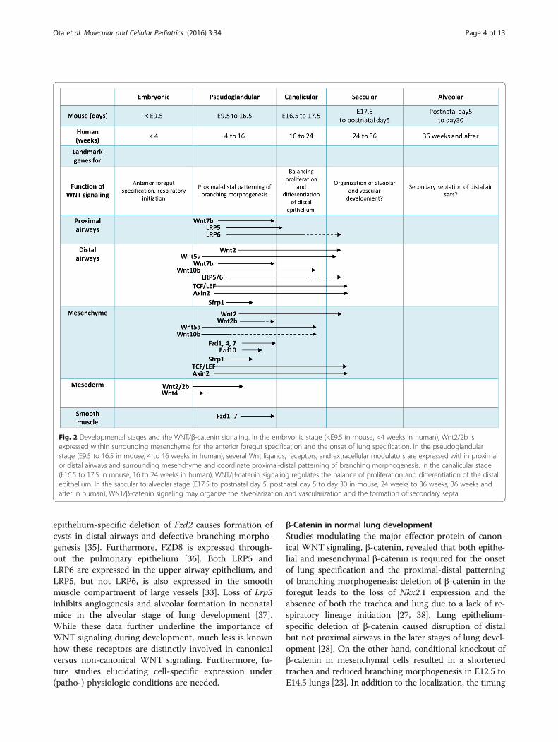
Lung development and WNT signalingHistorically, a large portion of our knowledge about lungdevelopment has been obtained by using wild-type ortransgenic mice [21]. In the mouse lung, embryonic lungdevelopment starts as early as E9.5 (equivalent to 4 weeksin human gestation), with tightly coordinated epithelialand mesenchymal differentiation processes, and is com-pleted postnatally. At this time point, Nkx2.1, a criticalhomeodomain-containing transcription factor for initialrespiratory specification, is expressed within endodermprogenitors in the anterior foregut [21]. Dorsal-ventralspecification occurs according to signals, such as BMPs,FGFs, or WNTs, from the surrounding mesenchyme,endoderm, or mesoderm. Primary lung buds generatetree-like structures for branching morphogenesis fromE9.5 to E16.5 (in human, 4 to 16 weeks, historicallycalled the “pseudoglandular stage”), followed by the“canalicular stage” (E16.5–17.5 in mouse, 16 to 24 weeksin humans) when terminal sacs are formed, the “saccularstage” (E17.5 to postnatal day 5 in mouse, 24 to 36 weeksin human) when distal airways are developed for the
alveoli, and the “alveolar stage” (postnatal day 5 to 30 inmouse, 36 weeks and after delivery in human) when sec-ondary alveolar septa are formed to further divide theairspaces into definitive alveoli (Fig. 2).WNT signaling is active and highly controlled in a
spatio-temporal fashion throughout murine lung endo-derm specification in the foregut as well as cellular pro-liferation and differentiation during lung development[21–24]. In the mouse lung, several loss- or gain-of-function studies revealed the importance of WNT signal-ing in lung morphogenesis [25, 26]. Here, we review therole of active WNT signaling during murine lung devel-opment (summarized in Table 1).
WNT ligands and their receptors in the developing mouselungA number of WNT ligands and receptors have beenidentified as being critical for various stages of develop-ment. Deletion of the canonical Wnt2 ligand causesmouse lung hypoplasia whereas Wnt2/2b double knock-out leads to complete lung agenesis in mice with a lossof Nkx2.1 in early embryonic development in the regionwhere the lung buds are derived from the foregut. ThusWnt2 and 2b are required to specify the Nkx2.1-expressed lung progenitors in the foregut through ca-nonical WNT/β-catenin signaling [27, 28]. Similarly,deletion of murine Wnt7b results in hypoplastic lungswith a proportionate decrease in the replication of bothepithelial and mesenchymal progenitors [29]. The non-canonical Wnt4 was reported to be expressed in the an-terior trunk mesoderm and was found to be essential forproper lung morphogenesis and trachea formation. InWnt4 knockout mice, reduced mesodermal proliferationin the lung bud leads to severe lung hypoplasia and tra-cheal abnormalities [30]. Moreover, Wnt5a, another lig-and of non-canonical WNT signaling, has been detectedas early as E12 at both epithelial and mesenchymal com-partment of the developing lung. The absence of Wnt5aactivity is associated with the overbranching of distal air-ways in murine E15–16 lung together with an architec-tural immaturity of the capillaries and alveolar airspaces[31]. Vice versa, Wnt5a overexpression in the distal epi-thelium results in reduced epithelial branching and di-lated distal airways [32]. These data highlight that bothcanonical as well as non-canonical signal aberrationsaffect normal lung development.In addition to WNT ligands, the receptors have also
been shown to be important for proper lung develop-ment. Tissue-specific analysis of the WNT receptorsfrom E12.5 to E16.5 revealed FZD1, 4, and 7 to be pri-marily expressed in the developing mouse lung mesen-chyme and FZD10 in distal airway epithelium and theexpression of those receptors decreases after E14.5 [33].FZD2 is also highly expressed in distal airways [34], and
Ota et al. Molecular and Cellular Pediatrics (2016) 3:34 Page 3 of 13
epithelium-specific deletion of Fzd2 causes formation ofcysts in distal airways and defective branching morpho-genesis [35]. Furthermore, FZD8 is expressed through-out the pulmonary epithelium [36]. Both LRP5 andLRP6 are expressed in the upper airway epithelium, andLRP5, but not LRP6, is also expressed in the smoothmuscle compartment of large vessels [33]. Loss of Lrp5inhibits angiogenesis and alveolar formation in neonatalmice in the alveolar stage of lung development [37].While these data further underline the importance ofWNT signaling during development, much less is knownhow these receptors are distinctly involved in canonicalversus non-canonical WNT signaling. Furthermore, fu-ture studies elucidating cell-specific expression under(patho-) physiologic conditions are needed.
β-Catenin in normal lung developmentStudies modulating the major effector protein of canon-ical WNT signaling, β-catenin, revealed that both epithe-lial and mesenchymal β-catenin is required for the onsetof lung specification and the proximal-distal patterningof branching morphogenesis: deletion of β-catenin in theforegut leads to the loss of Nkx2.1 expression and theabsence of both the trachea and lung due to a lack of re-spiratory lineage initiation [27, 38]. Lung epithelium-specific deletion of β-catenin caused disruption of distalbut not proximal airways in the later stages of lung devel-opment [28]. On the other hand, conditional knockout ofβ-catenin in mesenchymal cells resulted in a shortenedtrachea and reduced branching morphogenesis in E12.5 toE14.5 lungs [23]. In addition to the localization, the timing
Fig. 2 Developmental stages and the WNT/β-catenin signaling. In the embryonic stage (<E9.5 in mouse, <4 weeks in human), Wnt2/2b isexpressed within surrounding mesenchyme for the anterior foregut specification and the onset of lung specification. In the pseudoglandularstage (E9.5 to 16.5 in mouse, 4 to 16 weeks in human), several Wnt ligands, receptors, and extracellular modulators are expressed within proximalor distal airways and surrounding mesenchyme and coordinate proximal-distal patterning of branching morphogenesis. In the canalicular stage(E16.5 to 17.5 in mouse, 16 to 24 weeks in human), WNT/β-catenin signaling regulates the balance of proliferation and differentiation of the distalepithelium. In the saccular to alveolar stage (E17.5 to postnatal day 5, postnatal day 5 to day 30 in mouse, 24 weeks to 36 weeks, 36 weeks andafter in human), WNT/β-catenin signaling may organize the alveolarization and vascularization and the formation of secondary septa
Ota et al. Molecular and Cellular Pediatrics (2016) 3:34 Page 4 of 13
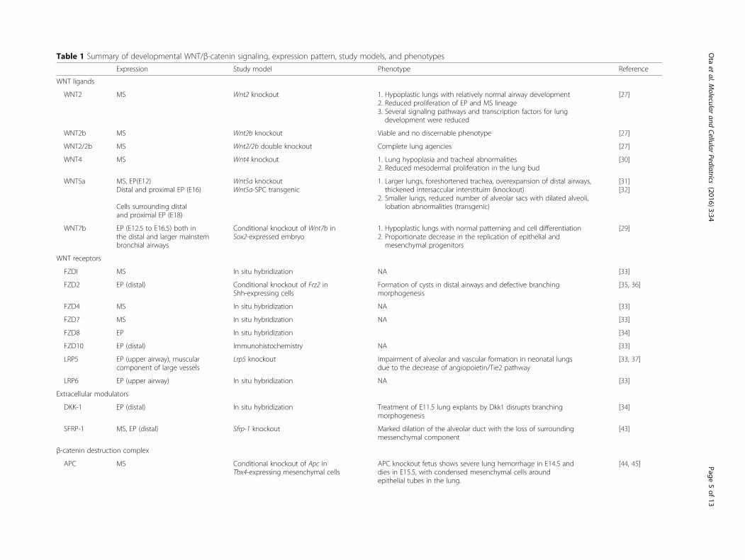
Table 1 Summary of developmental WNT/β-catenin signaling, expression pattern, study models, and phenotypes
Expression Study model Phenotype Reference
WNT ligands
WNT2 MS Wnt2 knockout 1. Hypoplastic lungs with relatively normal airway development2. Reduced proliferation of EP and MS lineage3. Several signaling pathways and transcription factors for lungdevelopment were reduced
[27]
WNT2b MS Wnt2b knockout Viable and no discernable phenotype [27]
WNT2/2b MS Wnt2/2b double knockout Complete lung agencies [27]
WNT4 MS Wnt4 knockout 1. Lung hypoplasia and tracheal abnormalities2. Reduced mesodermal proliferation in the lung bud
[30]
WNT5a MS, EP(E12)Distal and proximal EP (E16)
Cells surrounding distaland proximal EP (E18)
Wnt5a knockoutWnt5a-SPC transgenic
1. Larger lungs, foreshortened trachea, overexpansion of distal airways,thickened intersaccular interstituim (knockout)
2. Smaller lungs, reduced number of alveolar sacs with dilated alveoli,lobation abnormalities (transgenic)
[31][32]
WNT7b EP (E12.5 to E16.5) both inthe distal and larger mainstembronchial airways
Conditional knockout of Wnt7b inSox2-expressed embryo
1. Hypoplastic lungs with normal patterning and cell differentiation2. Proportionate decrease in the replication of epithelial andmesenchymal progenitors
[29]
WNT receptors
FZDI MS In situ hybridization NA [33]
FZD2 EP (distal) Conditional knockout of Frz2 inShh-expressing cells
Formation of cysts in distal airways and defective branchingmorphogenesis
[35, 36]
FZD4 MS In situ hybridization NA [33]
FZD7 MS In situ hybridization NA [33]
FZD8 EP In situ hybridization [34]
FZD10 EP (distal) Immunohistochemistry NA [33]
LRP5 EP (upper airway), muscularcomponent of large vessels
Lrp5 knockout Impairment of alveolar and vascular formation in neonatal lungsdue to the decrease of angiopoietin/Tie2 pathway
[33, 37]
LRP6 EP (upper airway) In situ hybridization NA [33]
Extracellular modulators
DKK-1 EP (distal) In situ hybridization Treatment of E11.5 lung explants by Dkk1 disrupts branchingmorphogenesis
[34]
SFRP-1 MS, EP (distal) Sfrp-1 knockout Marked dilation of the alveolar duct with the loss of surroundingmessenchymal component
[43]
β-catenin destruction complex
APC MS Conditional knockout of Apc inTbx4-expressing mesenchymal cells
APC knockout fetus shows severe lung hemorrhage in E14.5 anddies in E15.5, with condensed mesenchymal cells aroundepithelial tubes in the lung.
[44, 45]
Ota
etal.M
olecularand
CellularPediatrics
(2016) 3:34 Page
5of
13
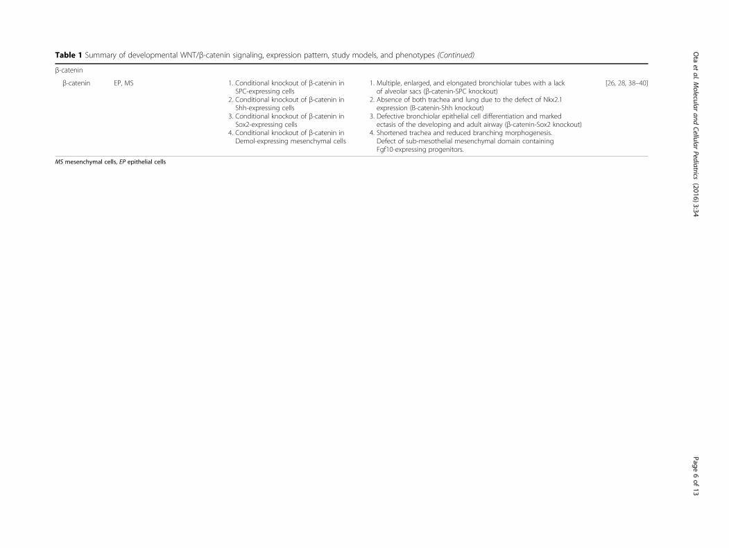
Table 1 Summary of developmental WNT/β-catenin signaling, expression pattern, study models, and phenotypes (Continued)
β-catenin
β-catenin EP, MS 1. Conditional knockout of β-catenin inSPC-expressing cells
2. Conditional knockout of β-catenin inShh-expressing cells
3. Conditional knockout of β-catenin inSox2-expressing cells
4. Conditional knockout of β-catenin inDemol-expressing mesenchymal cells
1. Multiple, enlarged, and elongated bronchiolar tubes with a lackof alveolar sacs (β-catenin-SPC knockout)
2. Absence of both trachea and lung due to the defect of Nkx2.1expression (B-catenin-Shh knockout)
3. Defective bronchiolar epithelial cell differentiation and markedectasis of the developing and adult airway (β-catenin-Sox2 knockout)
4. Shortened trachea and reduced branching morphogenesis.Defect of sub-mesothelial mesenchymal domain containingFgf10-expressing progenitors.
[26, 28, 38–40]
MS mesenchymal cells, EP epithelial cells
Ota
etal.M
olecularand
CellularPediatrics
(2016) 3:34 Page
6of
13
and duration of β-catenin expression are important forproper lung development. Prolonged activation of a β-catenin-Lef1 fusion protein in distal lung endoderm led tothe development of an undifferentiated distal airway epi-thelium and resulted in ectopic expression of gene charac-teristic of intestinal epithelial lineages [26]. Another studyrevealed that sustained β-catenin activity within the distallung endoderm in early lung development results in theloss of Sox2, a regulator of developing proximal airwayprogenitors, and defective bronchiolar epithelial cell differ-entiation and marked ectasia of the developing and adultairway [39]. β-Catenin appears to be an essential mediatorwith critical links to its target transcription factors as wellas various signaling pathways associated with lung devel-opment, such as, FGF [40], BMP4, or N-myc [41]signaling.
WNT signal antagonists in the developing mouse lungActivation of WNT signaling is highly controlled by sev-eral intra- and extracellular proteins. Dickkopf (DKK1-4)proteins antagonize WNT/β-catenin signaling by bind-ing to LRP5/6. Dkk-1, 2, and 3 are expressed in distallung epithelium from E11.5 (Dkk-2), E12.5 (Dkk-3), andE13.5 (Dkk-1) onward [36]. WNT/β-catenin signalingwas decreased in distal airways of TOPGAL WNT-reporter mouse at E14.5 after the onset of Dkk-1 expres-sion in the distal lung [36]. Furthermore, the DKK-1-treated lung explants from E11.5 mice showed impairedbranching, failed cleft formation, and enlarged terminalbuds with fibronectin deposition [36]. Retinoic acid (RA)is a known canonical WNT activator. Using mice with anRA-deficient lung foregut, it was shown that WNT/β-ca-tenin signal activation, at the onset of lung specification, isdependent on the repression of Dkk-1 by endogenous RA[42], indicating that repression and expression of DKKstakes part in WNT/β-catenin signaling coordination dur-ing lung development.SFRP-1, which antagonizes WNT/β-catenin signaling
by binding extracellular WNT ligands, can also be de-tected in the distal epithelium and the surrounding mes-enchyme from E13.5 to 15.5; however, it has not beenobserved in the later stages of lung development [43]. InSfrp-1 knockout mice, it was shown that nuclear β-catenin levels were enhanced after E16.5 to E18.5 andthat the alveolar ducts were dilated postnatally com-pared with the wild-type mice [43], indicating thatSFRP-1 is involved in the coordination of nuclear β-catenin translocation for proper alveolarization.APC, a component of the intracellular β-catenin de-
struction complex, is highly expressed in mesenchymalcells surrounding the large airways at E14.5 lung andcan be detected in both mesenchymal as well as epithe-lial cells at E18.5 [44]. Lung mesenchyme-specificconditional Apc knockdown results in hyperactivation of β-
catenin in embryonic lung mesenchyme at E10.5 and fetaldeath at E15.5 due to massive pulmonary hemorrhage.Histological analysis of Apc knockout mice revealed abnor-mal proliferation and disrupted differentiation of pulmon-ary mesenchymal cells and inhibition of branchingmorphogenesis and vasculogenesis [45].Taken together, WNT signal components, including
several antagonists, are involved in a variety of criticalprocesses during murine lung development, includingthe onset of lung specification, branching morphogen-esis, alveolar formation, and angiogenesis with tightlycoordinated epithelial and mesenchymal expression pat-terns for each time point and developmental stage. Assuch, it is reasonable to speculate that perturbation ofWNT signaling on several levels during lung develop-ment may lead to the arrest of alveolarization andvascularization as observed clinically in BPD.In contrast to mouse lung development, our know-
ledge about WNT signaling in human lung developmentremains sparse. To date, only two studies exist that re-ports on WNT signaling expression in developing hu-man lung. Zhang et al. performed quantitative PCR andin situ hybridization using a developing human lung andshowed that messenger RNA (mRNA) expression ofWNT2, WNT7B, FZD4, FZD7, LRP5, and LRP6 was re-stricted to the alveolar and bronchial epithelium in thehuman lungs at 7, 12, 17, and 21 weeks of gestation [46].Most WNT components were up-regulated graduallyuntil 17 weeks and subsequently decreased in 21 weeksof gestation [46]. Sharma et al. performed differentialgene expression analysis using human lung tissue sam-ples across pseudoglandular and canalicular stages of de-velopment and DNA samples obtained from two cohortsof childhood bronchial asthma [47]. They showed thatboth WIF1 and Wnt1-inducible signaling pathwayprotein-1 (WISP1) are associated with intrauterine air-way development and lung function impairment inchildhood asthmatic patients [47].Notably, there are several differences between mouse
and human lung development in terms of cellular com-position, timing of branching morphogenesis, or alveolarmaturation [21, 48]. In the mouse lung, a pseudostrati-fied epithelial layer including basal cells are only foundin the trachea and main stem bronchus, whereas it is ex-tended into terminal bronchioles in the human lung. Inaddition, club cells are found throughout mouse airways,while they are only found in the bronchiolar epitheliumin the human lung. The number of the branches in thebronchial tree is higher in humans compared to mouselungs. The lobation or branch pattern also differs be-tween human and mouse [49]. Alveolar formationstarted from saccular stage, E17.5, in mouse, and around24 weeks, late canalicular stage in human [48]. Consider-ing these differences, further approaches to decipher the
Ota et al. Molecular and Cellular Pediatrics (2016) 3:34 Page 7 of 13
role of developmental signaling pathways in human lungdevelopment are required, using, e.g., human histologicalsamples [50] or human induced pluripotent stem (iPS)cells [48].
Involvement of WNT signaling in early lung injury andadult chronic lung diseasesA number of environmental insults during pre- andpostnatal development are known to induce derangedlung morphogenesis and have also been shown to affectWNT signaling. Several studies have been conducted toinvestigate whether insults to the developing lung byincidental environmental factors, such as postnatal infec-tion or maternal cigarette smoke as well as medicalinterventions, such as ventilation and oxygen supple-mentation, affect lung morphogenesis and repair processvia WNT signaling. Neonatal hyperoxia is one of thewidely used animal (mainly rodent) models to mimicBPD, i.e., causing impaired alveolarization/vascularizationin neonatal lungs [51]. Neonatal hyperoxia following ma-ternal bacterial infection is another rodent model for BPD[51, 52]. Since BPD is caused by a variety of factor, includ-ing hyperoxia exposure, intrauterine infection, high-pressure ventilation, or prematurity of the lungs, rodentmodels of BPD, including hyperoxia exposure, do notcompletely recapitulate the BPD observed in clinical set-tings. However, because the lung samples from human ne-onates are rare, those rodent models of BPD, includinghyperoxia exposure models focusing especially on alveo-larization/vascularization, are important tools to revealthe pathophysiology of new BPD.These pre- and postnatal insults can result in a variety
of early lung diseases next to BPD. In particular, bron-chial asthma exhibits a high incidence in childhood andadolescence. Bronchial asthma is considered to be highlyinfluenced by maternal smoking [53], diet [54], intra-uterine growth restriction [55, 56], or exposure to patho-gens [57, 58]. In this next section, we discuss theenvironmental insult-associated with altered WNT/β-ca-tenin signaling in lung injury occurring during early lungdevelopment and its potential contribution to adultchronic lung diseases.
Infection and inflammationPrenatal infections are known to impact lung develop-ment. Premature rupture of the amniotic membrane re-sults in increased susceptibility to intrauterine infections.Antenatal inflammation of chorioamniotic membranescauses premature birth and adversely affects lung develop-ment [59]. Intra-amniotic lipopolysaccharide (LPS) expos-ure, which mimics amniotic bacterial infection, decreasesthe expression of Lef-1, Wnt1, Wnt4, and β-catenin in thecanalicular stage of lung development [60]. Similarly, inadult mice, acute lung injury caused by intra-tracheal
application of LPS and followed by high tidal volumemechanical ventilation results in the activation of DKK1and the subsequent down-regulation of active β-catenin inthe lung alveolar epithelium. It was shown that DKK-1 isreleased from activated platelets, and the binding affinityof DKK-1 to alveolar epithelial cells was increased duringacute lung inflammation [61]. Although only a few reportshave shown the relationship between early lung infection/inflammation and WNT signaling so far, the data todate are intriguing and further studies investigatinghuman lungs undergoing prenatal infections, such aschorioamnionitis-induced neonatal lung injury, will beimportant.
Smoking-related injuryMaternal smoking is one of the important risk factorsfor chronic lung diseases in children including recurrentrespiratory infection, infantile wheezing, bronchialasthma, and lower respiratory function in early adult-hood [53, 62, 63]. It has also been shown that maternalsmoking affects alveolarization/vascularization in devel-oping lung in vivo [64, 65]. Bronchial asthma sharessome similarities with BPD (e.g., pathologic airways andthe presence of clinical symptoms like wheezing) andhas been associated with aberrant WNT signaling as well[47, 66]. Impaired lung growth by these and other envir-onmental factors may cause the formation of smallerairways and decreased lung capacity contributing tochildhood asthma and lower respiratory function in earlyadulthood [67]. In the adult lung, dysfunction of WNTsignaling contributes to the impaired epithelial repairprocesses in disease [17, 68]. WNT signaling is reducedin the lungs from COPD patients, a smoking-related dis-ease, and the pharmacological activation of the signalingpathway through GSK3β inhibition activates epithelialrepair properties and attenuates known pathologicalfeatures of emphysema ex vivo [68] and in vivo [17, 69].Recently, Jiang et al. reported that FAM13A, a gene as-sociated with COPD susceptibility [69], might lead toemphysema development by facilitating β-catenin deg-radation [70]. Furthermore, WNT/β-catenin signalingcomponents, including canonical WNT ligands, FZDs,signal transducers, and target genes are down-regulated,while antagonists such as SFRP-1 and DKK-1 are up-regulated, in human lung tissue [43] and in particularin the small airway epithelium [71, 72] from COPDpatients.Much less is known about the effects of maternal
smoking on WNT signaling during lung development.Maternal smoking during pregnancy has been shown todecrease Fzd7 and Ctnnb1 (β-catenin) mRNA in neo-natal Balb/c mice [73]. Furthermore, it was recently re-ported that protein and mRNA expression of SFRP-1were significantly up-regulated in the placental tissues in
Ota et al. Molecular and Cellular Pediatrics (2016) 3:34 Page 8 of 13

smoking women compared with those from non-smokers [74]. Furthermore, a carbon monoxide analog,one of the components in cigarettes, increased SFRP-1expression accompanied by decreased WNT/β-cateninsignaling in a human trophoblast cell line. In addition,maternal Sfrp-1 overexpression causes fetal growth re-striction in mice [74]. Altogether, these studies stronglysuggest that (maternal) smoking and components ofcigarette smoke significantly impact WNT signaling ac-tivity. However, cigarette smoking is also known to gen-erally inhibit fetal growth and thus it remains an openquestion whether maternal smoking/nicotine exposuredirectly increases extracellular modulators of WNT anddecreases WNT/β-catenin signaling in the lung to affectfetal lung development or whether maternal smoking/nicotine exposure induces fetal growth restriction tocause premature birth and subsequent BPD.Given these studies, it is plausible that chronic lung
diseases, such as COPD, develop as a result of early lunginsults leading to aberrant WNT/β-catenin signaling andthus lung repair capacity. A gradual decline in lungfunction in early adulthood might be due to aberrantWNT/β-catenin signaling, which is retained over time,eventually resulting in adult chronic lung diseases. Gen-eration of experimental models to follow this hypothesisand long-term follow-up studies of new BPD patientsare needed. In particular, emerging evidences suggestthat epigenetic alterations, i.e., modified gene expressionvia DNA methylation, histone modification, or micro-RNA, of WNT signaling represents an important area ofinvestigation. Recently, it has been shown that cigarettesmoke exposure epigenetically altered WNT/β-cateninsignaling in lung cancer cells by histone modification ormicroRNA expression [75–77]. In the developing lung,differential methylation of WNT/β-catenin signal geneshave been reported in neonatal and adult mouse lungs[78]. Although there is no study addressing epigeneticalterations of WNT signaling by cigarette smoke expos-ure in the developing lung, it is reported that psycho-logical stress during pregnancy caused altered DNAmethylation of non-canonical, WNT5a/Ca2+ pathwayand postnatal wheeze of the affected children [66]. Assuch, further investigations on how environmental fac-tors, including maternal smoking, alter WNT signalingby epigenetic modifications and thus affect lung develop-ment of the neonate will be important.
WNT/β-catenin and TGF-β signaling in new BPDIn addition to WNT/β-catenin, transforming growth fac-tor (TGF)-β signaling is a critical pathway for lung de-velopment [21]. It has been shown that TGF-β is animportant mediator for the development of BPD [79–81]and is activated in neonatal rat lungs after hyperoxia ex-posure [82] as well as in neonatal mouse lungs after
mechanical ventilation with mild hyperoxia [83]. Thereare also reports regarding the dual activation of WNT/β-catenin and TGF-β signaling in hyperoxia exposuremodels, but the crosstalk between the two pathways isincompletely understood [82, 84]. Active WNT/β-ca-tenin signaling has been reported in fibrotic adult lungdiseases, such as IPF [14–16], in which TGF-β signalingis highly involved in epithelial cell reprogramming andmyofibroblast activation [85]. Furthermore, TGF-β re-sults in enhanced expression of WNT ligands andactivation of β-catenin in vitro [86]. TGF-β-induced acti-vation of WNT/β-catenin signaling [87, 88] may alsoplay a key role during developing BPD as well as adult fi-brotic lung diseases, including IPF.
WNT signaling in BPDTo date, only a few studies addressed WNT signaling innew BPD patients. In the lungs of patients who diedfrom BPD, nuclear β-catenin, which is used as a surro-gate marker for WNT/β-catenin activity, along withphosphorylated (inactivated) GSK-3β was found in thethickened alveolar septa [89, 90]. Notably, whole exomesequencing using blood spots from twin neonate pairswith and without BPD revealed that genes associatedwith WNT/β-catenin signaling were up-regulated inBPD [91].Nuclear translocation of β-catenin and increased Lef1
expression was observed in the lung from neonatal ratsexposed to hyperoxia (95 % oxygen) in the alveolar stage(postnatal days 0 to 7) [82]. Moreover, it has been shownthat neonatal hyperoxia increased nuclear β-catenin anddecreased alveolar epithelial type II (ATII) cell to ATIcell transdifferentiation [84, 92], which is generallyconsidered as a repair process of alveolar epithelial cellsfollowing injury. Furthermore, hyperoxia-induced inhib-ition of ATII to ATI transdifferentiation was recoveredby small interfering RNA (siRNA)-mediated knockdownof Wnt3a in vitro [92]. However, it has been reportedthat in the adult mouse lung, β-catenin was inducedduring ATII cell to ATI cell transdifferentiation in nor-moxia condition [93, 94]. This discrepancy might be dueto hyperoxia condition or using neonatal lung in theformer studies. Further studies are needed to clarify thisissue. Another study showed the enhancement of WNT/β-catenin signaling in impaired vascularization [95].Taken together, canonical WNT/β-catenin signaling isactivated in lung samples from BPD patients and neo-natal rodent model of hyperoxia exposure in the lung.This activation of WNT signaling might be a result of“attempted (and failed)” regeneration after injury of al-veolar epithelial cells, which is a hypothesized processesmodel in IPF lungs [11].It is unclear whether BPD contributes to the onset of
adult chronic lung diseases, such as COPD or IPF. In
Ota et al. Molecular and Cellular Pediatrics (2016) 3:34 Page 9 of 13
adult chronic lung diseases, canonical WNT/β-cateninsignaling is down-regulated in emphysematous lungs[17] while up-regulated in fibrotic lungs [15]. In BPDlungs, as mentioned above, enhanced expression ofTGF-β [79] and/or WNT/β-catenin signaling was ob-served [89, 90]. In contrast, intrauterine infection orcigarette smoke decreased WNT/β-catenin signaling.Longitudinal studies showed that along with decreasedFEV1, a hallmark of obstructive lung diseases, forcedvital capacity (FVC), a hallmark of restrictive lungdiseases, was also lower in BPD survivors [96, 97]. Al-though characterizing a disease entity as either emphy-sema or fibrosis is oversimplification, it seems like BPDfeatures a co-existence of emphysema and fibrosis asreported [98, 99]. It is possible that different environmentalinsults at different time points during lung developmentmight cause different expression patterns of WNT signal-ing. Also, if new BPD survivors with impaired alveolariza-tion/vascularization are exposed to a “second hit,” such ascigarette smoke, pathogens, or hyperoxia, at a later timepoint, they might develop adult chronic lung diseases withaberrant (increased/decreased) WNT signaling. Longitu-dinal studies are needed to study whether BPD survivorsare more susceptible to developing adult chronic lung dis-eases. Establishing animal models to mimic BPD and followthe outcome of the developing lung is needed.
Clinical implications and limitations of WNT/β-cateninsignaling in lung developmentSeveral studies indicated that targeting WNT/β-cateninsignaling may be a therapeutic strategy in BPD. VitaminA, whose metabolite is RA, has been used to preventBPD progression [100–102] although the effect is stillcontroversial. As mentioned previously, RA activatedWNT/β-catenin signaling via inhibition of DKK-1 [42].In this context, active WNT/β-catenin signaling mightbe beneficial for arrested alveolarization, as it has beenreported to maintain alveolar stem/progenitor cell popu-lations [11, 103], such as ATII cells [94].On the other hand, it is reported that intraperitoneal
administration of Mesd, a specialized chaperone forLRP5/6 to inhibit WNT/β-catenin signaling, attenuatedhyperoxia-induced pulmonary hypertension and rightventricular hypertrophy in neonatal rats [95]. Anotherstudy showed that ICG-001, a small molecule which in-hibits WNT/β-catenin signaling via interaction betweenβ-catenin and CREB-binding protein (CBP), an intrinsichistone acetyltransferase to activate gene transcription,increased alveolarization and decreased vascular remod-eling to develop pulmonary hypertension [104]. Resvera-trol, a polyphenol found in several fruits and nuts, wasalso shown to attenuate hyperoxia-induced model ofBPD in neonatal rats [64, 84]. It is important to addressthe question when or where WNT/β-catenin signaling
should be inactivated/activated for physiologic lung de-velopment and perinatal lung injury/repair processes.Thus, it is worth exploring whether the attenuation ofalveolar repair process or epigenetic modification alter-ing WNT signaling will be candidates for clinicalimplications.However, as most of the data shown here originate
from rodent experiments, limitations for translation ofthe findings have to be considered. Given the difficultyof obtaining human neonatal lung tissue for analysis, re-cent approaches using 3D lung tissue cultures [68] oriPS cells [105, 106] from BPD patients represent promis-ing tools to further explore signaling pathways involvedin the pathogenesis of disease, such as WNT/β-cateninsignaling. Collecting more evidence from preterm in-fants will be needed to identify new therapeutic targetsin WNT/β-catenin signaling pathway.
ConclusionsHere, we discussed the potential role of the developmen-tal WNT signaling pathway as a potential missing linkbetween early impairment of lung development and theoutcome in the adult lung. First, WNT/β-catenin signal-ing is essential for lung development in utero, which hasbeen elegantly investigated using the advantage of sev-eral wild-type and transgenic animals. Second, growingdata suggest that WNT/β-catenin signaling is involvedin pre- and postnatal lung injury and repair process; andthird, several lines of evidence exists that highlight theimpact of impaired WNT/β-catenin signaling on the de-velopment of adult chronic lung diseases, which seemssimilar to lung injury-repair processes in the developinglung. Additional studies are needed to advance ourcurrent knowledge of the pathogenesis of perinatal lungdiseases, such as BPD, to shed further light into signal-ing pathways involved that ultimately might lead tonovel therapeutic options for lung injury-repair processor epigenetic modifications in WNT signaling.
FundingThis work is supported by a postdoctoral research fellowship from the EuropeanRespiratory Society (Ota C, LTRF-2015-4822, Baarsma HA, LTRF-79-2012)and Helmholtz Postdoctoral Programme (Baarsma HA, PD-135), a WhitakerInternational Scholar Fellowship, and the Helmholtz Munich PostdoctoralProgramme (Wagner DE).
Authors’ contributionsOC drafted the manuscript and contributed to the conception and thedesign and the acquisition of the data. BHA, WDE, HA, KM contributed to theacquisition of the data and revised the manuscript critically for importantintellectual content. All authors read and approved the final manuscript.
Competing interestsThe authors declare that they have no competing interests.
Author details1Comprehensive Pneumology Center, Helmholtz Center Munich,Ludwig-Maximilians-University, University Hospital Grosshadern, German
Ota et al. Molecular and Cellular Pediatrics (2016) 3:34 Page 10 of 13
Center of Lung Research (DZL), Munich, Germany. 2The Perinatal Center,Campus Grosshadern, Ludwig-Maximilians-University, Munich, Germany.
Received: 10 March 2016 Accepted: 25 September 2016
References1. Northway WH Jr, Rosan RC, Porter DY (1967) Pulmonary disease following
respirator therapy of hyaline-membrane disease. Bronchopulmonarydysplasia. N Engl J Med 276(7):357–368. doi:10.1056/NEJM196702162760701
2. Baraldi E, Filippone M (2007) Chronic lung disease after premature birth. NEngl J Med 357(19):1946–1955. doi:10.1056/NEJMra067279
3. Madurga A, Mizikova I, Ruiz-Camp J, Morty RE (2013) Recent advances inlate lung development and the pathogenesis of bronchopulmonarydysplasia. American journal of physiology Lung cellular and molecularphysiology 305(12):L893–L905. doi:10.1152/ajplung.00267.2013
4. Vollsaeter M, Roksund OD, Eide GE, Markestad T, Halvorsen T (2013) Lungfunction after preterm birth: development from mid-childhood toadulthood. Thorax 68(8):767–776. doi:10.1136/thoraxjnl-2012-202980
5. Ronkainen E, Dunder T, Peltoniemi O, Kaukola T, Marttila R, Hallman M(2015) New BPD predicts lung function at school age: follow-up study andmeta-analysis. Pediatr Pulmonol 50(11):1090–1098. doi:10.1002/ppul.23153
6. Gibson AM, Reddington C, McBride L, Callanan C, Robertson C, Doyle LW(2015) Lung function in adult survivors of very low birth weight, with andwithout bronchopulmonary dysplasia. Pediatr Pulmonol 50(10):987–994. doi:10.1002/ppul.23093
7. Stocks J, Hislop A, Sonnappa S (2013) Early lung development: lifelongeffect on respiratory health and disease. Lancet Respir Med 1(9):728–742.doi:10.1016/S2213-2600(13)70118-8
8. Harding R, Maritz G (2012) Maternal and fetal origins of lung disease inadulthood. Seminars in fetal & neonatal medicine 17(2):67–72. doi:10.1016/j.siny.2012.01.005
9. Kranenburg AR, De Boer WI, Van Krieken JH, Mooi WJ, Walters JE, Saxena PR,Sterk PJ, Sharma HS (2002) Enhanced expression of fibroblast growth factorsand receptor FGFR-1 during vascular remodeling in chronic obstructivepulmonary disease. Am J Respir Cell Mol Biol 27(5):517–525. doi:10.1165/rcmb.4474
10. Morino S, Nakamura T, Toba T, Takahashi M, Kushibiki T, Tabata Y, Shimizu Y(2005) Fibroblast growth factor-2 induces recovery of pulmonary blood flow incanine emphysema models. Chest 128(2):920–926. doi:10.1378/chest.128.2.920
11. Konigshoff M, Eickelberg O (2010) WNT signaling in lung disease: a failure ora regeneration signal? Am J Respir Cell Mol Biol 42(1):21–31. doi:10.1165/rcmb.2008-0485TR
12. Myllarniemi M, Lindholm P, Ryynanen MJ, Kliment CR, Salmenkivi K, Keski-Oja J, Kinnula VL, Oury TD, Koli K (2008) Gremlin-mediated decrease in bonemorphogenetic protein signaling promotes pulmonary fibrosis. Am J RespirCrit Care Med 177(3):321–329. doi:10.1164/rccm.200706-945OC
13. Beers MF, Morrisey EE (2011) The three R’s of lung health and disease:repair, remodeling, and regeneration. J Clin Invest 121(6):2065–2073.doi:10.1172/JCI45961
14. Chilosi M, Poletti V, Zamo A, Lestani M, Montagna L, Piccoli P, Pedron S,Bertaso M, Scarpa A, Murer B, Cancellieri A, Maestro R, Semenzato G,Doglioni C (2003) Aberrant Wnt/beta-catenin pathway activation inidiopathic pulmonary fibrosis. Am J Pathol 162(5):1495–1502
15. Konigshoff M, Kramer M, Balsara N, Wilhelm J, Amarie OV, Jahn A, Rose F,Fink L, Seeger W, Schaefer L, Gunther A, Eickelberg O (2009) WNT1-inducible signaling protein-1 mediates pulmonary fibrosis in mice and isupregulated in humans with idiopathic pulmonary fibrosis. J Clin Invest119(4):772–787. doi:10.1172/JCI33950
16. Konigshoff M, Balsara N, Pfaff EM, Kramer M, Chrobak I, Seeger W, EickelbergO (2008) Functional Wnt signaling is increased in idiopathic pulmonaryfibrosis. PLoS One 3(5):e2142. doi:10.1371/journal.pone.0002142
17. Kneidinger N, Yildirim AO, Callegari J, Takenaka S, Stein MM, Dumitrascu R,Bohla A, Bracke KR, Morty RE, Brusselle GG, Schermuly RT, Eickelberg O,Konigshoff M (2011) Activation of the WNT/beta-catenin pathwayattenuates experimental emphysema. Am J Respir Crit Care Med 183(6):723–733. doi:10.1164/rccm.200910-1560OC
18. Krauss-Etschmann S, Bush A, Bellusci S, Brusselle GG, Dahlen SE, Dehmel S,Eickelberg O, Gibson G, Hylkema MN, Knaus P, Konigshoff M, Lloyd CM,Macciarini P, Mailleux A, Marsland BJ, Postma DS, Roberts G, Samakovlis C,Stocks J, Vandesompele J, Wjst M, Holloway J (2013) Of flies, mice and men:
a systematic approach to understanding the early life origins of chroniclung disease. Thorax 68(4):380–384. doi:10.1136/thoraxjnl-2012-201902
19. Baarsma HA, Konigshoff M, Gosens R (2013) The WNT signaling pathwayfrom ligand secretion to gene transcription: molecular mechanisms andpharmacological targets. Pharmacol Ther 138(1):66–83. doi:10.1016/j.pharmthera.2013.01.002
20. Volckaert T, De Langhe SP (2015) Wnt and FGF mediated epithelial-mesenchymal crosstalk during lung development. Dev Dyn 244(3):342–366.doi:10.1002/dvdy.24234
21. Morrisey EE, Hogan BL (2010) Preparing for the first breath: genetic andcellular mechanisms in lung development. Dev Cell 18(1):8–23. doi:10.1016/j.devcel.2009.12.010
22. Tebar M, Destree O, de Vree WJ, Ten Have-Opbroek AA (2001) Expression ofTcf/Lef and sFrp and localization of beta-catenin in the developing mouselung. Mech Dev 109(2):437–440
23. De Langhe SP, Carraro G, Tefft D, Li C, Xu X, Chai Y, Minoo P,Hajihosseini MK, Drouin J, Kaartinen V, Bellusci S (2008) Formationand differentiation of multiple mesenchymal lineages during lungdevelopment is regulated by beta-catenin signaling. PLoS One3(1):e1516. doi:10.1371/journal.pone.0001516
24. Al Alam D, Green M, Tabatabai Irani R, Parsa S, Danopoulos S, Sala FG, Branch J,El Agha E, Tiozzo C, Voswinckel R, Jesudason EC, Warburton D, Bellusci S (2011)Contrasting expression of canonical Wnt signaling reporters TOPGAL, BATGALand Axin2(LacZ) during murine lung development and repair. PLoS One 6(8):e23139. doi:10.1371/journal.pone.0023139
25. van Amerongen R, Berns A (2006) Knockout mouse models to study Wntsignal transduction. Trends Genet 22(12):678–689. doi:10.1016/j.tig.2006.10.001
26. Okubo T, Hogan BL (2004) Hyperactive Wnt signaling changes thedevelopmental potential of embryonic lung endoderm. J Biol 3(3):11.doi:10.1186/jbiol3
27. Goss AM, Tian Y, Tsukiyama T, Cohen ED, Zhou D, Lu MM, Yamaguchi TP,Morrisey EE (2009) Wnt2/2b and beta-catenin signaling are necessary andsufficient to specify lung progenitors in the foregut. Dev Cell 17(2):290–298.doi:10.1016/j.devcel.2009.06.005
28. Mucenski ML, Wert SE, Nation JM, Loudy DE, Huelsken J, Birchmeier W,Morrisey EE, Whitsett JA (2003) beta-Catenin is required for specification ofproximal/distal cell fate during lung morphogenesis. J Biol Chem 278(41):40231–40238. doi:10.1074/jbc.M305892200
29. Rajagopal J, Carroll TJ, Guseh JS, Bores SA, Blank LJ, Anderson WJ, Yu J,Zhou Q, McMahon AP, Melton DA (2008) Wnt7b stimulates embryonic lunggrowth by coordinately increasing the replication of epithelium andmesenchyme. Development 135(9):1625–1634. doi:10.1242/dev.015495
30. Caprioli A, Villasenor A, Wylie LA, Braitsch C, Marty-Santos L, Barry D, KarnerCM, Fu S, Meadows SM, Carroll TJ, Cleaver O (2015) Wnt4 is essential tonormal mammalian lung development. Dev Biol 406(2):222–234. doi:10.1016/j.ydbio.2015.08.017
31. Li C, Xiao J, Hormi K, Borok Z, Minoo P (2002) Wnt5a participates in distallung morphogenesis. Dev Biol 248(1):68–81
32. Li C, Hu L, Xiao J, Chen H, Li JT, Bellusci S, Delanghe S, Minoo P (2005)Wnt5a regulates Shh and Fgf10 signaling during lung development. DevBiol 287(1):86–97. doi:10.1016/j.ydbio.2005.08.035
33. Wang Z, Shu W, Lu MM, Morrisey EE (2005) Wnt7b activates canonicalsignaling in epithelial and vascular smooth muscle cells throughinteractions with Fzd1, Fzd10, and LRP5. Mol Cell Biol 25(12):5022–5030. doi:10.1128/MCB.25.12.5022-5030.2005
34. Zhang Y, Goss AM, Cohen ED, Kadzik R, Lepore JJ, Muthukumaraswamy K,Yang J, DeMayo FJ, Whitsett JA, Parmacek MS, Morrisey EE (2008) A Gata6-Wnt pathway required for epithelial stem cell development and airwayregeneration. Nat Genet 40(7):862–870. doi:10.1038/ng.157
35. Kadzik RS, Cohen ED, Morley MP, Stewart KM, Lu MM, Morrisey EE (2014)Wnt ligand/Frizzled 2 receptor signaling regulates tube shape and branch-point formation in the lung through control of epithelial cell shape. ProcNatl Acad Sci U S A 111(34):12444–12449. doi:10.1073/pnas.1406639111
36. De Langhe SP, Sala FG, Del Moral PM, Fairbanks TJ, Yamada KM, WarburtonD, Burns RC, Bellusci S (2005) Dickkopf-1 (DKK1) reveals that fibronectin is amajor target of Wnt signaling in branching morphogenesis of the mouseembryonic lung. Dev Biol 277(2):316–331. doi:10.1016/j.ydbio.2004.09.023
37. Mammoto T, Chen J, Jiang E, Jiang A, Smith LE, Ingber DE, Mammoto A(2012) LRP5 regulates development of lung microvessels and alveolithrough the angiopoietin-Tie2 pathway. PLoS One 7(7):e41596. doi:10.1371/journal.pone.0041596
Ota et al. Molecular and Cellular Pediatrics (2016) 3:34 Page 11 of 13

38. Harris-Johnson KS, Domyan ET, Vezina CM, Sun X (2009) beta-Cateninpromotes respiratory progenitor identity in mouse foregut. Proc Natl AcadSci U S A 106(38):16287–16292. doi:10.1073/pnas.0902274106
39. Hashimoto S, Chen H, Que J, Brockway BL, Drake JA, Snyder JC, Randell SH,Stripp BR (2012) beta-Catenin-SOX2 signaling regulates the fate of developingairway epithelium. J Cell Sci 125(Pt 4):932–942. doi:10.1242/jcs.092734
40. Cohen ED, Wang Z, Lepore JJ, Lu MM, Taketo MM, Epstein DJ, Morrisey EE(2007) Wnt/beta-catenin signaling promotes expansion of Isl-1-positivecardiac progenitor cells through regulation of FGF signaling. J Clin Invest117(7):1794–1804. doi:10.1172/JCI31731
41. Shu W, Guttentag S, Wang Z, Andl T, Ballard P, Lu MM, Piccolo S,Birchmeier W, Whitsett JA, Millar SE, Morrisey EE (2005) Wnt/beta-catenin signaling acts upstream of N-myc, BMP4, and FGF signalingto regulate proximal-distal patterning in the lung. Dev Biol 283(1):226–239. doi:10.1016/j.ydbio.2005.04.014
42. Chen F, Cao Y, Qian J, Shao F, Niederreither K, Cardoso WV (2010) A retinoicacid-dependent network in the foregut controls formation of the mouselung primordium. J Clin Invest 120(6):2040–2048. doi:10.1172/JCI40253
43. Foronjy R, Imai K, Shiomi T, Mercer B, Sklepkiewicz P, Thankachen J, BodineP, D’Armiento J (2010) The divergent roles of secreted frizzled relatedprotein-1 (SFRP1) in lung morphogenesis and emphysema. Am J Pathol177(2):598–607. doi:10.2353/ajpath.2010.090803
44. Li A, Xing Y, Chan B, Heisterkamp N, Groffen J, Borok Z, Minoo P, Li C (2010) Celltype-specific expression of adenomatous polyposis coli in lung development,injury, and repair. Dev Dyn 239(8):2288–2297. doi:10.1002/dvdy.22364
45. Luo Y, El Agha E, Turcatel G, Chen H, Chiu J, Warburton D, Bellusci S, QianBP, Menke DB, Shi W (2015) Mesenchymal adenomatous polyposis coliplays critical and diverse roles in regulating lung development. BMC Biol 13:42. doi:10.1186/s12915-015-0153-1
46. Zhang M, Shi J, Huang Y, Lai L (2012) Expression of canonical WNT/beta-CATENIN signaling components in the developing human lung. BMC DevBiol 12:21. doi:10.1186/1471-213X-12-21
47. Sharma S, Tantisira K, Carey V, Murphy AJ, Lasky-Su J, Celedon JC, Lazarus R,Klanderman B, Rogers A, Soto-Quiros M, Avila L, Mariani T, Gaedigk R,Leeder S, Torday J, Warburton D, Raby B, Weiss ST (2010) A role for Wntsignaling genes in the pathogenesis of impaired lung function in asthma.Am J Respir Crit Care Med 181(4):328–336. doi:10.1164/rccm.200907-1009OC
48. Snoeck HW (2015) Modeling human lung development and disease usingpluripotent stem cells. Development 142(1):13–16. doi:10.1242/dev.115469
49. Metzger RJ, Klein OD, Martin GR, Krasnow MA (2008) The branchingprogramme of mouse lung development. Nature 453(7196):745–750.doi:10.1038/nature07005
50. Suzuki T, Suzuki S, Fujino N, Ota C, Yamada M, Suzuki T, Yamaya M, KondoT, Kubo H (2014) c-Kit immunoexpression delineates a putative endothelialprogenitor cell population in developing human lungs. American journal ofphysiology Lung cellular and molecular physiology 306(9):L855–L865. doi:10.1152/ajplung.00211.2013
51. Berger J, Bhandari V (2014) Animal models of bronchopulmonary dysplasia.The term mouse models. American journal of physiology Lung cellular andmolecular physiology 307(12):L936–L947. doi:10.1152/ajplung.00159.2014
52. Velten M, Heyob KM, Rogers LK, Welty SE (2010) Deficits in lungalveolarization and function after systemic maternal inflammation andneonatal hyperoxia exposure. J Appl Physiol 108(5):1347–1356. doi:10.1152/japplphysiol.01392.2009
53. den Dekker HT, Sonnenschein-van der Voort AM, de Jongste JC, Reiss IK,Hofman A, Jaddoe VW, Duijts L (2015) Tobacco smoke exposure, airwayresistance, and asthma in school-age children: the generation R study.Chest 148(3):607–617. doi:10.1378/chest.14-1520
54. Thorburn AN, McKenzie CI, Shen S, Stanley D, Macia L, Mason LJ, Roberts LK,Wong CH, Shim R, Robert R, Chevalier N, Tan JK, Marino E, Moore RJ, WongL, McConville MJ, Tull DL, Wood LG, Murphy VE, Mattes J, Gibson PG,Mackay CR (2015) Evidence that asthma is a developmental origin diseaseinfluenced by maternal diet and bacterial metabolites. Nat Commun 6:7320.doi:10.1038/ncomms8320
55. Tedner SG, Ortqvist AK, Almqvist C (2012) Fetal growth and risk ofchildhood asthma and allergic disease. Clinical and experimental allergy:journal of the British Society for Allergy and Clinical Immunology 42(10):1430–1447. doi:10.1111/j.1365-2222.2012.03997.x
56. Mu M, Ye S, Bai MJ, Liu GL, Tong Y, Wang SF, Sheng J (2014) Birth weightand subsequent risk of asthma: a systematic review and meta-analysis.Heart, lung & circulation 23(6):511–519. doi:10.1016/j.hlc.2013.11.018
57. Duijts L (2012) Fetal and infant origins of asthma. Eur J Epidemiol27(1):5–14. doi:10.1007/s10654-012-9657-y
58. Beasley R, Semprini A, Mitchell EA (2015) Risk factors for asthma: isprevention possible? Lancet 386(9998):1075–1085. doi:10.1016/S0140-6736(15)00156-7
59. Goldenberg RL, Hauth JC, Andrews WW (2000) Intrauterine infection andpreterm delivery. N Engl J Med 342(20):1500–1507. doi:10.1056/NEJM200005183422007
60. Kuypers E, Willems MG, Collins JJ, Wolfs TG, Nitsos I, Jane Pillow J, PolglaseGR, Kemp MW, Newnham JP, Delhaas T, Jobe AH, Kallapur SG, Kramer BW(2014) Altered canonical Wingless-Int signaling in the ovine fetal lung afterexposure to intra-amniotic lipopolysaccharide and antenatalbetamethasone. Pediatr Res 75(2):281–287. doi:10.1038/pr.2013.226
61. Guo Y, Mishra A, Howland E, Zhao C, Shukla D, Weng T, Liu L (2015)Platelet-derived Wnt antagonist Dickkopf-1 is implicated in ICAM-1/VCAM-1-mediated neutrophilic acute lung inflammation. Blood 126(19):2220–2229.doi:10.1182/blood-2015-02-622233
62. Strachan DP, Cook DG (1998) Health effects of passive smoking. 6. Parentalsmoking and childhood asthma: longitudinal and case-control studies.Thorax 53(3):204–212
63. Svanes C, Sunyer J, Plana E, Dharmage S, Heinrich J, Jarvis D, de Marco R,Norback D, Raherison C, Villani S, Wjst M, Svanes K, Anto JM (2010) Early lifeorigins of chronic obstructive pulmonary disease. Thorax 65(1):14–20. doi:10.1136/thx.2008.112136
64. Ozdemir OM, Gozkeser E, Bir F, Yenisey C (2014) The effects of resveratrolon hyperoxia-induced lung injury in neonatal rats. Pediatrics andneonatology 55(5):352–357. doi:10.1016/j.pedneo.2013.11.004
65. Manoli SE, Smith LA, Vyhlidal CA, An CH, Porrata Y, Cardoso WV, Baron RM,Haley KJ (2012) Maternal smoking and the retinoid pathway in thedeveloping lung. Respir Res 13:42. doi:10.1186/1465-9921-13-42
66. Trump S, Bieg M, Gu Z, Thurmann L, Bauer T, Bauer M, Ishaque N, Roder S,Gu L, Herberth G, Lawerenz C, Borte M, Schlesner M, Plass C, Diessl N,Eszlinger M, Mucke O, Elvers HD, Wissenbach DK, von Bergen M, HerrmannC, Weichenhan D, Wright RJ, Lehmann I, Eils R (2016) Prenatal maternalstress and wheeze in children: novel insights into epigenetic regulation.Scientific reports 6:28616. doi:10.1038/srep28616
67. Canoy D, Pekkanen J, Elliott P, Pouta A, Laitinen J, Hartikainen AL, Zitting P,Patel S, Little MP, Jarvelin MR (2007) Early growth and adult respiratoryfunction in men and women followed from the fetal period to adulthood.Thorax 62(5):396–402. doi:10.1136/thx.2006.066241
68. Uhl FE, Vierkotten S, Wagner DE, Burgstaller G, Costa R, Koch I, Lindner M,Meiners S, Eickelberg O, Konigshoff M (2015) Preclinical validation andimaging of Wnt-induced repair in human 3D lung tissue cultures. Eur RespirJ 46(4):1150–1166. doi:10.1183/09031936.00183214
69. Wang B, Liang B, Yang J, Xiao J, Ma C, Xu S, Lei J, Xu X, Liao Z, Liu H, Ou X,Feng Y (2013) Association of FAM13A polymorphisms with COPD andCOPD-related phenotypes in Han Chinese. Clin Biochem 46(16-17):1683–1688. doi:10.1016/j.clinbiochem.2013.07.013
70. Jiang Z, Lao T, Qiu W, Polverino F, Gupta K, Guo F, Mancini JD, Naing ZZ,Cho MH, Castaldi PJ, Sun Y, Yu J, Laucho-Contreras ME, Kobzik L, Raby BA,Choi AM, Perrella MA, Owen CA, Silverman EK, Zhou X (2016) A chronicobstructive pulmonary disease susceptibility gene, FAM13A, regulatesprotein stability of beta-catenin. Am J Respir Crit Care Med. doi: 10.1164/rccm.201505-0999OC
71. Heijink IH, de Bruin HG, van den Berge M, Bennink LJ, Brandenburg SM, GosensR, van Oosterhout AJ, Postma DS (2013) Role of aberrant WNT signalling in theairway epithelial response to cigarette smoke in chronic obstructive pulmonarydisease. Thorax 68(8):709–716. doi:10.1136/thoraxjnl-2012-201667
72. Wang R, Ahmed J, Wang G, Hassan I, Strulovici-Barel Y, Hackett NR, CrystalRG (2011) Down-regulation of the canonical Wnt beta-catenin pathway inthe airway epithelium of healthy smokers and smokers with COPD. PLoSOne 6(4):e14793. doi:10.1371/journal.pone.0014793
73. Blacquiere MJ, Timens W, van den Berg A, Geerlings M, Postma DS, HylkemaMN (2010) Maternal smoking during pregnancy decreases Wnt signalling inneonatal mice. Thorax 65(6):553–554. doi:10.1136/thx.2009.120154
74. Wang A, Zsengeller ZK, Hecht JL, Buccafusca R, Burke SD, Rajakumar A,Weingart E, Yu PB, Salahuddin S, Karumanchi SA (2015) Excess placentalsecreted frizzled-related protein 1 in maternal smokers impairs fetal growth.J Clin Invest 125(11):4021–4025. doi:10.1172/JCI80457
75. Hussain M, Rao M, Humphries AE, Hong JA, Liu F, Yang M, Caragacianu D,Schrump DS (2009) Tobacco smoke induces polycomb-mediated repression
Ota et al. Molecular and Cellular Pediatrics (2016) 3:34 Page 12 of 13
of Dickkopf-1 in lung cancer cells. Cancer Res 69(8):3570–3578. doi:10.1158/0008-5472.CAN-08-2807
76. Liu F, Killian JK, Yang M, Walker RL, Hong JA, Zhang M, Davis S, Zhang Y,Hussain M, Xi S, Rao M, Meltzer PA, Schrump DS (2010) Epigenomic alterationsand gene expression profiles in respiratory epithelia exposed to cigarettesmoke condensate. Oncogene 29(25):3650–3664. doi:10.1038/onc.2010.129
77. Xi S, Xu H, Shan J, Tao Y, Hong JA, Inchauste S, Zhang M, Kunst TF,Mercedes L, Schrump DS (2013) Cigarette smoke mediates epigeneticrepression of miR-487b during pulmonary carcinogenesis. J Clin Invest123(3):1241–1261. doi:10.1172/JCI61271
78. Cuna A, Halloran B, Faye-Petersen O, Kelly D, Crossman DK, Cui X, Pandit K,Kaminski N, Bhattacharya S, Ahmad A, Mariani TJ, Ambalavanan N (2015)Alterations in gene expression and DNA methylation during murine andhuman lung alveolar septation. Am J Respir Cell Mol Biol 53(1):60–73. doi:10.1165/rcmb.2014-0160OC
79. Gauldie J, Galt T, Bonniaud P, Robbins C, Kelly M, Warburton D (2003)Transfer of the active form of transforming growth factor-beta 1 gene tonewborn rat lung induces changes consistent with bronchopulmonarydysplasia. Am J Pathol 163(6):2575–2584
80. Ahlfeld SK, Wang J, Gao Y, Snider P, Conway SJ (2016) Initial suppression oftransforming growth factor-beta signaling and loss of TGFBI causes earlyalveolar structural defects resulting in bronchopulmonary dysplasia. Am JPathol 186(4):777–793. doi:10.1016/j.ajpath.2015.11.024
81. Morty RE, Konigshoff M, Eickelberg O (2009) Transforming growth factor-beta signaling across ages: from distorted lung development to chronicobstructive pulmonary disease. Proc Am Thorac Soc 6(7):607–613. doi:10.1513/pats.200908-087RM
82. Dasgupta C, Sakurai R, Wang Y, Guo P, Ambalavanan N, Torday JS, Rehan VK(2009) Hyperoxia-induced neonatal rat lung injury involves activation ofTGF-{beta} and Wnt signaling and is protected by rosiglitazone. Americanjournal of physiology Lung cellular and molecular physiology 296(6):L1031–L1041. doi:10.1152/ajplung.90392.2008
83. Hilgendorff A, Parai K, Ertsey R, Jain N, Navarro EF, Peterson JL,Tamosiuniene R, Nicolls MR, Starcher BC, Rabinovitch M, Bland RD (2011)Inhibiting lung elastase activity enables lung growth in mechanicallyventilated newborn mice. Am J Respir Crit Care Med 184(5):537–546. doi:10.1164/rccm.201012-2010OC
84. Xu W, Zhao Y, Zhang B, Xu B, Yang Y, Wang Y, Liu C (2015) Resveratrolattenuates hyperoxia-induced oxidative stress, inflammation and fibrosisand suppresses Wnt/beta-catenin signalling in lungs of neonatal rats. ClinExp Pharmacol Physiol 42(10):1075–1083. doi:10.1111/1440-1681.12459
85. Fernandez IE, Eickelberg O (2012) New cellular and molecular mechanismsof lung injury and fibrosis in idiopathic pulmonary fibrosis. Lancet380(9842):680–688. doi:10.1016/S0140-6736(12)61144-1
86. Baarsma HA, Engelbertink LH, van Hees LJ, Menzen MH, Meurs H, Timens W,Postma DS, Kerstjens HA, Gosens R (2013) Glycogen synthase kinase-3 (GSK-3) regulates TGF-beta(1)-induced differentiation of pulmonary fibroblasts. BrJ Pharmacol 169(3):590–603. doi:10.1111/bph.12098
87. Spanjer AI, Baarsma HA, Oostenbrink LM, Jansen SR, Kuipers CC, Lindner M,Postma DS, Meurs H, Heijink IH, Gosens R, Konigshoff M (2016) TGF-beta-induced profibrotic signaling is regulated in part by the WNT receptorFrizzled-8. FASEB journal: official publication of the Federation of AmericanSocieties for Experimental Biology 30(5):1823–1835. doi:10.1096/fj.201500129
88. Zhou Q, Chen T, Bozkanat M, Ibe JC, Christman JW, Raj JU, Zhou G (2014)Intratracheal instillation of high dose adenoviral vectors is sufficient toinduce lung injury and fibrosis in mice. PLoS One 9(12):e116142. doi:10.1371/journal.pone.0116142
89. Popova AP, Bentley JK, Anyanwu AC, Richardson MN, Linn MJ, Lei J, WongEJ, Goldsmith AM, Pryhuber GS, Hershenson MB (2012) Glycogen synthasekinase-3beta/beta-catenin signaling regulates neonatal lung mesenchymalstromal cell myofibroblastic differentiation. American journal of physiologyLung cellular and molecular physiology 303(5):L439–L448. doi:10.1152/ajplung.00408.2011
90. Hummler SC, Rong M, Chen S, Hehre D, Alapati D, Wu S (2013) Targetingglycogen synthase kinase-3beta to prevent hyperoxia-induced lung injury inneonatal rats. Am J Respir Cell Mol Biol 48(5):578–588. doi:10.1165/rcmb.2012-0383OC
91. Li J, Yu KH, Oehlert J, Jeliffe-Pawlowski LL, Gould JB, Stevenson DK, SnyderM, Shaw GM, O’Brodovich HM (2015) Exome sequencing of neonatal bloodspots and the identification of genes implicated in bronchopulmonary
dysplasia. Am J Respir Crit Care Med 192(5):589–596. doi:10.1164/rccm.201501-0168OC
92. Xu W, Zhao Y, Zhang B, Xu B, Yang Y, Wang Y, Liu C (2015) Wnt3a mediatesthe inhibitory effect of hyperoxia on the transdifferentiation of AECIIs toAECIs. The journal of histochemistry and cytochemistry: official journal ofthe Histochemistry Society 63(11):879–891. doi:10.1369/0022155415600032
93. Flozak AS, Lam AP, Russell S, Jain M, Peled ON, Sheppard KA, Beri R, MutluGM, Budinger GR, Gottardi CJ (2010) Beta-catenin/T-cell factor signaling isactivated during lung injury and promotes the survival and migration ofalveolar epithelial cells. J Biol Chem 285(5):3157–3167. doi:10.1074/jbc.M109.070326
94. Mutze K, Vierkotten S, Milosevic J, Eickelberg O, Konigshoff M (2015) Enolase1 (ENO1) and protein disulfide-isomerase associated 3 (PDIA3) regulateWnt/beta-catenin-driven trans-differentiation of murine alveolar epithelialcells. Disease models & mechanisms 8(8):877–890. doi:10.1242/dmm.019117
95. Alapati D, Rong M, Chen S, Lin C, Li Y, Wu S (2013) Inhibition of LRP5/6-mediated Wnt/beta-catenin signaling by Mesd attenuates hyperoxia-induced pulmonary hypertension in neonatal rats. Pediatr Res 73(6):719–725. doi:10.1038/pr.2013.42
96. Filbrun AG, Popova AP, Linn MJ, McIntosh NA, Hershenson MB (2011)Longitudinal measures of lung function in infants with bronchopulmonarydysplasia. Pediatr Pulmonol 46(4):369–375. doi:10.1002/ppul.21378
97. Gough A, Linden M, Spence D, Patterson CC, Halliday HL, McGarvey LP(2014) Impaired lung function and health status in adult survivors ofbronchopulmonary dysplasia. The European respiratory journal 43(3):808–816. doi:10.1183/09031936.00039513
98. Tonson la Tour A, Spadola L, Sayegh Y, Combescure C, Pfister R, Argiroffo CB,Rochat I (2013) Chest CT in bronchopulmonary dysplasia: clinical and radiologicalcorrelations. Pediatr Pulmonol 48(7):693–698. doi:10.1002/ppul.22714
99. Walkup LL, Tkach JA, Higano NS, Thomen RP, Fain SB, Merhar SL, Fleck RJ,Amin RS, Woods JC (2015) Quantitative magnetic resonance imaging ofbronchopulmonary dysplasia in the neonatal intensive care unitenvironment. Am J Respir Crit Care Med 192(10):1215–1222. doi:10.1164/rccm.201503-0552OC
100. Laughon MM (2014) Vitamin A shortage and risk of bronchopulmonarydysplasia. JAMA Pediatr 168(11):995–996. doi:10.1001/jamapediatrics.2014.1416
101. Hutten MC, Wolfs TG, Kramer BW (2016) Can the preterm lung recover fromperinatal stress? Molecular and cellular pediatrics 3(1):15. doi:10.1186/s40348-016-0043-9
102. Couroucli XI, Placencia JL, Cates LA, Suresh GK (2016) Should we still usevitamin A to prevent bronchopulmonary dysplasia? Journal of perinatology:official journal of the California Perinatal Association 36(8):581–585. doi:10.1038/jp.2016.76
103. Crosby LM, Waters CM (2010) Epithelial repair mechanisms in the lung.American journal of physiology Lung cellular and molecular physiology298(6):L715–L731. doi:10.1152/ajplung.00361.2009
104. Alapati D, Rong M, Chen S, Hehre D, Hummler SC, Wu S (2014) Inhibition ofbeta-catenin signaling improves alveolarization and reduces pulmonaryhypertension in experimental bronchopulmonary dysplasia. Am J Respir CellMol Biol 51(1):104–113. doi:10.1165/rcmb.2013-0346OC
105. Huang SX, Islam MN, O’Neill J, Hu Z, Yang YG, Chen YW, Mumau M, GreenMD, Vunjak-Novakovic G, Bhattacharya J, Snoeck HW (2014) Efficientgeneration of lung and airway epithelial cells from human pluripotent stemcells. Nat Biotechnol 32(1):84–91. doi:10.1038/nbt.2754
106. Firth AL, Menon T, Parker GS, Qualls SJ, Lewis BM, Ke E, Dargitz CT, Wright R,Khanna A, Gage FH, Verma IM (2015) Functional gene correction for cysticfibrosis in lung epithelial cells generated from patient iPSCs. Cell Rep12(9):1385–1390. doi:10.1016/j.celrep.2015.07.062
Ota et al. Molecular and Cellular Pediatrics (2016) 3:34 Page 13 of 13