limulus ventral eye physiological properties of ... · limulus ventral eye physiological properties...
TRANSCRIPT
Limulus Ventral Eye
Physiological Properties of Photoreceptor Cells in an Organ Culture Medium
D. SCOTT BAYER and ROBERT B. BARLOW, JR.
From the Institute for Sensory Research, Syracuse University, Syracuse, New York 13210
A B S TRA C T Ventral photoreceptor cells bathed in an organ culture medium typically have resting potentials of -85 mV and membrane resistances of 35 M12 and, when dark-adapted, exhibit large potential fluctuations (LPFs) of 60 mV and small potential fluctuations (SPFs) of <30 mV. LPFs appear to be regenerative events triggered by SPFs, the well-known quantum humps. In the dark, SPFs and LPFs occur spontaneously. At intensities near threshold, the rate of occurrence is directly proportional to light intensity, indicating that SPFs and LPFs are elicited by single photon events. At higher intensities, SPFs and LPFs sum to produce a receptor potential that is graded over approximately a 9-log-unit range of light intensity. Amplitude histograms of the discrete potential waves are bimodal, reflecting the SPF and LPF populations. Histograms of current waves are unimo- dal. SPFs and LPFs are insensitive to 1 /zM tetrodotoxin. I-V characteristics show initial inward currents of -15 nA for voltage clamps to -40 mV and steady-state outward currents for all clamp potentials. Photoreceptor cells bathed in organ culture medium retain these properties for periods of at least 75 days.
I N T R O D U C T I O N
T h e ventral p h o t o r e c e p t o r cells o f Limulus are well suited for the s tudy o f basic pho to t ransduc t ion mechan isms (Millecchia et al., 1966). Located a long the lateral olfactory nerve , these cells are of ten isolated f r o m one ano the r and are usually large e n o u g h to pe rmi t i m p a l e m e n t with several microelectrodes . In addi t ion, they may be i l luminated directly without the in te r fe rence o f screening p igments or a lens s t ructure . Partly because o f these features , a grea t deal is now known abou t the physiology o f the pho to recep to r s , including in fo rmat ion on the ionic mechan i sms cont r ibut ing to the basic light response (Millecchia and Mauro, 1969 a, b), cellular processes under ly ing adapta t ion (Lisman and Brown, 1972, 1975; Brown and Blinks, 1974; Fein and DeVoe, 1973; Fein and Char l ton , 1975; Fein and Lisman, 1975), and the sensitivity o f the cells to chemical and pharmacologica l agents (Millecchia, 1969).
In the presen t s tudy we examine the proper t ies o f the ventral p h o t o r e c e p t o r cells in an o rgan cul ture m e d i u m . T h e results are c o m p a r e d with what we and
J. GzN. PHYSIOL. �9 The Rockefeller University Press �9 0022-1295/78/1001-053951.00 539
Volume 72 October 1978 539-563
540 THE JOURNAL OF GENERAL PHYSIOLOGY'VOLUME 7 2 " 1978
others have found when recording f rom the cells in artificial seawater or Limulus Ringer's solutions. The impetus for our study came f rom experiments which showed that the physiological propert ies of the Limulus lateral eye in situ are significantly altered when the eye is excised f rom the animal (Barlow and Kaplan, 1971, 1977; Kaplan and Barlow, 1975). Later experiments by Kaplan et al. (1973) and Bayer (1975) demons t ra ted that the deleterious effects of excision can be averted by bathing the lateral eye in an organ culture medium.
We show that the same organ culture medium can maintain the physiological characteristics of ventral photoreceptor cells for periods of at" least 75 days. Typical physiological characteristics in o rgan culture include resting potentials of - 8 5 mV, cell membrane resistances of 35 M~, and large regenerative potentials at low levels o f illumination. These properties are not typical o f cells bathed in seawater.
M E T H O D S
Biological Preparation
The lateral olfactory nerves, along which lie the ventral photoreceptors, were dissected from the animal and de-sheathed either in organ culture medium or in artificial seawater depending upon the experiment. The nerve preparation was then immersed in the appropriate bathing medium in a petri dish which was covered and stored in the dark at 2~ On the experiment day, usually 1-7 days after the dissection, the nerve was removed from the petri dish and pinned with fine glass needles to the Sylgard base (Dow Corning Corp., Midland, Mich.) of a Plexiglas chamber. The connective tissue remaining on the cell bodies was softened by a 60-s treatment with 2% Pronase (Calbiochem, San Diego, Calif.) in buffered seawater (pH 7.3). The nerve was then placed in the recording chamber containing the desired bathing medium.
Composition of Bathing Media
Table I gives the composition of a 100-ml vol of the organ culture medium used in our experiments. This medium proved to be the most effective of the seven tested on the excised lateral eye of Limulus (Bayer, 1975). The effectiveness of a medium was assessed by the extent to which intracellular records from the lateral eye bathed in the medium resembled those from the lateral eye in situ (Kaplan et al., 1973; Barlow and Kaplan, 1977). We settled upon this indirect method for screening culture media primarily because ventral photoreceptor cells in situ are not accessible for intracellular recording. It appears reasonable to assume that the medium found most effective for lateral eye cells will also be effective for ventral photoreceptor cells.
The final salt concentrations in the culture medium in Table I are 410 mM NaC1, 9.0 mM KC1, 9.3 mM CaC12, 7.0 mM MgCla, and 23.8 mM MgSO4. These values are based on the concentrations of major ions of Limulus serum (Parker and Cole, 1940). The medium had a pH of 7.3 and osmolality of 900 mosmol (Bayer, 1975). The concentrations of retinal and tocopherol are derived from the tissue culture studies of the ocular discs of Drosophila (Gottschewski, 1960). The glucose and penicillin-streptomycin concentrations are those used by Wolff (1963) for studying the structural integrity of explanted Limulus organs.
The final salt concentrations for the artificial seawater perfusate are 430 mM NaCI, 10 mM KC1, 10 mM CaCI~, 20 mM MgCI~, and 27 mM MgSO4. The perfusate was buffered to pH 7.3 with 36 mM Tris-OH and HCI. The recording chamber for all experiments had a volume of -1 ml and was perfused with a gravity feed system kept at 20~
BAYEn AND BARLOW Photoreceptor Cells of Limulus in Organ Culture Medium 541
Electrical Recording and Voltage Clamp
Glass micropipettes were made from parti t ion Theta tubing (William Dehn, Spencerville, Md.) and filled with 3 M KCI. Electrodes for recording the cell potential had resistances of 20-30 MI-I, measured in seawater. Electrodes for passing current under voltage-clamp conditions had a resistance o f - 1 0 MI-I. The bath was connected to ground through a 3 M KCl-agar salt br idge, Ag-AgCI wire electrode combination. For exper iments not involving voltage clamp, the recording electrode was connected to a high impedance DC bridge amplif ier with capacitance compensat ion and 10-fold gain (Electronics Laborato- ries, The Rockefeller University). The amplif ier contained a constant current source for passing currents into the cell through the recording electrode. The output of the
T A B L E I
ORGAN CULTURE MEDIUM Component Volume
ml
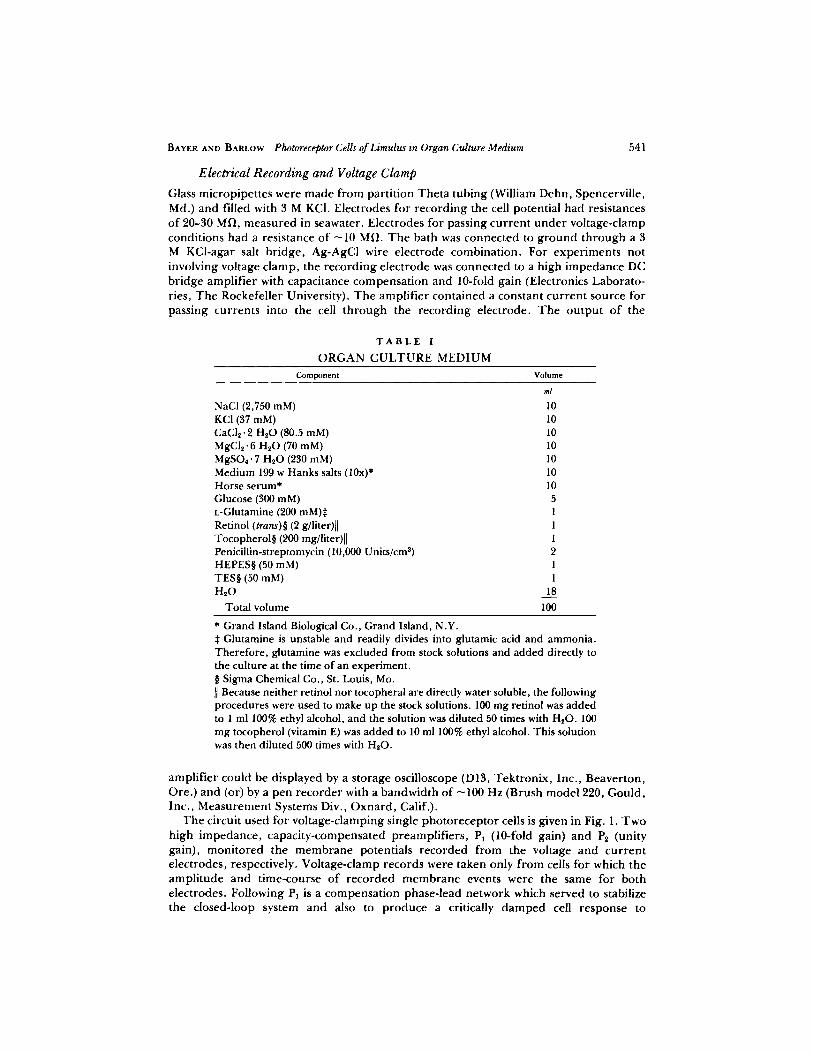
NaCl (2,750 mM) 10 KCI (37 mM) 10 CaCI2.2 H20 (80.5 raM) 10 MgCI,-6 H20 (70 raM) 10 MgSO4.7 H,O (230 raM) 10 Medium 199 w Hanks salts (10x)* 10 Horse serum* 10 Glucose (300 raM) 5 L-Glutamine (200 mM)* 1 Retinol (trans)w (2 g/liter)]l 1 Tocopherolw (200 rag/liter)[] 1 Penicillin-streptomycin (10,000 Units/cm 8) 2 HEPESw (50 mM) 1 TESw (50 raM) 1 H20 18
Total volume 100
* Grand Island Biological Co., Grand Island, N.Y. Glutamine is unstable and readily divides into glutamic acid and ammonia.
Therefore, glutamine was excluded from stock solutions and added directly to the culture at the time of an experiment. w Sigma Chemical Co., St. Louis, Mo. II Because neither retinol nor tocopheral are directly water soluble, the following procedures were used to make up the stock solutions. 100 mg retinol was added to 1 ml 100% ethyl alcohol, and the solution was diluted 50 times with H20. 100 mg tocopherol (vitamin E) was added to 10 ml 100% ethyl alcohol. This solution was then diluted 500 times with H20.
amplif ier could be displayed by a storage oscilloscope (DI3, Tektronix , Inc. , Beaverton, Ore.) and (or) by a pen recorder with a bandwidth of ~100 Hz (Brush model 220, Gould, Inc., Measurement Systems Div., Oxnard , Calif.).
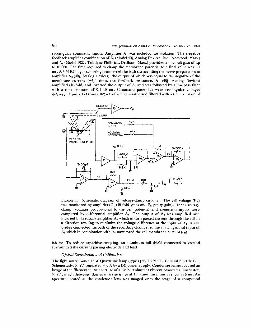
I 'he circuit used for voltage-clamping single photoreceptor cells is given in Fig. 1. Two high impedance, capacity-compensated preamplif iers , P~ (10-fold gain) and P~ (unity gain), moni tored the membrane potentials recorded from the voltage and current electrodes, respectively. Voltage-clamp records were taken only f rom cells for which the ampli tude and time-course of recorded membrane events were the same for both electrodes. Following PI is a compensat ion phase-lead network which served to stabilize the closed-loop system and also to produce a critically damped cell response to
542 T H E JOURNAL OF G E N E R A L PHYSIOLOGY " VOLUME 7 2 - 1 9 7 8
rectangular command inputs. Amplif ier A3 was included for isolation. The negative feedback amplif ier combination of A4 (Model 40j, Analog Devices, Inc., Norwood, Mass.) and As (Model 1022, Teledyne Philbrick, Dedham, Mass.) provided an overall gain of up to 10,000. The time required to clamp the membrane potential to a final value was ~ 1 ms. A 3 M KCl-agar salt br idge connected the bath sur rounding the nerve prepara t ion to amplifier A6 (40j, Analog Devices), the output of which was equal to the negative of the membrane current (--IM) times the feedback resistance. A7 (40j, Analog Devices) amplified (10-fold) and inverted the output of An and was followed by a low pass filter with a time constant o f 0.I-10 ms. Command potentials were rectangular voltages delivered from a Tekt ronix 162 waveform genera tor and filtered with a time constant of
RECORD �9 = V M :-J i
CLAMP. //9- COMMAND 4 , .
~ ' ~ l / X INPUT ~'-~r | HOLDING 47K
f POTENTIAL " ~ ' ' V ~ VENTRAL PHOTORECEPTOR
VMX I0 IM O-500K
IOK III
2.- ,oo ,o,< C ,o.,v
FIGURE 1. Schematic d iagram of voltage-clamp circuitry. The cell voltage (VM) was moni tored by amplifiers P1 (10-fold gain) and P2 (unity gain). Under voltage clamp, voltages proport ional to the cell potential and command inputs were compared by differential amplif ier A4. The output of A4 was amplif ied and inverted by feedback amplif ier A5 which in turn passed current through the cell in a direction tending to minimize the voltage difference at the input o f A4. A salt br idge connected the bath of the recording chamber to the virtual g round input of A6 which in combination with A7 moni tored the cell membrane current (lu).
0.5 ms. To reduce capacitive coupling, an a luminum foil shield connected to ground sur rounded the current passing electrode and lead.
Optical Stimulation and Calibration
The light source was a 45 W Quartzline lamp (type Q 45 T 2Wz CL, General Electric Co., Schenectady, N.Y.) regulated at 6 A by a DC power supply. Condenser lenses focused an image of the fi lament in the aper ture of a Uniblitz shutter (Vincent Associates, Rochester, N.Y.), which del ivered flashes with rise times of l ms and durat ions as short as 5 ms. An aper ture located at the condenser lens was imaged onto the stage of a compound
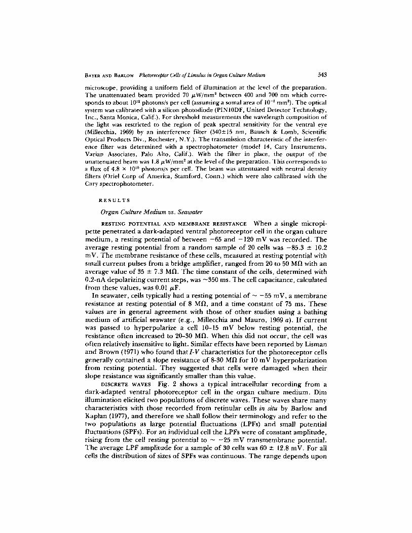
BAYER AND BARLOW Photoreceptor Cells of Limulus in Organ Culture Medium 543
microscope, providing a uniform field of illumination at the level of the preparation. The unattenuated beam provided 70 /zW/mm 2 between 400 and 700 nm which corre- sponds to about 10 .2 photons/s per cell (assuming a somal area of 10 -2 ram2). The optical system was calibrated with a silicon photodiode (PIN10DF, United Detector Technology, Inc., Santa Monica, Calif.). For threshold measurements the wavelength composition of the light was restricted to the region of peak spectral sensitivity for the ventral eye (Millecchia, 1969) by an interference filter (540-.+15 nm, Bausch & Lomb, Scientific Optical Products Div., Rochester, N.Y.). The transmission characteristic of the interfer- ence filter was determined with a spectrophotometer (model 14, Cary Instruments, Varian Associates, Palo Alto, Calif.). With the filter in place, the output of the unattenuated beam was 1.8/zW/mm 2 at the level of the preparation. This corresponds to a flux of 4.8 • 10 l~ photons/s per cell. The beam was attentuated with neutral density filters (Oriel Corp of America, Stamford, Conn.) which were also calibrated with the Cary spectrophotometer.
R E S U L T S
Organ Culture Medium vs. Seawater
R E S T I N G P O T E N T I A L A N D M E M B R A N E R E S I S T A N C E When a single micropi- pette pene t ra ted a dark-adapted ventral pho to recep to r cell in the organ cul ture medium, a rest ing potential o f between - 6 5 and - 1 2 0 mV was recorded . T h e average rest ing potential f rom a r a n d o m sample o f 20 cells was -85 .3 -+ 10.2 inV. T h e m e m b r a n e resistance o f these cells, measured at rest ing potential with small cu r ren t pulses f rom a br idge amplif ier , ranged f rom 20 to 50 M ~ with an average value o f 35 --- 7.3 MI~. T h e t ime constant o f the cells, de te rmined with 0.2-nA depolar iz ing cur ren t steps, was - 3 5 0 ms. T h e cell capacitance, calculated f rom these values, was 0.01/~F.
In seawater, cells typically had a resting potential o f - - 5 5 mV, a m e m b r a n e resistance at rest ing potential o f 8 MI~, and a time constant o f 75 ms. These values are in general agreement with those o f o the r studies using a bathing medium o f artificial seawater (e.g., Millecchia and Mauro, 1969 a). I f cu r ren t was passed to hyperpolar ize a cell 10-15 mV below rest ing potential , the resistance of ten increased to 20-30 MI-I. When this did not occur, the cell was of ten relatively insensitive to light. Similar effects have been repor ted by Lisman and Brown (1971) who found that I-V characteristics for the pho to recep to r cells general ly conta ined a slope resistance o f 8-30 MI-I for 10 mV hyperpolar iza t ion f rom resting potential. T h e y suggested that cells were damaged when their slope resistance was significantly smaller than this value.
DISCRETE WAVES Fig. 2 shows a typical intracellular record ing f rom a dark-adapted ventral pho to recep to r cell in the organ cul ture medium. Dim illumination elicited two populat ions o f discrete waves. These waves share many characteristics with those recorded f rom ret inular cells in situ by Barlow and Kaplan (1977), and the re fo re we shall follow their terminology and re fe r to the two popula t ions as large potential f luctuations (LPFs) and small potential f luctuations (SPFs). For an individual cell the LPFs were o f constant ampl i tude , rising f rom the cell resting potential to - - 2 5 mV t r ansmembrane potential. T h e average LPF ampl i tude for a sample o f 30 cells was 60 + 12.8 inV. For all cells the distribution o f sizes of SPFs was continuous. T h e range depends upon

5 4 4 T h E JOURNAL OF" GENERAL PHYSIOLOGY �9 VOLUME 72 " 1 9 7 8
the resting potential o f the cell and extends roughly f rom 0 to {{Resting Potential (mY) I -60}. SPFs decayed exponential ly with a time constant o f 200-500 ms which appea red to be related to the m e m b r a n e resistance of the cell. Both SPFs and LPFs occur red spontaneously in the dark and could be elicited with dim illumination.
In artificial seawater the pho to recep to r cells typically exhibited a single popula t ion o f discrete waves with ampli tudes < - 1 5 mV. However , cells having a membrane resistance that was strongly d e p e n d e n t on m em b ran e potential (see Lisman and Brown, 1971) could be hyperpolar ized to p roduce both SPFs and LPFs. Hyperpolar iza t ion p roduced no change in the ampli tudes of discrete waves for cells with membrane resistances which were relatively independen t of membrane potential.
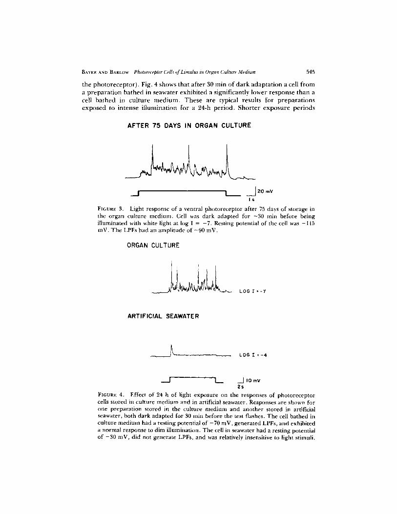
VIABILITY MEASUREMENTS Fig. 3 shows the response o f a ventral photo- receptor cell that was stored for 75 days in organ cul ture med ium in darkness at
\ I0 mV
" i s
FXGURE 2. LPFs and SPFs of a ventral photoreceptor cell bathed in the organ culture medium. Record taken after 30 min of dark adaptation. Cell was illumi- nated with white light at log I = -8.5. Shown are three LPFs of ~60 mV and a population of SPFs-the well-known quantum bumps. The cell resting potential was -85 mV.
2~ T h e only apparen t physiological change in this and in o ther prepara t ions stored for more than 30 days was a slight decrease in the response to a f ixed intensity flash. T h e decline in sensitivity for such preparat ions was usually <1 log unit. No changes in sensitivity were detected for cells stored <30 days in cul ture medium.
Photorecep tor cells stored in artificial seawater at 2~ in the dark remained viable for up to 4 days. Longer storage periods generally yielded cells with reduced resting potentials and lower responses to fixed test flashes. After ~6 days in seawater at 2~ in the dark, cells had no detectable resting potentials or light responses. These viability results are based on preparat ions s tored in cul ture med ium and in artificial seawater for various periods in the dark.
T o de te rmine the possible effects of light exposure on pho torecep to r viability, preparat ions were stored at 20~ in cul ture medium and in artificial seawater for 24 h u n d e r relatively intense i l lumination (~ 10 ~~ photons/s at the surface o f
BAYER AND BARLOW Photoreceptor Cells of Limulu.~ in Organ Culture Medium 545
the p h o t o r e c e p t o r ) . Fig . 4 shows tha t a f t e r 30 rain o f d a r k a d a p t a t i o n a cell f r o m a p r e p a r a t i o n b a t h e d in s e a w a t e r e x h i b i t e d a s ign i f i can t ly l o w e r r e s p o n s e t h a n a cell b a t h e d in c u l t u r e m e d i u m . T h e s e a r e typica l r esu l t s fo r p r e p a r a t i o n s e x p o s e d to i n t e n s e i l l u m i n a t i o n fo r a 24-h p e r i o d . S h o r t e r e x p o s u r e p e r i o d s
AFTER 75 DAYS IN ORGAN CULTURE
_..I I J 20 r n V
Is FIGURE 3. Light response of a ventral photoreceptor after 75 days of storage in the organ culture medium. Cell was dark adapted for ~30 rain before being i l luminated with white light at log I = - 7 . Resting potential of the cell was -115 inV. The LPFs had an ampli tude of - 9 0 inV.
ORGAN CULTURE
I ~
LOG I =-7
ARTIFICIAL SEAWATER
_ ]~- ' ~ LOG T = - 4
I__ _._] JOmV 2s
FIGURE 4. Effect of 24 h of light exposure on the responses of photoreceptor cells stored in culture medium and in artificial seawater. Responses are shown for one prepara t ion stored in the culture medium and another stored in artificial seawater, both dark adapted for 30 min before the test flashes. The cell bathed in culture medium had a resting potential of - 7 0 mV, generated LPFs, and exhibited a normal response to dim il lumination. The cell in seawater had a resting potential of - 3 0 mV, did not generate LPFs, and was relatively insensitive to light stimuli.
546 T H E J O U R N A L O F G E N E R A L P H Y S I O L O G Y " V O L U M E 72 �9 1978
generally yielded smaller reduct ions in the responses f rom cells in seawater and near normal responses f rom ceils in cul ture med ium. Longer exposure per iods were not tested.
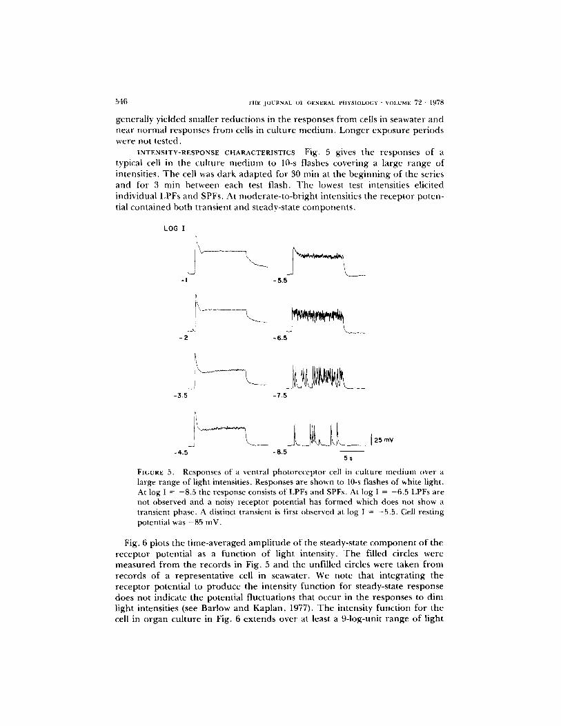
INTENSITY-RESPONSE CHARACTERISTICS Fig. 5 gives the responses o f a typical cell in the cul ture m e d i u m to 10-s flashes cover ing a large range of intensities. T h e cell was dark adap ted for 30 rain at the beg inn ing of the series and for 3 min between each test flash. T h e lowest test intensities elicited individual LPFs and SPFs, At modera te - to -b r igh t intensities the receptor poten- tial conta ined both t ransient and steady-state componen t s ,
LOG I
-I - 5 . 5
- 2 - 6 . 5
-3 .5 -7 .5
-4 .5 -8 .5 5 s
FIGURE 5. R e s p o n s e s o f a v e n t r a l p h o t o r e c e p t o r cell in c u l t u r e m e d i u m o v e r a
large range of light intensities. Responses are shown to 10-s flashes of white light. At log I = -8.5 the response consists of LPFs and SPFs. At log I = -6.5 LPFs are not observed and a noisy receptor potential has formed which does not show a transient phase. A distinct transient is first observed at log I = -5.5. Cell resting potential was -85 mV.
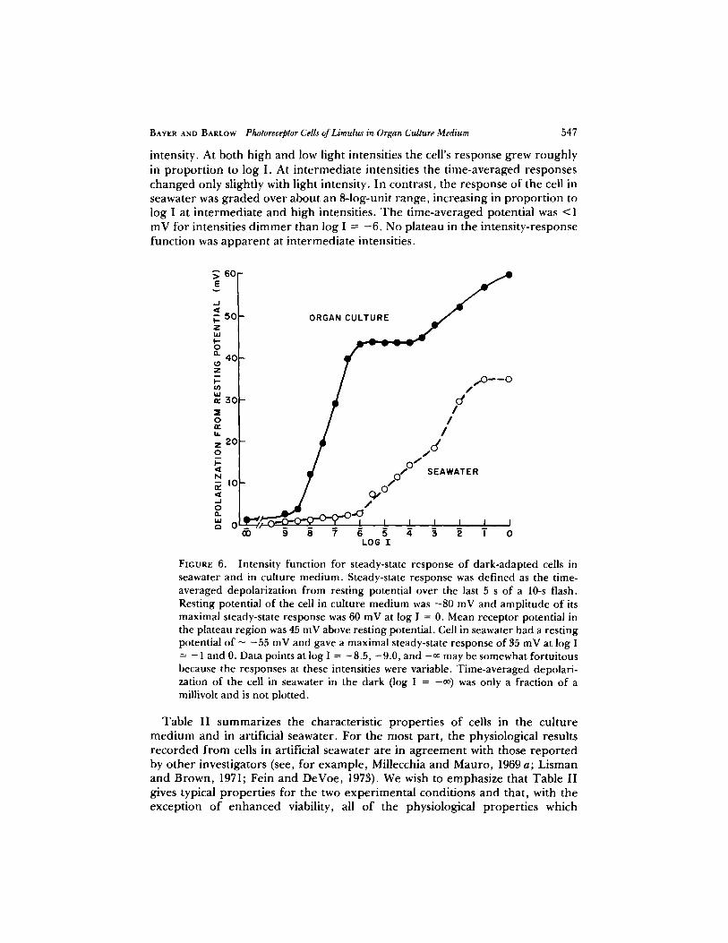
Fig. 6 plots the t ime-averaged ampl i tude of the steady-state c o m p o n e n t o f the recep tor potential as a funct ion of light intensity. T h e filled circles were measu red f r o m the records in Fig. 5 and the unfil led circles were taken f r o m records of a representa t ive cell in seawater. We note that in tegra t ing the receptor potential to p roduce the intensity funct ion for steady-state response does not indicate the potential f luctuations that occur in the responses to dim light intensities (see Barlow and Kaplan , 1977). T h e intensity funct ion for the cell in o rgan cul ture in Fig. 6 ex tends over at least a 9-log-unit range of light
BAYER AND BARLOW Photoreceptor Cells of Limul~ in Organ Culture Medium 547
intensity. At both high and low light intensities the cell's response grew roughly in p ropor t ion to log I. At in te rmedia te intensities the t ime-averaged responses changed only slightly with light intensity. In contrast , the response of the cell in seawater was g r aded over about an 8-log-unit range , increasing in p ropo r t i on to log I at in te rmedia te and high intensities. T h e t ime-averaged potential was <1 mV for intensities d i m m e r than log I = - 6 . No plateau in the intensi ty-response funct ion was a p p a r e n t at in te rmedia te intensities.
60 E
,,_1
V- 5 c Z ILl I - O a .
40 Z I--
W '~ 3 0
O
U.
z 2 0 O I--
N
J O
h i ,'- 0
ORGAN~ / /,,o--o
- / / - / J .0/0" SEAWATER
,_ ,_ _, _, _, , OD 9 8 7 6 5 4 3, 2 I 0
LOG I
FIGURE 6. Intensity function for steady-state response of dark-adapted cells in seawater and in culture medium. Steady-state response was defined as the time- averaged depolarization from resting potential over the last 5 s of a 10-s flash. Resting potential of the cell in culture medium was -80 mV and amplitude of its maximal steady-state response was 60 mV at log I = 0. Mean receptor potential in the plateau region was 45 mV above resting potential. Cell in seawater had a resting potential of - -55 mV and gave a maximal steady-state response of 35 mV at log I = - 1 and 0. Data points at log I = -8.5, -9.0, and -00 may be somewhat fortuitous because the responses at these intensities were variable. Time-averaged depolari- zation of the cell in seawater in the dark (log I = -09 was only a fraction of a millivoh and is not plotted.
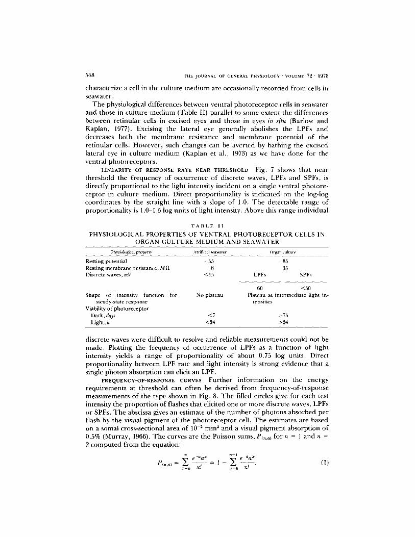
Tab le I I summar izes the characterist ic p roper t ies of cells in the cul ture m e d i u m and in artificial seawater . For the most par t , the physiological results r eco rded f r o m cells in artificial seawater are in a g r e e m e n t with those r e p o r t e d by o the r investigators (see, for example , Millecchia and Mauro , 1969 a; L isman and Brown, 1971; Fein and DeVoe, 1973). We wish to emphas ize that Tab le I I gives typical p roper t i es for the two expe r imen ta l condit ions and that , with the except ion o f enhanced viability, all o f the physiological p roper t ies which
5 4 8 THE JOURNAL OF GENERAL PHYSIOLOGY " VOLUME 72 �9 1978
character ize a cell in the cul ture m e d i u m are occasionally r ecorded f rom cells in seawater.
T h e physiological d i f ferences between ventral pho to recep to r cells in seawater and those in cul ture m e d i u m (Table II) parallel to some extent the d i f ferences between re t inular cells in excised eyes and those in eyes in situ (Barlow and Kaplan, 1977). Excising the lateral eye generally abolishes the LPFs and decreases both the m e m b r a n e resistance and m e m b r a n e potential o f the re t inular cells. However , such changes can be aver ted by bathing the excised lateral eye in cul ture m e d i u m (Kaplan et al., 1973) as we have done fi)r the ventral pho to recep to r s .
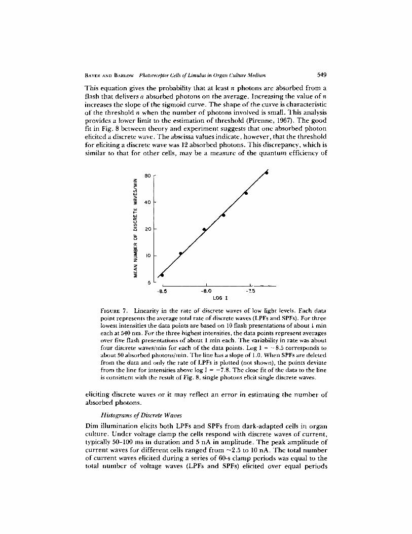
L I N E A R I T Y O F R E S P O N S E R A T E N E A R T H R E S H O L D Fig. 7 shows that near threshold the f requency o f occur rence o f discrete waves, LPFs and SPFs, is directly p ropor t iona l to the light intensity incident on a single ventral photore- ceptor in cul ture m ed i um . Direct propor t ional i ty is indicated on the log-log coordinates by the straight line with a slope o f 1.0. T h e detectable range o f propor t ional i ty is 1,0-1.5 log units o f light intensity. Above this range individual
T A B L E I I
PHYSIOLOGICAL PROPERTIES OF VENTRAL PHOTORECEPTOR CELLS IN ORGAN CULTURE MEDIUM AND SEAWATER
Physiological property Artificial seawater Organ culture
Resting potential -55 -85 Resting membrane resistance, M l) 8 35 Discrete waves, rnV < 15 LPFs SPFs
60 <30 Shape of intensity function for No plateau Plateau at intermediate light in-
steady-state response tensities Viability of photoreceptor
Dark, days <7 >75 Light, h <24 >24
discrete waves were difficult to resolve and reliable m e a s u r e m e n t s could not be made . Plotting the f requency o f occur rence of LPFs as a funct ion of light intensity yields a range of propor t ional i ty of about 0.75 log units. Direct propor t ional i ty between LPF rate and light intensity is s t rong evidence that a single pho ton absorpt ion can elicit an LPF.
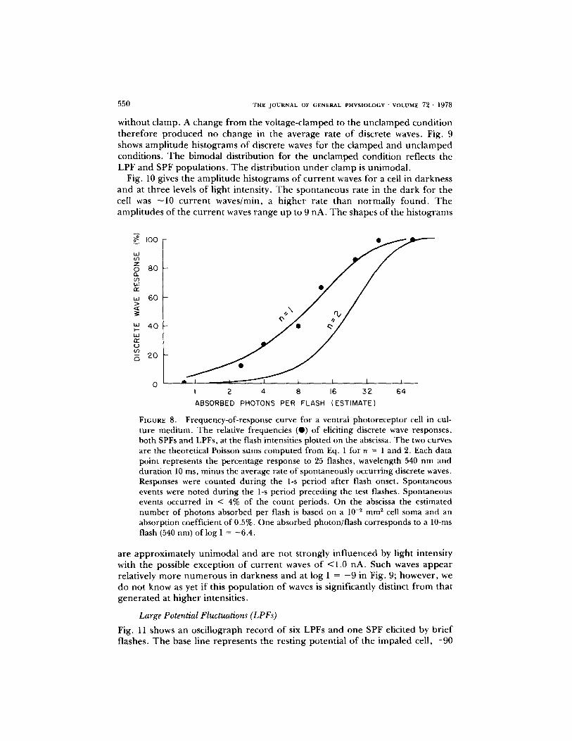
FREQUENCV-OE-RESPONSE CURVES Fur the r in fo rmat ion on the ene rgy requ i remen t s at threshold can of ten be der ived f rom f requency-of - response measu remen t s of the type shown in Fig. 8. T h e filled circles give for each test intensity the p ropo r t i on of flashes that elicited one or more discrete waves, LPFs or SPFs. T h e abscissa gives an est imate of the n u m b e r of photons absorbed per flash by the visual p igmen t o f the pho to recep to r cell. T h e estimates are based on a somal cross-sectional area of 10 -2 m m 2 and a visual p igmen t absorp t ion of 0.5% (Murray , 1966). T h e curves are the Poisson sums, P~,,a~ for n = 1 and n = 2 c o m p u t e d f rom the equation:
~__n e_aa ~ n-i e_aa ~ Pc,.o,= = x., = I - Z �9
a:'=0
549
80
This equat ion gives the probabil i ty that at least n photons are absorbed f r o m a flash that delivers a absorbed pho tons on the average, Increas ing the value o f n increases the slope o f the sigmoid curve. T h e shape o f the curve is characterist ic o f the threshold n when the n u m b e r o f photons involved is small. This analysis provides a lower limit to the est imation o f threshold (Pirenne, 1967). T h e good fit in Fig. 8 between theory and e x p e r i m e n t suggests that one absorbed pho ton elicited a discrete wave. T h e abscissa values indicate, however , that the threshold for eliciting a discrete wave was 12 absorbed photons . This discrepancy, which is similar to that for o the r cells, may be a measure of the q u a n t u m efficiency of
40
20
I0
1 ! |
-8.5 -8.0 -Z5
LOG I
BAYER AND B A R L O W Photoreceptor CelL~ of Limul~ in Organ Culture Medium
FIGURE 7. Linearity in the rate of discrete waves of low light levels. Each data point represents the average total rate of discrete waves (LPFs and SPFs). For three lowest intensities the data points are based on 10 flash presentations of about 1 rain each at 540 nm. For the three highest intensities, the data points represent averages over five flash presentations of about 1 rain each. The variability in rate was about four discrete waves/rain for each of the data points. Log I = -8.5 corresponds to about 50 absorbed photons/rain. The line has a slope of 1.0. When SPFs are deleted from the data and only the rate of LPFs is plotted (not shown), the points deviate from the line for intensities above log I = -7.8. The close fit of the data to the line is consistent with the result of Fig. 8, single photons elicit single discrete waves.
eliciting discrete waves or it may reflect an e r ro r in es t imat ing the n u m b e r o f absorbed photons .
Histograms of Discrete Waves
Dim il lumination elicits both LPFs and SPFs f rom da rk -adap t ed cells in o rgan cul ture. U n d e r voltage c lamp the cells r e spond with discrete waves o f cu r ren t , typically 50-100 ms in dura t ion and 5 nA in ampl i tude . T h e peak ampl i tude o f cu r ren t waves for d i f fe ren t cells r anged f r o m ~2.5 to 10 hA. T h e total n u m b e r o f cu r ren t waves elicited du r ing a series o f 60-s c lamp per iods was equal to the total n u m b e r o f voltage waves (LPFs and SPFs) elicited over equal per iods
550 T H E J O U R N A L OF G E N E R A L P H Y S I O L O G Y " V O L U M E 72 �9 1978
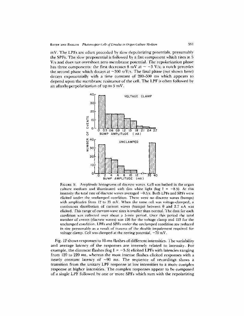
without clamp. A change from the voltage-clamped to the unclamped condition therefore produced no change in the average rate of discrete waves. Fig. 9 shows amplitude histograms of discrete waves for the clamped and unclamped conditions. The bimodal distribution for the unclamped condition reflects the LPF and SPF populations. The distribution under clamp is unimodal.
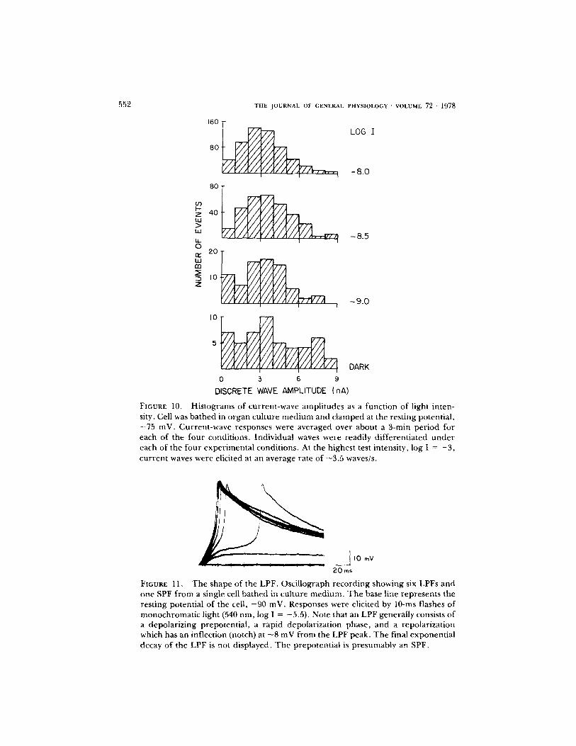
Fig. 10 gives the amplitude histograms of current waves for a cell in darkness and at three levels of light intensity. The spontaneous rate in the dark for the cell was - 1 0 current waves/min, a higher rate than normally found. The amplitudes of the current waves range up to 9 nA. The shapes of the histograms
IO0 �9 .....
Z o 8O
W w
60 -
" ' 4 0 ~" �9 I - - UA rr" 0 O0
2 0
i
0 ~ L I 1 I 2 4 8 16 5 2 64
ABSORBED PHOTONS PER FLASH (ESTIMATE)
FIGURE 8. Frequency-of-response curve for a ventral photoreceptor cell in cul- ture medium. The relative frequencies (O) of eliciting discrete wave responses, both SPFs and LPFs, at the flash intensities plotted on the abscissa. The two curves are the theoretical Poisson sums computed from Eq. 1 for n = 1 and 2. Each data point represents the percentage response to 25 flashes, wavelength 540 nm and durat ion 10 ms, minus the average rate of spontaneously occurr ing discrete waves. Responses were counted dur ing the 1-s period after flash onset. Spontaneous events were noted dur ing the 1-s per iod preceding the test flashes. Spontaneous events occurred in < 4% of the count periods. On the abscissa the est imated number of photons absorbed per flash is based on a 10 -~ mm ~ cell soma and an absorption coefficient of 0.5%. One absorbed photon/flash corresponds to a 10-ms flash (540 nm) of log I = -6 .4 .
a r e a p p r o x i m a t e l y u n i m o d a l a n d a r e n o t s t r o n g l y i n f l u e n c e d by l igh t i n t ens i t y wi th t he poss ib le e x c e p t i o n o f c u r r e n t waves o f < l . 0 n A . Such waves a p p e a r r e la t ive ly m o r e n u m e r o u s in d a r k n e s s a n d at log I = - 9 in Fig . 9; h o w e v e r , we d o no t k n o w as ye t i f this p o p u l a t i o n o f waves is s ign i f i can t ly d i s t inc t f r o m tha t g e n e r a t e d at h i g h e r in tens i t i es .
Large Potential Fluctuations (LPFs)
Fig. 11 shows an o s c i l l o g r a p h r e c o r d o f six LPFs a n d o n e SPF e l ic i ted by b r i e f f lashes . T h e base l ine r e p r e s e n t s t he r e s t i n g p o t e n t i a l o f t he i m p a l e d cel l , - 9 0
BAYER AND BARLOW Photoreceptor (,'elL~" of Limulus in Organ Culture Medium 551
inV. The LPFs are often preceded by slow depolarizing potentials, presumably the SPFs. The slow prepotential is followed by a fast component which rises at 5 V/s and does not overshoot zero membrane potential. The repolarization phase has three components: the first decreases 8 mV at ~ - 3 V/s; a notch precedes the second phase which decays at ~300 mV/s. The final phase (not shown here) decays exponentially with a time constant of 200-500 ms which appears to depend upon the membrane resistance of the cell. The LPF is often followed by an afterhyperpolarization of up to 5 inV.
40
30
20
0 3
io W >
uJ 0
n ' 4 0 I L l m
30 Z
VOLTAGE CLAMP ; ; ; ; ;
U//A
I l i i /
~44 r
0 0.3 0.6 0,9 1.2 1.5 I.O 21 2.4 2.7 BUMP AMPLITUDE ( nA)
UNCLAMPED ~'/'/'/'/'/'/'/'/'~ . . . . .
- v / x / / . . . . . V/X// �9 / i i , I f / I l l . . . . .
~ 1 1 1 1 " 1 1 1 , V / / / . " 1 1 1 1
I I I I i , 1 1 I , 2 0 - ~ 1 / / / , . . . . .
V / / l l , i / i , V I l l i " 1 1 1 1 V I l l i
I 0 . . . . . . V / l /
t r
0 2: 4 6 8 I0 12 35 40 BUMP AMPLITUDE (mY)
FmURE 9. A m p l i t u d e h is tograms o f d iscrete waves. Cel l was ba thed in the o r g a n cu l t u re m e d i u m and i l l u m i n a t e d w i t h d i m wh i te l i gh t ( log I = - 8 . 5 ) . A t this i n tens i t y the to ta l ra te of" d iscrete waves ave raged - 0 . 5 / s . B o t h LPFs and SPFs were elicited under the unclamped condition. There were no discrete waves (bumps) with ampli tudes from 12 to 35 inV. When the same cell was voltage-clamped, a continuous distribution of current waves (bumps) between 0 and 2.7 nA was elicited. This range of current-wave sizes is smaller than normal. The data for each condition was collected over about a 5-rain period. Over this period the total number of events (discrete waves) was 126 for the voltage clamp and 123 for the unclamped condition. LPFs and SPFs under the unclamped condition are reduced in size presumably as a result of t rauma of the double impalement required for voltage clamp. Cell was clamped at the resting potential , - 7 0 inV.
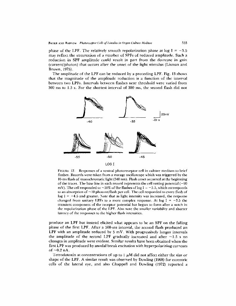
Fig. 12 shows r e s p o n s e s to 10-ms f lashes o f d i f f e r e n t in tens i t i es . T h e variability a n d a v e r a g e l a t ency o f the r e s p o n s e s a r e i nve r se ly r e l a t e d to i n t ens i ty . F o r e x a m p l e , the d i m m e s t f lashes ( log I = - 5 . 5 ) e l ic i ted L P F s with la tenc ies r a n g i n g f r o m 120 to 220 ms, w h e r e a s the mos t i n t e n s e f lashes e l i c i t ed r e s p o n s e s wi th a n e a r l y c o n s t a n t l a t ency o f ~ 9 0 ms. T h e s e q u e n c e o f r e c o r d i n g s shows a t r a n s i t i o n f r o m the u n i t a r y L P F r e s p o n s e at low in tens i t i e s to a m o r e c o m p l e x r e s p o n s e at h i g h e r in tens i t i es . T h e c o m p l e x r e s p o n s e s a p p e a r to be c o m p o s e d of a single LPF followed by one or more SPFs which sum with the repolarizing
552
160
80
t J ) b'-
Z W > ILl
t.t. o fw t.o rn
Z
8 0
4 0
THE JOURNAl. OF GENERAL PHYSIOLOGY - VOLUME 72 �9 1978
LOG I
-8 .0
2o IO
l i I
IO
0 3 6 9
-8.5
-9.0
DARK
DISCRETE WAVE AMPLITUDE (nA)
FIGURE 10. Histograms of current-wave amplitudes as a function of light inten- sity. Cell was bathed in organ culture medium and clamped at the resting potential, -75 inV. Current-wave responses were averaged over about a 3-rain period for each of the four conditions. Individual waves were readily differentiated under each of the four experimental conditions. At the highest test intensity, log I = - 3 , current waves were elicited at an average rate of ~3.5 waves/s.
#== I0 mY
20ms
FIGURE 11, The shape of the LPF. Oscillograph recording showing six LPFs and one SPF from a single cell bathed in culture medium. The base line represents the resting potential of the cell, - 90 mV. Responses were elicited by 10-ms flashes of monochromatic light (540 nm, log I = -5.5) . Note that an LPF generally consists of a depolarizing prepotential, a rapid depolarization phase, and a repolarization which has an inflection (notch) at - 8 mV from the LPF peak. The final exponential decay of the LPF is not displayed. The prepotentia| is presumably an SPF.
BAYER AND BARLOW Photoreceptor Cells of Limulus in Organ Culture Medium 553
phase o f the LPF. T h e relatively smooth repolar izat ion phase at log I = - 3 . 5 may reflect the summat ion of a n u m b e r o f SPFs o f r educed ampl i tude . Such a reduct ion in SPF ampl i tude could result in par t f rom the decrease in gain (cur ren t /pho ton) that occurs af ter the onset o f the light st imulus (Lisman and Brown, 1975).
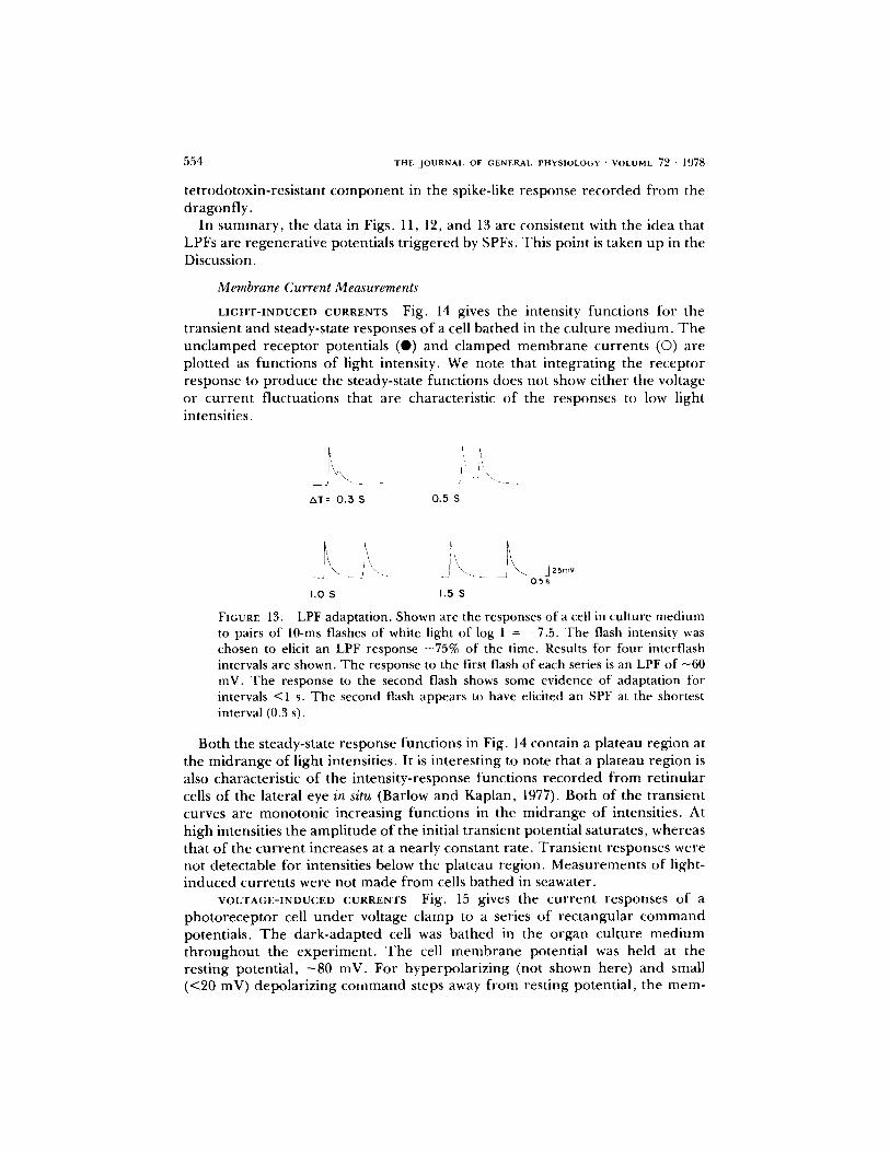
T h e ampl i tude o f the LPF can be r educed by a p reced ing LPF. Fig. 13 shows that the magn i tude o f the ampl i tude reduct ion is a funct ion of the interval between two LPFs. Intervals between flashes near threshold were varied f rom 300 ms to 1.5 s. For the shortest interval o f 300 ms, the second flash did not
-4.0 -3.5
20mY
-0.1 s
-5.5 -5.0 -4.5
LOG I
FIGURE 12. Responses of a ventral photoreceptor cell in culture medium to brief flashes. Records were taken from a storage oscilloscope which was triggered by the 10-ms flash of monochromatic light (540 nm). Flash onset occurred at the beginning of the traces. The base line in each record represents the cell resting potential ( -90 mV). The cell responded to -50% of the flashes of log I = -5.5, which corresponds to an absorption of -10 photons/flash per cell. The cell responded to every flash of log I = -4.5 and greater. Note that as light intensity was increased, the response changed from unitary LPFs to a more complex response. At log I = -3.5 the transient component of the receptor potential has begun to form after a notch in the repolarization phase of the LPF. Also note the smaller variability and shorter latency of the responses to the higher flash intensities.
p roduce an LPF but instead elicited what appea r s to be an SPF on the falling phase o f the first LPF. Af ter a 500-ms interval, the second flash p roduced an LPF with an ampl i tude reduced by 5 inV. With progressively longer intervals the ampl i tude o f the second LPF gradual ly increased and af ter - 1 . 5 s no changes in ampl i tude were evident . Similar results have been obta ined when the first LPF was p r o d u c e d by anodal b reak excitation with hyperpo la r iz ing cur ren ts o f - 0 . 2 nA.
T e t r o d o t o x i n at concentra t ions o f up to 1 /~M did not affect e i ther the size or shape o f the LPF. A similar result was observed by Dowling (1968) for eccentric cells o f the lateral eye, and also Chappel l and Dowling (1972) r epo r t ed a
5 5 4 T H E J O U R N A L O F G E N E R A L P H Y S I O L O ( ; Y ' V O L U M E 72 �9 1978
te t rodotoxin-res is tant c o m p o n e n t in the spike-like response recorded f rom the dragonf ly .
In s u m m a r y , the data in Figs. 11, 12, and 13 are consistent with the idea that LPFs are regenera t ive potentials t r iggered by SPFs. This point is taken up in the Discussion.
Membrane Current Measurements
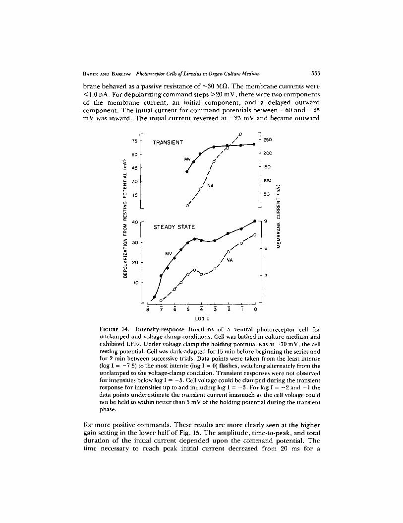
LIGHT-INDUCED CURRENTS Fig. 14 gives the intensity functions for the t ransient and steady-state responses o f a cell ba thed in the cul ture m e d i u m . T h e unc l amped recep to r potentials (0) and c lamped m e m b r a n e currents (�9 are plot ted as funct ions of light intensity. We note that in tegra t ing the recep to r response to p roduce the steady-state funct ions does not show ei ther the voltage or cur ren t f luctuations that are characterist ic o f the responses to low light intensities.
AT= 0 .3 S 0.5 S
I ~, j,
i' _j25mv J _i
05s 1,0 S 1.5 S
FIGURE 13, LPF adaptation. Shown are the responses of a cell in culture medium to pairs of 10-ms flashes of white light of log I -= -7.5. The flash intensity was chosen to elicit an LPF response --75% of the time. Results for four interflash intervals are shown. The response to the first flash of each series is an LPF of -60 mV. The response to the second flash shows some evidence of adaptation for intervals <1 s. The second flash appears to have elicited an SPF at the shortest interval (0.3 s).
Both the steady-state response funct ions in Fig. 14 contain a plateau region at the mid range of light intensities. It is interest ing to note that a plateau region is also characteristic o f the intensi ty-response functions r eco rded f r o m re t inular cells o f the lateral eye in situ (Barlow and Kaplan, 1977). Both of the t ransient curves are monotonic increasing funct ions in the mid range o f intensities. At high intensities the ampl i tude o f the initial t ransient potential saturates, whereas that o f the cu r ren t increases at a nearly constant rate. T rans ien t responses were not detectable for intensities below the plateau region. Measuremen t s of light- induced currents were not made f r o m cells ba thed in seawater.
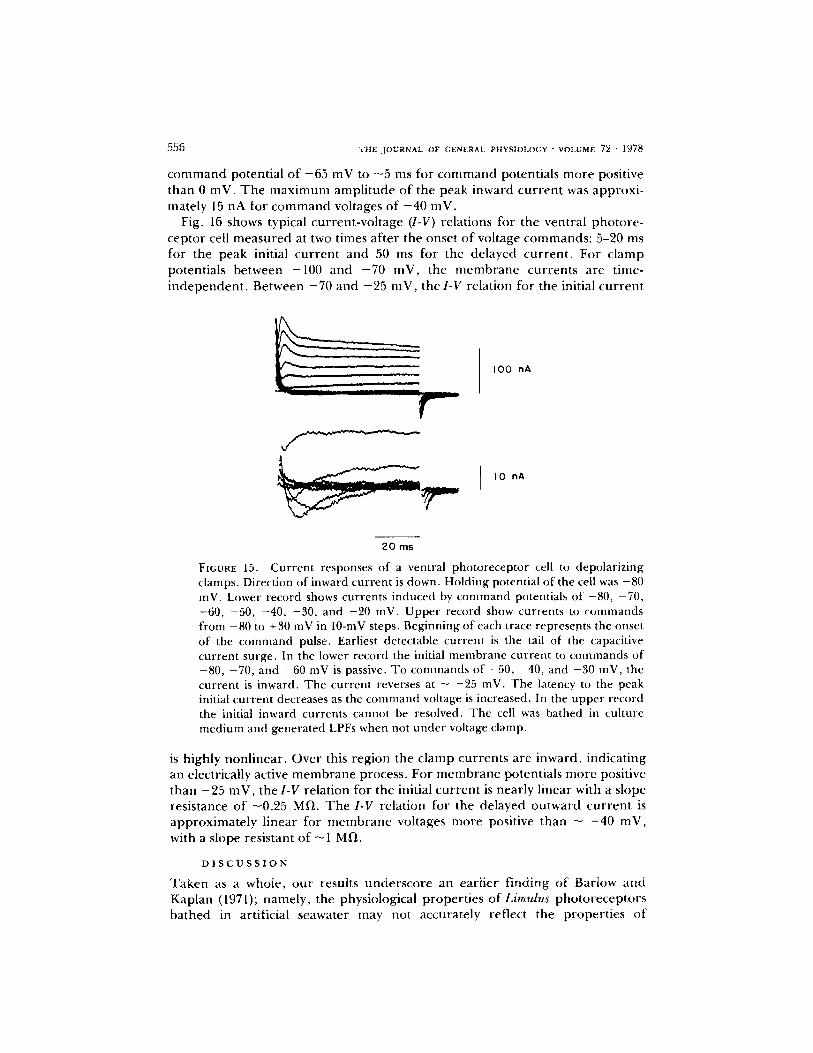
VOLTAGE-INDUCED CURRENTS Fig, 15 gives the cu r ren t responses of a pho to recep to r cell u n d e r voltage c lamp to a series of rec tangular c o m m a n d potentials. T h e da rk -adap ted cell was ba thed in the o rgan culture m e d i u m t h r o u g h o u t the exper imen t . T h e cell m e m b r a n e potential was held at the rest ing potential , - 8 0 inV. For hyperpola r iz ing (not shown here) and small (<20 mV) depolar iz ing COulmand steps away f rom rest ing potential , the mere-
B A Y E R AND B A R L O W Photoreceptor Cells of Limul~ in Organ Culture Medium 555
brane b e h a v e d as a passive resistance o f - 3 0 MD. T h e m e m b r a n e currents were < 1 . 0 nA. For depo lar i z ing c o m m a n d steps > 2 0 m V , there were two c o m p o n e n t s o f the m e m b r a n e current , an initial c o m p o n e n t , and a de layed outward c o m p o n e n t . T h e initial current for c o m m a n d potentials be tween - 6 0 and - 2 5 m V was inward. T h e initial current reversed at - 2 5 m V and became outward
E _J t,- z oJ 0
(.9 Z I-- 09
0
z 0
N
_1
W Q
75
6 0
45
30
15
P / T R A N S I E N T /
/
MV
/ , /
/ /
,d NA /
/ /
0
~250
2OO
150
I00
5O v
t - z
r~ r
bJ
. 0 STEADY STATE
f l 0
30 Mv / - o / ~ 6
ZO ~ / NA
/ ~176176 J~ I0 / 3
")" ~ , .-~ , i , , --.-J
LOG I
FIGURE 14. Intensity-response functions of a ventral photoreceptor cell for unclamped and voltage-clamp conditions. Cell was bathed in culture medium and exhibited LPFs. Under voltage clamp the holding potential was at - 7 0 mV, the cell resting potential. Cell was dark-adapted for 15 min before beginning the series and for 2 rain between successive trials. Data points were taken from the least intense (log I = -7 .5) to the most intense (log I = 0) flashes, switching alternately from the unclamped to the voltage-clamp condition. Transient responses were not observed for intensities below log I = - 5 . Cell voltage could be clamped during the transient response for intensities up to and including log I = - 3 . For log I = - 2 and - 1 the data points underestimate the transient current inasmuch as the cell voltage could not be held to within better than 5 mV of the holding potential during the transient phase.
for m o r e positive c o m m a n d s . T h e s e results are m o r e clearly seen at the h igher gain sett ing in the lower half o f Fig. 15. T h e ampl i tude , t ime-to-peak, and total durat ion o f the initial current d e p e n d e d u p o n the c o m m a n d potential . T h e t ime necessary to reach peak initial current decreased f rom 20 ms for a
556 T H E J O U R N A L OF GEN~.RAL P H V S I O L O G Y - V O L U M E 7 ~ " [ ~ 7 ~
command potential of - 6 5 mV to - 5 ms for command potentials more positive than 0 mV, The maximum ampli tude of the peak inward current was approxi- mately 15 nA for comm and voltages of - 4 0 inV.
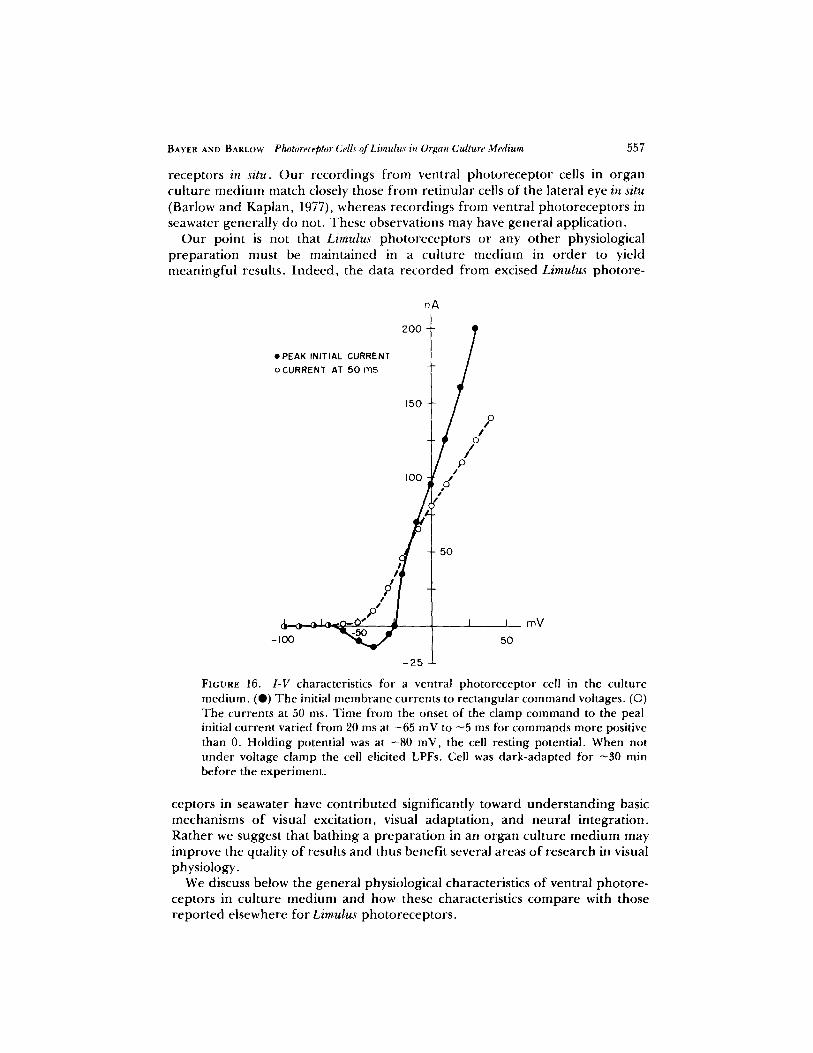
Fig. 16 shows typical current-voltage (I-V) relations for the ventral photore- ceptor cell measured at two times after the onset of voltage commands: 5-20 ms for the peak initial current and 50 ms for the delayed current , For clamp potentials between -100 and - 7 0 mV, the membrane currents are time- independent . Between - 7 0 and - 2 5 mV, the I-V relation for the initial cur rent
I 0 0 nA
I I0 nA
20 ms
FIGURE 15. Current responses of a ventral photoreceptor cell to depolarizing clamps. Direction of inward current is down. Holding potential of the cell was -80 mV. Lower record shows currents induced by command potentials of -80, -70, -60, -50, -40, -30, and -20 inV. Upper record show currents to commands from -80 to +30 mV in 10-mV steps. Beginning of each trace represents the onset of the command pulse. Earliest detectable current is the tail of the capacitive current surge. In the lower record the initial membrane current to commands of -80, -70, and -60 mV is passive. To commands of -50, -40, and -30 mV, the current is inward. The current reverses at - -25 inV. The latency to the peak initial current decreases as the conmaand w)ltage is increased. In the upper record the initial inward currents cannot be resolved. The cell was bathed in culture medium and generated LPFs when not under voltage clamp.
is highly nonlinear. Over this region the clamp currents are inward, indicating an electrically active membrane process. For membrane potentials more positive than - 2 5 mV, the I-V relation for the initial current is nearly linear with a slope resistance o f -0 .25 MI-I. The I-V relation for the delayed outward current is approximately linear for membrane voltages more positive than - - 4 0 mV, with a slope resistant of - 1 M~.
D I S C U S S I O N
Taken as a whole, our results underscore an earlier f inding of Barlow and Kaplan (1971); namely, the physiological properties of Limulus photoreceptors bathed in artificial seawater may not accurately reflect the properties of
BAVEg AND BARLOW Photoreceptor CelLs" of Limulus in Organ Culture Medium 557
receptors in situ. O u r recordings f rom ventral pho to recep to r cells in o rgan cul ture m e d i u m match closely those f rom ret inular cells of the lateral eye in situ (Barlow and Kaplan , 1977), whereas recordings f rom ventral pho to recep to r s in seawater general ly do not. These observat ions may have general applicat ion.
O u r point is not that Limulus pho to recep to r s or any o ther physiological p repa ra t ion must be mainta ined in a cul ture m e d i u m in o r d e r to yield meaningfu l results. I ndeed , the data r eco rded f rom excised Limulus photore -
�9 PEAK INITIAL CURRENT
oCURRENT AT 5 0 m s
/J
? ,o,
nA
2OO
150 o
I00
/,~ I
-25
J mV 50
FIGURE 16. I-V characteristics for a ventral photoreceptor cell in the culture medium. (Q) The initial membrane currents to rectangular command voltages. (�9 The currents at 50 ms. Time fi'om the onset of the clamp command to the peal initial current varied from 20 ms at -65 mV to ~5 ms for commands more positive than 0. Holding potential was at -80 mV, the cell resting potential. When not under voltage clamp the cell elicited LPFs. Cell was dark-adapted for ~30 min before the experiment.
ceptors in seawater have cont r ibu ted significantly toward unde r s t and ing basic mechanisms o f visual excitation, visual adapta t ion , and neural integrat ion. Ra ther we suggest that ba th ing a p repa ra t ion in an o rgan cul ture m e d i u m may improve the quality o f results and thus benefi t several areas o f research in visual physiology.
We discuss below the general physiological characteristics o f ventral pho to re - ceptors in cul ture m e d i u m and how these characteristics c o m p a r e with those r epo r t ed elsewhere for Limulus pho to recep to r s .

5 5 8 T H E J O U R N A L OF G E N E R A L P H Y S I O L O G Y ' V O L U M E 72 �9 1 9 7 8
Discrete Waves
Fig. 2 shows that a ventral photoreceptor cell in culture medium exhibits two types of discrete waves, LPFs and SPFs. Kaplan et al. (1973) recorded LPFs and SPFs from retinular cells of preparations bathed in an organ culture medium, and Barlow and Kaplan (1977) recorded both types of potentials from the lateral eye in situ. Under optimal conditions, Dowling (1968) was able to record similar potentials from retinular cells of excised lateral eyes.
Most studies of discrete wave activity in Limulus photoreccptors, however, have not reported LPFs and SPFs (for a review, see Fuortes and O'Bryan, 1972). These studies, which were carried out on excised eyes in artificial seawater, frequently reported two types of discrete waves: either "fast" and "slow" waves (Adolph, 1964), or "S" and "L" waves (Borsellino and Fuortes, 1968; Srebro and Behbehani, 1971). The results are not in general agreement. Adolph (1964) proposed that the fast wave was a regenerative response based on the fact that it appeared to be preceded by a prepotential. Borscllino and Fuortes (1968) found that L waves predominated in some cells, S waves predominated in others, and frequently one wave type would be absent altogether. They found a greater dispersion of latencies for L waves than for S waves. To explain the properties of the generator potential, they proposed a model of discrete wave activity which basically was an extension of an earlier compartment model proposed by Fuortes and Hodgkin 0964).
In a similar study Srehro and Behbehani (1971) found the latency distributions for S and L waves to be the same. They indicated that the model proposed by Borsellino and Fuortes was insufficient to account for this result and proposed instead a scheme in which L waves represent propagated, regenerative re- sponses, and S waves, nonpropagated responses. They subsequently rejected the idea that the L wave was a regenerative response (Behbehani and Srebro, 1974).
In each of the above studies the data were recorded from photoreceptors bathed in artificial seawater. The different results and interpretations may therefore reflect the physiological state of the individual preparations. Our results in Figs. 4 and 6 indicate that the characteristics of a ventral photoreceptor cell change after the cell is placed in artificial seawater. Both membrane potential and cell resistance are generally rcduced in size (see Table II), and as a consequence a given current wave would produce a reduced voltage wave. The relatively small sizes of fast and slow waves reported in the above studies may therefore result from a shift in resting potential. For example, a shift in resting potential entirely into or through the negative resistance region of the I- V characteristic (Fig. 16) would reduce or perhaps eliminate the effectiveness of a current wave in eliciting a regenerative response. The deteriorated condition of some cells in seawater may be sufficient to eliminate the electrical excitability of the photoreceptor membrane.
Behbehani and Srebro (1974) reported bimodal distributions of discrete wave sizes for both voltage-clamp and unclamped recordings from ventral photorc- ceptors in seawater. They found that the percentage of small waves increased when a second electrode was inserted into a cell and suggested that the
BAYER ANt) BARLOW Photoreceptor Cells of Limulgs in Organ Culture Medium 559
population of smaller waves resulted from damaged patches of cell membrane. Our results in Fig. 9 show that ventral photoreceptors in an organ culture medium yield a bimodal distribution of discrete waves for the unclamped condition and a unimodal distribution under voltage clamp. However, we also found an increase in small waves (-<1 mV) upon penetration of a cell with a second electrode, but the increase was not significant relative to the frequency of SPFs and LPFs. One effect of bathing cells in an organ culture medium may be to minimize the relative number of damaged membrane patches.
Yeandle and Spiegler (1973) reported that a ventral photoreceptor cell generates two statistically independent classes of waves, large light-evoked waves and small spontaneous waves. They found that increases in light intensity increased the relative proportion of large waves. For singly impaled cells in organ culture, we found that both classes of discrete waves, LPFs and SPFs, occur spontaneously in the dark and may be elicited by light. The frequency of occurrence of LPFs relative to SPFs decreases with light intensity, an effect which appears to reflect the regenerative nature and refractory characteristic of the LPF (Fig. 13). For doubly impaled cells we observed a slight increase in the proportion of small waves, an effect which may result from membrane damage. The relative frequency of the smallest population of current waves (-<1 nA) decreases with increasing light intensity (Fig. 10), a result similar to that reported by Yeandle and Spiegler (1973).
Nature of the Large Potential Fluctuations (LPFs)
LPFs appear to be regenerative events produced by the electrical excitability of the photoreceptor membrane. Properties which point to a regenerative process are as follows. First, the peak of the depolarizing potential of the LPF is followed initially by a rapid phase of repolarization (8 mV) and then by a slow phase of repolarization (Fig. 11). Second, the peak amplitude of the LPF depolarization has a fixed value of - -25 mV. Third, depolarizing voltage clamps reveal substantial inward currents indicative of an electrically excitable membrane (Fig. 16). And finally, small current pulses which depolarize the photoreceptor membrane potential into the "negative resistance" region of the I-V characteristic, between -60 and -40 mV, generate all-or-none LPFs. The membrane behaves passively for depolarizations from resting potential to < -60 mV, and LPFs are not elicited.
The peak amplitude of the LPF depolarization ( -25 mV; Fig. 11) has about the same value as the reversal potential for the initial inward current observed under voltage clamp (Fig. 16). Lisman and Brown (1971) reported a similar value for the reversal potential of inward current. Such a reversal potential is not readily interpreted in terms of a single ion. The equilibrium potentials of sodium and calcium are too high and those of potassium and chloride appear too low. It is possible, however, that the observed reversal potential reflects the increase in conductance to two ions such as calcium and potassium.
LPFs appear to be triggered by SPFs. This result is suggested by the finding that LPFs are often preceded by slow depolarizing potentials (Figs. 2, 11, and 12). Several LPFs in Fig. 12 (-5.5 log I) appear to be triggered by depolarizing

5 6 0 T H E , J O U R N A L OF G E N E R A L P H Y S I O L O G Y " V O L U M E 72 �9 1 9 7 8
prepotentials of --20 mV which is approximately the peak amplitude of the SPFs recorded from this cell. Barlow and Kaplan (1977) also observed that SPFs appear to trigger LPFs in retinular cells of the Limulus lateral eye.
THE SPIKE-LIKE RESPONSE Responses of ventral photoreceptors to in- creases in flash intensity change from a unitary LPF to a response with the fast rising phase of the LPF followed by a graded, depolarizing component (Fig. 12). At the highest test intensities the graded component overshadows the LPF. What remains is an inflection on the rising phase of the response. For flashes of greater duration, this combined response is the transient of the receptor potential.
Others have detected the initial fast rising phase of receptor potential and generally termed it the spike-like response (Benolken, 1959; Yeandle, 1967; Millecchia, 1969; Wulff and Mueller, 1973), A similar spike-like response has been reported for retinular cells of the honeybee drone eye (Bauman, 1968) and for cells of the dragonfly eye (Chappell and Dowling, 1972). In Limulus the differences between the spike-like responses and LPFs appear minor. Both potentials may result from the same regenerative membrane process. The fact that the spike-like response can be recorded from Limulus cells which do not exhibit LPFs may reflect changes in the membrane characteristics. Specifically, in healthy cells with large membrane resistance, small currents (5 nA) can shift the transmembrane potential rapidly through the negative resistance region of the I-V characteristics and thereby elicit a regenerative response. In partially deteriorated cells having lower membrane resistance, small currents are ineffec- tive, but large currents can elicit regenerative responses for cell resting potential > - -40 mV, the potential at which the slope of the initial current function changes sign.
Photon Sensitivity at Threshold
Yeandle (1958) first observed discrete waves in the lateral eye of Limulus and suggested that they resulted from the absorption of single photons. He based his conclusion on the random occurrence of discrete waves at low light levels and the fact that the probability of their occurrence as a function of flash intensity could be described approximately by a Poisson sum of n = 1. Supporting evidence has been reported by Adolph, 1964; Fuortes and Yeandle, 1964; Borsellino and Fuortes, 1968; and Barlow and Kaplan, 1977.
The generation of discrete waves by single photon events is also apparent in our records from ventral photoreceptor cells. Note that in Fig. 8 the Poisson curves indicate a threshold of one photon, but the abscissa gives a value of 12 absorbed photons. Such discrepancies were common and may indicate a low value for the quantum efficiency of eliciting discrete waves or an error in estimating the number of absorbed photons.
Stronger evidence that a single photon event can elicit a discrete wave is the linearity of the response rate in Fig. 7. Rough estimates of the number of absorbed photons in Figs. 7 and 8 yield quantum efficiencies of -0.1. Measure- ments from other ventral eye preparations yield values ranging from 0.03 to 0.13. Quantum efficiencies of 0.05-0.2 have been reported for cells of the lateral eye (Kaplan and Barlow, 1976).
BAYER AND BARLOW Photoreceptor Cells of Limul~ in Organ Culture Medium 561
Intensity-Response Function
T h e intensity funct ion for the steady-state response o f cells in the cul ture medium contains a p rominen t plateau (Fig. 6). This characteristic shape may have several cont r ibut ing factors. First, the intensity funct ion for m em b ran e cu r ren t contains a plateau over the same range o f intensities (Fig. 14). Second, the steady-state I -V characteristic exhibits substantial rectification between - 7 0 and - 4 0 mV (Fig. 16). Over this voltage range the cell resistance decreases f rom - 3 0 MI~ to 1 MI) and thereby reduces the effectiveness o f l ight-induced cu r ren t in p roduc ing membrane depolarizat ion. Th i rd , LPFs are the principal compo- nent o f the receptor potential at low intensities and SPFs p redomina te at high intensities (Fig. 5). T h e shift f rom LPFs to SPFs at in termediate intensities may influence the shape o f the intensity function (Barlow and Kaplan, 1977). T h e first of these possibilities may not be an impor tan t factor because cells have been recorded which exhibit a plateau in the intensity funct ion for recep tor cu r ren t but do not show a plateau in the funct ion for receptor potential (Lisman, 1971). Membrane rectification could be a contr ibut ing factor because cells which exhibit substantial rectification for hyperpolar iz ing currents p roduce distinct plateaus. T h e shift f rom LPFs to SPFs may contr ibute in conjunct ion with membrane rectification inasmuch as small depolar izing currents genera te LPFs only in cells with high membrane resistance.
We thank A. Fein, E. Kaplan, E. A. Kravitz,J. E. Lisman, W. G. Sokolich, B. R. Talamo, and A. E. Stuart for helpful suggestions. We also thank J. M. Brophy and B. E. Klock for technical assistance. This study was supported by grant EY-00667 from the National Institutes of Health. Parts of this study appeared in a dissertation by Dr. Bayer as partial fulfillment of the requirements for the degree of Doctor of Philosophy at Syracuse University.
Received for publication 13 June 1977.
R E F E R E N C E S
ADOLPH, A. R. 1964. Spontaneous slow potential fluctuations in the Limulus photorecep- tor.J. Gen. Physiol. 48:297-322.
BARLOW, R. B., JR., and E. KAPLAN. 1971. Limulus lateral eye: properties of receptor units in the unexcised eye. Science (Wash. D.C.). 174:1027-1029.
BARLOW, R. B., .JR., and E. KAPLAN. 1977. Properties of visual cells in the lateral eye of Limulus in situ : intracellular recordings.J. Gen. Physiol. 69:203-29-0.
BAt:MAN, F. 1968. Slow and spike potentials recorded from retinular cells of the honeybee drone in response to light.J. Gen. Physiol. 52:855-875.
BAYER, D. S. 1975. Limulus ventral eye: physiological properties of photoreceptor cells in organ culture. Doctoral Dissertation. Syracuse University, Syracuse, N.Y.
BEHBEHANI, M., and R. SREBRO. 1974. Discrete waves and phototransduction in voltage- clamped ventral photoreceptors.J. Gen. Physiol. 64:186-200.
BENOLKEN, R. M. 1959. Light- and dark-adaptation studies on the graded receptor potential of the Limulus eye. Doctoral Dissertation. The Johns Hopkins University, Baltimore, Md.
BORSELLINO, A., and M. G. F. FUORTES. 1968. Responses to single photons in visual cells of Limulus. J. Physiol. (Lond.). 196:507-539.
BROWN,J. E., andJ. R. BLINgS. 1974. Changes in intracellular free calcium concentration
. ~ 6 2 T H E J O U R N A L OF GENERAL P H Y S I O L O ( ; Y �9 VOLUME 7 2 " I978
during illumination of invertebrate photoreceptors. Detection with aequorin..]. Gen. Physiol. 64:643-665.
CrtAPI'ELL, R. L., and J. E. DOWLINC;. 1972. Neural organization of the median ocellus of the dragonfly.J . Gen. Physiol. 60:121-165.
DOWLING, J. E. 1968. Discrete potentials in the dark-adapted eye of Limulus. ~mture (Lond.). 217:28-31.
FEIN, A., and J. S. CHARLTOY. 1975. Local adaptation in the ventral photoreceptors of Limulus..]. Gen. Physiol. 66:823-836.
FEIN, A., and R. DEVoE. 1973. Adaptation in the ventral eye of Limulus is functionally independent of the photochemical cycle, membrane potential, and membrane resist- ance../. Gen. Physiol. 61:273-289.
FEIN, A., and J. LISMAN. 1975. Localized desensitization of Limulus photoreceptors produced by light or intracellular calcium ion injection. Science (Wash. D.C.). 187:1094- 1096.
FUORTES, M. G. F., and A. L. HODGKIN. 1964. Changes in time scale and sensitivity in the mnmatidia of Limulus. J. Physiol. (Loud.)., 172:239-263.
FUORTES, M. G. F., and P. M. O'BRvA~. 1972. Responses to single photons, lt~ Physiology of Photoreceptor Organs, Handbook of Sensory Physiology, Vol. VII/2. M. G. F. Fuortes, editor. Springer-Verlag, Berlin. 321-338.
FUORTV.S, M. G. F., and S. S. YEANDL~. 1964. Probability of occurrence of discrete potential waves in the eye ofLimulus. J. Gem Physiol. 47:443-463.
GOTTSCHEWSKI, G. H. M. 1960. Morphogenetische Untersuchungen an It~ Vitro wachsen- den Augenanlagen wm Drosophila melanogaster. Wilhelm Roux' Arch. Entwieklungsmeeh. Org. 152:204.
KAeLAY, E., and R. B. BARLOW, JR. 1975. Properties of visual cells in the lateral eye of Limulus in situ. Extracellular recordings.J. Gen. Physiol. 66:303-326.
KAPLAN, E., and R. B. BARLOW, JR. 1976. Energy, quanta, and Limulus vision. Vision Res. 16:745-751.
KAeLAN, E., D. S, BAWR, and R. B. BARLOW, JR. 1973. Limulus lateral eye: intracellular recordings in situ and in organ culture. Biol. Bull. (Woods Hole). 145:442.
LISMAN, J. 197 l. An electrophysiological investigation of the ventral eye of the horseshoe crab, Limulus polyphemus. Doctoral Dissertation. Massachusetts Institute of Technology, Cambridge, Mass.
L I S M A N , J. E., and J. E. BROWN. 1971. Two light-induced processes in the photoreceptor cells of Limulus ventral eye..]. Gen. Physiol. 58:544-561.
LI'SMAN, J. E., and J. E. BRowN. 1972. The effects of intracellular iontophoretic injection of calcium and sodium ions on the light response of Limulus ventral photoreceptors../. Gen. Physiol. 59:701-719.
LISMAN, J. E., and J. E. BROWN. 1975. Light-induced sensitivity changes in Limulus photoreceptor.J . Gen. Phy.siol. 66:473-488.
MILLECCHIA, R. 1969. The ventral photoreceptor cells of Limulus: an electrophysiological study. Doctoral Dissertation. The Rockefeller University, New York.
Mn.LECCHIA, R., R. B~ADBURV, and A. MAURO. 1966. Simple photoreceptors in Limulus polyphemus. Science (Wash. D.C.). 154:1199-1201.
MILLECCHIA, R., and A. MAURO. 1969 a. The ventral photoreceptor cells of Limulus. It . The basic photoresponse.J. Gen. Physiol. 54:310-330.
M1LLECCmA, R., and A. MAURO. 1969 b. The ventral photoreceptm" cells of Limulus. I l l . A voltage-clamp study.J. Gen. Physiol. 44:331-351.
BAYER AND BARLOW Photoreceptor Cells ~'Limulus in Organ Culture Medium 563
MURRAY, G. 1966. lntracel lular absorption difference spectrum o f Limulus extra-ocular photolabile pigment. Science (Wash. D.C.). 154:1182-1183.
PARKER, R. C., and W. H. COLE. 1940. Studies of the body fluids and sera of some marine invertebrates. Bull. MT. Desert 1,~l. Biol. Lab.
PIRENN~, M. H. 1967. Vision and the Eye. Chapman and Hall Ltd., London. 224 pp. SREBRO, R., and M. BEHBEHANI. 1971. A stochastic model fl)r discrete waves in the
Limulus photorecep tor , J . Gen. Physiol, 58:267-286. WOLVF, E. M. 1963. Sur l 'explanation in vitro de fragments d 'organes de limules
(Xiphosura polyphemus). Ann, Epiphyt. (Paris). 14"113. WVLVF, V. J. , and W . J . MUELLER. 1973. On the origin of the receptor potential in the
lateral eye o f Limulus. Vision Res. 13:661-671. YEANDLE, S. 1958. Evidence of quantized slow potentials in the eye of Limulus. Am. J.
Ophthalmol. 46:82-87. YEANDLE, S. S. 1967. Some propert ies of the components of the Limulus ommatidial
potential. Kybernetik 3:250-254. YEANDLE, S., and J. B. SPIEC, LER. 1973. Light-evoked and spontaneous discrete waves in
the ventral nerve photoreceptor ofLimulus. J. Gen. Physiol. 61"552-571.