life cycle stages of the amphibian chytrid ... · tologically prepared slides of infected skin or...
TRANSCRIPT
DISEASES OF AQUATIC ORGANISMSDis Aquat Org
Vol. 68: 51–63, 2005 Published December 30
INTRODUCTION
Batrachochytrium dendrobatidis causes a potentiallyfatal epidermal infection of amphibians and hascaused mass mortality, population declines and extinc-tions (Berger et al. 1999, Speare et al. 2001, McDonaldet al. 2005). Chytridiomycosis has been recorded fromAustralia, New Zealand, Europe, Africa, and South,Central and North America, from a broad range ofhabitats (Berger et al. 1999, Lips 1999, Mutschmann etal. 2000, Bosch et al. 2001, Fellers et al. 2001, Speare etal. 2001, Bradley et al. 2002, Weldon et al. 2004). Mor-tality rates of up to 100% occurred during natural out-breaks in captivity and in transmission experimentsin captive amphibians of susceptible species (Berger etal. 1998, Longcore et al. 1999, Berger 2001, Nichols etal. 2001), although other species can survive infection(Ardipradja 2001, Davidson et al. 2003). Death in sus-
ceptible experimental animals usually occurs frombetween 18 and 70 d post exposure and incubationtime varies with dose, fungal strain, temperature andamphibian species (Ardipradja 2001, Berger 2001,Nichols et al. 2001, Woodhams et al. 2003, Berger et al.2004). Amphibians with no clinical signs frequentlycarry light infections in the wild (Hopkins & Channing2003, Hanselmann et al. 2004, Retallick et al. 2004,McDonald et al. 2005).
The amphibian chytrid was placed in a new genus,Batrachochytrium (Phylum Chytridiomycota, ClassChytridiomycetes, Order Chytridiales) (Longcore et al.1999). Photographs of an isolate from a captive bluepoison dart frog Dendrobates azureus that died at theNational Zoological Park in Washington, DC, USA,were designated as the nomenclatural type of the spe-cies, which was named B. dendrobatidis (Longcore etal. 1999). The ultrastructural morphology of its zoospore,
© Inter-Research 2005 · www.int-res.com*Email: [email protected]
Life cycle stages of the amphibian chytridBatrachochytrium dendrobatidis
Lee Berger1, 2,*, Alex D. Hyatt2, Rick Speare1, Joyce E. Longcore3
1Amphibian Disease Ecology Group, School of Public Health, Tropical Medicine and Rehabilitation Science,James Cook University, Townsville, Queensland 4811, Australia
2Australian Animal Health Laboratory, CSIRO Livestock Industries, Private Bag 24, Geelong, Victoria 3220, Australia3Department of Biological Sciences, University of Maine, Orono, Maine 04469-5722, USA
ABSTRACT: An overview of the morphology and life cycle of Batrachochytrium dendrobatidis, thecause of chytridiomycosis of amphibians, is presented. We used a range of methods to examine stagesof the life cycle in culture and in frog skin, and to assess ultrastructural pathology in the skin of2 frogs. Methods included light microscopy, transmission electron microscopy with conventionalmethods as well as high pressure freezing and freeze substitution, and scanning electron microscopywith critical point drying as well as examination of bulk-frozen and freeze-fractured material.Although chytridiomycosis is an emerging disease, B. dendrobatidis has adaptations that suggest ithas long been evolved to live within cells in the dynamic tissue of the stratified epidermis. Sporangiadeveloped at a rate that coincided with the maturation of the cell, and fungal discharge tubes usuallyopened onto the distal surface of epidermal cells of the stratum corneum. A zone of condensed,fibrillar, host cytoplasm surrounded some sporangia. Hyperkeratosis may be due to (1) a hyperplasticresponse that leads to an increased turnover of epidermal cells, and (2) premature keratinization anddeath of infected cells.
KEY WORDS: Batrachochytrium dendrobatidis · Chytridiomycosis · Fungus · Morphology ·Ultrastructure · Transmission and scanning electron microscopy · Pathology · Amphibian
Resale or republication not permitted without written consent of the publisher
Dis Aquat Org 68: 51–63, 2005
its occurrence on an amphibian host and small subunitribosomal DNA (ssu-rDNA) sequence of B. dendro-batidis indicate that this fungus is distinctly differentfrom other known chytrids (Berger et al. 1998, Long-core et al. 1999, James et al. 2000). It is the only mem-ber of this phylum to cause disease in a vertebrate.
The life cycle of Batrachochytrium dendrobatidis isa simple progression from zoospore to the growingorganism, called a thallus, which produces a singlezoosporangium (= container for zoospores). The con-tents of the zoosporangium (also known as a spo-rangium) cleave into new zoospores which exit thesporangium through one or more papillae. Sexualreproduction has not been observed. Colonial develop-ment resulting from the formation of more than 1 spo-rangium from 1 zoospore is the only known variation ofthe cycle (Longcore et al. 1999). The life cycle seems tobe the same in culture and in skin. The duration of thelife cycle in vitro is 4 to 5 d at 22°C and is assumed tobe the same in amphibian skin, although this has notbeen tested.
Batrachochytrium dendrobatidis discharges zoosporesthrough an inoperculate opening and exhibits mono-centric or colonial growth (Longcore et al. 1999). Long-core et al. (1999) comprehensively described the taxo-nomic features of B. dendrobatidis and gave detailedmorphology based on light microscopic observationsof cultures and ultrastructure of serially sectionedzoospores, but did not describe the appearance of lifecycle stages using scanning electron microscopy (SEM).
Significant ultrastructural differences were notobserved among zoospores of isolates from Australia,the USA or Central America (Berger et al. 1998, Long-core et al. 1999). Multilocus sequence typing (MLST)has been used to examine genetic diversity amongfungal strains from North America, Panama, Australiaand from frogs imported from Africa, and only 5 vari-able nucleotide positions were detected among 10 loci(5918 bp)(Morehouse et al. 2003). These results sug-gest that Batrachochytrium dendrobatidis is a recentlyemerged clone and support the epidemiological datashowing that chytridiomycosis has been introducedinto many countries from a common source. There isevidence that Africa is the origin (Weldon et al. 2004).
In amphibians sporangia infect cells in the stratumgranulosum and stratum corneum in the superficialepidermis. Immature sporangia occur within thedeeper, more viable cells while mature zoosporangiaand empty sporangia are more prevalent in the outerkeratinized layers. Discharge tubes generally projecttowards the skin surface and zoospores may bereleased to the environment. The distribution of spo-rangia in developing tadpoles follows the changesin the distribution of keratinized epidermis (Marantelliet al. 2004).
Resistant resting spores have not been found in his-tologically prepared slides of infected skin or duringexamination of fresh skin in the process of isolating thefungus from more than 80 amphibians (J. E. Longcoreunpubl. data). MLST sequencing studies indicate thatBatrachochytrium dendrobatidis reproduces clonally,which supports the lack, or uncommon occurrence,of a sexually produced resting stage (Morehouse et al.2003).
The histopathology of chytridiomycosis based onexamination of haematoxylin and eosin stained sec-tions has been described (Berger et al. 1998, Pessier etal. 1999, Berger 2001). Infection almost always causeshyperkeratosis in the region of the thalli. Otherchanges include irregular multifocal hyperplasia, dis-ordered epidermal cell layers, spongiosis, erosions andoccasional ulcerations of the skin. Epidermal width ishighly variable with diffuse or focal thickening in someareas, as well as large areas of thinning. Individualepidermal cell pyknosis and vacuolation may occur inscattered cells in the stratum basale or more superficiallayers near infection foci. Occasionally vacuolateddegenerate cells appear to coalesce into vesicles thatresult in lifting of the epidermis and erosion. Theremay be a mild inflammatory response with a slightincrease in mononuclear cells in the dermis, but in-flammatory response in the epidermis is uncommon.
Two hypotheses have been proposed to explain howa fungus that is restricted to the superficial epidermishas the capacity to kill frogs (Berger et al. 1998, Pessieret al. 1999). (1) The chytrid might release proteolyticenzymes or other active compounds that are absorbedthrough the permeable skin of the frog or, possibly,(2) damage to skin function results in disturbance ofoxygen, water or electrolyte balance which results indeath. By examining the ultrastructural pathology inepidermal cells we aim to improve our understandingof the effect of Batrachochytrium dendrobatidis onepidermal cells.
Cultures of Batrachochytrium dendrobatidis arehighly susceptible to a range of antifungal drugs andantifungal peptides produced by frogs (Berger 2001,Rollins-Smith et al. 2002, Johnson et al. 2003). How-ever, since topical antifungal treatment of frogs hasvariable efficacy, it appears that B. dendrobatidis isless susceptible when present in amphibian skin(Berger 2001). The reason for this is not known, butone hypothesis is that thalli are protected by theirintracellular location in epidermal cells. One aim of ourtransmission electron microscopy (TEM) studies is todetermine if the epidermal cell undergoes modifica-tions that may explain this protection.
This study was undertaken to improve our under-standing of the morphology of Batrachochytrium den-drobatidis by using TEM and SEM of all fungal stages
52
Berger et al.: Life cycle of Batrachochytrium dendrobatidis
in culture and in epidermis. To understand the patho-genesis of chytridiomycosis and why chytridiomycosisappears resistant to antifungal drugs we used ultra-structural studies of infected epidermis to examine theeffect of the fungus on amphibian cells.
MATERIALS AND METHODS
Samples. Light microscopic studies of Batrachochy-trium dendrobatidis in culture were of isolate Mel-bourne-Ldumerilii-98-LB-1 from a captive, ill Limno-dynastes dumerilii. For ultrastructural studies weexamined the isolate Tully-Ndayi-98-LB-1 from afree-living, ill Nyctimystes dayi collected from Tully,Queensland. Cultures were isolated and maintainedon tryptone/gelatin hydrolysate/lactose (TGhL) agar orbroth using routine methods (Longcore et al. 1999).Infected skin samples were from a wild Litoria lesueurifrom Goomburra, Queensland (AAHL 97 574/1) thatdied and was fixed in 2.5% glutaraldehyde, a captiveBufo marinus that died and was fixed in 10% formalin,and 2 captive L. gracilenta were used for high pressurefreezing. The latter samples were a toe-clip taken froma healthy infected frog with a light infection, and skinfrom a frog that had recently died with severe chy-tridiomycosis. These samples were kept on ice forabout 2 h before processing.
TEM. Conventional processing: Samples of skinwere fixed in 2.5% (v/v) glutaraldehyde in 0.1 Mcacodylate buffer (pH 6.8, 300 mOsm kg–1) for 1 h,washed in buffer (3 × 20 min), post-fixed in 1% (w/v)osmium tetroxide in 0.1 M cacodylate buffer for 1 hfollowed by washing (3 × 5 min) in reverse osmosiswater. Samples were then dehydrated in gradedalcohols (70 to 100%), infiltrated with Spurrs resin byplacing them in 50/50 Spurrs/100% ethanol and thenin 2 changes of 100% Spurrs followed by embeddingin Spurrs epoxy resin at 65°C (overnight). Fixation ofcultures of Batrachochytrium dendrobatidis differed inthat glutaraldehyde was added to the culture medium(TGhL broth) to a final concentration of 2.5%. Pre-liminary experiments revealed that zoospores weresensitive to the osmolality of the buffer, but TGhLbroth was found to cause no discernible artifacts in thecytoplasm.
High pressure freezing and freeze substitution:Samples of Batrachochytrium dendrobatidis cultureand of infected frog skin were rapidly frozen underhigh pressure (2000 bars) within a Leica high-pressurefreezer. Brass planchets were soaked in a lecithin-chloroform solution and allowed to dry before packingwith agar cultures or pieces of toe skin. A rangeof embedding media was used: TGhL broth, 2.3 Msucrose, hexadecene and hexene. The frozen plan-
chets were stored in liquid nitrogen until processing.Substitution was initiated by placing planchets andsamples within perforated beam capsules in 1% (w/v)osmium tetroxide/2.5% (v/v) glutaraldehyde in 100%acetone at –90°C in the presence of a molecular sievewithin a ‘Leica CS auto’ for 8 d. The temperature wasraised to –60°C at 2.5°C h–1 and kept at –60°C for 2 d.The temperature was then raised to –18°C at 2.5°C h–1
then up to 0°C at 10°C h–1 and the media replaced with100% acetone. Samples were then warmed to roomtemperature (24°C) in 1 h. The substitution mediumwas replaced with 1:1 propylene oxide (PO):acetone,100% PO, 1:1 PO:epon, 100% epon × 2 and embeddedat 60°C for 24 h. All media used in planchets resultedin excellent preservation of samples.
Cutting and examination of samples: Thick sectionswere examined with methylene blue stain by lightmicroscopy to identify and select the sections contain-ing sporangia. Ultra-thin sections (70 nm) were cut ona Leica-Reichert-Jung Ultracut E microtome, floatedand adhered onto a grid, double stained in uranylacetate and lead citrate and examined with a HitachiH7000 or Philips CM 120 transmission electron micro-scope at 75 or 100 kV.
SEM. Critical point drying: Skin was fixed in phos-phate buffered 2.5% glutaraldehyde for 1 h, washed inphosphate buffer (pH 6.8, 300 mOsm kg–1) for 15 min,post-fixed in buffered 1% (w/v) osmium tetroxide for3 h, and rinsed in the same buffer (× 10). Samples werethen placed in saturated aqueous filtered thiocarbo-hydrazide (1% w/v) for 10 min, rinsed in distilled water(×10), then placed in 1% (w/v) aqueous osmiumtetroxide at 0°C (30 min), rinsed in reverse osmosiswater and sequentially dehydrated in graded alcohol(70 to 100 %). Following dehydration, tissues werecritically point dried from liquid carbon dioxide,mounted on a stub with carbon dag, and sputter coatedwith gold. Samples were viewed with a JEOL JSM 840scanning electron microscope at 5 to 15 kV with aworking distance of between 16 and 22 mm.
Bulk-frozen hydrated samples (agar): Cultures wereexamined with a JEOL 6340F field emission scanningelectron microscope fitted with an Oxford 1500 cryosystem. Blocks of 5 d old agar cultures were adhered tostubs with OCT compound cryo adhesive (Tissue Tek)and then plunged into melting nitrogen and trans-ferred under vacuum to the microscope at liquidnitrogen temperature. Samples were etched at –95°Cfor 60 s, then coated with gold/palladium in the trans-fer chamber. Samples were examined at 2 kV and aworking distance of 20 mm.
Bulk-frozen hydrated samples (coverslip): To obtaina cleaner view of the base of sporangia, cultures weregrown on sterile, round, plastic, 13 mm coverslips(Thermanox), by placing coverslips in petri dishes of
53
Dis Aquat Org 68: 51–63, 2005
active TGhL broth culture. Between 6 and 10 d, brothwas removed by rinsing the coverslips through 3changes of distilled water. Excess water was removedby touching the edges with filter paper, and leavingthem to dry for about 1 min. To reduce charging, cover-slips were cut into quarters before they were adheredto the stub with carbon dag. Stubs were plunged intomelting nitrogen then transferred under vacuum atliquid nitrogen temperature to the Hexland chamberand into the JEOL JSM-840 at –170°C. Extended dry-ing at –80°C was necessary to sublimate ice, and goodresults were obtained on a sample that was desiccatedovernight and examined as a freeze-dried sample;however, an effect of freeze drying is shrinkage. Somesporangia did not withstand this form of dehydrationand shrank, presenting a crumpled appearance. Sam-ples were gold-coated within the antechamber of themicroscope. Samples were examined at 3 to 5 kV and aworking distance of 20 mm.
Freeze fracture: Freeze fracture preparations wereproduced and examined using a Philips XL30 FEG or
JEOL JSM-840 SEM. Six to 14 d old agar cultures werepacked into rivets that were joined upright. Afterplunging in melting nitrogen and placing in thetransfer chamber of the microscope, the top rivet wasknocked off, exposing the fractured surface of theculture. The sample was etched at –96°C for 2 min toremove about 1200 nm water, and coated with plat-inum or gold. The sample was etched again at –100°Cfor 2 min. Samples were examined at 6 kV with aworking distance of 20 mm.
RESULTS AND REVIEW OF MORPHOLOGY
Zoospore
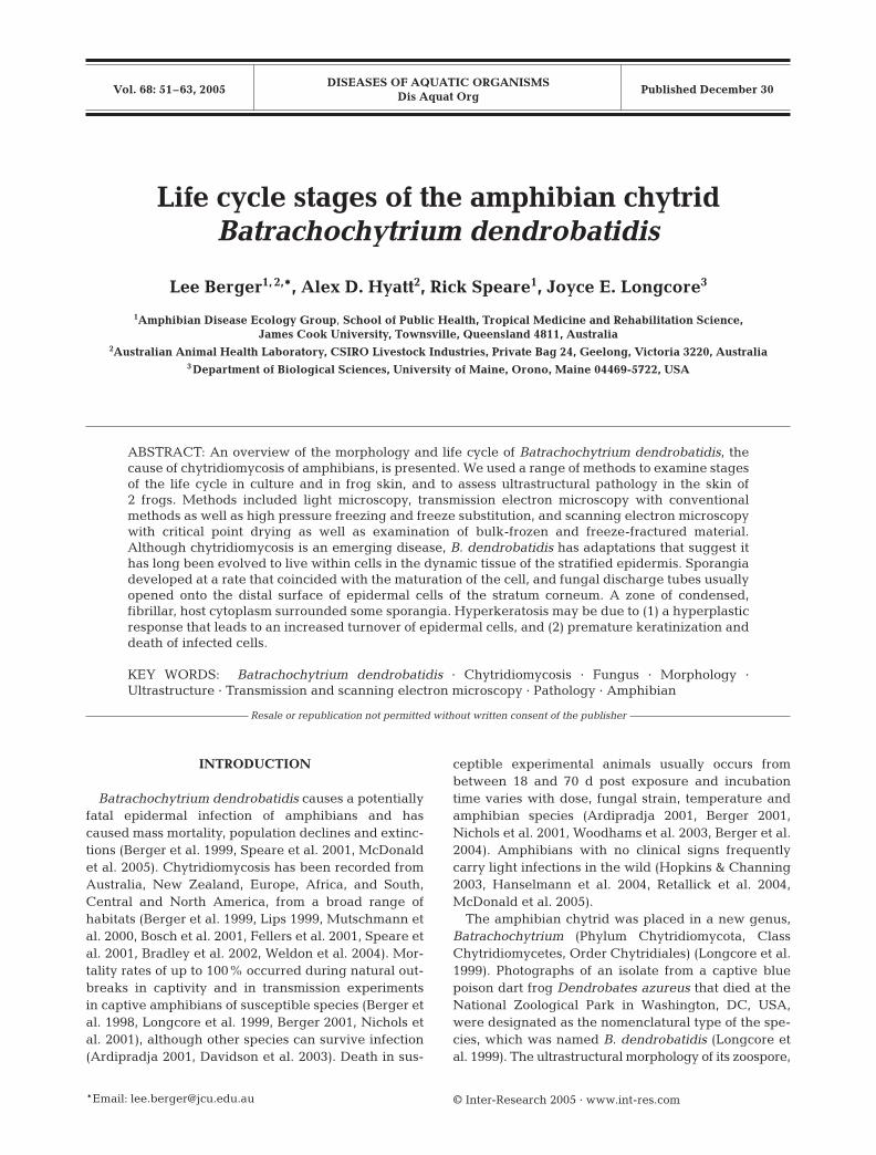
Zoospores are the unwalled, waterborne, motile,flagellated stage (Figs. 1 to 4). Zoospores of Batra-chochytrium dendrobatidis are mostly spherical butcan be elongate and amoeboid when first releasedfrom the zoosporangium (Longcore et al. 1999). They
54
Figs. 1 to 4. Batrachochytrium dendro-batidis. Fig. 1. Light micrograph of livecultured zoospore. Dark droplets areprobably lipid globules. Scale bar =6 µm. Figs. 2 to 4. B. dendrobatidis.Transmission electron micrographs ofzoospores. Fig. 2. Formalin-fixed zoo-spores within a zoosporangium in theskin of Bufo marinus. Zoospores arebeing released and contain numerouslipid globules that are partially sur-rounded by the microbody and occurat the edge of the ribosomal mass.Scale bar = 2 µm. Fig. 3. Glutar-aldehyde-fixed cultured zoospore.The nonflagellate centriole (NFC) isparallel to the kinetosome. Micro-tubule root runs parallel to the kineto-some and is embedded in a cone ofribosomes. Scale bar = 0.6 µm. Fig. 4.Glutaraldehyde-fixed cultured zoo-spore. Nucleus is not associated withthe kinetosome and is nested in theribosomal mass which is surroundedby endoplasmic reticulum. Mitochon-dria are adjacent to the ribosomalmass. Scale bar = 1 µm. F = flagellum;N = nucleus; R = ribosomes; Mb =microbody; L = lipid droplet; K = kine-tosome; M = mitochondria; Tp = termi-nal plate; V = vacuole; ER = endo-plasmic reticulum; MT = microtubules
Berger et al.: Life cycle of Batrachochytrium dendrobatidis
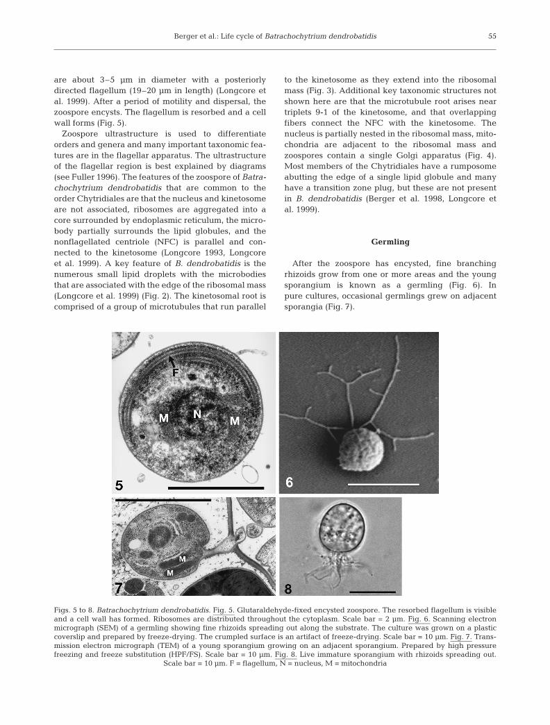
are about 3–5 µm in diameter with a posteriorlydirected flagellum (19–20 µm in length) (Longcore etal. 1999). After a period of motility and dispersal, thezoospore encysts. The flagellum is resorbed and a cellwall forms (Fig. 5).
Zoospore ultrastructure is used to differentiateorders and genera and many important taxonomic fea-tures are in the flagellar apparatus. The ultrastructureof the flagellar region is best explained by diagrams(see Fuller 1996). The features of the zoospore of Batra-chochytrium dendrobatidis that are common to theorder Chytridiales are that the nucleus and kinetosomeare not associated, ribosomes are aggregated into acore surrounded by endoplasmic reticulum, the micro-body partially surrounds the lipid globules, and thenonflagellated centriole (NFC) is parallel and con-nected to the kinetosome (Longcore 1993, Longcoreet al. 1999). A key feature of B. dendrobatidis is thenumerous small lipid droplets with the microbodiesthat are associated with the edge of the ribosomal mass(Longcore et al. 1999) (Fig. 2). The kinetosomal root iscomprised of a group of microtubules that run parallel
to the kinetosome as they extend into the ribosomalmass (Fig. 3). Additional key taxonomic structures notshown here are that the microtubule root arises neartriplets 9-1 of the kinetosome, and that overlappingfibers connect the NFC with the kinetosome. Thenucleus is partially nested in the ribosomal mass, mito-chondria are adjacent to the ribosomal mass andzoospores contain a single Golgi apparatus (Fig. 4).Most members of the Chytridiales have a rumposomeabutting the edge of a single lipid globule and manyhave a transition zone plug, but these are not presentin B. dendrobatidis (Berger et al. 1998, Longcore etal. 1999).
Germling
After the zoospore has encysted, fine branchingrhizoids grow from one or more areas and the youngsporangium is known as a germling (Fig. 6). Inpure cultures, occasional germlings grew on adjacentsporangia (Fig. 7).
55
Figs. 5 to 8. Batrachochytrium dendrobatidis. Fig. 5. Glutaraldehyde-fixed encysted zoospore. The resorbed flagellum is visibleand a cell wall has formed. Ribosomes are distributed throughout the cytoplasm. Scale bar = 2 µm. Fig. 6. Scanning electronmicrograph (SEM) of a germling showing fine rhizoids spreading out along the substrate. The culture was grown on a plasticcoverslip and prepared by freeze-drying. The crumpled surface is an artifact of freeze-drying. Scale bar = 10 µm. Fig. 7. Trans-mission electron micrograph (TEM) of a young sporangium growing on an adjacent sporangium. Prepared by high pressurefreezing and freeze substitution (HPF/FS). Scale bar = 10 µm. Fig. 8. Live immature sporangium with rhizoids spreading out.
Scale bar = 10 µm. F = flagellum, N = nucleus, M = mitochondria
Dis Aquat Org 68: 51–63, 2005
Developing zoosporangia
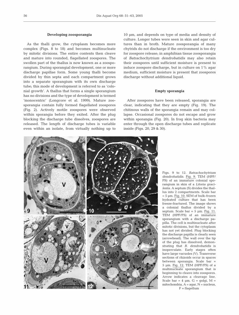
As the thalli grow, the cytoplasm becomes morecomplex (Figs. 8 to 18) and becomes multinucleateby mitotic divisions. The entire contents then cleaveand mature into rounded, flagellated zoospores. Theswollen part of the thallus is now known as a zoospo-rangium. During sporangial development, one or moredischarge papillae form. Some young thalli becomedivided by thin septa and each compartment growsinto a separate sporangium with its own dischargetube; this mode of development is referred to as ‘colo-nial growth’. A thallus that forms a single sporangiumhas no divisions and the type of development is termed‘monocentric’ (Longcore et al. 1999). Mature zoo-sporangia contain fully formed flagellated zoospores(Fig. 2). Actively motile zoospores were observedwithin sporangia before they exited. After the plugblocking the discharge tube dissolves, zoospores arereleased. The length of discharge tubes is variableeven within an isolate, from virtually nothing up to
10 µm, and depends on type of media and density ofculture. Longer tubes were seen in skin and agar cul-tures than in broth. Mature zoosporangia of manychytrids do not discharge if the environment is too dryfor zoospore release; in amphibian tissue zoosporangiaof Batrachochytrium dendrobatidis may also retaintheir zoospores until sufficient moisture is present toinduce zoospore discharge, but in culture on 1% agarmedium, sufficient moisture is present that zoosporesdischarge without additional liquid.
Empty sporangia
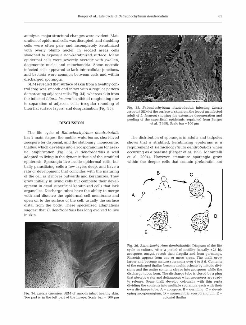
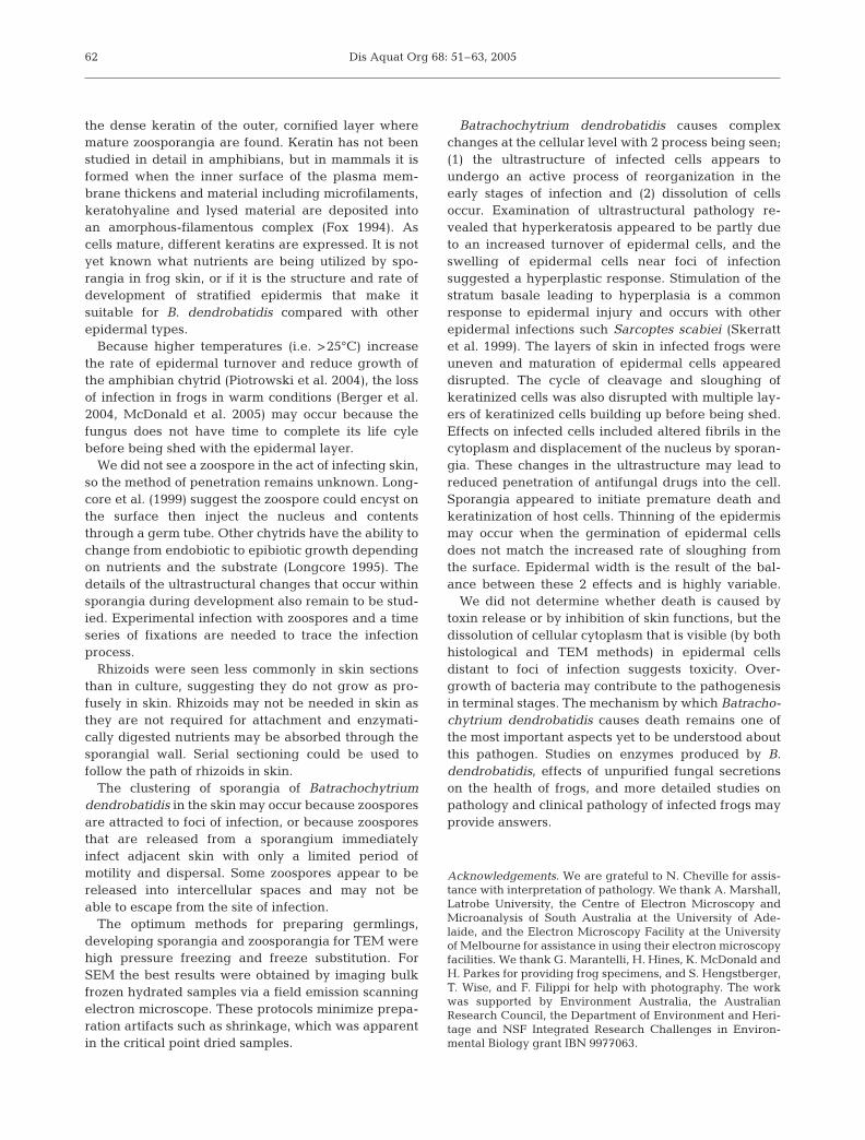
After zoospores have been released, sporangia areclear, indicating that they are empty (Fig. 19). Thechitinous walls of the sporangia remain and may col-lapse. Occasional zoospores do not escape and growwithin sporangia (Fig. 20). In frog skin bacteria mayenter through the open discharge tubes and replicateinside (Figs. 20, 29 & 30).
56
Figs. 9 to 12. Batrachochytriumdendrobatidis. Fig. 9. TEM (HPF/FS) of an immature colonial spo-rangium in skin of a Litoria graci-lenta. A septum (S) divides the thal-lus into 2 compartments. Scale bar= 5 µm. Fig. 10. SEM of bulk-frozenhydrated culture that has beenfreeze-fractured. The image showsa colonial thallus divided by aseptum. Scale bar = 5 µm. Fig. 11.TEM (HPF/FS) of an immaturesporangium with a discharge pa-pilla. The cell is multinucleate aftermitotic divisions, but the cytoplasmhas not yet divided. Plug blockingthe discharge papilla is clearly seen(arrowhead). The wall over the tipof the plug has dissolved, demon-strating that B. dendrobatidis isinoperculate. Early stages oftenhave large vacuoles (V). Transversesections of rhizoids occur in spacesbetween sporangia. Scale bar =5 µm. Fig. 12. TEM (HPF/FS) of amultinucleate sporangium that isbeginning to cleave into zoospores.Arrow indicates a cleavage line.Scale bar = 4 µm. G = golgi, M =mitochondria, A = agar, N = nucleus,
F = flagellum
Berger et al.: Life cycle of Batrachochytrium dendrobatidis
Colonies in culture
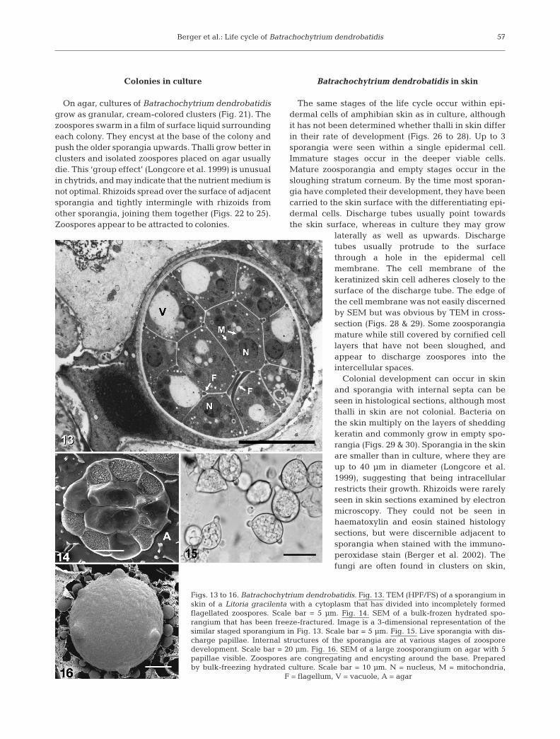
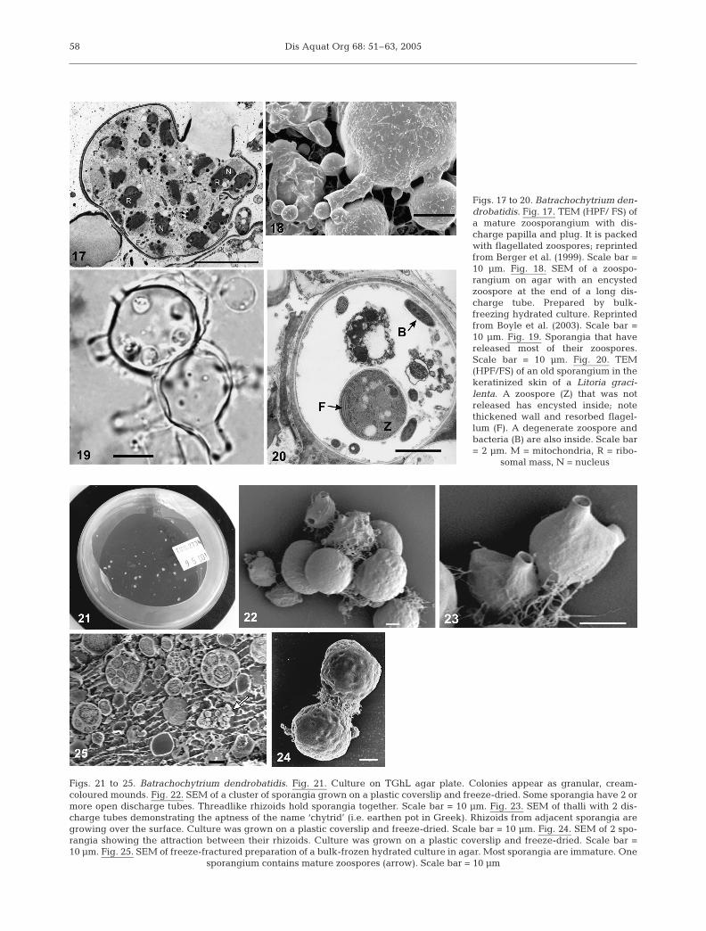
On agar, cultures of Batrachochytrium dendrobatidisgrow as granular, cream-colored clusters (Fig. 21). Thezoospores swarm in a film of surface liquid surroundingeach colony. They encyst at the base of the colony andpush the older sporangia upwards. Thalli grow better inclusters and isolated zoospores placed on agar usuallydie. This ‘group effect’ (Longcore et al. 1999) is unusualin chytrids, and may indicate that the nutrient medium isnot optimal. Rhizoids spread over the surface of adjacentsporangia and tightly intermingle with rhizoids fromother sporangia, joining them together (Figs. 22 to 25).Zoospores appear to be attracted to colonies.
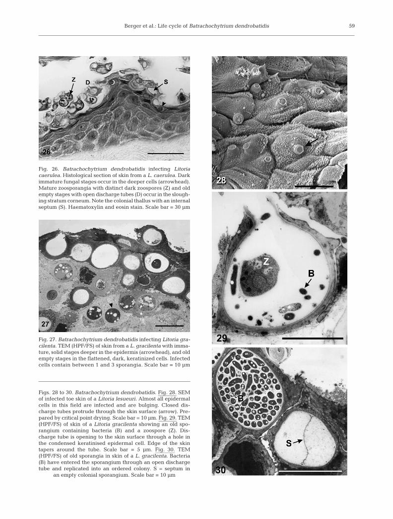
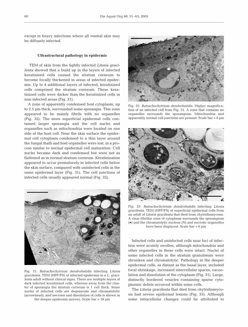
Batrachochytrium dendrobatidis in skin
The same stages of the life cycle occur within epi-dermal cells of amphibian skin as in culture, althoughit has not been determined whether thalli in skin differin their rate of development (Figs. 26 to 28). Up to 3sporangia were seen within a single epidermal cell.Immature stages occur in the deeper viable cells.Mature zoosporangia and empty stages occur in thesloughing stratum corneum. By the time most sporan-gia have completed their development, they have beencarried to the skin surface with the differentiating epi-dermal cells. Discharge tubes usually point towardsthe skin surface, whereas in culture they may grow
laterally as well as upwards. Dischargetubes usually protrude to the surfacethrough a hole in the epidermal cellmembrane. The cell membrane of thekeratinized skin cell adheres closely to thesurface of the discharge tube. The edge ofthe cell membrane was not easily discernedby SEM but was obvious by TEM in cross-section (Figs. 28 & 29). Some zoosporangiamature while still covered by cornified celllayers that have not been sloughed, andappear to discharge zoospores into theintercellular spaces.
Colonial development can occur in skinand sporangia with internal septa can beseen in histological sections, although mostthalli in skin are not colonial. Bacteria onthe skin multiply on the layers of sheddingkeratin and commonly grow in empty spo-rangia (Figs. 29 & 30). Sporangia in the skinare smaller than in culture, where they areup to 40 µm in diameter (Longcore et al.1999), suggesting that being intracellularrestricts their growth. Rhizoids were rarelyseen in skin sections examined by electronmicroscopy. They could not be seen inhaematoxylin and eosin stained histologysections, but were discernible adjacent tosporangia when stained with the immuno-peroxidase stain (Berger et al. 2002). Thefungi are often found in clusters on skin,
57
Figs. 13 to 16. Batrachochytrium dendrobatidis. Fig. 13. TEM (HPF/FS) of a sporangium inskin of a Litoria gracilenta with a cytoplasm that has divided into incompletely formedflagellated zoospores. Scale bar = 5 µm. Fig. 14. SEM of a bulk-frozen hydrated spo-rangium that has been freeze-fractured. Image is a 3-dimensional representation of thesimilar staged sporangium in Fig. 13. Scale bar = 5 µm. Fig. 15. Live sporangia with dis-charge papillae. Internal structures of the sporangia are at various stages of zoosporedevelopment. Scale bar = 20 µm. Fig. 16. SEM of a large zoosporangium on agar with 5papillae visible. Zoospores are congregating and encysting around the base. Preparedby bulk-freezing hydrated culture. Scale bar = 10 µm. N = nucleus, M = mitochondria,
F = flagellum, V = vacuole, A = agar
Dis Aquat Org 68: 51–63, 200558
Figs. 17 to 20. Batrachochytrium den-drobatidis. Fig. 17. TEM (HPF/ FS) ofa mature zoosporangium with dis-charge papilla and plug. It is packedwith flagellated zoospores; reprintedfrom Berger et al. (1999). Scale bar =10 µm. Fig. 18. SEM of a zoospo-rangium on agar with an encystedzoospore at the end of a long dis-charge tube. Prepared by bulk-freezing hydrated culture. Reprintedfrom Boyle et al. (2003). Scale bar =10 µm. Fig. 19. Sporangia that havereleased most of their zoospores.Scale bar = 10 µm. Fig. 20. TEM(HPF/FS) of an old sporangium in thekeratinized skin of a Litoria graci-lenta. A zoospore (Z) that was notreleased has encysted inside; notethickened wall and resorbed flagel-lum (F). A degenerate zoospore andbacteria (B) are also inside. Scale bar= 2 µm. M = mitochondria, R = ribo-
somal mass, N = nucleus
Figs. 21 to 25. Batrachochytrium dendrobatidis. Fig. 21. Culture on TGhL agar plate. Colonies appear as granular, cream-coloured mounds. Fig. 22. SEM of a cluster of sporangia grown on a plastic coverslip and freeze-dried. Some sporangia have 2 ormore open discharge tubes. Threadlike rhizoids hold sporangia together. Scale bar = 10 µm. Fig. 23. SEM of thalli with 2 dis-charge tubes demonstrating the aptness of the name ‘chytrid’ (i.e. earthen pot in Greek). Rhizoids from adjacent sporangia aregrowing over the surface. Culture was grown on a plastic coverslip and freeze-dried. Scale bar = 10 µm. Fig. 24. SEM of 2 spo-rangia showing the attraction between their rhizoids. Culture was grown on a plastic coverslip and freeze-dried. Scale bar =10 µm. Fig. 25. SEM of freeze-fractured preparation of a bulk-frozen hydrated culture in agar. Most sporangia are immature. One
sporangium contains mature zoospores (arrow). Scale bar = 10 µm
Berger et al.: Life cycle of Batrachochytrium dendrobatidis 59
Fig. 26. Batrachochytrium dendrobatidis infecting Litoriacaerulea. Histological section of skin from a L. caerulea. Darkimmature fungal stages occur in the deeper cells (arrowhead).Mature zoosporangia with distinct dark zoospores (Z) and oldempty stages with open discharge tubes (D) occur in the slough-ing stratum corneum. Note the colonial thallus with an internalseptum (S). Haematoxylin and eosin stain. Scale bar = 30 µm
Fig. 27. Batrachochytrium dendrobatidis infecting Litoria gra-cilenta. TEM (HPF/FS) of skin from a L. gracilenta with imma-ture, solid stages deeper in the epidermis (arrowhead), and oldempty stages in the flattened, dark, keratinized cells. Infectedcells contain between 1 and 3 sporangia. Scale bar = 10 µm
Figs. 28 to 30. Batrachochytrium dendrobatidis. Fig. 28. SEMof infected toe skin of a Litoria lesueuri. Almost all epidermalcells in this field are infected and are bulging. Closed dis-charge tubes protrude through the skin surface (arrow). Pre-pared by critical point drying. Scale bar = 10 µm. Fig. 29. TEM(HPF/FS) of skin of a Litoria gracilenta showing an old spo-rangium containing bacteria (B) and a zoospore (Z). Dis-charge tube is opening to the skin surface through a hole inthe condensed keratinised epidermal cell. Edge of the skintapers around the tube. Scale bar = 5 µm. Fig. 30. TEM(HPF/FS) of old sporangia in skin of a L. gracilenta. Bacteria(B) have entered the sporangium through an open dischargetube and replicated into an ordered colony. S = septum in
an empty colonial sporangium. Scale bar = 10 µm
Dis Aquat Org 68: 51–63, 2005
except in heavy infections where all ventral skin maybe diffusely infected.
Ultrastructural pathology in epidermis
TEM of skin from the lightly infected Litoria graci-lenta showed that a build up in the layers of infectedkeratinized cells caused the stratum corneum tobecome focally thickened in areas of infected epider-mis. Up to 4 additional layers of infected, keratinizedcells comprised the stratum corneum. These kera-tinized cells were darker than the keratinized cells innon-infected areas (Fig. 31).
A zone of apparently condensed host cytoplasm, upto 2.5 µm thick, surrounded some sporangia. This zoneappeared to be mainly fibrils with no organelles(Fig. 32). The more superficial epidermal cells con-tained larger sporangia and the cell nuclei andorganelles such as mitochondria were located on oneside of the host cell. Near the skin surface the epider-mal cell cytoplasm condensed to a thin layer aroundthe fungal thalli and host organelles were lost, in a pro-cess similar to normal epidermal cell maturation. Cellnuclei became dark and condensed but were not asflattened as in normal stratum corneum. Keratinizationappeared to occur prematurely in infected cells belowthe skin surface, compared with uninfected cells in thesame epidermal layer (Fig. 31). The cell junctions ofinfected cells usually appeared normal (Fig. 32).
Infected cells and uninfected cells near foci of infec-tion were acutely swollen, although mitochondria andother organelles in these cells were intact. Nuclei ofsome infected cells in the stratum granulosum wereshrunken and chromatolytic. Pathology in the deeperepidermal cells, as distant as the basal layer, includedfocal shrinkage, increased intercellular spaces, vacuo-lation and dissolution of the cytoplasm (Fig. 31). Large,distinctly bordered vesicles containing sparse cyto-plasmic debris occurred within some cells.
The Litoria gracilenta that died from chytridiomyco-sis had severe epidermal lesions (Fig. 33). Althoughsome intracellular changes could be attributed to
60
Fig. 31. Batrachochytrium dendrobatidis infecting Litoriagracilenta. TEM (HPF/FS) of infected epidermis in a L. graci-lenta adult without clinical signs. There are multiple layers ofdark infected keratinized cells, whereas away from the clus-ter of sporangia the stratum corneum is 1 cell thick. Somenuclei of infected cells are degenerate and chromatolytic(arrowhead), and necrosis and dissolution of cells is shown in
the deeper epidermis (arrow). Scale bar = 18 µm
Fig. 32. Batrachochytrium dendrobatidis. Higher magnifica-tion of an infected cell from Fig. 31. A zone that contains noorganelles surrounds the sporangium. Mitochondria andapparently normal cell junctions are present. Scale bar = 4 µm
Fig. 33. Batrachochytrium dendrobatidis infecting Litoriagracilenta. TEM (HPF/FS) of superficial epidermal cells froman adult of Litoria gracilenta that died from chytridiomycosis.A clear fibrillar zone of cytoplasm surrounds the sporangium(*) and the chromatolytic nucleus (N) and necrotic organelles
have been displaced. Scale bar = 6 µm
Berger et al.: Life cycle of Batrachochytrium dendrobatidis
autolysis, major structural changes were evident. Mat-uration of epidermal cells was disrupted, and sheddingcells were often pale and incompletely keratinizedwith overly plump nuclei. In eroded areas cellssloughed to expose a non-keratinized surface. Manyepidermal cells were severely necrotic with swollen,degenerate nuclei and mitochondria. Some necroticinfected cells appeared to lack intercellular junctions,and bacteria were common between cells and withindischarged sporangia.
SEM revealed that surface of skin from a healthy con-trol frog was smooth and intact with a regular patterndemarcating adjacent cells (Fig. 34), whereas skin fromthe infected Litoria lesueuri exhibited roughening dueto separation of adjacent cells, irregular rounding oftheir flat surface layers, and desquamation (Fig. 35).
DISCUSSION
The life cycle of Batrachochytrium dendrobatidishas 2 main stages: the motile, waterborne, short-livedzoospore for dispersal, and the stationary, monocentricthallus, which develops into a zoosporangium for asex-ual amplification (Fig. 36). B. dendrobatidis is welladapted to living in the dynamic tissue of the stratifiedepidermis. Sporangia live inside epidermal cells, ini-tially parasitizing cells a few layers deep, and have arate of development that coincides with the maturingof the cell as it moves outwards and keratinizes. Theygrow initially in living cells but complete their devel-opment in dead superficial keratinized cells that lackorganelles. Discharge tubes have the ability to mergewith and dissolve the epidermal cell membrane andopen on to the surface of the cell, usually the surfacedistal from the body. These specialized adaptationssuggest that B. dendrobatidis has long evolved to livein skin.
The distribution of sporangia in adults and tadpolesshows that a stratified, keratinizing epidermis is arequirement of Batrachochytrium dendrobatidis whenoccurring as a parasite (Berger et al. 1998, Marantelliet al. 2004). However, immature sporangia growwithin the deeper cells that contain prekeratin, not
61
Fig. 34. Litoria caerulea. SEM of smooth intact healthy skin.Toe pad is in the left part of the image. Scale bar = 100 µm
Fig. 35. Batrachochytrium dendrobatidis infecting Litorialesueuri. SEM of the surface of skin from the foot of an infectedadult of L. lesueuri showing the extensive degeneration andpeeling of the superficial epidermis; reprinted from Berger
et al. (1999). Scale bar = 100 µm
Fig. 36. Batrachochytrium dendrobatidis. Diagram of the lifecycle in culture. After a period of motility (usually <24 h),zoospores encyst, resorb their flagella and form germlings.Rhizoids appear from one or more areas. The thalli growlarger and become mature sporangia over 4 to 5 d. Contentsof the enlarged thallus become multinucleate by mitotic divi-sions and the entire contents cleave into zoospores while thedischarge tubes form. The discharge tube is closed by a plugthat absorbs water and deliquesces when zoospores are readyto release. Some thalli develop colonially with thin septadividing the contents into multiple sporangia each with theirown discharge tube. A = zoospore, B = germling, C = devel-oping zoosporangium, D = monocentric zoosporangium, E =
colonial thallus
Dis Aquat Org 68: 51–63, 2005
the dense keratin of the outer, cornified layer wheremature zoosporangia are found. Keratin has not beenstudied in detail in amphibians, but in mammals it isformed when the inner surface of the plasma mem-brane thickens and material including microfilaments,keratohyaline and lysed material are deposited intoan amorphous-filamentous complex (Fox 1994). Ascells mature, different keratins are expressed. It is notyet known what nutrients are being utilized by spo-rangia in frog skin, or if it is the structure and rate ofdevelopment of stratified epidermis that make itsuitable for B. dendrobatidis compared with otherepidermal types.
Because higher temperatures (i.e. >25°C) increasethe rate of epidermal turnover and reduce growth ofthe amphibian chytrid (Piotrowski et al. 2004), the lossof infection in frogs in warm conditions (Berger et al.2004, McDonald et al. 2005) may occur because thefungus does not have time to complete its life cylebefore being shed with the epidermal layer.
We did not see a zoospore in the act of infecting skin,so the method of penetration remains unknown. Long-core et al. (1999) suggest the zoospore could encyst onthe surface then inject the nucleus and contentsthrough a germ tube. Other chytrids have the ability tochange from endobiotic to epibiotic growth dependingon nutrients and the substrate (Longcore 1995). Thedetails of the ultrastructural changes that occur withinsporangia during development also remain to be stud-ied. Experimental infection with zoospores and a timeseries of fixations are needed to trace the infectionprocess.
Rhizoids were seen less commonly in skin sectionsthan in culture, suggesting they do not grow as pro-fusely in skin. Rhizoids may not be needed in skin asthey are not required for attachment and enzymati-cally digested nutrients may be absorbed through thesporangial wall. Serial sectioning could be used tofollow the path of rhizoids in skin.
The clustering of sporangia of Batrachochytriumdendrobatidis in the skin may occur because zoosporesare attracted to foci of infection, or because zoosporesthat are released from a sporangium immediatelyinfect adjacent skin with only a limited period ofmotility and dispersal. Some zoospores appear to bereleased into intercellular spaces and may not beable to escape from the site of infection.
The optimum methods for preparing germlings,developing sporangia and zoosporangia for TEM werehigh pressure freezing and freeze substitution. ForSEM the best results were obtained by imaging bulkfrozen hydrated samples via a field emission scanningelectron microscope. These protocols minimize prepa-ration artifacts such as shrinkage, which was apparentin the critical point dried samples.
Batrachochytrium dendrobatidis causes complexchanges at the cellular level with 2 process being seen;(1) the ultrastructure of infected cells appears toundergo an active process of reorganization in theearly stages of infection and (2) dissolution of cellsoccur. Examination of ultrastructural pathology re-vealed that hyperkeratosis appeared to be partly dueto an increased turnover of epidermal cells, and theswelling of epidermal cells near foci of infectionsuggested a hyperplastic response. Stimulation of thestratum basale leading to hyperplasia is a commonresponse to epidermal injury and occurs with otherepidermal infections such Sarcoptes scabiei (Skerrattet al. 1999). The layers of skin in infected frogs wereuneven and maturation of epidermal cells appeareddisrupted. The cycle of cleavage and sloughing ofkeratinized cells was also disrupted with multiple lay-ers of keratinized cells building up before being shed.Effects on infected cells included altered fibrils in thecytoplasm and displacement of the nucleus by sporan-gia. These changes in the ultrastructure may lead toreduced penetration of antifungal drugs into the cell.Sporangia appeared to initiate premature death andkeratinization of host cells. Thinning of the epidermismay occur when the germination of epidermal cellsdoes not match the increased rate of sloughing fromthe surface. Epidermal width is the result of the bal-ance between these 2 effects and is highly variable.
We did not determine whether death is caused bytoxin release or by inhibition of skin functions, but thedissolution of cellular cytoplasm that is visible (by bothhistological and TEM methods) in epidermal cellsdistant to foci of infection suggests toxicity. Over-growth of bacteria may contribute to the pathogenesisin terminal stages. The mechanism by which Batracho-chytrium dendrobatidis causes death remains one ofthe most important aspects yet to be understood aboutthis pathogen. Studies on enzymes produced by B.dendrobatidis, effects of unpurified fungal secretionson the health of frogs, and more detailed studies onpathology and clinical pathology of infected frogs mayprovide answers.
Acknowledgements. We are grateful to N. Cheville for assis-tance with interpretation of pathology. We thank A. Marshall,Latrobe University, the Centre of Electron Microscopy andMicroanalysis of South Australia at the University of Ade-laide, and the Electron Microscopy Facility at the Universityof Melbourne for assistance in using their electron microscopyfacilities. We thank G. Marantelli, H. Hines, K. McDonald andH. Parkes for providing frog specimens, and S. Hengstberger,T. Wise, and F. Filippi for help with photography. The workwas supported by Environment Australia, the AustralianResearch Council, the Department of Environment and Heri-tage and NSF Integrated Research Challenges in Environ-mental Biology grant IBN 9977063.
62
Berger et al.: Life cycle of Batrachochytrium dendrobatidis
LITERATURE CITED
Ardipradja K (2001) A study of resistance towards theemerging pathogen Batrachochytrium dendrobatidis in 4species of Australian frogs. BSc, University of Melbourne,Melbourne
Berger L (2001) Diseases in Australian frogs. PhD thesis,James Cook University, Townsville
Berger L, Speare R, Daszak P, Green DE and 10 others (1998)Chytridiomycosis causes amphibian mortality associatedwith population declines in the rain forests of Australiaand Central America. Proc Natl Acad Sci USA 95:9031–9036
Berger L, Speare R, Hyatt A (1999) Chytrid fungi and amphib-ian declines: overview, implications and future directions.In: Campbell A (ed) Declines and disappearances of Aus-tralian frogs. Environment Australia, Canberra, p 23–33
Berger L, Hyatt AD, Olsen V, Hengstberger SG, Boyle D,Marantelli G, Humphreys K, Longcore JE (2002) Productionof polyclonal antibodies to Batrachochytrium dendrobatidisand their use in an immunoperoxidase test for chytridio-mycosis in amphibians. Dis Aquat Org 48:213–220
Berger L, Speare R, Hines H, Marantelli G and 10 others(2004) Effect of season and temperature on mortality inamphibians due to chytridiomycosis. Aust Vet J 82:31–6
Bosch J, Martínez-Solano I, García-París M (2001) Evidenceof a chytrid fungus infection involved in the decline of thecommon midwife toad (Alytes obstetricans) in protectedareas of central Spain. Biol Conserv 97:331–337
Boyle DG, Hyatt AD, Daszak P, Berger L, Longcore JE, PorterD, Hengstberger SG, Olsen V (2003) Cryo-archivingof Batrachochytrium dendrobatidis and other chytridio-mycetes. Dis Aquat Org 56:59–64
Bradley GA, Rosen PC, Sredl MJ, Jones TR, Longcore JE(2002) Chytridiomycosis in native Arizona frogs. J WildlDis 38:206–212
Davidson EW, Parris M, Collins JP, Longcore JE, PessierAP, Brunner J (2003) Pathogenicity and transmission ofchytridiomycosis in tiger salamanders (Ambystomatigrinum). Copeia 2003:601–607
Fellers GM, Green DE, Longcore JE (2001) Oral chytridiomy-cosis in the mountain yellow-legged frog (Rana muscosa).Copeia 2001:945–953
Fox H (1994) The structure of the integument. In: Heatwole HG,Barthalmus T (eds) Amphibian biology, Vol 1. The integu-ment. Surrey Beatty and Sons, Chipping Norton, p 1–32
Fuller MS (1996) The flagellated fungal spore. In: Sutton B(ed) A century of mycology. Cambridge University Press,Cambridge, p 161–192
Hanselmann R, Rodríguez A, Lampo M, Fajardo-Ramos L and4 others (2004) Presence of an emerging pathogen ofamphibians in introduced bullfrogs Rana catesbeiana inVenezuela. Biol Conserv 120:115–119
Hopkins S, Channing A (2003) Chytrid fungus in northernand western cape frog populations, South Africa. HerpRev 34:334–336
James TY, Porter D, Leander CA, Vilaglys R, Longcore JE(2000) Molecular phylogenetics of the Chytridiomycotasupports the utility of ultrastructural data in chytridsystematics. Can J Bot 78:336–350
Johnson M, Berger L, Philips L, Speare R (2003) Fungicidaleffects of chemical disinfectants, UV light, desiccation andheat on the amphibian chytrid, Batrachochytrium dendro-batidis. Dis Aquat Org 57:255–260
Lips KR (1999) Mass mortality and population declines ofanurans at an upland site in western Panama. ConservBiol 13:117–125
Longcore JE (1993) Morphology and zoospore ultrastructureof Lacustromyces hiemalis gen. et sp. nov. (Chytridiales).Can J Bot 71:414–425
Longcore JE (1995) Morphology and zoospore ultrastructureof Entophlyctis luteolus sp. nov. (Chytridiales): implica-tions for chytrid taxonomy. Mycologia 87:25–33
Longcore JE, Pessier AP, Nichols DK (1999) Batrachochytriumdendrobatidis gen. et sp. nov., a chytrid pathogenic toamphibians. Mycologia 91:219–227
Marantelli G, Berger L, Speare R, Keegan L (2004) Distribu-tion of Batrachochytrium dendrobatidis and keratin dur-ing tadpole development. Pac Conserv Biol 10:173–179
McDonald KR, Méndez D, Müller R, Freeman AB, Speare R(2005) Decline in the prevalence of chytridiomycosis inupland frog populations in North Queensland, Australia.Pac Conserv Biol 11:114–120
Morehouse EA, James TY, Ganley ARD, Vilgaly R, Berger L,Murphy PJ, Longcore JE (2003) Multilocus sequencetyping suggests the chytrid pathogen of amphibians is arecently emerged clone. Mol Ecol 12:395–403
Mutschmann F, Berger L, Zwart P, Gaedicke C (2000)Chytridiomycosis on amphibians — first report fromEurope. Berl Münch Tierärzt Wochenschr 113:380–383
Nichols DK, Lamirande EW, Pessier AP, Longcore JE (2001)Experimental transmission of cutaneous chytridiomycosisin dendrobatid frogs. J Wildl Dis 37:1–11
Pessier AP, Nichols DK, Longcore JE, Fuller MS (1999) Cuta-neous chytridiomycosis in poison dart frogs (Dendrobatesspp.) and White’s tree frogs (Litoria caerulea). J Vet DiagnInvest 11:194–199
Piotrowski JS, Annis SL, Longcore JE (2004) Physiology ofBatrachochytrium dendrobatidis, a chytrid pathogen ofamphibians. Mycologia 96:9–15
Retallick RW, McCallum H, Speare R (2004) Endemic in-fection of the amphibian chytrid fungus in a frog com-munity post-decline. PLoS Biol 2:e351(doi: 10.1371/journal.pbio.0020351)
Rollins-Smith LA, Carey C, Longcore J, Doersam JK, BoutteA, Bruzgal JE, Conlon J M (2002) Activity of antimicrobialskin peptides from ranid frogs against Batrachochytriumdendrobatidis, the chytrid fungus associated with globalamphibian declines. Dev Comp Immunol 26:471–479
Skerratt LF, Middleton D, Beveridge I (1999) Distribution oflife cycle stages of Sarcoptes scabiei var wombati andeffects of severe mange on common wombats in Victoria.J Wildl Dis 35:633–646
Speare R, Alford R, Aplin R, Berger L and 11 others (2001)Nomination for listing of amphibian chytridiomycosis as akey threatening process under the Environment Protec-tion and Biodiversity Conservation Act 1999. In: Speare R,Alford R, Aplin R, Berger L and 10 others (eds) Developingmanagement strategies to control amphibian diseases: de-creasing the risks due to communicable diseases. Schoolof Public Health and Tropical Medicine, James CookUniversity, Townsville, p 186–208
Weldon C, du Preez LH, Hyatt AD, Muller R, Speare R (2004)Origin of the amphibian chytrid fungus. Emerg Infect Dis10:2100–2105
Woodhams DC, Alford RA, Marantelli G (2003) Emerging dis-ease cured by elevated body temperature. Dis Aquat Org55:65–67
63
Editorial responsibility: Peernel Zwart,Utrecht, The Netherlands
Submitted: March 27, 2005; Accepted: June 22, 2005Proofs received from author(s): November 25, 2005