lemastm/teaching/bi334/unit 03... · muscle physiology muscle mechanics: muscle fibers can be...
TRANSCRIPT
1
Page 1
Muscle
Physio
log
y
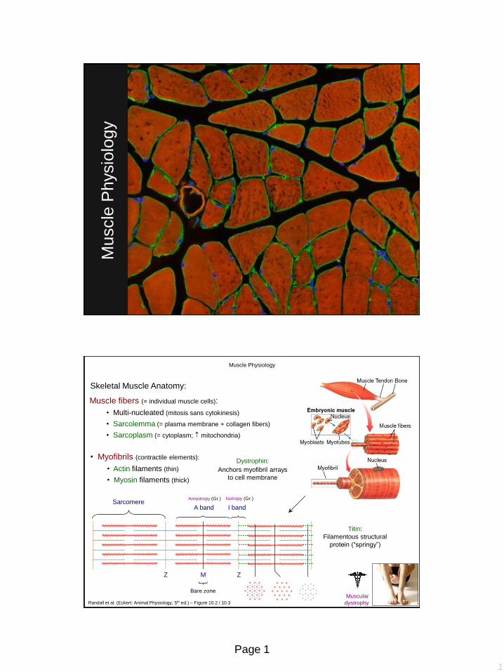
Muscle Physiology
Skeletal Muscle Anatomy:
Muscle fibers (= individual muscle cells):
• Multi-nucleated (mitosis sans cytokinesis)
• Sarcolemma (= plasma membrane + collagen fibers)
• Sarcoplasm (= cytoplasm; mitochondria)
• Myofibrils (contractile elements):
• Actin filaments (thin)
• Myosin filaments (thick)
Sarcomere
Z Z
Bare zone
M
Titin:
Filamentous structural
protein (“springy”)
I band
Isotropy (Gr.)
A band
Anisotropy (Gr.)
Dystrophin:
Anchors myofibril arrays
to cell membrane
Muscular
dystrophy Randall et al. (Eckert: Animal Physiology, 5th ed.) – Figure 10.2 / 10.3

2
Page 2
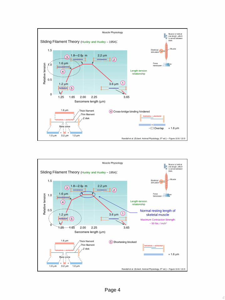
Sliding Filament Theory (Huxley and Huxley – 1954):
Contraction results from sliding
action of inter-digitating actin
and myosin filaments
Evidence?
Myosin head interacts with
actin (cross-bridging)
Each cross-bridge generates
force independent of other
cross-bridges
Thus
Total tension developed by
sarcomere proportional to number
of cross-bridges (proportional to filament overlap)
Muscle Physiology
Randall et al. (Eckert: Animal Physiology, 5th ed.) – Figure 10.8
Length-tension
relationship
Muscle Physiology
Sliding Filament Theory (Huxley and Huxley – 1954):
Randall et al. (Eckert: Animal Physiology, 5th ed.) – Figure 10.8 / 10.9

3
Page 3
Muscle Physiology
Length-tension
relationship
Randall et al. (Eckert: Animal Physiology, 5th ed.) – Figure 10.8 / 10.9
Sliding Filament Theory (Huxley and Huxley – 1954):
Muscle Physiology
Length-tension
relationship
Randall et al. (Eckert: Animal Physiology, 5th ed.) – Figure 10.8 / 10.9
Sliding Filament Theory (Huxley and Huxley – 1954):
4
Page 4
Muscle Physiology
Length-tension
relationship
Randall et al. (Eckert: Animal Physiology, 5th ed.) – Figure 10.8 / 10.9
Sliding Filament Theory (Huxley and Huxley – 1954):
Maximum Contraction Strength:
~ 50 lbs. / inch2
Normal resting length of
skeletal muscle
Muscle Physiology
Length-tension
relationship
Randall et al. (Eckert: Animal Physiology, 5th ed.) – Figure 10.8 / 10.9
Sliding Filament Theory (Huxley and Huxley – 1954):

5
Page 5
The geometry of myofilaments in a sarcomere strongly affects
the contractile properties of the muscle
Muscle Physiology
Randall et al. (Eckert: Animal Physiology, 5th ed.) – Spotlight 10.1
1) Myosin:
• Two heavy chains (tail)
• Four light chains (head)
• Actin-binding sites
• ATPase activity
• Myosin filament composed of 200+ individual
myosin molecules (~1.6 m in length)
2) Actin:
• Two double-stranded helixes of G-actin polymers
woven to form F-actin (~ 1 m in length)
• ADP attached to G-actin (active site)
• Tropomyosin: Spiral around F-actin; cover
active sites
• Troponin: Attaches tropomyosin to F-actin
Muscle Physiology
Myofilament Anatomy:
Guyton & Hall (Textbook of Medical Physiology, 12th ed.) – Figure 6.5

6
Page 6
1) Myosin:
• Two heavy chains (tail)
• Four light chains (head)
• Actin-binding sites
• ATPase activity
• Myosin filament composed of 200+ individual
myosin molecules (~1.6 m in length)
2) Actin:
Muscle Physiology
Myofilament Anatomy:
Guyton & Hall (Textbook of Medical Physiology, 12th ed.) – Figure 6.5
Troponin (sub-units):
1) Troponin C: Binds calcium (up to 4 Ca++)
2) Troponin T: Binds tropomyosin
3) Troponin I: Binds actin (covers active site on actin)
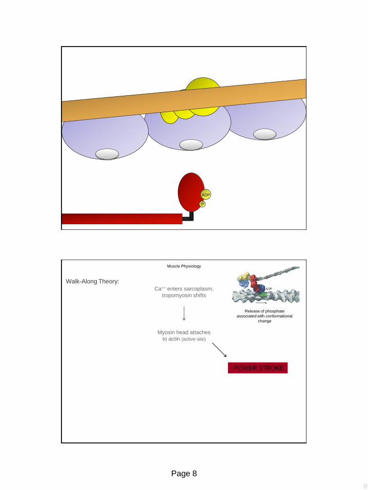
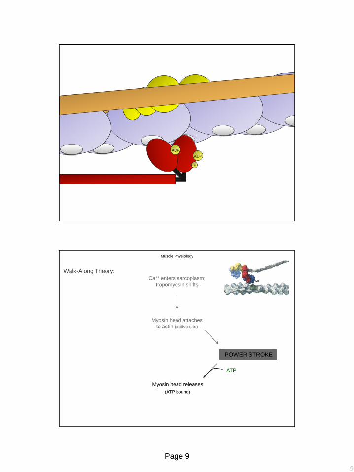
Walk-Along Theory:
Ca++ enters sarcoplasm;
tropomyosin shifts
Muscle Physiology

7
Page 7
Troponin
Actin
Tropomyosin
Myosin
Head
Ca++
ADP
P
Myosin head attaches
to actin (active site)
Muscle Physiology
Walk-Along Theory:
Ca++ enters sarcoplasm;
tropomyosin shifts
8
Page 8
ADP
P
POWER STROKE
Muscle Physiology
Release of phosphate
associated with conformational
change
Myosin head attaches
to actin (active site)
Walk-Along Theory:
Ca++ enters sarcoplasm;
tropomyosin shifts
9
Page 9
ADP
P
ADP
Myosin head releases
ATP
(ATP bound)
Muscle Physiology
POWER STROKE
Myosin head attaches
to actin (active site)
Walk-Along Theory:
Ca++ enters sarcoplasm;
tropomyosin shifts
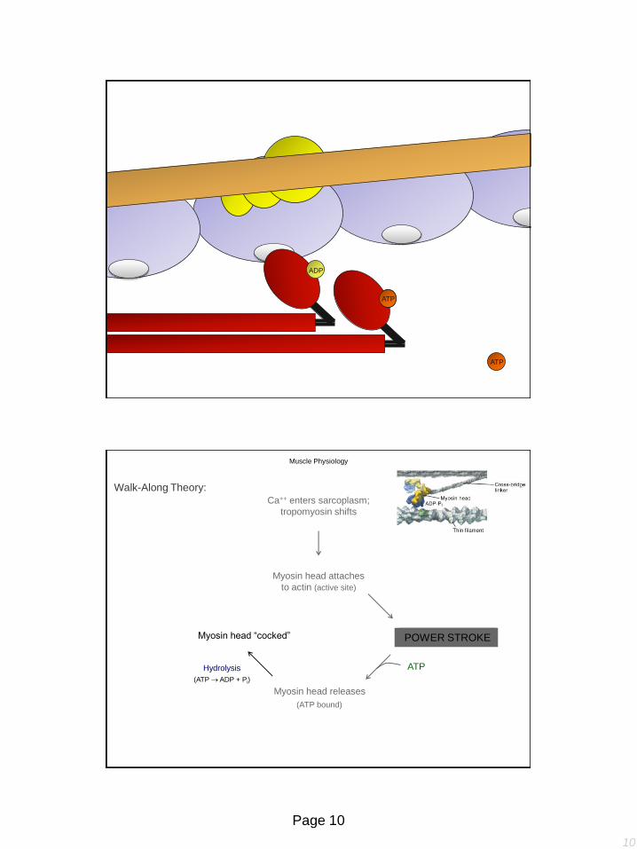
10
Page 10
ATP
ATP ATP
ADP
Muscle Physiology
Myosin head “cocked”
Hydrolysis
(ATP ADP + Pi)
Myosin head releases
ATP
(ATP bound)
POWER STROKE
Myosin head attaches
to actin (active site)
Walk-Along Theory:
Ca++ enters sarcoplasm;
tropomyosin shifts
11
Page 11
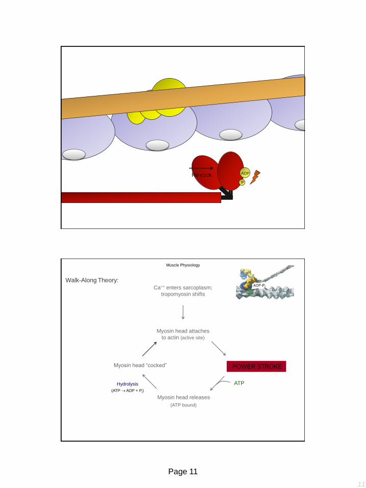
ATP ATP Re-cock ADP
P
Muscle Physiology
Myosin head “cocked”
Hydrolysis
(ATP ADP + Pi)
Myosin head releases
ATP
(ATP bound)
POWER STROKE
Myosin head attaches
to actin (active site)
Walk-Along Theory:
Ca++ enters sarcoplasm;
tropomyosin shifts
12
Page 12
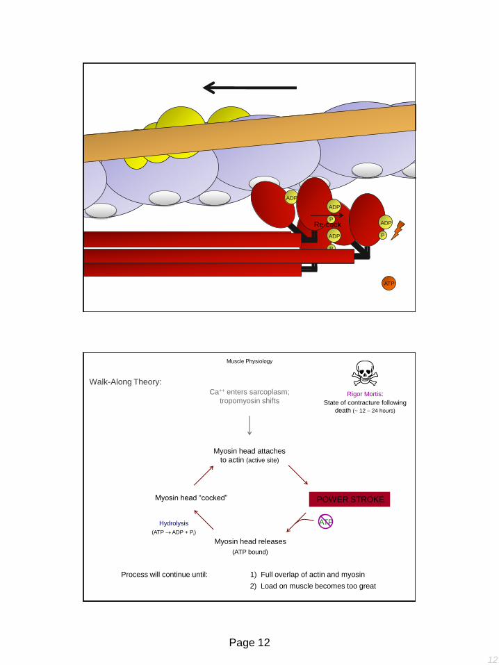
ATP
ATP ADP
P
ADP
ATP ADP
P
ATP
Re-cock ATP ADP
P
Muscle Physiology
Myosin head “cocked”
Hydrolysis
(ATP ADP + Pi)
Myosin head releases
ATP
(ATP bound)
POWER STROKE
Myosin head attaches
to actin (active site)
Walk-Along Theory:
Ca++ enters sarcoplasm;
tropomyosin shifts Rigor Mortis:
State of contracture following
death (~ 12 – 24 hours)
Process will continue until: 1) Full overlap of actin and myosin
2) Load on muscle becomes too great

13
Page 13
Neuromuscular Junction:
Neuron Muscle fiber
Mo
tor N
eu
ron
Muscle Fiber
Subneural cleft ( surface area)
Synaptic cleft
20 –
30 n
m
STEP 1:
Secretion of acetylcholine
by nerve terminals
Muscle Physiology
Excitation – Contraction Coupling:
1 connection / muscle fiber
Motor End Plate
Guyton & Hall (Textbook of Medical Physiology, 12th ed.) – Figure 7.1
Neuromuscular Junction:
Mo
tor N
eu
ron
Muscle Fiber
Muscle Physiology
Excitation – Contraction Coupling:
~ 300,000
A) Small vesicles formed in stoma of neuron;
shuttled to axon terminal
B) Acetylcholine (ACh) synthesized in terminal;
transported into vesicles (~ 10,000 Ach / vesicle)
C) Action potential travels down axon; activates
voltage-gated Ca++ channels at terminal
Ca++
Ca++
D) Ca++ influx triggers vesicles to fuse with
membrane (~ 125 vesicles / AP); ACh released
E) ACh binds with ACh-gated ion channels at
mouth of subneural clefts (muscle fiber)
Choline + Acetyl CoA Acetylcholine
choline
acetyltransferase
Nicotinic
receptors
14
Page 14
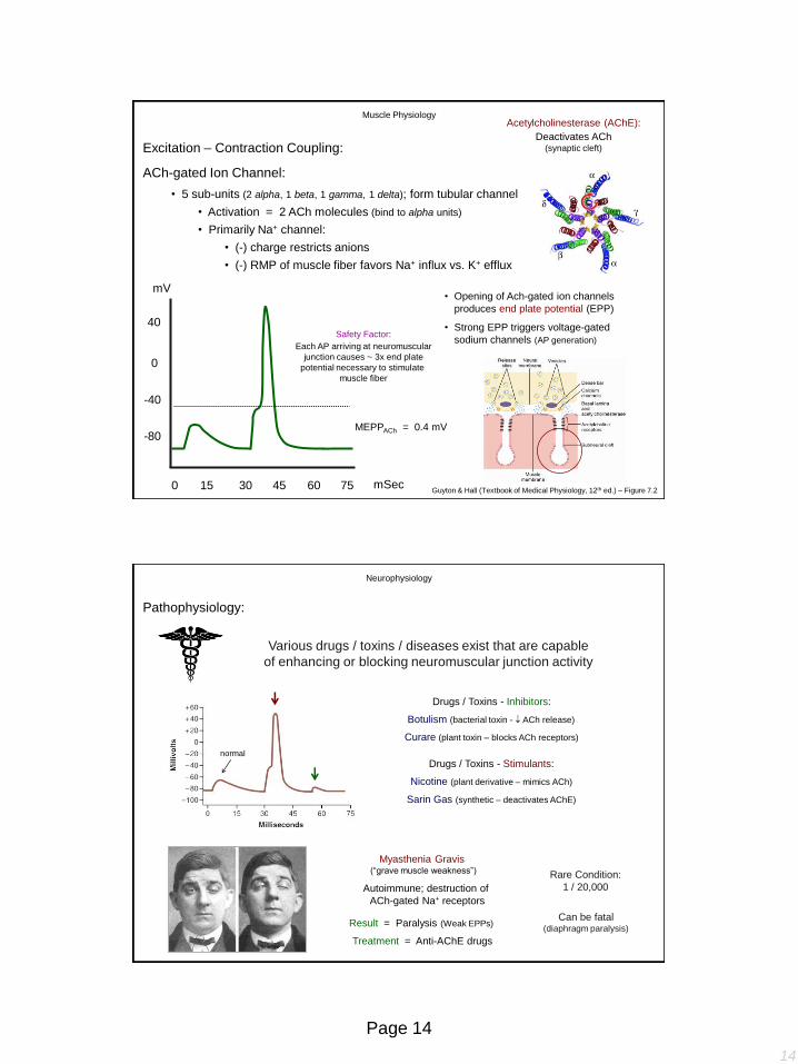
ACh-gated Ion Channel:
• 5 sub-units (2 alpha, 1 beta, 1 gamma, 1 delta); form tubular channel
40
0
-40
-80
mV
0 15 30 45 60 75 mSec
• Opening of Ach-gated ion channels
produces end plate potential (EPP)
• Strong EPP triggers voltage-gated
sodium channels (AP generation) Safety Factor:
Each AP arriving at neuromuscular
junction causes ~ 3x end plate
potential necessary to stimulate
muscle fiber
Acetylcholinesterase (AChE):
Deactivates ACh (synaptic cleft)
Muscle Physiology
MEPPACh = 0.4 mV
Excitation – Contraction Coupling:
Guyton & Hall (Textbook of Medical Physiology, 12th ed.) – Figure 7.2
• Activation = 2 ACh molecules (bind to alpha units)
• Primarily Na+ channel:
• (-) charge restricts anions
• (-) RMP of muscle fiber favors Na+ influx vs. K+ efflux
Pathophysiology:
Various drugs / toxins / diseases exist that are capable
of enhancing or blocking neuromuscular junction activity
Neurophysiology
normal
Drugs / Toxins - Inhibitors:
Botulism (bacterial toxin - ACh release)
Curare (plant toxin – blocks ACh receptors)
Nicotine (plant derivative – mimics ACh)
Sarin Gas (synthetic – deactivates AChE)
Drugs / Toxins - Stimulants:
Myasthenia Gravis (“grave muscle weakness”)
Autoimmune; destruction of
ACh-gated Na+ receptors
Treatment = Anti-AChE drugs
Result = Paralysis (Weak EPPs)
Rare Condition:
1 / 20,000
Can be fatal (diaphragm paralysis)

15
Page 15
Role of Calcium:
Ringer’s
Solution
Isolated Frog heart stopped
beating if Ca++ omitted from bath
• Interacts with troponin in
thin filament:
When Ca++ binds:
(uncovers active sites)
2) Troponin I / actin bond weakens
1) Troponin T / I / C bonds strengthen
10-4
Muscle Physiology
Excitation – Contraction Coupling:
Randall et al. (Eckert: Animal Physiology, 5th ed.) – Figure 10.15
Sidney Ringer (1836 – 1910)
Solution:
Sarcoplasmic Reticulum
For a muscle contraction to occur, there must be a link between
electrical excitation and increased intracellular Ca++ levels…
Problem 1:
Rate of diffusion from Ca++ to interior of cell
(~ 25 – 50 m) several orders of magnitude too
slow to explain observed latent period
AP triggers voltage-gated Ca++ channels in
plasma membrane which flood cell with Ca++…
Terminal cisterna:
Hollow collars around
myofibril; neighbor Z lines
• Specialized ER; stores Ca++
The only source of regulatory Ca++ in
skeletal muscle is from the SR
• SR membrane contains Ca++ pumps
• Maintain < [10-7 M Ca++]
• Calsequestrin: Binds Ca++ in SR
• Reduces [gradient]
Muscle Physiology
Excitation – Contraction Coupling:
Randall et al. (Eckert: Animal Physiology, 5th ed.) – Figure 10.12 / 10.15

16
Page 16
Problem 2:
A potential difference across the plasma
membrane of a muscle fiber affects an
intracellular region a fraction of a m deep
(Myofibrils 50 – 100 m thick)
Solution:
Transverse Tubules
OK… intracellular Ca++ stores released by
AP spreading along surface of muscle cell…
Cytoplasmic extensions continuous with
plasma membrane (~ 0.1 µm diameter); provide link between plasma membrane and
myofibrils deep inside muscle fiber
Muscle Physiology
For a muscle contraction to occur, there must be a link between
electrical excitation and increased intracellular Ca++ levels…
Excitation – Contraction Coupling:
Guyton & Hall (Textbook of Medical Physiology, 12th ed.) – Figure 7.5
How does Ca++ escape the SR?
Ryanodine Receptors:
Dihydropyridine Receptors:
• Located in T-tubule; voltage-gated Ca++
channels
• Only ½ of the ryanodine receptors linked
with dihyropyridine receptors
Calcium-induced Calcium Release (Positive feedback mechanism)
Muscle Physiology
Excitation – Contraction Coupling:
Randall et al. (Eckert: Animal Physiology, 5th ed.) – Figure 10.25
Plunger Model
• Located in SR; Ca++ channels

17
Page 17
AP
Ca2+ pump
calsequestrin
Transverse
tubule
-
-
-
-
-
-
-
-
+
+
+
+
+
+
+
+
+
+
+
+
+
+
+
+
-
-
-
-
-
-
-
-
Muscle Physiology
Excitation – Contraction Coupling:
Ca2+ pump
calsequestrin
Transverse
tubule
Muscle Physiology
Excitation – Contraction Coupling:
+
+
+
+
+
+
+
+
-
-
-
-
-
-
-
-

18
Page 18
Ca2+ pump
calsequestrin
Transverse
tubule
Clinical Oddity:
Malignant Hyperthermia
Trigger:
Anesthetics (e.g., halothane)
Familial tendency… can be tested
for by muscle biopsy
• Skeletal muscle rigidity
Body is only 45% energy efficient; 55% of
the energy appears as heat
• Spontaneous combustion
• Metabolic acidosis (hypermetabolism)
Muscle Physiology
Excitation – Contraction Coupling:
Muscle Energetics:
Major processes requiring energy:
[ATP] in stimulated muscles = [ATP] in unstimulated muscles - ???
ATP usage: ~ 600 trillion / second
• Creatine phosphokinase
• ~ 5 – 8 seconds of fuel…
(100 m)
• Anaerobic respiration
• ~ 1 minutes (“poisons” system)
(400 m)
• Oxidative metabolism (aerobic respiration)
• Primary food source = glycogen / lipids
(5000 m)
4x
2x
1x
Rate:
Muscle Physiology
1) Cross-bridging
2) Ca++ and Na+ / K+ pumps
Guyton & Hall (Textbook of Medical Physiology, 12th ed.) – Figure 84.1

19
Page 19
Muscle Mechanics:
1) Cross-bridge detachment rate (fast detachment = fast contraction)
• Chemical nature of myosin head (Vmax of ATPase)
2) Density of Ca++ pumps (affects clearance of Ca++)
3) Mitochondria # / vasculature (affects oxidative ATP production capacities)
Fast Glycolytic Fibers:
• Rapid cross-bridge cycling
• Rapid Ca++ clearance
• Low endurance (anaerobic respiration)
• () glycolytic enzyme content
• () glycogen reserves
Large diameter (powerful)
Muscle Physiology
Muscle fibers can be divided into two primary types based on
anatomical and physiological properties
Marieb & Hoehn (Human Anatomy and Physiology, 9th ed.) – Figure 9.14
Slow Oxidative Fibers:
• Slow cross-bridge cycling
• Slow Ca++ clearance
• High endurance
• () mitochondria / capillaries
• () myoglobin content
Small diameter
Muscle Physiology
Muscle Mechanics:
1) Cross-bridge detachment rate (fast detachment = fast contraction)
• Chemical nature of myosin head (Vmax of ATPase)
2) Density of Ca++ pumps (affects clearance of Ca++)
3) Mitochondria # / vasculature (affects oxidative ATP production capacities)
Muscle fibers can be divided into two primary types based on
anatomical and physiological properties
Marieb & Hoehn (Human Anatomy and Physiology, 9th ed.) – Figure 9.14
20
Page 20
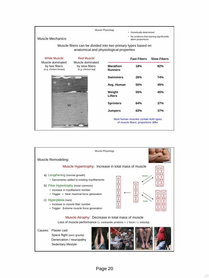
White Muscle:
Muscle dominated
by fast fibers (e.g. chicken breast)
Red Muscle:
Muscle dominated
by slow fibers (e.g. chicken leg)
Most human muscles contain both types
of muscle fibers; proportions differ
Fast Fibers Slow Fibers
Marathon 18% 82%
Runners
Swimmers 26% 74%
Avg. Human 55% 45%
Weight 55% 45%
Lifters
Sprinters 64% 37%
Jumpers 63% 37%
• Genetically determined
• No evidence that training significantly
alters proportions
Muscle Physiology
Muscle Mechanics:
Muscle fibers can be divided into two primary types based on
anatomical and physiological properties
Muscle Remodeling:
Muscle Hypertrophy: Increase in total mass of muscle
b) Fiber Hypertrophy (most common)
• Increase in myofilament number
• Trigger = Near maximal force generation
• Increase in muscle fiber number
• Trigger: Extreme muscle force generation
c) Hyperplasia (rare)
a) Lengthening (normal growth)
• Sarcomeres added to existing myofilaments
Loss of muscle performance ( contractile proteins = force / velocity)
Causes: Plaster cast
Muscle Atrophy: Decrease in total mass of muscle
Weeks
Years
Muscle Physiology
Sedentary lifestyle
Denervation / neuropathy
Space flight (zero gravity)
21
Page 21
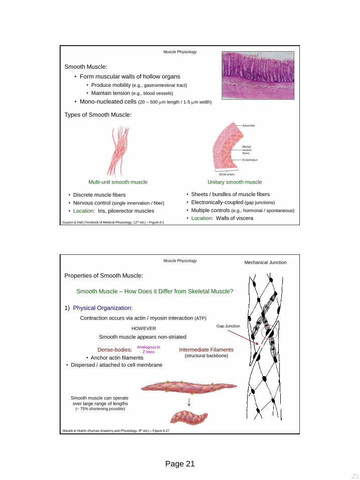
• Discrete muscle fibers
• Nervous control (single innervation / fiber)
• Location: Iris, piloerector muscles
Unitary smooth muscle
• Sheets / bundles of muscle fibers
• Electronically-coupled (gap junctions)
• Multiple controls (e.g., hormonal / spontaneous)
Multi-unit smooth muscle
Muscle Physiology
Types of Smooth Muscle:
• Form muscular walls of hollow organs
• Location: Walls of viscera Guyton & Hall (Textbook of Medical Physiology, 12th ed.) – Figure 8.1
Smooth Muscle:
• Produce mobility (e.g., gastrointestinal tract)
• Maintain tension (e.g., blood vessels)
• Mono-nucleated cells (20 – 500 m length / 1-5 m width)
Properties of Smooth Muscle:
Contraction occurs via actin / myosin interaction (ATP)
Smooth Muscle – How Does it Differ from Skeletal Muscle?
1) Physical Organization:
Dense-bodies: Analogous to
Z lines Intermediate Filaments (structural backbone)
Gap Junction
Mechanical Junction
Smooth muscle can operate
over large range of lengths (~ 75% shortening possible)
• Dispersed / attached to cell membrane
Muscle Physiology
HOWEVER
Smooth muscle appears non-striated
• Anchor actin filaments
Marieb & Hoehn (Human Anatomy and Physiology, 9th ed.) – Figure 9.27
22
Page 22
Properties of Smooth Muscle:
Smooth Muscle – How Does it Differ from Skeletal Muscle?
2) Neuromuscular Junction:
3) Mechanical Operation:
• Slow cycling of myosin cross-bridges (1/10 – 1/300 of skeletal)
• ATPase activity ( = energy required: ~ 1% of skeletal muscle)
• Slow onset of contraction / relaxation (0.2 – 30 sec.)
• Slow cross-bridge action; Slow Ca++ influx / efflux
• Prolonged contraction periods (hours / days / weeks)
• “Latch” mechanism (poorly understood…)
Muscle Physiology
Diffuse junctions present in smooth muscle
Varicosities:
Bulbous swellings along
innervating neuron
Properties of Smooth Muscle:
Smooth Muscle – How Does it Differ from Skeletal Muscle?
4) Ca++ Source:
5) Activation Mechanism:
Muscle Physiology
• Primarily extracellular (poorly developed SR)
• More extensive SR = More rapid contraction
• Caveolae (T.T. analogs)
• Ca++ pumps (S.R. / plasma membrane) clear Ca++ (slow-acting)
10-3 M 10-7 M
Guyton & Hall (Textbook of Medical Physiology, 12th ed.) – Figure 8.6
• Regulation is myosin-based (not actin-based)
• Troponin complex absent
• Myosin must be phosphorylated before it can hydrolyze ATP (become activated)
• Regulatory chain = Myosin light chain phosphorylated
• Latent period = 200 – 300 ms (50x longer than skeletal muscle)
• Force of contraction dependent on [extracellular Ca++]

23
Page 23
Ca++
Calmodulin
Myosin light
chain kinase
MLC active (phosphorylated)
MLC inactive (dephosphorylated)
Contraction
Excitation – Contraction Coupling:
Events:
1) Voltage-gated Ca++ channels open
2) Ca++ binds with calmodulin Similar in structure
to troponin C
3) Ca++ - calmodulin complex activates
myosin light chain kinase
4) When Ca++ levels fall; myosin
phosphatase deactivates myosin
Relaxation
Myosin
phosphatase
Amount of active myosin
phosphatase can greatly affect
the time required for relaxation
Ca++
Muscle Physiology
Excitation – Contraction Coupling:
Muscle Physiology
Additional Sources of Ca++:
G-protein
coupled system
G-protein
coupled system
Costanzo (Physiology, 4th ed.) – Figure 1.29