leishmania braziliensis and leishmania amazonensis amastigote extracts differ in their enhancement...
TRANSCRIPT

RESEARCH ARTICLE Open Access
Leishmania braziliensis and Leishmaniaamazonensis amastigote extracts differ in theirenhancement effect on Leishmania infectionwhen injected intradermallyCintia F de Araújo1, Virgínia MG Silva2, Andre Cronemberger-Andrade1, Luciana S Aragão-França1,Viviane CJ Rocha1, Priscila SL Santos1 and Lain Pontes-de-Carvalho1*
Abstract
Background: It has been reported that repeated intravenous injections of a relatively large amount of Leishmaniaamazonensis amastigote extract (LaE) in BALB/c mice exacerbates the infection of these mice by Leishmaniabraziliensis. The identification of the extract active principle(s) through physicochemical purification often involvesdilution and losses of protein in the course of successive purification procedures. The large amount of the extractrequired to induce the phenomenon, therefore, hinders the carrying out of experiments aimed at identifying theactive molecule(s) through extract purification. In the present work, a dose–response experiment was done to findout if smaller amounts of LaE than that necessary to be used by the intravenous route would reproduce thephenomenon when injected by the intradermal route. In addition, it was also investigated whether a Leishmaniabraziliensis amastigote extract (LbE) would exert the same effect and whether the effect would occur in C57BL/6 mice.
Results: It was found that a single injection of either LaE or LbE containing 5 μg of protein was capable of enhancingthe infection in BALB/c but not in C57BL/6 mice. In addition, it was observed that the largest tested doses of LbE(containing 30 and 180 μg of protein) failed to enhance the infection by L. braziliensis, whereas all doses of LaEenhanced equally that infection.
Conclusions: Those results indicate the possible existence in LbE, and not in LaE, of molecules that interfere with theextract infection-enhancing activity when it is injected in large amounts, and that the inoculation of Leishmania extractsthrough the intravenous and intradermal routes potentiate the infection by L. braziliensis through the same mechanism.
Keywords: Leishmania amazonensis, Leishmania braziliensis, Intradermal antigen, Infection enhancement, Leishmaniasis
BackgroundLeishmaniases are zoonosis caused by different speciesof protozoa of the genus Leishmania. In Brazil, Leish-mania braziliensis is the most prevalent etiologic agentin the northeast region and Leishmania amazonensis isthe most widely distributed species [1,2]. Both species cancause American cutaneous leishmaniasis (ACL), which isin geographic expansion in Brazil, with the incidence in-creasing from 10.5/100,000 inhabitants in 1985 to 18.6/
100,000 inhabitants in 2005. The disease has currentlybeen reported in all regions of the country, but, with anaverage of 28,568 annual cases, the northeastern region,one of the poorest of the country, had the highest inci-dences between 1985 and 2005. In 2001, for example, theincidence in that region was 93.8 cases/100,000 inhabi-tants [3].ACL has four distinct clinical manifestations: cutaneous,
mucosal, disseminated cutaneous, and diffuse cutaneous.Cutaneous, mucosal, and disseminated leishmaniases arecaused in Brazil mainly by L. braziliensis, while L. amazo-nensis, in addition to cutaneous leishmaniasis, causes, in aminority of cases, diffuse cutaneous leishmaniasis [4].
* Correspondence: [email protected] de Pesquisas Gonçalo Moniz. Fundação Oswaldo Cruz, RuaWaldemar Falcão 121, Salvador, BrazilFull list of author information is available at the end of the article
© 2014 de Araújo et al.; licensee BioMed Central Ltd. This is an Open Access article distributed under the terms of the CreativeCommons Attribution License (http://creativecommons.org/licenses/by/2.0), which permits unrestricted use, distribution, andreproduction in any medium, provided the original work is properly cited. The Creative Commons Public Domain Dedicationwaiver (http://creativecommons.org/publicdomain/zero/1.0/) applies to the data made available in this article, unless otherwisestated.
de Araújo et al. BMC Research Notes 2014, 7:70http://www.biomedcentral.com/1756-0500/7/70
BALB/c mice are partially resistant to infection with L.braziliensis: they do not develop severe injuries and usuallycure the infection with a mixed cellular immune response[5]. On the other hand, when infected by Leishmaniamajor or L. amazonensis, either subcutaneously in the footpad [6-8] or intradermally in the external ear [1,9], they de-velop a Th2-cell response and progressive skin lesions,which tend to be more necrotic in L. major-infected mice[10,11], and eventually die of the disease [11].The susceptibility to the leishmanial infection depends
not only on the Leishmania species, but also on the gen-etic background of the mouse. Thus, C57BL/6, CBA andBALB/c mice are resistant to L. braziliensis and suscep-tible to L. amazonensis [12,13]. On the other hand, con-trasting with BALB/c mice, C57BL/6 and CBA mice areresistant to L. major [10].The outcome of the infection of BALB/c mice by L.
braziliensis, however, can be completely changed by treat-ing them with four biweekly intravenous injections of L.amazonensis amastigote extract containing 200 μg of pro-tein and starting one week before the infection: the animalsthus treated become fully susceptible to the disease. On theother hand, this phenomenon did not take place when a L.braziliensis amastigote extract or a L. amazonensis promas-tigote extract was used instead of the L. amazonensisamastigote extract [8], or when lower amounts of L.amazonensis amastigote extract were used [Silva VMG,unpublished].The identification of the extract active principle by its
purification to homogeneity very often entails its dilu-tion or partial loss during two or more consecutive puri-fication steps. For this reason, it would be important tofind out whether the infection-enhancing phenomenoncould be observed with lower amounts of the extractthan that required by the intravenous route if it wasinjected by another route. The aim of the present studywas to investigate whether lower doses of the L. amazo-nensis amastigote extract (LaE) could modulate the L.braziliensis infection in BALB/c and C57BL/6 mice wheninjected by the intradermal route and whether this effectcould be observed with L. braziliensis amastigote extract(LbE). It was found that a single injection of either LaE orLbE in a dose two orders of magnitude lower than thatneeded intravenously [8] induced the phenomenon inBALB/c mice. In addition, the highest doses of LbE failedto enhance the infection in BALB/c mice, and none of thetested doses enhanced the infection in C57BL/6 mice.
ResultsEffect of the intradermal injection of LaE on L. braziliensisinfection in BALB/c miceIn previous experiments carried out by our researchgroup, an amount of LaE containing at least 200 μg ofprotein was required to enhance a L. braziliensis cutaneous
infection in BALB/c mice ([8]; Silva et al., unpublishedresults). The requirement to inject this relatively largeamount of extract in each mouse, in experiments with 6to 8 mice per group, practically precludes the purifica-tion of the active principle from research laboratory’sparasite batches by means of biophysical techniques,such as electrophoresis and liquid chromatography. Oneof the aims of the present work, therefore, was to find outwhether smaller amounts of extract, when injected by an-other route, would also mediate the phenomenon.In a first experiment, doses of extract containing 1.25,
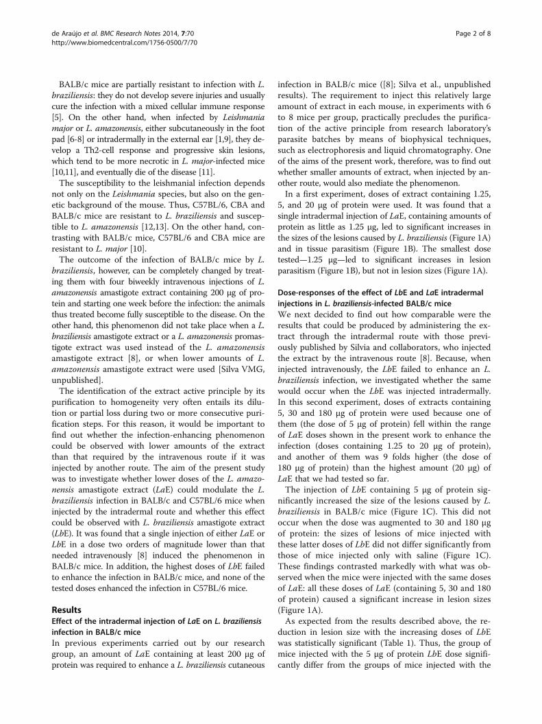
5, and 20 μg of protein were used. It was found that asingle intradermal injection of LaE, containing amounts ofprotein as little as 1.25 μg, led to significant increases inthe sizes of the lesions caused by L. braziliensis (Figure 1A)and in tissue parasitism (Figure 1B). The smallest dosetested—1.25 μg—led to significant increases in lesionparasitism (Figure 1B), but not in lesion sizes (Figure 1A).
Dose-responses of the effect of LbE and LaE intradermalinjections in L. braziliensis-infected BALB/c miceWe next decided to find out how comparable were theresults that could be produced by administering the ex-tract through the intradermal route with those previ-ously published by Silvia and collaborators, who injectedthe extract by the intravenous route [8]. Because, wheninjected intravenously, the LbE failed to enhance an L.braziliensis infection, we investigated whether the samewould occur when the LbE was injected intradermally.In this second experiment, doses of extracts containing5, 30 and 180 μg of protein were used because one ofthem (the dose of 5 μg of protein) fell within the rangeof LaE doses shown in the present work to enhance theinfection (doses containing 1.25 to 20 μg of protein),and another of them was 9 folds higher (the dose of180 μg of protein) than the highest amount (20 μg) ofLaE that we had tested so far.The injection of LbE containing 5 μg of protein sig-
nificantly increased the size of the lesions caused by L.braziliensis in BALB/c mice (Figure 1C). This did notoccur when the dose was augmented to 30 and 180 μgof protein: the sizes of lesions of mice injected withthese latter doses of LbE did not differ significantly fromthose of mice injected only with saline (Figure 1C).These findings contrasted markedly with what was ob-served when the mice were injected with the same dosesof LaE: all these doses of LaE (containing 5, 30 and 180of protein) caused a significant increase in lesion sizes(Figure 1A).As expected from the results described above, the re-
duction in lesion size with the increasing doses of LbEwas statistically significant (Table 1). Thus, the group ofmice injected with the 5 μg of protein LbE dose signifi-cantly differ from the groups of mice injected with the
de Araújo et al. BMC Research Notes 2014, 7:70 Page 2 of 8http://www.biomedcentral.com/1756-0500/7/70
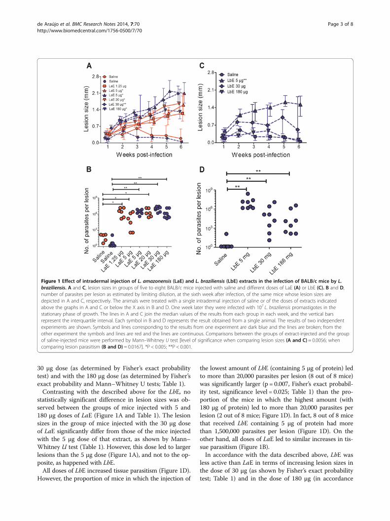
30 μg dose (as determined by Fisher’s exact probabilitytest) and with the 180 μg dose (as determined by Fisher’sexact probability and Mann–Whitney U tests; Table 1).Contrasting with the described above for the LbE, no
statistically significant difference in lesion sizes was ob-served between the groups of mice injected with 5 and180 μg doses of LaE (Figure 1A and Table 1). The lesionsizes in the group of mice injected with the 30 μg doseof LaE significantly differ from those of the mice injectedwith the 5 μg dose of that extract, as shown by Mann–Whitney U test (Table 1). However, this dose led to largerlesions than the 5 μg dose (Figure 1A), and not to the op-posite, as happened with LbE.All doses of LbE increased tissue parasitism (Figure 1D).
However, the proportion of mice in which the injection of
the lowest amount of LbE (containing 5 μg of protein) ledto more than 20,000 parasites per lesion (8 out of 8 mice)was significantly larger (p = 0.007, Fisher’s exact probabil-ity test, significance level = 0.025; Table 1) than the pro-portion of the mice in which the highest amount (with180 μg of protein) led to more than 20,000 parasites perlesion (2 out of 8 mice; Figure 1D). In fact, 8 out of 8 micethat received LbE containing 5 μg of protein had morethan 1,500,000 parasites per lesion (Figure 1D). On theother hand, all doses of LaE led to similar increases in tis-sue parasitism (Figure 1B).In accordance with the data described above, LbE was
less active than LaE in terms of increasing lesion sizes inthe dose of 30 μg (as shown by Fisher’s exact probabilitytest; Table 1) and in the dose of 180 μg (in accordance
Figure 1 Effect of intradermal injection of L. amazonensis (LaE) and L. braziliensis (LbE) extracts in the infection of BALB/c mice by L.braziliensis. A and C, lesion sizes in groups of five to eight BALB/c mice injected with saline and different doses of LaE (A) or LbE (C). B and D,number of parasites per lesion as estimated by limiting dilution, at the sixth week after infection, of the same mice whose lesion sizes aredepicted in A and C, respectively. The animals were treated with a single intradermal injection of saline or of the doses of extracts indicatedabove the graphs in A and C or below the X axis in B and D. One week later they were infected with 107 L. braziliensis promastigotes in thestationary phase of growth. The lines in A and C join the median values of the results from each group in each week, and the vertical barsrepresent the interquartile interval. Each symbol in B and D represents the result obtained from a single animal. The results of two independentexperiments are shown. Symbols and lines corresponding to the results from one experiment are dark blue and the lines are broken; from theother experiment the symbols and lines are red and the lines are continuous. Comparisons between the groups of extract-injected and the groupof saline-injected mice were performed by Mann–Whitney U test [level of significance when comparing lesion sizes (A and C) = 0.0056; whencomparing lesion parasitism (B and D) = 0.0167]. *P < 0.005; **P < 0.001.
de Araújo et al. BMC Research Notes 2014, 7:70 Page 3 of 8http://www.biomedcentral.com/1756-0500/7/70
both with Fisher’s exact probability and with Mann–Whitney U tests; Table 1). In terms of enhancing lesionparasitism, again LbE was less active than LaE in the doseof 30 μg (as shown by Mann–Whitney U test; Table 1) andin the dose of 180 μg (in accordance with both Fisher’sexact probability and Mann–Whitney U tests; Table 1). Nostatistically significant differences between LbE and LaEwas observed when they were administered in the dose of5 μg of protein (Table 1).
Effect of the intradermal injection of LaE or LbE on L.braziliensis infection in C57BL/6 miceWhen injected intravenously, LaE failed to potentiate theinfection by L. braziliensis in C57BL/6 mice [8]. In orderto further compare the results that could be obtained
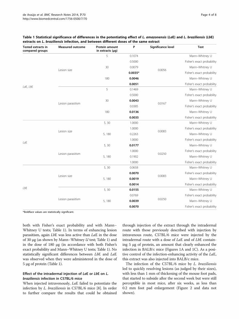
through injection of the extract through the intradermalroute with those previously described with injection byintravenous route, C57BL/6 mice were injected by theintradermal route with a dose of LaE and of LbE contain-ing 5 μg of protein, an amount that clearly enhanced theinfection in BALB/c mice (Figures 1A and 1C). As a posi-tive control of the infection-enhancing activity of the LaE,this extract was also injected into BALB/c mice.The infection of the C57BL/6 mice by L. braziliensis
led to quickly resolving lesions (as judged by their sizes),with less than 1 mm of thickening of the mouse foot pads,that started to subside after the second week but were stillperceptible in most mice, after six weeks, as less than0.2 mm foot pad enlargement (Figure 2 and data notshown).
Table 1 Statistical significance of differences in the potentiating effect of L. amazonensis (LaE) and L. braziliensis (LbE)extracts on L. braziliensis infection, and between different doses of the same extract
Tested extracts incompared groups
Measured outcome Protein amountin extracts (μg)
P Significance level Test
LaE, LbE
Lesion size
5 0.1074
0.0056
Mann–Whitney U
0.5000 Fisher’s exact probability
30 0.0079 Mann–Whitney U
0.0035* Fisher’s exact probability
180 0.0046 Mann–Whitney U
0.0051 Fisher’s exact probability
Lesion parasitism
5 0.1469
0.0167
Mann–Whitney U
0.5000 Fisher’s exact probability
30 0.0043 Mann–Whitney U
0.0385 Fisher’s exact probability
180 0.0136 Mann–Whitney U
0.0035 Fisher’s exact probability
LaE
Lesion size
5, 30 1.0000
0.0083
Mann–Whitney U
1.0000 Fisher’s exact probability
5, 180 0.2263 Mann–Whitney U
1.0000 Fisher’s exact probability
Lesion parasitism
5, 30 0.0177
0.0250
Mann–Whitney U
1.0000 Fisher’s exact probability
5, 180 0.1902 Mann–Whitney U
1.0000 Fisher’s exact probability
LbE
Lesion size
5, 30 0.0658
0.0083
Mann–Whitney U
0.0070 Fisher’s exact probability
5, 180 0.0019 Mann–Whitney U
0.0014 Fisher’s exact probability
Lesion parasitism
5, 30 0.0155
0.0250
Mann–Whitney U
0.0769 Fisher’s exact probability
5, 180 0.0039 Mann–Whitney U
0.0070 Fisher’s exact probability
*Boldface values are statistically significant.
de Araújo et al. BMC Research Notes 2014, 7:70 Page 4 of 8http://www.biomedcentral.com/1756-0500/7/70
Neither the intradermal injection of LaE nor of LbE ex-acerbated the cutaneous disease caused by L. braziliensisin those mice (Figure 2).
DiscussionAs has been shown for the intravenous injection, theintradermal injection of LaE in BALB/c mice potentiatedthe infection by L. braziliensis. However, when adminis-tered by the intradermal route, a dose of LaE two ordersof magnitude lower (1.25 μg) than the dose that was ableto promote the infection by the intravenous route(200 μg; [8]) was enough to produce the effect. Differ-ences in Leishmania infection outcomes depending onthe route of administration of the antigen have been ob-served [4,14,15]. However, comparisons between immuni-zations with Leishmania antigens by the intravenous andthe intradermal routes have not been so far reported.The effect on the size of the lesions caused by the
1.25 μg of protein dose was very apparent. However, theeffect, at this low dose of the extract, was not statisticallysignificant (p = 0.0102, Mann–Whitney U test), perhapsdue to the fact that three different comparisons had beenmade (namely 1.25 μg vs saline, 5 μg vs saline, and 20 μgvs saline) in three different occasions (in which differencesin lesion sizes could be expected: the 4th, the 5th, and the6th weeks). These multiple comparisons reduced the levelof significance to 0.0056. The conspicuousness of the
extract effect in this low dose, however, increases the feasi-bility of the physicochemical purification of its activeprinciple from amounts of extract easily obtainable in aresearch facility.When injected intravenously, the enhancing effect on
the infection of Leishmania extracts depends on thepresence of IL-4 [8]. The mechanism of this enhance-ment when the antigen is injected into the intradermalspace, however, has still to be clarified. Preliminary ex-periments showed no differences in the production ofeither IL-4 or IL-10 by anti-CD3 - stimulated cells oflesion-draining lymph nodes (unpublished). The possibil-ity that the effect could be mediated by TGF-β-producingTreg cells is open to investigation. Supporting this hypoth-esis, it has been reported that TGF-β exacerbates the in-fection of BALB/c mice by L. amazonensis [16,17]. If themost likely underlying mechanism, i. e., the biasing of theBALB/c mouse immune response towards an infection-permissive one by antigens in the LaE is true, the C57BL/6 immune system would respond to these antigens in adifferent way, as the LaE did not increase lesion sizes inC57BL/6 mice.It was also demonstrated in the present work that the
injection of LbE by the intradermal route potentiates theinfection by L. braziliensis. However, contrasting to whatwas observed with LaE, a statistically significant effectwas only seen with a lower dose of the extract and notwith the larger doses. The infection-enhancing activityof the largest doses of LbE was indeed significantly lowerthan the activivities of the same doses of LaE. This findingis consistent with that reported by Silva and collaborators[8], who found that LbE, when repeatedly administered bythe intravenous route in a relatively high dose, did not ex-acerbate the infection of BALB/c mice by L. braziliensis.This dose-dependent difference between LaE and LbE inaffecting the course of the disease could be ascribed to thecoexistence in LbE of molecules that could induce pro-tective immune responses and of others that could inducea permissive immune response, which would have dif-ferent dose (of the extract)-response curves. Thus, anti-gens that could induce a protective immune responsewould be recognized by the immune system only whenhigh amounts of LbE was introduced in the organism(and identical or similar antigens would not be presentor be present in an insufficient amount in the LaE),whereas in low amounts of LbE other antigens, which couldbe present in higher concentrations in the extract, wouldinduce a susceptibility-associated immune response. Thishypothesis is amenable to investigation, e.g. through experi-ments using LbE fractions and/or by testing the effect ofmixtures of low amounts of LaE with high amounts of LbE.These results indicate, therefore, that the two Leishmania
species may differ in terms of the presence, or of the rela-tive amounts, of antigens against which the BALB/c
Figure 2 Lesions sizes in L. braziliensis-infected C57BL/6 mice inwhich L. amazonensis and L. braziliensis extracts had beeninjected. The animals (n = 4–6 per group) were treated withintradermal injections of the vehicle (Saline C57BL/6) or of L.amazonensis (LaE BALB/c and LaE C57BL/6) or L. braziliensis (LbEC57BL/6) extracts containing 5 μg of protein, one week before beinginfected with 107 L. braziliensis promastigotes in the stationary phaseof growth. The lines in A and C join the median values of the resultsfrom each group in each week, and the vertical bars represent theinterquartile interval. Comparisons between the groups of extract-injected and the group of saline-injected mice were performed byMann–Whitney U test (level of significance = 0.0056). *P < 0.001.
de Araújo et al. BMC Research Notes 2014, 7:70 Page 5 of 8http://www.biomedcentral.com/1756-0500/7/70
immune system would mount a protective immune re-sponse. Whether this difference would account for thefact that a minority of human beings with cutaneous lesionscaused by L. amazonensis develops a progressive disease, aphenomenon that does not occur when the cutaneous le-sions are caused by L. braziliensis, is open to speculation.The progressive disease would be accounted by L. amazo-nensis parasites producing mainly infection-promotingmolecules, whose recognition as such would also dependon the genetic background of the host. It is tempting tomake a parallel between the fact that only the BALB/c, andnot the C57BL/6 mice, had their disease worsened byLeishmania extracts and the fact that only a minority ofhuman beings with clinically manifested L. amazonensisinfection develop the diffuse, progressive disease.Evidence for the existence of different immunomodu-
latory Leishmania molecules that might potentiate theinfection have been reported [17-22]. A preliminary frac-tionation of the LaE by polyacrylamide gel electrophoresisfollowed by electroelution of proteins from gel fragmentsand analysis of the eluted protein fractions by mass spec-trometry led to the conclusion that at least three differentLeishmania proteins are able to enhance the Leishmaniainfection in BALB/c mice when injected intradermally.One of the proteins present in a fraction with infection-enhancing activity was the Leishmania homologue of re-ceptors for activated C kinase (LACK), which has beendescribed as having immunomodulatory activity [19]. Twoother fractions with Leishmania infection-enhancing ac-tivity contained proteins to which no immunomodulatoryactivity has been attributed so far (unpublished).Different L. braziliensis isolates may cause different inten-
sities of cutaneous disease and lesions with distinct histopa-tological patterns [5,23]. The isolate we used causes aself-healing cutaneous disease when inoculated in the footpad of BALB/c mice, with lesions smaller than 1 mm thatusually subside after two or three weeks and are bearlymeasurable after six weeks. However, about one in everythree or four mice develops a larger lesion, that also sub-sides after six weeks (unpublished observations). It wouldnot be surprising if extracts prepared from different L. bra-ziliensis isolates would differ in their infection-enhancingactivity.Moreover, the fact that a high amount of LbE is less ac-
tive in terms of inducing disease exacerbation than loweramounts of the same extract and than the same highamount of LaE can be used as evidence against the hy-pothesis that the simple introduction of any Leishmaniaantigen in the intradermal compartment would aggravatethe disease.
ConclusionsIn conclusion, it was demonstrated in the present workthat a few micrograms of LaE or LbE, when injected
intradermally, are capable of aggravating the cutaneousdisease caused by L. braziliensis in BALB/c mice, and thatthis phenomenon is not seen with larger amounts of LbE.As has been reported previously, LaE failed to enhance
the infection in C57BL/6 mice when injected intraven-ously [8]. As shown in the present work, this failure alsooccurs when it is injected intradermally. In addition, theLbE was less active in promoting the infection than theLaE when injected in relatively high doses by either oneof the two routes ([8] and the present work). Taken to-gether, these findings indicate that the same mechanismis underlying the observed phenomena when the extractis injected intravenously or intradermally.
MethodsMice and ethical considerationsSpecific-pathogen-free, 8 to 12 week-old BALB/c andC57BL/6 mice were maintained at the animal facilities ofthe Gonçalo Moniz Research Center, Oswaldo CruzFoundation, Salvador, Brazil, and were provided with ro-dent diet and water ad libitum. All procedures were ap-proved and conducted according to the institutionalCommittee for Animal Care and Utilization.
Parasites and parasite extractsL. amazonensis (MHOM/Br87/Ba125) and L. braziliensis(MHOM/Br/3456) strains were used. Their infectivitieswere maintained by regular inoculations of promasti-gotes into susceptible BALB/c mice and golden ham-sters, respectively.Promastigotes, derived from tissue amastigotes, were
cultured at 23°C in Schneider’s medium (Sigma ChemicalCo., St. Louis, MO, USA), pH 7.2, supplemented with50 μg/mL of gentamicin and 10% heat-inactivated fetal bo-vine serum (FBS; Gibco Laboratories, Grand Island, NY)for L. amazonensis or 20% FBS for L. braziliensis.L. amazonensis and L. braziliensis axenic amastigotes
were obtained by the differentiation of promastigotes inaxenic cultures, as described elsewhere [24]. The amasti-gotes were washed three times in ice cold sterile saline,resuspended in saline, and lysed by exposure to ultra-sound (10 1-minute, 300-W pulses, with 30-second inter-vals in between, on ice; Sonifier Cell Disruptor; BransonSonic Power Company, Danbury, CT, USA). The lysateswere centrifuged at 16,000 g for 10 min at 4°C, the super-natants were filtered on membranes with 0.22-μm diam-eter pores (Millipore, São Paulo, Brazil) and immediatelyaliquoted and stored at −70°C.
Infection and treatment of animals, determination oflesion size and experimental designL. braziliensis promastigotes (107), obtained from stationary-phase cultures, were subcutaneously inoculatedinto one of the hind footpads of BALB/c or C57BL/6
de Araújo et al. BMC Research Notes 2014, 7:70 Page 6 of 8http://www.biomedcentral.com/1756-0500/7/70
mice [16,24] one week after their being injected in thedermis of the dorsal region, with 50 μL of LaE or LbEcontaining the amounts of protein that are indicated inthe Figures. The hind pad thicknesses were weekly mon-itored with a digital caliper, until the sixth week post-infection and the lesion sizes estimated by subtractingthe thickness of the uninfected pad from the thicknessof the infected pad. Parasite loads in the footpads wereestimated by limiting dilution [25] at the end of the ex-periments, as described below.
Quantification of tissue parasitismBriefly, the infected pads were macerated in Schneider’smedium and centrifuged at 50 g for 10 min, at 4°C. Thesupernatants were recentrifuged at 1,540 g for 10 min at4°C, and the pellets were resuspended in Schneider’smedium supplemented with 50 μg/mL gentamicin and20% FBS. The suspension was serially diluted in 2-folddilutions and distributed in triplicate in 96-well cultureplates. The number of viable parasites in each footpadwas determined from the reciprocal of the highest dilu-tion at which promastigotes could be detected after 7days at 23°C and was expressed as the number of para-sites per lesion.
Statistical analysesThe type of data distribution was determined by theShapiro-Wilk test. Because the distribution was found tobe non-Gaussian, non-parametric tests were used.The statistical significance of differences in lesion size
and parasitism between pairs of groups of mice subjectedto different treatments were estimated by the Mann–Whitney U test. As explained below, the number of com-pared pairs in each experiment was taken into account tocalculate the significance level.To additionally assess the statistical significance of dif-
ferences in lesion sizes, and in intensity of lesion parasit-ism, between the groups of mice that had received 5 and30 μg of extract, between the groups of mice that had re-ceived 5 and 180 μg of extract, and between the groups ofmice that had received LbE and LaE, these groups of micewere stratified into two pairs of categories: mice with le-sions larger than 1 mm and mice with lesions smaller than1 mm; mice with lesions with more than 20,000 parasitesand mice with lesions with less than 20,000 parasites.Two × 2 contingency tables associating the numbers ofmice falling within the categories with one or another oftwo experimental protocols (the protocol in which themice received 5 μg of extract and the protocol in whichthey received 30 μg of the same extract; the protocol inwhich the mice received 5 μg of extract and the protocolin which they received 180 μg of the same extract; and theprotocol in which the mice received a given amount ofLaE and the protocol in which they received the same
amount of LbE) were constructed. Fisher’s exact probabil-ity test was used to assess whether the frequency distribu-tions of data within these tables were due to chance, sincethat test was designed for contingency tables with a smallsample size, like the one in the present study.Results were considered significant when the value of
P was ≤0.05 divided by the number of pairs of compareddatum sets in the statistical test.
AbbreviationsACL: American cutaneous leishmaniasis; FBS: Fetal bovine serum; IL-: Interleukin;LaE: Leishmania amazonensis amastigote extract; LbE: Leishmania braziliensisamastigote extract; TGF-β: Transforming growth factor beta; Th2: T helper type 2.
Competing interestsThe authors declare that they have no competing interests.
Authors’ contributionsCFde-A carried out most of the experiments and wrote a first draft of themanuscript; VMGS helped in designing the experiments; ACA, LSA-F, VCJR,and PSLS carried out some of the experimental work; LP-de-C devised anddesigned the experiments and wrote the final version of the manuscript. Allauthors read and approved the final manuscript.
AcknowledgementsThis work was financially supported by the PRONEX, Fundação de Amparo aPesquisa do Estado da Bahia (FAPESB), Brazil.
Author details1Centro de Pesquisas Gonçalo Moniz. Fundação Oswaldo Cruz, RuaWaldemar Falcão 121, Salvador, Brazil. 2State University of Southwest Bahia,Rua José Moreira Sobrinho, Jequié, Brazil.
Received: 11 September 2013 Accepted: 27 January 2014Published: 1 February 2014
References1. Belkaid Y, Kamhawi S, Modi G, Valenzuela J, Noben-Trauth N, Rowron E,
Ribeiro J, Sacks DL: Development of a natural model of cutaneous leish-maniasis: powerful effects of vector saliva and saliva pre exposure onthe long-term outcome of Leishmania major infection in the mouse eardermis. J Exp Med 1998, 188:1941–1953.
2. De Oliveira C, Barral-Netto M: O modelo experimental nas infecções causadaspor L. amazonensis e L. braziliensis. Gaz Med Bahia 2005, 75:35–45.
3. Departamento de Vigilância Epidemiológica, Secretaria de Vigilância emSaúde, Ministério da Saúde: Manual de controle da leishmaniose tegumentaramericana. Brasília: MS Editora; 2007.
4. Carvalho PL, Passos TS, Jesus RA: Immunopathogenesis of tegumentaryleishmaniasis. Gaz Med Bahia 2005, 1:57–65.
5. Indiani de Oliveira C, Teixeira MJ, Teixeira CR, Ramos de Jesus J,Bomura Rosato A, Santa da Silva J, Brodskyn C, Barral-Netto M, Barral A:Leishmania braziliensis isolates differing at the genome level displaydistinctive features in BALB/c mice. Microbes Infect 2004, 6:977–984.
6. Scott PA, Farrell JP: Experimental cutaneous leishmaniasis: disseminatedleishmaniasis in genetically susceptible and resistant mice. Am J TropMed Hyg 1982, 31:230–238.
7. Granfell RFQ, Marques-da-Silva EA, Souza-testasicca MC, Coelho EA,Fernandes AP, Afonso LC, Rezende SA: Antigenic extracts of Leishmaniabraziliensis and Leishmania amazonensis associated with saponin partiallyprotects BALB/c mice against Leishmania chagasi infection by suppressingIL-10 and IL-4 production. Mem Inst Oswaldo Cruz 2010, 105:818–822.
8. Silva VMG, Larangeira DF, Oliveira PRS, Sampaio RB, Suzart P,Biointervention Student Group, Nihei J, Teixeira MCA, Mengel JO,Dos Santos WLC, Pontes-de-Carvalho L: Enhancement of experimentalcutaneous leishmaniasis by leishmania molecules with serine proteaseactivity. I. Requirement of IL-4. Infect Immun 2011, 79:1236–1243.
9. Courret N, Lang T, Milon G, Antoine JC: Intradermal inoculations of lowdoses of Leishmania major and Leishmania amazonensis metacyclic
de Araújo et al. BMC Research Notes 2014, 7:70 Page 7 of 8http://www.biomedcentral.com/1756-0500/7/70

promastigotes induce different immunoparasitic processes and status ofprotection in BALB/c mice. Int J Parasitol 2003, 33:1373–1380.
10. Silva-Almeida M, Carvalho POL, Abreu-Silva LA, Souza FSC, Hardoim JD:Extracellular matrix in experimental Leishmania amazonensis infection insusceptible and resistant mice. Vet Res 2012, 43:1–9.
11. Weiss R, Scheiblhofer S, Thalhamer J, Bickert T, Richardt U, Fleischer B, Ritter U:Epidermal inoculation of Leishmania-antigen by gold bombardment resultsin a chronic form of leishmaniasis. Vaccine 2007, 25:25–33.
12. Childs GE, Lightner LK, Mckinney L, Groves MG, Price EE, Hendricks LD:Inbred mice as model hosts for cutaneous leishmaniasis. I. Resistanceand susceptibility to infection with Leishmania braziliensis, L. mexicana,and L. aethiopica. Ann Trop Med Parasitol 1984, 78:25–34.
13. Felizardo TC, Gaspar-Elsas MI, Lima GM, Abrahamsohn IA: Lack of signalingby IL-4 or by IL-4/IL-13 has more attenuating effects on Leishmaniaamazonensis dorsal skin- than on footpad-infected mice. Exp Parasitol2012, 130:48–57.
14. Aebischer T, Morris L, Handman E: Intravenous injection of irradiatedLeishmania major into susceptible BALB/c mice: immunization orprotective tolerance. Int Immunol 1994, 10:1535–1543.
15. Bhowmick S, Mazumdar T, Ali N: Vaccination route that inducestransforming growth factor β production fails to elicit protectiveimmunity against Leishmania donovani infection. Infect Immun 2009,77:1514–1523.
16. Sacks D, Noben-Trauth N: The immunology of susceptibility and resistanceto Leishmania major in mice. Nat Rev Immunol 2002, 2:845–858.
17. Pinheiro RO, Pinto EF, Lopes JRC, Guedes HLM, Fertanes FR, Rossi-Bergmann B:TGF-β-associated enhanced susceptibility to leishmaniasis followingintramuscular vaccination of mice with Leishmania amazonensis antigens.Microbes Infect 2005, 7:1317–1323.
18. de Assis RR, Ibraim IC, Nogueira PM, Soares RP, Turco SJ: Glycoconjugatesin New World species of Leishmania: polymorphisms inlipophosphoglycan and glycoinositolphospholipids and interaction withhosts. Biochim Biophys Acta 1820, 2012:1354–1365.
19. Julia V, Rassoulzadegan M, Glaichenhaus N: Resistance to Leishmania majorinduced by tolerance to a single antigen. Science 1996, 274:421–423.
20. Lacerda ID, Cysne-Finkelstein L, Numed PM, de-Luca PM, Genestra SM,Leon PLL, Pinho BM, Lima ML, Matos SCD, Medeiros AM, MENDONçA SFC:Kinetoplastid membrane protein-11 exacerbates infection withLeishmania amazonensis in murine macrophages. Memórias InstitutoOswaldo Cruz 2012, 107:238–245.
21. Yao C, Donelson JE, Wilson ME: The major surface protease (MSP or GP63)of Leishmania sp. Biosynthesis, regulation of expression, and function.Mol Biochem Parasitol 2003, 132:1–16.
22. Wanderlei MLJ, Costa FJ, Borges MV, Barcinski M: Subversion of Immunityby Leishmania amazonensis Parasites: Possible role ofphosphatidylserine as a main regulator. J Parasitol Res 2012, 2012:981186.
23. Pereira CG, Silva AL, de Castilhos P, Mastrantonio EC, Souza RA, Romão RP,Rezende RJ, Pena JD, Beletti ME, Souza MA: Different isolates fromLeishmania braziliensis complex induce distinct histopathologicalfeatures in a murine model of infection. Vet Parasitol 2009, 165:231–240.
24. Teixeira MC, De Jesus SR, Sampaio RB, Pontes De Carvalho LC, Dos Santos WC:A simple and reproducible method to obtain large numbers of axenicamastiotes of different Leishmania species. Parasitology 2002, 88:963–968.
25. Lima HC, Bleyenberg JA, Titus RG: A simple method for quantifyingLeishmania in tissues of infected animals. Parasitol Today 1997, 13:80–82.
doi:10.1186/1756-0500-7-70Cite this article as: de Araújo et al.: Leishmania braziliensis andLeishmania amazonensis amastigote extracts differ in theirenhancement effect on Leishmania infection when injectedintradermally. BMC Research Notes 2014 7:70.
Submit your next manuscript to BioMed Centraland take full advantage of:
• Convenient online submission
• Thorough peer review
• No space constraints or color figure charges
• Immediate publication on acceptance
• Inclusion in PubMed, CAS, Scopus and Google Scholar
• Research which is freely available for redistribution
Submit your manuscript at www.biomedcentral.com/submit
de Araújo et al. BMC Research Notes 2014, 7:70 Page 8 of 8http://www.biomedcentral.com/1756-0500/7/70