leaf mitochondria modulate whole cell redox homeostasis ... · leaf mitochondria modulate whole...
TRANSCRIPT

The Plant Cell, Vol. 15, 1212–1226, May 2003, www.plantcell.org © 2003 American Society of Plant Biologists
Leaf Mitochondria Modulate Whole Cell Redox Homeostasis, Set Antioxidant Capacity, and Determine Stress Resistance through Altered Signaling and Diurnal Regulation
Christelle Dutilleul,
a,1
Marie Garmier,
a,1
Graham Noctor,
b,2
Chantal Mathieu,
a
Philippe Chétrit,
a
Christine H. Foyer,
b
and Rosine de Paepe
a,3
a
Institut de Biotechnologie des Plantes, Laboratoire Mitochondries et Métabolisme, Centre National de la Recherche Scientifique Unité Mixte de Recherche 8618, Université Paris-Sud XI, Orsay 91405, France
b
Crop Performance and Improvement Division, Rothamsted Research, Harpenden, Hertfordshire AL52JQ, United Kingdom
To explore the role of plant mitochondria in the regulation of cellular redox homeostasis and stress resistance, we exploiteda
Nicotiana sylvestris
mitochondrial mutant. The cytoplasmic male-sterile mutant (CMSII) is impaired in complex I functionand displays enhanced nonphosphorylating rotenone-insensitive [NAD(P)H dehydrogenases] and cyanide-insensitive (alter-native oxidase) respiration. Loss of complex I function is not associated with increased oxidative stress, as shown by de-creased leaf H
2
O
2
and the maintenance of glutathione and ascorbate content and redox state. However, the expression andactivity of several antioxidant enzymes are modified in CMSII. In particular, diurnal patterns of alternative oxidase expres-sion are lost, the relative importance of the different catalase isoforms is modified, and the transcripts, protein, and activityof cytosolic ascorbate peroxidase are enhanced markedly. Thus, loss of complex I function reveals effective antioxidantcrosstalk and acclimation between the mitochondria and other organelles to maintain whole cell redox balance. This reor-chestration of the cellular antioxidative system is associated with higher tolerance to ozone and
Tobacco mosaic virus
.
INTRODUCTION
Active oxygen species (AOS) such as the superoxide anion rad-ical (O
2.
�
), the hydroxyl radical (OH·), and hydrogen peroxide(H
2
O
2
) are universal products of aerobic metabolism. In plants,AOS are generated at significant rates by reactions intrinsic tophotosynthesis, photorespiration, oxidative phosphorylation,fatty acid
�
-oxidation, and many types of oxidases (Alscher etal., 1997). H
2
O
2
participates widely in plant metabolism (e.g.,cell wall biosynthesis) and also has a signal function as a sec-ondary messenger regulating growth and development as wellas stress responses (Foyer et al., 1997). However, H
2
O
2
can beconverted via Fenton-type reactions to the dangerous hydroxylradical, which exerts toxic effects within all cellular compart-ments via lipid peroxidation, protein degradation/modification,and DNA damage (Fridovich, 1986). The accumulation of AOSincreases the probability of hydroxyl radical formation. Failureto control AOS accumulation leads to the phenomenon knownas oxidative stress (Bartosz, 1997; Foyer and Noctor, 2000).
An appropriate intracellular balance between AOS generationand scavenging exists in all cells. This “redox homeostasis” re-quires the efficient coordination of reactions in different cellcompartments and is governed by a complex network of
prooxidant and antioxidant systems. The latter include nonen-zymatic scavengers such as ascorbate, glutathione, and hydro-phobic molecules (tocopherols, carotenoids, and xanthophylls)and detoxifying enzymes that operate in the different cellularorganelles (Noctor and Foyer, 1998a). These enzymes includesuperoxide dismutases (SODs; EC 1.15.1.1), which catalyze thedismutation of O
2.
�
to H
2
O
2
and O
2
(Scandalios, 1993). Threeclasses of SODs have been identified in plants on the basisof their metal cofactor content. FeSODs are found only in chlo-roplasts, MnSODs are found mainly in mitochondria, and Cu/ZnSODs are located in chloroplasts, cytosol, apoplast, andperoxisomes (Bowler et al., 1992). The main enzymatic H
2
O
2
scavengers of photosynthetic cells are catalases (CAT; EC1.11.1.6), which convert H
2
O
2
to H
2
O and O
2
(Scandalios,1987), and ascorbate peroxidases (APX; EC 1.11.1.11), whichuse ascorbate as the electron donor for H
2
O
2
reduction (Asada,1992). In peroxisomes/glyoxysomes, CAT predominates. CATisoforms are distinguished on the basis of organ specificity andresponses to environmental stress (Willekens et al., 1994a). ACAT isoform has been reported to be present in maize mito-chondria (Scandalios et al., 1980), but no mitochondrial formhas been reported in C3 species (Foyer and Noctor, 2000). APXis part of the ascorbate-glutathione cycle, which uses reducedglutathione to regenerate ascorbate (Foyer and Halliwell, 1976),and glutathione is regenerated by glutathione reductase (GR;EC 1.6.4.2). The ascorbate/glutathione cycle is the most impor-tant H
2
O
2
-detoxifying system in the chloroplast, but it also hasbeen identified in the cytosol (Nakano and Asada, 1981), perox-isomes, and mitochondria (Jimenez et al., 1997).
Very little is known about interorganellar redox crosstalk andhomeostasis in plants. In particular, the role of mitochondria in
1
These authors contributed equally to this work.
2
Current address: Institut de Biotechnologie des Plantes, LaboratoireSignalisation Redox, Centre National de la Recherche Scientifique UnitéMixte de Recherche 8618, Université Paris-Sud XI, Orsay 91405, France.
3
To whom correspondence should be addressed. E-mail [email protected]; fax 33-169153425.Article, publication date, and citation information can be found atwww.plantcell.org/cgi/doi/10.1105/tpc.009464.

Mitochondrial Redox Signaling 1213
these processes remains unclear. Indeed, unlike in animals, inwhich the mitochondrial electron transport chain is the majorsite of AOS generation (Liu et al., 2002), in plants the rates ofphotosynthetic electron transport and peroxisomal H
2
O
2
forma-tion are far higher than the capacity for H
2
O
2
generation in mi-tochondria (Foyer and Noctor, 2000). Thus, despite the fact thatsome examples of AOS generation by plant mitochondrial elec-tron transport enzymes (complexes I, II, and III) have been re-ported (Purvis et al., 1995; Braidot et al., 1999; reviewed byMøller, 2001), this production has been considered to be ofsecondary importance. Nevertheless, the recent demonstrationof the crucial role of mitochondria in cell death in animals hasled to an increased interest in parallel functions in plants. Sev-eral recent reports have suggested that plant mitochondria maybe involved in the tolerance to oxidative stress induced by bi-otic and abiotic treatments (reviewed by Millar et al., 2001;Møller, 2001). In particular, mitochondria may play a key role inthe hypersensitive response, which is a form of programmedcell death induced in response to pathogens (Jones, 2000;Swidzinski et al., 2002).
However, one of the key differences between plant and animalmitochondria is the presence of plant-specific alternative respi-ratory pathways, which may play a role in the control of AOSformation and scavenging. These include non-proton-pumpingNAD(P)H dehydrogenases that bypass complex I (Rasmussonet al., 1998) and an alternative oxidase (AOX) that accepts elec-trons directly from the ubiquinone pool without the interventionof the cytochrome
c
oxidase pathway through complexes IIIand IV. These alternative pathways allow the uncoupling ofelectron transfer from ATP production, preventing the overre-duction of the respiratory electron transport chain that other-wise could occur in situations of major flux restrictions (Day andWiskich, 1995; Vanlerberghe and McIntosh, 1997; Wagner andMoore, 1997). This type of regulation diminishes the risk of elec-tron transfer to O
2
and AOS generation. Accordingly, AOX hasbeen shown to be induced by H
2
O
2
(Wagner, 1995), and inhibi-tion or underexpression of the alternative oxidase stimulatesH
2
O
2
production (Popov et al., 1997; Maxwell et al., 1999).To explore the role of plant mitochondria in the regulation of
cellular redox homeostasis and stress resistance, we exploiteda
Nicotiana sylvestris
mitochondrial mutant, cytoplasmic malesterile II (CMSII) (Li et al., 1988). This mutant carries a stable mi-tochondrial DNA mutation that affects the respiratory electrontransport chain: the mitochondrial
nad7
gene encoding theNAD7 subunit of complex I is deleted (Pla et al., 1995), and mi-tochondria are impaired in complex I structure and function(Gutierres et al., 1997). In the mutant, the activity of nonphos-phorylating NAD(P)H dehydrogenases is enhanced (Sabar etal., 2000). This effect allows increased leaf respiration in CMSII(Dutilleul et al., 2003). Despite impaired photosynthesis andslower growth, the plants eventually attain biomass similar tothat of the wild type and undergo reproductive development,although they are conditionally male sterile (Li et al., 1988; DePaepe et al., 1990).
Here, we show that acclimation in response to the loss ofcomplex I function is associated with a marked spatial and tem-poral reorganization of defense metabolism that affords en-hanced protection to oxidative stress. Diurnal patterns of antiox-
idant transcript abundance are modified. In addition, transcriptsand activities are redistributed between cell compartments suchthat the oxidant/antioxidant balance is maintained, as shown bydecreased H
2
O
2
and ascorbate and glutathione redox statessimilar to those in the wild type. Furthermore, we show that thereorganization of the antioxidant system confers enhanced resis-tance to ozone and
Tobacco mosaic virus
(TMV). Thus, ratherthan inducing a state of oxidative stress, mitochondrial signalsassociated with the loss of complex I function switch leaves froma stress-sensitive to a stress-tolerant state.
RESULTS
The diploid tobacco
N. sylvestris
is a long-day species with apattern of development very different from that of most com-mon cultivars of its close relative
Nicotiana tabacum
, a tetrap-loid species. Under a photoperiod of 16 h,
N. sylvestris
wild-type plants remain at the vegetative rosette stage for
�
2 to 3months (according to the season and environmental condi-tions) before bolting. The same growth pattern is followed byCMSII, but its rosette stage lasts for
�
4 to 5 months, as a resultof its lower growth rate (Li et al., 1988; De Paepe et al., 1990).Unless stated otherwise, all analyses described here were per-formed using the second well-developed leaves of wild-type andCMSII rosette plants just before bolting. Although older thanwild-type plants, CMSII plants had similar overall shape and di-mensions (i.e., the plants were at similar developmental stages)(Figure 1). It must be emphasized that CMSII had undergonegrowth and development in the absence of a competent com-plex I and therefore was acclimated to this defect. Key featuresof this long-term acclimation process are described below.
The Complex I Defect Does Not Result in Higher H
2
O
2
Content or Increased Oxidation of Major Soluble Antioxidant Pools
First, we analyzed the amount and distribution of key AOS inleaf tissue from wild-type and mutant plants. In situ detection of
Figure 1. N. sylvestris Wild-Type (WT) and CMSII Plants at the RosetteStage.
A 3-month-old wild-type plant is shown next to a 4-month-old CMSIImutant.

1214 The Plant Cell
O
2.
�
was performed using the nitroblue tetrazolium (NBT) stain-ing method (Jabs et al., 1996), whereas H
2
O
2
was analyzed using3,3
�
-diaminobenzidine (DAB) (Thordal-Christensen et al., 1997).For both stains, the degree of staining, and hence the amountsof superoxide and H
2
O
2
in leaf parenchyma tissue, were similarin the wild type and CMSII (Figure 2). For leaf veins, however, astronger coloration was observed for DAB and NBT staining inCMSII (Figure 2). However, the interpretation of this finding isproblematic. Because of the nature of the DAB assay, which re-lies on in vivo peroxidase activity (Thordal-Christensen et al.,1997), the stronger staining in the CMSII veins could be attribut-able to higher H
2
O
2
, greater activities of peroxidases, or both.Similarly, the degree of staining could be affected by the relativeaccumulation of DAB and NBT in different cell types.
To determine in vivo AOS levels more precisely, we quantifiedleaf H
2
O
2
in protein-free extracts from flash-frozen samples us-ing an improved spectrophotometric assay (Veljovic-Jovanovicet al., 2002). This method showed that global leaf H
2
O
2
concen-trations were lower in CMSII (Figure 3), strongly suggesting thatthe enhanced DAB staining in CMSII veins reflects greater per-oxidase activity. Because the proportional contribution of leafmitochondria to whole cell AOS production is likely to be differ-ent in the light and in the dark, we examined leaf H
2
O
2
contentsthroughout the day/night cycle. In both genotypes, global leafH
2
O
2
did not change significantly during the day but decreasedslightly in the dark period (Figure 3). At all times, the H
2
O
2
con-tent was lower in CMSII than in the wild type, the difference be-ing most significant in the light.
The abundance and redox state of the major nonenzymaticantioxidants of plant cells, ascorbate and glutathione, also
were examined during the day/night cycle. The increase in leafascorbate observed between 1 and 6 h of light was similar inCMSII and the wild type (Figure 4A). Total glutathione did notshow significant variation during the day/night cycle (Figure4C). The difference between CMSII and the wild type was sig-nificant only at one time point (6 h of light). The redox state (100
�
reduced/total) of both the ascorbate and glutathione pools wassimilar in the light and the dark and comparable in the wild typeand CMSII (Figures 4B and 4D).
Together, these results strongly suggest that the oxidant/an-tioxidant balance is maintained in CMSII photosynthetic cells inspite of the lack of complex I function.
The Abundance and Diurnal Pattern of AOX and Antioxidant Transcripts Is Modified in CMSII
Although no evidence was obtained for oxidative stress in CM-SII, transcripts that encode several major components involvedin maintaining cellular redox balance were increased. AOX andMnSOD were used as marker enzymes for mitochondria; Fe-SOD, chlAPX, and chlGR were used as marker enzymes forchloroplasts; cAPX was used as a marker enzyme for the cyto-sol; and three CAT isoforms were used as marker enzymes forperoxisomes/glyoxysomes. The abundance of transcripts thatencode these enzymes was measured throughout the diurnalcycle. Figure 5 shows representative RNA gel blots (Figure 5A)together with relative RNA abundance levels computed from atleast three independent experiments (Figure 5B). In the wildtype, the expression of most isoforms showed specific dailyrhythms. The pattern of these rhythms was perturbed in CMSII.
AOX
transcripts showed a marked diurnal rhythm in the wildtype, with maximum values occurring in the middle of the lightperiod and decreasing to nearly undetectable levels in dark-ness (Figure 5). By contrast, CMSII
AOX
transcripts were more
Figure 2. In Situ Detection of Leaf H2O2 and O2.�.
DAB and NBT stains were used to detect H2O2 and O2.�, respectively.
The samples shown are representative of three independent experi-ments (six samples per experiment). In all cases, wild-type (WT) andCMSII leaf discs were harvested at the middle of the light period andstained immediately. No specific coloration was observed in controlswith ascorbic acid for the DAB staining and with SOD for the NBT stain-ing (data not shown).
Figure 3. Quantitative Comparison of Diurnal Changes in H2O2 Accu-mulation in CMSII and Wild-Type Leaves.
Leaf samples were harvested during the day/night cycle at the times in-dicated. The white horizontal bar indicates the light period, and theblack horizontal bar indicates the dark period. Open columns indicatethe wild type, and closed columns indicate CMSII. Values are means �SE from five independent experiments. The difference between wild-type and CMSII leaf H2O2 contents is significant in both the light (P �0.01) and the dark (P � 0.05). FW, fresh weight.

Mitochondrial Redox Signaling 1215
abundant in the dark. Hence, the CMSII mutant had 10 timesmore
AOX
transcripts than the wild type at the beginning of thelight period and during the night (Figure 5).
Although mitochondrial
MnSOD
transcripts did not changeduring the photoperiod in either genotype, CMSII values weretwice those measured in the wild type (Figure 5). By contrast, theamount of chloroplastic
FeSOD
transcripts clearly decreased atthe end of the day in the wild type (Figure 5). In the mutant, thisdecrease was less apparent, and in the dark,
FeSOD
transcriptlevels were higher than those in the wild type.
CAT1
transcript abundance decreased during the light pe-riod in both genotypes (Figure 5). Transcripts were very low atthe end of the day and increased during the dark period.
CAT2
and
CAT3
transcripts (Figure 5) showed inverse diurnalchanges than those observed for
CAT1
. They increased by asmuch as sixfold to sevenfold during the light period and de-creased to very low levels during darkness. The increase in thelight was much more apparent in CMSII, and at the end of thelight period, the abundance of
CAT2
and
CAT3
transcripts wasthreefold to fourfold higher than that in the wild type (Figure 5).
In both genotypes,
cAPX
transcript levels increased during thelight period and were significantly higher in CMSII than in thewild type (Figure 5). By contrast, values for
chlAPX
transcriptsdid not change markedly during the photoperiod and were simi-lar in both genotypes (Figure 5). Amounts of
chlGR
transcriptsincreased slightly during the light period in both wild-type andCMSII leaves but showed no significant differences between thetwo genotypes (Figure 5). Cytosolic
GR
transcript levels, ana-lyzed using a heterologous pea probe (Stevens et al., 1997),were similar in the wild type and CMSII (data not shown).
CMSII Leaves Have Greater Amounts of AOX and cAPX Proteins and Increased APX and GR Activities
We examined whether differences in transcripts also were re-flected in the amounts of proteins and corresponding enzymeactivities. Figure 6 shows protein gel blot analyses of AOX,CAT, and cAPX proteins throughout the diurnal cycle. Very lowlevels of AOX protein were found in wild-type leaves during thelight period, and no protein at all was detected in darkness (Fig-ure 6A), even using the very sensitive chemiluminescencemethod. By contrast, high amounts of AOX protein were foundin CMSII leaves at all times (Figure 6A). cAPX protein abun-dance was similar in the light and in the dark in both wild-typeand CMSII leaves (Figure 6B). However, cAPX protein alwayswas more abundant in CMSII than in the wild type (Figure 6B).The rye anti-CAT antibody revealed a single protein band underour experimental conditions (Figure 6B), which did not changeduring the day/night cycle and was similar in both genotypes.
Changes in maximum extractable CAT, APX, and GR activi-ties were measured throughout the diurnal cycle (Figure 7). Inagreement with CAT protein abundance data, CAT activity wasconstant through the day/night cycle and was similar in bothgenotypes. Although values for CAT activity in CMSII wereslightly lower than in the wild type, the only significant differ-ence was recorded at one time point (6 h of light; Figure 7A).GR activity, which also was stable throughout the diurnal timecourse, was greater in CMSII than in wild-type leaves (Figure
Figure 4. Leaf Ascorbate and Glutathione Contents and Redox Statesthroughout the Day/Night Cycle.
The white horizontal bar indicates the light period, and the black hori-zontal bar indicates the dark period. Open columns indicate the wildtype, and closed columns indicate CMSII. Values are means � SE fromtwo independent experiments. FW, fresh weight.(A) Ascorbate content. The increase in ascorbate content between 1and 5 h of light is significant in CMSII.(B) Percent reduced ascorbate.(C) Glutathione content. After 5 h of light, total glutathione content wassignificantly lower in CMSII than in the wild type.(D) Percent reduced glutathione.

1216 The Plant Cell
Figure 5. Effect of the CMSII Mutation on the Abundance and Diurnal Variation of Antioxidant Transcripts.
The white horizontal bar indicates the light period, and the black horizontal bar indicates the dark period. Open columns indicate the wild type, andclosed columns indicate CMSII. Ten micrograms of total RNA from each sample was subjected to RNA gel blot analysis on the same blot. WT, wild type.(A) Autoradiograms were obtained using AOX, MnSOD, FeSOD, CAT1, CAT2, CAT3, chlAPX, cAPX, and chlGR probes with 18S rRNA as a standard.(B) Relative abundance of antioxidant gene transcripts in wild-type and CMSII leaves. RNA gel blot signals were scanned with the Scanalytics Master-Scan software program. Results are expressed as the values of the following ratio: integrated density of the signal/integrated density of the 18S rRNAsignal (arbitrary units). Values are means � SE from three independent experiments. The abundance of the following transcripts was significantly dif-ferent in CMSII compared with the wild type: AOX, MnSOD, cAPX (in the light and the dark), FeSOD (in the dark), CAT2, and CAT3 (at 12 h of light).

Mitochondrial Redox Signaling 1217
7B). This increase of 30 to 40% was significant at all timepoints except at the end of the night period (Figure 7B).
For the measurement of leaf APX activity, samples were ex-tracted in the presence or absence of added ascorbate. Cyto-solic APX isoforms are considered to be much more resistant toascorbate depletion than their chloroplastic counterparts, whichare inactivated rapidly in the absence of ascorbate (Miyake andAsada, 1996). Therefore, a comparison of the APX activities de-termined in extracts prepared in the absence or presence ofascorbate provides a measure of the proportion of activity attrib-utable to chloroplastic APX (Amako et al., 1994). In both geno-types, total soluble leaf APX activity extracted in the presence ofascorbate decreased slightly in darkness (Figure 7C). At alltimes, values for soluble APX activity from CMSII leaves weresignificantly higher than those measured in wild-type leaves (Fig-ure 7C). This increase in total APX activity was accompanied bya marked increase in nonchloroplastic APX activity (i.e., activityresistant to ascorbate depletion; Figure 7D). This finding is con-sistent with the increase in cytosolic APX protein (Figure 6B) andalso with the increased DAB staining in vascular tissue (Figure 2).
Transcript levels and activities also were measured in plants attwo other stages of development: young plants with four well-developed leaves (2-month-old wild-type plants and 3-month-oldCMSII plants) and plants at the appearance of the first flowerbud (4-month-old wild-type plants and 6-month-old CMSIIplants). At all three stages analyzed, transcripts for AOX,
MnSOD, FeSOD, cAPX, APX, and GR activities (Table 1) weresignificantly more abundant in CMSII. With the exception ofAOX, the difference increased with plant age.
Complex I Deficiency Is Associated with Improved Tolerance to Stress Generated by Both Abiotic andBiotic Treatments
To determine whether the modulation of the antioxidant systemdescribed above results in modified tolerance to abiotic stress,we exposed CMSII and wild-type plants to ozone, an air pollut-ant known to induce oxidative stress (Pell et al., 1997). Plantswere given up to three exposures to acute ozone (1000 partsper billion) for 4 h. Wild-type plants developed necrotic areas atthe top of the most exposed leaf after a single exposure (datanot shown). Two treatments resulted in severe damage, withnecrotic areas first appearing at the top of the leaves and nearthe middle vein (Figures 8A and 8B). After the third treatment,the most damaged leaf was completely necrotic. By contrast,CMSII leaves did not show any visible damage after two treat-ments, and only very small necrotic points were detected afterthe third exposure (Figures 8A and 8B). To test the effect ofozone on antioxidant expression, we examined AOX and cAPXtranscript levels 1 day after the third exposure. Samples wereharvested after one or three exposures from the central part ofthree neighboring leaves, including the most damaged leaf. In-duction of cAPX was observed in both genotypes, with a stron-ger induction in CMSII. Ozone treatment significantly inducedAOX transcripts only in CMSII.
To establish whether changes in the antioxidant systemmodify tolerance to biotic stress, we examined susceptibility toattack by TMV. N. sylvestris does not possess the resistancegene N, which confers hypersensitivity to the TMV U1 strain(Dawson, 1999). Thus, this gene was introduced into wild-typeand CMSII cytoplasms by crossing diploid N. sylvestris plantsas female with tetraploid N. tabacum cv Xanthi NN plants asmale. The presence/absence of the complex I nad7 gene in thesterile triploid hybrid plants, named WT(N) and CMSII(N), re-spectively, was checked by DNA gel blot analyses (data notshown). Both hybrids were similar in morphology to their N.tabacum parent, but CMSII(N) plants developed more slowlythan WT(N) hybrid plants, with smaller leaves and flowers. Inboth hybrids, inoculation with TMV U1 led to small localized ne-crotic lesions on the leaves after 2 days. The number of lesionsper leaf was, on average, 30% lower in CMSII(N) than in WT(N)(Figure 8C). Seven days after inoculation, the average diameterof CMSII(N) lesions was 40 to 60% less (depending on leafrank) than that of WT(N) lesions (Figures 8D and 8F). These vis-ible effects were associated with lower abundance of the viruscoat protein in inoculated CMSII(N) leaves (Figure 8E).
Together, these results show that the absence of complex I isassociated with improved resistance to both abiotic (ozone)and biotic (the incompatible TMV interaction) stresses.
DISCUSSION
The role of plant mitochondria in cell death and stress resis-tance is receiving progressively more attention. Although our
Figure 6. Effect of the CMSII Mutation on the Abundance of AOX, CAT,and cAPX Proteins throughout the Day/Night Cycle.
The white horizontal bar indicates the light period, and the black hori-zontal bar indicates the dark period. Protein gel blot analysis was per-formed on total proteins (10 �g) extracted from wild-type (W) and CMSII(C) leaf samples. Results are representative of three independent exper-iments. The bands correspond to the following approximate molecularmasses: 35 kD (AOX), 30 kD (cAPX), and 50 kD (CAT). The large subunitof ribulose-1,5-bisphosphate carboxylase/oxygenase (RBCL; 55 kD)was used as a control.(A) AOX and RBCL proteins visualized by the chemiluminescence method.(B) cAPX, CAT, and RBCL proteins visualized by the peroxidase reaction.

1218 The Plant Cell
knowledge is increasing in this domain, most informationcomes from studies with respiratory inhibitors, and the interpre-tation of such studies must be done with caution because ofthe lack of specificity and the inherent problem of distinguish-ing the engagement and capacity of respiratory pathways. Veryrecently, Lee et al. (2002) reported that an Arabidopsis mutantlacking the 18-kD subunit of complex I is affected in cold accli-mation, as indicated by increased membrane damage. By con-trast, no such damage was detected in the N. sylvestris com-plex I mutant analyzed in this study (our unpublished results).Furthermore, the data presented here show unambiguouslythat the lack of complex I activity in N. sylvestris was associ-ated with enhanced tolerance to oxidative stress induced byozone and TMV. Comparison of these two studies is rendereddifficult by the absence of complete information on the struc-tural and functional effects of the lesion in the Arabidopsiscomplex.
It is possible that, in contrast to the deletion of the mitochon-drial nad7 gene in CMSII, disruption of the nuclear gene thatencodes the 18-kD subunit has no effect on complex I assem-bly and activity, as was found in several complex I mutants ofNeurospora (Videira, 1998); therefore, it may not be associatedwith the activation of AOX and antioxidant components, as ob-served here. This would explain the differences between Arabi-dopsis and N. sylvestris mutants. It is not possible to speculatefurther, in the absence of information on the actual perturbationof respiration in the Arabidopsis mutant. For CMSII, we re-ported previously that the mutant shows modified metabolism(e.g., photosynthesis) and enhanced rotenone-resistant respi-ration (Sabar et al., 2000; Dutilleul et al., 2003), demonstrating
Figure 7. CAT, GR, and APX Activities in CMSII and Wild-Type Plantsthroughout the Day/Night Cycle.
The white horizontal bar indicates the light period, and the black hori-zontal bar indicates the dark period. Open columns indicate the wildtype, and closed columns indicate CMSII. Values are means � SE fromthree to six independent experiments.(A) Total CAT activity (�mol H2O2·min�1·mg�1 protein).(B) Total GR activity (�mol NADPH oxidized·min�1·mg�1 protein).(C) Total APX activity (nmol ascorbate oxidized·min�1·mg�1 protein).(D) Ascorbate depletion-insensitive APX (nonchloroplastic APX) activity(nmol ascorbate oxidized·min�1·mg�1 protein).
Table 1. Abundance of Transcripts and Total Antioxidant Enzyme Activities in Leaves of the Wild Type and CMSII at ThreeDevelopmental Stages
Variable Young Rosette Bud
TranscriptsAOX 3.0a 2.3a 2.3a
MnSOD 2.2a 2.2a 3.1a
FeSOD 0.8 1.2 1.7CAT3 1.1a 2.1a 2.9a
cAPX 1.5a 1.7a 3.2a
Enzyme activitiesCAT 1.1 0.9 1.0APX 1.4a 2.0a 3.4a
GR 1.6a 1.5a 2.0a
Data are given for three developmental stages: young plants with fourfully developed leaves; rosette plants (Figure 1); and mature plants atthe floral bud stage. In each case, data (for a time point 6 h into the lightperiod) are given for the second fully developed leaf. Values are meansof two (young and floral stages) or three (rosette stage) independent ex-periments. For transcripts, results are quantified as described in the leg-end to Figure 5. The data show the ratios of the integrated densities ofthe bands in CMSII compared with those in the wild type. For enzymeactivities, results are expressed as activities in CMSII divided by activi-ties in the wild type.a Values that were significantly different between the wild type and CMSII.
Mitochondrial Redox Signaling 1219
agreement with our results, Pitkanen and Robinson (1996) haveshown the induction of MnSOD in patients suffering from com-plex I dysfunction. Thus, our observations strongly suggest thatin CMSII the expression of both MnSOD and AOX is responsiveto local mitochondrial production of AOS and that signals emit-ted as a result cause the acclimation that increases the capac-ity for scavenging to prevent AOS accumulation. In any aerobicsystem, redox homeostasis depends on the balance betweenproduction and removal. The striking upregulation of AOX rep-resents a local avoidance strategy, whereas that of MnSOD ispart of a local defense (scavenging) tactic.
Even more importantly, our data show that the signaled re-dox acclimation extends far beyond the mitochondria. The in-duction of local mitochondrial components in CMSII was ac-companied by the remote upregulation of AOS-processingsystems in other cell compartments. Steady state transcriptlevels for CAT2 and CAT3 (targeted to microbodies such asperoxisomes and glyoxysomes), FeSOD (targeted to chloro-plasts), and cAPX (targeted to the cytosol) all were more abun-dant in CMSII than in wild-type leaves. This finding suggeststhat mitochondrial signals control either rates of transcription orrates of turnover of mRNA that encodes these enzymes (orboth). We recently discussed the modified redox dialog be-tween compartments with regard to primary metabolism andthe increased activation state of NADP–malate dehydrogenaseactivity in CMSII (Dutilleul et al., 2003). Although the sensing/signaling factors that control antioxidative gene expression re-main elusive, it is generally accepted that these genes are up-regulated to control AOS. Indeed, because AOS formation isvirtually impossible to measure, induction of the antioxidantsystem frequently is taken as a marker for enhanced AOS pro-duction. If this is so, our data suggest that reactive oxygen spe-cies sensors in the mitochondria markedly influence the ex-pression of antioxidative genes throughout the cell.
Signals Arising from the Mitochondria Cause the Loss of Wild-Type Diurnal Patterns of Antioxidant Expression
Not only is AOX induced in CMSII, but its diurnal regulation isinverted. In the wild type, AOX transcript abundance was verylow during the night, and the AOX protein was hardly detect-able. The light/dark rhythm in AOX expression was disrupted inthe mutant, in which the AOX gene was highly expressed indarkness at both the transcript and protein levels. The dailyrhythms of some other antioxidant transcripts also were al-tered, although to a lesser extent than that of AOX. Numerousplant antioxidant genes showed diurnal rhythms interactingwith the circadian clock. Similar to N. sylvestris cAPX (Figure 5), theArabidopsis cAPX transcripts peak during the light period (Kubo etal., 1995), and diurnal rhythms of maize CAT3 (Redinbaugh etal., 1990) and Nicotiana plumbaginifolia CAT1 (Willekens et al.,1994a) have been shown to be under the influence of a circa-dian clock. In wild-type N. sylvestris, the inverse relationshipbetween the expression of CAT1 and CAT2/CAT3 was quite re-markable: transcript levels of CAT1 increased during darknessand decreased in the light, whereas those of CAT2 and CAT3increased in the light. Clearly, the relative contributions of thedifferent isoforms to total leaf CAT activity (which remains
that although complex I function is lost, respiratory electron flowis sustained through alternative pathways. Data presented hereshow that this readjustment of respiratory metabolism is associ-ated with a temporal and spatial redistribution of the antioxidantsystem coupled with increased antioxidant capacity that mayaccount for the enhanced resistance to oxidative stress. Thepresent data allow us to draw the following conclusions.
Complex I Dysfunction Does Not Cause Oxidative Stress in N. sylvestris
Much less information is available for plants than for animalsabout the role of mitochondria in the maintenance of the wholecell redox state. In plants, many studies have focused on therole of the AOX, which has been proposed to be involved in thecontrol of H2O2 levels in plant cells (Wagner and Moore, 1997).Accordingly, H2O2 levels were decreased in tobacco cells over-expressing the AOX gene (Maxwell et al., 1999). A previousstudy has indicated that the loss of complex I function in CMSIIis associated with the induction of AOX (Gutierres et al., 1997).The present study demonstrates that the induction of AOX isassociated with lower global H2O2 levels, whereas there are nomarked changes in glutathione and ascorbate pools. Most im-portantly, these key antioxidants remain in the highly reducedstate compatible with continued cell function. Because en-hanced nonphosphorylating electron transport via AOX whencomplex I is nonfunctional is expected to decrease mitochon-drial ATP yields further, our data suggest that maintenance ofthe cellular redox state takes precedence over ATP production.Together, these results strongly suggest that despite complex Idysfunction, there is no constitutive oxidative stress in CMSIIphotosynthetic tissues. Therefore, these results are in contrastto those reported for the Arabidopsis complex I mutant, inwhich increased DAB staining was detected (Lee et al., 2002),but they are consistent with very recent results showing nosigns of protein oxidation in several mitochondrial mutants ofmaize (Karpova et al., 2002).
Redistribution of Compartment-Specific Antioxidants throughout the Cell Implicates Mitochondria in Whole Cell Redox Signal Transduction
As in animal mitochondria, two types of mechanisms limit AOSaccumulation in plants. The first involves the avoidance of AOSproduction and is facilitated in plants by the presence of thenonphosphorylating respiratory enzymes NAD(P)H dehydroge-nases and AOX (Wagner and Moore, 1997; Møller, 2001). In-duction of AOX under stress conditions has been reported in anumber of plant systems, particularly in relation to H2O2 accu-mulation. For example, AOX has been shown to be induced byH2O2 (Wagner, 1995), and inhibition of the alternative oxidasestimulates H2O2 production (Popov et al., 1997). Moreover,H2O2 content was decreased in N. tabacum cells overexpress-ing the AOX gene (Maxwell et al., 1999). The second defensestrategy consists of the recruitment of AOS-scavenging en-zymes. Components relating to both strategies are activated inCMSII mitochondria, because both AOX and MnSOD tran-scripts were increased in association with decreased H2O2. In

1220 The Plant Cell
Figure 8. The CMSII Mutant Is More Resistant Than the Wild Type to Ozone and TMV.
(A) Differences in symptoms in wild-type (WT) and CMSII (CMS) plants subjected to ozone (1 treatment 1000 parts per billion, 4 h per day).(B) Relative abundance and size of necrotic lesions developed on N. sylvestris wild-type and CMSII leaves after two or three ozone treatments. Onlythe wild-type leaves show extensive damage, although arrows indicate small necrotic points on the CMSII leaf after the third treatment.(C) Induction of AOX and cAPX transcripts by ozone exposure. Ozone treatments were as in (A) and (B). 1T indicates one ozone treatment, and 3T in-dicates three ozone treatments. Samples were harvested 1 day after the third ozone exposure from the middle part of three successive leaves: theleaf immediately younger than the most damaged leaf (a); the most damaged leaf (b); and the leaf immediately older than the most damaged leaf (c).

Mitochondrial Redox Signaling 1221
fairly constant) change during the day/night cycle. This diurnalpattern became even more pronounced in CMSII, in whichthe relative contributions of CAT2 and CAT3 were increased,especially at the end of the light period. By contrast, the di-urnal rhythms of chlFeSOD and cAPX were attenuated in themutant. These data suggest, first, that engagement of redoxsignaling overrides the control of diurnal rhythms by the cir-cadian clock, and second, that mitochondria provide trig-gers for the network involved in diurnal rhythms in gene ex-pression.
Although our data clearly show upregulation of the antioxi-dant system at the transcript level, several aspects suggestthat antioxidant capacity can be influenced by post-transcrip-tional and/or post-translational events. One example is thedaily rhythm of AOX transcripts, which is less apparent at theprotein level. The increase in GR activity in CMSII, which is un-related to increases in transcripts, also indicates that the read-justment of the antioxidative system in CMSII is a multilevelphenomenon that involves transcriptional and post-transcrip-tional events.
Although higher amounts of antioxidant transcripts/activitiesin CMSII are not specific to the rosette stage, the differencesbetween the genotypes tend to increase with age (Table 1).This effect could be related to the slower growth and delayedsenescence in the mutant. At the flowering stage, CMSII ma-ture leaves remain healthy for several weeks, whereas they rap-idly become senescent in the wild type. Again, these featuresare indicative of the absence of oxidative stress in the mutant.The relationships between aging, AOS generation, and induc-tion of the antioxidative system have been known for manyyears (Thompson et al., 1987), particularly in peroxisomes andmitochondria (Droillard and Paulin, 1990). Our data indicatethat mitochondria-linked control of redox state is a significantplayer in the regulation of leaf senescence.
Mitochondria-Driven Antioxidant Crosstalk Markedly Influences Oxidative Stress Tolerance
A number of specific antioxidant genes are induced to cope withenhanced AOS production during oxidative stress (Bartosz,1997). For example, Arabidopsis cAPX transcripts are in-creased after exposure to high light (Karpinski et al., 1997),methyl viologen (Yoshimura et al., 2000), or ozone (Kubo et al.,1995; Ranieri et al., 2000). By contrast, chlAPX expression is
not changed by these stresses. Enhanced tolerance to ozonehas been shown in tobacco plants that overexpress MnSOD(Van Camp et al., 1994) but not in those that overexpresschlCu/ZnSOD (Pitcher et al., 1991), highlighting the importanceof the intracellular localization of antioxidants. In good agree-ment, our data show that CMSII, with increased expressionof AOX, MnSOD, and cAPX, has enhanced tolerance toozone. Although photosynthesis and transpiration are de-creased by �20 to 30% in CMSII compared with the wildtype (Dutilleul et al., 2003), it is unlikely that ozone exclusionaccounts for the very marked difference in sensitivity. Theresponse of cAPX and AOX transcripts shows that ozone isnot excluded from the leaves of either genotype and sug-gests that resistance to ozone is linked mainly to increasedantioxidant capacity.
In addition to increased resistance to ozone, lack of complexI activity is associated with differences in hypersensitive re-sponse–induced responses to both viral and bacterial elicitors.We recently reported differences in antioxidant and defensegene expression between wild-type and CMSII leaves inocu-lated with a bacterial elicitor (Boccara et al., 2001; Garmier etal., 2002). Here, we show that the number and size of N gene–induced lesions were attenuated markedly in N. sylvestris � N.tabacum (N) hybrids carrying the CMSII cytoplasm. Moreover,amounts of TMV capsidial protein were reduced in the lesions.This finding suggests that CMSII(N) plants are more resistant toTMV than wild-type plants, because multiplication of the virusis hindered (Figure 8E) by the high level of leaf antioxidant en-zymes.
N. tabacum NN plants, in which antioxidant enzyme levelswere increased after exposure to oxidative stress, were shown tobe more resistant to TMV in an incompatible interaction (Mittleret al., 1999). The possible involvement of AOX in plant resis-tance and programmed cell death also has given rise to muchinterest and debate. Increased AOX protein levels have beenfound in both bacterial (Simons et al., 1999) and viral (Chivasaand Carr, 1998) incompatible plant–pathogen interactions. How-ever, although AOX protein was more abundant in N. tabacumNN inoculated with TMV, in vivo engagement of the enzyme re-mained unaffected (Lennon et al., 1997). Recently, the proportionof cell death was found to be related to the level of AOX expres-sion in tobacco cell cultures (Robson and Vanlerberghe, 2002;Vanlerberghe et al., 2002), and TMV-induced lesions wereslightly smaller in N. tabacum NN plants overexpressing AOX
Ten micrograms of total RNA from each sample was subjected to RNA gel blot analysis on the same blot. AOX and cAPX were as in Figure 5. 18SrRNA was used as a standard.(D) Hypersensitive response induced by the U1 strain of TMV in N. sylvestris � N. tabacum hybrid plants carrying the N gene of resistance with eitherthe wild-type [WT(N)] or the mutant [CMS(N)] cytoplasm. All of the developed leaves of plants at the pre-floral-bud stage were inoculated with virus (1�g/mL). The middle-rank leaf (5; see [F]) is shown, photographed 7 days after inoculation.(E) Immunodetection of the TMV coat protein (CP) in inoculated WT(N) and CMS(N) plants. At each time point, protein was extracted from 10 lesionson inoculated leaf number 3. RBCL, large subunit of ribulose-1,5-bisphosphate carboxylase/oxygenase.(F) Average TMV-induced lesion diameter in consecutive leaves from bottom (bottom leaf) to top (top leaf). At all ranks, lesion size was reduced signif-icantly in CMS(N) compared with WT(N). Results are representative of two independent experiments, with at least 50 lesions measured per experiment.
Figure 8. (continued).
1222 The Plant Cell
(Ordog et al., 2002). By contrast, TMV-susceptible N. tabacumplants overexpressing AOX did not show improved resistanceto the virus (Ordog et al., 2002). Accordingly, we did not detectdifferences in viral replication between the wild type and CMSIIin the compatible TMV interaction (our unpublished results).Thus, it seems that although they are possibly involved in theestablishment of the hypersensitive response, AOX and otherantioxidants do not necessarily confer better resistance to acompatible pathogen. It is possible that the upregulation ofAOX that we observed in CMSII may be accompanied by de-creased cytochrome pathway components, which may influ-ence cell death susceptibility, because, as in animals, cyto-chrome c release is involved in programmed cell death in plants(Balk et al., 1999)
Concluding Remarks
Here, we have demonstrated that leaf mitochondria play a ma-jor role in the control of leaf oxidant/antioxidant balance andstress resistance. Although considered a major source of AOSin nonphotosynthetic tissues and in photosynthetic tissues inthe dark, mitochondria are expected to contribute only a smallfraction of total leaf AOS production in the light. Despite thisfact, CMSII leaves show induction of the antioxidative systemin both the light and the dark. Associated with this induction isenhanced stress resistance. Recent data show that the re-sponse to oxidative stress involves numerous genes, and notonly those involved directly in the control of AOS (Vranova etal., 2002). Therefore, we expect that enhanced stress resis-tance may not result solely from the upregulation of antioxida-tive components. However, this upregulation is a clear markerfor the long-term acclimatory activation of stress resistance inresponse to the loss of a major mitochondrial NADH dehydro-genase.
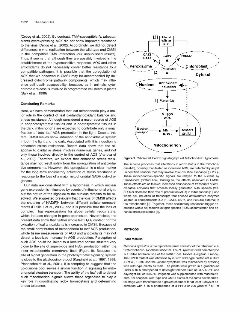
Our data are consistent with a hypothesis in which nucleargene expression is influenced by events of mitochondrial origin,but the nature of the signal(s) to the nucleus remains to be re-solved. We suggested previously that the loss of CMSII affectsthe shuttling of NAD(P)H between different cellular compart-ments (Dutilleul et al., 2003), and it is possible that the loss ofcomplex I has repercussions for global cellular redox state,which induces changes in gene expression. Nevertheless, thepresent data show that neither whole leaf H2O2 content nor theoxidation of leaf antioxidants is increased in CMSII. Because ofthe small contribution of mitochondria to leaf AOS production,whole tissue measurements of AOS and antioxidants may notdetect a localized increase in AOS production. Perception ofsuch AOS could be linked to a localized sensor situated veryclose to the site of superoxide and H2O2 production within theinner mitochondrial membrane itself (Figure 9). Because thesite of signal generation in the photosynthetic signaling systemis close to the plastoquinone pool (Karpinski et al., 1997, 1999;Pfannschmidt et al., 2001), it is tempting to suggest that theubiquinone pool serves a similar function in signaling for mito-chondrial electron transport. The ability of the leaf cell to detectsuch mitochondrial signals allows these organelles to play akey role in coordinating redox homeostasis and determiningstress tolerance.
METHODS
Plant Material
Nicotiana sylvestris is the diploid maternal ancestor of the tetraploid cul-tivated tobacco, Nicotiana tabacum. The N. sylvestris wild parental typeis a fertile botanical line of the Institut des Tabacs (Bergerac, France).The CMSII mutant was obtained by in vitro wild-type protoplast culture(Li et al., 1988), and the variant cytoplasm was maintained by crossingwith wild-type plants as male. The plants were grown in a greenhouseunder a 16-h photoperiod at day/night temperatures of 23.5/17.5C andday/night RH of 60/50%. Irrigation was supplemented with macronutri-ents. For analyses, wild-type and CMSII plants at the same developmen-tal stage were transferred to a growth chamber for at least 3 days of ac-climation with a 16-h photoperiod at a PPFD of 200 �mol·m�2·s�1 at
Figure 9. Whole Cell Redox Signaling by Leaf Mitochondria: Hypothesis.
The scheme proposes that alterations in redox status in the mitochon-dria (Mit), possibly manifested as increased AOS, are detected by as yetunidentified sensors that may involve thiol-disulfide exchange (SH/SS).These mitochondrion-specific signals are relayed to the nucleus bytransducers (dotted line), leading to the effects observed in CMSII.These effects are as follows: increased abundance of transcripts of anti-oxidative enzymes that process locally generated AOS species (Mn-SOD) or decrease their rate of production (AOX) in mitochondria [1]; andwhole cell induction of transcripts that encode antioxidative enzymeslocated in compartments (CAT1, CAT3, cAPX, and FeSOD) external tothe mitochondria [2]. Together, these acclimatory responses trigger de-creased whole cell reactive oxygen species (ROS) accumulation and en-hance stress resistance [3].

Mitochondrial Redox Signaling 1223
day/night temperatures of 24/17C and RH of 70%. Samples taken werefrom the second well-developed leaves.
RNA Isolation and Gel Blot Analysis
For RNA isolation, leaf tissue pieces (0.1 g) were harvested in liquid ni-trogen during the light/dark cycle and stored at �80C. Total RNAs wereextracted by the Trizol-chloroform procedure (Gibco BRL). Ten micro-grams of total RNA was fractionated on a 1.2% agarose gel, blotted ontonylon-based membranes (Appligène, Illkirsh, France), and hybridizedwith 32P-labeled probes. Homologous probes were Nicotiana plum-baginifolia CAT1, CAT2, and CAT3 (Willekens et al., 1994b), N. tabacumplastidial GR (Creissen and Mullineaux, 1995), N. tabacum cAPX (Orvarand Ellis, 1995), N. plumbaginifolia MnSOD1 (Bowler et al., 1989), N.plumbaginifolia FeSOD (Van Camp et al., 1990), and a N. sylvestris AOXprobe (Sabar et al., 2000). An N. sylvestris chlAPX probe was generatedusing primers 5�-TCCAGGAAACCCTGGAGGAC-3� and 5�-TCAGGA-GGATCAAATTTGGC-3� designed from the N. tabacum chlAPX se-quence (Yoshimura et al., 1999). All procedures for blot analysis wereperformed as described previously (Sabar et al., 2000). Quantification ofthe relative abundance of transcripts was determined using a scanningdensitometer (MasterScan; Scanalytics, Billerica, MA). Results were ex-pressed as the values of the following ratio: integrated density of the sig-nal/integrated density of the 18S rRNA signal.
Total Protein Extraction
Leaf material (0.1 g) was ground in liquid nitrogen, and total proteinswere extracted into 0.3 mL of 0.1 M Tris-HCl, pH 8.1, 10% sucrose, and0.05% �-mercaptoethanol. To distinguish between chloroplastic andnonchloroplastic extractable APX activities, samples were extracted inthe presence or absence of 5 mM ascorbate in the medium. The extractwas centrifuged for 5 min at 15,000g, and protein was determined ac-cording to Bradford (1976).
Protein Gel Blot Immunodetection
Total leaf proteins (10 �g per lane) were separated on a 12% SDS-poly-acrylamide gel and transferred to nitrocellulose membranes as de-scribed by Gutierres et al. (1997). The following antisera were used: micemonoclonal antiserum against Sauromatum guttatum AOX (Elthon et al.,1989) at a dilution of 1:200; rabbit antiserum directed against rye leafcatalase (Hertwig et al., 1992) at a dilution of 1:3000; rabbit antiserum di-rected against spinach cAPX at a dilution of 1:3000 (Saji et al., 1990);rabbit antiserum directed against sorghum ribulose-1,5-bisphosphatecarboxylase/oxygenase large subunit (RBCL); and antiserum directedagainst the viral coat protein (17.5 kD) of Tobacco mosaic virus (TMV)strain U1. Horseradish peroxidase–conjugated goat anti-mouse or anti-rabbit IgG was used as a secondary antibody (at a dilution of 1:2000),and immune complexes were visualized by the color reaction of peroxi-dase as described by Gutierres et al. (1997), except for AOX (and thecorresponding RBCL detection), which was visualized by chemilumines-cence (West Dura Trial Kit; Pierce).
Antioxidant Enzyme Activities
Catalase activity was determined by monitoring H2O2 removal as the de-crease in A240 (� 36 M�1 cm�1) for 30 s, as described by Dorey et al.(1998). GR activity was determined by monitoring the oxidation ofNADPH at 340 nm (� 6.22 M�1 cm�1), as described by Donahue et al.(1997). APX activity was determined by monitoring the oxidation of
ascorbate at 290 nm (� 2.88 mM�1 cm�1), as described by Nakano andAsada (1981).
Determination of O2.� and H2O2 Content
Global leaf H2O2 was determined according to the method of Veljovic-Jovanovic et al. (2002) using the chromogenic peroxidase-coupled oxi-dation of 3-methyl 2-benzothiazolinone hydrazone and 3-dimethyl ami-nobenzoic acid.
In situ O2.� was estimated using the nitroblue tetrazolium (NBT) staining
method as described by Jabs et al. (1996), with the following modifica-tions. Leaf discs were punched out with a cork borer (2 cm in diameter)from the central area of the second fully developed leaf and vacuum-infiltrated (three cycles of 5 min) in 0.5 mg/mL NBT prepared in 10 mMpotassium phosphate buffer, pH 7.8. As a control, superoxide dismutase(10 units/mL) and 10 mM MnCl2 were added to the staining medium be-fore infiltration. Samples were incubated for 1 h in the dark at room tem-perature and then cleared in 90% ethanol at 70C until complete removalof chlorophyll. O2
.� was visualized as a blue color at the site of NBT pre-cipitation. Samples were stored and examined in 70% glycerol. In situH2O2 was estimated using the 3,3�-diaminobenzidine (DAB) stainingmethod as described by Thordal-Christensen et al. (1997) and modifiedas follows. Leaf discs were vacuum-infiltrated (three cycles of 5 min) with1 mg/mL DAB solution, pH 3.8. As a control, DAB solution was supple-mented with 10 mM ascorbic acid before infiltration. After incubation inthe dark at room temperature for 14 h, samples were transferred in 90%ethanol at 70C until complete removal of chlorophyll. H2O2 was visual-ized as brown color at the site of DAB polymerization. Samples werestored and examined in 70% glycerol.
Ascorbate and Glutathione Determination
Leaf discs (2.4 cm in diameter) were harvested, ground in liquid nitrogen,and then ground in 1 mL of 1 M HClO4. After thawing, samples were cen-trifuged for 15 min at 15,000g and 4C. The pH of the clarified superna-tant was adjusted to 5.6 with K2CO3, and insoluble KClO4 was removedby centrifugation. Ascorbate and glutathione were measured in the samesupernatant. Total and reduced ascorbate contents were measured asthe ascorbate oxidase–dependent decrease in A265 before (reducedascorbate) and after (total ascorbate) treatment of the sample for 15 minwith 0.1 M DTT (modified from Foyer et al., 1983). The 1-mL reactionmixture contained 0.12 M NaH2PO4, pH 5.6, and 0.1 mL of extract. Thedifference in A265 before and after the addition of 1 unit of ascorbate ox-idase was converted to ascorbate concentration (A265 12.6 mM�1
cm�1). Total and oxidized glutathione contents were measured using theenzymatic recycling assay, which involves the NADPH-driven glu-tathione-dependent reduction of 5,5-dithiobis 2-nitrobenzoic acid at 412nm (Noctor and Foyer, 1998b).
Ozone Treatment
Wild-type and CMSII plants were exposed to acute ozone doses (1000parts per billion) for up to 3 successive days for 4 h per day in a controlledenvironment, 21C and 75% RH, as described by Cotovio et al. (2001).
TMV Inoculation
Wild-type and CMSII N. sylvestris � N. tabacum hybrid plants, at thesame developmental stage (first flower bud), were inoculated with thecommon U1 strain of TMV. Using Carborundum as an abrasive, nineleaves of each plant were inoculated with 1 mL of 1 �g/mL viral proteinprepared in 0.01 M phosphate buffer, pH 7.0. Lesions were evaluated 7days after inoculation.

1224 The Plant Cell
Statistical Analyses
The significance of differences was determined using Student’s t test. Val-ues are denoted as significant (P � 0.05) or highly significant (P � 0.01).
Upon request, all novel materials described in this article will be madeavailable in a timely manner for noncommercial research purposes.
ACKNOWLEDGMENTS
We gratefully acknowledge the following for the gifts of probes: D. Inzé(SOD and CAT cDNA probes), B. Ellis (cAPX probe), G. Creissen (chlGRprobe), T. Elthon (AOX antibody), J. Feierabend (CAT antibody), and A.Kubo (cAPX antibody). We thank S. Kauffmann for providing the TMV U1strain and Roland Boyer for photographic work. Many thanks to M. Boccarafor helpful discussion and comments. This work was supported by theCentre National de la Recherche Scientifique (France), the Biotechnologyand Biological Science Research Council (United Kingdom), the BritishCouncil (Alliance program funding), the European Science Foundation(fellowship award to C.D.), and the French Ministère de la Recherche etde la Technologie (award to M.G.).
Received November 22, 2002; accepted February 21, 2003.
REFERENCES
Alscher, R.G., Donahue, J.L., and Cramer, C.L. (1997). Reactive oxy-gen species and antioxidants: Relationships in green cells. Physiol.Plant. 100, 224–233.
Amako, K., Chen, G.X., and Asada, K. (1994). Separate assays spe-cific for ascorbate peroxidase and guaiacol peroxidase and for chlo-roplastic and cytosolic isozymes of ascorbate peroxidase in plants.Plant Cell Physiol. 35, 497–504.
Asada, K. (1992). Ascorbate peroxidase: A hydrogen peroxide-scav-enging enzyme in plants. Physiol. Plant. 85, 235–241.
Balk, J., Leaver, C.J., and McCabe, P.F. (1999). Translocation of cyto-chrome c from the mitochondria to the cytosol occurs during heat-induced programmed cell death in cucumber plants. FEBS Lett. 463,151–154.
Bartosz, G. (1997). Oxidative stress in plants. Acta Physiol. Plant. 19, 47–64.Boccara, M., Boué, C., Garmier, M., De Paepe, R., and Boccara,
A.C. (2001). IR thermography revealed a role for mitochondria inpresymptomatic cooling during harpin-induced hypersensitive response.Plant J. 28, 663–670.
Bowler, C., Alliotte, T., De Loose, M., Van Montagu, M., and Inzé, D.(1989). The induction of manganese superoxide dismutase inresponse to stress in Nicotiana plumbaginifolia. EMBO J. 8, 31–38.
Bowler, C., Van Montagu, M., and Inzé, D. (1992). Superoxide dismu-tase and stress tolerance. Annu. Rev. Plant Physiol. Plant Mol. Biol.43, 83–116.
Bradford, M.M. (1976). A rapid and sensitive method for the quantita-tion of microgram quantities of protein utilizing the principle of pro-tein-dye binding. Anal. Biochem. 7, 248–254.
Braidot, E., Petrussa, E., Vianello, A., and Macri, F. (1999). Hydrogenperoxide generation by higher plant mitochondria oxidizing complex Ior complex II substrates. FEBS Lett. 451, 347–350.
Chivasa, S., and Carr, J.P. (1998). Cyanide restores N gene–mediatedresistance to tobacco mosaic virus in transgenic tobacco expressingsalicylic acid hydroxylase. Plant Cell 10, 1489–1498.
Cotovio, J., Onno, L., Justine, P., Lamure, S., and Catroux, P. (2001).Generation of oxidative stress in human cutaneous models followingin vitro ozone exposure. Toxicol. in Vitro 15, 357–362.
Creissen, G., and Mullineaux, P. (1995). Cloning and characterisation
of glutathione reductase cDNAs and identification of two genesencoding the tobacco enzyme. Planta 197, 422–425.
Dawson, W.O. (1999). Tobacco mosaic virus virulence and avirulence.Philos. Trans. R. Soc. Lond. B 354, 645–651.
Day, D.A., and Wiskich, J.T. (1995). Regulation of alternative oxidaseactivity in higher plants. J. Bioenerg. Biomembr. 27, 379–385.
De Paepe, R., Chétrit, P., Vitart, V., Ambard-Bretteville, F., Prat, D.,and Vedel, F. (1990). Several nuclear genes control both sterility andmitochondrial protein synthesis in Nicotiana sylvestris protoclones.Mol. Gen. Genet. 222, 206–210.
Donahue, J.L., Okpodu, C.M., Cramer, C.L., Grabau, E.A., andAlscher, R.G. (1997). Responses of antioxidants to paraquat in pealeaves. Plant Physiol. 113, 249–257.
Dorey, S., Baillieul, F., Saindrenan, P., Fritig, B., and Kauffmann, S.(1998). Tobacco class I and II catalases are differentially expressedduring elicitor-induced hypersensitive cell death and localizedacquired resistance. Mol. Plant-Microbe Interact. 11, 1102–1109.
Droillard, M.J., and Paulin, A. (1990). Isozymes of superoxide dismu-tase in mitochondria and peroxisomes isolated from petals of carna-tion (Dianthus caryophyllus) during senescence. Plant Physiol. 94,1187–1192.
Dutilleul, C., Driscoll, S., Cornic, G., De Paepe, R., Foyer, C.H., andNoctor, G. (2003). Tobacco leaves require functional mitochondrialcomplex I for optimal photosynthetic performance in photorespiratoryconditions and during transients. Plant Physiol. 131, 264–275.
Elthon, T.E., Nickels, R.L., and McIntosh, L. (1989). Monoclonal anti-bodies to the alternative oxidase of higher plant mitochondria. PlantPhysiol. 89, 1311–1317.
Foyer, C.H., and Halliwell, B. (1976). The presence of glutathione andglutathione reductase in chloroplasts: A proposed role in ascorbicacid metabolism. Planta 133, 21–25.
Foyer, C.H., Lopez-Delgado, H., Dat, J.F., and Scott, I.M. (1997).Hydrogen peroxide and glutathione–associated mechanisms of accli-matory stress tolerance and signalling. Physiol. Plant. 100, 241–254.
Foyer, C.H., and Noctor, G. (2000). Tansley Review No. 112. Oxygenprocessing in photosynthesis: Regulation and signalling. New Phytol.146, 359–388.
Foyer, C.H., Rowell, J., and Walker, D. (1983). Measurement of theascorbate content of spinach leaf protoplasts and chloroplasts duringillumination. Planta 157, 239–244.
Fridovich, I. (1986). Biological effects of the superoxide radical. Arch.Biochem. Biophys. 247, 1–11.
Garmier, M., Dutilleul, C., Mathieu, C., Chétrit, P., Boccara, M., andDe Paepe, R. (2002). Changes in antioxidant expression and harpin-induced hypersensitive response in a Nicotiana sylvestris mitochon-drial mutant. Plant Physiol. Biochem. 40, 561–566.
Gutierres, S., Sabar, M., Lelandais, C., Chétrit, P., Diolez, P.,Degand, H., Boutry, M., Vedel, F., de Kouchkovsky, Y., and DePaepe, R. (1997). Lack of mitochondrial and nuclear-encoded sub-units of complex I and alteration of the respiratory chain in Nicotianasylvestris mitochondrial deletion mutants. Proc. Natl. Acad. Sci. USA94, 3436–3441.
Hertwig, B., Streb, P., and Feierabend, J. (1992). Light dependence ofcatalase synthesis and degradation in leaves and the influence ofinterfering stress conditions. Plant Physiol. 100, 1547–1553.
Jabs, T., Dietrich, R.A., and Dangl, J.L. (1996). Initiation of runawaycell death in an Arabidopsis mutant by extracellular superoxide. Sci-ence 27, 1853–1856.
Jimenez, A., Hernandez, J.A., del Rio, L.A., and Sevilla, F. (1997). Evi-dence for the presence of the ascorbate-glutathione cycle in mitochon-dria and peroxisomes of pea leaves. Plant Physiol. 114, 275–284.
Jones, A. (2000). Does the plant mitochondrion integrate cellular stressand regulate programmed cell death? Trends Plant Sci. 5, 225–230.

Mitochondrial Redox Signaling 1225
Karpinski, S., Escobar, C., Karpinska, B., Creissen, G., and Mullineaux,P.M. (1997). Photosynthetic electron transport regulates the expres-sion of cytosolic ascorbate peroxidase genes in Arabidopsis duringexcess light stress. Plant Cell 9, 627–640.
Karpinski, S., Reynolds, H., Karspinka, B., Wingsle, G., Creissen, G.,and Mullineaux, P. (1999). Systemic signalling and acclimation inresponse to excess excitation energy. Science 23, 654–657.
Karpova, O.V., Kuzmin, E.V., Elthon, T.E., and Newton, K.J. (2002).Differential expression of alternative oxidase genes in maize mito-chondrial mutants. Plant Cell 14, 3271–3284.
Kubo, A., Saji, H., Tanaka, K., and Kondo, N. (1995). Expression ofcytosolic ascorbate peroxidase gene in response to ozone or sulfurdioxide. Plant Mol. Biol. 29, 479–489.
Lee, B.H., Lee, H., Xiong, L., and Zhu, J.K. (2002). A mitochondrialcomplex I defect impairs cold-regulated nuclear gene expression.Plant Cell 14, 1235–1251.
Lennon, A.M., Neuenschwander, U.H., Ribas-Carbo, M., Giles, L.,Ryals, J.A., and Siedow, J.N. (1997). The effects of salicylic acid andtobacco mosaic virus infection on the alternative oxidase of tobacco.Plant Physiol. 115, 783–791.
Li, X.Q., Chétrit, P., Mathieu, C., Vedel, F., De Paepe, R., Rémy, R.,and Ambard-Bretteville, F. (1988). Regeneration of cytoplasmicmale sterile protoclones of Nicotiana sylvestris with mitochondrialvariations. Curr. Genet. 13, 261–266.
Liu, Y., Fiskum, G., and Schubert, D. (2002). Generation of reactiveoxygen species by the mitochondrial electron transport chain. J. Neu-rochem. 80, 780–787.
Maxwell, D.P., Wang, Y., and McIntosh, L. (1999). The alternative oxi-dase lowers mitochondrial reactive oxygen production in plant cells.Proc. Natl. Acad. Sci. USA 96, 8271–8276.
Millar, H., Considine, M.G., Day, D.A., and Whelan, J. (2001). Unravel-ing the role of mitochondria during oxidative stress in plants. IUBMBLife 51, 201–205.
Mittler, R., Herr, E.H., Orvar, B.J., van Camp, W., Willekens, H., Inzé,D., and Ellis, B.E. (1999). Transgenic tobacco plants with reducedcapability to detoxify reactive oxygen intermediates are hyperrespon-sive to pathogen infection. Proc. Natl. Acad. Sci. USA 96, 14165–14170.
Miyake, C., and Asada, K. (1996). Inactivation mechanism of ascorbateperoxidase at low concentrations of ascorbate: Hydrogen peroxidedecomposes compound I of ascorbate peroxidase. Plant Cell Physiol.37, 423–430.
Møller, I.M. (2001). Plant mitochondria and oxidative stress: Electrontransport, NADPH turnover, and metabolism of reactive oxygen spe-cies. Annu. Rev. Plant Physiol. Plant Mol. Biol. 52, 561–591.
Nakano, Y., and Asada, K. (1981). Hydrogen peroxide is scavenged byascorbate-specific peroxidase in spinach chloroplasts. Plant CellPhysiol. 22, 867–880.
Noctor, G., and Foyer, C.H. (1998a). Ascorbate and glutathione: Keep-ing active oxygen under control. Annu. Rev. Plant Physiol. Plant Mol.Biol. 49, 249–279.
Noctor, G., and Foyer, C.H. (1998b). Simultaneous measurement offoliar glutathione, �-glutamylcysteine, and amino acids by high-per-formance liquid chromatography: Comparison with two other assaymethods for glutathione. Anal. Biochem. 264, 98–110.
Ordog, S.H., Higgins, V.J., and Vanlerberghe, G.C. (2002). Mitochon-drial alternative oxidase is not a critical component of plant viral resis-tance but may play a role in the hypersensitive response. Plant Phys-iol. 129, 1858–1865.
Orvar, B.L., and Ellis, B.E. (1995). Isolation of a cDNA encoding cyto-solic ascorbate peroxidase in tobacco. Plant Physiol. 108, 839–840.
Pell, E.J., Schlagnhaufer, C.D., and Arteca, R.N. (1997). Ozone-induced oxidative stress: Mechanisms of action and reaction. Physiol.Plant. 100, 264–273.
Pfannschmidt, T., Schütze, K., Brost, M., and Oelmüller, R. (2001). Anovel mechanism of nuclear photosynthesis gene regulation by redoxsignals from the chloroplast during photosystem stoichiometryadjustment. J. Biol. Chem. 276, 36125–36130.
Pitcher, L.H., Brennan, E., Hurley, A., Dunsmuir, P., Tepperman,J.M., and Zilinskas, B.A. (1991). Overproduction of petunia copper/zinc superoxide dismutase does not confer ozone tolerance in trans-genic tobacco. Plant Physiol. 97, 452–455.
Pitkanen, S., and Robinson, B.H. (1996). Mitochondrial complex I defi-ciency leads to increased production of superoxide radicals andinduction of superoxide dismutase. J. Clin. Invest. 98, 345–351.
Pla, M., Mathieu, C., De Paepe, R., Chétrit, P., and Vedel, F. (1995).Deletion of the last two exons of the mitochondrial nad7 gene resultsin lack of the NAD7 polypeptide in a Nicotiana sylvestris CMS mutant.Mol. Gen. Genet. 248, 279–288.
Popov, V.N., Simonian, R.A., Skulachev, V.P., and Starkov, A.A.(1997). Inhibition of the alternative oxidase stimulates H2O2 produc-tion in plant mitochondria. FEBS Lett. 415, 87–90.
Purvis, A.C., Shewfelt, R.L., and Gegogeine, J.W. (1995). Superoxideproduction by mitochondria isolated from green bell pepper fruit.Physiol. Plant. 94, 743–749.
Ranieri, A., Castagna, A., and Soldatini, G.F. (2000). Differential stimu-lation of ascorbate peroxidase isoforms by ozone exposure in sun-flower plants. J. Plant Physiol. 156, 266–271.
Rasmusson, A.G., Heiser, V., Zabaleta, E., Brennicke, A., and Grohmann,L. (1998). Physiological, biochemical and molecular aspects of mitochon-drial complex I in plants. Biochim. Biophys. Acta 1364, 1401–1411.
Redinbaugh, M.G., Sabre, M., and Scandalios, J.G. (1990). Expres-sion of the maize Cat3 catalase gene is under the influence of a circa-dian rhythm. Proc. Natl. Acad. Sci. USA 87, 6853–6857.
Robson, C.A., and Vanlerberghe, G.C. (2002). Transgenic plant cellslacking mitochondrial alternative oxidase have increased susceptibil-ity to mitochondria-dependent and -independent pathways of pro-grammed cell death. Plant Physiol. 129, 1908–1920.
Sabar, M., De Paepe, R., and de Kouchkovsky, Y. (2000). Complex Iimpairment, respiratory compensations, and photosynthetic decreasein nuclear and mitochondrial male sterile mutants of Nicotiana sylves-tris. Plant Physiol. 124, 1239–1250.
Saji, H., Tanaka, K., and Kondo, N. (1990). Monoclonal antibodies tospinach ascorbate peroxidase and immunochemical detection of theenzyme in eight different plant species. Plant Sci. 69, 1–9.
Scandalios, J.G. (1987). Isozymes. In Current Topics in Biological andMedical Research, Vol. 14, M.C. Rattazi, J.G. Scandalios, and G.S.Whitt, eds (New York: Liss), pp. 19–44.
Scandalios, J.G. (1993). Oxygen stress and superoxide dismutases.Plant Physiol. 101, 7–12.
Scandalios, J.G., Tong, W.F., and Roupakias, D.G. (1980). Cat3, athird gene locus coding for a tissue-specific catalase in maize: Genet-ics, intracellular location, and some biochemical properties. Mol. Gen.Genet. 179, 33–41.
Simons, B.H., Millenaar, F.F., Mulder, L., Van Loon, L.C., and Lambers,H. (1999). Enhanced expression and activation of the alternative oxi-dase during infection of Arabidopsis with Pseudomonas syringae pvtomato. Plant Physiol. 120, 529–538.
Stevens, R.G., Creissen, G.P., and Mullineaux, P.M. (1997). Cloningand characterisation of a cytosolic glutathione reductase cDNA frompea (Pisum sativum L.) and its expression in response to stress. PlantMol. Biol. 5, 641–654.
Swidzinski, J.A., Sweetlove, L.J., and Leaver, C.J. (2002). A custommicroarray analysis of gene expression during programmed celldeath in Arabidopsis thaliana. Plant J. 30, 431–446.
Thompson, J.E., Ledge, R.L., and Barber, R.F. (1987). The role of freeradicals in senescence and wounding. New Phytol. 105, 317–344.

1226 The Plant Cell
Thordal-Christensen, H., Zhang, Z., Wei, Y., and Collinge, D.B.(1997). Subcellular localization of H2O2 in plants: H2O2 accumulationin papillae and hypersensitive response during the barley-powderymildew interaction. Plant J. 11, 1187–1194.
Van Camp, W., Bowler, C., Villarroel, R., Tsang, E.W., Van Montagu,M., and Inzé, D. (1990). Characterization of iron superoxide dismu-tase cDNAs from plants obtained by genetic complementation inEscherichia coli. Proc. Natl. Acad. Sci. USA 87, 9903–9907.
Van Camp, W., Willekens, H., Bowler, C., Van Montagu, M., Inzé, D.,Reupold-Popp, P., Sandermann, H., Jr., and Langebartels, C.(1994). Elevated levels of superoxide dismutase protect transgenicplants against ozone damage. Biotechnology 12, 165–168.
Vanlerberghe, G.C., and McIntosh, L.C. (1997). Alternative oxidase: Fromgene to function. Annu. Rev. Plant Physiol. Plant Mol. Biol. 48, 703–734.
Vanlerberghe, G.C., Robson, C.A., and Yip, J.Y.H. (2002). Induction ofmitochondrial alternative oxidase in response to a cell signal pathwaydown-regulating the cytochrome pathway prevents programmed celldeath. Plant Physiol. 129, 1829–1842.
Veljovic-Jovanovic, S.D., Noctor, G., and Foyer, C.H. (2002). Are leafhydrogen peroxide concentrations commonly over-estimated? Thepotential influence of artefactual interference by tissue phenolics andascorbate. Plant Physiol. Biochem. 40, 501–508.
Videira, A. (1998). Complex I from the fungus Neurospora crassa. Bio-chim. Biophys. Acta 1364, 89–100.
Vranova, E., Atichartpongkul, S., Villarroel, R., Van Montagu, M.,Inzé, D., and Van Camp, W. (2002). Comprehensive analysis of geneexpression in Nicotiana tabacum leaves acclimated to oxidativestress. Proc. Natl. Acad. Sci. USA 99, 10870–10875.
Wagner, A.M. (1995). A role for active oxygen species as second mes-sengers in the induction of alternative oxidase gene expression inPetunia hybrida cells. FEBS Lett. 368, 339–342.
Wagner, A.M., and Moore, A.L. (1997). Structure and function of theplant alternative oxidase: Its putative role in the oxygen defencemechanism. Biosci. Rep. 17, 319–333.
Willekens, H., Langebartels, C., Tire, C., Van Montagu, M., Inzé, D.,and Van Camp, W. (1994a). Differential expression of catalase genesin Nicotiana plumbaginifolia (L.). Proc. Natl. Acad. Sci. USA 91,10450–10454.
Willekens, H., Villarroel, R., Van Montagu, M., Inze, D., and VanCamp, W. (1994b). Molecular identification of catalases from Nicoti-ana plumbaginifolia (L.). FEBS Lett. 352, 79–83.
Yoshimura, K., Yabuta, Y., Ishikawa, T., and Shigeoka, S. (2000).Expression of spinach ascorbate peroxidase isoenzymes in responseto oxidative stresses. Plant Physiol. 123, 223–234.
Yoshimura, K., Yabuta, Y., Tamoi, M., Ishikawa, T., and Shigeoka,S. (1999). Alternative spliced mRNA variants of chloroplast ascor-bate peroxidase isoenzymes in spinach leaves. Biochem. J. 338,41–48.

DOI 10.1105/tpc.009464; originally published online March 28, 2003; 2003;15;1212-1226Plant Cell
Foyer and Rosine de PaepeChristelle Dutilleul, Marie Garmier, Graham Noctor, Chantal Mathieu, Philippe Chétrit, Christine H.
Determine Stress Resistance through Altered Signaling and Diurnal RegulationLeaf Mitochondria Modulate Whole Cell Redox Homeostasis, Set Antioxidant Capacity, and
This information is current as of March 7, 2019
References /content/15/5/1212.full.html#ref-list-1
This article cites 83 articles, 28 of which can be accessed free at:
Permissions https://www.copyright.com/ccc/openurl.do?sid=pd_hw1532298X&issn=1532298X&WT.mc_id=pd_hw1532298X
eTOCs http://www.plantcell.org/cgi/alerts/ctmain
Sign up for eTOCs at:
CiteTrack Alerts http://www.plantcell.org/cgi/alerts/ctmain
Sign up for CiteTrack Alerts at:
Subscription Information http://www.aspb.org/publications/subscriptions.cfm
is available at:Plant Physiology and The Plant CellSubscription Information for
ADVANCING THE SCIENCE OF PLANT BIOLOGY © American Society of Plant Biologists