lapidus procedure - j&j medical devices · the lapidus procedure* designed to reduce the risk...
TRANSCRIPT
DEPUY SYNTHES IS WITH YOU—AND YOUR PATIENTS
EVERY STEP OF THE WAY
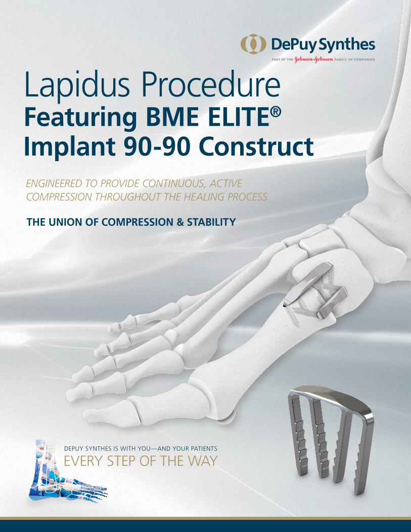
Lapidus ProcedureFeaturing BME ELITE®
Implant 90-90 ConstructENGINEERED TO PROVIDE CONTINUOUS, ACTIVE COMPRESSION THROUGHOUT THE HEALING PROCESS
THE UNION OF COMPRESSION & STABILITY
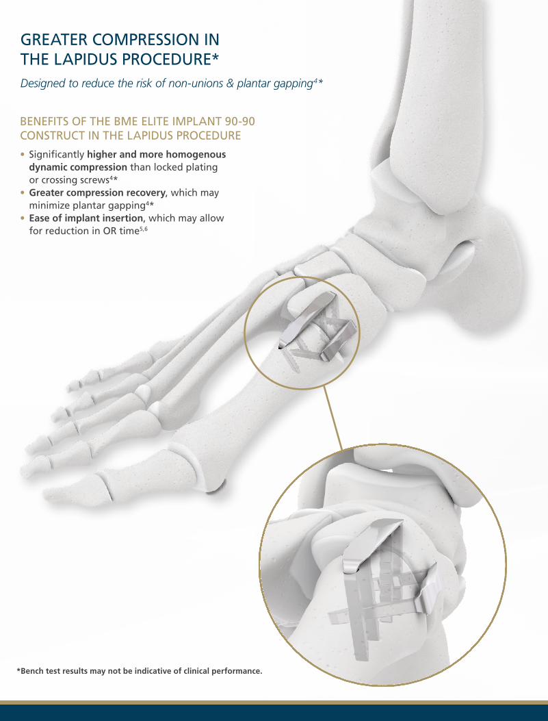
GREATER COMPRESSION IN THE LAPIDUS PROCEDURE*Designed to reduce the risk of non-unions & plantar gapping 4*
BENEFITS OF THE BME ELITE IMPLANT 90-90 CONSTRUCT IN THE LAPIDUS PROCEDURE
• Significantly higher and more homogenous dynamic compression than locked plating or crossing screws4*
• Greater compression recovery, which may minimize plantar gapping4*
• Ease of implant insertion, which may allow for reduction in OR time5,6
*Bench test results may not be indicative of clinical performance.
GREATER COMPRESSION RECOVERY MINIMIZES PLANTAR GAPPINGCompression extends plantarly through the legs of the CCI, achieving significantly higher contact area after repetitive loading compared to a 4-hole compression plate and to 2 crossing screws.4*
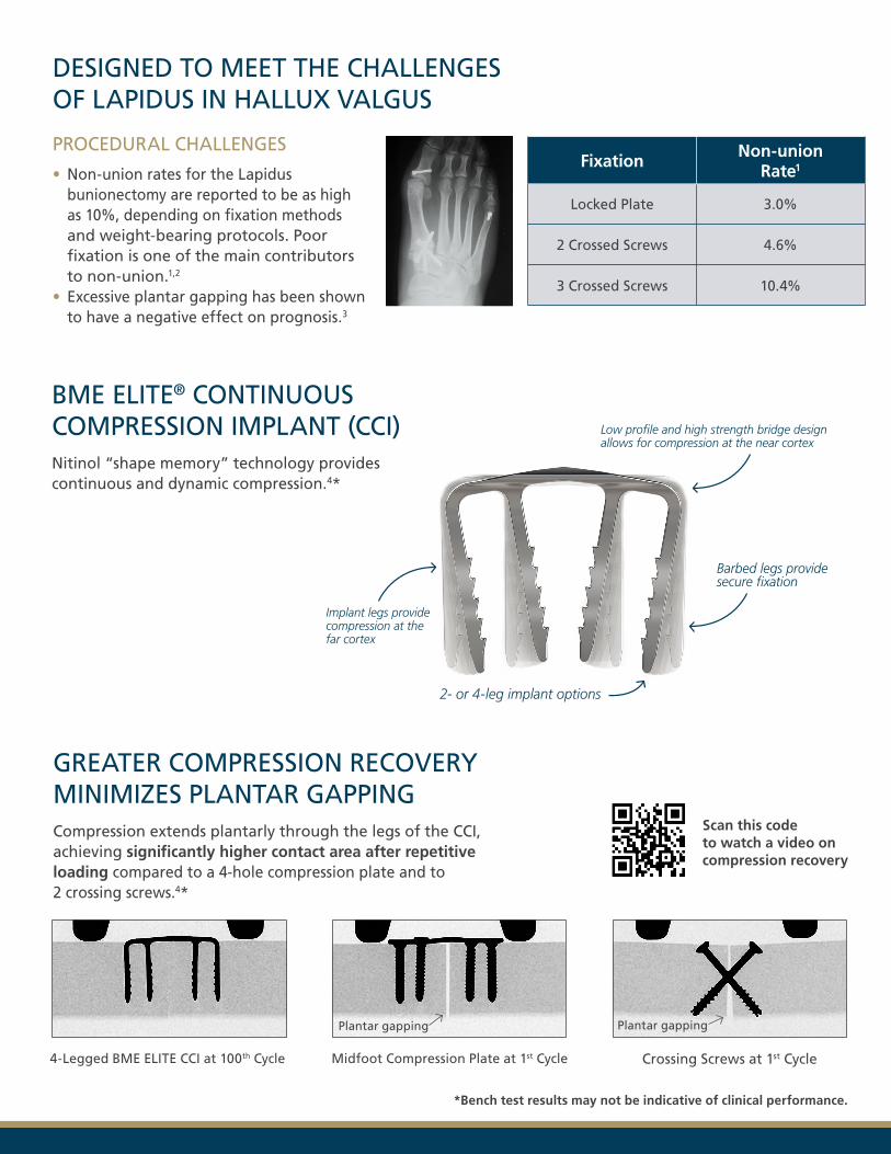
DESIGNED TO MEET THE CHALLENGES OF LAPIDUS IN HALLUX VALGUS
PROCEDURAL CHALLENGES
• Non-union rates for the Lapidus bunionectomy are reported to be as high as 10%, depending on fixation methods and weight-bearing protocols. Poor fixation is one of the main contributors to non-union.1,2
• Excessive plantar gapping has been shown to have a negative effect on prognosis.3
Fixation Non-union Rate1
Locked Plate 3.0%
2 Crossed Screws 4.6%
3 Crossed Screws 10.4%
Low profile and high strength bridge design allows for compression at the near cortex
Barbed legs provide secure fixation
Implant legs provide compression at the far cortex
2- or 4-leg implant options
4-Legged BME ELITE CCI at 100th Cycle Midfoot Compression Plate at 1st Cycle
Plantar gapping Plantar gapping
Crossing Screws at 1st Cycle
*Bench test results may not be indicative of clinical performance.
BME ELITE® CONTINUOUS COMPRESSION IMPLANT (CCI)Nitinol “shape memory” technology provides continuous and dynamic compression.4*
Scan this code to watch a video on compression recovery
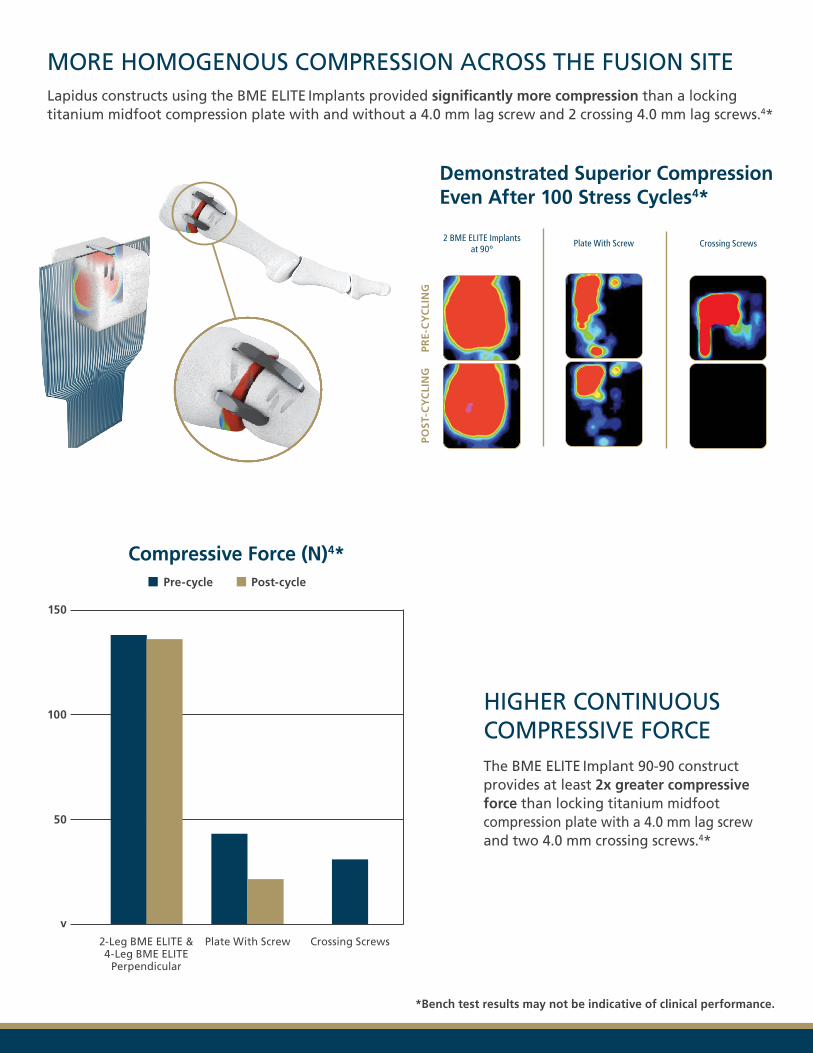
The BME ELITE Implant 90-90 construct provides at least 2x greater compressive force than locking titanium midfoot compression plate with a 4.0 mm lag screw and two 4.0 mm crossing screws.4*
HIGHER CONTINUOUS COMPRESSIVE FORCE
MORE HOMOGENOUS COMPRESSION ACROSS THE FUSION SITELapidus constructs using the BME ELITE Implants provided significantly more compression than a locking titanium midfoot compression plate with and without a 4.0 mm lag screw and 2 crossing 4.0 mm lag screws.4*
150
50
Crossing ScrewsPlate With Screw2-Leg BME ELITE & 4-Leg BME ELITE
Perpendicular
*Bench test results may not be indicative of clinical performance.
Pre-cycle Post-cycle
Compressive Force (N)4*
Demonstrated Superior Compression Even After 100 Stress Cycles4*
2 BME ELITE Implants at 90°
Plate With Screw Crossing Screws
PRE-
CY
CLI
NG
POST
-CY
CLI
NG
v
100
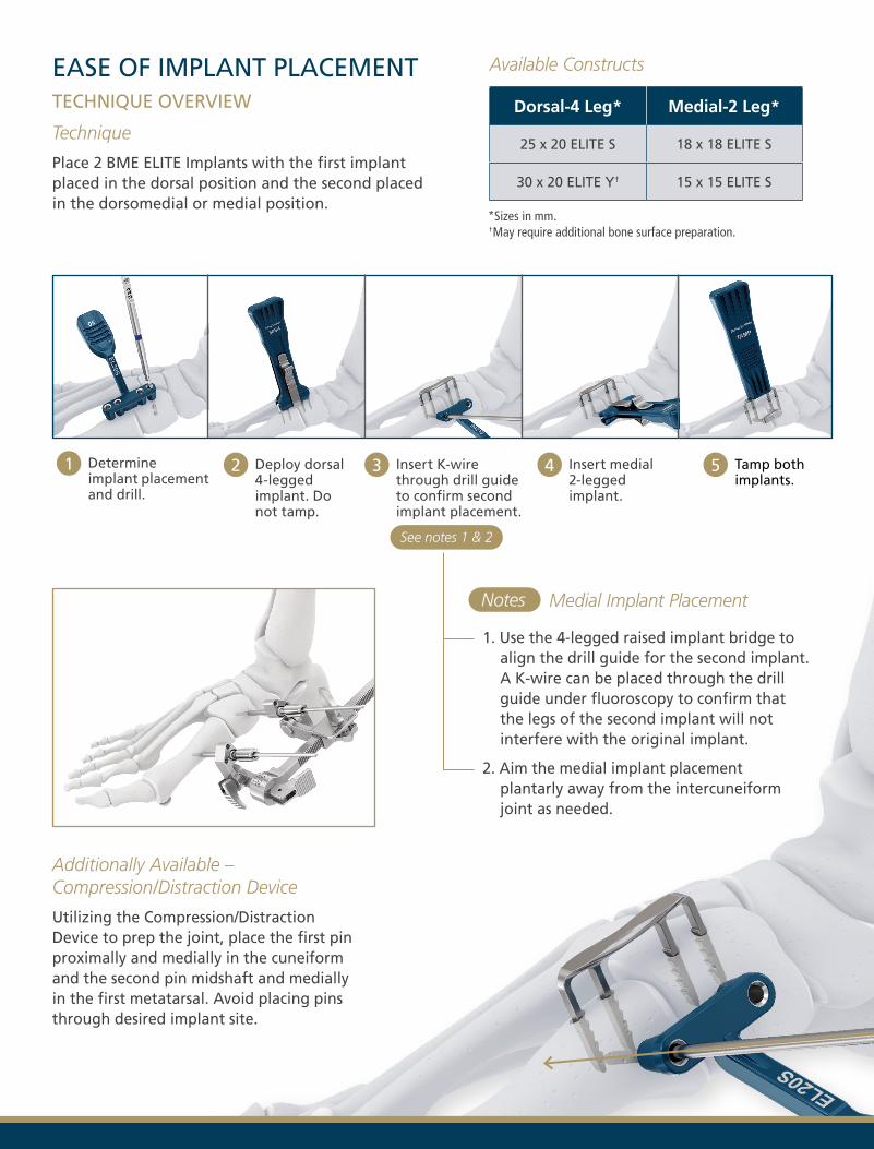
EASE OF IMPLANT PLACEMENTTECHNIQUE OVERVIEW
Technique
Place 2 BME ELITE Implants with the first implant placed in the dorsal position and the second placed in the dorsomedial or medial position.
1. Use the 4-legged raised implant bridge toalign the drill guide for the second implant.A K-wire can be placed through the drillguide under fluoroscopy to confirm thatthe legs of the second implant will notinterfere with the original implant.
2. Aim the medial implant placementplantarly away from the intercuneiformjoint as needed.
Additionally Available – Compression/Distraction Device
Utilizing the Compression/Distraction Device to prep the joint, place the first pin proximally and medially in the cuneiform and the second pin midshaft and medially in the first metatarsal. Avoid placing pins through desired implant site.
Determine implant placement and drill.
Deploy dorsal 4-legged implant. Do not tamp.
Insert K-wire through drill guide to confirm second implant placement.
Insert medial 2-legged implant.
Tamp both implants.
Dorsal-4 Leg* Medial-2 Leg*
25 x 20 ELITE S 18 x 18 ELITE S
30 x 20 ELITE Y† 15 x 15 ELITE S
*Sizes in mm.† May require additional bone surface preparation.
Available Constructs
See notes 1 & 2
Medial Implant PlacementNotes
JOINT PREPARATION SET
Adequate joint preparation is a requirement for fusion success. DePuy Synthes offers a variety of chisel shapes and a cartilage remover to facilitate proper joint preparation.
COMPRESSION/DISTRACTION DEVICE
The Compression/Distraction Device provides an adjustable and minimally invasive approach for manipulation of the fusion site to achieve precise indirect reduction and compression.7
VIVIGEN® AND VIVIGEN FORMABLE® CELLULAR BONE MATRIX†
ViviGen® Cellular Bone Matrix provides an alternative to autograft bone to pack the fusion site. It contains viable, lineage committed bone cells within a corticocancellous bone matrix and demineralized bone, delivering all of the properties necessary for bone formation.8
† ViviGen and ViviGen Formable are registered trademarks of LifeNet Health.
© DePuy Synthes 2020. All rights reserved. 131262-200127 DSUS 2/20
Please also refer to the Instructions For Use, surgical technique, or other labeling associated with the devices identified in this brochure for additional information. CAUTION: Federal law restricts these devices to sale by or on the order of a physician. Complete information regarding indications, contraindications, warnings, care, and caution can be found in the Instructions For Use.
THE UNION OF COMPRESSION AND STABILITY IN LAPIDUS
Implant Kit Bridge* Legs*
Dorsal-4 Leg EL-2520S4 25 20
Medial-2 Leg EL-1818S2 18 18
Implant Kit Bridge* Legs*
Dorsal-4 Leg EL-302007Y4 30 20
Medial-2 Leg EL-1515S2 15 15
BME ELITE IMPLANT CONSTRUCT BME ELITE Y IMPLANT CONSTRUCT
PROCEDURAL ENHANCEMENTS
Synthes USA, LLC 1101 Synthes Avenue Monument, CO 80132 www.jnjmedicaldevices.com
BioMedical Enterprises, Inc. 14785 Omicron Dr., Suite 205 San Antonio, TX 78245
Manufactured or Distributed by:
References: 1. Prissel MA, Hyer CF, Grambart ST, et al. A multicenter, retrospective study of early weightbearing for modified Lapidus arthrodesis. J Foot Ankle Surg. 2016;55(2):226-229. 2. Crowell A, Van JC, Meyr AJ. Early weightbearing after arthrodesis of the first metatarsal-medial cuneiform joint: a systematic review of the incidence of nonunion. J Foot Ankle Surg. 2018;57(6):1204-1206. 3. Lee KT, Park YU, Jegal H, Park JW, Choi JP, Kim JS. Prognostic classification of fifth metatarsal stress fracture using plantar gap. Foot Ankle Int. 2013;34(5):691-696. 4. DePuy Synthes Test Report. Compression Heat Map Test. #TR-132-03-15131. 2015. 5. Harden JL, Hiscox JA. Cost savings and quality improvement—single-use suture instruments? Scott Med J. 2006;51(3):30-33. 6. Wong J, Khu KJ, Kaderali Z, Bernstein M. Delays in the operating room: signs of an imperfect system. Can J Surg. 2010;53(3):189-195. 7. Porteous M, Bauerle S. Techniques and Principles for the Operating Room. AO Foundation; 2010:210-213. 8. Data on file. LifeNet Health 65-0347.
Utilizes the DK-300 drill kit and EL-DTS template Utilizes the DK-300 drill kit and EL-DTY template
*Sizes in mm.