laboratory diagnosis of parasitic infections lecturer. mohamed el-sakhawy 1
TRANSCRIPT
LABORATORY LABORATORY DIAGNOSIS DIAGNOSIS
OF OF PARASITIC PARASITIC
INFECTIONSINFECTIONS
Lectu
rer.
Moh
am
ed
El-
Sakh
aw
y
1
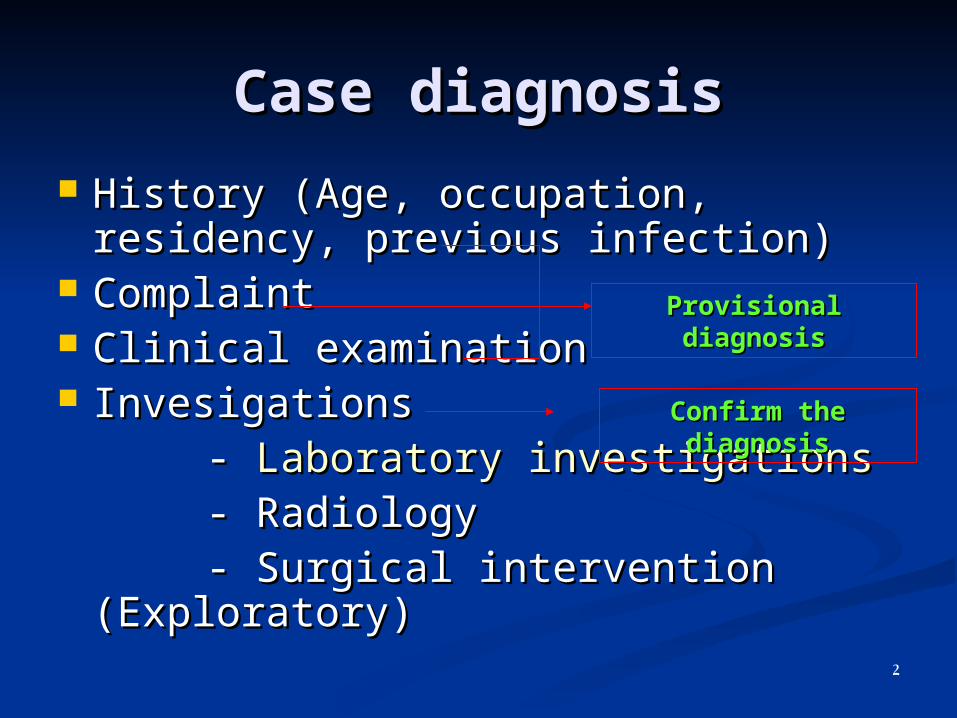
Case diagnosisCase diagnosis
History (Age, occupation, residency, History (Age, occupation, residency, previous infection)previous infection)
ComplaintComplaint Clinical examinationClinical examination InvesigationsInvesigations - - Laboratory investigationsLaboratory investigations - Radiology- Radiology - Surgical intervention - Surgical intervention
(Exploratory)(Exploratory)
Provisional Provisional diagnosisdiagnosis
Confirm the Confirm the diagnosisdiagnosis
2
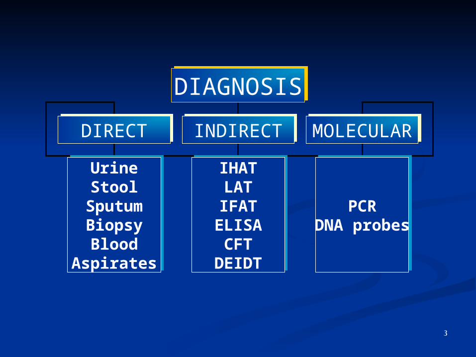
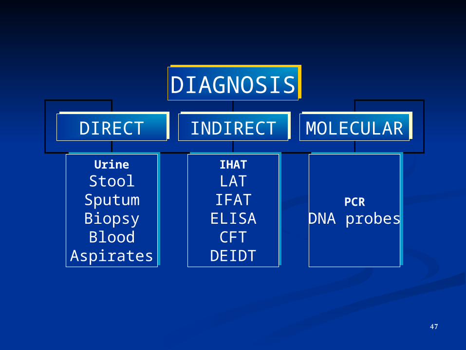
DIAGNOSISDIAGNOSIS
DIRECTDIRECT INDIRECTINDIRECT MOLECULARMOLECULAR
UrineStool
SputumBiopsyBlood
Aspirates
UrineStool
SputumBiopsyBlood
Aspirates
PCRDNA probes
PCRDNA probes
IHATLATIFATELISACFT
DEIDT
IHATLATIFATELISACFT
DEIDT
3
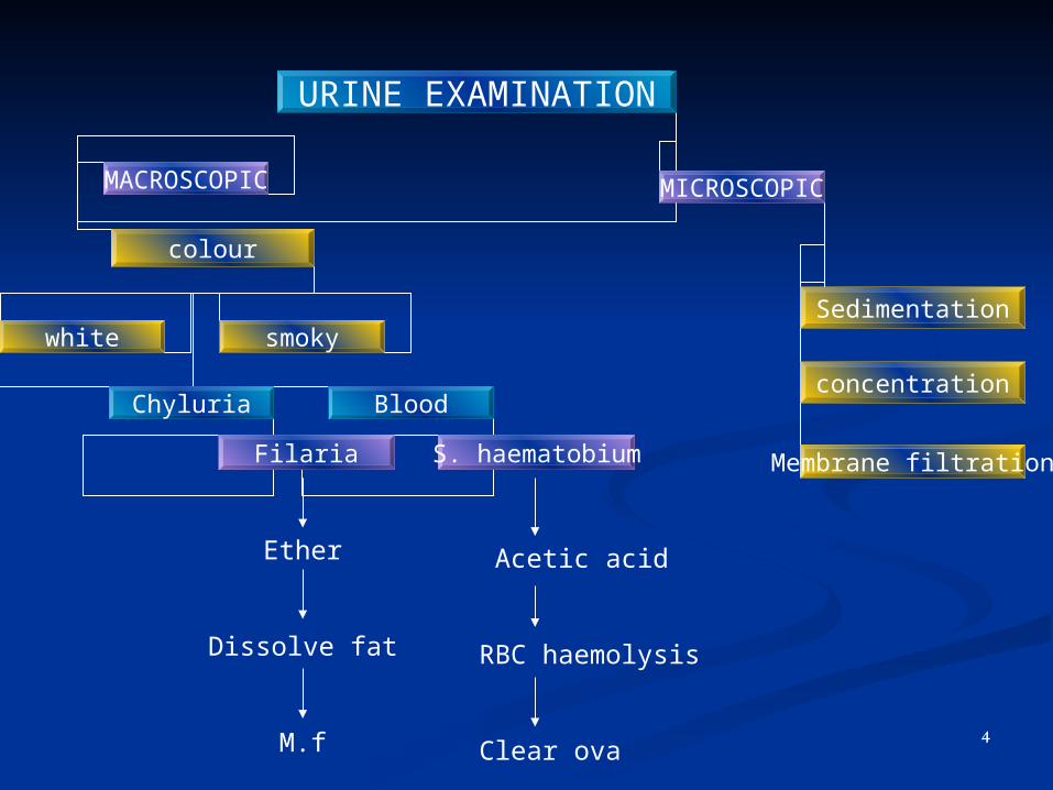
URINE EXAMINATION
MACROSCOPIC MICROSCOPIC
colour
white smokySedimentation
concentration
Membrane filtration
Chyluria Blood
Filaria S. haematobium
Acetic acid
RBC haemolysis
Clear ova
Ether
Dissolve fat
M.f4
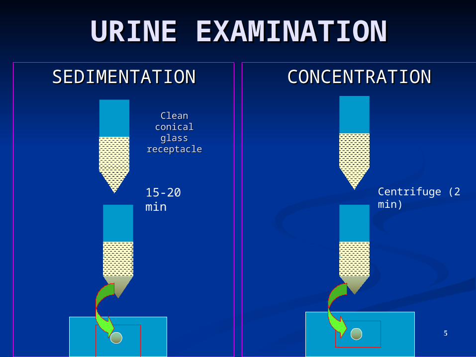
URINE EXAMINATIONURINE EXAMINATION
SEDIMENTATIONSEDIMENTATION CONCENTRATIONCONCENTRATION
15-20 min Centrifuge (2 min)
Clean conical Clean conical glass glass
receptaclereceptacle
5
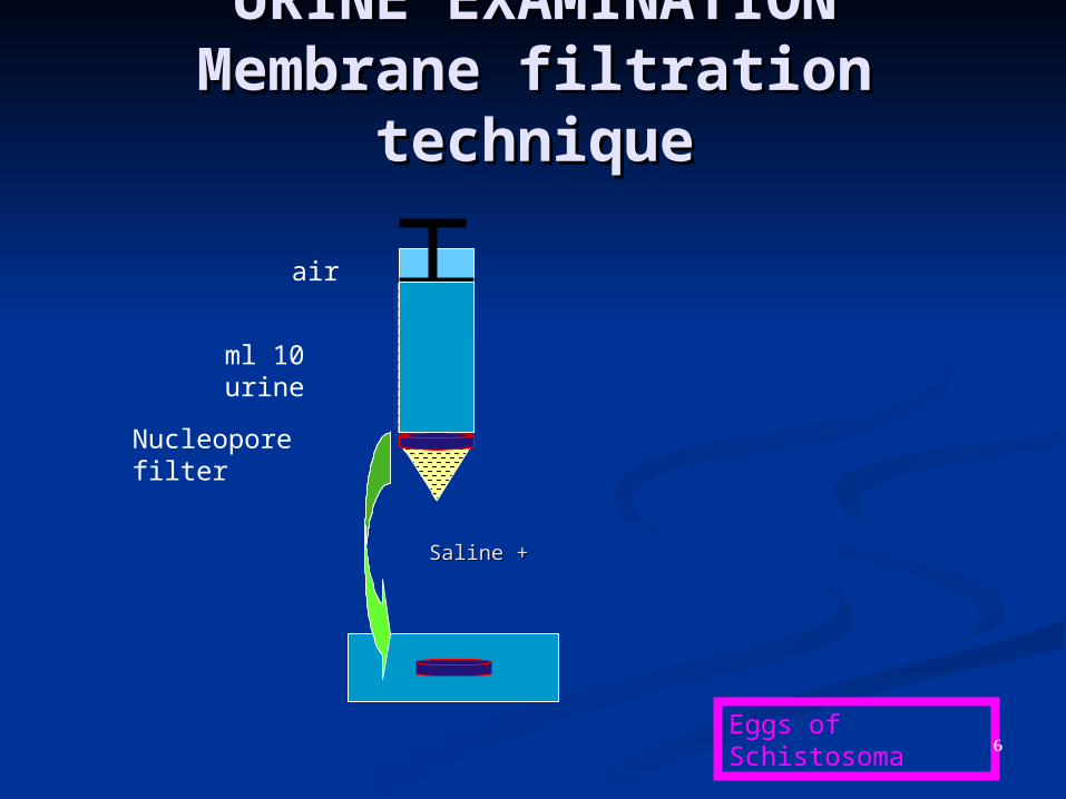
URINE EXAMINATIONURINE EXAMINATIONMembrane filtration Membrane filtration
techniquetechnique
air
10 ml urine
Nucleopore filter
Eggs of Schistosoma
+ +SalineSaline
6

URINE EXAMINATION
HELMINTHES PROTOZOA ARTHROPODES
• S. haem.egg• E. vermic. egg• S. mansoni egg• Micrfilaria (Ov, Wb)• H sand
Tricomonas. Vaginalis troph
• Pthirus pubis• L. higher deptera
7
URINE EXAMINATIONURINE EXAMINATION
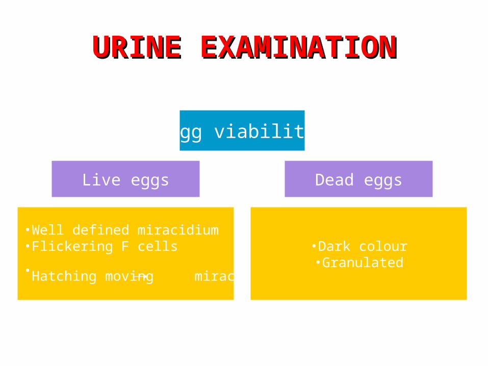
Egg viability
Live eggs Dead eggs
•Well defined miracidium•Flickering F cells
•Hatching moving miracidium •Dark colour•Granulated
8
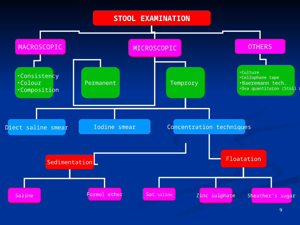
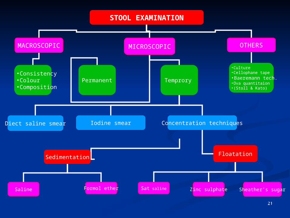
STOOL EXAMINATION
MACROSCOPIC MICROSCOPIC OTHERS
•Consistency•Colour•Composition
•Culture•Cellophane tape•Baeremann tech.•Ova quantitaion (Stoll & Kato)
TemproryPermanent
Diect saline smear Iodine smear Concentration techniques
Sedimentation Floatation
Saline Formol ether Sat saline Zinc sulphate Sheather’s sugar
9
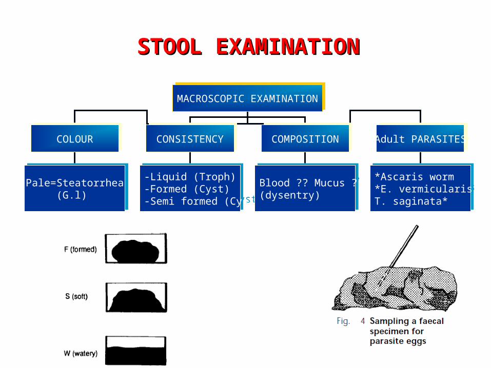
MACROSCOPIC EXAMINATIONMACROSCOPIC EXAMINATION
COLOURCOLOUR CONSISTENCYCONSISTENCY COMPOSITIONCOMPOSITION Adult PARASITESAdult PARASITES
Pale=Steatorrhea( G.l)
Pale=Steatorrhea( G.l)
-Liquid (Troph)-Formed (Cyst)-Semi formed (Cyst)
-Liquid (Troph)-Formed (Cyst)-Semi formed (Cyst)
??Blood ?? Mucus(dysentry)
??Blood ?? Mucus(dysentry)
*Ascaris worm*E. vermicularis
*T. saginata
Ascaris worm*E. vermicularis*
*T. saginata
STOOL EXAMINATIONSTOOL EXAMINATION
10

STOOL EXAMINATION
MACROSCOPIC MICROSCOPIC OTHERS
•Consistency•Colour•Composition
•Culture•Cellophane tape•Baeremann tech.•Ova quantitaion •(Stoll & Kato)
TemproryPermanent
Diect saline smear Iodine smear Concentration techniques
Sedimentation Floatation
Saline Formol ether Sat saline Zinc sulphate Sheather’s sugar
11
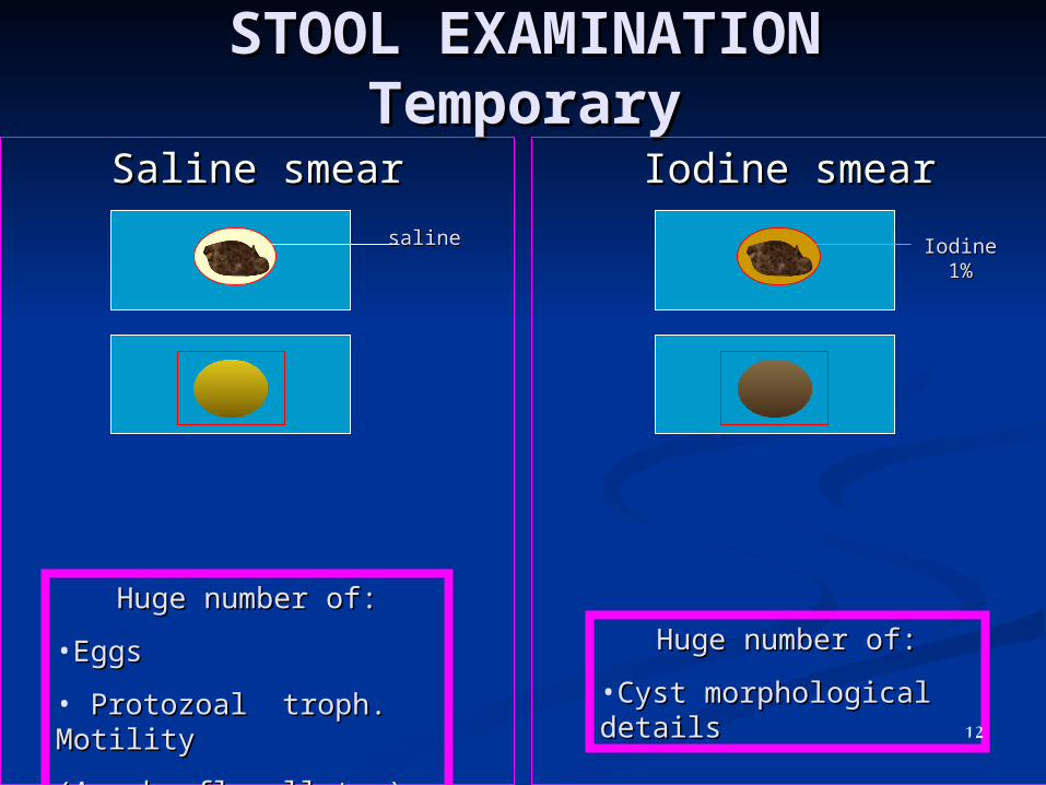
STOOL EXAMINATIONSTOOL EXAMINATIONTemporaryTemporary
Saline smearSaline smear Iodine smearIodine smear
salinesaline Iodine Iodine 1%1%
Huge number of:Huge number of:
•EggsEggs
• Protozoal troph. Protozoal troph. Motility Motility
(Amoeb, flagellates)(Amoeb, flagellates)
Huge number of:Huge number of:
•Cyst morphological Cyst morphological detailsdetails
12
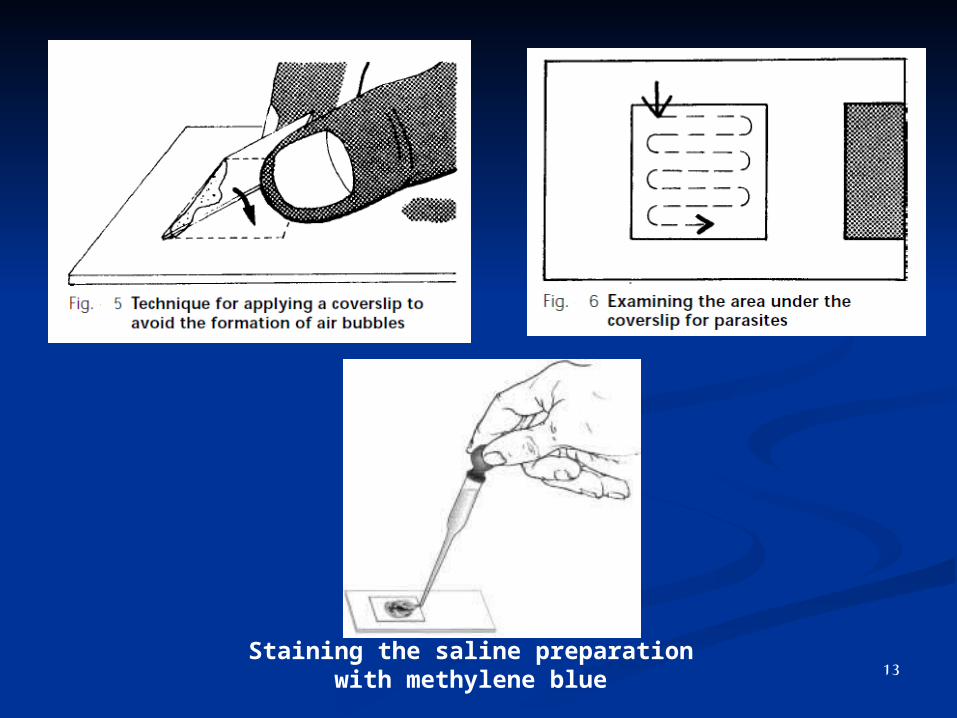
Staining the saline preparation with methylene blue 13

Lugol iodine–acetic acid solution causes the Lugol iodine–acetic acid solution causes the trophozoite forms to become nonmotile. trophozoite forms to become nonmotile.
Using a fine Pasteur pipette, allow a drop of methylene Using a fine Pasteur pipette, allow a drop of methylene blue solution to run under the coverslip over the saline blue solution to run under the coverslip over the saline preparation (Fig. 7). This will stain the nuclei of any preparation (Fig. 7). This will stain the nuclei of any cells present and distinguish the lobed nuclei of cells present and distinguish the lobed nuclei of polymorphs from the large single nuclei of mucosal polymorphs from the large single nuclei of mucosal cells.cells.
If a drop of eosin solution is added, the whole field If a drop of eosin solution is added, the whole field becomes stained except for the protozoa (particularly becomes stained except for the protozoa (particularly amoebae), which remain colourless and are thus easily amoebae), which remain colourless and are thus easily recognized.recognized.
14

STOOL EXAMINATIONSTOOL EXAMINATIONScanty infectionScanty infection
Concentration techniquesConcentration techniques
SedimentatioSedimentationn
FloatationFloatation
• Heavy eggs (Ascaris egg)Heavy eggs (Ascaris egg)
• Operculated eggs Operculated eggs (Trematodes)(Trematodes)
• Larvae (Strong sterc.)Larvae (Strong sterc.)
• CystsCysts
• Non Operculated eggs
Trematodes ( S. m.)
Cestode Nematode(Hookworms,TrichosHookworms,Trichostong)tong)
• Cysts 15

STOOL EXAMINATION
MACROSCOPIC MICROSCOPIC OTHERS
•Consistency•Colour•Composition
•Culture•Cellophane tape•Baeremann tech.•Ova quantitaion •(Stoll & Kato)
TemproryPermanent
Diect saline smear Iodine smear Concentration techniques
Sedimentation Floatation
Saline Formol ether Sat saline Zinc sulphate Sheather’s sugar
16
17
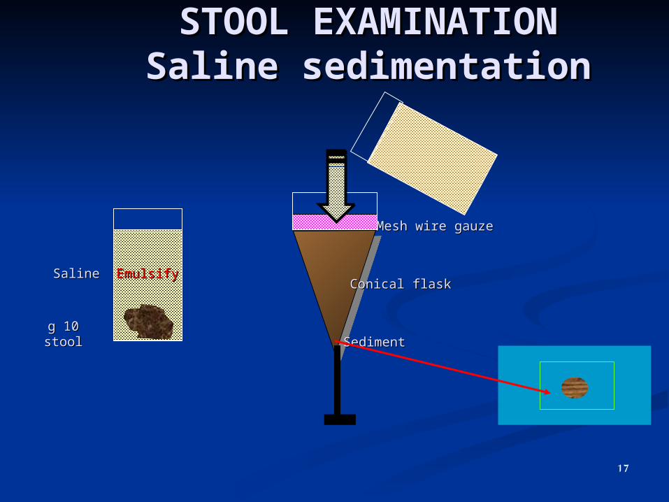
STOOL EXAMINATIONSTOOL EXAMINATIONSaline sedimentationSaline sedimentation
1010 g stoolg stool
SalineSaline
Mesh wire gauzeMesh wire gauze
Conical flaskConical flask
SedimentSediment
EmulsifyEmulsify
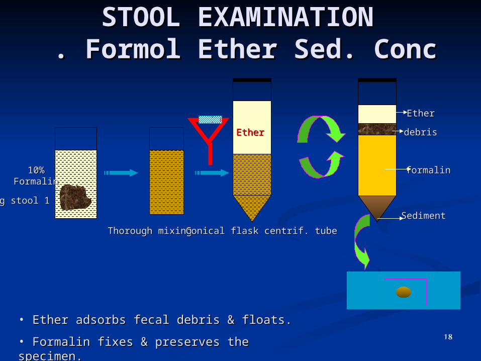
STOOL EXAMINATIONSTOOL EXAMINATION Formol Ether Sed. Conc Formol Ether Sed. Conc . .
10%10% FormalinFormalin
11 g stoolg stool
SedimentSediment
formalinformalin
debrisdebris
EtherEther
Thorough mixingThorough mixing
EtherEther
• Ether adsorbs fecal debris & floats.Ether adsorbs fecal debris & floats.
• Formalin fixes & preserves the specimen.Formalin fixes & preserves the specimen.
Conical flask centrif. tubeConical flask centrif. tube
18

STOOL EXAMINATION
MACROSCOPIC MICROSCOPIC OTHERS
•Consistency•Colour•Composition
•Culture•Cellophane tape•Baeremann tech.•Ova quantitaion •(Stoll & Kato)
TemproryPermanent
Diect saline smear Iodine smear Concentration techniques
Sedimentation Floatation
Saline Formol ether Sat saline Zinc sulphate Sheather’s sugar
19

STOOL EXAMINATIONSTOOL EXAMINATION
Floatation concentrationFloatation concentration
Sat salineSat saline Zn sulphateZn sulphate Sheather’s sugarSheather’s sugar
• Cestode eggs (non op)Cestode eggs (non op)•Nematode eggs?????Nematode eggs?????•Hookworms???????Hookworms???????•TrichostongTrichostong؟؟؟؟؟؟؟؟؟؟؟؟؟؟؟؟؟؟؟؟؟؟
•Egg of S.m.Egg of S.m.•Eggs of small tapewormsEggs of small tapeworms•CystsCysts
• Crypto, Iso. oocystsCrypto, Iso. oocysts
Tin Tin containercontainer
2020 minmin Centrif. 2 Centrif. 2 minmin
SeiveSeive
Clean light eggs & Clean light eggs & cystscysts
20
STOOL EXAMINATION
MACROSCOPIC MICROSCOPIC OTHERS
•Consistency•Colour•Composition
•Culture•Cellophane tape•Baeremann tech.•Ova quantitaion •(Stoll & Kato)
TemproryPermanent
Diect saline smear Iodine smear Concentration techniques
Sedimentation Floatation
Saline Formol ether Sat saline Zinc sulphate Sheather’s sugar
21
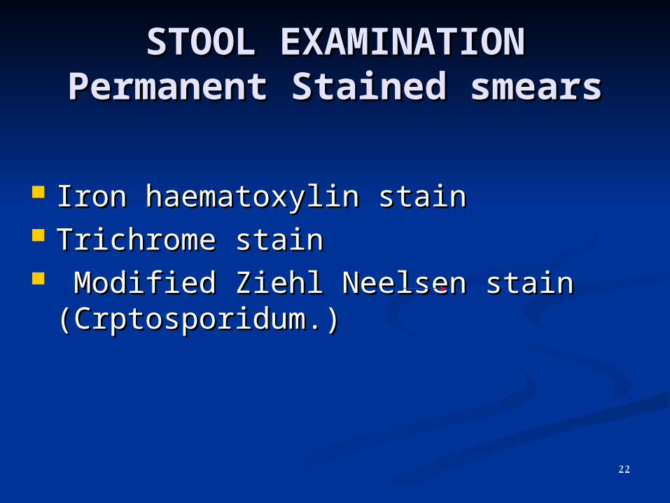
STOOL EXAMINATIONSTOOL EXAMINATIONPermanent Stained smearsPermanent Stained smears
Iron haematoxylin stainIron haematoxylin stain Trichrome stainTrichrome stain Modified Ziehl Neelsen stain Modified Ziehl Neelsen stain
(Crptosporidum.)(Crptosporidum.)
22

STOOL EXAMINATION
MACROSCOPIC MICROSCOPIC OTHERS
•Consistency•Colour•Composition
• Cellophane tape• Culture•Baeremann tech.•Ova quantitaion •(Stoll & Kato)
TemproryPermanent
Diect saline smear Iodine smear Concentration techniques
Sedimentation Floatation
Saline Formol ether Sat saline Zinc sulphate Sheather’s sugar
23
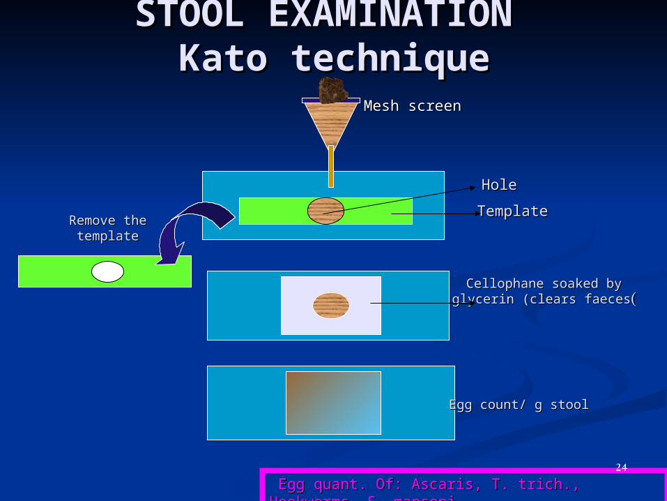
STOOL EXAMINATION STOOL EXAMINATION Kato techniqueKato technique
Mesh screenMesh screen
TemplateTemplate
HoleHole
Remove the Remove the templatetemplate
Cellophane soaked by Cellophane soaked by glycerin (clears faecesglycerin (clears faeces((
Egg count/ g stoolEgg count/ g stool
Egg quant. Of: Ascaris, T. trich., Hookworms, S. Egg quant. Of: Ascaris, T. trich., Hookworms, S. mansonimansoni
24
25
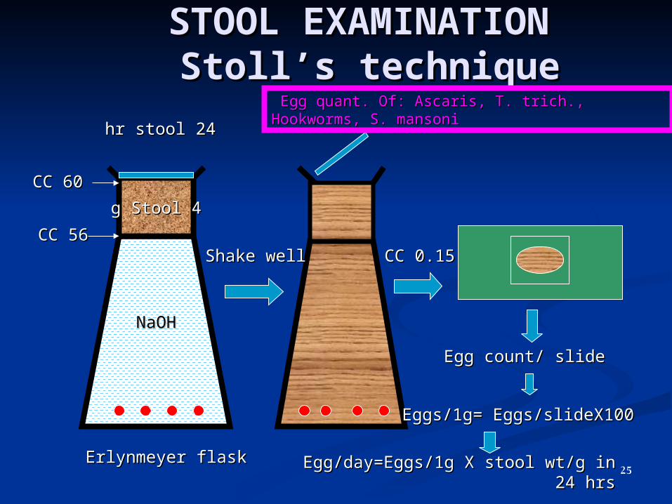
STOOL EXAMINATION STOOL EXAMINATION StollStoll’’s techniques technique
NaOHNaOH
44 g Stoolg Stool
Erlynmeyer flaskErlynmeyer flask
5656 CCCC
6060 CCCC
Shake wellShake well 0.150.15 CCCC
Egg count/ slideEgg count/ slide
Eggs/1g= Eggs/slideX100Eggs/1g= Eggs/slideX100
Egg/day=Eggs/1g X stool wt/g in 24 Egg/day=Eggs/1g X stool wt/g in 24 hrshrs
2424 hr stoolhr stool
Egg quant. Of: Ascaris, T. trich., Hookworms, S. Egg quant. Of: Ascaris, T. trich., Hookworms, S. mansonimansoni
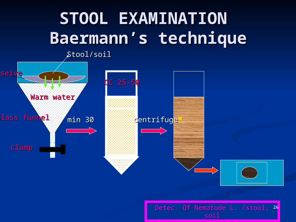
STOOL EXAMINATION STOOL EXAMINATION BaermannBaermann’’s techniques technique
WarmWarm waterwater
Stool/soilStool/soil
seiveseive
Glass funnelGlass funnel
clampclamp
3030 minmin
25-5025-50 CCCC
centrifugecentrifuge
Detec. Of Nematode L. /stool, soilDetec. Of Nematode L. /stool, soil26
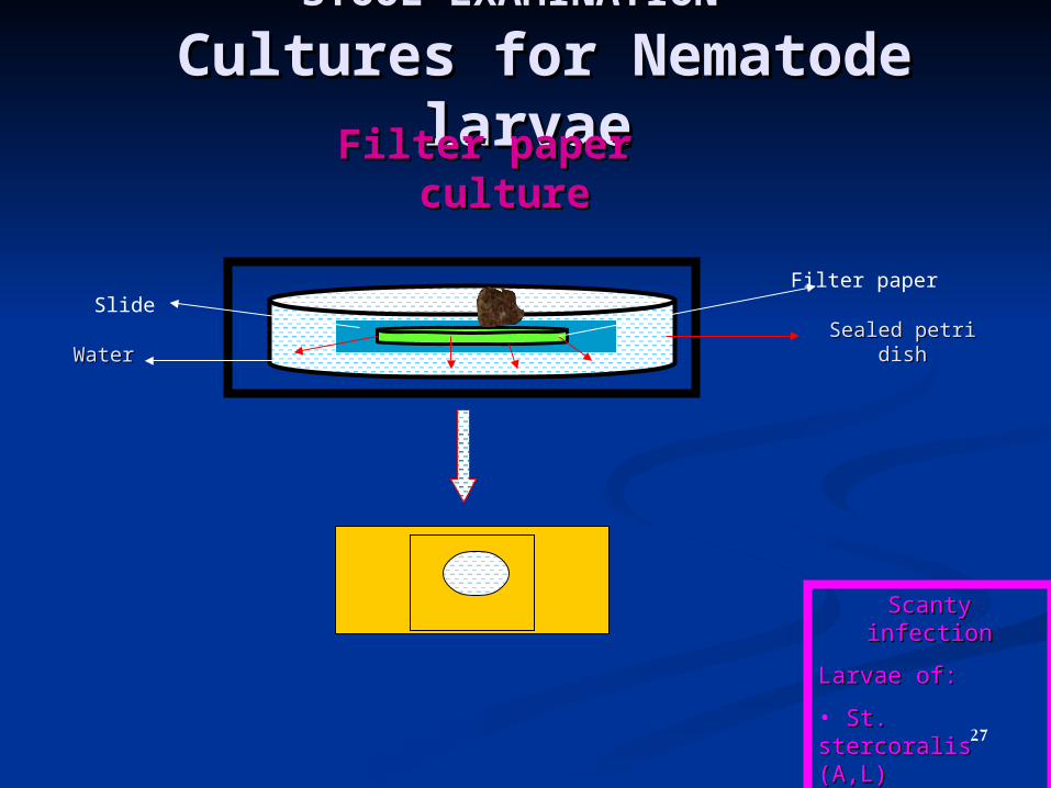
STOOL EXAMINATIONSTOOL EXAMINATION Cultures for Nematode Cultures for Nematode
larvaelarvaeFilter paper Filter paper cultureculture
Scanty infectionScanty infection
Larvae of:Larvae of:
• St. stercoralis St. stercoralis (A,L)(A,L)
• HookwormsHookworms
• TrichostrongTrichostrong
WaterWaterSealed petri Sealed petri
dishdish
Filter paperSlide
27

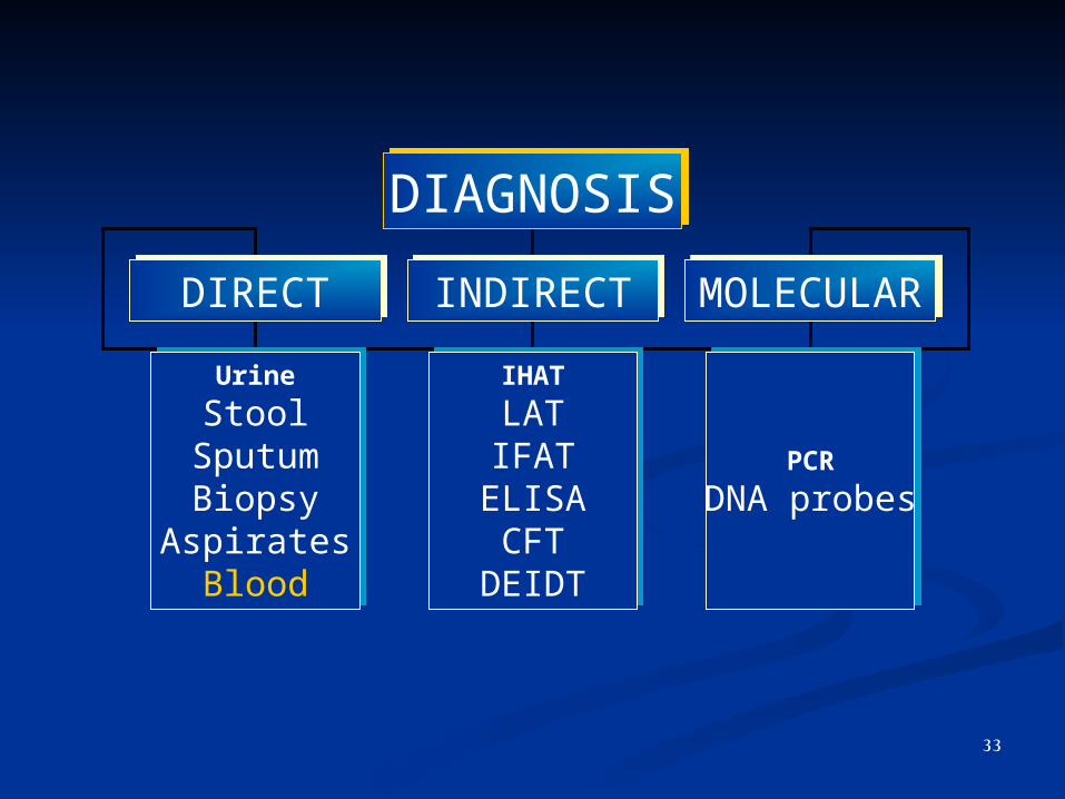
DIAGNOSISDIAGNOSIS
DIRECTDIRECT INDIRECTINDIRECT MOLECULARMOLECULAR
Urine
StoolSputumBiopsy
AspiratesBlood
Urine
StoolSputumBiopsy
AspiratesBlood
PCR
DNA probesPCR
DNA probes
IHAT
LATIFAT
ELISACFT
DEIDT
IHAT
LATIFAT
ELISACFT
DEIDT
28
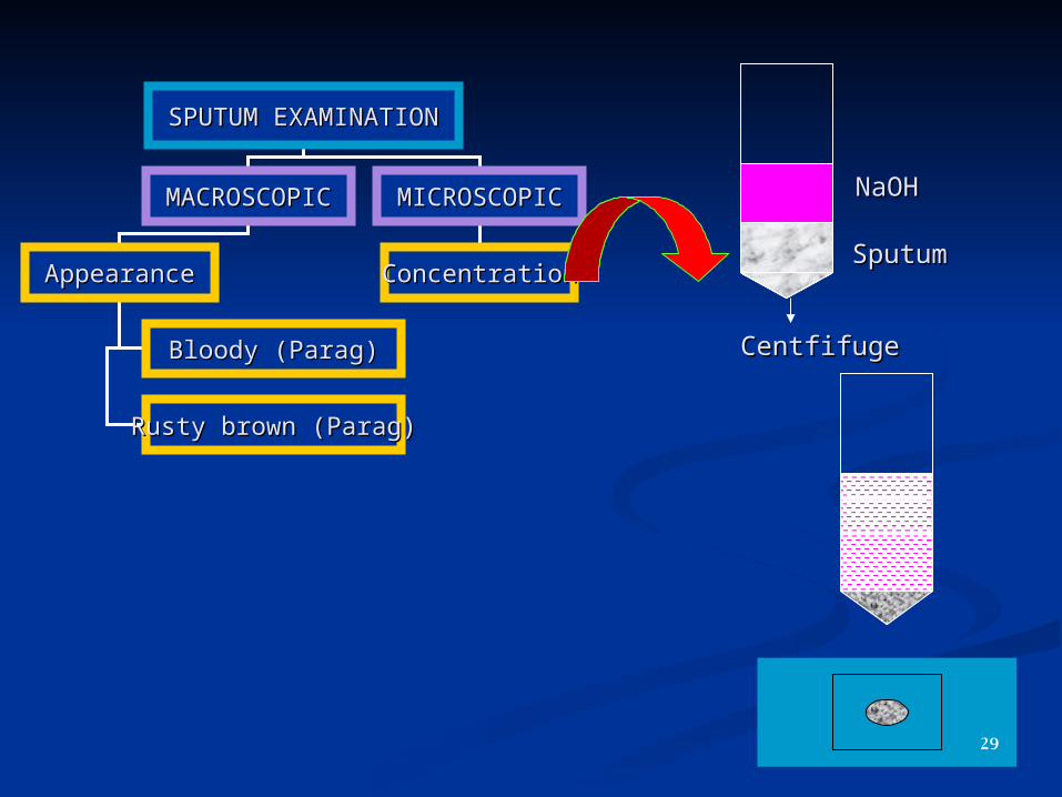
SPUTUM EXAMINATIONSPUTUM EXAMINATION
MACROSCOPICMACROSCOPIC MICROSCOPICMICROSCOPIC
AppearanceAppearance ConcentrationConcentration
Bloody (Parag)Bloody (Parag)
Rusty brown (Parag)Rusty brown (Parag)
NaOHNaOH
SputumSputum
CentfifugeCentfifuge
29
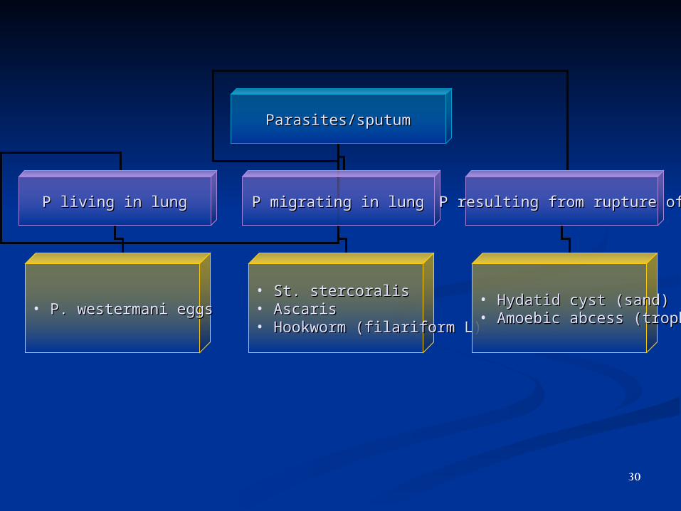
Parasites/sputumParasites/sputum
P living in lungP living in lung P migrating in lungP migrating in lung P resulting from rupture ofP resulting from rupture of
• P. westermani eggsP. westermani eggs• St. stercoralisSt. stercoralis• AscarisAscaris• Hookworm (filariform L)Hookworm (filariform L)
• Hydatid cyst (sand)Hydatid cyst (sand)• Amoebic abcess (troph)Amoebic abcess (troph)
30
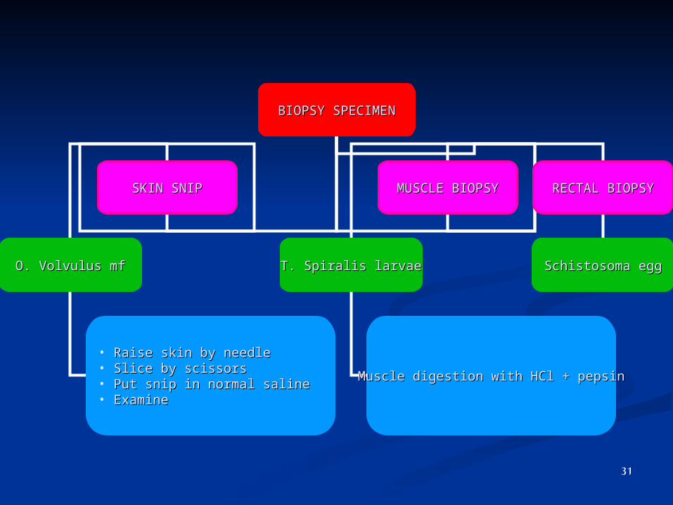
BIOPSY SPECIMENBIOPSY SPECIMEN
SKIN SNIPSKIN SNIP MUSCLE BIOPSYMUSCLE BIOPSY RECTAL BIOPSYRECTAL BIOPSY
O. Volvulus mfO. Volvulus mf T. Spiralis larvaeT. Spiralis larvae Schistosoma eggSchistosoma egg
• Raise skin by needleRaise skin by needle• Slice by scissorsSlice by scissors• Put snip in normal salinePut snip in normal saline• ExamineExamine
Muscle digestion with HCl + pepsinMuscle digestion with HCl + pepsin
31
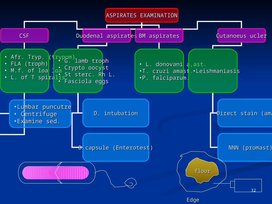
ASPIRATES EXAMINATIONASPIRATES EXAMINATION
CSFCSF Duodenal aspiratesDuodenal aspirates BM aspiratesBM aspirates Cutanoeus uclerCutanoeus ucler
• Afr. Tryp. (trypom)Afr. Tryp. (trypom)• FLA (troph)FLA (troph)• M.f. of loa loaM.f. of loa loa• L. of T spiralisL. of T spiralis
• G. lamb trophG. lamb troph• Crypto oocystCrypto oocyst• St sterc. Rh L.St sterc. Rh L.• Fasciola eggsFasciola eggs
• L. donovani a,ast.L. donovani a,ast.•T. cruzi amast.T. cruzi amast.•P. falciparum.P. falciparum.
•LeishmaniasisLeishmaniasis
D. intubationD. intubation
D capsule (Enterotest)D capsule (Enterotest)
Direct stain (amast)Direct stain (amast)
NNN (promast)NNN (promast)
•Lumbar puncutreLumbar puncutre• CentrifugeCentrifuge•Examine sed.Examine sed.
floorfloor
EdgeEdge32
DIAGNOSISDIAGNOSIS
DIRECTDIRECT INDIRECTINDIRECT MOLECULARMOLECULAR
Urine
StoolSputumBiopsy
AspiratesBlood
Urine
StoolSputumBiopsy
AspiratesBlood
PCR
DNA probesPCR
DNA probes
IHAT
LATIFAT
ELISACFT
DEIDT
IHAT
LATIFAT
ELISACFT
DEIDT
33
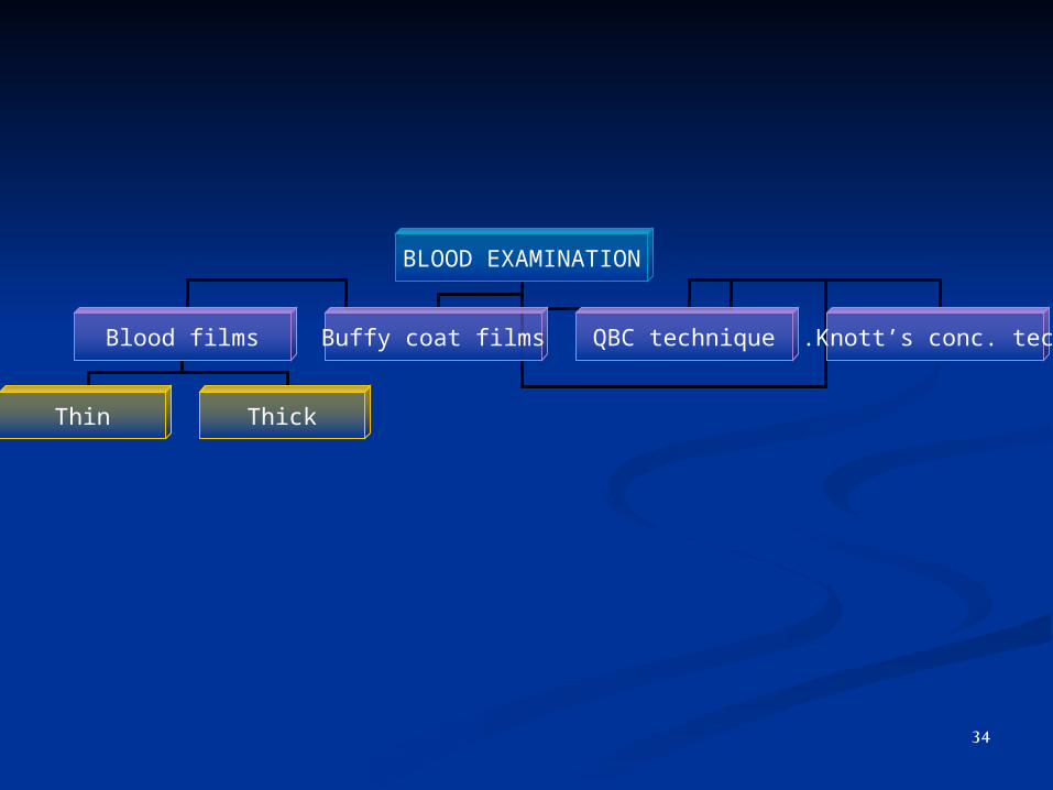
BLOOD EXAMINATION
Blood films Buffy coat films QBC technique Knott’s conc. tech.
Thin Thick
34
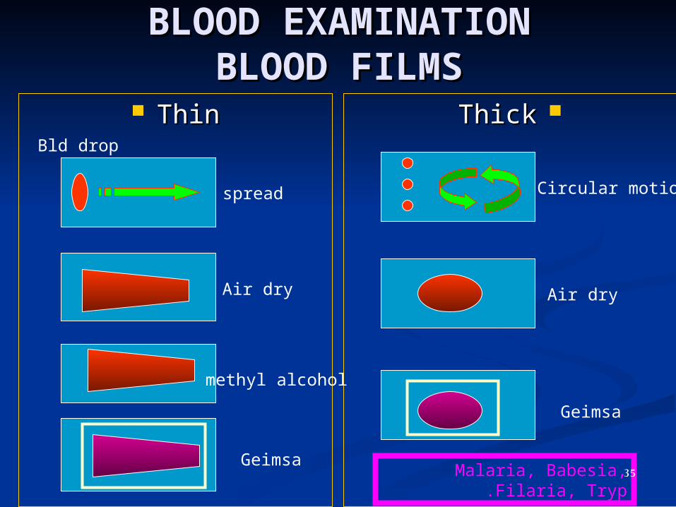
BLOOD EXAMINATIONBLOOD EXAMINATIONBLOOD FILMSBLOOD FILMS
ThinThin ThickThickBld drop
spread
Air dry
methyl alcohol
Geimsa
Air dry
Geimsa
Circular motion
Malaria, Babesia, Filaria, Tryp. 35
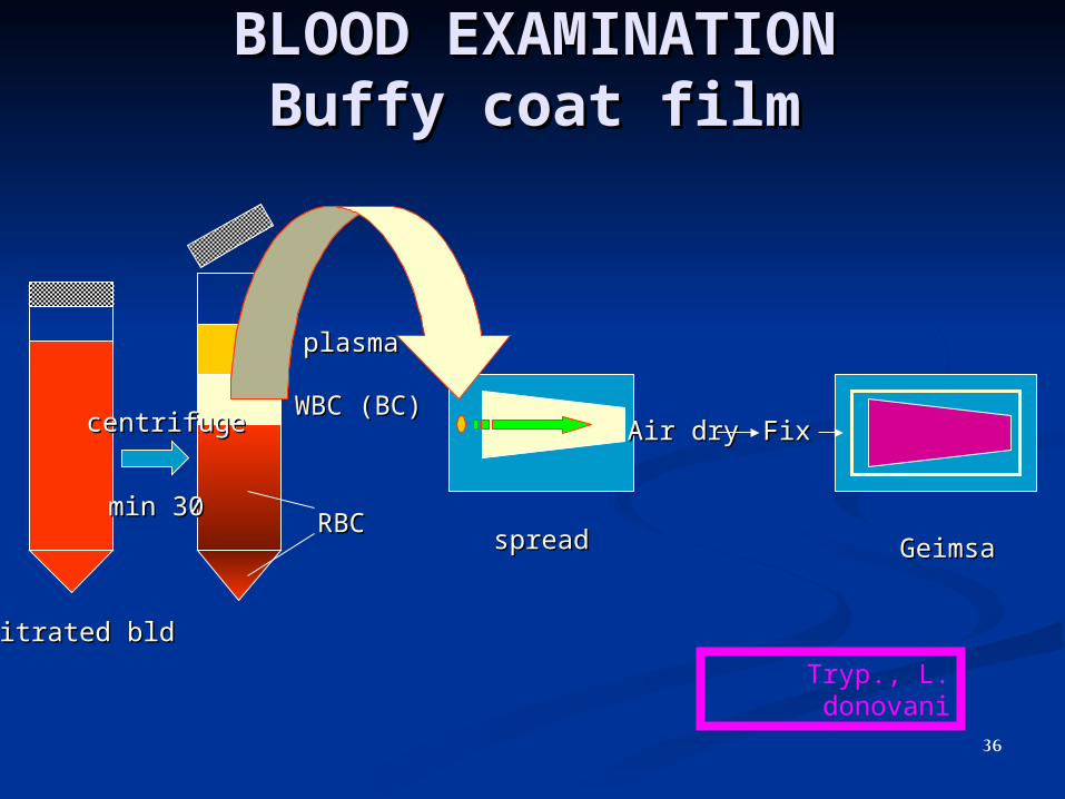
BLOOD EXAMINATIONBLOOD EXAMINATIONBuffy coat filmBuffy coat film
centrifugecentrifuge
RBCRBC
WBC (BC)WBC (BC)
plasmaplasma
Citrated bldCitrated bld
3030 minmin
Air dryAir dry FixFix
spreadspread GeimsaGeimsa
Tryp., L. donovani
36
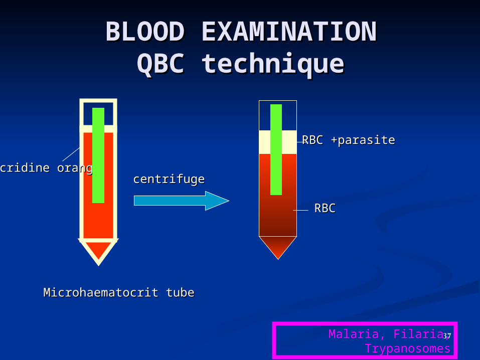
BLOOD EXAMINATIONBLOOD EXAMINATIONQBC techniqueQBC technique
centrifugecentrifuge
RBCRBC
RBC +parasiteRBC +parasite
Microhaematocrit tubeMicrohaematocrit tube
Acridine orangeAcridine orange
Malaria, Filaria, Trypanosomes37

BLOOD EXAMINATIONBLOOD EXAMINATIONKNOTTKNOTT’’S CONC. TECHNIQUES CONC. TECHNIQUE
1010 mlml
11 mlml
Air dryAir dry fixfix GeimsaGeimsa
Citrated Citrated bldbld
Formalin 2Formalin 2% % sedimentsediment
22 minmin
centrifugecentrifuge
Filaria38
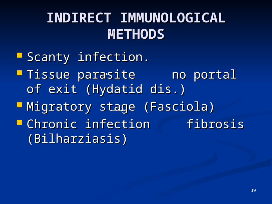
INDIRECT IMMUNOLOGICAL INDIRECT IMMUNOLOGICAL METHODSMETHODS
Scanty infection.Scanty infection. Tissue parasite no portal of exit Tissue parasite no portal of exit
(Hydatid dis.)(Hydatid dis.) Migratory stage (Fasciola)Migratory stage (Fasciola) Chronic infection fibrosis Chronic infection fibrosis
(Bilharziasis)(Bilharziasis)
39

INDIRECT IMMUNOLOGICAL METHODSINDIRECT IMMUNOLOGICAL METHODS
Antigen detection Antibody detection
• More specific More specific • More accurate.More accurate.• Active infectionActive infection• EarlyEarly• QuantitativeQuantitative
Ab remain in serum forAb remain in serum for months even after curemonths even after cure
40
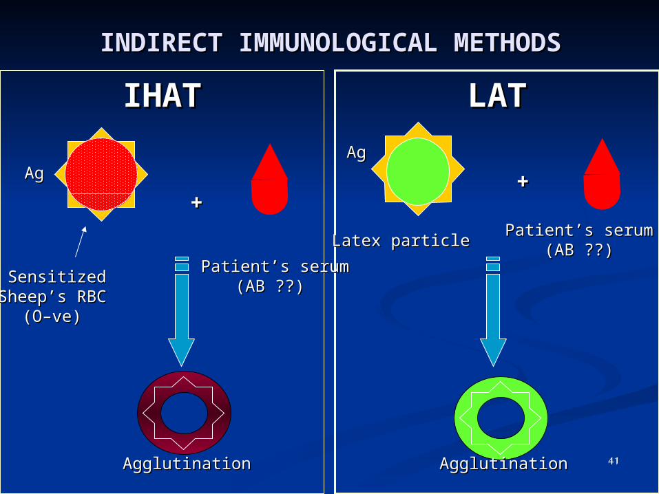
INDIRECT IMMUNOLOGICAL INDIRECT IMMUNOLOGICAL METHODSMETHODS
IHATIHAT LATLAT
++
SensitizedSensitized SheepSheep’’s RBCs RBC
((OO––veve))
AgAg
PatientPatient’’s serums serum ??( ??(ABAB))
AgglutinationAgglutination
++
AgglutinationAgglutination
AgAg
Latex particleLatex particlePatientPatient’’s serums serum
??( ??(ABAB))
41
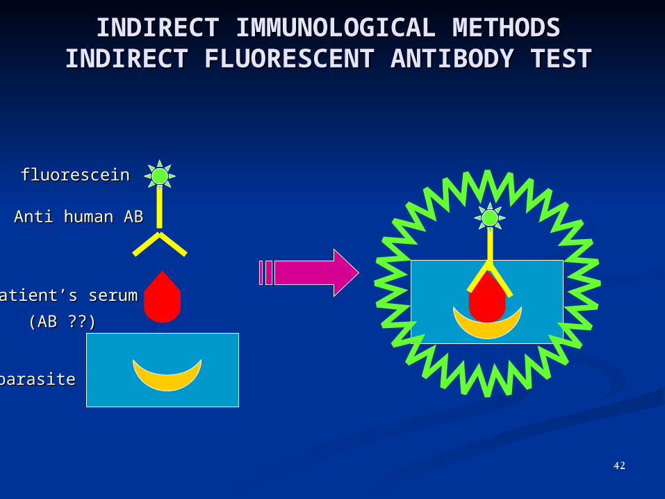
INDIRECT IMMUNOLOGICAL INDIRECT IMMUNOLOGICAL METHODSMETHODS
INDIRECT FLUORESCENT ANTIBODY INDIRECT FLUORESCENT ANTIBODY TESTTEST
parasiteparasite
PatientPatient’’s serums serum
??( ??(ABAB))
Anti human ABAnti human AB
fluoresceinfluorescein
42

INDIRECT IMMUNOLOGICAL INDIRECT IMMUNOLOGICAL METHODSMETHODS
ELISAELISA
OPDOPD
OPDOPD
Flat bottom plastic micrititre plateFlat bottom plastic micrititre plate
AgAg
PatientPatient’’s serums serum ??( ??(ABAB))
Anti human ABAnti human AB
Peroxidase EPeroxidase E
ABAB
43

INDIRECT IMMUNOLOGICAL INDIRECT IMMUNOLOGICAL METHODSMETHODS
CFTCFT
AgAg
PatientPatient’’s serums serum ??( ??(ABAB))
complement
Anti sheep ABAnti sheep AB
Sheep’s RBC
-ve Ab
+
ve Ab
haemolysis
No Sheep
RBChaemolysis
Tube / Tube / microplatemicroplate
ABAB
44
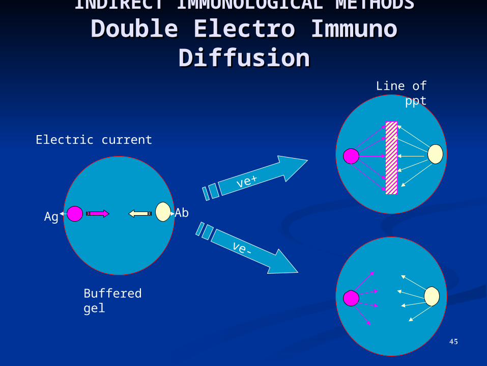
INDIRECT IMMUNOLOGICAL INDIRECT IMMUNOLOGICAL METHODSMETHODS
Double Electro Immuno Double Electro Immuno DiffusionDiffusion
+ve
-ve
Ag Ab
Buffered gel
Electric current
Line of ppt
45

INDIRECT IMMUNOLOGICAL INDIRECT IMMUNOLOGICAL METHODSMETHODS
Immunodiagnostic Strip Test (Dip Stick Test) Immunodiagnostic Strip Test (Dip Stick Test) Ag Ag
Nitrocellulose strip
Monoclonal Ab
Coloured dye
Pt bld (?Ag)
+ve
-ve
Malaria, Filaria, African tryp.
46
DIAGNOSISDIAGNOSIS
DIRECTDIRECT INDIRECTINDIRECT MOLECULARMOLECULAR
Urine
StoolSputumBiopsyBlood
Aspirates
Urine
StoolSputumBiopsyBlood
Aspirates
PCR
DNA probesPCR
DNA probes
IHAT
LATIFAT
ELISACFT
DEIDT
IHAT
LATIFAT
ELISACFT
DEIDT
47
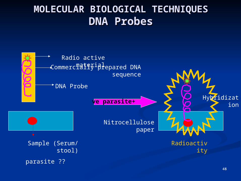
MOLECULAR BIOLOGICAL MOLECULAR BIOLOGICAL TECHNIQUESTECHNIQUESDNA ProbesDNA Probes
DNA Probe
Commercially prepared DNA sequence
Radio active material
Nitrocellulose paper
Sample (Serum/ stool)
??parasite
+ve parasiteHybridizatio
n
Radioactivity
48
MOLECULAR BIOLOGICAL MOLECULAR BIOLOGICAL TECHNIQUESTECHNIQUES
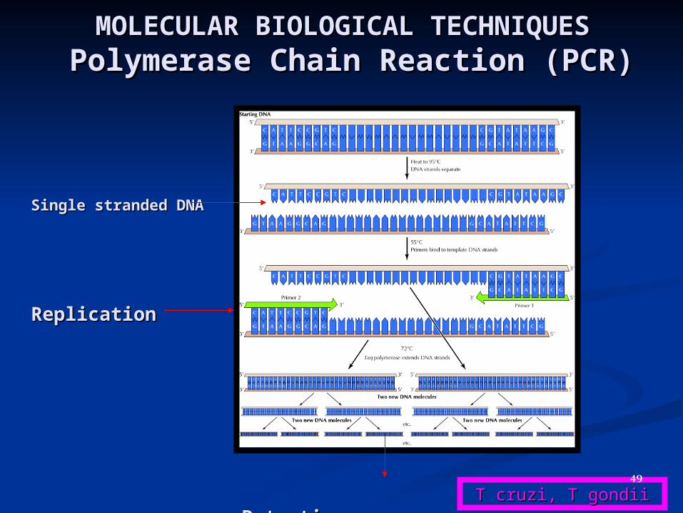
Polymerase Chain Reaction (PCR) Polymerase Chain Reaction (PCR)
Single stranded DNA Single stranded DNA
ReplicationReplication
DetectionDetection T cruzi, T gondiiT cruzi, T gondii49
10 X Objective
50
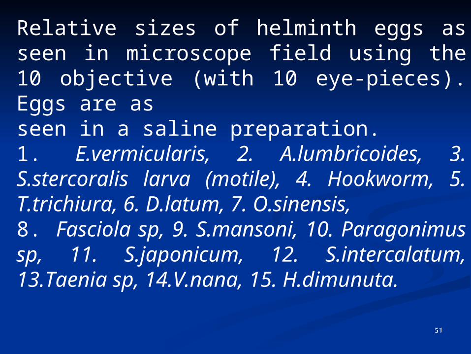
Relative sizes of helminth eggs as seen in microscope field using the 10 objective (with 10 eye-pieces). Eggs are asseen in a saline preparation.1. E.vermicularis, 2. A.lumbricoides, 3. S.stercoralis larva (motile), 4. Hookworm, 5. T.trichiura, 6. D.latum, 7. O.sinensis,8. Fasciola sp, 9. S.mansoni, 10. Paragonimus sp, 11. S.japonicum, 12. S.intercalatum, 13.Taenia sp, 14.V.nana, 15. H.dimunuta.
51
40 X Objective
52
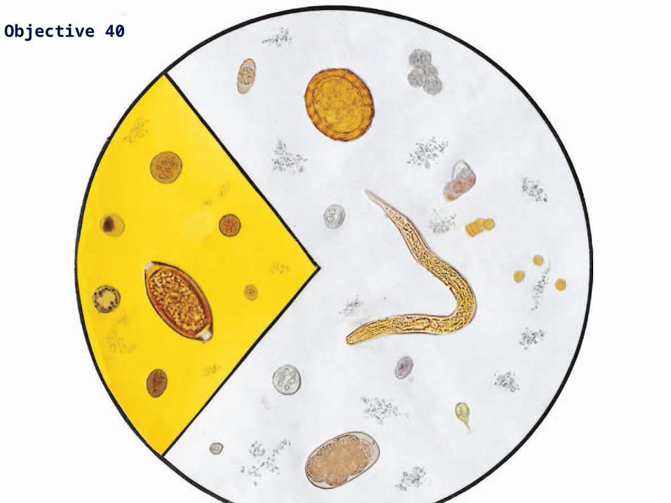
Relative sizes of trophozoites and cysts of intestinal protozoa, common nematode eggs and larva of Strongyloides as seen in microscope field using the 40 objective (with 10 eyepieces).1. I.belli oocyst, 2. A lumbricoides egg, 3. Leucocytes, 4. E.histolytica/E.dispar cyst, 5. E.histolytica trophozoite (motile),6. Red cells, 7. S.stercoralis larva (motile), 8. E.coli cyst (mature), 9. G.lamblia cyst, 10. C.mesnili cyst, 11. Hookwormegg, 12. G.lamblia trophozoite (motile).Iodine preparation: 13. E.coli cyst, 14. I.buetschlii cyst, 15. E.histolytica/E.dispar cyst, 16. V.nana cyst, 17. T.trichiura egg,18. Blastocystis hominis, 19. G.lamblia cyst.
53
Non-parasitic structures found Non-parasitic structures found in faeces: Carein faeces: Care
must be taken not to report as parasites those structures must be taken not to report as parasites those structures that can be normally found in faeces such as:that can be normally found in faeces such as:
muscle fibres, vegetable fibres, starch cells (stain muscle fibres, vegetable fibres, starch cells (stain blue-black blue-black with iodine), pollen grains, fatty acid crystals, soaps, spores, with iodine), pollen grains, fatty acid crystals, soaps, spores, yeasts, and hairs .yeasts, and hairs .
Large numbers of fat globules may be seen in faeces when Large numbers of fat globules may be seen in faeces when there is malabsorption. there is malabsorption.
Charcot Leyden crystals (breakdown products of eosinophils) Charcot Leyden crystals (breakdown products of eosinophils) can sometimes be seen in faeces (also in sputum) in parasitic can sometimes be seen in faeces (also in sputum) in parasitic infections. They appear as slender crystals with pointed infections. They appear as slender crystals with pointed ends, about 30–40m in lengthends, about 30–40m in length
54

Structures found in faeces that Structures found in faeces that required differentiation from required differentiation from
parasitesparasites..
Structures found in faeces that required differentiation from parasites. 55
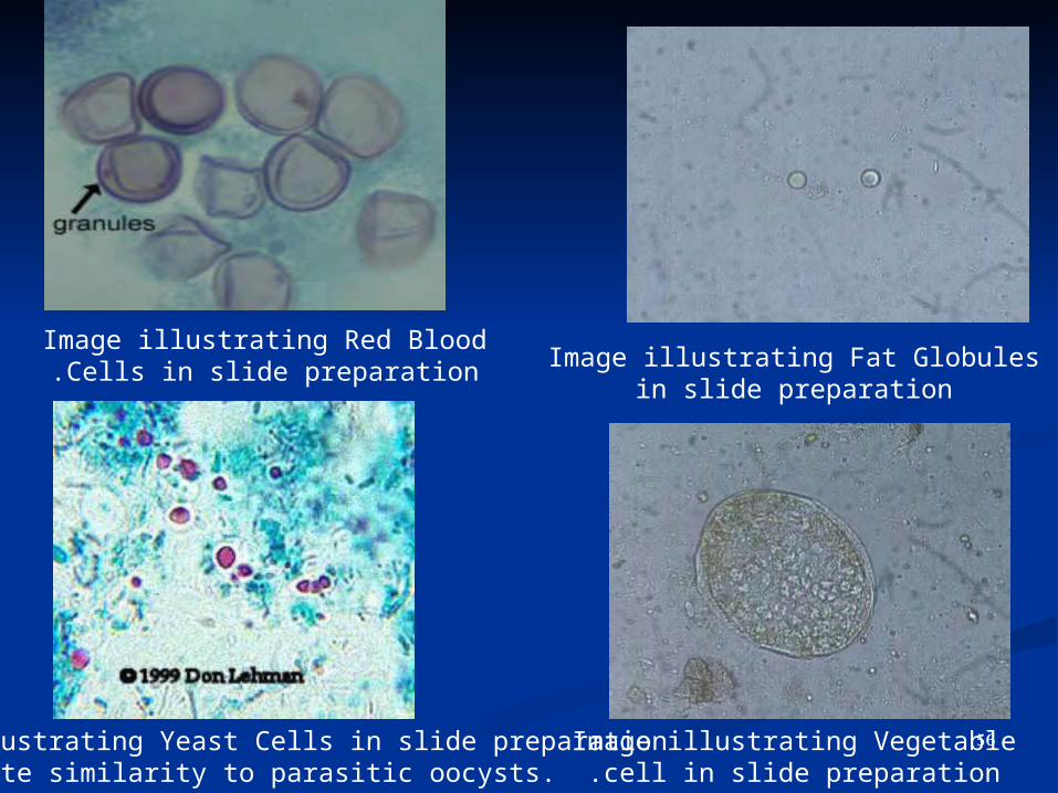
Image illustrating Yeast Cells in slide preparationNote similarity to parasitic oocysts.
Image illustrating Vegetable cell in slide preparation.
Image illustrating Red Blood Cells in slide preparation. Image illustrating Fat Globules in
slide preparation
56
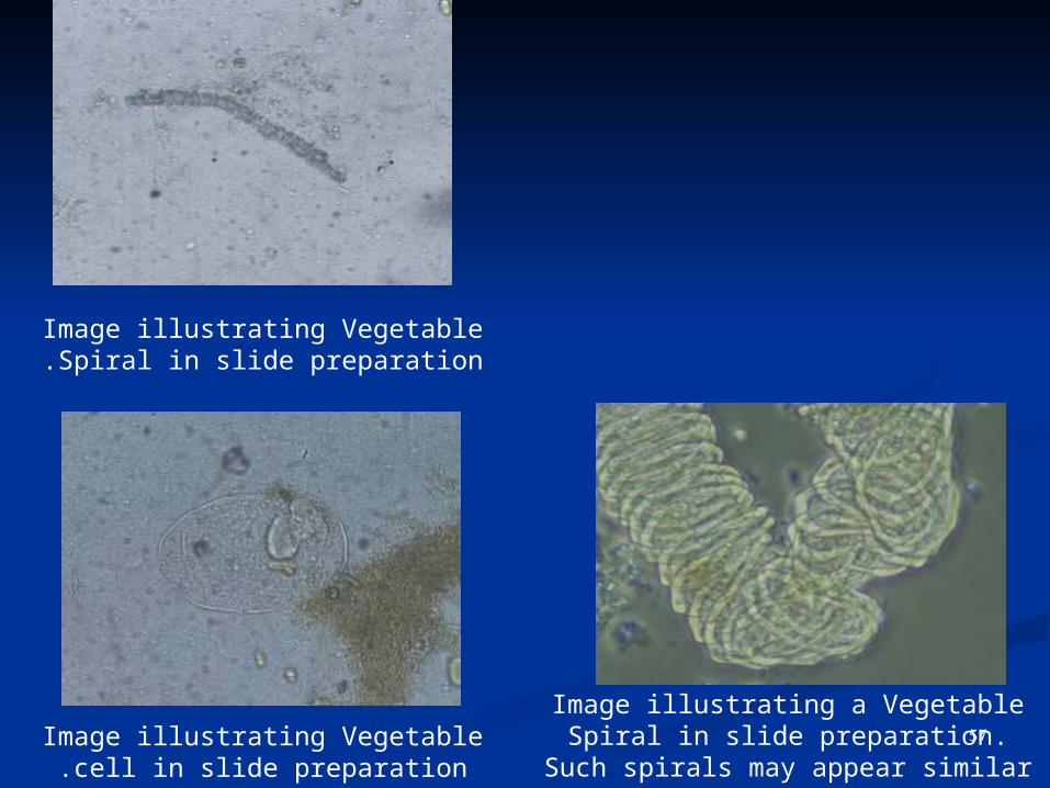
Image illustrating Vegetable cell in slide preparation.
Image illustrating a Vegetable Spiral in slide preparation. Such spirals may appear similar to proglottids.
Image illustrating Vegetable Spiral in slide preparation.
57
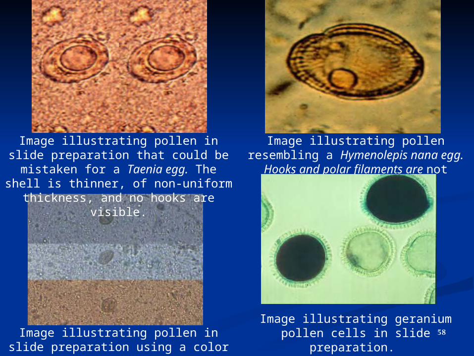
Image illustrating pollen in slide preparation using a color filter
Image illustrating pollen in slide preparation that could be mistaken
for a Taenia egg. The shell is thinner, of non-uniform thickness, and no
hooks are visible.
Image illustrating pollen resembling a Hymenolepis nana egg. Hooks and
polar filaments are not visible.
Image illustrating geranium pollen cells in slide preparation.
58
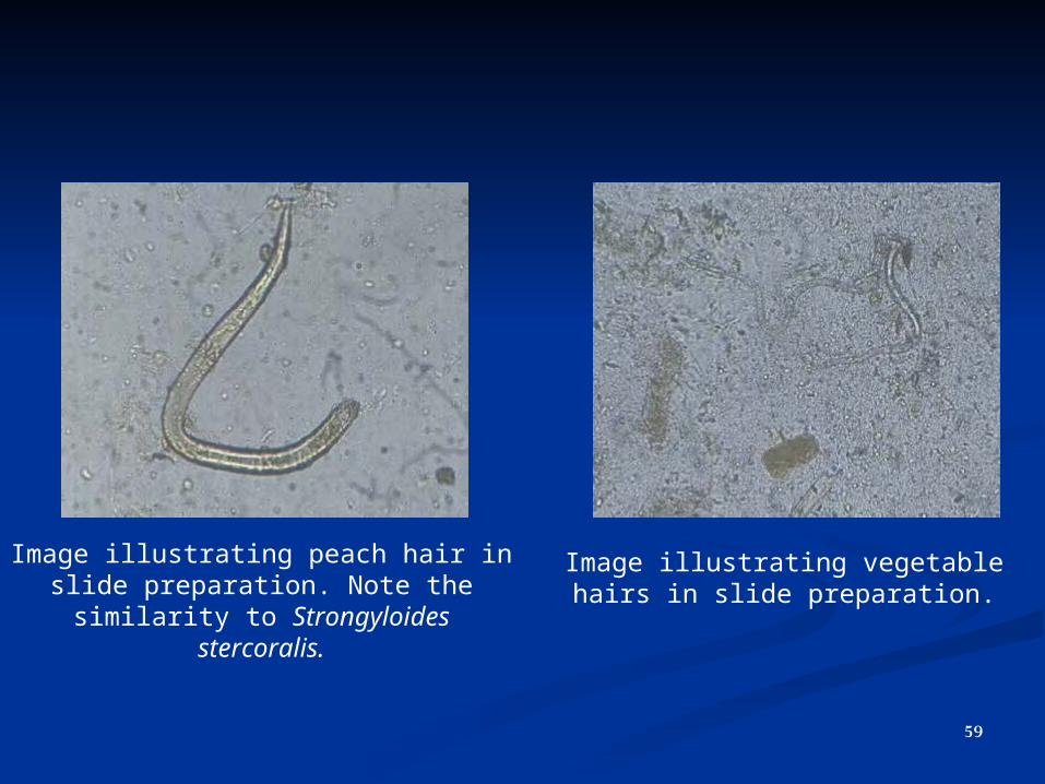
Image illustrating peach hair in slide preparation. Note the similarity to
Strongyloides stercoralis.
Image illustrating vegetable hairs in slide preparation.
59