kinetics of tissue rrr-α-tocopherol depletion and repletion. effect of cold exposure
TRANSCRIPT

Kinetics of tissue RRR-cx-tocopherol depletion and repletion. Effect of cold exposure
Willy A. Behrens and RenC Mad&e
Bureau of Nutritional Sciences, Food Directorate, Health Protection Branch, Health and Welfare Canada, Tunney’s Pasture, Ottawa, Ontario, Canada KlA OL2
Vitamin E was estimated in plasma and tissues ofrats kept for three months on a low vitamin E diet or
a high vitamin E diet. Some of the animals from each group were switched to the opposite diet, and the
kinetics of uptake and depletion of vitamin E were followed 3, 8, and 15 days ufter the diet change.
Some rats tisere also submitted to cold exposure (6°C) for three days. During repletion plasma, red
blood cells, liver, spleen, und adrenul gland were the only tissues that responded rapidly to the diet
change; after three days, their vitumin E levels corresponded to that of the new diet. Heart, brain, lung, muscle, and thymus M’ere slow in reacting to diet change. Fifteen days after the change in diet, white
adipose tissue did not respond. The rate of repletion for all tissues was more rapid than the rate oj
depletion, but liver was the only tissue that after three days had litumin E lelsels corresponding to the
low-vitamin diet. Cold exposure for three days did not produce any sign&ant change in the vitamin E
content of any tissue, indicating that despite high oxygen consumption by the animal, vitumin E was
not consumed or mobilized.
Keywords: Vitamin E; d-a-tocopherol; cold exposure; deplenon; repletion
Introduction
It is known that different tissues have different kinet- ics of cw-tocopherol depletion and repletion. Draper and Csallany’ reported that in rats and rabbits fed a vitamin E deficient diet, liver a-tocopherol decreased rapidly for several weeks, then reached a plateau, whereas the muscle concentration of a-tocopherol de- clined much more slowly. Studies of vitamin E ac- cumulation in the body have generally been restricted to one tissue or have considered only one or two . .
2*3 Two more extensive studies have ~~~t~~%otI!~~~.4*S The first one studied effects of the addition of low levels of vitamin E to a diet deficient in vitamin E over a 25-week period in rats.4 The second one studied the effect of long-term administration of high levels of vitamin E.’ However, the rates of deple- tion and repletion were measured at different ages in the two studies. Both investigations used an interval of
Bureau of Nutritional Sciences Publication No. 319. Address reprint requests to Dr. Willy A. Behrens, Bureau of Nutri- tional Sciences, Food Directorate, Health Protection Branch, Bant- ing Research Centre, Tunney’s Pasture, Ottawa, Ontario, Canada KlA 0L2. Received March 6, 1990; accepted May 8, 1990.
one or more weeks. In the experiments described in this paper, measurements were taken 3, 8, and 15 days after starting depletion or repletion. In addition, kinet- ics of a-tocopherol depletion and repletion were esti- mated in animals at the same age by using rats fed a low- or a high-vitamin E diet and then switching diets in these two groups. This study was also extended to some important tissues, such as adrenal gland, spleen, and thymus, that were not included in previous stud- ies.4,5
A secondary aim of this experiment was to deter- mine if the tissue uptake and depletion of a-tocopherol could be modified by subjecting the animal to an in- creased oxidative stimulus. Instead of using toxic drugs such as CCL or an iron-overload, the animal was submitted to a cold exposure in a chamber at 6°C for three days. It is well known that in this condition, the animal maintains its body temperature by “shivering thermogenesis” in which its oxygen consumption rate is increased. This increase could be up to a maximum value of 5 times the basal metabolic rate, depending on the species and the experimental conditions.6,7 The rationale was that under this physiological stimulus, the chance to produce more “free radicals” in vivo increases markedly; therefore, a substantial catabo- lism or use of vitamin E could potentially be observed.
528 J. Nutr. Biochem., 1990, vol. 1, October 0 1990 Buttetworth-Heinemann
Tissue RRR-e-tocopherol depletion and repletion: Behrens and Madere
An increase in the rate of depletion of ~-tocopherol and a decrease in the rate of uptake of the vitamin were expected.
Materials and methods
Animals and diets
Thirty-nine male Wistar rats (Charles River Canada, St. Constant, Quebec) weighing 100 g + 10% were used in all the experiments and for three months were fed a modified AIN-76 diet 8 as follows. The diet for the first group (18 rats) (Group LE) contained the AIN-76 diet in which vitamin E was omitted from the vitamin mixture. In the second group (21 rats) (Group HE), d- a-tocopherol acetate (RRR-~-tocopherol-acetate; ICN Nutritional Biochemicals, Cleveland, OH) was added to the vitamin mixture to give 1.0 g d-c~-tocopherol acetate (1360 IU)/kg diet. Twelve rats from each LE and HE groups were switched to the opposite diet and fed the new diet for 3, 8, or 15 days. In addition, three of these rats from each group also were transferred to a cold room (6°C +_ I°C) for three days.
Blood and tissue collection
Blood and tissues were obtained from animals anes- thetized with halothane (2% in oxygen) (Fluothane, Ayerst Laboratory, Montreal). Plasma was obtained from heparinized blood after centrifugation at 3000 x g. Red blood cells (RBC) were obtained after washing the pellet with 0.9% cold saline followed by centrifuga- tion.
Analytical methods
Alpha- and ~-tocopherols (vitamin E) were determined in plasma and tissues with the high performance liquid chromatography (HPLC) method of Thompson and Hatina 9 as previously described.t°
Statistical analysis
Student's t-test zz was used for statistical evaluation of the results.
Results
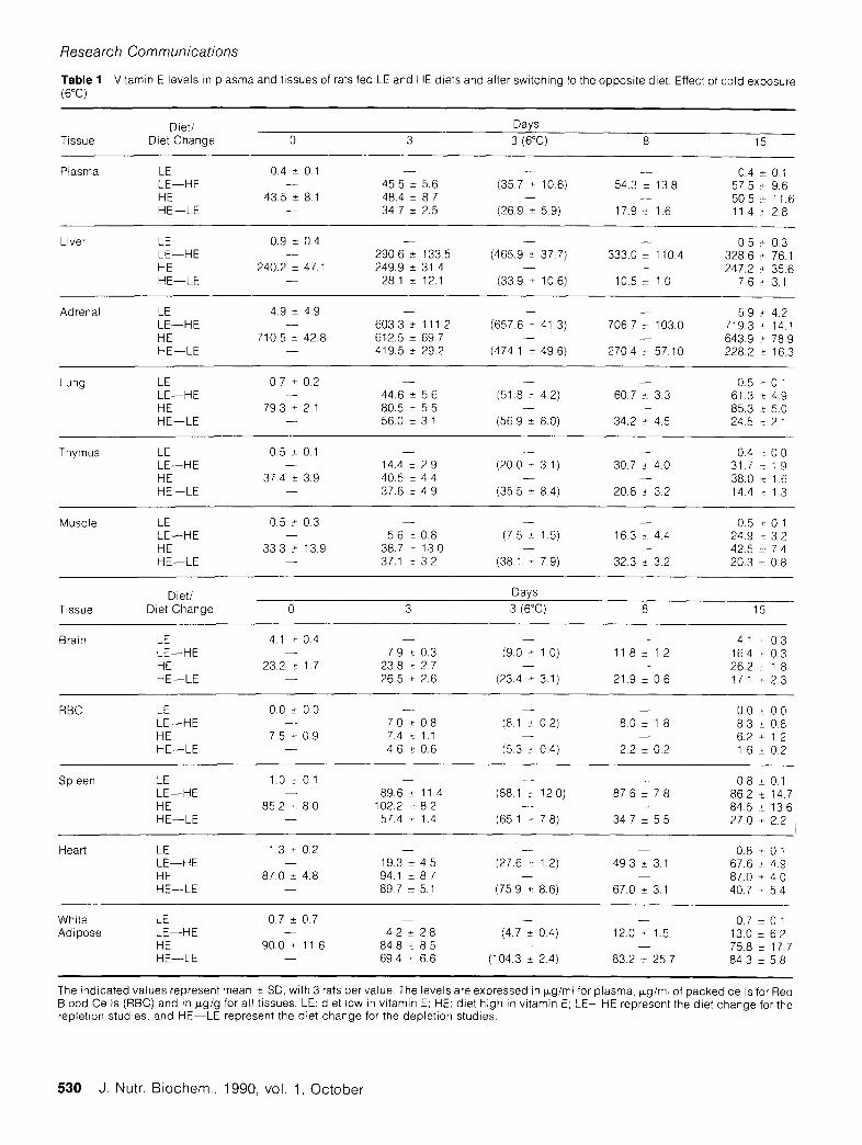
Table I shows vitamin E levels in plasma and tissues of rats fed LE and HE diets for 3 months, and after they were switched to the opposite diet. The effect of cold exposure at 6°C for three days is also included. As expected, very low levels of vitamin E were observed in all tissues from rats fed the LE diet; of the studied tissues, only adrenal gland and brain retained more than 4.0 p~g/g of tissue. Adrenal gland was the tissue that accumulated the most vitamin E in rats fed the HE diet; second in this regard was liver. At the other ex- treme, red blood cell was the rat tissue that retained the least vitamin E.
Changing animals to the opposite diet produced in each tissue a different result. Rates of repletion and depletion were calculated for the different time inter-
vals and are shown in Table 2. Adrenal gland and liver, followed closely by spleen, were the tissues that ac- cumulated c~-tocopherol at a faster rate than any of the other tissues. After only three days on the HE diet, these organs had vitamin E levels comparable to those from rats fed the HE diet for 3 months. They may have reached these levels even earlier. The same tissues were also the ones that lost ~x-tocopherol the fastest. Other tissues maintained relatively unchanged rates of repletion and depletion during the intervals of 3 to 8 and 8 to 15 days. In general, in all tissues, rates of repletion were higher than rates of depletion, the ex- ception being white adipose tissue in the interval 0 to 3 days, but there was a great variability in this tissue (Table 1). It seems that liver, as indicated in Table 2, is the only tissue that accumulates and loses vitamin E at a similar rate. The rate of repletion in lung was approx- imately twice that of depletion during the first 8 days of the experiment (Figure 1). Plasma had a relatively high repletion rate but a very slow depletion rate. All the other tissues examined had slow repletion and deple- tion rates. Muscle, white adipose tissue, and brain showed a very small change or no change at all, even after 15 days of diet change.
Cold exposure at 6°C for three days failed to de- crease the rate of repletion and to accelerate the rate of depletion in any significant way in any of the tissues investigated (Tables 1 and 2). Figure 1 shows this graphically for lung, the tissue in which the high oxy- gen uptake during cold exposure takes place first. In general, cold exposure produced higher levels of vita- min E than in the corresponding control tissues at three days. This was translated into a higher rate of repletion (Table 2), which was the inverse of what was expected. The only exception was plasma in which the rate of repletion was slightly smaller but not sig- nificantly different than that at 3 days at room tem- perature. Table 2 shows that rate of depletion was smaller or did not change with the cold exposure in comparison with the rate of depletion at room temper- ature. Once again, the exception was plasma, which was the only site where the rate of depletion under cold exposure was higher than the control at room temperature.
Discussion
In the present study, tocopherol apparently reached saturation levels after rats were fed the HE diet (1,360 IU/kg) for three months, because no further increases were observed after an extra 15 days. In other experi- ments in which higher doses of dietary vitamin E were given (10,000 IU/kg), tocopherol levels continued to increase in all tissues examined for the duration of the supplementation. 5 Also, Yang and Desai reported that there was a continued increase in both liver and plasma after 8 and 16 months of feeding high levels of vitamin E. 12 These observations suggest that satura- tion of tissues with tocopherol depends on the level in the diet.
Adrenal glands and liver followed closely by spleen
J. Nutr. Biochem., 1990, vol. 1, October 529
Research Communications
T a b l e 1 V i tamin E leve ls in p l asma and t issues of rats fed LE and HE diets and af ter sw i t ch ing to the oppos i t e diet. Effect of co ld exposu re (6°C)
Die t / Days
T issue Diet C h a n g e 0 3 3 (6°C) 8 15
P lasma LE 0.4 ± 0.1 - - - - - - 0.4 ± 0.1 L E - - H E - - 45.5 _+ 5.6 (35.7 _+ 10.6) 54.3 ÷ 13.8 57.5 ± 9.6 HE 43.5 + 81 48.4 _+ 8.7 - - - - 50.5 ~ 11.6 H E I L E - - 34.7 +_ 2.5 (26.9 ± 5.9) 17.9 ± 1.6 1 1 4 ± 2 8
L iver LE 0.9 +- 0.4 - - - - - - 0.5 ± 0 3 L E - - H E - - 290.6 ± 1 3 3 5 (465.9 _+ 37.7) 333.0 ± 110.4 328.6 ± 76.1 HE 240.2 _+ 47.1 249.9 ± 31.4 - - - - 247.2 ± 35.6 H E I L E - - 28.1 ~ 12.1 (33.9 _+ 10.6) 10.5 ± 1.0 7.6 ± 3.1
Adrena l LE 4.9 ± 4 9 - - - - - - 5.9 ± 4.2 L E - - H E - - 603.3 ± 111.2 (657.6 + 41.3) 706.7 ± 103.0 719.3 ± 14.1 HE 710.5 ± 42.8 612.5 ± 69.7 - - - - 643.9 ± 78.9 H E - - L E - - 419.5 ± 29.2 (4741 _+ 49.6) 270.4 ~* 57.10 228.2 ~ 16.3
Lung LE 0 7 _+ 0.2 - - - - - - 0.5 _~ 0.1 L E - - H E - - 44.6 + 5 6 (51.8 ± 4.2) 60.7 ± 3.3 61.3 = 4.9 HE 79.3 ± 2.1 80.5 + 5.5 - - - - 85.3 ± 5.0 H E - - L E - - 56.0 ± 31 (56.9 _+ 8.0) 34.2 ± 4.5 24.5 _* 21
Thymus LE 0.5 + 0.1 - - - - - - 0.4 ± O0 L E - - H E - - 14.4 ± 2.9 (20.0 _+ 3.1) 3 0 7 ± 4.0 31.7 ± 1.9 HE 37.4 ± 3.9 40.5 ± 4.4 - - - - 38.0 ~ 1.6 H E - - L E - - 37.6 ± 4.9 (35.5 ~ 8.4) 20.8 ± 3.2 14.4 z 1.3
Musc le LE 0.5 ± 0.3 - - - - - - 0.5 _* 0.1 L E - - H E - - 5.6 ± 0.8 (7.5 ± 1.5) 16.3 ~ 4.4 24.9 ± 3.2 HE 33.3 ± 13.9 38.7 ± 13.0 - - - - 42.5 _+ 7.4 H E - - L E - - 37.1 ± 3.2 (38.1 ~ 7.9) 3 2 3 ± 3.2 20.3 _* 0.8
T issue
Die t / Days
Diet Change 0 3 3 (6°C) 8 15
Brain LE 4.1 _* 0.4 - - - - - - 4.1 ~ 0.3 L E - - H E - - 7.9 ± 0.3 (9.0 ± 1.0) 1 1 8 ± 1.2 16.4 _+ 0.3 HE 23.2 ~ 1.7 23.8 ± 2.7 - - - - 26.2 ± 1.8 H E - - L E - - 26.5 ± 2.6 (23.4 ~ 3.1) 21.9 + 0.6 17.1 + 2 3
RBC LE 0.0 ± 0.0 - - - - - - 0.0 ± 0.0 L E - - H E - - 7.0 ± 0.8 (81 + 0.2) 8.0 _+ 1.8 8.3 _+ 0.8 HE 7.5 ± 0.9 7.4 _* 1.1 - - - - 6.2 ± 1 2 H E - - L E - - 4.6 _* 0 6 (5.3 ± 0.4) 2.2 ± 0.2 1.6 ± 0.2
Spleen LE 1.0 ± 0.1 - - - - - - 0.8 _+ 0.1 L E - - H E - - 89.6 _+ 11.4 (88.1 ± 12.0) 87.6 ± 7.8 86.2 ± 14.7 HE 85.2 _* 8.0 102.2 ± 8.2 - - - - 84.5 ± 1 3 6 H E - - L E - - 57.4 _* 1.4 (65.1 ± 7 8 ) 34.7 + 5.5 27.0 + 2.2 ,
I
Heart LE 1.3 ± 0.2 - - - - - - 0.8 _+ 0.1 L E - - H E - - 19.3 ± 4.5 (27.6 : 1.2) 49.3 +_ 3.1 67.6 ± 4.9 HE 87.0 _+ 4.8 94.1 _+ 8.7 - - - - 87.0 _+ 4.0 H E - - L E - - 89.7 - 5.1 (75.9 ± 8.6) 67.0 ± 3.1 40.7 ± 5.4
White LE 0.7 _+ 0.7 - - - - - - 0.7 ~- 0.1 A d i p o s e L E - - H E - - 4.2 + 2.8 (4.7 ± 0.4) 12.0 + 1.5 13.0 _* 6.2
HE 90.0 + 11.6 84.8 ± 8.5 - - - - 75.8 _+ 17.7 H E - - L E - - 69.4 _+ 6.6 (104.3 _+ 2.4) 83.2 ± 25.7 84.3 ÷ 5.8
The ind ica ted va lues represen t mean ± SD, wi th 3 rats per value. The leve ls are exp ressed in p.g/ml for p lasma, Fg /m l of p a c k e d ce l l s for Red B lood Ce l ls (RBC) and in ~ g / g for al l t issues. LE: d iet l ow in v i tamin E; HE: d ie t h igh in v i tamin E; L E - - H E represen t the d ie t change for the rep le t ion studies; and H E - - L E represen t the d ie t change for the dep le t i on studies.
5 3 8 J. N u t r . B i o c h e m . , 1 9 9 0 , v o l . 1, O c t o b e r

Table 2 tissues
Repletion and depletion rates of e-tocopherol in rat
Time Intervals Tissue 0-3 Days 3-8 Days 8-15 Days
Adrenal (R) 199.5 (217.6) a 20.7 (D) 97,0 (78.8) 29.8
Liver (R) 96,6 (155.0) 8.5 (D) 70.7 (68.8) 3.5
Spleen (R) 29.5 (29.0) 0.0 (D) 9.3 (6.7) 4.5
Plasma (R) 15,0 (11.8) b 1.8 (D) 2.9 (5.5) 3.4
Lung (R) 14,6 (17.0) 12.0 (U) 7.8 (7.5) 4.4
Thymus (R) 4,6 (6.5) 3.3 (U) 0.0 (0,6) 3.4
Heart (R) 6.0 (8.8) 6,0 (U) 0.0 (3.7) 4,5
RBC (R) 2.3 (2.7) c 0.2 (D) 1.0 (0,7) 0.5
Muscle (R) 1.7 (2,3) 2.1 (D) 0.0 (0.0) 1,0
White (R) 1.2 (1.3) 1,6 Adipose (D) 6,9 (0.0) 0.0 Brain (R) 1,2 (1.6) 0.8
(D) 0.0 (0.0) 0.9
a Values expressed in i~g/g tissue per day. R: repletion; D: deple- tion. The numbers in parentheses are those corresponding to the cold exposure at 6°C for three days, b I~g/ml plasma per day. ° ~,g/ ml packed cells per day.
were the tissues that accumulated ~-tocopherol at a faster rate than any of the other tissues. This could be an indication that an active transport-and-uptake sys- tem is operating in these tissues. Machlin and Gabriel 5 reported that adipose tissue accumulated vitamin E at a very rapid rate, but this was not the case in the present experiment. However, their calculated rate of accumulation was for a longer period. It seems that an accelerated uptake occurred only after 4 weeks when the diet was supplemented with 1,000 IU/kg. There- fore, this indicated that uptake mechanisms in this tis- sue take more time to develop in comparison to liver and other tissues.
The present work demonstrated that tissues ac- cumulate vitamin E at a characteristic rate. It seems that different mechanisms of uptake, depending on the metabolism of vitamin E, operate in each tissue. It is known that this mechanism is specific for ~-tocopherol in comparison to ~-tocopherol, 13 and even for RRR-a- tocopherol in comparison to its stereoisomer SRR.14
On the other hand, in some tissues, a high rate of depletion could indicate utilization or could be an indi- rect measurement of the involvement of the vitamin in metabolic processes that take place in the tissue, nota- bly in adrenals, liver, and spleen where the rate of depletion is very high compared to other tissues. It has been suggested that liver is the main storage organ for tocopherol and that liver helps to maintain plasma levels when the intake becomes inadequate. 5 How- ever, the high rate of depletion to levels of LE diet reported here, while plasma and other tissues take a
Tissue RRR-e~-tocopherol depletion and repletion: Behrens and Mad&e
long time to decrease the level of vitamin, argue against this possibility. In contrast, in white adipose tissue the rate of release is so slow that it cannot main- tain plasma levels. In fact, it has been documented that in guinea pig, the rate of release or depletion from adipose tissue is so slow that blood levels of vitamin E
1.8 are not maintained, and animals develop a myopathy 6.0 0.6 even though adipose tissue stores are still high.15 0.4 It seems that those tissues that lose o~-tocopherol o.o rapidly, such as adrenals and liver, lack mechanisms 1.1 for regeneration of vitamin E. Nevertheless, these tis- 0.5 0,9 sues also have high levels of vitamin C. 16 This vitamin 0.1 has been postulated to form part of a system that re- ~.4 generates vitamin E in vivo, due to the interaction of o.1 these two vitamins in vitro.IV Experiments performed 0.9 in this laboratory, in which the vitamin C status (as- 2.6 3.8 corbic and dehydroascorbic acids) of rats fed diets 00 varying in vitamin E was measured, indicated that this 0.1 interaction does not take place in vivo. z8 1.2 Therefore, the high depletion rate of vitamin E in 1.7 adrenals, liver, and spleen could be attributed to a 0.1 00 higher utilization in situ; this could be a strong indica- 0.7 tion that in these tissues, vitamin E plays a specific and 0.7 urgent metabolic role that needs to be elucidated.
The lack of effect of the cold exposure on the levels of a-tocopherol in all tissues, and on the rate of re- pletion and depletion, is important in view of the proposed antioxidant role of the vitamin. 19 It was expected that when mammals exposed to a cold environment increased their oxygen consumption, the production of free radicals would also increase, thereby resulting in an increased utilization of vitamin E, at least in those tissues that would directly handle this extra oxygen, such as lung, red blood cells, and muscle. However, other tissues under the stress of
120
100
~ SO
e¢.
~: 6o o
~ 4D I
LUNG
I c I
20
o
DAYS ON DIET
Figure 1 Vitamin E levels in lung of rats fed LE (O---O) and HE diets (I-3 FI) and after switching to the opposite diet (closed symbols). Symbols represent mean _+ SD expressed as i~g e- tocopherol/g tissue. There were 3 rats for each value, The letter C beside a half-open symbol denotes the levels of tocopherol after the 3 days' cold exposure. (More details in footnote in Table 1.)
J. Nutr. Biochem., 1990, vol. 1, October 531

Research Communications
cold would also respond actively with their normal and physiological responses to the stress; therefore, changes in vitamin E would be expected there also. Nevertheless, none of this occurred under the stimulus of cold during three days.
Several other stimuli characterized by imposing a high oxygen consumption have failed to produce significant decreases in some tissues of a-tocopherol. Kihlstr6m et al. 2° found decreased levels of several cardiac antioxidants in endurance-trained rats (200 hours' swimming), but a-tocopherol levels were de- creased only by a small amount, and not correlated with the severity of the training. This agrees with the results of Gohil et al. 2j who found that there was not a significant decrease in the concentration of vitamin E in the skeletal muscle or in the myocardium of rats after a long-lasting running program. Oikawa et al. re- ported lower levels of vitamin E in muscle of rats undergoing training for 9 weeks. 22 However, as the vitamin content was expressed for g of wet tissue, a small reduction in muscle vitamin E could well result from lipid mobilization during long training, leading to a leaner muscle. No decreases in vitamin E were ob- served in liver, muscle, or brown adipose tissue of rats submitted to endurance exercise. 23 Even strenuous ex- ercise that induces necrotic myopathy in skeletal mus- cle did not produce any changes in the concentration of vitamin E in this tissue. 24 Moreover, iron adminis- tration, an oxidative stress, failed to produce a signifi- cant decrease in the vitamin content in skeletal muscle of rats. 25
Therefore, it seems that the metabolism of vitamin E is not related or interconnected with oxidative pro- cesses. This, of course, casts a shadow of doubt upon the proposed antioxidant role of this vitamin. J9 The other possibility is that free radicals are not produced in vivo in sufficient amounts to consume this vitamin, as suggested several years ago by Green. 26 It could be added, that the amount of a-tocopherol in membranes is so small in comparison to that of the phospholipid to be protected, 27 that the antioxidant action of the vita- min is difficult to conceive in vivo. Hence, the main metabolic role of a-tocopherol needs further elucida- tion.
References
1 Draper, H.H. and Csallany, A.S. (1958). Action of N,N'- diphenyl-p-phenylenediamine in tocopherol deficiency dis- eases. Proc. Soc. Exp. Biol. Med. 99, 739-742
2 Edwin, E.E., Diplock, A.A., Bunyan, J. and Green, J. (1961). Studies on vitamin E. The distribution of vitamin E in the rat and the effect of a-tocopherol and dietary selenium on ubi- quinone and ubichromenol in tissues. Biochem. J. 79, 91-105
3 Weglicki, W.B., Luna, Z. and Nair, P.P. (1969). Sex and tissue specific differences in concentrations of a-tocopherol in ma- ture and senescent rats. Nature 221, 185-187
4 Bieri, J.G. (1972). Kinetics of tissue a-tocopherol depletion and repletion. Ann. NYAcad . Sci. 203, 181-191
5 Machlin, L.J. and Gabriel, E. (1982). Kinetics of tissue c~- tocopherol uptake and depletion following administration of high levels of vitamin E. Ann. N Y Acad. Sci. 393, 48-59
6 Hemingway, A. (1963). Shivering thermogenesis. Physiol. Rev. 43, 397-422
7 Himms-Hagen, J. (1967). Sympathetic regulation of metabo- lism. Pharmacol. Rev. 19, 367-461
8 American Institute of Nutrition (1977). Report of the American Institute of Nutrition Ad Hoc Committee on standards for nu- tritional studies. J. Nutr. 107, 1340-1348
9 Thompson, J.N. and Hatina, G. (1979). Determination of to- copherols and tocotrienols in foods and tissues by high per- formance liquid chromatography. J. Liquid Chromato.~. 2, 327-344
10 Behrens, W.A. and Madere, R. (1982). Occurrence of a rat liver a-tocopherol binding protein in vivo. Nutr. Rep. Int. 25, 107-112
11 Snedecor, G.W. and Cochran, W. G. (1980). Statistical methods. 7th ed., Iowa State Univ. Press, Ames, IA
12 Yang, N.Y.J. and Desai, D. (1977). Effect of high dietary levels of vitamin E on liver and plasma lipids and fat soluble vitamins in rats. J. Nutr. 107, 1418-1426
13 Behrens, W.A. and Madere. R. (1987). Mechanisms of absorp- tion, transport and tissue uptake of RRR-c~-tocopherol and d- y-tocopherol in the white rat. J. Nutr. 117, 1562-1569
14 Ingold, K.U., Burton, G.W., Foster, D.O., Hughes, L., Lind- say, D.A. and Webb, A. (1987). Biokinetics of and discrimina- tion between dietary RRR- and SRR-a-tocopherols in the male rat. Lipids 22, 163-172
15 Machlin, L.J., Keating, J., Nelson, J., Brin, M., Filipski, R. and Miller, O.N. (1979). Availability of adipose tissue to- copherol in the guinea pig. J. Nutr. 109, 105-109
16 Behrens, W.A., and Madere, R. (1987). A highly sensitive high-performance liquid chromatography method for the esti- mation of ascorbic and dehydroascorbic acid in tissues, biolog- ical fluids and foods. Anal. Biochem. 165, 102-107
17 Bendich, A., Machlin, L.J. and Scandurra, O. (1986). The antioxidant role of vitamin C. Adv. Free Radical Biol. Med, 2, 419- 444
18 Behrens, W.A., and Mad~re, R. (1989). Ascorbic and dehy- droascorbic acid status in rats fed diets varying in vitamin E levels. Int. J. Vit, Nutr. Res. 59, 360-364
19 Tappel. A.L. (1972). Vitamin E and free radical peroxidation of lipids. Ann. NYAead . Sci. 203, 12-28
20 Kihlstr6m, M., Ojala, J. and Salminen, A. (1989). Decreased level of cardiac antioxidants in endurance-trained rats. Acta Physiol. Scan. 135, 549-554
21 Gohil, K., Rothfuss, L., Lang, J. and Packer, L. (1987). Effect of exercise training on tissue vitamin E and ubiquinone con- tent. J. Appl. Physiol. 63, 1638-1641
22 Aikawa, K.M., Quintanilha. A.T.. de Lumen, B.O., Brooks, G.A. and Packer, L. (1984). Exercise endurance-training alters vitamin E tissue levels and red-blood-cell hemolysis in ro- dents. Bioscience Rep. 4, 253-257
23 Lang, J., Gohil, K., Packer, L. and Burk, R.F. (1987). Selenium deficiency, endurance exercise capacity, and antiox- idant status in rats. J. Appl. Physiol. 63, 2532-2535
24 Salminen, A. and Vikko. V. (1983). Lipid peroxidation in exer- cise myopathy. Exp. Mol. Pathol. 38, 380-388
25 Kagan, V.E., Bakalova, R.A., Rangelova, D.S., Stoya- novsky, D.A., Koynova, G,M. and Wolinksy, I. (1989). Ox- idative stress leads to inhibition of calcium transport by sarco- plasmic reticulum in skeletal muscle. Proc. So¢. Exp. Biol. Med. 190, 365-368
26 Green, J. (1972). Vitamin E and the biological antioxidant the- ory. Ann. N Y Acad. Sci. 203, 29-44
27 Buttriss, J.L. and Diplock, A.T. (1988). The relationship be- tween a-tocopherol and phospholipid fatty acids in rat liver subcellular membrane fractions. Biochim. Biophys. Acta. 962, 81 - 90
532 J. Nutr. Biochem., 1990, vol. 1, October