kinetic analysis of subunit oligomerization of the legume lectin soybean agglutinin ...
TRANSCRIPT
Kinetic Analysis of Subunit Oligomerization of the Legume Lectin SoybeanAgglutinin†
Manjeer Chatterjee and Dipak K. Mandal*
Department of Chemistry, Presidency College, Calcutta 700 073, India
ReceiVed April 23, 2003; ReVised Manuscript ReceiVed July 15, 2003
ABSTRACT: The reconstitution of soybean agglutinin (SBA), a tetrameric GalNAc/Gal-specific legumelectin, after denaturation in urea has been studied using fluorescence, far-UV CD, a hemagglutinationassay, and chemical cross-linking with glutaraldehyde as a bifunctional reagent. The reconstituted proteinexhibits similar quaternary structure and activity as of native lectin. The kinetics of subunit oligomerizationhas been determined from the cross-linking reaction of the reconstituting protein followed by sodiumdodecyl sulfate-polyacrylamide gel electrophoresis (SDS-PAGE). Monomers and tetramers could bequantitatively analyzed during reconstitution. Dimers are not detectable. The reassociation reaction followssecond-order kinetics. The results are described by a kinetic mechanism in which the monomer-to-dimerassociation (characterized by a second-order rate constant (k1) of 1.4× 10⁴ M-1 s-1 at 37°C) is involvedin the rate-determining step of the oligomerization reaction.
Legume lectins, a class of homologous oligomeric carbo-hydrate-binding proteins, exhibit a large variation in theirquaternary structure arising from small alterations due tosequence variations in essentially the same tertiary structuralfold of the molecule (1-3). Subunit oligomerization and themultivalency that develops as a consequence are of consider-able importance to the structure and biological activity ofthese proteins (4-6). The tertiary structure of each subunitof legume lectin describes a jelly roll motif that comprisesthree antiparallelâ-sheets: a nearly flat six-stranded backâ-sheet, a curved seven-membered frontâ-sheet, and a five-membered topâ-sheet that forms a roof-like structure abovethe other two (5). The process of oligomerization in legumelectins involves the six-stranded backâ-sheet in variousways, and the resulting mutual disposition of theseâ-sheetsin the participating monomers leads to diverse quaternarystructures. For example, in concanavalin A (ConA) (7, 8),the two backâ-sheets interact side-by-side to form a 12-stranded contiguous sheet, which has been described as thecanonical mode of legume lectin dimerization. Tetramericassembly of ConA then results from back-to-back associationof the two side-by-side dimers that are inclined in perpen-dicular manner. On the other hand, the dimeric structure ofthe lectin fromErythrina corallodendron(EcorL), the wingedbean basic agglutinin (WBA I), and the winged bean acidicagglutinin (WBA II) involves a handshake kind of quaternarystructure with a relatively reduced buried intersubunitinterface (9-11). These observations reveal that legumelectins, which display various dimeric as well as tetramericquaternary structures, can serve as excellent model systemsfor the investigation of folding and association reactions of
oligomeric proteins addressing the effect of subunit oligo-merization on their stability, structure, and function.
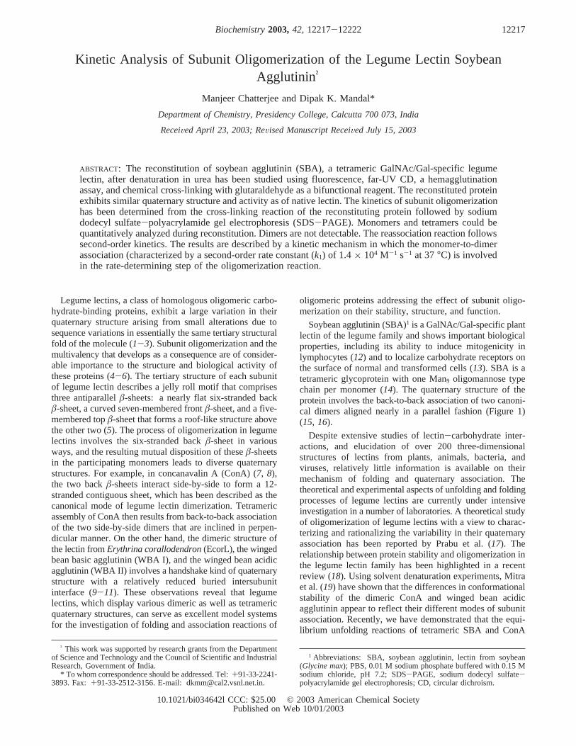
Soybean agglutinin (SBA)1 is a GalNAc/Gal-specific plantlectin of the legume family and shows important biologicalproperties, including its ability to induce mitogenicity inlymphocytes (12) and to localize carbohydrate receptors onthe surface of normal and transformed cells (13). SBA is atetrameric glycoprotein with one Man9 oligomannose typechain per monomer (14). The quaternary structure of theprotein involves the back-to-back association of two canoni-cal dimers aligned nearly in a parallel fashion (Figure 1)(15, 16).
Despite extensive studies of lectin-carbohydrate inter-actions, and elucidation of over 200 three-dimensionalstructures of lectins from plants, animals, bacteria, andviruses, relatively little information is available on theirmechanism of folding and quaternary association. Thetheoretical and experimental aspects of unfolding and foldingprocesses of legume lectins are currently under intensiveinvestigation in a number of laboratories. A theoretical studyof oligomerization of legume lectins with a view to charac-terizing and rationalizing the variability in their quaternaryassociation has been reported by Prabu et al. (17). Therelationship between protein stability and oligomerization inthe legume lectin family has been highlighted in a recentreview (18). Using solvent denaturation experiments, Mitraet al. (19) have shown that the differences in conformationalstability of the dimeric ConA and winged bean acidicagglutinin appear to reflect their different modes of subunitassociation. Recently, we have demonstrated that the equi-librium unfolding reactions of tetrameric SBA and ConA
† This work was supported by research grants from the Departmentof Science and Technology and the Council of Scientific and IndustrialResearch, Government of India.
* To whom correspondence should be addressed. Tel:+91-33-2241-3893. Fax: +91-33-2512-3156. E-mail: [email protected].
1 Abbreviations: SBA, soybean agglutinin, lectin from soybean(Glycine max); PBS, 0.01 M sodium phosphate buffered with 0.15 Msodium chloride, pH 7.2; SDS-PAGE, sodium dodecyl sulfate-polyacrylamide gel electrophoresis; CD, circular dichroism.
12217Biochemistry2003,42, 12217-12222
10.1021/bi034642l CCC: $25.00 © 2003 American Chemical SocietyPublished on Web 10/01/2003
involve the formation of a structured monomeric intermediate(20, 21). These studies have shown that the dissociationprocess can be isolated from the gross unfolding of thepolypeptide chains. Thus, dissociation-association phenom-ena may be studied without the added complication ofmonomer unfolding and refolding.
Reconstitution studies of oligomeric proteins, using kineticapproaches, give access to intermediates of association that,in turn, serve to delineate the pathway of reconstitution ofnative quaternary structure (22). In this paper, we presentsome aspects of kinetics of subunit oligomerization of SBA.To determine whether the rate-limiting step of the association
reaction may be attributed to dimer or tetramer formation,we have used an approach based on a fast cross-linkingreaction during reassociation (23). The present study revealsthe sequence of events that are involved in the subunitassociation of SBA during its reconstitution from thedenatured state.
MATERIALS AND METHODS
Materials. Soybean meal was purchased from Sigma.Cross-linked guar gum matrix was prepared as described(20). Sephadex G-100 was obtained from Pharmacia. Urea(AR, E. Merck, India) was further crystallized from hotethanol to remove possible contamination by cyanate ions,and its stock solution was prepared on a dry weight basis.Glutaraldehyde [50% (m/v) aqueous solution] and sodiumdeoxycholate were purchased from Sigma. All other reagentsused were of analytical grade. Double distilled deionizedwater was used throughout.
Protein Purification. SBA was purified from the crudeextract of soybean meal by affinity chromatography on cross-linked guar gum matrix (24). The integrity of the tetramericstructure of SBA was confirmed from gel filtration analysison a Sephadex G-100 column when the protein was elutedas a single peak corresponding to its tetrameric molecularmass. The purity of the preparation was also checked bysodium dodecyl sulfate-polyacrylamide gel electrophoresis(SDS-PAGE) (25). Protein concentration was determinedspectrophotometrically using A1%,1cm) 12.8 at 280 nm andexpressed in terms of monomer (Mr ) 30 000) (26).
ActiVity Assay. The hemagglutinating activity of the lectinwas assayed by the 2-fold serial dilution technique (27) inPBS (0.01 M sodium phosphate, 0.15 M sodium chloride,pH 7.2) containing 0.1 mM Mn2+ and 0.1 mM Ca2+. Theassay was done in microtiter plates using 25µL of lectinsolution and 25µL of a 3% suspension of trypsin-treatedrabbit erythrocytes. A unit of activity is defined as the lowestconcentration of lectin giving visible agglutination (28).
Spectroscopic Measurements.Absorption spectra wererecorded on a Hitachi U 3210 UV-vis spectrophotometerusing a Sigma cuvette (volume: 2 mL; path length: 1 cm).
Fluorescence spectroscopy was performed on a Hitachi4010 spectrofluorimeter equipped with a constant temperaturecell holder. The spectra were measured at 37°C in PBScontaining 0.1 mM Mn2+ and 0.1 mM Ca2+, pH 7.2 using aSigma fluorimeter cuvette (volume: 2 mL; path length: 1cm). The excitation wavelength was fixed at 280 nm, andthe emission was scanned from 300 to 400 nm. The relativechange (%) of the emission maximum was calculated on thebasis of the difference of the wavelength maximum betweenthe structured monomeric state and the native tetrameric statetaken as 100%.
Far-UV CD measurements were made on a JASCO 600spectropolarimeter at ambient temperature in a 1 mmpathlength cell using a scan speed of 50 nm/min and a responsetime of 2 s. The spectra were measured at a proteinconcentration of 0.33µM and averaged over five scans toeliminate signal noise. The buffer used during measurementwas PBS containing 0.1 mM Mn2+ and 0.1 mM Ca2+, pH7.2. The data are represented as molar ellipticity [θ], whichis defined as [θ] ) 100θobs/(lc), whereθobs is the observedellipticity in degrees,l is the length of the light path in
FIGURE 1: Structure of the SBA tetramer. (Top panel) Schematicdiagram of two SBA dimers that compose the tetramer from theperspective of thez axis (first sheet: yellow and blue subunits;second sheet: green and pink subunits). The tetramerizationinvolves back-to-back association of two side-by-side dimers thatare nearly parallel to each other. (Bottom panel) The representationof the SBA tetramer from the perspective of thex axis of the unitcell (a 90° rotation from the top panel). (Reprinted with permissionfrom ref 15.)
12218 Biochemistry, Vol. 42, No. 42, 2003 Chatterjee and Mandal
centimeters, andc is the molar concentration of the protein.The values obtained were normalized by subtracting thebaseline recorded for the buffer having the same concentra-tion of denaturant under similar conditions.
Denaturation and Reconstitution. The protein solution (20µM) was denatured in 8 M urea in PBS for 30 min at 37°C.Reconstitution of the protein was initiated by dilution withreconstitution buffer (PBS containing 0.1 mM Mn2+ and 0.1mM Ca2+, pH 7.2) to a residual denaturant concentration of0.08-0.2 M urea. The final protein concentration duringrenaturation was 0.2-0.5 µM. Immediately after dilution,the mixtures were vigorously stirred in a vortex mixer, anda series of tubes containing the mixtures, in duplicate, wasincubated at 37°C for different periods of time up to a totaltime of 3 h. The kinetics of reassociation was examined,using aliquots taken at defined times during reconstitution,by fluorescence as mentioned previously and by a cross-linking reaction with glutaraldehyde described next.
Cross-Linking Reaction and SDS-PAGE. Cross-linkingof native SBA and the reassociating protein at various timeswas achieved by a rapid reaction with glutaraldehyde as abifunctional reagent as described (23). The cross-linkingreaction was optimized with respect to the glutaraldehydeconcentration and the time required for cross-linking. It wasalso found that residual denaturant (0.14 M urea) present inthe solution during reconstitution did not interfere with thecross-linking reaction. Briefly, the procedure is as follows.To 3 mL aliquots of native or reassociating protein (30µg)was added an aliquot of 50% (m/v) glutaraldehyde so as tomake a final concentration of 1% glutaraldehyde. After 2min, the cross-linking reaction was quenched by adding a∼10-fold molar excess of NaBH4 (dissolved in 0.1 MNaOH). From this solution, the cross-linked protein wasprecipitated by a combined sodium deoxycholate/trichloro-acetic acid precipitation. After NaBH4 reduction (15 min),aqueous sodium deoxycholate (0.15%) was added so as tomake a final concentration of 0.015% deoxycholate followedby a careful addition of trichloroacetic acid to its finalconcentration of 7% when excess NaBH4 was destroyed andthe cross-linked protein was precipitated. After centrifugation(4000g, 30 min), the obtained precipitate was redissolved in0.1 M Tris-HCl buffer, pH 6.8 containing 1% SDS andheated at 100°C for 5 min.
Samples were analyzed by 10% SDS-PAGE (25), andthe electrophoretic run was calibrated with marker proteinsof known molecular mass. Densitometric scanning of the gelswas performed in a Thermal Imaging System (Image MasterVDSFTI-500), and the relative amounts of the cross-linkedspecies involved in the reconstitution pathway were deter-mined from the densitometric plots.
RESULTS
We have studied kinetically the process of quaternaryassociation of SBA from its structured monomer monitoredby fluorescence and far-UV CD and by a cross-linkingreaction with glutaraldehyde as a bifunctional reagent thatpermits the detection and quantitative evaluation of inter-mediate(s) of association. We have previously shown thatthe equilibrium unfolding of SBA involves a structuredmonomeric intermediate having characteristic fluorescenceproperties (20). The restoration of structure at the monomeric
level, that is, the formation of structured monomer duringreconstitution from the completely denatured state, takesplace in a rapid reaction within the dead time of measurement(<10 s). Thus, the process of subunit association that occursin a much longer time scale (hours) can be investigated aftermanual mixing of the denatured protein in 8 M urea withthe reconstitution buffer (PBS containing 0.1 mM Mn2+ and0.1 mM Ca2+, pH 7.2).
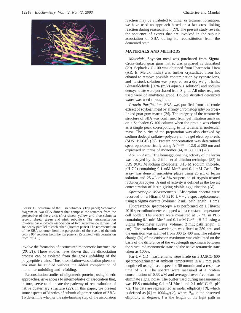
Reassociation Monitored by Fluorescence and CircularDichroism. The fluorescence spectra of native SBA, unfoldedmonomer, structured monomer, and the renatured protein areshown in Figure 2A. The unfolded protein in 8 M ureaexhibits an emission maximum at 351( 1 nm with a relativeintensity of∼2.5-fold higher than that of native SBA havingan emission maximum at 330( 1 nm. When the denaturedprotein is subjected to reconstitution by dilution with
FIGURE 2: (A) Fluorescence spectra at 37°C of native SBA (N4),unfolded monomer (U), reconstituting monomer (N) within 10 sof reconstitution, and renatured SBA (R). The protein concentrationwas 0.33µM. The spectra were corrected for the buffers containingrequisite concentrations of urea. Excitation wavelength, 280 nm;excitation and emission band-pass, 5 nm each; scan rate, 60 nm/min. (B) A plot of relative change (%) of the emission wavelengthmaximum as a function of time during reconstitution. The percentchange was calculated on the basis of the difference of thewavelength maximum between the structured monomeric state(within 10 s of reconstitution) and the native tetrameric state takenas 100%. Each data point represents an average of three determina-tions.
Kinetics of Subunit Association of Soybean Agglutinin Biochemistry, Vol. 42, No. 42, 200312219
renaturation buffer, the emission maximum shifts im-mediately (<10 s) to 340( 1 nm with an emission intensitysimilar to that of native SBA, which is the characteristicproperty of the structured monomer (20). As there was noappreciable difference in the relative fluorescence intensitiesof native tetramer and the structured monomer in the entireemission envelope, the fluorescence intensity parameter couldnot serve as a suitable probe to monitor reassociation.However, the emission wavelength maximum showed agradual blue shift to that of native protein during the processof reassociation; thus, the relative change (%) of thewavelength maximum was used to examine reassociationwith time (Figure 2B). The reconstitution was found to becomplete in about 80 min. It may be mentioned that theemission wavelength parameter can give a qualitative indica-tion of the oligomerization reaction, and the use of emissionmaximum data for quantitative analysis of reassociationkinetics would be inappropriate (29, 30).
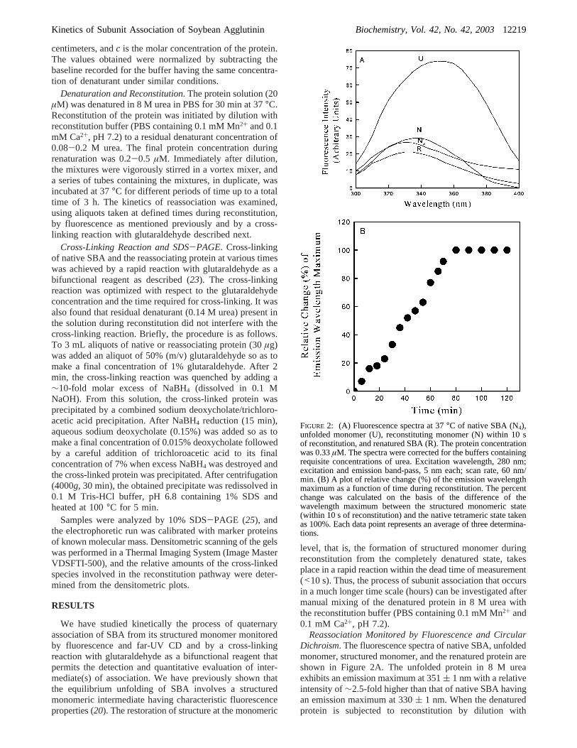
The far-UV CD spectra of SBA in native, unfolded, andrenatured states are shown in Figure 3. While the spectrumin the presence of 8 M urea shows the loss of secondarystructures for the completely unfolded state of the protein,the spectrum of native SBA and that of renatured proteinare similar. It may be mentioned that the far-UV CDspectrum of the reconstituting protein, at the initial stage ofreconstitution, closely resembles that of native protein (datanot shown), implying that the secondary conformation wasrestored immediately. The near-UV CD experiments, how-ever, failed to provide any conclusive evidence about therestoration of tertiary structure due to the minute intensitiesand changes of the near-UV signals (data not shown).
Hemagglutinating ActiVity. The minimal hemagglutinatingconcentration of the reconstituted lectin was the same as thatof the native SBA (1.2µg/mL). These results show that theactivity of the protein was restored completely upon re-association. It may be mentioned that the usual hemagglu-tination assay for the measurement of lectin activity requiressmall amounts of protein and can thus be carried out under
the conditions of low protein concentrations during recon-stitution; however, the low accuracy of this method (28) doesnot entail quantitative analysis of reactivation kinetics.
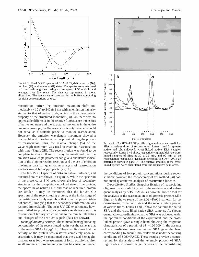
Cross-Linking Studies. Snapshot fixation of reassociatingoligomer by cross-linking with glutaraldehyde and subse-quent analysis by SDS-PAGE is a powerful kinetic tool forthe analysis of the reassociation of oligomeric proteins (23).Figure 4A shows some of the SDS-PAGE patterns for thecross-linking of native SBA and the reconstituting proteinat various times. Lanes 1 and 2 show the patterns for nativeSBA and the cross-liked native SBA samples. As shown,quantitative cross-linking of native SBA was achieved underthe optimized conditions of the experiment, and the cross-linked protein gave a single band showing the migrationcharacteristics of a protein ofMr ∼120 000. In the absenceof a cross-linking reaction, native SBA gave the bandcorresponding to subunit molecular mass under denaturingconditions of SDS-PAGE. These results provide a controlsystem for the analysis of the assembly process of SBA.Figure 4A also shows the gel patterns of the reconstituting
FIGURE 3: Far-UV CD spectra of SBA (0.33µM) in native (N4),unfolded (U), and renatured (R) states. The spectra were measuredin 1 mm path length cell using a scan speed of 50 nm/min andaveraged over five scans. The data are represented in molarellipticities. The spectra were corrected for the buffers containingrequisite concentrations of urea.
FIGURE 4: (A) SDS-PAGE profile of glutaraldehyde cross-linkedSBA at various times of reconstitution. Lanes 1 and 2 representnative and glutaraldehyde cross-linked native SBA samples,respectively. Lanes 3-5 show, respectively, glutaraldehyde cross-linked samples of SBA at 30 s, 32 min, and 90 min of thereassociation reaction. (B) Densitometric plots of SDS-PAGE gelpatterns as shown in panel A. The relative amounts of the cross-linked species were quantitated from the respective peak areas.
12220 Biochemistry, Vol. 42, No. 42, 2003 Chatterjee and Mandal
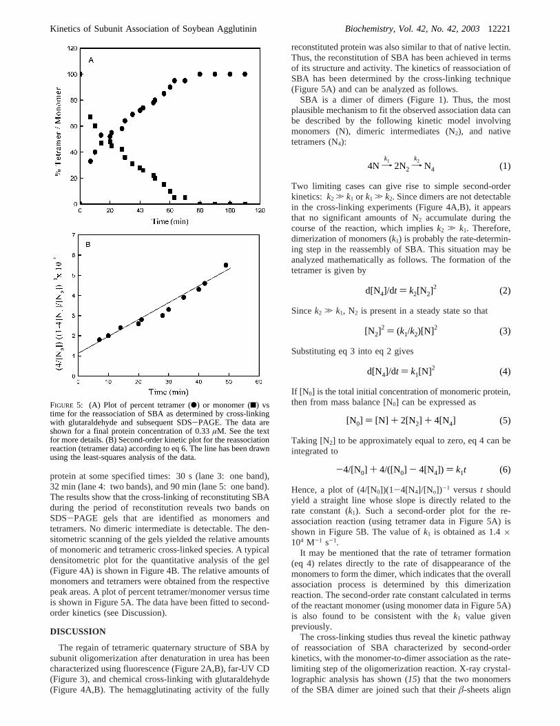
protein at some specified times: 30 s (lane 3: one band),32 min (lane 4: two bands), and 90 min (lane 5: one band).The results show that the cross-linking of reconstituting SBAduring the period of reconstitution reveals two bands onSDS-PAGE gels that are identified as monomers andtetramers. No dimeric intermediate is detectable. The den-sitometric scanning of the gels yielded the relative amountsof monomeric and tetrameric cross-linked species. A typicaldensitometric plot for the quantitative analysis of the gel(Figure 4A) is shown in Figure 4B. The relative amounts ofmonomers and tetramers were obtained from the respectivepeak areas. A plot of percent tetramer/monomer versus timeis shown in Figure 5A. The data have been fitted to second-order kinetics (see Discussion).
DISCUSSION
The regain of tetrameric quaternary structure of SBA bysubunit oligomerization after denaturation in urea has beencharacterized using fluorescence (Figure 2A,B), far-UV CD(Figure 3), and chemical cross-linking with glutaraldehyde(Figure 4A,B). The hemagglutinating activity of the fully
reconstituted protein was also similar to that of native lectin.Thus, the reconstitution of SBA has been achieved in termsof its structure and activity. The kinetics of reassociation ofSBA has been determined by the cross-linking technique(Figure 5A) and can be analyzed as follows.
SBA is a dimer of dimers (Figure 1). Thus, the mostplausible mechanism to fit the observed association data canbe described by the following kinetic model involvingmonomers (N), dimeric intermediates (N2), and nativetetramers (N4):
Two limiting cases can give rise to simple second-orderkinetics: k2 . k1 or k1 . k2. Since dimers are not detectablein the cross-linking experiments (Figure 4A,B), it appearsthat no significant amounts of N2 accumulate during thecourse of the reaction, which impliesk2 . k1. Therefore,dimerization of monomers (k1) is probably the rate-determin-ing step in the reassembly of SBA. This situation may beanalyzed mathematically as follows. The formation of thetetramer is given by
Sincek2 . k1, N2 is present in a steady state so that
Substituting eq 3 into eq 2 gives
If [N 0] is the total initial concentration of monomeric protein,then from mass balance [N0] can be expressed as
Taking [N2] to be approximately equal to zero, eq 4 can beintegrated to
Hence, a plot of (4/[N0])(1-4[N4]/[No])-1 versust shouldyield a straight line whose slope is directly related to therate constant (k1). Such a second-order plot for the re-association reaction (using tetramer data in Figure 5A) isshown in Figure 5B. The value ofk1 is obtained as 1.4×10⁴ M-1 s-1.
It may be mentioned that the rate of tetramer formation(eq 4) relates directly to the rate of disappearance of themonomers to form the dimer, which indicates that the overallassociation process is determined by this dimerizationreaction. The second-order rate constant calculated in termsof the reactant monomer (using monomer data in Figure 5A)is also found to be consistent with thek1 value givenpreviously.
The cross-linking studies thus reveal the kinetic pathwayof reassociation of SBA characterized by second-orderkinetics, with the monomer-to-dimer association as the rate-limiting step of the oligomerization reaction. X-ray crystal-lographic analysis has shown (15) that the two monomersof the SBA dimer are joined such that theirâ-sheets align
FIGURE 5: (A) Plot of percent tetramer (b) or monomer (9) vstime for the reassociation of SBA as determined by cross-linkingwith glutaraldehyde and subsequent SDS-PAGE. The data areshown for a final protein concentration of 0.33µM. See the textfor more details. (B) Second-order kinetic plot for the reassociationreaction (tetramer data) according to eq 6. The line has been drawnusing the least-squares analysis of the data.
4N 98k1
2N2 98k2
N4 (1)
d[N4]/dt ) k2[N2]2 (2)
[N2]2 ) (k1/k2)[N]2 (3)
d[N4]/dt ) k1[N]2 (4)
[N0] ) [N] + 2[N2] + 4[N4] (5)
-4/[N0] + 4/([N0] - 4[N4]) ) k1t (6)
Kinetics of Subunit Association of Soybean Agglutinin Biochemistry, Vol. 42, No. 42, 200312221
to form a 12-stranded sheet spanning one face of the dimer(Figure 1, top panel). At the dimer interface, the firstâ-strandof the two subunits interacts through six main chain hydrogenbonds involving the uncharged Ser and Thr residues. Furtherintersubunit contacts occur through side chain-side chainand side chain-main chain interactions from Ala, Ser, andAsn residues within the interface. In the SBA tetramer, the12-strandedâ-sheets of each dimer face each other, and thetwo dimers interact with their two outermost strands, creatinga large channel in the middle of the tetramer (Figure 1,bottom panel). The interface formed by these two outermoststrands consists mainly of a number of relatively short sidechains that intercalate in a zipper-like fashion. This type ofdimeric structure of SBA resembles the canonical dimer ofconcanavalin A (ConA) and other legume lectins (5);however, its tetrameric assemblage is different from that inConA, and unlike ConA, the SBA tetramer does notdissociate into two dimeric species at more acidic pH. Thepresent study provides the kinetic mechanism for thetetrameric association of SBA with the dimerization of SBAdimers occurring faster than the dimerization of SBAmonomers, which may arise from the requirement of differentarchitecture of theâ-sheet packing in the dimeric andtetrameric interface of the lectin. These results are also inagreement with the mechanism proposed previously for theequilibrium denaturation of SBA (20).
The reconstitution kinetics of oligomeric proteins includingsome tetrameric enzymes such as lactate dehydrogenase andphosphofructokinase offer several kinetic mechanisms inrespect to the rate-limiting step involved in the kineticpathway of reassociation (22, 31-33). SBA seems to be thefirst multimeric protein in the legume lectin family for whichsuch a kinetic analysis is being reported.
ACKNOWLEDGMENT
We thank Prof. A. Chatterjee, Principal, for his supportand Prof. S. Ghosh, Head of the Department of Chemistry,for providing the necessary facilities.
REFERENCES
1. Sharon, N., and Lis, H. (1990)FASEB J. 4, 3198-3208.2. Lis, H., and Sharon, N. (1998)Chem. ReV. 98, 637-674.3. Vijayan, M., and Chandra, N. (1999)Curr. Opin. Struct. Biol. 9,
707-714.4. Rini, J. M. (1995)Annu. ReV. Biophys. Biomol. Struct. 24, 551-
557.
5. Loris, R., Hamelryck, T., Bouckaert, J., and Wyns, L. (1998)Biochim. Biophys. Acta 1383, 9-36.
6. Brewer, C. F. (1996)Chemtracts Biochem. Mol. Biol. 6, 165-179.
7. Hardman, K. D., Agarwal, R. C., and Freiser, M. J. (1982)J. Mol.Biol. 157, 69-86.
8. Parkin, S., Rupp, B., and Hope, H. (1996)Acta Crystallogr. D52,1161-1168.
9. Shaanan, B., Lis, H., and Sharon, N. (1991)Science 254, 862-866.
10. Schwarz, F. P., Puri, K. D., Bhat, R. G., and Surolia, A. (1993)J.Biol. Chem. 268, 7668-7677.
11. Prabu, M. M., Sankaranarayanan, R., Puri, K. D., Sharma, V.,Surolia, A., Vijayan, M., and Suguna, K. (1998)J. Mol. Biol.276, 787-796.
12. Novogrodski, A., and Katchalski, E. (1973)Proc. Natl. Acad. Sci.U.S.A. 70, 2515-2518.
13. Sharon, N. (1983)AdV. Immunol. 34, 213-398.14. Lis, H., and Sharon, N. (1978)J. Biol. Chem. 253, 3468-3476.15. Dessen, A., Gupta, D., Sabesan, S., Brewer, C. F., and Sacchettini,
J. C. (1995)Biochemistry 34, 4933-4942.16. Olsen, L. R., Dessen, A., Gupta, D., Sabesan, S., Sacchettini, J.
C., and Brewer, C. F. (1997)Biochemistry 36, 15073-15080.17. Prabu, M. M., Suguna, K., and Vijayan, M. (1999)Proteins:
Struct., Funct., Genet. 35, 58-69.18. Srinivas, V. R., Reddy, G. B., Ahmad, N., Swaminathan, C. P.,
Mitra, N., and Surolia, A. (2001)Biochim. Biophys. Acta 1527,102-111.
19. Mitra, N., Srinivas, V. R., Ramya, T. N. C., Ahmad, N., Reddy,G. B., and Surolia, A. (2002)Biochemistry 41, 9256-9263.
20. Ghosh, M., and Mandal, D. K. (2001)Int. J. Biol. Macromol. 29,273-280.
21. Chatterjee, A., and Mandal, D. K. (2003)Biochim. Biophys. Acta1648, 174-183.
22. Jaenicke, R., and Lilie, H. (2000)AdV. Protein Chem. 53, 329-397.
23. Jaenicke, R., and Rudolph, R. (1986)Methods Enzymol. 131, 218-250.
24. Mandal, D. K., and Brewer, C. F. (1992)Biochemistry 31, 12602-12609.
25. Laemmli, U. K. (1970)Nature 227, 680-685.26. Lotan, R., Siegelman, H. W., Lis, H., and Sharon, N. (1974)J.
Biol. Chem. 246, 1219-1224.27. Osawa, T., and Matsumoto, I. (1972)Methods Enzymol. 28, 323-
327.28. Arango, R., Adar, R., Rozenblatt, S., and Sharon, N. (1992)Eur.
J. Biochem. 205, 575-581.29. Eftink, M. R. (1994)Biophys. J. 66, 482-501.30. Fan, Y.-X., Zhou, J.-M., Kihara, H., and Tsou, C.-L. (1998)Protein
Sci. 7, 2631-2641.31. Hermann, R., Jaenicke, R., and Rudolph, R. (1981)Biochemistry
20, 5195-5201.32. Zettlmeissl, G., Rudolph, R., and Jaenicke, R. (1982)Biochemistry
21, 3946-3950.33. Parr, G. R., and Hammes, G. G. (1976)Biochemistry 15, 857-862.
BI034642L
12222 Biochemistry, Vol. 42, No. 42, 2003 Chatterjee and Mandal