keyword: amyloid, nsaid, anti-amyloidogenic activity, … · draft 1 inhibition of islet amyloid...
TRANSCRIPT
Draft
Inhibition of Islet Amyloid Polypeptide Aggregation and
Associated Cytotoxicity by Non-steroidal Anti-Inflammatory Drugs
Journal: Canadian Journal of Physiology and Pharmacology
Manuscript ID cjpp-2015-0117.R2
Manuscript Type: Article
Date Submitted by the Author: 26-May-2015
Complete List of Authors: Fortin, Jessica; University of Montreal, Faculté de médecine vétérinaire, Département de pathologie et microbiologie Benoit-Biancamano, Marie-Odile; Université de montreal,
Keyword: Amyloid, NSAID, anti-amyloidogenic activity, IAPP, fibrils
https://mc06.manuscriptcentral.com/cjpp-pubs
Canadian Journal of Physiology and Pharmacology
Draft
1
Inhibition of Islet Amyloid Polypeptide Aggregation and Associated
Cytotoxicity by Non-steroidal Anti-Inflammatory Drugs
Jessica S. Fortin1 and Marie-Odile Benoit-Biancamano
1
1Département de Pathologie et de Microbiologie, Faculté de Médecine Vétérinaire,
Université de Montréal, Saint-Hyacinthe, QC, Canada, J2S 2M2.
Corresponding author : Marie-Odile Benoit-Biancamano, Faculté de médecine
vétérinaire, Département de pathologie et microbiologie, Université de Montréal, 3200
Sicotte, St-Hyacinthe, QC, J2S 2M2. Tel.: 450-773-8521 ext. 8539, Fax: 450-778-8116.
E-mail: [email protected] (MOBB),
[email protected] (JF).
Page 1 of 48
https://mc06.manuscriptcentral.com/cjpp-pubs
Canadian Journal of Physiology and Pharmacology
Draft
2
Inhibition of Islet Amyloid Polypeptide Aggregation and Associated
Cytotoxicity by Non-steroidal Anti-Inflammatory Drugs
Jessica S. Fortin and Marie-Odile Benoit-Biancamano
Abstract: Non-steroidal anti-inflammatory drugs (NSAIDs) constitute an important
pharmacotherapeutic class which, over the past decade, have expanded in application to a
panoply of medical conditions. They have been tested for neurodegenerative diseases
such as Alzheimer’s to reduce the inflammation and also in the attempt to abrogate
amyloid deposition. However, the use of NSAID as aggregation inhibitors has not been
extensively studied in pancreatic amyloid deposition. Pancreatic amyloidosis involves the
misfolding of islet amyloid polypeptide (IAPP) and contributes to the progression of type
2 diabetes in the human and feline. To ascertain their anti-amyloidogenic activity, several
NSAIDs were tested using fluorometric ThT assays, circular dichroism, photo-induced
cross-linking assays and cell culture. Celecoxib, diclofenac, indomethacin, meloxicam,
niflumic acid, nimesulide, phenylbutazone, piroxicam, sulindac, tenoxicam reduced
fibrillization at a molar ratio of 1:10. The circular dichroism spectra of diclofenac,
piroxicam and sulindac showed characteristic spectral signatures found in predominantly
α-helical structures. The oligomerization of hIAPP was abrogated with diclofenac and
sulindac at a molar ratio of 1:5. The cytotoxic effects of pre-incubated hIAPP on cultured
INS-1 cells were noticeably reduced in the presence of diclofenac, meloxicam,
phenylbutazone, sulindac and tenoxicam at a molar ratio of 1:10. Our results demonstrate
that NSAIDs can provide chemistry scaffolds to generate new promissing anti-
amyloidogenic agents that can be used alone or as a coadjuvant therapy.
Page 2 of 48
https://mc06.manuscriptcentral.com/cjpp-pubs
Canadian Journal of Physiology and Pharmacology
Draft
3
Key words: Amyloid, anti-amyloidogenic activity, degenerative disease, fibrils, IAPP,
NSAID, protein misfolding, type 2 diabetes.
Introduction
Diseases involving abnormal folding of proteins and conversion to an insoluble
state are numerous in human and veterinary medicine, with more than thirty diseases
related specifically to amyloid formation, including Alzheimer's disease (AD), type 2
diabetes, Parkinson's disease, prion disease and familial amyloidosis (Sipe et al. 2014).
Protein misfolding, in amyloidosis, is characterized by a monomeric protein which is
assembled into β-sheet insoluble fibrils (Hard and Lendel 2012). These amyloid deposits
damage the cell membrane, inducing reactive oxygen species and apoptosis (Abedini and
Schmidt 2013; Jayasinghe and Langen 2007; Jelinek and Sheynis 2010). Amyloid
deposits may be localized in a tissue or generalized in different organs depending on the
origin of the constitutive protein (Hard and Lendel 2012; Sipe et al. 2014).
Amyloidogenic proteins do not share a common amino acid sequence nor a similar
secondary or tertiary conformation in the native state. However, all types of amyloid have
in common the formation of fibrils, with unique physicochemical characteristics as
follows: a diameter of 5-15 nm, a cross-β sheet quaternary structure, a poor solubility in
acidic or basic environments, and a resistance to extreme temperatures (Cheng et al.
2013; Hard and Lendel 2012). Several amyloidogenic proteins, such as TTR, are rich in
beta-sheet in their soluble native conformation.
Page 3 of 48
https://mc06.manuscriptcentral.com/cjpp-pubs
Canadian Journal of Physiology and Pharmacology
Draft
4
In the past few decades, extensive effort has been expended to understand the
pathophysiology of amyloidosis related to degenerative diseases such as Alzheimer's
disease (AD) and type 2 diabetes, potentially leading to new therapeutic strategies.
Although the actual details of many of the steps are unclear, the assembly of
amyloidogenic proteins into a fibrillar structure occurs in stepwise fashion. An
apparent early step in the aggregation is the formation of a set of conformationally
diverse oligomers (Brender et al. 2015; Buchanan et al. 2014; Mishra et al. 2009), present
at low abundance (Suzuki et al. 2012), with structures different from either the native
largely unstructured peptide (Reddy et al. 2010; Williamson and Miranker 2007;
Yonemoto et al. 2008) or the mature amyloid fiber (Luca et al. 2007). Subsequently, the
oligomers assemble into a mature fibril. At this point, the proteins have a β-sheet
secondary structure (Luca et al. 2007; Westermark et al. 2011). In parallel, these
oligomers act as a seed, thereby initiating the aggregation and propagation of fibrils
(Westermark et al. 2011). Although several factors can influence the fibrillar aggregation,
the precise mechanisms are not clearly identified. More research on the mechanisms
underlying the fibrillar aggregation will be crucial in the discovery of new anti-
amyloidogenic agents. The development of an inhibitor is also challenging considering
that it doesn’t require to stabilize solely the native protein or the oligomers (Cheng et al.
2013; Hard and Lendel 2012; Porat et al. 2006). The most common approach in
pharmaceutical discovery research against amyloidogenic proteins involves exploring
inhibitors of fibrillar deposition (Cheng et al. 2013; Hard and Lendel 2012; Porat et al.
2006). Perhaps, the best way to inhibit amyloid formation is by stabilizing the native
conformation by small molecules, i.e. Tafamidis and TTR.
Page 4 of 48
https://mc06.manuscriptcentral.com/cjpp-pubs
Canadian Journal of Physiology and Pharmacology
Draft
5
Substantive clinical research has focused on inflammation as a critical component
in amyloidogenic diseases. The use of NSAIDs has been demonstrated to delay the onset
of Alzheimer’s disease (AD) (Dong et al. 2014; Lim et al. 2000; Yan et al. 2003).
Whether selective COX-2 inhibitors have clinical benefits with direct effects on brain
amyloid deposition remain to be elucidated. Interestingly, the diarylamine classes,
including diclofenac, were effective in inhibiting fibril formation related to other
diseases, such as familial amyloid cardiomyopathy (FAC) and familial amyloid
polyneuropathy (FAP), that involve transthyretin (TTR) amyloid formation (Oza et al.
2002). Others studies were performed in the attempt to inhibit amyloid formation related
to type 2 diabetes. Amyloid deposits localized in the islets involve the misfolding of islet
amyloid polypeptide (IAPP) and contribute to the progression of type 2 diabetes in
human and diabetes mellitus in adult cats (Hoppener et al. 2002; Jaikaran and Clark
2001; O'Brien et al. 1993; Westermark et al. 2011). The effect of aspirin and ketoprofen
were examined in pancreatic amyloidosis (Tu et al. 2014). However, these drugs failed to
inhibit amyloid formation or to disaggregate preformed fibrils (Tu et al. 2014).
The implication of NSAIDs as IAPP aggregation inhibitors has not been
extensively explored and their clinical benefits remain to be elucidated. This study
focusses on the investigation of NSAIDs in inhibiting amyloidosis related to type 2
diabetes, with a potential therapeutic target of hIAPP fibrils. We describe the inhibition
activity of a number of commercially available NSAIDs on hIAPP fibrils formation. The
information gained from this study will help further structure-activity relationship studies
for designing new derivatives with improved potency.
Page 5 of 48
https://mc06.manuscriptcentral.com/cjpp-pubs
Canadian Journal of Physiology and Pharmacology
Draft
6
Materials and Methods
Chemicals
Aceclofenac, diclofenac, DMSO, Hexafluoroisopropanol (HFIP), ibuprofen,
ketoprofen, meloxicam, naproxen, nimesulide, phenylbutazone, resveratrol, resazurin,
silibinin, sulindac, tenoxicam and thioflavin-T (ThT) were obtained from Alfa Aesar
(Ward Hill, MA). Celecoxib, etoricoxib, indometacin, ketorolac, flurbiprofen, niflumic
acid, oxaprozin, piroxicam, rofecoxib and valdecoxib were purchased from Sigma-
Aldrich (St. Louis, MO).
Cell line and culture
INS-1 (rat insulinoma) cells were purchased from AddexBio (San Diego, CA) and
cultured in RPMI1640 medium supplemented with 10 mM HEPES, 2 mM L-glutamine,
1X sodium pyruvate, 50 µM 2-mercaptoethanol, 100 U/mL streptomycin, 100 U/mL of
penicillin G and 10 % fetal bovine serum (Wisent Inc., St-Bruno, Qc, Canada). Cells
were maintained in a moisture saturated atmosphere at 37 °C under 5% CO2.
Peptide synthesis
Synthetic hIAPP (1-37) was obtained from Peptidogen International Corp
(Brossard, Qc, Canada). Peptides were prepared, as previously published, with a
microwave peptide synthesizer, using 9-fluornylmethoxycarbonyl (Fmoc) chemistry, and
Fmoc-protected pseudoproline dipeptide derivatives were incorporated to facilitate the
synthesis (Cao et al. 2013). The peptides were purified by reverse-phase HPLC using a
Page 6 of 48
https://mc06.manuscriptcentral.com/cjpp-pubs
Canadian Journal of Physiology and Pharmacology
Draft
7
C18 preparative column. The identity of the pure products was confirmed by mass
spectrometry using a Bruker MALDI-TOF MS. Analytical HPLC was used to check the
purity of the peptides (≥ 95%) before each experiment. The purified peptide was
lyophilized.
Sample preparation
hIAPP stock solutions were prepared by dissolving the hIAPP peptide at 1 mM in
100% HFIP and incubating for at least 12 h. For the ThT kinetics, circular dichroism
spectrometry and photo-induced cross-linking based oligomerization assays, hIAPP
peptide stock solutions were prepared by dissolving peptides at 1 mM in 100% HFIP. For
the cytotoxicity assays, the IAPP peptide stock solutions of 1 mM were divided in
aliquots to obtain the desired final concentration and were air-dried. Aliquots of the
stock solutions were dried to remove organic solvents and suspended in 10 mM PBS
buffer (pH 7.4) at the desired concentration. hIAPP was dissolved in DMSO and PBS.
DMSO was used at a final concentration lower than 0.1 % (v/v) to avoid cell toxicity.
Thioflavin T (ThT) fluorescence assay
hIAPP peptide from the stock solution of 1mM were added to 10 mM PBS buffer
(pH 7.4) and transferred to a black 96-well microplate with transparent bottom (Corning
Incorporated Costar 3603). Each well contained a final volume of 150 µL with a
peptide final concentration of 10 µM. Experiments in the presence of hIAPP were
initiated by adding a solution of ThT at a final concentration of 10 µM ThT with different
drug treatments. Final drug concentrations ranged from 25 to 100 µM. All drugs were
Page 7 of 48
https://mc06.manuscriptcentral.com/cjpp-pubs
Canadian Journal of Physiology and Pharmacology

Draft
8
dissolved in DMSO at a final concentration lower than 0.25 % (v/v). The vehicle DMSO
was tested on hIAPP, without drug treatment, and did not change the kinetics.
The background signal consisted of 1 % HFIP without peptide. Thioflavin T-
based fluorescence assays were used to detect the formation of amyloid. The fluorescence
emission experiments were performed with the excitation and emission wavelengths set
at 440 and 485 nm, respectively, with a Synergy HT multi-mode microplate reader
(BioTek, Winooski, VT). Measurements were taken with two different periods of
agitation prior to plate reading. Agitation during incubation leads to shorter aggregation
times due to the increased rate of nucleation due to the formation of new nuclei by the
breakage of amyloid fibers during agitation (Padrick and Miranker 2002; Ruschak and
Miranker 2007; Shvadchak et al. 2015). For measurements with long agitation,
measurements were taken at room temperature every 2 min with 10 seconds shaking
prior measurements over 6 h. Short agitation measurements were obtained by shaking the
plate prior measurement for 5 sec to ensure proper mixing and subsequently measuring
the fluorescence intensity every 10 min. Samples were measured in three replicates and
the experiments were repeated three times using different IAPP stock solutions. For each
time point, arbitrary units of fluorescence were calculated from the mean values
normalized against the maximum value in each completed assay. Arbitrarily, the
maximum value (100 %) for the fluorescence intensity was established for hIAPP
peptide. The lag time and the T50 were calculated as previously described (Zhang et al.
2011).
Far-Ultraviolet (UV) circular discroism (CD)
Page 8 of 48
https://mc06.manuscriptcentral.com/cjpp-pubs
Canadian Journal of Physiology and Pharmacology
Draft
9
CD spectra of the secondary structure of hIAPP samples were recorded at 25 °C
under a constant flow of N2 using a JASCO-810 spectropolarimeter (Jasco, Easton, MD).
Spectra were recorded over a wavelength range of 190–250 nm using a quartz cuvette of
1 mm path length and an instrument scanning speed of 100 nm/min, with a response time
of 2 sec and a bandwidth of 1 nm. Samples were not agitated. All IAPP samples were
dissolved to a final concentration of 15 µM in 10 mM PBS (pH 7.4) and 1% HFIP, and
where appropriate, the samples contained different drug treatments at a final
concentration of 25 µM. Each result is given as the average scans taken of three
measurements at room temperature. The data were converted to mean residue ellipticity
(θ) and analyzed using the software CDPro as previously described (Sreerama and
Woody 2004). All CD spectra were averaged, smoothed and baseline-corrected for signal
contributions due to the buffer.
Photo-induced cross-linking (PICUP) assay
hIAPP was diluted in 10 mM phosphate buffer (pH 7.4) to a final concentration of
250 µM peptide and 1% HFIP. The peptide solution was sonicated for 1 min. Drugs were
also freshly dissolved in DMSO and added to the peptide solution at a molar ratio of 1:5
(hIAPP:compound). The control consisted of samples that were photo-cross-linked using
the PICUP method as described (Bitan and Teplow 2004). The reaction buffer consisted
of 938 µM Ru(bpy), 18.8 mM ammonium persulfate and 250 µM hIAPP. Cross-linking
was induced by irradiation, for 3 s, of the mixture, at a final volume of 60 µL, with a 150
W incandescent lamp installed in a homemade dark-box. After irradiation, 25 µL of
Lammeli loading buffer was immediately added to the solution, followed by incubation at
Page 9 of 48
https://mc06.manuscriptcentral.com/cjpp-pubs
Canadian Journal of Physiology and Pharmacology
Draft
10
37 °C for 15 min. The cross-linked samples were separated on a 16% Tricine-urea gel
and visualized by silver staining.
Cell viability assay
Cells were maintained in a 37 °C, 5% CO2 incubator, in exponential growth, for
the duration of experimentation. For this assay, cells were seeded in 96-well microtiter
plates at a density of 5 × 10³ Ins-1 cells per well for 24 h. Drugs freshly solubilized in
DMSO were diluted in culture medium and aliquots containing sequential dilution of
drugs were added subsequently after stimulation with 2.5 µM of hIAPP. hIAPP was
dissolved and incubated in 10 mM PBS (pH 7.4, 25 °C) 24 h before addition to cells.
Drugs were tested at 25 and 50 µM and DMSO concentration was maintained at 0.1 % to
avoid growth inhibition. Negative control consisted of DMSO with no added drug. Plates
were incubated for 24h in the presence of drugs and hIAPP. Resazurin-based reduction
assays were performed as previously described (Fortin et al. 2010). Briefly, Resazurin
(25 µg/mL) was added to the culture medium of each well for 1h30 at 37 °C. The cell
viability was calculated from fluorescence (excitation, 530 nm; emission, 590 nm)
measured with a Synergy HT multi-mode microplate reader (BioTek, Winooski, VT).
The data from experiments conducted in triplicate were corrected for the background
fluorescence of the medium and was expressed as the mean percentage of fluorescence
obtained for control DMSO-treated cells. The growth inhibition percentage was
calculated with reference to DMSO-treated cells for each drug concentrations. The results
were obtained from at least three separated experiments. The cell viability assay was
considered valid when the variability among data for a given set of conditions, within the
Page 10 of 48
https://mc06.manuscriptcentral.com/cjpp-pubs
Canadian Journal of Physiology and Pharmacology
Draft
11
same experiment, was less than 10% with respect to the mean value. All values represent
means ± SEM (n = 3).
Statistical analysis
All results in Table 2, Table 3 and Figure 4 were presented as mean ± SD. For the
cell viability assays, data were analyzed by the one-way analysis of variance with
Dunnett's multiple comparison between IAPP and several drug treatments. Differences
were considered statistically significant at p < 0.05 and p < 0.001.
Results
NSAIDs are effective inhibitors of hIAPP aggregation
Table 1 displays the classification and molecular structure of NSAIDs used in this
study. The effects of NSAIDs on the kinetics of hIAPP fibrillization using thioflavin T
(ThT) fluorescence assay were examined. ThT binds to the cross β-structure of amyloid
fibrils, theoretically in the surface grooves generated by stacking parallel β-sheets (Buell
et al. 2010). The self-quenching of the fluorescent dye is relieved by the conformational
changes induced by amyloid binding, resulting in fluorescence enhancement (Buell et al.
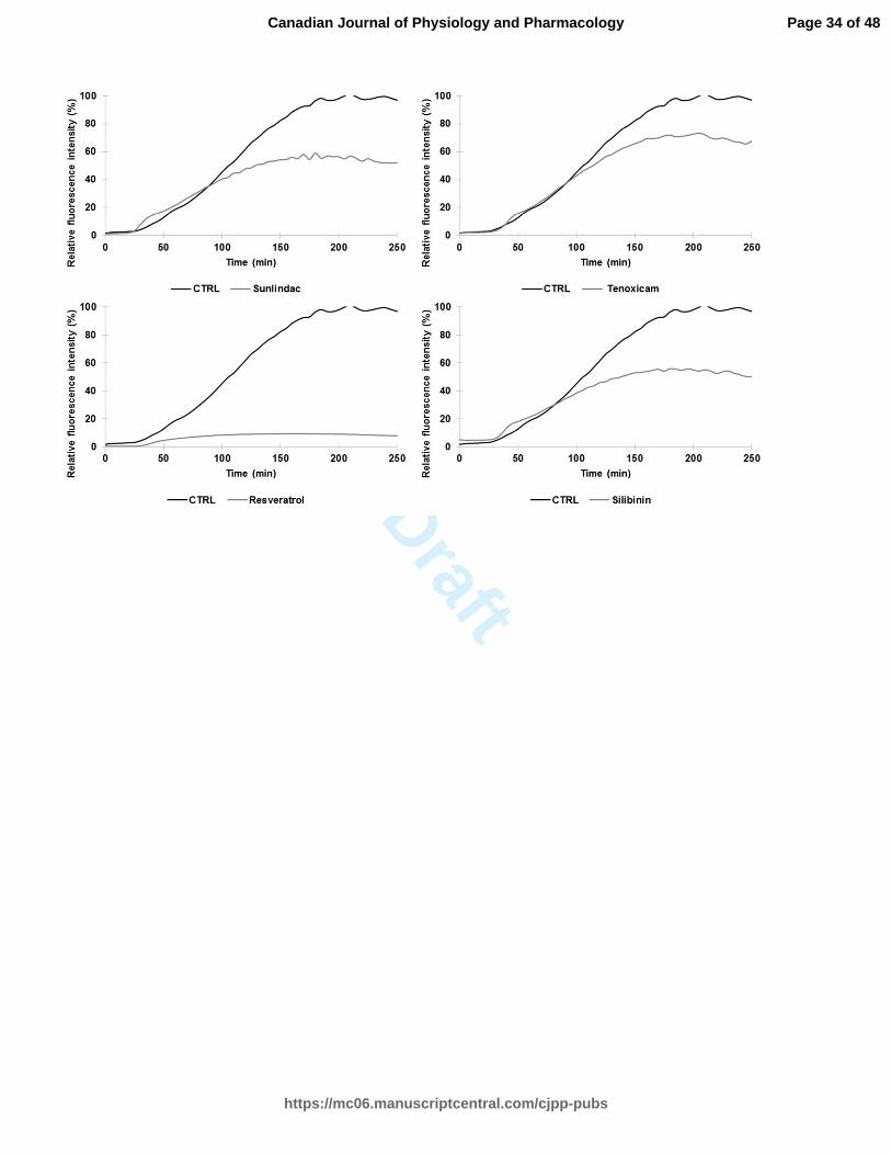
2010). The expected sigmoidal time course is observed when hIAPP is incubated without
drug treatments (Fig. 1) (Paulsson et al. 2011). In the absence of drugs, the time required
to reach half of the total fluorescence signal (T50) was 39.9 ± 0.2 min for samples with
longer agitation during measurement and 105,3 ± 0.7 min for samples with shorter
agitation during measurement. For both samples, diclofenac and phenylbutazone
significantly increased the T50 at a molar ratio of 1:10 (10 µM of hIAPP:100 µM of drug)
Page 11 of 48
https://mc06.manuscriptcentral.com/cjpp-pubs
Canadian Journal of Physiology and Pharmacology
Draft
12
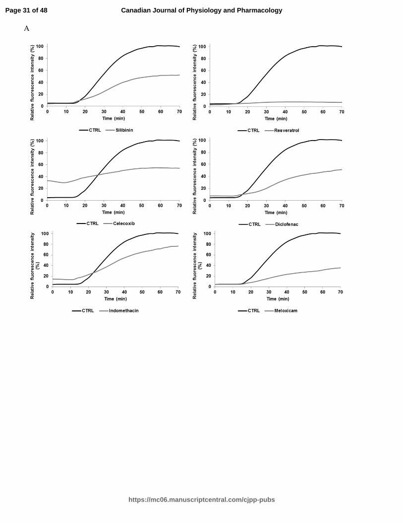
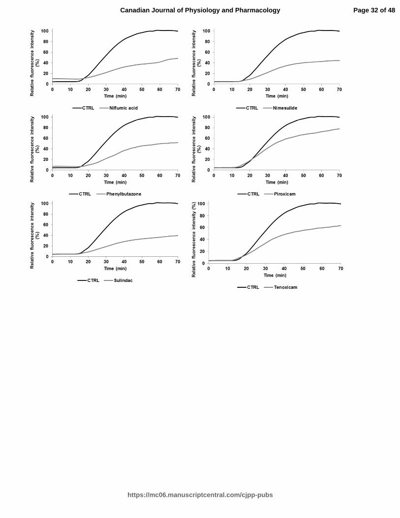
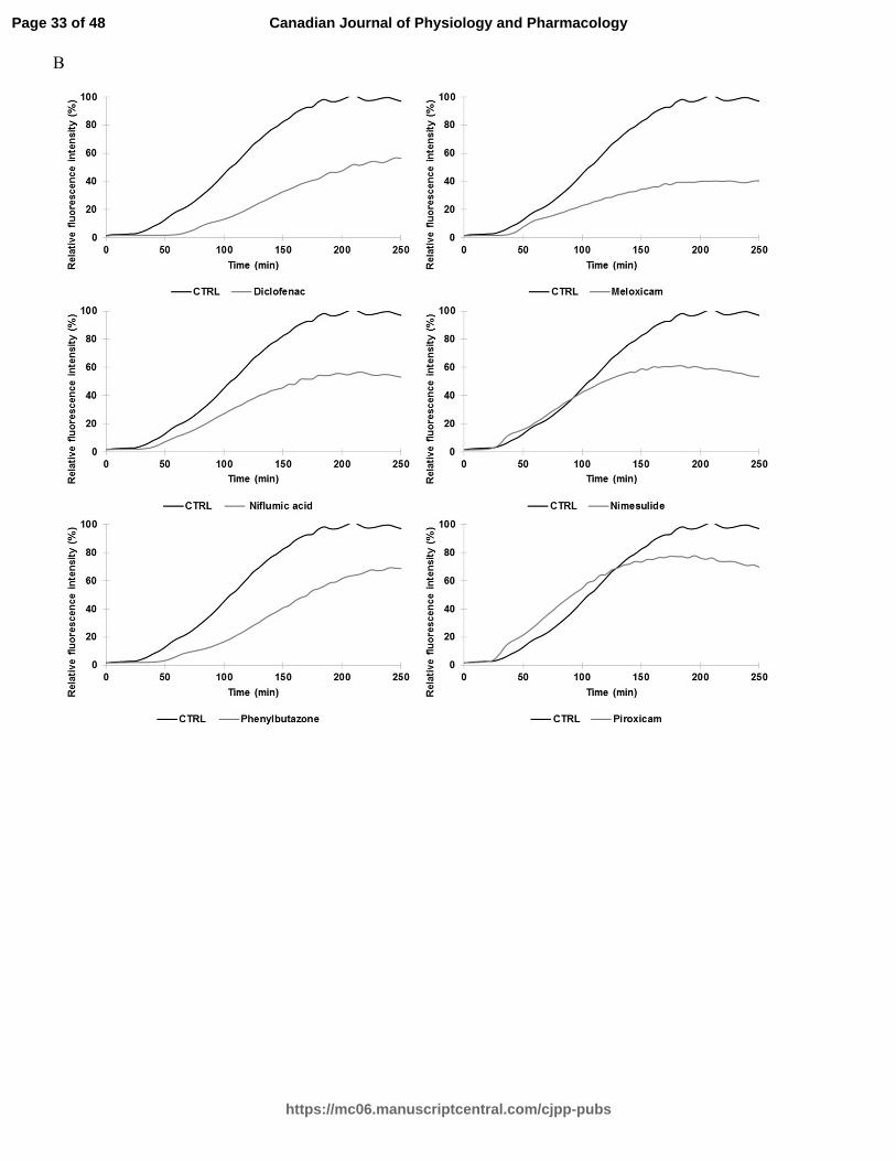
(Table 2 and Table 4). Positive controls (resveratrol, silibinin) (Cheng et al. 2012; Jiang
et al. 2011) and NSAIDs treatments shown in Fig. 1 (celecoxib, diclofenac,
indomethacin, meloxicam, niflumic acid, nimesulide, phenylbutazone, piroxicam,
sulindac, tenoxicam) significantly reduced the fluorescence intensity at a molar ratio of
1:10. The curves in these graphs exhibit a different slope for the treated and untreated
groups. The time during which no significant fibril formation occurs followed by a rapid
growth phase (lag time) was shorter for meloxicam, nimesulide, piroxicam, sulindac,
tenoxicam, resveratrol and silibinin in Table 4. Interestingly, diclofenac and
phenylbutazone prolonged the lag time. NSAID treatments such as niflumic acid,
nimesulide and sulindac maintained their efficacy in reducing the amyloidogenicity of
hIAPP fibrils using a molar ration of 1:5 (10 µM of hIAPP:50 µM of drug). The ThT
fluorescence of h-IAPP was reduced by the presence of 25 µM and 50 µM of meloxicam
(Table 3). Other NSAIDs, such as COX-2 inhibitors, had no detectable effect on the
kinetics of amyloid formation and the final fluorescence intensity.
NSAIDs induce secondary structure changes of hIAPP
Amyloid formation involves the oligomerization of hIAPP and the formation of
mature linear fibrils by the oligomer self-assembly (Buchanan et al. 2013; Cao et al.
2013; Hard and Lendel 2012). The first and the last steps are characterized by the α-
helical structure and β-sheet structure, respectively (Buchanan et al. 2013; Cao et al.
2013; Hard and Lendel 2012). To monitor the effect of NSAIDs on hIAPP
oligomerization and self-assembly into mature fibrils, the secondary structure was
determined by Far-UV CD. Spectra were recorded using hIAPP at 15 µM and drug
Page 12 of 48
https://mc06.manuscriptcentral.com/cjpp-pubs
Canadian Journal of Physiology and Pharmacology
Draft
13
treatments at 25 µM (molar ratio of 1:1.67). The assay is limited by the spectral
interference of aromatic compounds at higher concentration. Random coil structures were
predominant for the untreated condition at an incubation time of time 0 and 15 minutes
(data not shown). Spectra indicative of random coil structure are characterized by a single
minimum around 200-205 nm. A conformational transition to β-sheet occurred after an
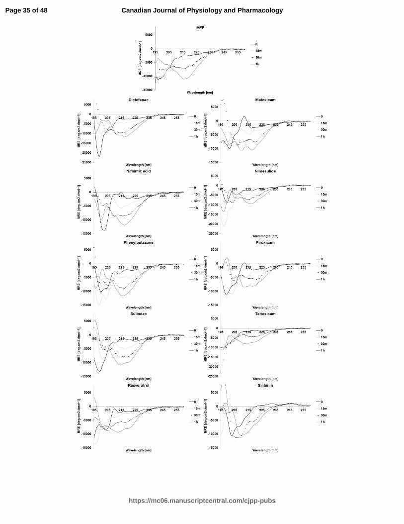
incubation time of 30 minutes (data not shown). After an incubation time of 1h, a major
negative peak at 220 nm was observed, indicative of a predominant β-sheet secondary
structure. The CD spectra recorded at an incubation time of 1h were shown to compare
the untreated and treated conditions (Fig. 2).
The CD spectra of niflumic acid, phenylbutazone, and nimesulide showed
evidence of extensive β-sheet formation at the final time point, suggesting the decrease in
ThT fluorescence intensity may be due to the displacement of the ThT dye (Suzuki et al.
2012) or to the formation of non-fibrillar but still β-sheet aggregates.
The spectrum of IAPP in presence of diclofenac, piroxicam and, to a lesser extent,
sulindac shows the two negative peaks at 208 nm and around 220-221 nm characteristic
of the exciton splitting found in predominantly α-helical structures. Silibinin, tenoxicam,
and meloxicam showed a mixed β-sheet/random coil spectra at the final time-point,
indicating a delayed transition or the formation of more disordered aggregates than the
fibers produced in the absence of these inhibitors. Resveratrol showed a more
complicated, less easily interpreted spectrum.
NSAIDs abrogate the oligomerization of hIAPP
Page 13 of 48
https://mc06.manuscriptcentral.com/cjpp-pubs
Canadian Journal of Physiology and Pharmacology
Draft
14
Soluble oligomeric species of hIAPP contribute to the deleterious cellular effect
by binding to bilipidic membranes leading to cell membrane leakage (Buchanan et al.
2013; Cao et al. 2013; Gurlo et al. 2010; Jayasinghe and Langen 2007; Jelinek and
Sheynis 2010; Khemtemourian et al. 2008). PICUP assays were performed to determine
the effect of NSAIDs on the oligomerization of hIAPP. The potent NSAIDs in affecting
hIAPP aggregate were tested at a molar ratio of 1:5. Without NSAID and irradiation
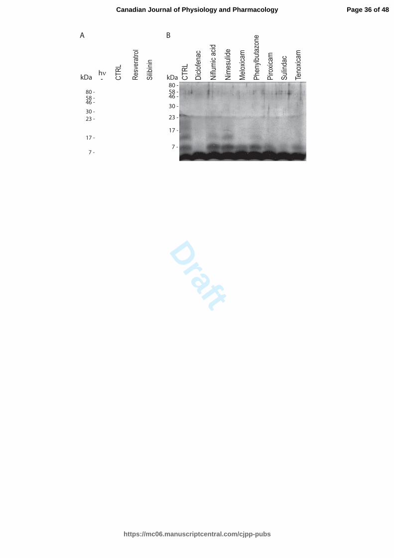
treatments, hIAPP was predominantly in a monomeric state. When hIAPP was subjected
to light exposure, a mixture of monomer, dimer, trimer, tetramer and higher oligomers
was obtained (Fig. 3), illustrating the strong oligomerization potential of hIAPP. The
oligomerization of hIAPP was inhibited for samples treated with resveratrol and silibinin,
consistent with previous experiments (Cheng et al. 2012; Jiang et al. 2011).
The oligomerization of hIAPP was abrogated following a treatment with
diclofenac. The second most potent inhibitor was sulindac. The dimeric state was
observed in samples treated with the oxicam family (meloxicam, piroxicam and
tenoxicam). Niflumic acid, nimesulide and phenylbutazone were able to partially block
the oligomerization process.
NSAIDs protect INS-1 cells from toxic hIAPP fibrils
The potential protective effect of NSAIDs against hIAPP fibrils cytotoxicity was
investigated. Pre-incubated hIAPP was added to INS-1 cells for 24 h in the presence or
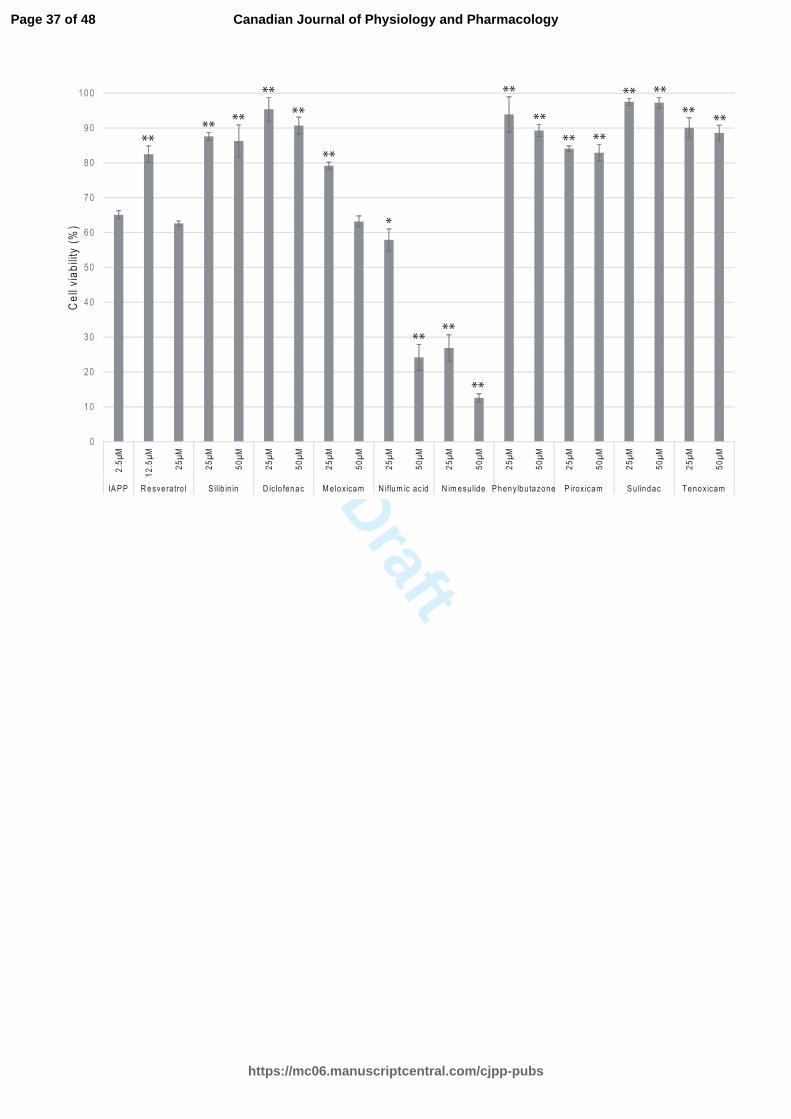
absence of increasing concentration of NSAIDs (Fig. 4). Resveratrol protected cells at
12.5 µM (molar ratio 1:5). The cell viability was significantly preserved with silibinin,
diclofenac, meloxicam, phenylbutazone, sulindac and tenoxicam at a molar ratio of 1:10.
Page 14 of 48
https://mc06.manuscriptcentral.com/cjpp-pubs
Canadian Journal of Physiology and Pharmacology
Draft
15
These drugs were not toxic at a higher concentration (50 µM, molar ratio of 1:20). Higher
concentrations of meloxicam and resveratrol (50 µM) did not have a protective effect,
whereas lower concentrations (25 µM) reduced the deleterious effect of hIAPP fibrils.
The protective effect of resveratrol and meloxicam against hIAPP aggregation has a
narrow therapeutic window. Niflumic acid and nimesulide are toxic at concentrations
affecting hIAPP aggregation. Thus, a protective effect of these drugs was not observed.
Discussion
The global prevalence of type 2 diabetes among adults population worldwide was
approximated to be 9 % in 2014 (www.who.int/mediacentre/factsheets/fs312/en/). Up to
1.5 million deaths were estimated in 2012 to be directly attributed to type 2 diabetes
(www.who.int/mediacentre/factsheets/fs312/en/). There is a compelling need to improve
the prevention and/or treatment of type 2 diabetes. IAPP possesses a potent ability to
aggregate into mature fibrils in the islet of Langerhans (Buchanan et al. 2013). Mature
linear fibrils and intermediate forms, such as oligomers, provoke cell damage, oxidative
stress and apoptosis (Abedini and Schmidt 2013; Cao et al. 2013; Gurlo et al. 2010;
Jayasinghe and Langen 2007; Khemtemourian et al. 2008). Amyloid deposition
contributes to the β-cell loss and the progression of the disease (Abedini and Schmidt
2013; Hoppener et al. 2002; Jaikaran and Clark 2001; Kamata et al. 2014). Treatment
with inhibitors of hIAPP fibril or oligomer formation presents a potential new
pharmacotherapeutic strategy for the management of type 2 diabetes (Cheng et al. 2013;
Frydman-Marom et al. 2011; Hard and Lendel 2012; Porat et al. 2006).
Page 15 of 48
https://mc06.manuscriptcentral.com/cjpp-pubs
Canadian Journal of Physiology and Pharmacology
Draft
16
Non-steroidal anti-inflammatory drugs (NSAIDs) have been used in a plethora of
inflammatory conditions based on the inhibition of COX-2. There are contradictory
reports in regard to the amyloid inhibitory effect of NSAIDs in Alzheimer’s disease (AD)
(Browne et al. 2006; Cole et al. 2004; Dong et al. 2014; Hillmann et al. 2012; Lim et al.
2000; McKee et al. 2008; Netland et al. 1998; Yan et al. 2003). Previous investigations
reported that the Aβ-amyloid induces inflammation, but the inflammatory responses
occur earlier than the formation of the senile plaque (Cole et al. 2004). The
physiopathology underlying AD involves an inflammatory process that has not been fully
understood and clinical studies have been reported conflicting data (Browne et al. 2006;
Cole et al. 2004; Dong et al. 2014; Hillmann et al. 2012; Lim et al. 2000; McKee et al.
2008; Netland et al. 1998; Yan et al. 2003). Rofecoxib, a selective COX-2 inhibitor,
failed to reduce the progression of cognitive impairment in patients suffering from mild
or moderate AD (Aisen et al. 2003). However, another study reported that COX-2
inhibition improves the suppression of memory associated with Aβ-amyloid deposition in
patients with AD. In a transgenic mouse model of AD, ibuprofen, a non-selective COX-2
inhibitor, reduced AD-type pathologic change after 6 months of treatment in comparison
with an untreated control group (Lim et al. 2000). Whether selective COX-2 inhibitors
have clinical benefit remains to be elucidated.
There are limits to the interpretation of each assay that was performed, hence it is
critical to consider the results of the different assays altogether before drawing
conclusions. This study highlights compounds and structures of potential interest. For
practical purposes, assays were completed within few hours (shorter and longer kinetics).
Page 16 of 48
https://mc06.manuscriptcentral.com/cjpp-pubs
Canadian Journal of Physiology and Pharmacology
Draft
17
ThT assays can be optimized by simple variations of initial testing conditions,
compromising between physiologically relevant conditions and the need for a relatively
fast reaction. When high-throughput screening of small molecules in relation to
aggregation is used, it is important to complement the study with other assays,
particularly a cell-based assay. Drugs may be capable of displacing ThT binding,
resulting in false positives (false hit compound). Which is why, we complemented the
Thioflavin T-based fluorescence assays with the Far-UV circular discroism spectrometry,
the photo-induced cross-linking (PICUP) assay and a biological assay. There are some
limits in the interpretation of CD spectra for the apparent decrease at 220 nm due to the
absorbance of the inhibitors, which stresses the importance of performing multiple assays
before drawing conclusions on the potential efficacy of compounds. The cell-based assay
used in our study involved pre-formed hIAPP amyloid before the drug challenge.
Consequently, the effective drugs must be able to destabilize the fibril state to
demonstrate a cytoprotective effect and/or impede the activation of apoptosis.
Few studies have evaluated the NSAIDs potential to abrogate the formation of
fibrils. A series of analogues of diclofenac were effective in vitro in inhibiting the
transthyretin fibrillization (Oza et al. 2002). In our study, several NSAIDs, such as
diclofenac, phenylbutazone, sulindac and the oxicam representatives, exerted a
significant inhibitory effect on fibril formation and delayed the structural transition of
hIAPP. Meloxicam and sulindac were the most potent inhibitors of fibrillization, whereas
diclofenac and sulindac were more potent in abrogating oligomerization of hIAPP. They
protected pancreatic INS-1 cell from the deleterious effect of hIAPP fibrils. Ketorolac
was not effective compounds, consistent with previous studies (Tu et al. 2014).
Page 17 of 48
https://mc06.manuscriptcentral.com/cjpp-pubs
Canadian Journal of Physiology and Pharmacology
Draft
18
The diclofenac derivatives prepared in the transthyretin amyloid fibril formation
study depicted structure-activity relationships, applicable to impedance of hIAPP
aggregation (Oza et al. 2002). Diclofenac and its related derivatives bind to the retinol
binding pocket of TTR via computational docking. However, other interactions and
targets are possible and we hypothesized that these molecules act as universal amyloid
inhibitors, such as polyphenols, by inhibiting aggregation with a combination of
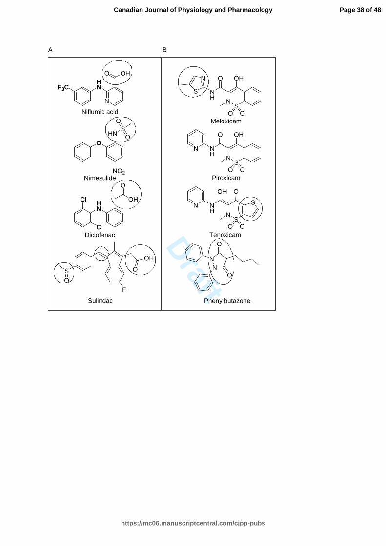
structural rigidity and aromatic stacking. This study highlighted the requirement of a
carboxylic acid directly attached to the benzene ring or separated with a methylene (Oza
et al. 2002). In the second column of Figure 5 display the potent molecules that bear a
carboxylic acid or sulfonamide in the current study. The protection of the carboxylic acid
moiety, found in aceclofenac, abolished the activity to compare with diclofenac. To gain
further information, it would be interesting to study the impact of acidic moieties with
various pKas on hIAPP amyloidosis. Another information arising from the transthyretin
study is the chlorines attached to the aromatic ring that bind to the protein (Oza et al.
2002). The chlorines were either positioned symmetrically in ortho or metha to provide
binding, whereas their absence abolished the activity (Oza et al. 2002). Adding a
carboxylic acid in para allowed an additional electrostatic interaction with the protein
transthyretin (Oza et al. 2002). Herein, the NSAIDs molecular screening on hIAPP fibrils
formation led to similar conclusions regarding the substitution patterns on the benzene
ring. Interestingly, the aromatic ring of nimesulide is devoid of halides and niflumic acid
benzene bears a trifluoromethyl group. Niflumic acid and nimesulide were less potent
than diclofenac. Sulindac has a similar fibril inhibition activity to diclofenac. The
aromatic ring has a methylsulfinyl moiety in para, not found in ketorolac, suggesting that
Page 18 of 48
https://mc06.manuscriptcentral.com/cjpp-pubs
Canadian Journal of Physiology and Pharmacology
Draft
19
an electron donor is required for fibril inhibition and could be important in the absence of
halides.
The structure-activity relationships between diclofenac, niflumic acid, nimesulide,
and their fibril inhibition activities against hIAPP underline the importance of a
negatively charged substituent between the aromatic rings (Fig. 5). Sulindac has a double
bound that can be delocalized easily to provide electrons, considering the electronic
resonance. Sulindac has similarity in structure with the exception of an indenyl moiety,
suggesting that the benzene ring could be substituted by a fused ring or heterocyclic
aromatic ring. Amyloid inhibitors bear aromatic moieties that can potentially be stacked
between other aromatic residues from the protein and abrogate the self-assembly of
oligomers (Cheng et al. 2013; Frydman-Marom et al. 2011; Hard and Lendel 2012; Porat
et al. 2006). The aromatic structure found in the diarylamine (second column Fig. 5) and
oxicam scaffold (first column Fig. 5) abrogates the oligomerization of hIAPP potentially
by preventing the aromatic interactions between the aromatic residues of hIAPP.
Phenylbutazone and the oxicam family provide a different chemistry scaffold than
the diarylamine scaffold (diclofenac), to prepare other derivatives (first column of Fig.
5). Among the oxicam family members, meloxicam had the strongest fibril inhibition
activity and tenoxicam provided the highest survival when INS-1 cells were challenged
with toxic fibrils. Tenoxicam bears a 2,3-dihydrothiophene that is substituted by a
benzene moiety in meloxicam and piroxicam. Meloxicam differs from Piroxicam and
tenoxicam, having a 5-methyl-2-thiazolyl rather than a pyridine moiety. However, the 5-
methyl-2-thiazolyl moiety could potentially explain the deleterious effect of meloxicam
Page 19 of 48
https://mc06.manuscriptcentral.com/cjpp-pubs
Canadian Journal of Physiology and Pharmacology
Draft
20
on INS-1 cells and further study is needed to confirm this hypothesis. The molecular
structure of phenylbutazone is quite similar to the oxicam family with a heterocycle
bearing two amines and two carbonyls, resulting in nucleophilicity. Phenylbutazone is
very toxic to human, causing anaplastic anemia and liver damage (Goodman et al. 2011).
However, it has been extensively used in veterinary medicine (Kahn et al. 2010).
Chemical modification will be required not only to improve its efficacy toward hIAPP
fibrillization but to modify the toxicologic effect in humans.
The present in vitro study demonstrates that several NSAIDs inhibit amyloid
formation. Multiple probes were used to study the inhibition of the toxic hIAPP
assembly. It has been reported that oligomers are important entities involved in the
mechanism of hIAPP amyloid cytotoxicity. The most potent drugs against hIAPP
oligomerization were diclofenac and sulindac. Diclofenac, meloxicam, phenylbutazone,
piroxicam, sulindac and tenoxicam conferred cell protection against toxic fibrils.
Chemical scaffold from the enolic acid class (oxicam and phenylbutazone) and the acetic
acid class (diclofenac, sulindac) could be modified to generate new inhibitors of hIAPP
aggregation. NSAID derived compound scaffolds are interesting potential candidates for
the development of type 2 diabetes treatment.
Acknowledgment
This work was supported by a grant from Diabètes Québec. We are very grateful
to Mr. Frédéric Berthiaume for the technical advice and Dr. Mostafa Hatam for the
peptide synthesis.
Page 20 of 48
https://mc06.manuscriptcentral.com/cjpp-pubs
Canadian Journal of Physiology and Pharmacology
Draft
21
References
Abedini, A., and Schmidt, A.M. 2013. Mechanisms of islet amyloidosis toxicity in type 2
diabetes. FEBS Lett. 587(8): 1119-1127. doi: 10.1016/j.febslet.2013.01.017.
Aisen, P.S., Schafer, K.A., Grundman, M., Pfeiffer, E., Sano, M., Davis, K.L., Farlow,
M.R., Jin, S., Thomas, R.G., Thal, L.J., and Alzheimer's Disease Cooperative, S. 2003.
Effects of rofecoxib or naproxen vs placebo on Alzheimer disease progression: a
randomized controlled trial. J. Am. Med. Assoc. 289(21): 2819-2826. doi:
10.1001/jama.289.21.2819.
Bitan, G., and Teplow, D.B. 2004. Rapid photochemical cross-linking--a new tool for
studies of metastable, amyloidogenic protein assemblies. Accounts of C Research,
37(6): 357-364. doi: 10.1021/ar000214l.
Brender, J. R., Krishnamoorthy, J., Sciacca, M. F. M., Vivekanandan, S., D'Urso, L.,
Chen, J., La Rosa, C., and Ramamoorthy, A. 2015. Probing the Sources of the
Apparent Irreproducibility of Amyloid Formation: Drastic Changes in Kinetics and a
Switch in Mechanism Due to Micelle like Oligomer Formation at Critical
Concentrations of IAPP. J. Phys. Chem. B 119(7), 2886-2896.
doi:10.1021/jp511758w.
Browne, K.D., Iwata, A., Putt, M.E., and Smith, D.H. 2006. Chronic ibuprofen
administration worsens cognitive outcome following traumatic brain injury in rats.
Exp. Neurol. 201(2): 301-307. doi: 10.1016/j.expneurol.2006.04.008.
Page 21 of 48
https://mc06.manuscriptcentral.com/cjpp-pubs
Canadian Journal of Physiology and Pharmacology
Draft
22
Buchanan, L.E., Dunkelberger, E.B., Tran, H.Q., Cheng, P.N., Chiu, C.C., Cao, P.,
Raleigh, D.P., de Pablo, J.J., Nowick, J.S., and Zanni, M.T. 2013. Mechanism of IAPP
amyloid fibril formation involves an intermediate with a transient beta-sheet. Proc.
Natl. Acad. Sci. U. S. A. 110(48): 19285-19290. doi: 10.1073/pnas.1314481110.
Buell, A.K., Dobson, C.M., Knowles, T.P., and Welland, M.E. 2010. Interactions
between amyloidophilic dyes and their relevance to studies of amyloid inhibitors.
Biophys. J. 99(10): 3492-3497. doi: 10.1016/j.bpj.2010.08.074.
Cao, P., Abedini, A., Wang, H., Tu, L.H., Zhang, X., Schmidt, A.M., and Raleigh, D.P.
2013. Islet amyloid polypeptide toxicity and membrane interactions. Proc. Natl. Acad.
Sci. U. S. A. 110(48): 19279-19284. doi: 10.1073/pnas.1305517110.
Cheng, B., Gong, H., Li, X., Sun, Y., Zhang, X., Chen, H., Liu, X., Zheng, L., and
Huang, K. 2012. Silibinin inhibits the toxic aggregation of human islet amyloid
polypeptide. Biochem. Biophys. Res. Commun. 419(3): 495-499. doi:
10.1016/j.bbrc.2012.02.042.
Cheng, B., Gong, H., Xiao, H., Petersen, R.B., Zheng, L., and Huang, K. 2013. Inhibiting
toxic aggregation of amyloidogenic proteins: a therapeutic strategy for protein
misfolding diseases. Biochim. Biophys. Acta, 1830(10): 4860-4871. doi:
10.1016/j.bbagen.2013.06.029.
Cole, G.M., Morihara, T., Lim, G.P., Yang, F., Begum, A., and Frautschy, S.A. 2004.
NSAID and antioxidant prevention of Alzheimer's disease: lessons from in vitro and
animal models. Ann. N. Y. Acad. Sci. 1035: 68-84. doi: 10.1196/annals.1332.005.
Page 22 of 48
https://mc06.manuscriptcentral.com/cjpp-pubs
Canadian Journal of Physiology and Pharmacology
Draft
23
Dong, Z., Yan, L., Huang, G., Zhang, L., Mei, B., and Meng, B. 2014. Ibuprofen partially
attenuates neurodegenerative symptoms in presenilin conditional double-knockout
mice. Neuroscience, 270: 58-68. doi: 10.1016/j.neuroscience.2014.03.048.
Fortin, J., Patenaude, A., Deschesnes, R.G., Cote, M.F., Petitclerc, E., and C-Gaudreault,
R. 2010. ASK1-P38 Pathway is Important for Anoikis Induced by Microtubule-
Targeting Aryl Chloroethylureas. J. Pharm. Pharm. Sci. 13(2): 175-190.
Frydman-Marom, A., Shaltiel-Karyo, R., Moshe, S., and Gazit, E. 2011. The generic
amyloid formation inhibition effect of a designed small aromatic beta-breaking
peptide. Amyloid, 18(3): 119-127. doi: 10.3109/13506129.2011.582902.
Goodman, L.S., Brunton, L.L., Chabner, B., and Knollmann, B.C. 2011. Goodman &
Gilman's the pharmacological basis of therapeutics. McGraw-Hill Medical,, New York
; Toronto.
Gurlo, T., Ryazantsev, S., Huang, C.J., Yeh, M.W., Reber, H.A., Hines, O.J., O'Brien,
T.D., Glabe, C.G., and Butler, P.C. 2010. Evidence for proteotoxicity in beta cells in
type 2 diabetes: toxic islet amyloid polypeptide oligomers form intracellularly in the
secretory pathway. Am. J. Pathol. 176(2): 861-869. doi: 10.2353/ajpath.2010.090532.
Hard, T., and Lendel, C. 2012. Inhibition of amyloid formation. J. Mol. Biol. 421(4-5):
441-465. doi: 10.1016/j.jmb.2011.12.062.
Hillmann, A., Hahn, S., Schilling, S., Hoffmann, T., Demuth, H.U., Bulic, B., Schneider-
Axmann, T., Bayer, T.A., Weggen, S., and Wirths, O. 2012. No improvement after
Page 23 of 48
https://mc06.manuscriptcentral.com/cjpp-pubs
Canadian Journal of Physiology and Pharmacology

Draft
24
chronic ibuprofen treatment in the 5XFAD mouse model of Alzheimer's disease.
Neurobiology of aging, 33(4): 833 e839-850. doi:
10.1016/j.neurobiolaging.2011.08.006.
Hoppener, J.W., Nieuwenhuis, M.G., Vroom, T.M., Ahren, B., and Lips, C.J. 2002. Role
of islet amyloid in type 2 diabetes mellitus: consequence or cause? Mol. Cell.
Endocrinol. 197(1-2): 205-212.
Jaikaran, E.T., and Clark, A. 2001. Islet amyloid and type 2 diabetes: from molecular
misfolding to islet pathophysiology. Biochim. Biophys. Acta, 1537(3): 179-203.
Jayasinghe, S.A., and Langen, R. 2007. Membrane interaction of islet amyloid
polypeptide. Biochim. Biophys. Acta, 1768(8): 2002-2009. doi:
10.1016/j.bbamem.2007.01.022.
Jelinek, R., and Sheynis, T. 2010. Amyloid - membrane interactions: experimental
approaches and techniques. Current protein & peptide science, 11(5): 372-384.
Jiang, P., Li, W., Shea, J.E., and Mu, Y. 2011. Resveratrol inhibits the formation of
multiple-layered beta-sheet oligomers of the human islet amyloid polypeptide segment
22-27. Biophys. J. 100(6): 1550-1558. doi: 10.1016/j.bpj.2011.02.010.
Kahn, C.M., Line, S., and Merck & Co. 2010. The Merck veterinary manual. 10th ed.
Merck & Co., Whitehouse Station, N.J.
Kamata, K., Mizukami, H., Inaba, W., Tsuboi, K., Tateishi, Y., Yoshida, T., and
Yagihashi, S. 2014. Islet amyloid with macrophage migration correlates with
Page 24 of 48
https://mc06.manuscriptcentral.com/cjpp-pubs
Canadian Journal of Physiology and Pharmacology
Draft
25
augmented beta-cell deficits in type 2 diabetic patients. Amyloid, 21(3): 191-201. doi:
10.3109/13506129.2014.937857.
Khemtemourian, L., Killian, J.A., Hoppener, J.W., and Engel, M.F. 2008. Recent insights
in islet amyloid polypeptide-induced membrane disruption and its role in beta-cell
death in type 2 diabetes mellitus. Exp. Diabetes Res. 2008: 421287. doi:
10.1155/2008/421287.
Lim, G.P., Yang, F., Chu, T., Chen, P., Beech, W., Teter, B., Tran, T., Ubeda, O., Ashe,
K.H., Frautschy, S.A., and Cole, G.M. 2000. Ibuprofen suppresses plaque pathology
and inflammation in a mouse model for Alzheimer's disease. J. Neurosci. 20(15):
5709-5714.
Luca, S., Yau, W. M., Leapman, R., and Tycko, R. 2007. Peptide conformation and
supramolecular organization in amylin fibrils: Constraints from solid-state NMR.
Biochemistry, 46(47): 13505-13522. doi: 10.1021/bi701427q.
Mishra, R., Geyer, M., and Winter, R. 2009. NMR spectroscopic investigation of early
events in IAPP amyloid fibril formation. Chembiochem, 10(11): 1769-1772. Doi:
10.1002/cbic.200900237.
McKee, A.C., Carreras, I., Hossain, L., Ryu, H., Klein, W.L., Oddo, S., LaFerla, F.M.,
Jenkins, B.G., Kowall, N.W., and Dedeoglu, A. 2008. Ibuprofen reduces Abeta,
hyperphosphorylated tau and memory deficits in Alzheimer mice. Brain Res. 1207:
225-236. doi: 10.1016/j.brainres.2008.01.095.
Page 25 of 48
https://mc06.manuscriptcentral.com/cjpp-pubs
Canadian Journal of Physiology and Pharmacology
Draft
26
Netland, E.E., Newton, J.L., Majocha, R.E., and Tate, B.A. 1998. Indomethacin reverses
the microglial response to amyloid beta-protein. Neurobiology of aging, 19(3): 201-
204.
O'Brien, T.D., Butler, P.C., Westermark, P., and Johnson, K.H. 1993. Islet amyloid
polypeptide: a review of its biology and potential roles in the pathogenesis of diabetes
mellitus. Vet. Pathol. 30(4): 317-332.
Oza, V.B., Smith, C., Raman, P., Koepf, E.K., Lashuel, H.A., Petrassi, H.M., Chiang,
K.P., Powers, E.T., Sachettinni, J., and Kelly, J.W. 2002. Synthesis, structure, and
activity of diclofenac analogues as transthyretin amyloid fibril formation inhibitors. J.
Med. Chem. 45(2): 321-332.
Padrick, S. B., and Miranker, A. D. 2002. Islet amyloid: Phase partitioning and secondary
nucleation are central to the mechanism of fibrillogenesis. Biochemistry, 41(14),
4694-4703. doi: 10.1021/bi0160462.
Paulsson, J.F., Benoit-Biancamano, M.O., Schaffer, L., and Dahl, K. 2011. Ferret islet
amyloid polypeptide (IAPP): characterization of in vitro and in vivo amyloidogenicity.
Amyloid, 18(4): 222-228. doi: 10.3109/13506129.2011.627956.
Porat, Y., Abramowitz, A., and Gazit, E. 2006. Inhibition of amyloid fibril formation by
polyphenols: structural similarity and aromatic interactions as a common inhibition
mechanism. Chemical biology & drug design, 67(1): 27-37. doi: 10.1111/j.1747-
0285.2005.00318.x.
Page 26 of 48
https://mc06.manuscriptcentral.com/cjpp-pubs
Canadian Journal of Physiology and Pharmacology
Draft
27
Reddy, A. S., Wang, L., Singh, S., Ling, Y. L., Buchanan, L., Zanni, M. T., Skinner, J.
L., and de Pablo, J. J. 2010. Stable and Metastable States of Human Amylin in
Solution. Biophysique journal, 99(7): 2208-2216. doi: 10.1016/j.bpj.2010.07.014.
Ruschak, A.M., and Miranker, A.D. 2007. Fiber-dependent amyloid formation as
catalysis of an existing reaction pathway. Proc. Natl. Acad. Sci. U. S. A. 104(30):
12341-12346. doi: 10.1073/pnas.0703306104.
Shvadchak, V.V., Claessens, M.M.A.E., and Subramaniam, V. 2015. Fibril Breaking
Accelerates alpha-Synuclein Fibrillization. J. Phys. Chem. B 119(5): 1912-1918. doi:
10.1021/jp5111604.
Sipe, J.D., Benson, M.D., Buxbaum, J.N., Ikeda, S., Merlini, G., Saraiva, M.J., and
Westermark, P. 2014. Nomenclature 2014: Amyloid fibril proteins and clinical
classification of the amyloidosis. Amyloid, 21(4): 221-224. doi:
10.3109/13506129.2014.964858.
Sreerama, N., and Woody, R.W. 2004. Computation and analysis of protein circular
dichroism spectra. Meth. Enzymol. 383: 318-351. doi: 10.1016/S0076-
6879(04)83013-1.
Suzuki, Y., Brender, J.R., Hartman, K., Ramamoorthy, A., and Marsh, E.N.G. 2012.
Alternative Pathways of Human Islet Amyloid Polypeptide Aggregation Distinguished
by F-19 Nuclear Magnetic Resonance-Detected Kinetics of Monomer Consumption.
Biochemistry, 51 (41), 8154-8162. doi: 10.1021/bi3012548.
Page 27 of 48
https://mc06.manuscriptcentral.com/cjpp-pubs
Canadian Journal of Physiology and Pharmacology
Draft
28
Tu, L.H., Noor, H., Cao, P., and Raleigh, D.P. 2014. Aspirin, diabetes, and amyloid: re-
examination of the inhibition of amyloid formation by aspirin and ketoprofen. ACS
Chemical Biology, 9(7): 1632-1637. doi: 10.1021/cb500162w.
Westermark, P., Andersson, A., and Westermark, G.T. 2011. Islet amyloid polypeptide,
islet amyloid, and diabetes mellitus. Physiol. Rev. 91(3): 795-826. doi:
10.1152/physrev.00042.2009.
Williamson, J.A., and Miranker, A.D. 2007. Direct detection of transient alpha-helical
states in islet amyloid polypeptide. Protein Science, 16(1): 110-117. doi:
10.1110/ps.062486907.
Yan, Q., Zhang, J., Liu, H., Babu-Khan, S., Vassar, R., Biere, A.L., Citron, M., and
Landreth, G. 2003. Anti-inflammatory drug therapy alters beta-amyloid processing
and deposition in an animal model of Alzheimer's disease. J. Neurosci. 23(20): 7504-
7509.
Yonemoto, I.T., Kroon, G.J., Dyson, H.J., Balch, W.E., and Kelly, J.W. 2008. Amylin
proprotein processing generates progressively more amyloidogenic peptides that
initially sample the helical state. Biochemistry, 47(37), 9900-9910. doi:
10.1021/bi800828u.
Zhang, X., Cheng, B., Gong, H., Li, C., Chen, H., Zheng, L., and Huang, K. 2011.
Porcine islet amyloid polypeptide fragments are refractory to amyloid formation.
FEBS Lett. 585(1): 71-77. doi: 10.1016/j.febslet.2010.11.050.
Page 28 of 48
https://mc06.manuscriptcentral.com/cjpp-pubs
Canadian Journal of Physiology and Pharmacology
Draft
29
Figure captions
Fig. 1. A Relative ThT fluorescence intensity of hIAPP with different effective drug
treatments (silibinin, resveratrol, celecoxib, diclofenac, indomethacin, meloxicam,
niflumic acid, nimesulide, phenylbutazone, piroxicam, sulindac, and tenoxicam) tested at
a concentration of 100 µM (longer agitation). ThT-fluorescence of hIAPP was assessed at
25°C in PBS buffer at a peptide concentration of 10 µM and a molar ratio of 1:10. B
Relative ThT fluorescence intensity of hIAPP with identical drug treatments obtained
from a shorter agitation.
Fig. 2. Far-UV circular dichroism spectra of hIAPP recorded in the presence of the
vehicle (1% HFIP) or different drug treatments. Spectra were recorded at 25°C in PBS
buffer using hIAPP at 15 µM and drug treatments at 25 µM (molar ratio of 1:1.67). The
incubation time were 0, 15, 30 and 60 min.
Fig. 3. Oligomerization status of hIAPP studied by the photo-induced cross linking
(PICUP) assay. (a) Lane 1, hIAPP without irradiation; lane 2, hIAPP irradiated for 3 sec;
lanes 3-4, hIAPP with positive controls irradiated for 3 sec. The molar ratio was 1:5, 0.25
mM of hIAPP and 1.25 mM of positive controls (resveratrol and silibinin). (b) Lane 1,
hIAPP irradiated for 3 sec; lanes 2-9, hIAPP with different drug treatment irradiated for 3
sec. The molar ratio was 1:5, 0.25 mM of hIAPP and 1.25 mM of drugs (diclofenac,
meloxicam, niflumic acid, nimesulide, phenylbutazone, piroxicam, sulindac and
tenoxicam).
Page 29 of 48
https://mc06.manuscriptcentral.com/cjpp-pubs
Canadian Journal of Physiology and Pharmacology
Draft
30
Fig. 4. INS-1 cell viability in the presence of hIAPP alone and hIAPP with different drug
treatments, such as resveratrol, silibinin, diclofenac, meloxicam, niflumic acid,
nimesulide, phenylbutazone, piroxicam, sulindac and tenoxicam, as determined by
resazurin-based assays. hIAPP was tested at a concentration of 2.5 µM and with
effective concentrations (12.5, 25 or 50 µM) of each drug. The molar ratios were 1:5,
1:10 and 1:20. The one-way analysis of variance with Dunnett's multiple comparison test
showed significant differences between IAPP and several drug treatments (* and * * are
indicative of p < 0.05 and p < 0.001, respectively).
Fig. 5. Proposed structure-activity relationships between the acetic acid class (A) and
between the enolic acid class (B).
Abbreviations list:
AD, Alzheimer's disease; COX, cyclooxygenase; hIAPP, human islet amyloid
polypeptide; NSAID, non-steroidal anti-inflammatory.
Page 30 of 48
https://mc06.manuscriptcentral.com/cjpp-pubs
Canadian Journal of Physiology and Pharmacology
Draft
A
Page 31 of 48
https://mc06.manuscriptcentral.com/cjpp-pubs
Canadian Journal of Physiology and Pharmacology
Draft
Page 32 of 48
https://mc06.manuscriptcentral.com/cjpp-pubs
Canadian Journal of Physiology and Pharmacology
Draft
B
Page 33 of 48
https://mc06.manuscriptcentral.com/cjpp-pubs
Canadian Journal of Physiology and Pharmacology
Draft
Page 34 of 48
https://mc06.manuscriptcentral.com/cjpp-pubs
Canadian Journal of Physiology and Pharmacology
Draft
Page 35 of 48
https://mc06.manuscriptcentral.com/cjpp-pubs
Canadian Journal of Physiology and Pharmacology
Draft
A B
Diclo
fenac
Niflu
mic a
cid
Nime
sulid
e
Melox
icam
Phen
ylbuta
zone
Piro
xicam
Sulin
dac
Teno
xicam
CTRL
kDa
17 -
23 -
30 -
46 -58 -80 -
7 -
h CTRL
- Resv
eratr
ol
Silib
inin
kDa
17 -
23 -30 -
46 -58 -80 -
7 -
Page 36 of 48
https://mc06.manuscriptcentral.com/cjpp-pubs
Canadian Journal of Physiology and Pharmacology
Draft0
10
20
30
40
50
60
70
80
90
100
2.5µ
M
12.5
µM
25µM
25µM
50µM
25µM
50µM
25µM
50µM
25µM
50µM
25µM
50µM
25µM
50µM
25µM
50µM
25µM
50µM
25µM
50µM
IA P P R esveratro l S ilib in in D ic lo fenac M e loxicam N iflum ic ac id N im esulide P heny lbu tazone P irox icam S u lindac Tenoxicam
Cel
l via
bilit
y (%
) *
**** **
****
**
****
**
**
**** **
** **** **
Page 37 of 48
https://mc06.manuscriptcentral.com/cjpp-pubs
Canadian Journal of Physiology and Pharmacology
Draft
NS
O O
OH
NH
ON
S
Meloxicam
NS
O O
OH
NH
O
N
Piroxicam
N NH
OH
NS
S
O
O OTenoxicam
HN
N
OHO
F3C
Niflumic acid
O
NO2
HNS
O
O
Nimesulide
NN
O
O
Phenylbutazone
OH
OSO
FSulindac
Cl HN
Cl
OH
O
Diclofenac
A B
Page 38 of 48
https://mc06.manuscriptcentral.com/cjpp-pubs
Canadian Journal of Physiology and Pharmacology

Draft
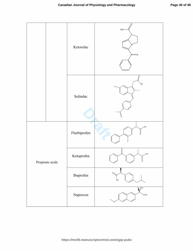
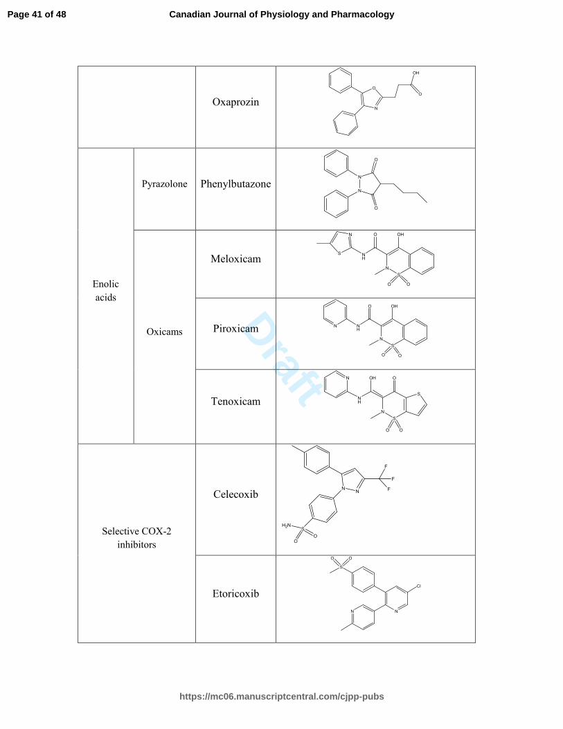
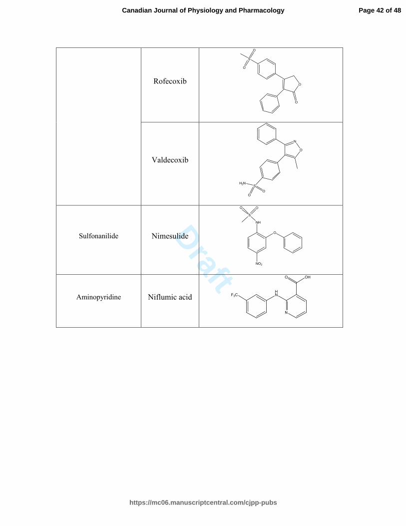
Table 1. Classification and molecular structure of positive controls and NSAIDs used in
this study.
Pharmacological class Generic name Molecular structure
Positive controls
Resveratrol HO
OH
OH
Silibinin O
HO
O
O
OH
O
HO
O OH
OH
Acetic
acids
Phenylacetic
acids
Aceclofenac
Cl
NH
Cl O
O
OH
O
Diclofenac
Cl
NH
Cl OH
O
Carbo- and
Hetero-
Cyclic
Acetic
Acids
Indomethacin N
OH
O
Cl
O
O
Page 39 of 48
https://mc06.manuscriptcentral.com/cjpp-pubs
Canadian Journal of Physiology and Pharmacology
Draft
Ketorolac
O
N
HO
O
Sulindac
OH
O
S
O
F
Propionic acids
Flurbiprofen
F
OH
O
Ketoprofen
O
OH
O
Ibuprofen OH
O
Naproxen
O
CO2H
H CH3
Page 40 of 48
https://mc06.manuscriptcentral.com/cjpp-pubs
Canadian Journal of Physiology and Pharmacology
Draft
Oxaprozin N
O
OH
O
Enolic
acids
Pyrazolone Phenylbutazone N
N
O
O
Oxicams
Meloxicam N
S
O O
OH
NH
ON
S
Piroxicam N
S
O O
OH
NH
O
N
Tenoxicam
N
NH
OH
N
S
S
O
O O
Selective COX-2
inhibitors
Celecoxib N
N
SH2N
O
O
F
F
F
Etoricoxib
NN
Cl
S
OO
Page 41 of 48
https://mc06.manuscriptcentral.com/cjpp-pubs
Canadian Journal of Physiology and Pharmacology
Draft
Rofecoxib O
O
S
O
O
Valdecoxib
O
N
SH2N
O
O
Sulfonanilide Nimesulide O
NO2
NH
S
OO
Aminopyridine Niflumic acid HN
N
OHO
F3C
Page 42 of 48
https://mc06.manuscriptcentral.com/cjpp-pubs
Canadian Journal of Physiology and Pharmacology
Draft
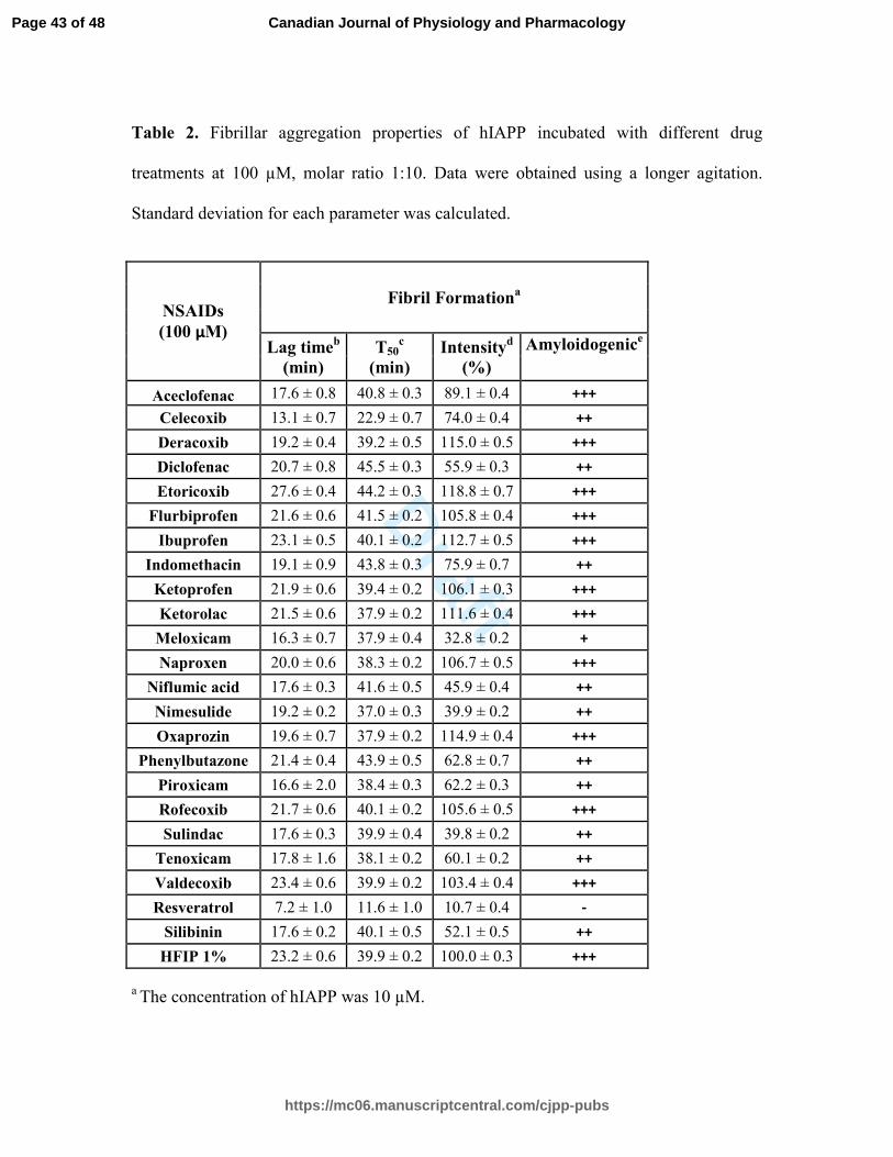
Table 2. Fibrillar aggregation properties of hIAPP incubated with different drug
treatments at 100 µM, molar ratio 1:10. Data were obtained using a longer agitation.
Standard deviation for each parameter was calculated.
NSAIDs
(100 µM)
Fibril Formationa
Lag timeb
(min)
T50c
(min)
Intensityd
(%)
Amyloidogenice
Aceclofenac 17.6 ± 0.8 40.8 ± 0.3 89.1 ± 0.4 +++
Celecoxib 13.1 ± 0.7 22.9 ± 0.7 74.0 ± 0.4 ++
Deracoxib 19.2 ± 0.4 39.2 ± 0.5 115.0 ± 0.5 +++
Diclofenac 20.7 ± 0.8 45.5 ± 0.3 55.9 ± 0.3 ++
Etoricoxib 27.6 ± 0.4 44.2 ± 0.3 118.8 ± 0.7 +++
Flurbiprofen 21.6 ± 0.6 41.5 ± 0.2 105.8 ± 0.4 +++
Ibuprofen 23.1 ± 0.5 40.1 ± 0.2 112.7 ± 0.5 +++
Indomethacin 19.1 ± 0.9 43.8 ± 0.3 75.9 ± 0.7 ++
Ketoprofen 21.9 ± 0.6 39.4 ± 0.2 106.1 ± 0.3 +++
Ketorolac 21.5 ± 0.6 37.9 ± 0.2 111.6 ± 0.4 +++
Meloxicam 16.3 ± 0.7 37.9 ± 0.4 32.8 ± 0.2 +
Naproxen 20.0 ± 0.6 38.3 ± 0.2 106.7 ± 0.5 +++
Niflumic acid 17.6 ± 0.3 41.6 ± 0.5 45.9 ± 0.4 ++
Nimesulide 19.2 ± 0.2 37.0 ± 0.3 39.9 ± 0.2 ++
Oxaprozin 19.6 ± 0.7 37.9 ± 0.2 114.9 ± 0.4 +++
Phenylbutazone 21.4 ± 0.4 43.9 ± 0.5 62.8 ± 0.7 ++
Piroxicam 16.6 ± 2.0 38.4 ± 0.3 62.2 ± 0.3 ++
Rofecoxib 21.7 ± 0.6 40.1 ± 0.2 105.6 ± 0.5 +++
Sulindac 17.6 ± 0.3 39.9 ± 0.4 39.8 ± 0.2 ++
Tenoxicam 17.8 ± 1.6 38.1 ± 0.2 60.1 ± 0.2 ++
Valdecoxib 23.4 ± 0.6 39.9 ± 0.2 103.4 ± 0.4 +++
Resveratrol 7.2 ± 1.0 11.6 ± 1.0 10.7 ± 0.4 -
Silibinin 17.6 ± 0.2 40.1 ± 0.5 52.1 ± 0.5 ++
HFIP 1% 23.2 ± 0.6 39.9 ± 0.2 100.0 ± 0.3 +++
a The concentration of hIAPP was 10 µM.
Page 43 of 48
https://mc06.manuscriptcentral.com/cjpp-pubs
Canadian Journal of Physiology and Pharmacology
Draft
b The lag time indicates the time during which no significant fibril formation occurs
followed by a rapid growth phase.
c The T50 represents the time to reach half of the maximum signal strength.
d The fluorescence intensity of hIAPP alone was set as 100 %.
e The amyloidogenic parameter is a semi-quantitative analysis of ThT-fluorescence based
amyloid formation: "+++" indicates samples with fluorescence intensity greater than 75
%, "++" represents values between 40 and 75 %, "+" is set for values between 10 and 40
%, and "-" underlines values lower than 10 %.
Page 44 of 48
https://mc06.manuscriptcentral.com/cjpp-pubs
Canadian Journal of Physiology and Pharmacology
Draft
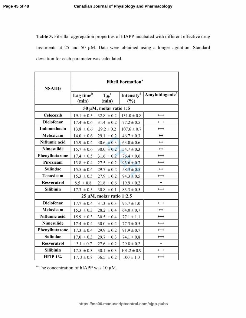
Table 3. Fibrillar aggregation properties of hIAPP incubated with different effective drug
treatments at 25 and 50 µM. Data were obtained using a longer agitation. Standard
deviation for each parameter was calculated.
NSAIDs
Fibril Formationa
Lag timeb
(min)
T50c
(min)
Intensityd
(%)
Amyloidogenice
50 µM, molar ratio 1:5
Celecoxib 19.1 ± 0.5 32.8 ± 0.2 131.0 ± 0.8 +++
Diclofenac 17.4 ± 0.6 31.4 ± 0.2 77.2 ± 0.5 +++
Indomethacin 13.8 ± 0.6 29.2 ± 0.2 107.6 ± 0.7 +++
Meloxicam 14.0 ± 0.6 29.1 ± 0.2 46.7 ± 0.3 ++
Niflumic acid 15.9 ± 0.4 30.6 ± 0.3 63.0 ± 0.6 ++
Nimesulide 15.7 ± 0.6 30.0 ± 0.2 54.7 ± 0.3 ++
Phenylbutazone 17.4 ± 0.5 31.6 ± 0.2 76.4 ± 0.6 +++
Piroxicam 13.8 ± 0.4 27.5 ± 0.2 93.6 ± 0.7 +++
Sulindac 15.5 ± 0.4 29.7 ± 0.2 58.3 ± 0.5 ++
Tenoxicam 15.3 ± 0.5 27.9 ± 0.2 94.3 ± 0.5 +++
Resveratrol 8.5 ± 0.8 21.8 ± 0.6 19.9 ± 0.2 +
Silibinin 17.3 ± 0.5 30.8 ± 0.1 83.3 ± 0.5 +++
25 µM, molar ratio 1:2.5
Diclofenac 17.7 ± 0.4 31.3 ± 0.3 95.7 ± 1.0 +++
Meloxicam 15.3 ± 0.3 28.2 ± 0.4 64.0 ± 0.7 ++
Niflumic acid 15.9 ± 0.3 30.5 ± 0.4 77.1 ± 1.1 +++
Nimesulide 17.4 ± 0.4 30.0 ± 0.2 77.3 ± 0.5 +++
Phenylbutazone 17.3 ± 0.4 29.9 ± 0.2 91.9 ± 0.7 +++
Sulindac 17.0 ± 0.3 29.7 ± 0.3 74.1 ± 0.8 +++
Resveratrol 13.1 ± 0.7 27.6 ± 0.2 29.8 ± 0.2 +
Silibinin 17.5 ± 0.3 30.1 ± 0.3 101.2 ± 0.9 +++
HFIP 1% 17. 3 ± 0.8 36.5 ± 0.2 100 ± 1.0 +++
a The concentration of hIAPP was 10 µM.
Page 45 of 48
https://mc06.manuscriptcentral.com/cjpp-pubs
Canadian Journal of Physiology and Pharmacology
Draft
b The lag time indicates the time during which no significant fibril formation occurs
followed by a rapid growth phase.
c The T50 represents the time to reach half of the maximum signal strength.
d The fluorescence intensity of hIAPP alone was set as 100 %.
e The amyloidogenic parameter is a semi-quantitative analysis of ThT-fluorescence based
amyloid formation: "+++" indicates samples with fluorescence intensity greater than 75
%, "++" represents values between 40 and 75 %, "+" is set for values between 10 and 40
%, and "-" underlines values lower than 10 %.
Page 46 of 48
https://mc06.manuscriptcentral.com/cjpp-pubs
Canadian Journal of Physiology and Pharmacology
Draft
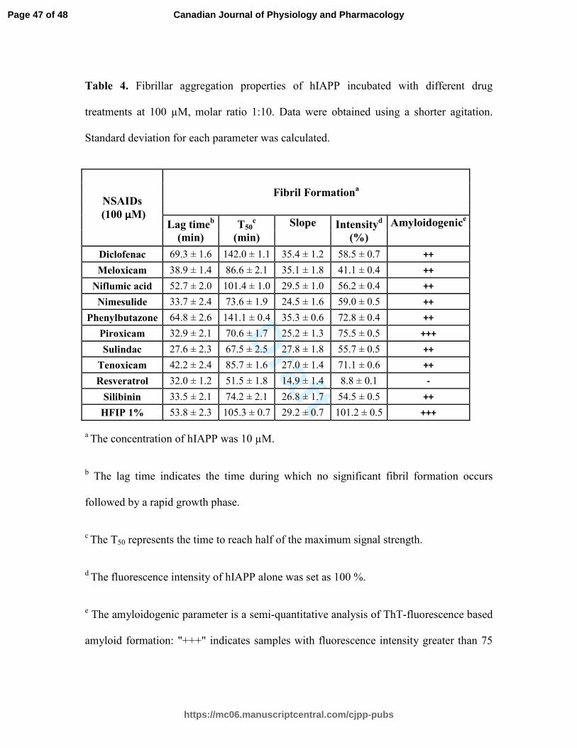
Table 4. Fibrillar aggregation properties of hIAPP incubated with different drug
treatments at 100 µM, molar ratio 1:10. Data were obtained using a shorter agitation.
Standard deviation for each parameter was calculated.
NSAIDs
(100 µM)
Fibril Formationa
Lag timeb
(min)
T50c
(min)
Slope Intensityd
(%)
Amyloidogenice
Diclofenac 69.3 ± 1.6 142.0 ± 1.1 35.4 ± 1.2 58.5 ± 0.7 ++
Meloxicam 38.9 ± 1.4 86.6 ± 2.1 35.1 ± 1.8 41.1 ± 0.4 ++
Niflumic acid 52.7 ± 2.0 101.4 ± 1.0 29.5 ± 1.0 56.2 ± 0.4 ++
Nimesulide 33.7 ± 2.4 73.6 ± 1.9 24.5 ± 1.6 59.0 ± 0.5 ++
Phenylbutazone 64.8 ± 2.6 141.1 ± 0.4 35.3 ± 0.6 72.8 ± 0.4 ++
Piroxicam 32.9 ± 2.1 70.6 ± 1.7 25.2 ± 1.3 75.5 ± 0.5 +++
Sulindac 27.6 ± 2.3 67.5 ± 2.5 27.8 ± 1.8 55.7 ± 0.5 ++
Tenoxicam 42.2 ± 2.4 85.7 ± 1.6 27.0 ± 1.4 71.1 ± 0.6 ++
Resveratrol 32.0 ± 1.2 51.5 ± 1.8 14.9 ± 1.4 8.8 ± 0.1 -
Silibinin 33.5 ± 2.1 74.2 ± 2.1 26.8 ± 1.7 54.5 ± 0.5 ++
HFIP 1% 53.8 ± 2.3 105.3 ± 0.7 29.2 ± 0.7 101.2 ± 0.5 +++
a The concentration of hIAPP was 10 µM.
b The lag time indicates the time during which no significant fibril formation occurs
followed by a rapid growth phase.
c The T50 represents the time to reach half of the maximum signal strength.
d The fluorescence intensity of hIAPP alone was set as 100 %.
e The amyloidogenic parameter is a semi-quantitative analysis of ThT-fluorescence based
amyloid formation: "+++" indicates samples with fluorescence intensity greater than 75
Page 47 of 48
https://mc06.manuscriptcentral.com/cjpp-pubs
Canadian Journal of Physiology and Pharmacology
Draft
%, "++" represents values between 40 and 75 %, "+" is set for values between 10 and 40
%, and "-" underlines values lower than 10 %.
Page 48 of 48
https://mc06.manuscriptcentral.com/cjpp-pubs
Canadian Journal of Physiology and Pharmacology