junctional epithelium
TRANSCRIPT
Dental College AzamgarhDepartment of Periodontology
Seminar On : Junctional Epithelium
Guided By: Submitted byDr. Leka Dr.Rahul KesharwaniDr. Kapil Garg PG 1st year Dr. RajeshDr. Anjum VishwasDr. Abhishek SinhaDr. Vivek TripathiDr. Payal Gupta
• Introduction
• Junctional epithelium
– Development of junctional epithelium
– Structure
– Epithelial attachment
– Dynamic aspects of junctional epithelium
– Expression of various molecules and their functions
– Permeability
– Functions
– Regeneration
– role of JE in initiation of pocket formation
• Junctional epithelium around implants
• Biologic width
• Conclusion
Introduction
• Teeth are trans-mucosal organs.
• This is a unique association in the human body where a hard
tissue emerges through the soft tissue.
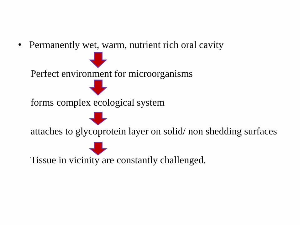
• Permanently wet, warm, nutrient rich oral cavity
Perfect environment for microorganisms
forms complex ecological system
attaches to glycoprotein layer on solid/ non shedding surfaces
Tissue in vicinity are constantly challenged.
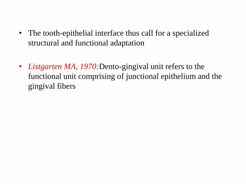
• The tooth-epithelial interface thus call for a specialized
structural and functional adaptation
• Listgarten MA, 1970:Dento-gingival unit refers to the
functional unit comprising of junctional epithelium and the
gingival fibers
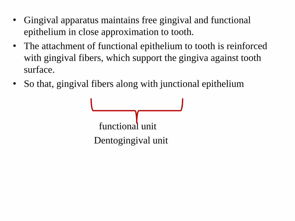
• Gingival apparatus maintains free gingival and functional
epithelium in close approximation to tooth.
• The attachment of functional epithelium to tooth is reinforced
with gingival fibers, which support the gingiva against tooth
surface.
• So that, gingival fibers along with junctional epithelium
functional unit
Dentogingival unit
History
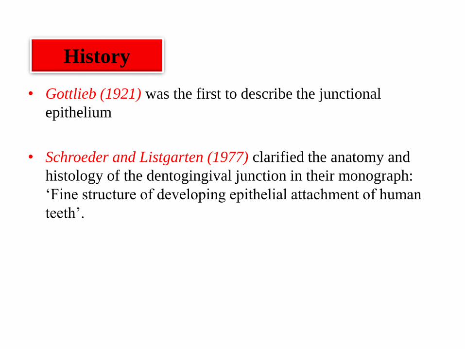
• Gottlieb (1921) was the first to describe the junctional
epithelium
• Schroeder and Listgarten (1977) clarified the anatomy and
histology of the dentogingival junction in their monograph:
‘Fine structure of developing epithelial attachment of human
teeth’.
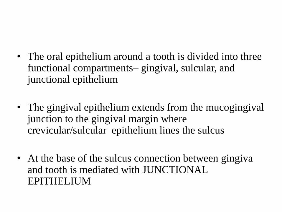
• The oral epithelium around a tooth is divided into three functional compartments– gingival, sulcular, and junctional epithelium
• The gingival epithelium extends from the mucogingivaljunction to the gingival margin where crevicular/sulcular epithelium lines the sulcus
• At the base of the sulcus connection between gingivaand tooth is mediated with JUNCTIONAL EPITHELIUM
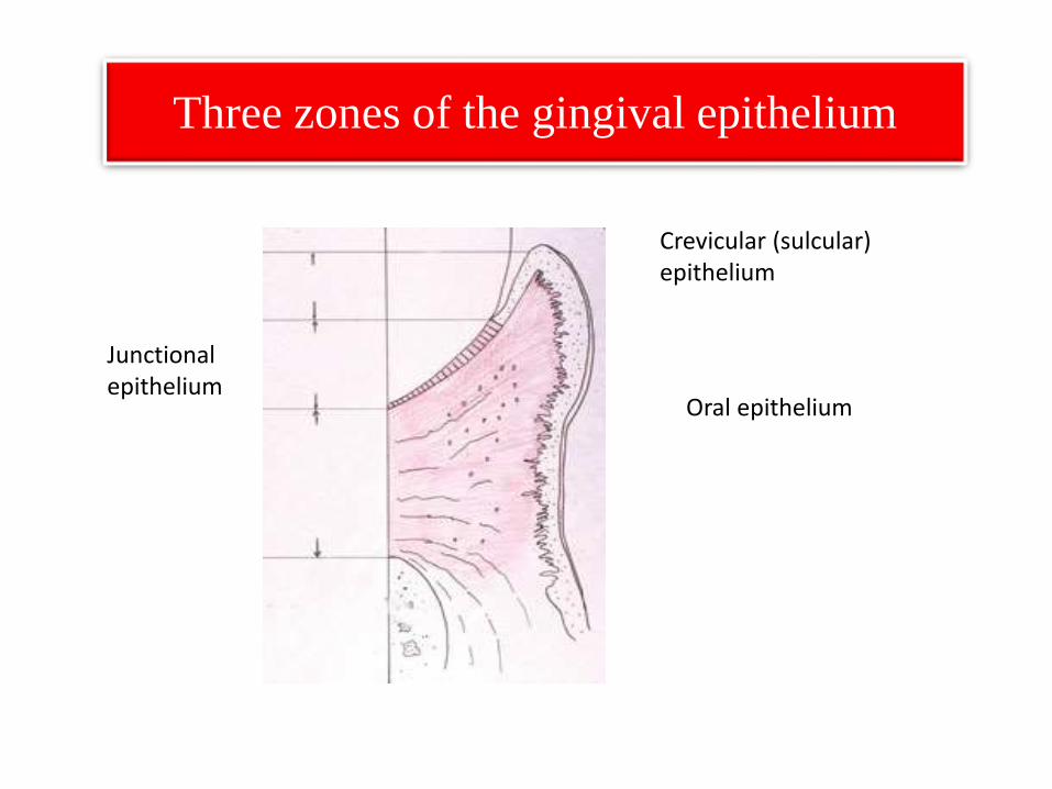
Three zones of the gingival epithelium
Crevicular (sulcular) epithelium
Oral epithelium
Junctionalepithelium
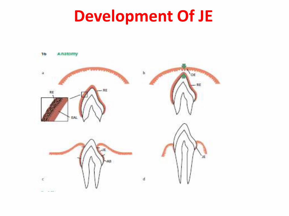
Development Of JE
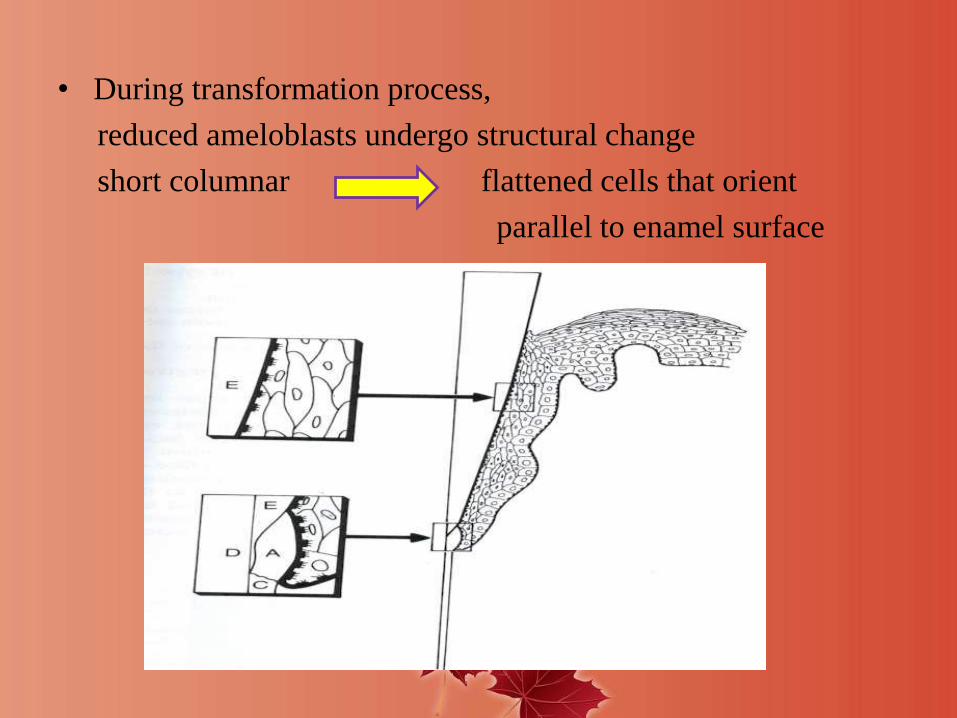
• During transformation process,
reduced ameloblasts undergo structural change
short columnar flattened cells that orient
parallel to enamel surface
Structure of junctional epithelium
• Anatomical aspects
• Junctional epithelium and interstitial cells
• Epithelial attachment
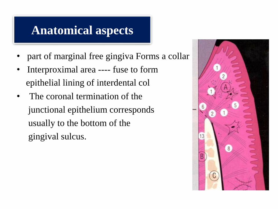
Anatomical aspects
• part of marginal free gingiva Forms a collar
• Interproximal area ---- fuse to form
epithelial lining of interdental col
• The coronal termination of the
junctional epithelium corresponds
usually to the bottom of the
gingival sulcus.
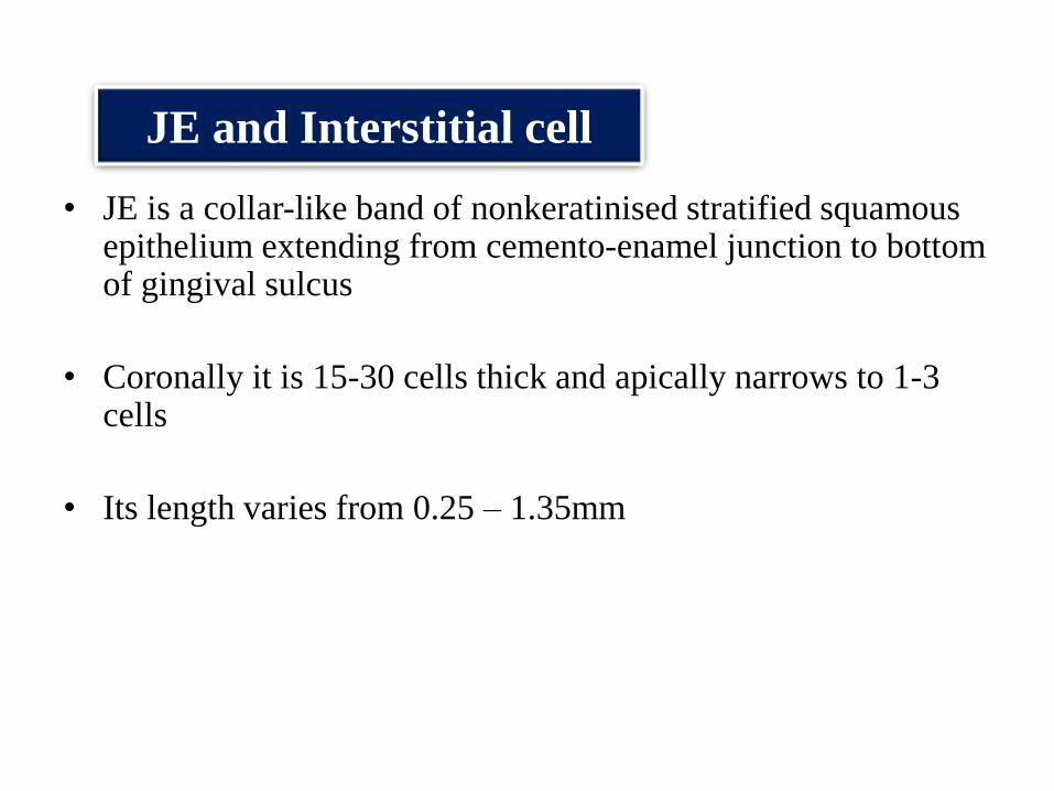
JE and Interstitial cell
• JE is a collar-like band of nonkeratinised stratified squamousepithelium extending from cemento-enamel junction to bottom of gingival sulcus
• Coronally it is 15-30 cells thick and apically narrows to 1-3 cells
• Its length varies from 0.25 – 1.35mm
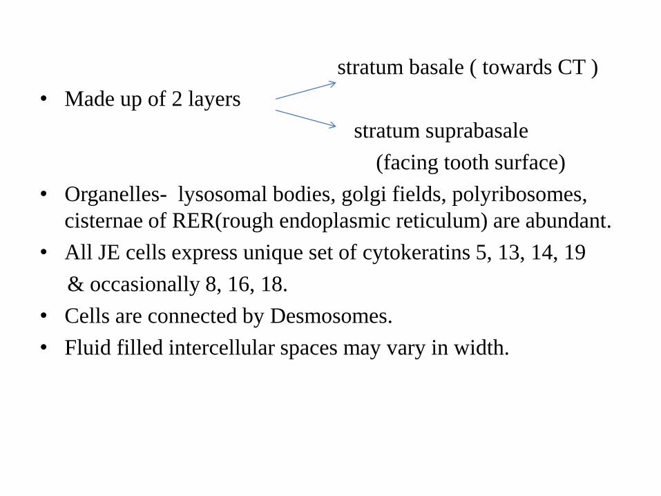
stratum basale ( towards CT )
• Made up of 2 layers
stratum suprabasale
(facing tooth surface)
• Organelles- lysosomal bodies, golgi fields, polyribosomes,
cisternae of RER(rough endoplasmic reticulum) are abundant.
• All JE cells express unique set of cytokeratins 5, 13, 14, 19
& occasionally 8, 16, 18.
• Cells are connected by Desmosomes.
• Fluid filled intercellular spaces may vary in width.
EPITHELIAL ATTACHMENT APPARATUS
• The term epithelial attachment refers to the attachment
apparatus, i.e. internal basal lamina & hemidesmosomes
that connects the junctional epithelium to the tooth surface.
• It consists of hemidesmosomes at the plasma membrane of the
cells directly attached to the tooth (DAT cells) and a basal
lamina-like extracellular matrix, termed the internal basal
lamina, on the tooth surface
HISTORICAL CONCEPTS OF
ATTACHMENT
Gottlieb’s concept (1921)
• Soft tissue of gingiva is organically united to enamel surface.
• He termed the epithelium contacting the tooth “epithelial
attachment”.
Orban’s concept (1953)
• He stated that the separation of the epithelial attachment cells
from the tooth surface involved preparatory degenerative
changes in the epithelium.
Waerhaug’s concept (1960)
• He presented the concept of epithelial cuff. This concept was
based on insertion of thin blades between the surface of tooth
and the gingiva
• Blades could be easily passed apically to the connective tissue
attachment at CEJ without resistance.
• It was concluded that gingival tissue and tooth are closely
adapted but not organically united.
Schroeder and Listgarten concept
(1971)
• The previous controversy was resolved after evolution of transmission electron microscopy.
• Primary epithelial attachment refers to the epithelial attachment lamina released by the REE. It lies in direct contact with enamel and epithelial cells attached to it by hemi-desmosomes.
• When REE cells transform into JE cells the primary epithelial attachment becomes secondary epithelial attachment . It is made of epithelial attachment between basal lamina and hemi-desmosomes.
• Basement membrane – specialized extracellular matrix
• Functions-
a. Compartmentalization
b. Filtration
(selective permeability barrier function)
c. Cell polarization, migration.
d. Cell adhesions
e. Cell differentiation.
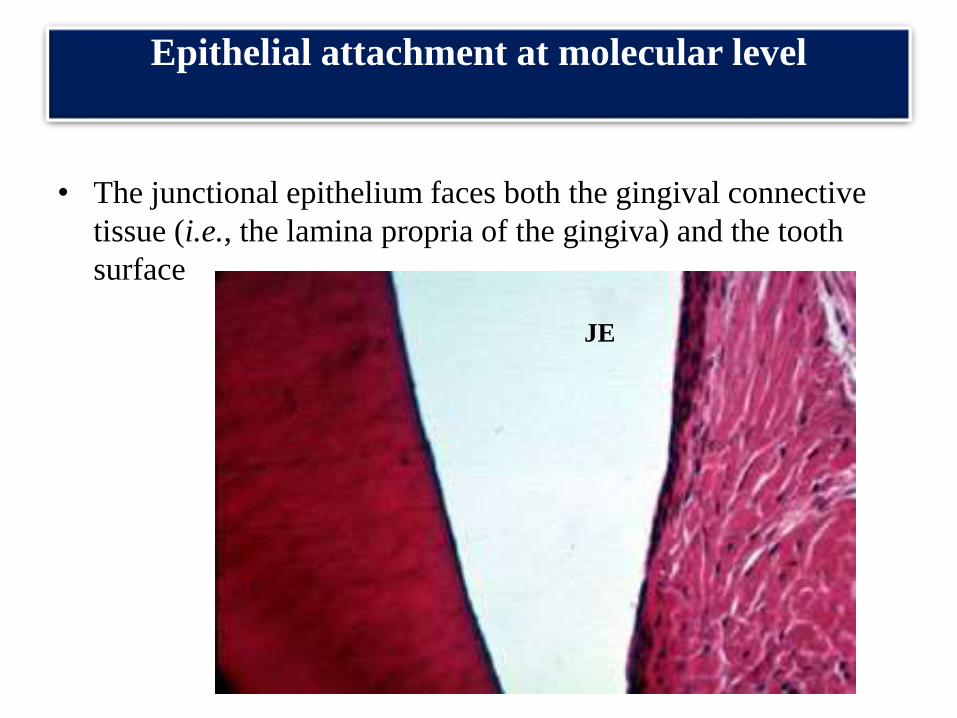
Epithelial attachment at molecular level
• The junctional epithelium faces both the gingival connective
tissue (i.e., the lamina propria of the gingiva) and the tooth
surface
JE
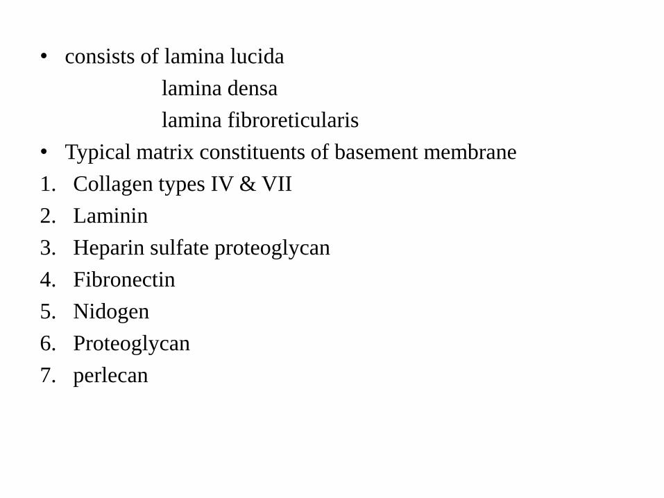
• consists of lamina lucida
lamina densa
lamina fibroreticularis
• Typical matrix constituents of basement membrane
1. Collagen types IV & VII
2. Laminin
3. Heparin sulfate proteoglycan
4. Fibronectin
5. Nidogen
6. Proteoglycan
7. perlecan
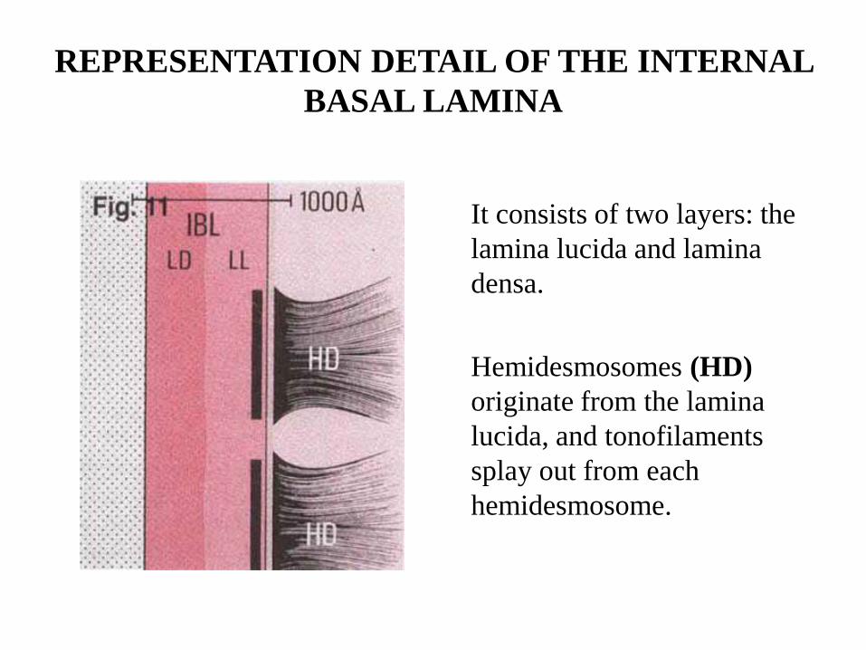
REPRESENTATION DETAIL OF THE INTERNAL
BASAL LAMINA
It consists of two layers: the
lamina lucida and lamina
densa.
Hemidesmosomes (HD)
originate from the lamina
lucida, and tonofilaments
splay out from each
hemidesmosome.
• The internal basement membrane was initially described as an
80-120nm wide homogeneous layer. It directly faces the enamel,
with an intervening laminated or non-laminated layer of cuticles
(Listgarten, 1966) or afibrillar cementum (Kobayashi et al.,
1976).
• There are numerous fine strands crossing the lamina densa of the
internal basement membrane at the hemidesmosomes. These
strands may have been the anchoring filaments of
hemidesmosomes (Eady, 1994; Garrod, 1993).
• In the cytoplasm of the cells of the junctional epithelium, the
tonofibrils are associated with hemidesmosomes.
• The internal basement membrane of the dentogingival border is
uniquely specialized for mechanical strength, sealing off the
periodontal tissues from the oral environment (Sawada &
Inoue, 1996).
• The internal basement membrane takes the form of both thin
and multilayered thick basement membranes
• Multilayered internal basement membrane may provide
mechanical strength for firm attachment of the tooth to the
gingiva and the sealing off of the periodontal tissues from the
oral environment.
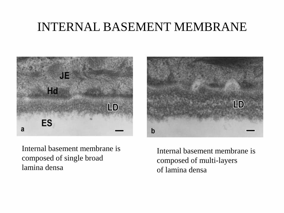
INTERNAL BASEMENT MEMBRANE
Internal basement membrane is
composed of single broad
lamina densa
Internal basement membrane is
composed of multi-layers
of lamina densa
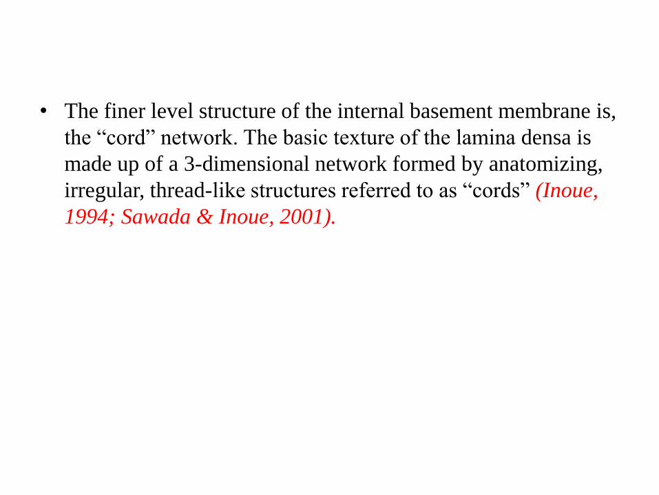
• The finer level structure of the internal basement membrane is,
the “cord” network. The basic texture of the lamina densa is
made up of a 3-dimensional network formed by anatomizing,
irregular, thread-like structures referred to as “cords” (Inoue,
1994; Sawada & Inoue, 2001).
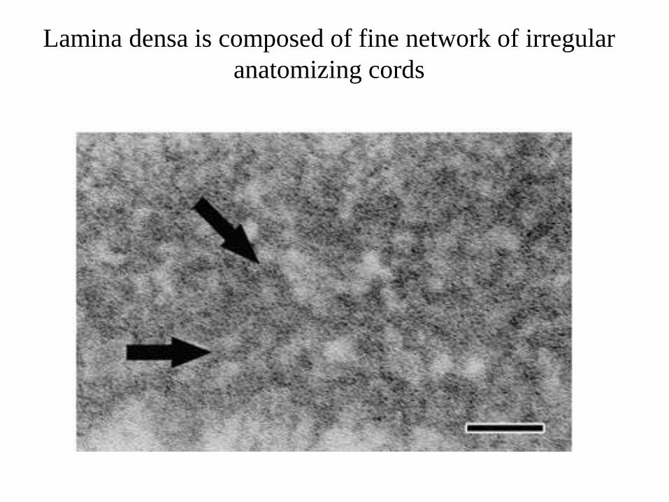
Lamina densa is composed of fine network of irregular
anatomizing cords
MECHANISM OF BINDING OF NORMAL TOOTH TO
GINGIVA THROUGH CORD LIKE STRUCTURES IN
LAMINA DENSA
• The lamina densa of the internal basement membrane is
closely associated with an additional layer referred to as the
supplementary lamina densa found on the enamel side of the
tooth.
• One part of the basement membrane, the supplementary
lamina densa, is mineralized. This mineral deposit is
continuous with that of the enamel of the tooth, and thus this
deposit on the supplementary lamina densa forms an
advancing edge of mineralization.
(Sawada & Inoue, 2003)
• In the mineralized portion of the lamina densa, mineral
crystals were arranged in a network pattern which was
comparable to the pattern of the cord network.
• This may assist more powerful gripping, and further
demonstrates the elaborate mechanism by which firm binding
of the mineral and organic phases is achieved.
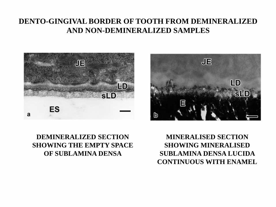
DENTO-GINGIVAL BORDER OF TOOTH FROM DEMINERALIZED
AND NON-DEMINERALIZED SAMPLES
DEMINERALIZED SECTION
SHOWING THE EMPTY SPACE
OF SUBLAMINA DENSA
MINERALISED SECTION
SHOWING MINERALISED
SUBLAMINA DENSA LUCIDA
CONTINUOUS WITH ENAMEL
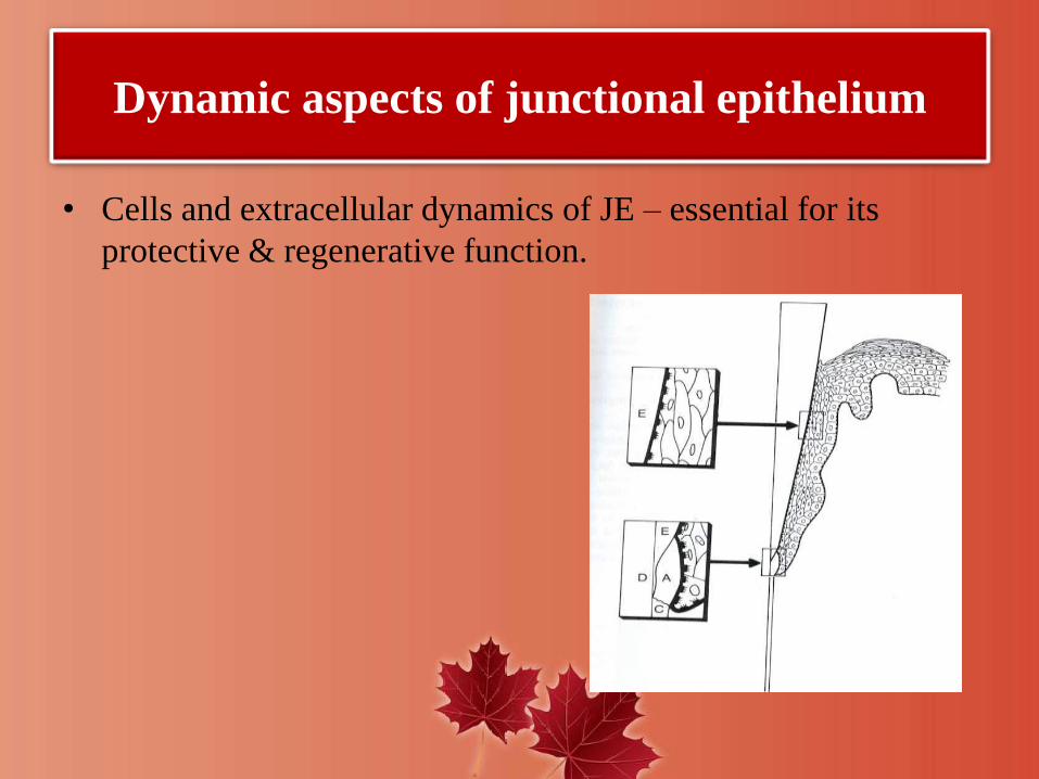
Dynamic aspects of junctional epithelium
• Cells and extracellular dynamics of JE – essential for its
protective & regenerative function.
• Exfoliation must occur at extremely high rate ( Loe & Karring
1969)
• Since DAT cells are connected to basal lamina via
hemidesmosomes, a remodelling of epithelial attachment must
occur.
• Thus epithelial attachment normally is not static but dynamic.
• Intercellular spaces of JE
provides pathway for fluid & transmigratory leukocytes
a variety of molecules + leukocytes ( host defense system)
Expression of various molecules and their
function
• JE cells have surface or cell membrane molecules that play a
role in cell matrix and cell-cell interactions. JE cells express
numerous cell adhesion molecules (CAM’s), such as integrins
and cadherins.
• Knowledge about structure and molecules involved in the
maintenance of cell-cell contact is particularly important in
view of the pathological changes that the epithelium undergoes
during its conversion to a pocket lining.
• Integrins – are cell surface receptors that mediate interactions
between cell and extracellular matrix, and also contribute to
cell to cell adhesion.
• The cadherins are responsible for tight contacts between cells.
• E-cadherin, an epithelium specific cell adhesion molecule,
plays a crucial role in maintaining the structural integrity.
• Intercellular adhesion molecule-1(ICAM-1) and lymphocytic
function antigen- 3(LFA-3) are additional cell adhesion
molecules.
• Cells in contact with the internal basal lamina express the
integrins.
(CAM1)—a transmembrane cell-adhesion molecule that is
expressed on leukocytes, epithelia, and blood vessel endothelia
.
high expression of interleukin-8 (IL-8), a chemotactic
cytokine, is seen in the coronal-most cells of the junctional
epithelium
interleukin-1α (IL-1α),
interleukin-1β (IL-1β),
tumor necrosis factor-α (TNF-α)—are strongly expressed in
the coronal half of the junctional epithelium
N-acetyllactosamine—the type 2 chain H precursor of the
blood group A-specific carbohydrate, which is usually
associated with the lowest level of cell differentiation.
• Antimicrobial molecules--- α and β defensins
cathelicidin family
calprotectin
DYNAMICS (TURNOVER RATE) OF JE
• The turnover rate of JE cells is rapid.
• The DAT cells express a high density of transferring receptors
supporting the idea of active metabolism and high turnover.
• DAT cells have an important role in tissue dynamics and
reparative capacity of the JE.
• The existence of a dividing population of DAT cells in a
suprabasal location in several layers from connective tissue is
a unique feature of JE.
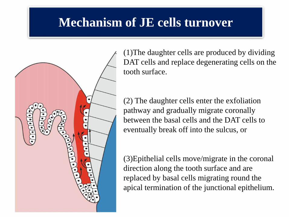
Mechanism of JE cells turnover
(1)The daughter cells are produced by dividing
DAT cells and replace degenerating cells on the
tooth surface.
(2) The daughter cells enter the exfoliation
pathway and gradually migrate coronally
between the basal cells and the DAT cells to
eventually break off into the sulcus, or
(3)Epithelial cells move/migrate in the coronal
direction along the tooth surface and are
replaced by basal cells migrating round the
apical termination of the junctional epithelium.
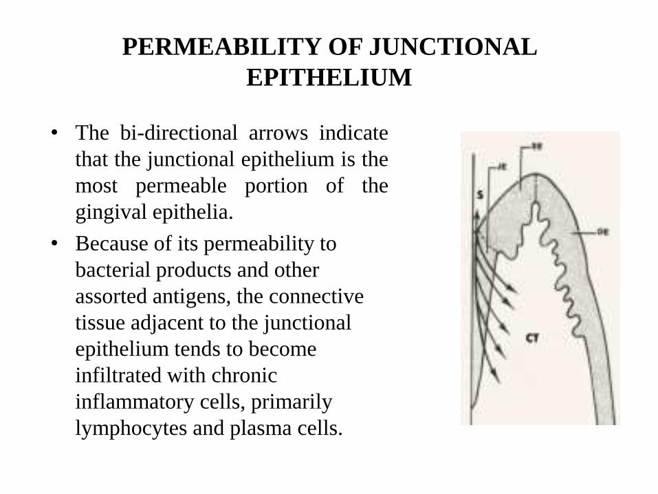
PERMEABILITY OF JUNCTIONAL
EPITHELIUM
• The bi-directional arrows indicate
that the junctional epithelium is the
most permeable portion of the
gingival epithelia.
• Because of its permeability to
bacterial products and other
assorted antigens, the connective
tissue adjacent to the junctional
epithelium tends to become
infiltrated with chronic
inflammatory cells, primarily
lymphocytes and plasma cells.
FUNCTIONS OF JUNCTIONAL EPITHELIUM
• Has attachment role and protective role.
• Permeability allows GCF and defence cells to pass across to
protect underlying tissues from disease processes (periodontal
disease).
• Helps maintain integrity of tooth/periodontium structure
• GCF contains gamma globulins and poly-morphonuclearleukocytes (PMNs) giving it immunological/phagocyticproperties to combat disease processes.
• Such molecules pass readily across JE to underlying tissues.
• JE may contain neutrophils & other inflammatory cells indicating disease & state of health of periodontium.
• The junctional epithelium plays a crucial role since it
essentially seals off periodontal tissues from the oral
environment.
• Its integrity is thus essential for maintaining a healthy
periodontium.
• Periodontal disease sets in when the structure of the junctional
epithelium starts to fail, an excellent example of how structure
determines function.
JE in antimicrobial defense(1) JE cells exfoliate because of rapid cell division
(2) funneling of junctional epithelial cells towards the sulcus hinder bacterial
colonization.
(3) Active antimicrobial substances are produced in junctional epithelial cells.
(4) Epithelial cells activated by microbial substances secrete chemokines, that
attract and activate professional defense cells, PMN.

• Role of JE in pocket formation
Role of JE in the initiation of pocket formation
• Conversion of the JE to pocket epithelium is regarded as a
hallmark in the development of periodontitis.
• Schroeder – 1996 pointed to a biologically relevant and
clinically important question that still awaits resolution: ‘what
happens to the JE under conditions of sub-gingival microbial
attack i.e. in context with pocket formation and deepening?’

• Schluger et al 1977: Pocket formation is attributed to a loss of
cellular continuity in the coronal most portion of the JE
• Thus the initiation of pocket formation may be attributed to the
detachment of the DAT cells from the tooth surface or to the
development of intraepithelial split.
• Takata and Donath (1988) observed degenerative changes in
the second or third layer of the DAT cells in the coronal most
portion of the JE cells facing the biofilm.
• Schroeder and Listgarten 1977: An increased number of
mononuclear leukocytes (T and B cells, macrophages) together
with PMNs are considered as factors contributing to the
disintegration of the JE.
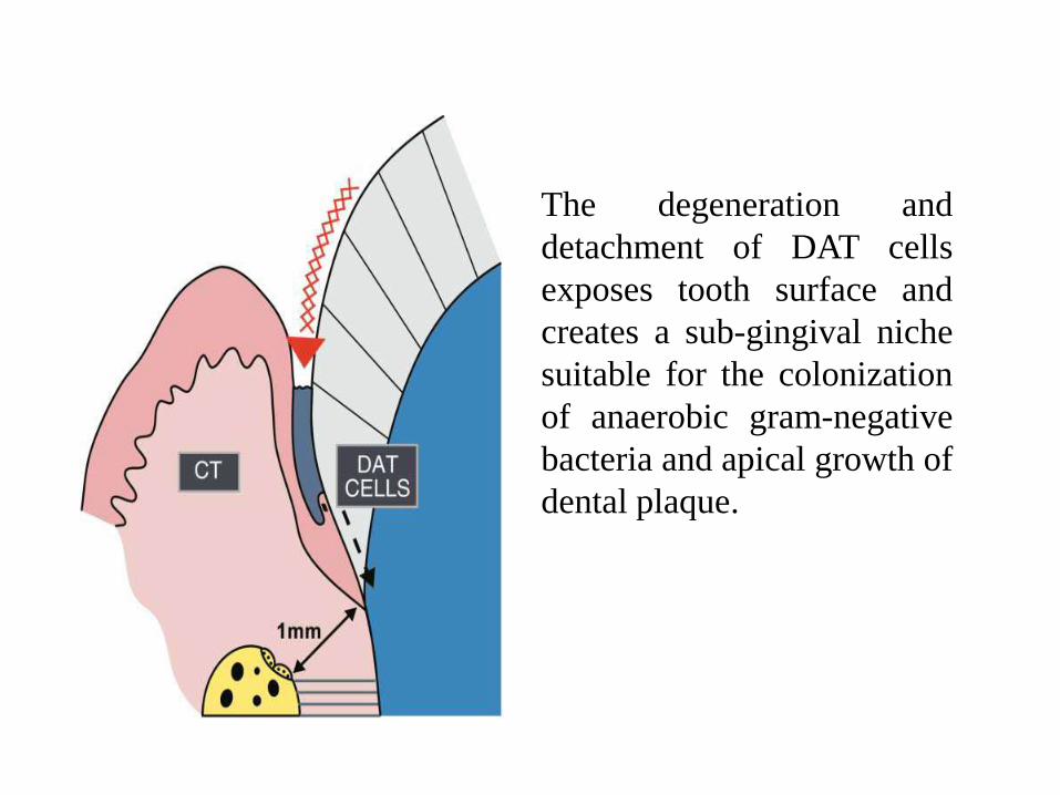
The degeneration and
detachment of DAT cells
exposes tooth surface and
creates a sub-gingival niche
suitable for the colonization
of anaerobic gram-negative
bacteria and apical growth of
dental plaque.
• Hintermann et al 2002: Gingipains degrade the epithelial cell-
cell junctional complexes and cells exposed to proteinases
derived from P.gingivalis showed reduced adhesion to
extracellular matrix.
• Destruction of cell-cell and cell to ECM perturbs the structural
and functional integrity of the JE.

• Regeneration of JE
• Injury to JE may occur due to intentional or accidental trauma.
• Accidental trauma can occur during probing, flossing or tooth
margin preparations for restorations.
• Intentional trauma occurs during periodontal surgeries where
the JE is completely lost.
• Many studies have been done to investigate the renewal of JE.
These include studies done on renewal of JE on tooth and
implant surface after mechanical detachment by probing.
• Studies have been done on mechanical trauma during flossing
and on regeneration of JE after gingivectomy procedure which
completely removes JE.
• Taylor and Campbell 1972: A new and complete attachment
indistinguishable from that in control was established 5 days
after complete separation of the JE from the tooth surface.
• Frank et al 1972: A study demonstrated that newly
differentiated attachment apparatus with normal
hemidesmosomal attachment is possible following surgery.
This new attachment apparatus was seen on cementum as well
as dentin.
• Listgarten 1972:Hemidesmosomes appeared to form prior to
the basal lamina. The basal lamina is initially formed in close
proximity to the hemidesmosomes at both the tooth and
connective tissue interface. At 4 to 7 weeks, the basal lamina
appeared complete. Studies have shown that regeneration of JE
after procedure usually occurs within 20 days.
• JE AROUND IMPLANTS
• The junctional epithelium around implants always originates
from epithelial cells of the oral mucosa, as opposed to the
junctional epithelium around teeth which originates from the
reduced enamel epithelium.
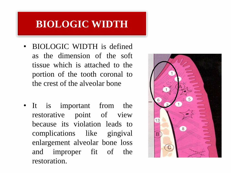
BIOLOGIC WIDTH
• BIOLOGIC WIDTH is defined
as the dimension of the soft
tissue which is attached to the
portion of the tooth coronal to
the crest of the alveolar bone
• It is important from the
restorative point of view
because its violation leads to
complications like gingival
enlargement alveolar bone loss
and improper fit of the
restoration.
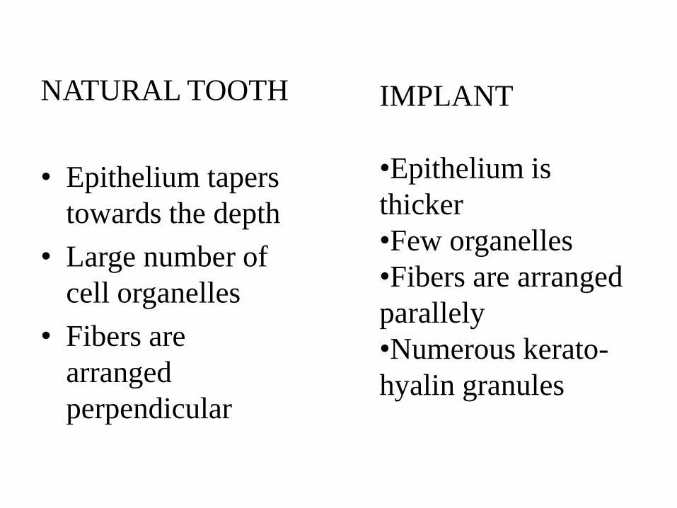
NATURAL TOOTH
• Epithelium tapers
towards the depth
• Large number of
cell organelles
• Fibers are
arranged
perpendicular
IMPLANT
•Epithelium is
thicker
•Few organelles
•Fibers are arranged
parallely
•Numerous kerato-
hyalin granules
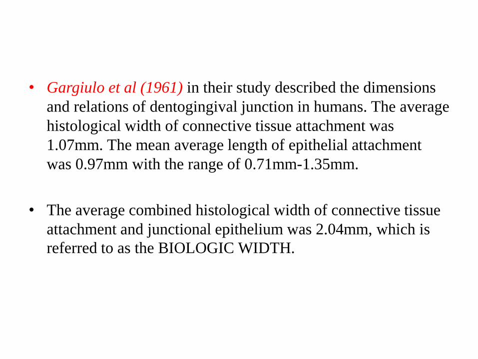
• Gargiulo et al (1961) in their study described the dimensions
and relations of dentogingival junction in humans. The average
histological width of connective tissue attachment was
1.07mm. The mean average length of epithelial attachment
was 0.97mm with the range of 0.71mm-1.35mm.
• The average combined histological width of connective tissue
attachment and junctional epithelium was 2.04mm, which is
referred to as the BIOLOGIC WIDTH.
CONCLUSION
• DENTOGINGIVAL UNIT is important because of its
anatomical location.
• It is the site of host-bacterial interaction in initiation of
periodontal disease.
• There is a constant presence of bacteria and their products in
the gingival sulcus which makes this an important structural
component of periodontal defense mechanism.
• The conversion of the junctional epithelium to pocket
epithelium is regarded as hallmark in the development of
periodontitis.
Future scope
• To find out the therapeutic strategies that stop the disease
progression at this important tooth-tissue interface.
References
• CARRANZA 11TH EDITION
• JAN LYNDHE 5TH EDITION
• DD Bosshardt and NP Lang. The Junctional Epithelium: from health to disease. J Dent Res 2005, 84 (1): 9-20
• Moon-Il Cho & Philias R. Garant. Development and general structure of the periodontium. Periodontology 2000, Vol. 24, 2000, 9–27.
• Mark Bartold, Laurence J. Walsh & A. Sampath Narayanan. Molecular and cell biology of the gingiva.P. Periodontology 2000, Vol. 24, 2000, 28–55.
• Thomas M Hassell. Tissues and cells of the periodontium. Periodontology2000, Vol. 3, 1993, 9-38
• Huberte . Schroede & R M Listgarten. The gingival tissues: The architecture of periodontal Protection. Periodontology 2000, Vol. 13, 1997, 91-120.
• Takashi Sawada1 and Sadayuki Inoue. Ultrastructure of DentogingivalBorder of Normal and Replanted Tooth and Dental Implant, chapter 11www.intechopen.com/books/implantdentistry