jowl cysts, multiplicity and taxonomy: ameloblastoma. · ameloblastoma is a rare, benign,...
TRANSCRIPT
Jowl cysts, multiplicity and taxonomy: ameloblastoma.
Anubha Bajaj*
Punjab University, Chandigarh, India
Keywords: Ameloblastoma, Cystosarcoma, Metamorphosis, Carcinoma, Inflammation.Accepted on March 28, 2018
Short CommunicationElaborated from “amel” (implying enamel) and “blastos”(connoting a germ) and initially chronicled in 1827 by Cusack,Ameloblastoma is a rare, benign, slow-growing, locallyinvasive neoplasm of odontogenic origin, affecting mandible(80%) and maxilla necessitating conservative therapy andelucidating considerable recurrences. Substitute terminologiesare cystosarcoma, adamantine epithelioma, adamantinoma andameloblastoma. The worldwide prevalence is of 0.5 cases permillion persons, a range of presentation between 30 to 60 yearsand a median at 40 years. The tumor classically manifests as apainless lump in the maxilla or mandible. Malignantmetamorphosis is heralded by a painful, accelerated growth.Tooth displacement, root resorption, paraesthesia areinfrequent. Mandibular lesions (80%) are predominate in theposterior mandibular zone and exceptionally in the sinonasalcavities. Maxillary ameloblastoma, in conjunction, maypreponderate the posterior molar region. Ameloblasticcarcinoma preferentially has a maxillary localization. Non-specific irritation from tooth extraction, dental caries, trauma,inflammation, nutritional deficiencies are factors whichpredispose to the genesis of ameloblastoma.
Diagnostic Approbation is with imaging and biopsy. The tumorarises in the bone and is incidentally discovered on dental or
plain x-rays delineating a lytic lesion with scallopedboundaries, resorption of tooth roots, impacted molars (theunicystic variant) and the classic soap bubble occurrence(characteristic of the multilocular /solid variant). Computerizedtomography (CT) is beneficial - a well delineated radiolucence(unilocular/ multilocular), an expansive lesion with corticaldestruction or soft tissue extension. CT can be utilized forcomputation of a planned surgery. Magnetic ResonanceImaging (MRI) assists in the identification of a soft tissue /marrow extension besides the classic, lytic bony participation.It expedites the detection of a typical lesion arising from themaxilla and extending to the orbit, paranasal sinuses, and baseof the skull [1]. Positron Emission Tomography (PET-CT) is amethod to evaluate the staging and distant metastasis inmetastatic ameloblastoma. Resection biopsy at the site ofcortical destruction (an aperture for biopsy sampling) isessential for preventing unnecessary surgery. Surrogatediagnoses are osteomyelitis, cystic fibrous dysplasia, and giantcell tumor, ossifying fibroma, multiple myeloma and raresarcomas. Fine needle aspiration cytology (FNAC) is suitablefor localized lesions displaying cortical erosions/ in a dentalsocket. Incisional biopsy is precise but necessitates mucosaldisruption. The specific locus is subsequently surgicallyeliminated. Peripheral ameloblastoma lacks a bony envelopeand is successfully biopsied (Table 1).
Table 1. Histology with ancillaries.
Epithelium of Origin Odontogenic /Enamel Cell rests of Malassez Sheetsof Hertwig
Epithelium of DentigerousCyst
Basal Layer of Oral Mucosa
WHO(2005) Classification Solid Multicystic Extraosseous Peripheral Desmoplastic Unicystic
Microscopic Patterns Follicular, Acanthomatous Spindle, Granular Basal Cell like Plexiform
Microscopic Features Stellate Reticulum Peripheral Palisading Reverse Polarization ColumnarEpithelium
Basal Layer
Clinical Subtypes Central- Osseous Peripheral- Extra osseous
Malignant Subtypes Metastatic Ameloblastoma Ameloblastic carcinoma
IHC staining CK5, CK6 CK13, CK14 CK 19, CD56 Calretinin
Molecular features SMO mutations BRAF mutations KRAS FGFR2
Electron Microscopy Epithelial Differentiation Tonofilament Desmosome
Supplementary Aspects the ectomesenchyme or the heterotopicepithelial rests from body components such as the pituitary areimplicated in the tumor ancestry. Clinico-pathological elementsdemonstrate that the solid/multicystic variant of ameloblastomais extremely common (91%) and locally destructive. Unicystictype is an infrequent (6%), benign version, demarcated into theintraluminal or intramural subdivisions. The intraluminal
subtype has a lower reoccurrence rate, minimal stromalinvasion and is amenable to conservative management. Solid/Multicystic/ Desmoplastic variant has the highest reappearancerate (90% with enucleation/curettage), can be centralized in thebone marrow and illustrates a bony circumscription, hence it isalso designated as Central ameloblastoma. Peripheralameloblastoma is extra-osseous (2%), may classically present
Short Communication http://www.alliedacademies.org/pathology-and-disease-biology/
J Pathol Dis Biol 2018 Volume 2 Issue 12
as s gingival pedunculated/ exophytic lesions. Desmoplasticvariant is the least common (1%). Cellular atypia and mitosis isinfrequent, an augmentation of these parameters is a harbingerof neoplastic conversion. Ameloblastic carcinoma/odontogenicsarcoma are malignant subtypes (excluded from the WHOclassification) commencing de novo or from benignameliorations. Metastatic ameloblastoma may display welldifferentiated benign histology similar to the preponderantsolid/multicystic type but adjunctive, similar benign foci arerecognized at a distant location from the initial primary focus,thus it is labelled as discussed. Ameloblastic carcinoma maydelineate a malignant outcome featuring atypical mitosis,cytological atypia or invasiveness which are detectable at theprimary locus. Distant metastasis are discerned at the lungs,bones, liver and brain. Provocation for a malignanttransformation are observed with lengthy duration of thetumor, numerous reoccurrences after conventional treatmentand delayed metastasis (Figures 1-9).
Histological Variants
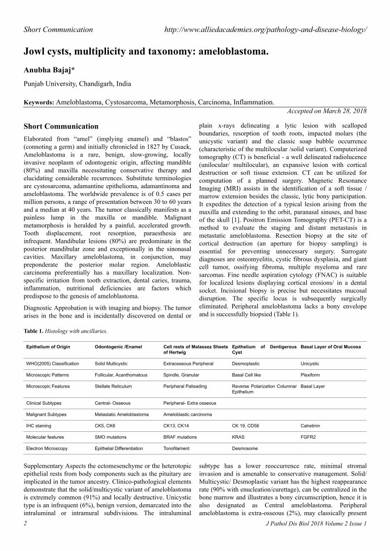
Figure 1. Plexiform ameloblastoma.
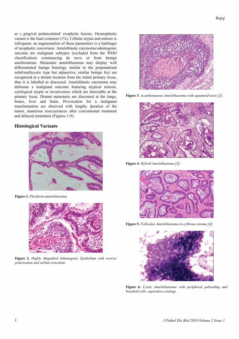
Figure 2. Highly Magnified Odontogenic Epithelium with reversepolarization and stellate reticulum.
Figure 3. Acanthomatous Ameloblastoma with squamoid nests [2].
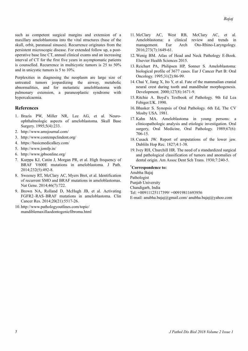
Figure 4. Hybrid Ameloblastoma [3].
Figure 5. Follicular Ameloblastoma in a fibrous stroma [4].
Figure 6. Cystic Ameloblastoma with peripheral palisading andbasaloid cells- aspiration cytology.
Bajaj
J Pathol Dis Biol 2018 Volume 2 Issue 13
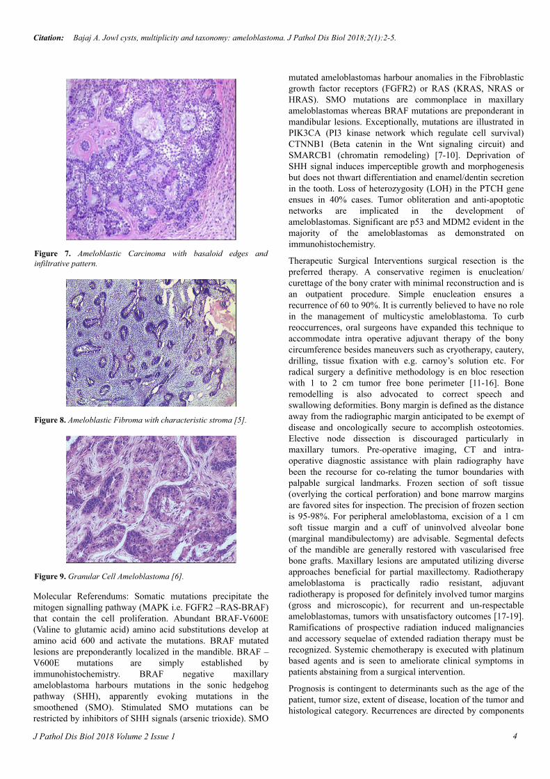
Figure 7. Ameloblastic Carcinoma with basaloid edges andinfiltrative pattern.
Figure 8. Ameloblastic Fibroma with characteristic stroma [5].
Figure 9. Granular Cell Ameloblastoma [6].
Molecular Referendums: Somatic mutations precipitate themitogen signalling pathway (MAPK i.e. FGFR2 –RAS-BRAF)that contain the cell proliferation. Abundant BRAF-V600E(Valine to glutamic acid) amino acid substitutions develop atamino acid 600 and activate the mutations. BRAF mutatedlesions are preponderantly localized in the mandible. BRAF –V600E mutations are simply established byimmunohistochemistry. BRAF negative maxillaryameloblastoma harbours mutations in the sonic hedgehogpathway (SHH), apparently evoking mutations in thesmoothened (SMO). Stimulated SMO mutations can berestricted by inhibitors of SHH signals (arsenic trioxide). SMO
mutated ameloblastomas harbour anomalies in the Fibroblasticgrowth factor receptors (FGFR2) or RAS (KRAS, NRAS orHRAS). SMO mutations are commonplace in maxillaryameloblastomas whereas BRAF mutations are preponderant inmandibular lesions. Exceptionally, mutations are illustrated inPIK3CA (PI3 kinase network which regulate cell survival)CTNNB1 (Beta catenin in the Wnt signaling circuit) andSMARCB1 (chromatin remodeling) [7-10]. Deprivation ofSHH signal induces imperceptible growth and morphogenesisbut does not thwart differentiation and enamel/dentin secretionin the tooth. Loss of heterozygosity (LOH) in the PTCH geneensues in 40% cases. Tumor obliteration and anti-apoptoticnetworks are implicated in the development ofameloblastomas. Significant are p53 and MDM2 evident in themajority of the ameloblastomas as demonstrated onimmunohistochemistry.
Therapeutic Surgical Interventions surgical resection is thepreferred therapy. A conservative regimen is enucleation/curettage of the bony crater with minimal reconstruction and isan outpatient procedure. Simple enucleation ensures arecurrence of 60 to 90%. It is currently believed to have no rolein the management of multicystic ameloblastoma. To curbreoccurrences, oral surgeons have expanded this technique toaccommodate intra operative adjuvant therapy of the bonycircumference besides maneuvers such as cryotherapy, cautery,drilling, tissue fixation with e.g. carnoy’s solution etc. Forradical surgery a definitive methodology is en bloc resectionwith 1 to 2 cm tumor free bone perimeter [11-16]. Boneremodelling is also advocated to correct speech andswallowing deformities. Bony margin is defined as the distanceaway from the radiographic margin anticipated to be exempt ofdisease and oncologically secure to accomplish osteotomies.Elective node dissection is discouraged particularly inmaxillary tumors. Pre-operative imaging, CT and intra-operative diagnostic assistance with plain radiography havebeen the recourse for co-relating the tumor boundaries withpalpable surgical landmarks. Frozen section of soft tissue(overlying the cortical perforation) and bone marrow marginsare favored sites for inspection. The precision of frozen sectionis 95-98%. For peripheral ameloblastoma, excision of a 1 cmsoft tissue margin and a cuff of uninvolved alveolar bone(marginal mandibulectomy) are advisable. Segmental defectsof the mandible are generally restored with vascularised freebone grafts. Maxillary lesions are amputated utilizing diverseapproaches beneficial for partial maxillectomy. Radiotherapyameloblastoma is practically radio resistant, adjuvantradiotherapy is proposed for definitely involved tumor margins(gross and microscopic), for recurrent and un-respectableameloblastomas, tumors with unsatisfactory outcomes [17-19].Ramifications of prospective radiation induced malignanciesand accessory sequelae of extended radiation therapy must berecognized. Systemic chemotherapy is executed with platinumbased agents and is seen to ameliorate clinical symptoms inpatients abstaining from a surgical intervention.
Prognosis is contingent to determinants such as the age of thepatient, tumor size, extent of disease, location of the tumor andhistological category. Recurrences are directed by components
Citation: Bajaj A. Jowl cysts, multiplicity and taxonomy: ameloblastoma. J Pathol Dis Biol 2018;2(1):2-5.
4J Pathol Dis Biol 2018 Volume 2 Issue 1
such as competent surgical margins and extension of amaxillary ameloblastoma into the vital structures (base of theskull, orbit, paranasal sinuses). Recurrence originates from thepersistent microscopic disease. For extended follow up, a post-operative base line CT, annual clinical exams and an increasinginterval of CT for the first five years in asymptomatic patientsis counselled. Recurrence in multicystic tumors is 25 to 50%and in unicystic tumors is 5 to 10%.
Perplexities in diagnosing the neoplasm are large size ofuntreated tumors jeopardizing the airway, metabolicabnormalities, and for metastatic ameloblastoma withpulmonary extension, a paraneoplastic syndrome withhypercalcaemia.
References1. Brazis PW, Miller NR, Lee AG, et al. Neuro-
ophthalmologic aspects of ameloblastoma. Skull BaseSurgery. 1995;5(4):233.
2. http://www.amsjournal.com/3. http://www.contempclindent.org/4. https://basicmedicalkey.com/5. http://www.jomfp.in/6. http://www.jpbsonline.org/7. Kurppa KJ, Catón J, Morgan PR, et al. High frequency of
BRAF V600E mutations in ameloblastoma. J Path.2014;232(5):492-8.
8. Sweeney RT, McClary AC, Myers Bret, et al. Identificationof recurrent SMO and BRAF mutations in ameloblastomas.Nat Gene. 2014;46(7):722.
9. Brown NA, Rolland D, McHugh JB, et al. ActivatingFGFR2–RAS–BRAF mutations in ameloblastoma. ClinCancer Res. 2014;20(21):5517-26.
10. http://www.pathologyoutlines.com/topic/mandiblemaxillaodontogenicfibroma.html
11. McClary AC, West RB, McClary AC, et al.Ameloblastoma: a clinical review and trends inmanagement. Eur Arch Oto-Rhino-Laryngology.2016;273(7):1649-61.
12. Wenig BM. Atlas of Head and Neck Pathology E-Book.Elsevier Health Sciences 2015.
13. Reichart PA, Philipsen HP, Sonner S. Ameloblastoma:biological profile of 3677 cases. Eur J Cancer Part B: OralOncology. 1995;31(2):86-99.
14. Chai Y, Jiang X, Ito Y, et al. Fate of the mammalian cranialneural crest during tooth and mandibular morphogenesis.Development. 2000;127(8):1671-9.
15. Ritchie A. Boyd’s Textbook of Pathology. 9th Ed LeaFebiger.UK. 1990.
16. Bhasker S. Synopsis of Oral Pathology. 6th Ed, The CVMosby USA. 1981.
17. Kahn MA. Ameloblastoma in young persons: aclinicopathologic analysis and etiologic investigation. Oralsurgery, Oral Medicine, Oral Pathology. 1989;67(6):706-15.
18. Cusack JW. Report of amputations of the lower jaw.Dubliln Hop Rec. 1827;4:1-38.
19. Ivey RH, Churchill HR. The need of a standardized surgicaland pathological classification of tumors and anomalies ofdental origin. Am Assoc Dent Sch Trans. 1930;7:240-5.
*Correspondence to:Anubha BajajPathologistPunjab UniversityChandigarh, IndiaTel: +00911125117399/ +00919811693956E-mail: [email protected]/ [email protected]
Bajaj
J Pathol Dis Biol 2018 Volume 2 Issue 15