joint stage recognition and anatomical annotation of...
TRANSCRIPT

Copyedited by: TRJ MANUSCRIPT CATEGORY:
[11:19 31/5/2012 Bioinformatics-bts220.tex] Page: i16 i16–i24
BIOINFORMATICS Vol. 28 ISMB 2012, pages i16–i24doi:10.1093/bioinformatics/bts220
Joint stage recognition and anatomical annotation of drosophilagene expression patternsXiao Cai, Hua Wang, Heng Huang∗ and Chris DingDepartment of Computer Science and Engineering, University of Texas at Arlington, Arlington, TX, 76019, USA
ABSTRACT
Motivation: Staining the mRNA of a gene via in situ hybridization(ISH) during the development of a Drosophila melanogasterembryo delivers the detailed spatio-temporal patterns of the geneexpression. Many related biological problems such as the detectionof co-expressed genes, co-regulated genes and transcription factorbinding motifs rely heavily on the analysis of these image patterns.To provide the text-based pattern searching for facilitating relatedbiological studies, the images in the Berkeley Drosophila GenomeProject (BDGP) study are annotated with developmental stage termand anatomical ontology terms manually by domain experts. Due tothe rapid increase in the number of such images and the inevitablebias annotations by human curators, it is necessary to developan automatic method to recognize the developmental stage andannotate anatomical terms.Results: In this article, we propose a novel computational modelfor jointly stage classification and anatomical terms annotationof Drosophila gene expression patterns. We propose a novelTri-Relational Graph (TG) model that comprises the data graph,anatomical term graph, developmental stage term graph, andconnect them by two additional graphs induced from stage orannotation label assignments. Upon the TG model, we introducea Preferential Random Walk (PRW) method to jointly recognizedevelopmental stage and annotate anatomical terms by utilizing theinterrelations between two tasks. The experimental results on tworefined BDGP datasets demonstrate that our joint learning methodcan achieve superior prediction results on both tasks than thestate-of-the-art methods.Availability: http://ranger.uta.edu/%7eheng/Drosophila/Contact: [email protected]
1 INTRODUCTIONThe mRNA in situ hybridization (ISH) provides an effective way tovisualize gene expression patterns. The ISH technique can preciselydocument the localization of gene expression at the cellular levelvia visualizing the probe by colorimetric or fluorescent microscopyto allow the production of high quality images recording thespatial location and intensity of the gene expression (Fowlkeset al., 2008; Hendriks et al., 2006; L’ecuyer et al., 2007; Megasonand Fraser, 2007). Such spatial and temporal characterizations ofexpressions paved the way for inferring regulatory networks basedon spatio-temporal dynamics. The raw data produced from suchexperiments includes digital images of the Drosophila embryo(examples are visualized in Fig. 1) showing a particular geneexpression pattern revealed by a gene-specific probe (Grumbling
∗To whom correspondence should be addressed.
et al., 2006; Lyne et al., 2007; Tomancak et al., 2002, 2007). Thefruit fly Drosophila melanogaster is one of the most used modelorganisms in developmental biology.
Traditionally, such ISH images are analyzed directly by theinspection of microscope images and available from well-knowndatabases, such as the Berkeley Drosophila Genome Project (BDGP)gene expression pattern database (Tomancak et al., 2002, 2007)and Fly-FISH (L’ecuyer et al., 2007). To facilitate spatio-temporalDrosophila gene expression pattern studies, researchers neededto solve two challenging tasks first: Drosophila gene expressionpattern stage recognition (temporal descriptions) and anatomicalannotation (spatial descriptions). As shown in Figure 1, Drosophilaembryogenesis has been subdivided into 17 embryonic stages. Thesestages are defined by prominent features that are distinguishable inliving Drosophila embryos (Weigmann et al., 2003). To recognizethe stages of the Drosophila, embryos provide their time coursepatterns. On the other hand, the Drosophila gene expression patternsare often recorded by controlled vocabularies from the biologist’sperspective (Tomancak et al., 2002). Such anatomical ontologyterms describe the spatial biological patterns and often cross stages.What is more, because the ISH images are attached to each othercollectively becoming bags of images, the corresponding stage labelas well as anatomical controlled terms are the descriptions of thewhole group of images instead of each individual image insidethe bag. A Drosophila embryo ISH image bag belongs to onlyone stage, but has multiple related anatomical terms. Previously,those two tasks are tackled by domain experts. However, due tothe rapid increase in the number of such images and the inevitablebias annotation by human curators, it is necessary to develop anautomatic method to classify the developmental stage and annotateanatomical structure using controlled vocabulary.
Recently, a lot of research works have been proposed to solvethe above two problems. They considered the stage recognitionas a single-label multi-class classification problem while theanatomical annotation was treated as a multi-label multi-classclassification problem. (Kumar et al., 2002) first developed anembryo enclosing algorithm to find the embryo outline andextract the binary expression patterns via adaptive thresholding.(Peng and Myers, 2004) and (Peng et al., 2007) developednew ways to represent ISH images based on Gaussian mixturemodels, principal component analysis and wavelet functions.Besides that, they utilized min-redundancy max-relevance to dothe feature selection and automatically classify gene expressionpattern developmental stages. Recently, (Puniyani et al., 2010)constructed a system (called SPEX2) and concluded that thelocal regression (LR) method taking advantage of the controlledterm–term interactions can get the best enhanced anatomicalcontrolled term annotation results. The LR method was proposedby Ji et al. and developed based on their previous works
© The Author(s) 2012. Published by Oxford University Press.This is an Open Access article distributed under the terms of the Creative Commons Attribution Non-Commercial License (http://creativecommons.org/licenses/by-nc/3.0), which permits unrestricted non-commercial use, distribution, and reproduction in any medium, provided the original work is properly cited.
at Colorado School of M
ines on March 24, 2013
http://bioinformatics.oxfordjournals.org/
Dow
nloaded from

Copyedited by: TRJ MANUSCRIPT CATEGORY:
[11:19 31/5/2012 Bioinformatics-bts220.tex] Page: i17 i16–i24
Drosophila gene expression patterns
Fig. 1. Examples of Drosophila embryo ISH images and associatedanatomical annotation terms in the stages 4–6, 7–8, 9–10, 11–12 and 13–16in the BDGP database. The darker stained region highlights the place wherethe gene is expressed. The darker color the region has, the higher the geneexpression level is
(Ji et al., 2008, 2010; Li et al., 2009; Shuiwang et al., 2009). All ofthe above methods have provided new inspirations and insightsfor classifying or annotating Drosophila gene expression patternscaptured by ISH. However, none of them considered doing those twotasks simultaneously. As we know, intuitively, anatomical controlledvocabulary terms provide evidence for the stage label and vice versa.For example, the early stage range is more likely annotated with thecontrolled terms such as ‘statu nascendi’ and ‘celluar’ than the terms‘embryonic’ and ‘epidermis’. Therefore, besides the image–stageand image–annotation relationships which have been well studiedand applied in the previous research, it is necessary to take advantageof the correlations between stage classes and annotation terms.
In this article, we propose a novel Tri-Relational Graph (TG)model that comprises the data graph, anatomical controlled termsgraph, developmental stage label graph to jointly classify thestage of images and annotate anatomical terms simultaneously.Upon the TG model, we introduce a Preferential RandomWalk (PRW) method to simultaneously produce image-to-stage,image-to-annotation, image-to-image, stage-to-image, stage-to-annotation, stage-to-stage, annotation-to-image, annotation-to-stageand annotation-to-annotation relevances to jointly learn the salientpatterns among images that are predictive of their stage labeland anatomical annotation terms. Our method achieves superiordevelopmental stage classification performance and anatomicalterms annotation results compared with the state-of-the-art methods.
We consider each image bag as a data point and extract the bag-of-word features that are widely used in computer vision research asthe corresponding descriptors. Since the real object is 3D and each
image can only provide 2D observation from a certain perspective,we integrate the bag-of-word features for different views. Wesummarize our contributions as follows:
(1) This article is the first one to propose a novel solution tothe questions ‘What is the developmental stage?’ and ‘Whatare the anatomical annotations’ simultaneously, given anunlabeled image bag.
(2) Via the new TG model that we constructed, the relationshipsbetween stage label and anatomical controlled terms as wellas the correlations among anatomical terms can be naturallyand explicitly exploited by the graph-based semi-supervisedlearning methods.
(3) We propose a new PRW method to seek the hiddenannotation–annotation and annotation–stage relevances.Other than only using image-to-image relevance conductedby existing methods, we can directly predict the stage labeland annotate anatomical controlled terms for unknown imagebags.
2 DATA DESCRIPTORSAs we known, the Drosophila embryos are 3D objects. However,the corresponding image data can only demonstrate 2D informationfrom a certain view. Since recent study has shown that incorporatingimages from different views can improve the classificationperformance consistently (Ji et al., 2008), we will use the imagestaken from multiple views instead of one perspective as the datadescriptor. We only consider the lateral, dorsal and ventral imagesin our experiment due to the fact that the number of images takenfrom other views is much less than that of the above three views. Allthe images from BDGP database have been pre-processed, includingalignment and resizing to 128×320 gray images. For the sake ofsimplicity, we extract the popular SIFT (Lowe, 2004) features fromthe regular patches with the radius as well as the spacing as 16 pixels(Shuiwang et al., 2009), which is shown in Figure 2. Specifically,we extract one SIFT descriptor with 128 dimensions on each patchand each image is represented by 133 (7×19) SIFT descriptors.Nevertheless, the above SIFT features cannot be directly used tomeasure similarity between data points (image bags), because thenumber of images in each image bag is different. In order to get adesired equal length descriptor to release the burden of later learningtask, we need to build codebook for all extracted SIFT features firstand then redo the data representations for each image bag based onthe constructed codebook.
2.1 Codebook constructionUsually the codebook is established by conducting the clusteringalgorithms on a subset of the local features, and the cluster centersare then chosen as the visual words of the codebook. In our study,we use K-means to do the clustering on the training image bags.Since the result of K-means depends on the initial centers, we repeatit with 10 random initializations from which the one resulting inthe smallest objective function value is selected. The number ofclusters is set to 1000, 500 and 250 for lateral, dorsal and ventralimages, respectively, according to the total number of images foreach view as shown in Table 1. (Other codebook sizes gave similarperformance.)
i17
at Colorado School of M
ines on March 24, 2013
http://bioinformatics.oxfordjournals.org/
Dow
nloaded from

Copyedited by: TRJ MANUSCRIPT CATEGORY:
[11:19 31/5/2012 Bioinformatics-bts220.tex] Page: i18 i16–i24
X.Cai et al.
Fig. 2. Demonstration of the regular patches. We extract one SIFT featureon one patch, where the radius and spacing of the regular patches are set to16 pixels
Table 1. The statistics summary of the refined BDGP images with 79 terms
Stage range 4–6 7–8 9–10 11–12 13–16 Total
Size of control term 11 12 12 20 31 79No. of image bags 500 500 500 500 500 2500No. of lateral images 1514 812 727 1356 1004 5414No. of dorsal images 226 324 431 447 724 2152No. of ventral images 164 137 81 214 216 812
2.2 Data (image bag) representationsAfter we get three codebooks, the images in each bag are quantizedseparately for each view. Features computed from patches on imageswith a certain view are compared with the visual words in thecorresponding codebook and the visual word closest to the featurein terms of Euclidean distance is utilized to represent it. Therefore,if an image bag encompasses the images from three views, thenit could be represented by three bags of words, one for eachview. We concatenate the three vectors so that the images withdifferent views (lateral, dorsal and ventral) in one bag can berepresented by one vector. To be specific, Let xl ∈R
1000,xd ∈R500
and xv ∈R250 denote the bag-of-words vector for images in a bag
with lateral, dorsal and ventral view, respectively. The descriptor forthis image bag can be represented as x=[xl;xd ;xv]∈R
1750. Sincenot all the image bags enclose the images from all three views, thecorresponding bag-of-words representation is a vector of zeroes if aspecific view is absent. Moreover, in order to capture the variabilityof the number of images in each view and each bag, we normalizedthe bag-of-words vector to unit length. At last, each image bag isrepresented by a normalized vector x.
3 METHODSIn this section, we first construct a TG to model Drosophila gene expressionpatterns followed by proposing a novel PRW method. Using PRW on TG, wejointly make stage classification and annotate anatomical terms of Drosophilagene expression patterns.
For the Drosophila gene expression pattern data, we have n geneexpression images bags X ={x1,··· ,xn}, where each image bag is abstractedas a data point xi ∈R
p. Each data point xi belongs to one of Kc stageclasses C={c1,··· ,cKc } represented by yc
i ∈{0,1}Kc , such that yci (k)=1 if
xi is classified into class ck , and 0 otherwise. Meanwhile, each imagebag xi is also annotated with a number of anatomical ontology termsA={a1,··· ,aKa } represented by ya
i ∈{0,1}Ka , such that yai (k)=1 if xi is
annotated with term ak , and 0 otherwise. Also, for convenience, we write
yi =[ycTi ,yaT
i ]T ∈{0,1}Kc+Ka . Without loss of generality, we assume the firstl <n image bags are already labeled, which are denoted as T ={xi,yi}l
i=1. Ourtask is to learn a function f :X →{0,1}Kc+Ka from T that is able to classify anunlabeled data point xi(l+1≤ i≤n) into one stage class in C and to annotateit with a number of anatomical terms in A at the same time. For simplicity, wewrite Yc =[yc
1,··· ,ycn], Ya =[ya
1,··· ,yan], and Y =[y1, ··· ,yn]. As introduced
in Section 1, the stage class and anatomical terms have some relations. Weutilize the following affinity matrix to model their interrelations, R∈R
Kc×Ka ,where R(i,j) indicates how closely class ci and term aj are related. In thiswork, we compute it as
R(i,j)=cos(yci ,y
aj )=< yc
i ,yaj >/(
∥∥∥yci
∥∥∥∥∥∥yaj
∥∥∥) (1)
where yci is the i-th row of Yc and ya
j is the j-th row of Ya . Throughout thisarticle, we denote a vector as a bold lowercase character and a matrix as anuppercase character. We denote the i-th entry of a vector v as v(i), and theentry at the i-th row and j-th column of a matrix M as M (i,j). ||v|| denotesthe Euclidian norm of vector v. And the inner product of two vector v1 andv2 is defined as <v1,v2 >=vT
1 v2.
3.1 The construction of TGGiven the dataset X , pairwise similarity WX ∈R
n×n between data points canbe computed using the Gaussian kernel function,
WX (i,j)={
exp(−∥∥xi −xj∥∥2
/2σ 2, i �= j0, otherwise
(2)
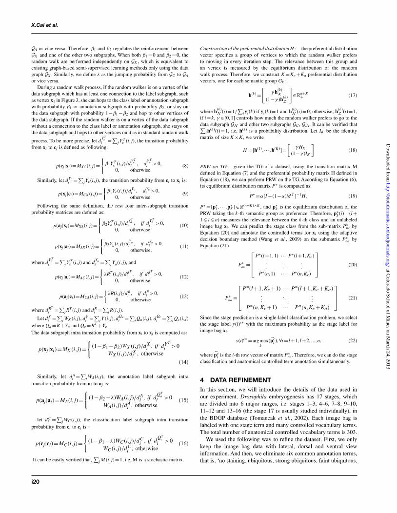
where the vector x is calculated using bag-of-word features for one imagebag. Regarding the parameter σ , we resort to self-tuning method (Zelnik-Manor and Perona, 2004). WX (i,j) indicates how closely xi and xj are related.From WX , a graph GX = (VX ,EX ) can be induced, where VX =X and EX ⊆VX ×VX . And we use kNN graph. To be specific, we connect xi,xj if oneof them is among the other’s k nearest neighbor and define the value ofthe edge connecting them by Equation (2). Because GX characterizes therelationships between data points, it is usually called as data graph, suchas the middle subgraph in Figure 3. Existing graph-based semi-supervisedlearning methods (Kang et al., 2006; Zha et al., 2008) only make use of thedata graph, on which the class label information is propagated.
Different from conventional single-label classification learning problem inwhich classes are mutual exclusive, the anatomical terms are interrelated withone another. Again, we resort to cosine similarity to calculate the controlledterm affinity matrix WA, where WA(i,j) indicates the correlation betweenai and aj . Thus, a graph GA = (VA,EA) is induced, where VA =A and EA ⊆VA ×VA. We call GA as annotation label subgraph, which is shown as theright subgraph in Figure 3. Similarly, stage classification label graph shownas the left subgraph in Figure 3, GC = (VC ,EC ) can be constructed fromstage classification labels, where VC =C and EC ⊆VC ×VC , where we definethe value of the edge connecting two stage labels as WC (i,j)=∥∥Sbi −Sbj
∥∥F ,
where ‖ ‖F means Frobenius norm and Sbi denotes the between class scattermatrix for stage i. Connecting GX and GA by the annotation associations viathe green dashed lines, connecting GX and GC by the class associations viathe blue dashed lines and connecting GC and GA by the stage-term associationvia the purple dashed lines, we construct a TG as following:
G= (VX ∪VC ∪VA,EX ∪EA ∪EXC ∪EXA ∪ECA), (3)
which is illustrated in Figure 3. Obviously, the subgraph GAX = (VX ,VA,EXA)connects GX and GA, whose adjacency matrix is Y T
a . Similarly, theadjacency matrix of GCX = (VX ,VC ,EXC ) is Y T
c . The subgraph(VC ,VA,ECA
)characterizes the associations between stage classes and anatomical termswhose adjacency matrix is R defined in Equation (1).
In contrast to existing graph-based semi-supervised learning methods thatonly use information conveyed by GX , we aim to simultaneously classify andannotate an unlabeled data point using all the information encoded in G. Sinceall data points (gene expression image bags), stage terms and annotationterms are equally regarded as vertices on G, our task is to measure the
i18
at Colorado School of M
ines on March 24, 2013
http://bioinformatics.oxfordjournals.org/
Dow
nloaded from

Copyedited by: TRJ MANUSCRIPT CATEGORY:
[11:19 31/5/2012 Bioinformatics-bts220.tex] Page: i19 i16–i24
Drosophila gene expression patterns
Cellularblastoderm
Foregutanlage
Foregutprimordium
Clypeolabrumanlage
Annotation labelsubgraphAAA EVG ,
Stage 7-8
Stage 13-16
Classification labelsubgraph
Stage 4-6
Datasubgraph
XXX EVG ,
? ?
Fig. 3. The TG constructed from the gene expression data. Solid lines indicate affinity between vertices within in a same subgraph, dashed lines indicatesassociations between vertices in two different subgraphs
relevance between a class/anntotation term vertex and a data point vertex.As each class/annotation term has a set of associated training data points,which convey the same biological record information as the class/annotationterm, we consider both a class/annotation term vertex and its labeled trainingimage bag vertices as a group set,
Gk =ck ∪{xi|yi(k)=1}, (4)
which is illustrated as the vertices with orange boundary in 3. As a result,instead of measuring vertex-to-vertex relevance between a class/annotatationterm vertex and an unlabeled data point vertex, we may measure the set-to-vertex relevance between the group set and the data point. Motivated by Brinand Page (1998); Tong et al. (2006), we consider to further develop standardrandom walk and use its equilibrium probability to measure the relevancebetween a group set and an unlabeled data point.
3.2 Preferential random walkStandard random walk on a graph W can be described as a Markov processwith transition probability M =D−1W , where di =∑
j W (i,j) is the degree
of vertex i and D=diag(d1,··· ,dn). Clearly, M T �=M and∑
j M (i,j)=1.When W is symmetric, it corresponds to an undirected graph. When Wis asymmetric, it corresponds to a directed graph and di is the out degreeof vertex i. Let p(t) be the distribution of the random walker at time t,the distribution at t+1 is given by p(t+1)(j)=∑
i p(t)(i)M (i,j). Thus, the
equilibrium (stationary) distribution of the random walk p∗ =p(t=∞) isdetermined by M T p∗ =p∗. It is well known that the solution is simply givenby p∗ =W e/(
∑i di)=d/(
∑i di), where d=[d1,··· ,dn]T .
It can be seen that the equilibrium distribution of a standard random walkis solely determined by the graph itself, but independent of the location wherethe random walk is initiated. In order to incorporate label information, wepropose the following PRW:
p(t+1)(j)= (1−α)∑
ip(t)(i)M (i,j)+αhj, (5)
where 0�α�1 is a fixed parameter, and h, called preferential distribution,is a probability distribution such that h(i)�0 and
∑i h(i)=1. Equation (5)
describes a random walk process in which the random walker hops on
the graph W according to the transition matrix M with probability 1−α,and meanwhile it takes a preference to go to other vertices specified by hwith probability α. The equilibrium distribution of PRW in Equation (5) isdetermined by p∗ = (1−α)M T p∗ +αh, which leads to:
p∗ =α[I −(1−α)M T ]−1h. (6)
Due to Perron-Frobenius theorem, the maximum eigenvalue of M is less thanmaxi
∑j M (i,j)=1. Thus, I −(1−α)M T is positive definite and invertible.
Equation (5) takes a similar form to two existing works: random walk withrestart (RWR) method (Tong et al., 2006) and PageRank algorithm (Brin andPage, 1998). In the former, h is a vector with all entries to be 0 except oneentry to be 1 indicating the vertex where the random walk could be restarted;while in the latter, h is a constant vector called as damping factor (Brin andPage, 1998). In contrast, the preferential distribution vector h in Equation (5)is a generic probability distribution, which is flexible thereby more powerful.Most importantly, through h we can assess group-to-vertex relevance, whileRWR and PageRank methods measure vertex-to-vertex relevance.
Similar to RWR (Tong et al., 2006), when we set the h to be a probabilitydistribution in which all the entries are 0 except for those corresponding toGk , p∗(i) measures how relevant the k-th group is to the i-th vertex on G.
3.3 Preferential random walk on TGIn order to classify and annotate unlabeled data points using the equilibriumprobabilities in Equation (6) of the PRW on TG, we need to construct thetransition matrix M and the preferential distribution h from G.Construction of the transition matrix M :
Let
M =⎡⎣ MX MXC MXA
MCX MC MCA
MAX MAC MA
⎤⎦, (7)
where MX , MC and MA are the intrasubgraph transition matrices of GX ,GC and GA respectively, and the rest six sub-matrices are the intersubgraphtransition matrices among GX , GC and GA. Let β1 ∈[0,1] be the jumpingprobability, i.e. the probability that a random walker hops from GX to GC
and vice versa. And let β2 ∈[0,1] be the jumping probability from GX to
i19
at Colorado School of M
ines on March 24, 2013
http://bioinformatics.oxfordjournals.org/
Dow
nloaded from
Copyedited by: TRJ MANUSCRIPT CATEGORY:
[11:19 31/5/2012 Bioinformatics-bts220.tex] Page: i20 i16–i24
X.Cai et al.
GA or vice versa. Therefore, β1 and β2 regulates the reinforcement betweenGX and one of the other two subgraphs. When both β1 =0 and β2 =0, therandom walk are performed independently on GX , which is equivalent toexisting graph-based semi-supervised learning methods only using the datagraph GX . Similarly, we define λ as the jumping probability from GC to GA
or vice versa.During a random walk process, if the random walker is on a vertex of the
data subgraph which has at least one connection to the label subgraph, suchas vertex x1 in Figure 3, she can hops to the class label or annotation subgraphwith probability β1 or annotation subgraph with probability β2, or stay onthe data subgraph with probability 1−β1 −β2 and hop to other vertices ofthe data subgraph. If the random walker is on a vertex of the data subgraphwithout a connection to the class label or annotation subgraph, she stays onthe data subgraph and hops to other vertices on it as in standard random walk
process. To be more precise, let dY T
ci =∑
j YTc (i,j), the transition probability
from xi to cj is defined as following:
p(cj|xi)=MXC (i,j)={
β1Y Tc (i,j)/d
Y Tc
i , dY T
ci >0,
0, otherwise.(8)
Similarly, let dYci =∑
j Yc(i,j), the transition probability from ci to xj is:
p(xj|ci)=MCX (i,j)={
β1Yc(i,j)/dYci , dYc
i >0,
0, otherwise.(9)
Following the same definition, the rest four inter-subgraph transitionprobability matrices are defined as:
p(aj|xi)=MXA(i,j)={
β2Y Ta (i,j)/d
Y Ta
i , if dY T
ai >0,
0, otherwise.(10)
p(xj|ai)=MAX (i,j)={
β2Ya(i,j)/dYai , if dYa
i >0,
0, otherwise.(11)
where dY T
ai =∑
j YTa (i,j) and d
Yai =∑
j Ya(i,j), and
p(cj|ai)=MAC (i,j)={
λRT (i,j)/dRT
i , if dRT
i >0,
0, otherwise.(12)
p(aj|ci)=MCA(i,j)={
λR(i,j)/dRi , if dR
i >0,
0, otherwise(13)
where dRT
i =∑j R
T (i,j) and dRi =∑
j R(i,j).
Let dXi =∑
j WX (i,j), dYi =∑
j Y (i,j), dQai =∑
j Qa(i,j), dQci =∑
j Qc(i,j)where Qa =R+Ya and Qc =RT +Yc.The data subgraph intra transition probability from xi to xj is computed as:
p(xj|xi)=MX (i,j)={
(1−β1 −β2)WX (i,j)/dXi , if dY T
i >0WX (i,j)/dX
i , otherwise
(14)
Similarly, let dAi =∑
j WA(i,j), the annotation label subgraph intratransition probability from ai to aj is:
p(aj|ai)=MA(i,j)={
(1−β2 −λ)WA(i,j)/dAi , if d
QTa
i >0WA(i,j)/dA
i , otherwise(15)
let dCi =∑
j WC (i,j), the classification label subgraph intra transitionprobability from ci to cj is:
p(cj|ci)=MC (i,j)={
(1−β1 −λ)WC (i,j)/dCi , if d
QTc
i >0WC (i,j)/dC
i , otherwise(16)
It can be easily verified that,∑
j M (i,j)=1, i.e. M is a stochastic matrix.
Construction of the preferential distribution H: the preferential distributionvector specifies a group of vertices to which the random walker prefersto moving in every iteration step. The relevance between this group andan vertex is measured by the equilibrium distribution of the randomwalk process. Therefore, we construct K =Kc +Ka preferential distributionvectors, one for each semantic group Gk :
h(k) =[
γ h(k)X
(1−γ )h(k)L
]∈R
n+K+ (17)
where h(k)X (i)=1/
∑i yi(k) if yi(k)=1 and h(k)
X (i)=0, otherwise; h(k)L (i)=1,
if i=k, γ ∈[0,1] controls how much the random walker prefers to go to thedata subgraph GX and other two subgraphs GC , GA. It can be verified that∑
i h(k)(i)=1, i.e, h(k) is a probability distribution. Let IK be the identity
matrix of size K ×K , we write
H =[h(1),··· ,h(K)]=[
γ HX
(1−γ )IK
](18)
PRW on TG: given the TG of a dataset, using the transition matrix Mdefined in Equation (7) and the preferential probability matrix H defined inEquation (18), we can perform PRW on the TG. According to Equation (6),its equilibrium distribution matrix P∗ is computed as:
P∗ =α[I −(1−α)M T ]−1H , (19)
P∗ =[p∗1,··· ,p∗
K ]∈R(n+K)×K , and p∗
k is the equilibrium distribution of thePRW taking the k-th semantic group as preference. Therefore, p∗
k (i) (l+1� i�n) measures the relevance between the k-th class and an unlabeledimage bag xi . We can predict the stage class from the sub-matrix P∗
nc byEquation (20) and annotate the controlled terms for xi using the adaptivedecision boundary method (Wang et al., 2009) on the submatrix P∗
na byEquation (21).
P∗nc =
⎡⎢⎢⎣P∗(l+1,1) ··· P∗(l+1,Kc)
.
.
.. . .
.
.
.
P∗(n,1) ··· P∗(n,Kc)
⎤⎥⎥⎦ (20)
P∗na =
⎡⎢⎢⎣P∗(l+1,Kc +1) ··· P∗(l+1,Kc +Ka)
.... . .
...
P∗(n,Kc +1) ··· P∗(n,Kc +Ka)
⎤⎥⎥⎦ (21)
Since the stage prediction is a single-label classification problem, we selectthe stage label y(i)c∗ with the maximum probability as the stage label forimage bag xi .
y(i)c∗ =argmaxk
(pci ),∀i= l+1,l+2,...,n. (22)
where pci is the i-th row vector of matrix P∗
nc. Therefore, we can do the stageclassification and anatomical controlled term annotation simultaneously.
4 DATA REFINEMENTIn this section, we will introduce the details of the data used inour experiment. Drosophila embryogenesis has 17 stages, whichare divided into 6 major ranges, i.e. stages 1–3, 4–6, 7–8, 9–10,11–12 and 13–16 (the stage 17 is usually studied individually), inthe BDGP database (Tomancak et al., 2002). Each image bag islabeled with one stage term and many controlled vocabulary terms.The total number of anatomical controlled vocabulary terms is 303.
We used the following way to refine the dataset. First, we onlykeep the image bag data with lateral, dorsal and ventral viewinformation. And then, we eliminate six common annotation terms,that is, ‘no staining, ubiquitous, strong ubiquitous, faint ubiquitous,
i20
at Colorado School of M
ines on March 24, 2013
http://bioinformatics.oxfordjournals.org/
Dow
nloaded from

Copyedited by: TRJ MANUSCRIPT CATEGORY:
[11:19 31/5/2012 Bioinformatics-bts220.tex] Page: i21 i16–i24
Drosophila gene expression patterns
maternal, rapidly degraded’, which can be regarded as outliersbecause they can neither provide stage-specific information norrecord anatomical structures. After that, we remove the anatomicalterms whose data sample is <50. We ignore the stage 1–3 data sincethe number of anatomical terms after the above procedure becomes2, too small to be compared with other stages. And finally we get 79anatomical annotation terms in total that we will consider to annotatethe unlabeled image bag.
We refine the data mainly based on the following two reasons.On one hand, the annotation terms which appear in too few imagebags are statistically too weak to be learned effectively. On the otherhand, since we will use 5-fold cross-validations in our experiments,we have to guarantee there is at least one data point associated witheach anatomical term in each fold. Moreover, in order to balance thenumber of image bags for different stages, we randomly sample 500image bags as the data points for each stage. At last, the summaryof the refined dataset is shown in Table 1.
5 EXPERIMENTIn this section, we will conduct experiments to evaluate PRWempirically on the refined dataset and compare it with otherstate-of-art classification methods. Since our method can do jointclassification, in order to evaluate the benefit of joint learning, wecompare its performance with that of the state-of-art multiclasssingle label or multiclass multilabel algorithms which can onlyhandle either stage classification or anatomical term annotationproblem. Our procedure is to train our model with stage labeledand anatomical term annotated image bags. All testing imagebags are unlabeled with developmental stage and unannotated withanatomical controlled terms.
5.1 Experimental setupWhen constructing PRW on TG, we used kNN graph setting k =9.We used ‘inverse’ 5-fold cross-validation to determine the valuesof the following five parameters, that is, using 1-fold for trainingand using the remaining 4-folds for testing to mimic the scenarioin the real application where the number of training data is muchless than the testing data. In our experiment, we found that thefollowing five parameters are not sensitive in certain ranges withgood performances. β1, β2 and λ controls the jumping betweendifferent subgraphs and cannot affect the result much if they areassigned in the range of (0.1,0.45). α controls initial preference ofthe random walker and will get stable result if it is assigned in therange of (0,0.1). γ controls how much the random walker prefersto go to the data subgraph or to go to two other subgraphs and it isusually in the range of (0.1,0.3).
Besides those parameters, we also need to initialize the stageas well as anatomical controlled terms for the testing image bagxi , where i= l+1,...,n, l is the number of training image bag. Inour experiment, we used k-nearest neighbor (KNN) method to dothe initializations for both stage classification and anatomical termannotations tasks because of its simplicity and clear intuition. Tobe specific, we use k =1 and we abbreviate it as 1NN. Our jointclassification framework will self-consistently amend the incorrectlabels for stage and controlled terms. We perform 10 random splits ofthe data and report the average performance over the 10 trials. Please
note that, in each trial, we still do ‘inverse’ 5-fold cross validationand record the average performance result as the result of that trial.
5.2 Image bag stage classificationDrosophila gene expression pattern stage categorization is a single-label multi-class problem. We compare the result of our method withthat of support vector machine (SVM) with radial basis function(RBF) kernel (Chang and Lin, 2001). We use the optimal parametervalues for C and γ got from cross-validation as well. We alsocompare the classification result of 1NN that we use to do theinitialization. We assess the classification in terms of the averageclassification accuracy and the average confusion matrices. Since thedata that we used is class balanced, the mean value of the entries onthe diagonal of the confusion matrix is also the average classificationaccuracy. From the resulting average confusion matrices shown inFigure 5, we can see that the average prediction accuracy of ourmethod is better than that of the other two state-of-art methods,especially in the last stage 13–16, where the number of anatomicalterms is greatly larger than that of the other stages.
5.3 Image bag controlled vocabulary terms annotationBesides the stage classification task, we also validate our methodby predicting the anatomical controlled terms for the Drosophilagene expression patterns, which can be considered as a multi-classmulti-label classification problem. The conventional classificationperformance metrics in statistical learning, precision and F1 score,are utilized to evaluate the proposed methods. For every anatomicalterm, the precision and F1 score are computed following the standarddefinition for the binary classification problem. To address the multi-label scenario, following Tsoumakas and Vlahavas (2007), macroand micro average of precision and F1 score are used to assess theoverall performance across multiple labels. We compared four stateof art multi-label classification methods: local shared subspace (LS)(Ji et al., 2008), local regression (LR) (Ji et al., 2009), harmonicfunction (HF) (Zhu et al., 2003) and random walk (RW) (Zhouand Schölkopf, 2004). All of them are proposed recently to solvethe multilabel annotation problem. In addition, we compare theresults of 1NN as well. For the first three methods we use thepublished codes posted on the corresponding author’s websites. Andwe implement the RW method following the original work (Zhouand Schölkopf, 2004). For HF and RW methods, we follow theoriginal work to solve the multilabel annotation only. Therefore, weonly evaluate those two methods on data subgraph and annotationlabel subgraph without using any information derived from theclassification label subgraph such as the stage–term correlation.Table 2 shows the average anatomical annotation performance of79-term dataset. Compared to the above five stat-of-the-art methods,our method has the best results by all metrics. Figure 6 illustratesthe average Micro F1 score of our method, 1NN, LS, LR, RW andHF approaches for all the anatomical terms on 79-term dataset. Andagain, our method consistently achieves best performance for mostof the anatomical controlled terms.
5.4 The advantage of joint learningUnlike the traditional work, our proposed method can take advantageof all the information to do the stage classification and anatomicalterm annotation simultaneously. Therefore, when the number oftraining data is scare, we can resort to both intrarelations and
i21
at Colorado School of M
ines on March 24, 2013
http://bioinformatics.oxfordjournals.org/
Dow
nloaded from

Copyedited by: TRJ MANUSCRIPT CATEGORY:
[11:19 31/5/2012 Bioinformatics-bts220.tex] Page: i22 i16–i24
X.Cai et al.
Fig. 4. The middle part demonstrates the terms–stages correlation and the right part shows the terms–terms correlation of 79 terms. The stage unknown testdata shown in the left part is classified correctly as Stage 13–16, because of the strong correlation between the predicted stage and its predicted anatomicalterms and vice versa, NOT the similarity of its first and second nearest neighboring data induced from the data graph only
Table 2. Annotation prediction performance comparisonon the 79-term dataset
Method Ma Pre Ma F1 Mi Pre Mi F1
1NN 0.3455 0.3595 0.2318 0.2230LS 0.5640 0.3778 0.3516 0.1903LR 0.6049 0.4425 0.3953 0.2243RW 0.4019 0.3385 0.2808 0.1835HF 0.3727 0.3296 0.2756 0.1733Our method 0.6125 0.4434 0.4057 0.2336
Ma Pre, Avg. Macro Precision; Ma F1, Avg. Macro F1; Mi Pre,Avg. Micro Precision; Mi F1, Avg. Micro F1.
interrelations to make the decision for stage classification andanatomical controlled term annotation simultaneously. When thereare strong correlation between those two tasks, we expect thatthe performance of both tasks will be enhanced by joint learningwork than treating them individually and independently. Figure 4shows the pairwise label correlations of the 79 terms and stage–termcorrelations between 5 stages and 79 terms. As highlighted by purplearrows, we can observe that there are high pairwise correlationsbetween the terms ‘embryonic brain’,‘ventral nerve cord’ as wellas ‘embryonic/larval muscle system’. Moreover, all the above threeterms have high correlations with the stage 13–16, which can providestrong evidence that the given testing image bag could belong tothe last developmental stage besides the induction from the datagraph only. If our joint classification framework annotates it with allthose three terms, although from the data similarity we cannot get
(a) (b) (c)
Fig. 5. Stage classification results in terms of confusion matrices on 79-termdataset: (a) the confusion matrix calculated by SVM (b) the confusion matrixcalculated by 1NN. (c) the confusion matrix calculated by our method. (a)SVM: acc. 84.50%; (b) 1NN: acc. 77.40%; (c) our: acc. 85.20%
strong evidence for the stage prediction, we can take advantage ofthe term–term as well as term–stage high correlations to adjust itsstage to stage 13–16. In other words, relevant anatomical terms couldhelp us to predict the stage label since they provide the spatial andtemporal information of local structure corresponding to a specificembryo development stage. Nevertheless, not all anatomical termswill definitely benefit stage classification, which is consistent withour stage classification result. From Figure 5, we can see that ourmethod may have competitive result compared with SVM withrespect to some certain stage. However, given more anatomicalterm information, the performance of our method will graduallyoutperform the other methods, especially for the prediction result ofstage 13–16.
i22
at Colorado School of M
ines on March 24, 2013
http://bioinformatics.oxfordjournals.org/
Dow
nloaded from

Copyedited by: TRJ MANUSCRIPT CATEGORY:
[11:19 31/5/2012 Bioinformatics-bts220.tex] Page: i23 i16–i24
Drosophila gene expression patterns
Fig. 6. The Avg. Micro F1 score of five methods on each term in 79-term dataset. (It is better to be viewed in colorful and zoomed in mode.)
5.5 The more meaningful asymmetric correlationmatrix
When we build the TG, at first, we assume the term–term correlationand stage–term correlation are both symmetric, since we usedcosine similarity to represent their correlations. However, the aboveassumption does not always hold in the real data. In Drosophilaembryo gene expression images, we found that the conditionalprobability of the occurrence of term ‘ventral nerve cord’ given term‘embryonic brain’ is higher than that of the ‘embryonic brain’ given‘ventral nerve cord’, which satisfies the biology meaning that ventralnerve cord occurs earlier than embryonic brain. After learning, ourmethod can automatically discover the above hidden asymmetriccorrelation information, that is,
P∗aa =
⎡⎢⎣ P∗(n+Kc +1,Kc +1) ··· P∗(n+Kc +1,K)...
. . ....
P∗(n+K,Kc +1) ... P∗(n+K,K)
⎤⎥⎦ (23)
In order to see the learned asymmetric term–term correlationmore clearly, in Figure 7, we show the difference matrix got byP∗
aa −P∗aa
T . Taking those more accurate asymmetric correlationinto consideration, our method can potentially improve both stageclassification and anatomical annotation results even more.
6 CONCLUSIONIn this article, we proposed a novel TG model to learn thetask interrelations between stage recognition and anatomical termsannotation of Drosophila gene expression patterns. The standardbag-of-word features and three major views (lateral, dorsal andventral) were used to describe the 3D Drosophila images. A newPRW method was introduced to simultaneously propagate thestage labels and anatomical controlled terms via TG model. Bothstage classification and anatomical controlled term annotation tasksare jointly completed. We evaluated the proposed method using onerefined BDGP dataset. The experimental results demonstrated inthe real application, when the number of training data is scarce,our joint learning method can achieve superior prediction results onboth tasks than the state-of-the-art methods. What is more, we candiscovery more accurate asymmetric term–term correlation, whichcan potentially improve the results of both tasks even more.
Fig. 7. The learned difference matrix. (It is better to be viewed in colorful andzoomed in mode.) In order to see the asymmetric entries more clearly, we plotP∗
aa −P∗aa
T . After PRW, the entries marked as brighter square have higherconditional probability (positive correlation) than its counterpart which ismarked as darker color. This asymmetric reflects more accurate term–termcorrelation than the original symmetric assumption
ACKNOWLEDGEMENTThe author would like to thank Dr Sudhir Kumar for his help in datacollection.
Funding: [This research was supported by National ScienceFoundation Grants CCF-0830780, CCF-0917274, DMS-0915228and IIS-1117965].
REFERENCESBrin,S. and Page,L. (1998) The anatomy of a large-scale hypertextual web search
engine. In International Conference on World Wide Web (WWW), Elsevier SciencePublishers, pp. 107–117.
Chang,C. and Lin,C. (2011) LIBSVM : a library for support vector machines. ACMTransactions on Intelligent Systems and Technology, 2, 1–27.
Fowlkes,C. et al. (2008) A quantitative spatiotemporal atlas of gene expression in theDrosophila blastoderm. Cell, 133, 364–374.
Grumbling,G. et al. (2006) FlyBase: anatomical data, images and queries. Nucleic AcidsRes., 34, D484–D488.
Hendriks,C. L. et al. (2006) Three dimensional morphology and gene expression in theDrosophila blastoderm at cellular resolution I: data acquisition pipeline. GenomeBiol., 7, R123.
i23
at Colorado School of M
ines on March 24, 2013
http://bioinformatics.oxfordjournals.org/
Dow
nloaded from

Copyedited by: TRJ MANUSCRIPT CATEGORY:
[11:19 31/5/2012 Bioinformatics-bts220.tex] Page: i24 i16–i24
X.Cai et al.
Ji,S. et al. (2008) Extracting shared subspace for multi-label classification. In ACMSIGKDD International Conference on Knowledge Discovery and Data Mining2009, pp. 381–389.
Ji,S. et al. (2009) Drosophila gene expression pattern annotation using sparsefeatures and term-term interactions. In ACM SIGKDD International Conferenceon Knowledge Discovery and Data Mining, pp. 407–416.
Ji,S. et al. (2010) A shared-subspace learning framework for multi-label classification.ACM Transactions on Knowledge Discovery from Data (TKDD), 4, 1–29.
Kang,F. et al. (2006) Correlated label propagation with application to multi-labellearning. In IEEE International Conference on Computer Vision and PatternRecognition (CVPR), pp. 1719–1726.
Kumar,S. et al. (2002) BEST: A novel computational approach for comparing geneexpression patterns from early stages of Drosophila melanogaster development.Genetics, 162, 2037–2047.
L’ecuyer,E. et al. (2007) Global analysis of mRNA localization reveals a prominentrole in organizing cellular architecture and function. Cell, 131, 174–187.
Li,Y. et al. (2009) Drosophila gene expression pattern annotation through multi-instancemulti-label learning. In Proceedings of the 21st International Joint Conference onArtificial Intelligence, AAAI press, pp. 1445–1450.
Lowe,D. (2004) Distinctive image features from scale-invariant keypoints. Int.J. Comput. Vis., 60, 91–110.
Lyne,R. et al. (2007) FlyMine: an integrated database for Drosophila and anophelesgenomics. Genome Biol., 8, R129.
Megason,S. and Fraser,S. (2007) Imaging in systems biology. Cell, 130, 784–795.Peng,H. and Myers,E.W. (2004) Comparing in situ mRNA expression patterns of
drosophila embryos. In International Conference on Research in ComputationalMolecular Biology (RECOMB), ACM, pp. 157–166.
Peng,H. et al. (2007) Automatic image analysis for gene expression patterns of flyembryos. BMC Cell Biol., 8(Suppl. 1), S7.
Puniyani,K. et al. (2010) SPEX2: Automated Concise Extraction of Spatial GeneExpression Patterns from Fly Embryo ISH Images. Intell. Sys. Mol. Biol., 26,i47–i56.
Shuiwang,J. et al. (2009) A bag-of-words approach for Drosophila gene expressionpattern annotation. BMC Bioinformatics, 10, 119.
Tomancak,P. et al. (2002) Systematic determination of patterns of gene expressionduring Drosophila embryogenesis. Genome Biol., 3, 88.
Tomancak,P. et al. (2007) Global analysis of patterns of gene expression duringDrosophila embryogenesis. Genome Biol., 8, R145.
Tong,H. et al. (2006) Fast random walk with restart and its applications. In IEEEInternational Conference on Data Mining (ICDM), pp. 613–622.
Tsoumakas,G. and Vlahavas,I.P. (2007) Random k-labelsets: An ensemble method formultilabel classification. In European conference on Machine Learning, Springer-Verlag, pp. 406–417.
Wang,H. et al. (2009) Image annotation using multi-label correlated Green’s function.In IEEE International Conference on Computer Vision, pp. 2029–2034.
Weigmann,K. et al. (2003) FlyMove – a new way to look at development of Drosophila.Trends Genet. 19, 310–311.
Zelnik-Manor,L. and Perona,P. (2004) Self-tuning spectral clustering. Advances inneural information processing systems, 17, 16.
Zha,Z. et al. (2008) Graph-based semi-supervised learning with multi-label.In IEEE International Conference on Multimedia and Expo (ICME),pp. 1321–1324.
Zhou,D. and Schölkopf,B. (2004) Learning from labeled and unlabeled data usingrandom walks. In Annual Symposium of the German Association for PatternRecognition (DAGM), Springer, pp. 237–244.
Zhu,X. et al. (2003) Semi-supervised learning using gaussian fields and harmonicfunctions. In International Conference on Machine Learning (ICML), ACM press,pp. 912–919.
i24
at Colorado School of M
ines on March 24, 2013
http://bioinformatics.oxfordjournals.org/
Dow
nloaded from