isolation of microorganism and enumeration
TRANSCRIPT
27
ISOLATION OF PURE CULTURES
A pure culture theoretically contains a single bacterial species. There are anumber of procedures available for the isolation of pure cultures from mixedpopulations. A pure culture may be isolated by the use of special media withspecific chemical or physical agents that allow the enrichment or selection of oneorganism over another. The differential and selective procedures will be utilizedlater in this course. Simpler methods for isolation of a pure culture include: (i)spread plating on solid agar medium with a glass spreader and (ii) streak platingwith a loop. The purpose of spread plating and streak plating is to isolateindividual bacterial cells (colony-forming units) on a nutrient medium.
Both procedures (spread plating and streak plating) require understanding ofthe aseptic technique. Asepsis can be defined as the absence of infectiousmicroorganisms. However, the term is usually applied to any technique designedto keep unwanted microorganisms from contaminating sterile materials.
FIRST PERIOD
Material:
1. Seven 9-ml dilution tubes of sterile saline2. Seven nutrient agar plates3. 1.0 ml and 0.1 ml pipets4. Glass spreader aka �hockey stick�5. 95% ethyl alcohol in glass beaker (WARNING: Keep alcohol away from
flame!!)6. Mixed overnight broth culture of Staphylococcus aureus and Serratia
marcescens
28
Procedure: (work in pairs)
A. Spread Plate Technique
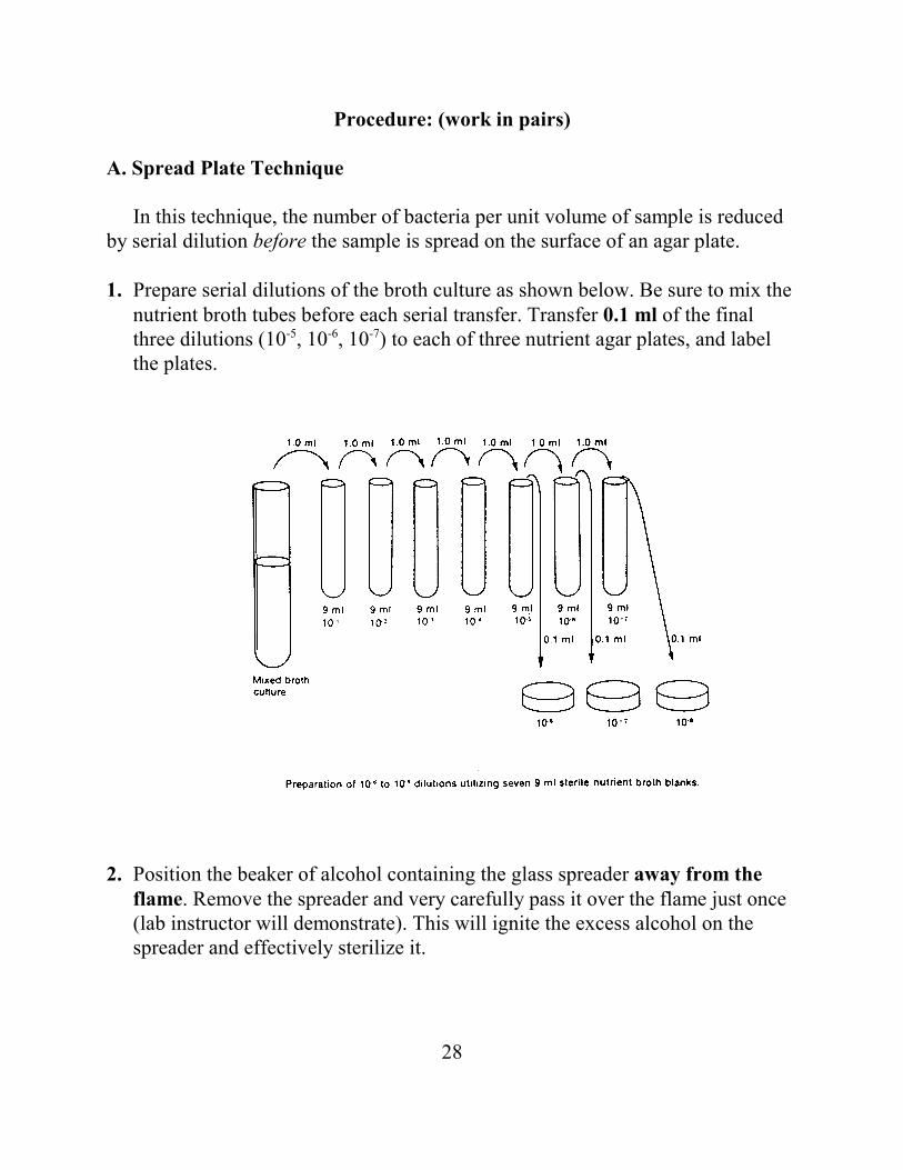
In this technique, the number of bacteria per unit volume of sample is reducedby serial dilution before the sample is spread on the surface of an agar plate.
1. Prepare serial dilutions of the broth culture as shown below. Be sure to mix thenutrient broth tubes before each serial transfer. Transfer 0.1 ml of the finalthree dilutions (10-5, 10-6, 10-7) to each of three nutrient agar plates, and labelthe plates.
2. Position the beaker of alcohol containing the glass spreader away from theflame. Remove the spreader and very carefully pass it over the flame just once(lab instructor will demonstrate). This will ignite the excess alcohol on thespreader and effectively sterilize it.
29
3. Spread the 0.1 ml inoculum evenly over the entire surface of one of the nutrientagar plates until the medium no longer appears moist. Return the spreader tothe alcohol.
4. Repeat the flaming and spreading for each of the remaining two plates.
5. Invert the three plates and incubate at room temperature until the next labperiod.
B. Streak Plate Technique
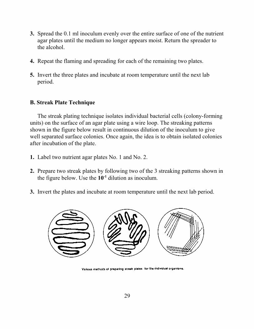
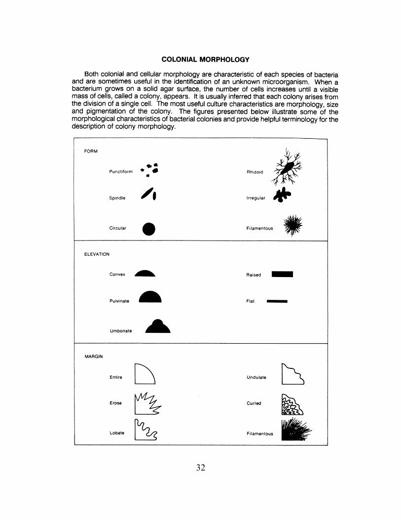
The streak plating technique isolates individual bacterial cells (colony-formingunits) on the surface of an agar plate using a wire loop. The streaking patternsshown in the figure below result in continuous dilution of the inoculum to givewell separated surface colonies. Once again, the idea is to obtain isolated coloniesafter incubation of the plate.
1. Label two nutrient agar plates No. 1 and No. 2.
2. Prepare two streak plates by following two of the 3 streaking patterns shown inthe figure below. Use the 10-1 dilution as inoculum.
3. Invert the plates and incubate at room temperature until the next lab period.
30
C. Exposure Plates
Exposure of sterile media to the environment will demonstrate the importanceof aseptic technique.
1. Label two nutrient agar plates as "Exposure I" and "Exposure II."
2. Uncover the plate marked "Exposure I" and allow it to remain exposed in thelab for about 5 minutes.
3. Expose the plate marked "Exposure II" to a source of possible contaminants.Use your imagination: cough or sneeze, place your fingers on the surface ofthe agar, etc.
4. Invert the plates and incubate at room temperature until the next lab period.
31
SECOND PERIOD
Material:
1. Colony counter
Procedure:
A. Spread Plate Technique
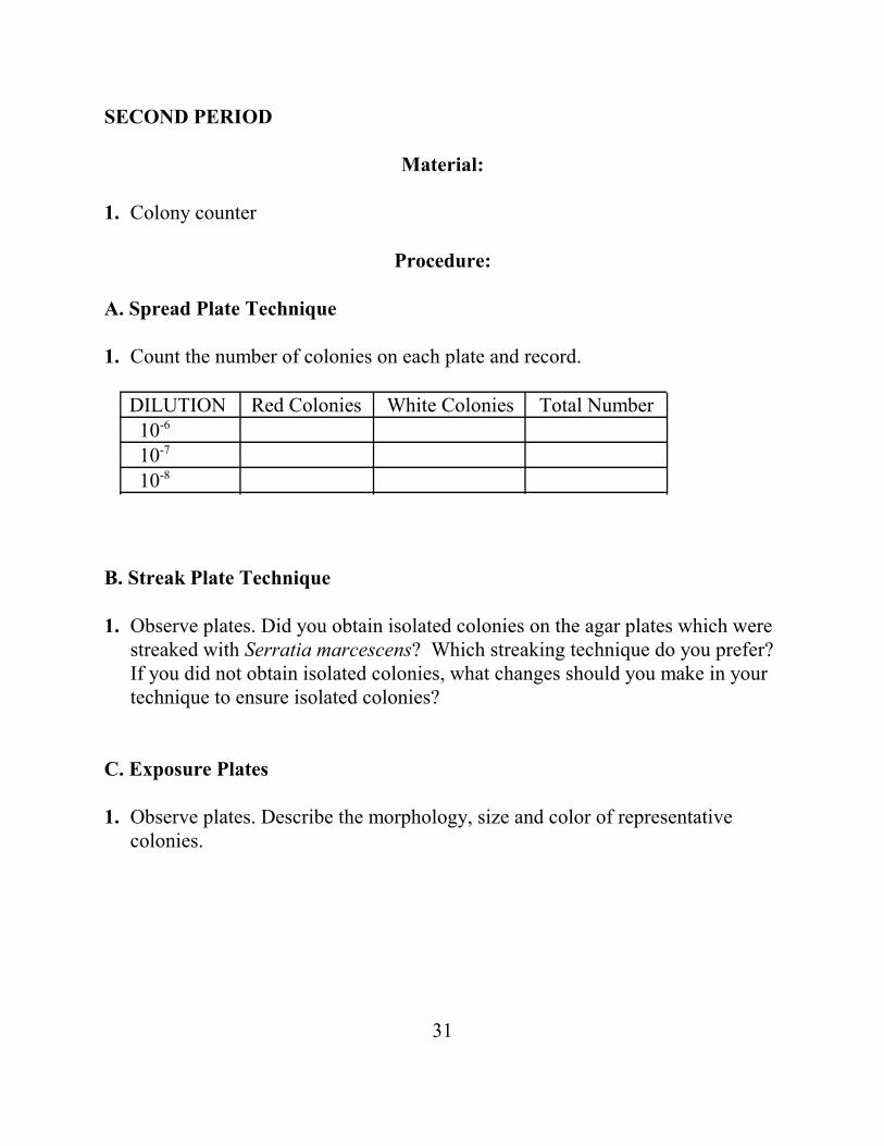
1. Count the number of colonies on each plate and record.
DILUTION Red Colonies White Colonies Total Number 10-6
10-7
10-8
B. Streak Plate Technique
1. Observe plates. Did you obtain isolated colonies on the agar plates which werestreaked with Serratia marcescens? Which streaking technique do you prefer?If you did not obtain isolated colonies, what changes should you make in yourtechnique to ensure isolated colonies?
C. Exposure Plates
1. Observe plates. Describe the morphology, size and color of representativecolonies.
32
33
BACTERIAL ENUMERATION
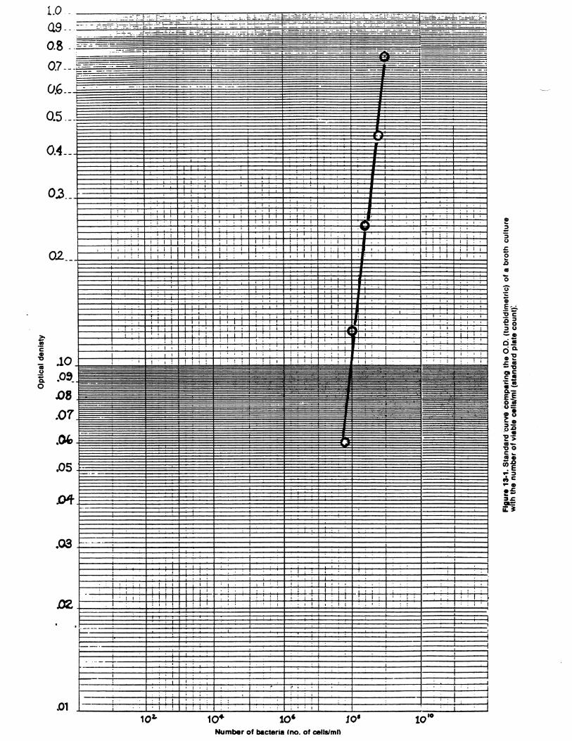
In the study of microbiology, there are numerous occasions when it isnecessary to either estimate or determine the number of bacterial cells in a brothculture or liquid medium. Determination of cell numbers can be accomplished bya number of direct or indirect methods. The methods include standard platecounts, turbidimetric measurements, visual comparison of turbidity with a knownstandard, direct microscopic counts, cell mass determination, and measurementof cellular activity. In this exercise, you will compare three methods of bacterialenumeration: the standard plate count, turbidimetric measurement and directmicroscopic counts.
Standard Plate Count (Viable Counts)
A viable cell is defined as a cell which is able to divide and form apopulation (or colony). A viable cell count is usually done by diluting theoriginal sample, plating aliquots of the dilutions onto an appropriate culturemedium, then incubating the plates under proper conditions so that colonies areformed. After incubation, the colonies are counted and, from a knowledge of thedilution used, the original number of viable cells can be calculated. For accuratedetermination of the total number of viable cells, it is critical that each colonycomes from only one cell, so chains and clumps of cells must be broken apart.However, since one is never sure that all such groups have been broken apart, thetotal number of viable cells is usually reported as colony-forming units (CFUs)rather than cell numbers. This method of enumeration is relatively easy toperform and is much more sensitive than turbidimetric measurement. A majordisadvantage, however, is the time necessary for dilutions, platings andincubations, as well as the time needed for media preparation.
Turbidimetric Measurement
A quick and efficient method of estimating the number of bacteria in aliquid medium is to measure the turbidity or cloudiness of a culture and translatethis measurement into cell numbers. This method of enumeration is fast and isusually preferred when a large number of cultures are to be counted.
34
Although measuring turbidity is much faster than the standard plate count,the measurements must be correlated initially with cell number. This is achievedby determining the turbidity of different concentrations of a given species ofmicroorganism in a particular medium and then utilizing the standard plate countto determine the number of viable organisms per milliliter of sample. A standardcurve can then be drawn (e.g., this lab protocol section), in which a specificturbidity or optical density reading is matched to a specific number of viableorganisms. Subsequently, only turbidity needs to be measured. The number ofviable organisms may be read directly from the standard curve, withoutnecessitating time-consuming standard counts.
Turbidity can be measured by an instrument such as a colorimeter orspectrophotometer. These instruments contain a light source and a light detector(photocell) separated by the sample compartment. Turbid solutions such as cellcultures interfere with light passage through the sample, so that less light hits thephotocell than would if the cells were not there. Turbidimetric methods can beused as long as each individual cell blocks or intercepts light; as soon as the massof cells becomes so large that some cells effectively shield other cells from thelight, the measurement is no longer accurate.
Before turbidimetric measurements can be made, the spectrophotometermust be adjusted to 100% transmittance (0% absorbance). This is done using asample of uninoculated medium. Percent transmittance of various dilutions of thebacterial culture is then measured and the values converted to optical density,based on the formula: Absorbance (O.D.) = 2 - log % Transmittance. Awavelength of 420 nm is used when the solution is clear, 540 nm when thesolution is light yellow, and 600-625 nm is used for yellow to brown solutions.
Direct Microscopic Count
Petroff-Hausser counting chambers can be used as a direct method todetermine the number of bacterial cells in a culture or liquid medium. In thisprocedure, the number of cells in a given volume of culture liquid is counteddirectly in 10-20 microscope fields. The average number of cells per field iscalculated and the number of bacterial cells ml-1 of original sample can then becomputed. A major advantage of direct counts is the speed at which results areobtained. However, since it is often not possible to distinguish living from dead
35
cells, the direct microscopic count method is not very useful for determining thenumber of viable cells in a culture.
FIRST PERIOD
Material:
1. Seven 9-ml dilution tubes of nutrient broth2. Six nutrient agar plates3. 1.0 and 10 ml pipets4. Glass spreader5. 95% ethyl alcohol in glass beaker (WARNING: Keep alcohol away from
flame!!)6. Overnight broth culture of Serratia marcescens
Procedure: (work in pairs)
A. Spread Plate Technique
1. Prepare serial dilutions of the broth culture as shown in the figure from aprevious lab exercise (Isolation of Pure Cultures). Be sure to mix thenutrient broth tubes before each serial transfer. Transfer 0.1 ml of the finalthree dilutions (10-5, 10-6, 10-7) to duplicate nutrient agar plates, and labelthe plates.
2. Spread the 0.1 ml inoculum evenly over the entire surface of one of thenutrient agar plates until the medium no longer appears moist. Return thespreader to the alcohol.
3. Repeat the flaming and spreading for each of the remaining five plates.
4. Invert the six plates and incubate at room temperature until the next labperiod (or ~ 48 hours, whichever is the shortest). Remember that onlyplates with 30 to 300 colonies are statistically valid.
36
B. Turbidimetric Method
1. Using the spectrophotometer, determine the optical density (O.D.) of theassigned broth culture at 600 nm. Note, you may have to use one of yourserial dilutions of the broth culture to get a good reading.
2. Record results.
C. Direct Microscopic Counts
Material:
1. Petroff-Hausser counting chamber 2. Cover slips3. Sterile diluent (nutrient broth or sterile saline)4. Pasteur pipets
Procedure: (work in pairs)
Be extremely careful handling Petroff-Hausser counting chambers!
1. Clean P-H counting chamber with 70% alcohol an let air dry.
2. Mix culture well and apply a single drop to counting chamber with Pasteurpipet. Examine the counting chamber using high power, oil immersionobjective.
3. Make a preliminary estimation of the concentration of cells from theovernight culture of Serratia marcescens using the following formula:
Total cells counted x 2.0x10⁷ x dilution factor = cells/ml# small squares counted
Therefore, if you counted an average of 15 cells per small square, then youwould have a final concentration of 3.0 x 10⁸ cells/ml.
37
4. You may have to adjust downward using one of your initial serial dilutionsso that the counts per small square are in the 5 to 15 cell range.
5. Once this is done, make sure to allow time for cells to settle and movefocus through the suspension (i.e., up and down) so as to count all cellswithin the small square “box”. Most cells will have attached to the bottomand/or top glass interface. You can also check the depth, which is 20 µm.The small square should also be 50 by 50 µm.
6. Count the number of bacterial cells in at least 10 small squares. Variabilityshould be less than +/- 10%.
SECOND PERIOD
Material:
1. Colony counter
Procedure:
1. Remember to pull plates and refrigerate after 48 hours max. Either then ornext lab period, count the number of colonies on each plate, calculate anaverage and record results.
2. Compare results from the standard plate counts with P-H directmicroscopic counts.
3. Compare results from the standard plate counts and direct microscopiccounts with that of optical density while considering the graph provided.Which data are the most robust and why? Which data yields the highestcounts and why?
38
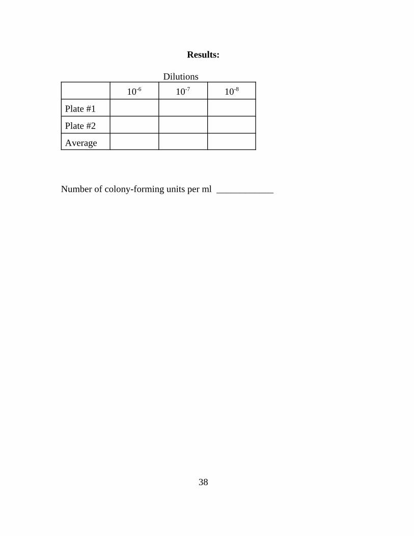
Results:
Dilutions10-6 10-7 10-8
Plate #1
Plate #2
Average
Number of colony-forming units per ml ____________