isolated maize zygotes mimicin vivoembryonic development and express microinjected genes when...
TRANSCRIPT
DEVELOPMENTAL BIOLOGY 177, 190–203 (1996)ARTICLE NO. 0155
Isolated Maize Zygotes Mimic in Vivo EmbryonicDevelopment and Express Microinjected GenesWhen Cultured in Vitro
Nathalie Leduc, Elisabeth Matthys-Rochon, Mireille Rougier,Lloyd Mogensen,* Preben Holm,† Jean-Louis Magnard,and Christian DumasLaboratoire de Reconnaissance Cellulaire et d’Amelioration des Plantes, Ecole NormaleSuperieure de Lyon, 46 allee d’Italie, 69364 Lyon Cedex 07, France; *Biological Sciences,Northern Arizona University, P.O. Box 5640, Flagstaff, Arizona 86011-5640; and †CarlsbergResearch Laboratory, 10 Gamle Carlsberg Vej, DK-2500 Copenhagen, Denmark
We established conditions for the regeneration of natural maize zygotes isolated from pollinated plants with the goal ofinvestigating the molecular control of early embryogenesis in higher plants. Viable zygotes were excised from embryo sacsby minimal enzymatic digestion and microdissection. Viable zygotes transferred to coculture with androgenic microsporesfrom barley developed into embryo-like structures in 61% of the cases. No development was observed when zygotes werecultured in the presence of maize anthers undergoing androgenetic embryogenesis. Zygote-derived embryo-like structuresregenerated into fertile plants through secondary embryogenesis when transferred to solid medium. The first zygoticdivision was asymmetrical and bipolar structures similar to pretransitional embryos observed in planta were later producedas observed using light and electron microscopy. Conditions for efficient microinjection of DNA into zygotes were estab-lished. Calcofluor and PATAG staining of zygotes showed that cell wall regeneration occurred as early as 20 min afterenzymatic isolation and that after 2 hr, each zygote was bordered with cell wall material. Through quantitative micropho-tometry, DNA synthesis during the first cell cycle of the zygote was shown to occur between isolation and 12 hr ofculture. Microinjection of two types of reporter genes (GUS gene and anthocyanin regulatory genes) demonstrates transientexpression in plant zygotes. On average, 3.5% of microinjected zygotes showed transgenic expression. Reporter geneexpression was observed in zygotes at different time points of their first cell cycle. q 1996 Academic Press, Inc.
INTRODUCTION to the complete understanding of embryo development inhigher plants.
Although the morphology and the cytology of develop-Zygotic embryogenesis is one of the most important mental stages are well documented in dicots (Mansfield et
events in plant biology. In higher plants, a zygote and an al., 1990; West and Harada, 1993) and in monocots (Abbeendosperm progenitor cell are produced concomitantly after and Stein, 1954; Meinke, 1991), information has only re-a double fertilization event. Zygotic embryogenesis corre- cently become available concerning the molecular controlsponds to the subsequent development of a zygote into an of embryogenic development in plants (Clark and Sheridan,embryo which is fed by the surrounding nutritive tissue: 1991; Mayer et al., 1993; Shevell et al., 1994; Yadegari etthe endosperm. The initial division of the polarized zygote al., 1994; Heck et al., 1995; Franzmann et al., 1995). How-is generally asymmetrical in angiosperms and leads to two ever, basic questions remain: (i) How is polarity establishedunequal cells. The smaller (apical cell) will produce the in the zygote and in the early embryonic stages? (ii) Howembryo proper and the larger (basal cell) the suspensor. The is cell differentiation governed in these tissues? and (iii)early stages of embryogenesis are defined by the shape of What genes are involved in these processes and how theythe embryos, and it is considered that the determination of are regulated?cell fate is mostly completed before the embryo reaches the This time lag in the field of plant embryogenesis as com-globular or heart stage (Goldberg et al., 1994; Jurgens, 1995). pared to animal embryogenesis is mainly due to the inacces-
sibility of the developing plant embryo to direct experimen-The study of early embryogenesis is, therefore, preliminary
190
0012-1606/96 $18.00Copyright q 1996 by Academic Press, Inc.
All rights of reproduction in any form reserved.
AID DB 8217 / 6x0f$$$161 06-20-96 10:06:21 dbal AP: Dev Bio
191Embryogenesis and Transformation of Isolated Maize Zygotes
in a growth chamber under the following controlled conditions:tation, and most of the knowledge gathered to date on earlyA photoperiod of 16/24 hr, a light intensity of 560 mErm02 sec01embryogenesis in plants has been gained through the study(provided by 400-W high-pressure sodium lamps MAZD-MAC 400of mutants (Clark and Sheridan, 1991; Mayer et al., 1993;E40), a day/night temperature of 24/197C and 75/95% humidity.Yadegari et al., 1994; Franzmann et al., 1995).Developing ears were covered with paper bags before silks emergedThe inaccessibility of the plant embryo could be over-from the husks. Control hand pollinations were made in the morn-
come through the use of a culture system in which isolated ing using freshly harvested pollen from tassels of the same geno-zygotes assume a normal pattern of growth and develop- type. Ears were collected 24 hr after pollination (hap).ment, thus allowing direct molecular analysis at any of the Highly androgenic maize plants from genotypes DH99 andearly embryonic stages (Dumas and Mogensen, 1993; Kranz DH5xDH7 (Barloy et al., 1989) were used as one source of nurseand Lorz, 1993; Holm et al., 1994). Strategies such as the cells for zygote culture. These plants were raised as previouslygenetic manipulation of isolated zygotes and of immature described for maize plants genotype A188 except that a photoperiod
of 16 hr and a light intensity of 800mErm02 sec01 were used. Tasselsembryos could contribute to a better understanding of thewere harvested just before emergence from the leaf whorl, wrappedprocesses involved in early embryogenic developmentin aluminium foil, and pretreated at 77C for 2 weeks prior to anther(Thomas, 1993). Previous studies using direct gene transferculture.techniques have already proven the value of monitoring
Barley plants (cultivar Igri) were also used as a source of andro-gene expression in cells (Olive et al., 1990; Simmonds etgenic material and were cultured as previously described in Holmal., 1992; Neuhaus et al., 1993; Pua and Lee, 1995) andet al. (1994).
embryos (Neuhaus et al., 1987; Kost et al., 1995). In this Androgenic cultures. Maize androgenetic material was ob-manner, microinjection of antisense RNAs or of plasmids tained from another cultures and prepared according to Vergne etcontaining genes of interest into zygotes or young embryos al. (1993). Briefly, anthers were excised from the tassels under amay help in the study of the processes of embryonic devel- sterile bench and transferred by batches of 30 into 6-cm-diameteropment (Thomas, 1993). Manipulation of plant zygotes and petri dishes (Falcon, Becton Dickinson and Co., Lincoln Park, NJ)embryos depends on the availability of an in vitro system containing 3 ml G0 medium (Dieu and Beckert, 1986). Cultures
were sealed with parafilm and kept in the dark at 257C for 6 tocoupled with a method for gene transfer. The culture and12 days prior to coculture. The day of the coculture, 2 ml of thetransformation method we have developed for maize zy-androgenetic culture as well as 10 anthers containing a high num-gotes contributes toward this goal. Cereals are known to beber of androgenetic embryos were transferred into a 35-mm-diame-recalcitrant to in vitro development. However, Kranz andter sterile petri dish (Corning, New York). The G0 medium wasLorz (1993) and Holm et al. (1994) have demonstrated theremoved and replaced by 2 ml NBM medium (Mol et al., 1994) asregeneration of plants from electrofused maize gametes anddescribed below but containing no growth regulator.
from isolated naturally fertilized barley and wheat zygotes, Barley androgenetic cultures were obtained from microspores iso-respectively. Using a nurse cell culture system (maize sus- lated and cultured as described in Holm et al. (1994) for 6 to 12pension cells or barley microspores), these authors obtained days. Prior to coculture, 0.5 ml of the androgenetic suspension waszygotic development producing embryo-like structures transferred into a well of a 12-well plate (Corning, New York).which regenerated into fertile plants. In this paper, we de- Microspore culture medium was removed and replaced by 0.5 ml
Kao medium supplemented with 2 mg/liter 2,4-D (see mediumscribe (1) a technical procedure for the isolation of viablecomposition below).maize zygotes produced in planta, (2) their development
Zygote isolation. Sterilization of the ears was not necessaryinto embryos that regenerate into fertile plants in the pres-prior to zygote isolation. The removal of the most internal husksence of barley microspores, and (3) histological evidence forin the laminar flow hood was sufficient to collect sterile material.the differentiation of suspensor-like and embryo-like struc-Ovule halves were isolated from the ear as described in Mol et al.tures during the in vitro development of zygotes. To exam-(1993) and collected in culture medium. When observed under anine the potential of these cells for gene transfer, we analyzedinverted stereomicroscope, ovule halves are translucent and the
the first zygotic cell cycle by determining the time course embryo sacs can easily be located. Using a scalpel blade, embryofor the resynthesis of the cell wall and for each phase of sacs bearing only a few cell layers of nucellus could thus be cutDNA synthesis during in vitro culture. These two aspects from the ovules. Embryo sacs were then transferred by batches ofproved to be essential for efficient gene transfer into isolated 30 into 1250 ml of culture medium and digested for exactly 2 minzygotes. Finally, we demonstrated for the first time the tran- after addition of 200 ml of enzyme solution (1% Macerozyme, 0.5%
pectolyase, 1.5% pectinase, 1% cellulase, 6.6% mannitol w/v; 600sient expression of transgenes in zygotes. Also reported ismOsm/kg H2O) (Leduc et al., 1995) and then rinsed twice in 1 mlthe expression of microinjected reporter genes in zygotesculture medium. Embryo sacs, which are fragile at this time, wereat different time points during their first cell cycle. Theleft for 1 hr at room temperature (227C) before further microdissec-importance of this in vitro system and the ability to transfertion. Using microneedles (Soc. Peter, Lyon, France), the remaininggenes into unique zygotic cells is discussed with respect tonucellus cells attached to the micropylar end of the embryo sacsthe study of early events in embryogenesis and to appliedwere removed, allowing the location of the zygote. The zygote was
research. released by gently tearing off the embryo sac envelope. Using aflame-drawn Pasteur pipette, the isolated zygote was collected andembedded in a drop of soft culture medium containing 0.75%MATERIAL AND METHODSw/v low-melting-point agarose (Type IX, Sigma Chemical Co., St.Louis, MO). This isolation procedure took about 4 hr/ear.Plant material. Maize (Zea mays L.) plants genotype A188 was
used as a source of natural maize zygotes. These plants were raised Zygote culture. Zygote development was obtained in the pres-
Copyright q 1996 by Academic Press, Inc. All rights of reproduction in any form reserved.
AID DB 8217 / 6x0f$$$162 06-20-96 10:06:21 dbal AP: Dev Bio
192 Leduc et al.
ence of androgenetic material from either maize or barley plants. Quantitative measurements of DNA levels. For quantitativemeasurements of DNA levels during the first cell cycle, zygotesIn experiments with maize nurse cells, NBM medium (Mol et al.,
1993) was supplemented with 120 g/liter sucrose, 1 mg/liter Zeatin, were cultured in the same manner as described above for wall detec-tion. At the end of the culture period, zygotes were rinsed in a dropand 1 mg/liter 2,4-D for zygote culture. This medium was shown
to support zygote development in isolated maize embryo sacs (Mol of low-melting-point agarose (1% in 19.4 g/liter NaCl solution,approx. 600 mOsm/kg H2O) and treated for quantitative micropho-et al., 1993). The osmolality of this medium was adjusted to 600
mOsm/kg H2O by addition of 18.5 g/liter mannitol. Using a flame- tometry following the procedures of Mogensen and Holm (1995)and Mogensen et al. (1995).drawn Pasteur pipette, zygotes embedded in agarose were trans-
ferred into a 12-mm-diameter Millicell-CM dish (Millipore, Bed- Light and electron microscopy. Fixation of zygotes was per-formed immediately or at different culture times after their isola-ford, MA) which contained 100 ml NBM medium. The Millicell-CM
dish was placed in the petri dish containing the maize androgenic tion. Prior to fixation, zygotes were rinsed and embedded into adrop of low-melting-point agarose (2% in 19.4 g/liter NaCl solution,culture, prepared as described above. In the experiments using bar-
ley androgenic material, zygotes were cultured in Kao medium approx. 600 mOsm/kg H2O). Zygotes were fixed in 2.5% glutaralde-hyde in sodium cacodylate buffer (0.1 M, pH 7.0) at room tempera-(Holm et al., 1994) supplemented with BAP (1 mg/liter) and 2,4-D
(2 mg/liter) unless otherwise stated. The osmolality of the Kao ture for 2 hr and at 47C for 4 hr, and then rinsed in the samebuffer for 1 night at 47C. Postfixation was achieved in 2% bufferedmedium was adjusted to 600 mOsm/kg H2O with 160 g/liter malt-
ose. Zygotes were transferred into a 12-mm-diameter transwell in- osmium tetroxide for 3 hr in the dark at room temperature. Follow-ing two brief rinses in buffer, zygotes were treated for 15 min withsert (Costar Corp., Cambridge, MA) containing 100 ml Kao medium
as described in Holm et al. (1994). The insert was placed in the a 1% solution of buffered tannic acid before dehydration in anethanol series and embedding in Spurr’s resin. Processing proce-well of the multiwell plate containing the microspore culture. The
cultures were sealed with parafilm and kept in the dark at 257C in dures for embryos collected at different culture times were similarexcept for increased fixation time (2 to 8 hr at room temperature,a growth chamber until observation or regeneration.
Plant regeneration. When proembryos and transition phase followed by 1 night at 47C). Semithin and ultrathin sections werecut with a diamond knife on a Reichert OmU2 ultramicrotome.embryos had reached a size of 1 to 2 mm, they were transferred to
solid MS medium (Murashige and Skoog (1962) modified after Semithin sections were stained with toluidine blue and ultrathinsections were stained with uranyl acetate and lead citrate using anOlsen (1987) and containing 90 g/liter maltose, 0.8% low gelling
temperature sea plaque agarose (FMC BioProducts, Rockland, ME), LKB automatic stainer. Additional ultrathin sections weremounted on gold grids and stained by the periodic acid–thiocarbo-and no growth regulator. Embryos were kept under low light condi-
tions until the development of green tissues. Shoot growth and hydrazide–silver proteinate (PATAg) method to visualize polysac-charides and cell walls (Roland, 1978). Ultrastructural observationsrooting were obtained after the transfer of the embryonic tissues
onto the same modified MS medium complemented with only 30 were made using an Hitachi 12A transmission electron microscopeat CMEABG (Centre de Microscopie Electronique Applique a lag/liter maltose. Young plants were kept in the light until the devel-
opment of two to three leaves and then transferred to soil. Plants Biologie et a la Geologie, Lyon I University).Microinjection. For microinjection, zygotes were embedded inwere grown to maturity in a climate-controlled chamber under the
same growth conditions as those used for seed-derived maize a drop of soft NBM medium containing Zeatin (1 mg/liter), 2,4-D(2 mg/liter), and 1.5% w/v low-melting-point agarose. This dropplants. Reciprocal crosses with seed-derived plants were made to
check the fertility of the in vitro grown plants. was made on the surface of a 1.6-ml-thick layer of culture mediumhardened with 0.8% w/v sea plaque agarose prepared in the lid ofViability test. The viability of the isolated zygotes and of the
pollen grains produced by the regenerated plants was tested by the a sterile petri dish (Corning, 3.5 mm diameter). The dishes weresealed and zygotes kept in the dark in the growth chamber untilfluorochromatic reaction (Heslop-Harrison and Heslop-Harrison,
1970). One microliter of a 2 mg/ml fluorescein diacetate stock solu- gene transfer. Just prior to microinjection, agarose drops were hard-ened by a 30-min cold treatment at 47C in order to firmly immobi-tion (Sigma Chem. Co.) was mixed with 1 ml culture medium. A
drop of this solution was added to a drop of agarose-containing lize the zygotes. Microinjections were performed using a micro-manipulator (Carl Zeiss, Germany) fixed to an inverted microscopemedium in which isolated zygotes were embedded or to a drop of
liquid BK (Brewbaker and Kwack, 1963) medium containing freshly (IM 35 Carl Zeiss, Germany) and by a microinjector (Eppendorf5242). Microcapillaries 0.5 mm in diameter (Femtotips, Eppendorf)harvested pollen grains. Zygotes and pollen grains were observed
10 min later using an inverted microscope (IM 35 Carl Zeiss, Ger- were loaded either with a solution of FITC–dextran (fluoresceinisothiocyanate dextran MW 4400, FD10S, Sigma Chemical Co.), ormany) with fluorescent light (495 nm).
Cell wall staining. The kinetics of cell wall regeneration was with a mix of plasmid DNA in FITC–dextran solution. FITC–dex-tran solutions were prepared at a concentration of 60 mg/ml inestablished from the observation of zygotes cultured in 35-mm-
diameter petri dishes and embedded in soft NBM medium supple- injection buffer as described in Schnorf et al. (1991). GUS (Jeffersonet al., 1987) and maize C1 and Bperu regulatory genes of the antho-mented with Zeatin (1 mg/liter), 2,4-D (2 mg/liter), and 0.75% low-
melting-point agarose. In this way, each zygote could be followed cyanin pathway (Goff et al., 1990) were used as reporter genes inour transformation experiments. Plasmid construct p109 con-during culture and the time culture precisely determined. From
isolation to wall detection, zygotes were kept in the dark at 257C taining the GUS gene under the control of the maize histone H3C4promoter followed by an actin intron was a gift of Rhone-Poulencin the growth chamber. Wall material on the surface of the zygotes
was detected after staining with calcofluor white (Sigma Chemical Agrochimie. Plasmids p35SBperu and p35SC1 as described in Goffet al. (1990) were kindly provided by Professor V. Chandler (Univer-Co.). Four microliters of a 0.1% w/v calcofluor white stock solution
was added to 1 ml culture medium and one drop of this mixture sity of Oregon). For transformation experiments, these three plas-mids were mixed together, each at a final concentration of 0.66 mg/was added to the zygote. Each zygote was observed immediately
using an inverted microscope (IM 35 Carl Zeiss, Germany) with ml FITC–dextran solution. The injections of DNA into zygote nu-clei were controlled using the fluorescence of FITC–dextran underfluorescent light. Wall material stained a white–blue color. Wall
regeneration was also observed at the ultrastructural level as de- UV light. Microinjected zygotes were kept for 2 hr in solid mediumprior to transfer to coculture.scribed below.
Copyright q 1996 by Academic Press, Inc. All rights of reproduction in any form reserved.
AID DB 8217 / 6x0f$$$162 06-20-96 10:06:21 dbal AP: Dev Bio
193Embryogenesis and Transformation of Isolated Maize Zygotes
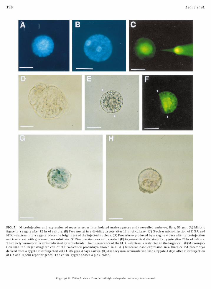
Detection of transgene expression. The expression of the re- in each reciprocal cross were full and contained normalporter genes was observed 4 days after microinjection. The expres- grains (Fig. 1G).sion of the anthocyanin reporter genes was detected by the accumu- Cytological characteristics of the embryos developed inlation of anthocyanin pigments in the microinjected zygotes which vitro. Different embryonic stages were observed duringthen showed a pink to red color. GUS-expressing zygotes were zygote development in culture. Upon isolation zygotes pre-visualized after an overnight incubation in X-Gluc substrate as
sented a polarized nucleus surrounded by a cytoplasmicdescribed by Mendel et al. (1989). The frequency of transformationcrown rich in organelles and numerous cytoplasmic strandsis presented as the number of zygotes showing reporter gene expres-(Fig. 1A). Following 3 hr of culture, this polarization in-sion per 100 microinjected zygotes.creased markedly, with a large vacuole being observed atthe opposite side of the cytoplasmic pole (Fig. 2A).
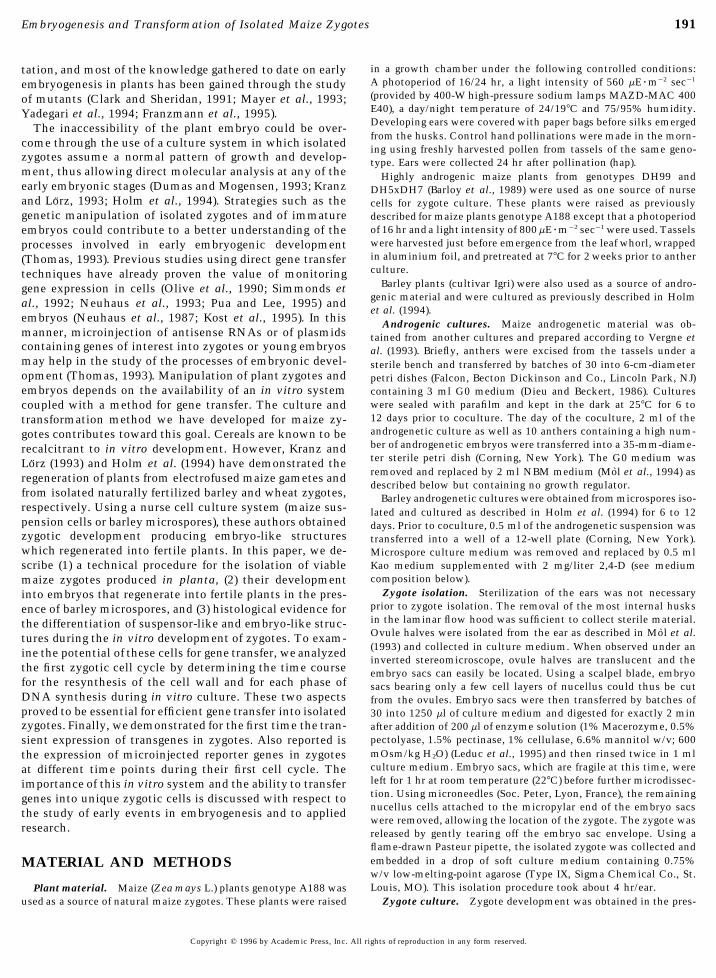
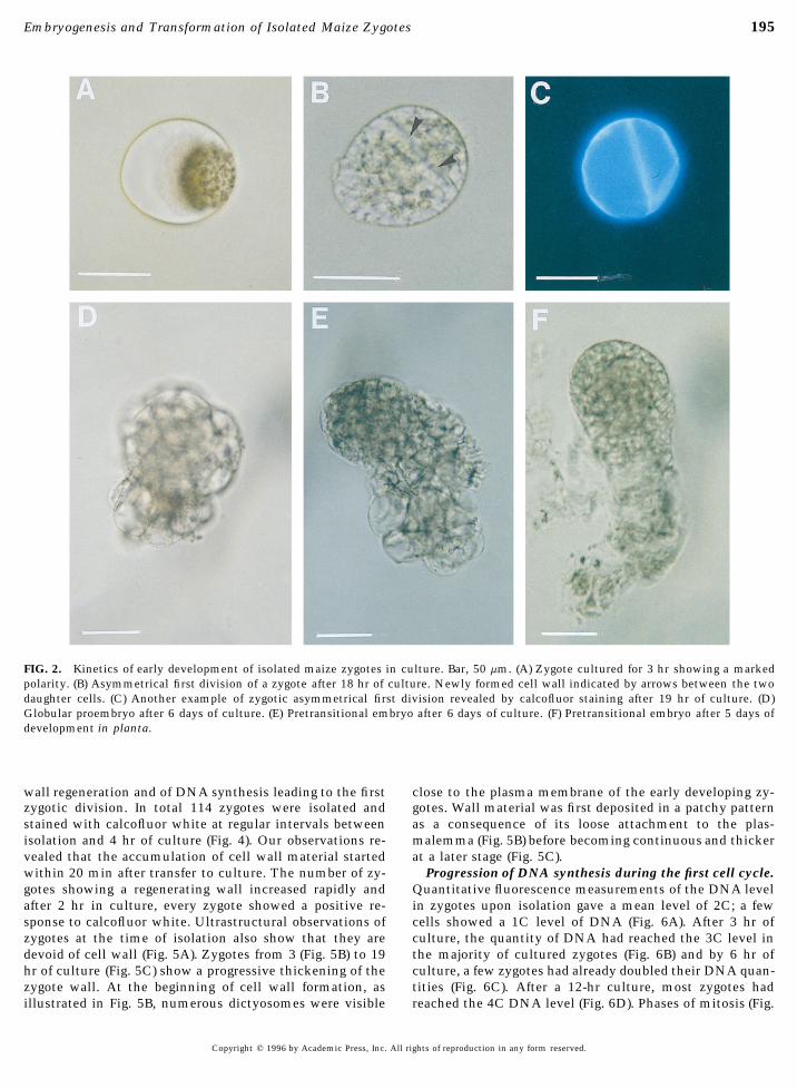
The first asymmetrical division of isolated zygotes wasRESULTS observed from 19 to 24 hr of zygote culture (Fig. 2B). The
difference in size between the two daughter cells was partic-ularly obvious after staining the cell walls with calcofluorIsolated Maize Zygotes Develop into Fertile Plantswhite as shown in Fig. 2C. The next developmental stagein Vitrowas globular to slightly elongated in shape. In these embryo-like structures, two to three additional vacuolated cellsKinetics and rate of in vitro development. Maize zy-
gotes obtained after in planta pollination could be isolated were often visible at one pole (Fig. 2D), which probablyrepresented the basal cells of the proembryo observed inafter the combination of a very short (2 min) enzymatic
treatment and microdissection of maternal tissues (Fig. 1A). planta 3 to 4 days postpollination (data not shown). Theseembryo-like structures elongated into a bipolar form inZygotes isolated in this manner produced globular struc-
tures within 4 to 5 days when cultured in the presence of which one pole was dense with small cells and the otherwas composed of a few elongated and vacuolated cells (Fig.androgenic tissues (Fig. 1B). We have observed that andro-
genic cultures from maize and barley stimulated zygote de- 2E). These structures were very similar to the pretransi-tional stage in planta (Fig. 2F). Further development of thevelopment. Table 1 summarizes the observations we have
made over several months and provides the mean values pretransitional embryos produced in vitro was characterizedby their enlargement and the differentiation of a protodermof several experiments concerning the frequency of zygote
development. Overall, 37 to 45% of the zygotes transferred (not shown). Cytological characteristics of zygote develop-ment were analyzed at the optical and ultrastructural levels.to culture continued to develop. However, one must note
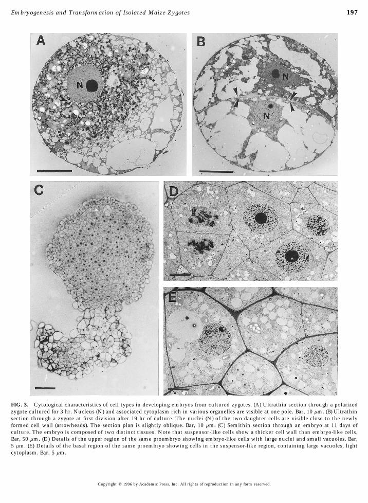
that there was a large variation in the percentage of devel- High zygote polarity was also clearly evidenced in ultrathinsectioned zygotes at the 3-hr culture stage (Fig. 3A). Cyto-oping zygotes from one experiment to another. In the pres-
ence of barley microspores, this ranged from 13 to 61%. plasmic organelles including numerous plastids, mitochon-dria, and dictyosomes were preferentially located aroundIn the presence of maize androgenic nurse cells, zygotes
produced multicellular structures 100 to 150 mm in diame- the polarized nucleus whereas vacuoles were concentratedat the opposite side. Observations of zygotes at the 19-hrter within a week, but these neither grew nor differentiated
further. However, differentiation of zygotes into further em- culture stage, after completion of their first division, didnot show marked differences in the ultrastructural charac-bryonic stages occurred in the presence of barley androgenic
cultures (Table 2, Fig. 1C). The addition of growth regulators teristics of the two daughter cells (Fig. 3B). Semithin sec-tions through proembryos confirmed the differentiation ofto the culture medium appears to be necessary for the differ-
entiation of globular structures (Table 2). This differentia- two distinct tissues located at two opposite poles (Fig. 3C).Cells at the upper dense pole had a meristematic appear-tion was obtained only in the presence of 2,4-D. In contrast,
the addition of BAP did not appear to be essential (Table 2). ance. They were poorly vacuolated, contained large nuclei,and were surrounded by thin cell walls (Fig. 3D). A largeAfter 1 month in culture, the embryo-like structures had
reached a size of 1 to 2 mm. When transferred to solid number of them were dividing. The elongated cells charac-teristic of the opposite basal pole showed a different cytolog-medium, direct germination was not observed. Instead, sec-
ondary embryos and multiple shoots rapidly developed on ical organization (Fig. 3E). These cells contained larger vacu-oles, light cytoplasm and nuclei, and were surrounded bytheir surface (Fig. 1D). Once isolated from the embryonic
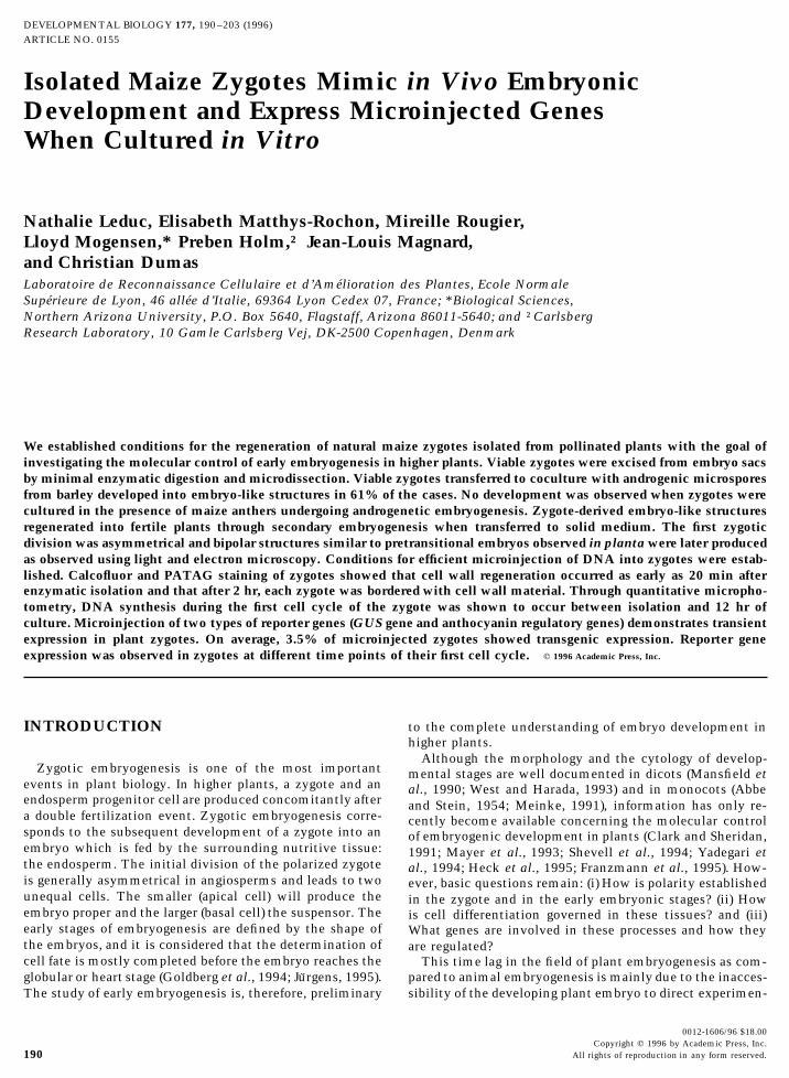
masses, these shoots rooted and developed in vitro into thicker cell walls.young plantlets (Fig. 1E). From these, five were transferredto soil in the greenhouse and 4 months after zygote isola- Cell Wall Regeneration and DNA Synthesistion, all five plants had reached maturity (Fig. 1F). Thesewere morphogenetically similar to maize plants raised from Cell wall regeneration in isolated zygotes after enzy-
matic isolation. Maize zygotes were isolated using enzy-seeds, being of a smaller size. Both male and female inflo-rescences were produced on each of these plants. Pollen matic digestion. When stained immediately after isolation
with calcofluor white, 84% of zygotes presented no wallviability was demonstrated by a positive FCR test and suc-cessful reciprocal crosses with maize plants (A188) raised material on their surface and thus were true protoplasts.
Only a very faint positive reaction was detected in the re-from seeds proved that the five plants derived from thezygote culture were male and female fertile. Ears pollinated maining 16% of zygotes. We followed the timing of cell
Copyright q 1996 by Academic Press, Inc. All rights of reproduction in any form reserved.
AID DB 8217 / 6x0f$$$162 06-20-96 10:06:21 dbal AP: Dev Bio
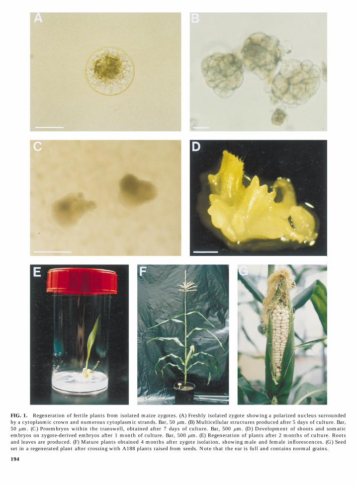
FIG. 1. Regeneration of fertile plants from isolated maize zygotes. (A) Freshly isolated zygote showing a polarized nucleus surroundedby a cytoplasmic crown and numerous cytoplasmic strands. Bar, 50 mm. (B) Multicellular structures produced after 5 days of culture. Bar,50 mm. (C) Proembryos within the transwell, obtained after 7 days of culture. Bar, 500 mm. (D) Development of shoots and somaticembryos on zygote-derived embryos after 1 month of culture. Bar, 500 mm. (E) Regeneration of plants after 2 months of culture. Rootsand leaves are produced. (F) Mature plants obtained 4 months after zygote isolation, showing male and female inflorescences. (G) Seedset in a regenerated plant after crossing with A188 plants raised from seeds. Note that the ear is full and contains normal grains.
194
06-20-96 10:06:21 dbal AP: Dev Bio
195Embryogenesis and Transformation of Isolated Maize Zygotes
FIG. 2. Kinetics of early development of isolated maize zygotes in culture. Bar, 50 mm. (A) Zygote cultured for 3 hr showing a markedpolarity. (B) Asymmetrical first division of a zygote after 18 hr of culture. Newly formed cell wall indicated by arrows between the twodaughter cells. (C) Another example of zygotic asymmetrical first division revealed by calcofluor staining after 19 hr of culture. (D)Globular proembryo after 6 days of culture. (E) Pretransitional embryo after 6 days of culture. (F) Pretransitional embryo after 5 days ofdevelopment in planta.
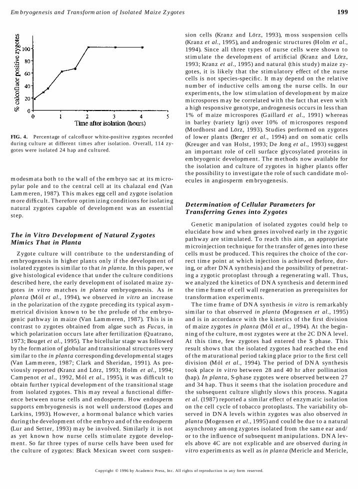
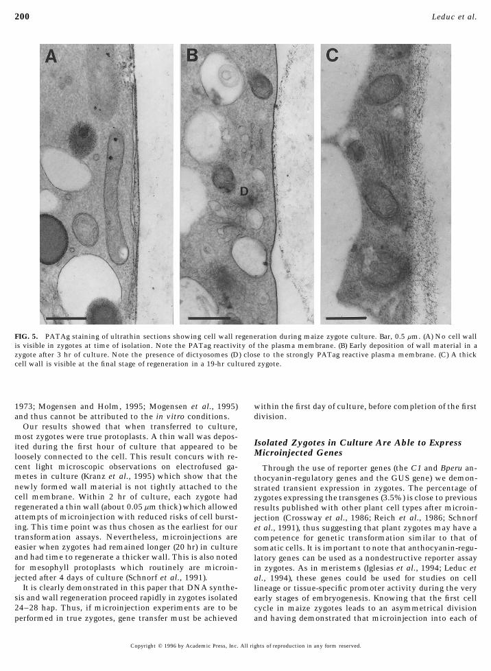
wall regeneration and of DNA synthesis leading to the first close to the plasma membrane of the early developing zy-gotes. Wall material was first deposited in a patchy patternzygotic division. In total 114 zygotes were isolated and
stained with calcofluor white at regular intervals between as a consequence of its loose attachment to the plas-malemma (Fig. 5B) before becoming continuous and thickerisolation and 4 hr of culture (Fig. 4). Our observations re-
vealed that the accumulation of cell wall material started at a later stage (Fig. 5C).Progression of DNA synthesis during the first cell cycle.within 20 min after transfer to culture. The number of zy-
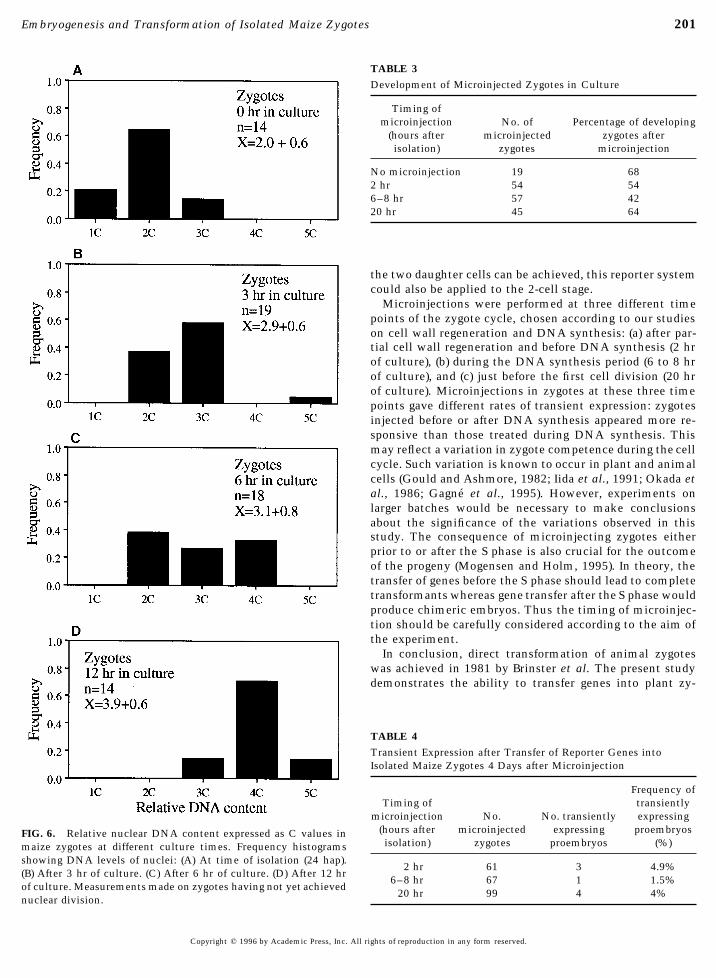
gotes showing a regenerating wall increased rapidly and Quantitative fluorescence measurements of the DNA levelin zygotes upon isolation gave a mean level of 2C; a fewafter 2 hr in culture, every zygote showed a positive re-
sponse to calcofluor white. Ultrastructural observations of cells showed a 1C level of DNA (Fig. 6A). After 3 hr ofculture, the quantity of DNA had reached the 3C level inzygotes at the time of isolation also show that they are
devoid of cell wall (Fig. 5A). Zygotes from 3 (Fig. 5B) to 19 the majority of cultured zygotes (Fig. 6B) and by 6 hr ofculture, a few zygotes had already doubled their DNA quan-hr of culture (Fig. 5C) show a progressive thickening of the
zygote wall. At the beginning of cell wall formation, as tities (Fig. 6C). After a 12-hr culture, most zygotes hadreached the 4C DNA level (Fig. 6D). Phases of mitosis (Fig.illustrated in Fig. 5B, numerous dictyosomes were visible
Copyright q 1996 by Academic Press, Inc. All rights of reproduction in any form reserved.
AID DB 8217 / 6x0f$$$163 06-20-96 10:06:21 dbal AP: Dev Bio
196 Leduc et al.
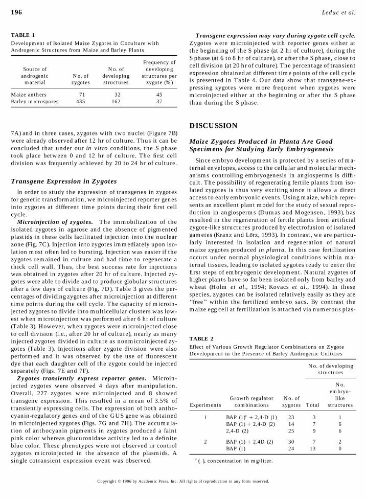
TABLE 1 Transgene expression may vary during zygote cell cycle.Development of Isolated Maize Zygotes in Coculture with Zygotes were microinjected with reporter genes either atAndrogenic Structures from Maize and Barley Plants the beginning of the S phase (at 2 hr of culture), during the
S phase (at 6 to 8 hr of culture), or after the S phase, close toFrequency of cell division (at 20 hr of culture). The percentage of transient
Source of No. of developingexpression obtained at different time points of the cell cycleandrogenic No. of developing structures peris presented in Table 4. Our data show that transgene-ex-material zygotes structures zygote (%)pressing zygotes were more frequent when zygotes were
Maize anthers 71 32 45 microinjected either at the beginning or after the S phaseBarley microspores 435 162 37 than during the S phase.
DISCUSSION7A) and in three cases, zygotes with two nuclei (Figure 7B)were already observed after 12 hr of culture. Thus it can be Maize Zygotes Produced in Planta Are Goodconcluded that under our in vitro conditions, the S phase Specimens for Studying Early Embryogenesistook place between 0 and 12 hr of culture. The first cell
Since embryo development is protected by a series of ma-division was frequently achieved by 20 to 24 hr of culture.ternal envelopes, access to the cellular and molecular mech-anisms controlling embryogenesis in angiosperms is diffi-
Transgene Expression in Zygotes cult. The possibility of regenerating fertile plants from iso-lated zygotes is thus very exciting since it allows a directIn order to study the expression of transgenes in zygotesaccess to early embryonic events. Using maize, which repre-for genetic transformation, we microinjected reporter genessents an excellent plant model for the study of sexual repro-into zygotes at different time points during their first cellduction in angiosperms (Dumas and Mogensen, 1993), hascycle.resulted in the regeneration of fertile plants from artificialMicroinjection of zygotes. The immobilization of thezygote-like structures produced by electrofusion of isolatedisolated zygotes in agarose and the absence of pigmentedgametes (Kranz and Lorz, 1993). In contrast, we are particu-plastids in these cells facilitated injection into the nuclearlarly interested in isolation and regeneration of naturalzone (Fig. 7C). Injection into zygotes immediately upon iso-maize zygotes produced in planta. In this case fertilizationlation most often led to bursting. Injection was easier if theoccurs under normal physiological conditions within ma-zygotes remained in culture and had time to regenerate aternal tissues, leading to isolated zygotes ready to enter thethick cell wall. Thus, the best success rate for injectionsfirst steps of embryogenic development. Natural zygotes ofwas obtained in zygotes after 20 hr of culture. Injected zy-higher plants have so far been isolated only from barley andgotes were able to divide and to produce globular structureswheat (Holm et al., 1994; Kovacs et al., 1994). In theseafter a few days of culture (Fig. 7D). Table 3 gives the per-species, zygotes can be isolated relatively easily as they arecentages of dividing zygotes after microinjection at different‘‘free’’ within the fertilized embryo sacs. By contrast thetime points during the cell cycle. The capacity of microin-maize egg cell at fertilization is attached via numerous plas-jected zygotes to divide into multicellular clusters was low-
est when microinjection was performed after 6 hr of culture(Table 3). However, when zygotes were microinjected closeto cell division (i.e., after 20 hr of culture), nearly as many
TABLE 2injected zygotes divided in culture as nonmicroinjected zy-Effect of Various Growth Regulator Combinations on Zygotegotes (Table 3). Injections after zygote division were alsoDevelopment in the Presence of Barley Androgenic Culturesperformed and it was observed by the use of fluorescent
dye that each daughter cell of the zygote could be injected No. of developingseparately (Figs. 7E and 7F). structures
Zygotes transiently express reporter genes. Microin-No.jected zygotes were observed 4 days after manipulation.
embryo-Overall, 227 zygotes were microinjected and 8 showedGrowth regulator No. of liketransgene expression. This resulted in a mean of 3.5% of
Experiments combinations zygotes Total structurestransiently expressing cells. The expression of both antho-cyanin-regulatory genes and of the GUS gene was obtained 1 BAP (1)a / 2,4-D (1) 23 3 1in microinjected zygotes (Figs. 7G and 7H). The accumula- BAP (1) / 2,4-D (2) 14 7 6
2,4-D (2) 25 9 6tion of anthocyanin pigments in zygotes produced a faintpink color whereas glucuronidase activity led to a definite
2 BAP (1) / 2,4D (2) 30 7 2blue color. These phenotypes were not observed in control BAP (1) 24 13 0zygotes microinjected in the absence of the plasmids. A
a ( ), concentration in mg/liter.single cotransient expression event was observed.
Copyright q 1996 by Academic Press, Inc. All rights of reproduction in any form reserved.
AID DB 8217 / 6x0f$$$163 06-20-96 10:06:21 dbal AP: Dev Bio
197Embryogenesis and Transformation of Isolated Maize Zygotes
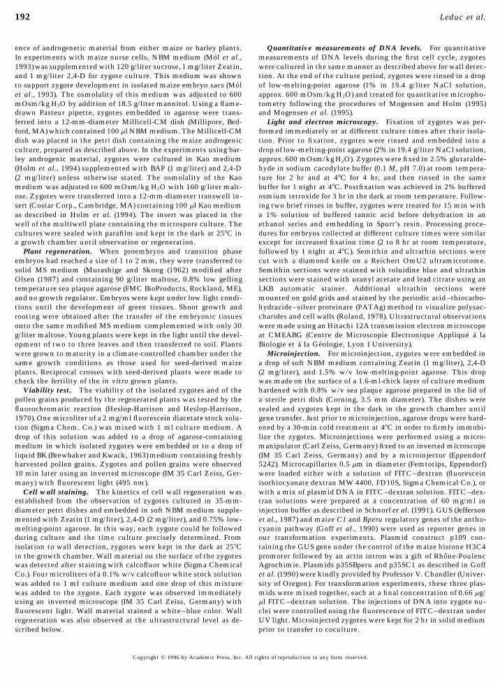
FIG. 3. Cytological characteristics of cell types in developing embryos from cultured zygotes. (A) Ultrathin section through a polarizedzygote cultured for 3 hr. Nucleus (N) and associated cytoplasm rich in various organelles are visible at one pole. Bar, 10 mm. (B) Ultrathinsection through a zygote at first division after 19 hr of culture. The nuclei (N) of the two daughter cells are visible close to the newlyformed cell wall (arrowheads). The section plan is slightly oblique. Bar, 10 mm. (C) Semithin section through an embryo at 11 days ofculture. The embryo is composed of two distinct tissues. Note that suspensor-like cells show a thicker cell wall than embryo-like cells.Bar, 50 mm. (D) Details of the upper region of the same proembryo showing embryo-like cells with large nuclei and small vacuoles. Bar,5 mm. (E) Details of the basal region of the same proembryo showing cells in the suspensor-like region, containing large vacuoles, lightcytoplasm. Bar, 5 mm.
Copyright q 1996 by Academic Press, Inc. All rights of reproduction in any form reserved.
AID DB 8217 / 6x0f$$8217 06-20-96 10:06:21 dbal AP: Dev Bio
198 Leduc et al.
FIG. 7. Microinjection and expression of reporter genes into isolated maize zygotes and two-celled embryos. Bars, 50 mm. (A) Mitoticfigure in a zygote after 12 hr of culture. (B) Two nuclei in a dividing zygote after 12 hr of culture. (C) Nuclear microinjection of DNA andFITC–dextran into a zygote. Note the brightness of the injected nucleus. (D) Proembryo produced by a zygote 4 days after microinjectionand treatment with glucuronidase substrate. GUS expression was not revealed. (E) Asymmetrical division of a zygote after 20 hr of culture.The newly formed cell wall is indicated by arrowheads. The fluorescence of the FITC–dextran is restricted to the larger cell. (F) Microinjec-tion into the larger daughter cell of the two-celled proembryo shown in E. (G) Glucuronidase expression in a three-celled proembryoderived from a zygote microinjected with GUS gene 4 days earlier. (H) Anthocyanin accumulation into a zygote 4 days after microinjectionof C1 and B-peru reporter genes. The entire zygote shows a pink color.
Copyright q 1996 by Academic Press, Inc. All rights of reproduction in any form reserved.
AID DB 8217 / 6x0f$$8217 06-20-96 10:06:21 dbal AP: Dev Bio
199Embryogenesis and Transformation of Isolated Maize Zygotes
sion cells (Kranz and Lorz, 1993), moss suspension cells(Kranz et al., 1995), and androgenic structures (Holm et al.,1994). Since all three types of nurse cells were shown tostimulate the development of artificial (Kranz and Lorz,1993; Kranz et al., 1995) and natural (this study) maize zy-gotes, it is likely that the stimulatory effect of the nursecells is not species-specific. It may depend on the relativenumber of inductive cells among the nurse cells. In ourexperiments, the low stimulation of development by maizemicrospores may be correlated with the fact that even witha high responsive genotype, androgenesis occurs in less than1% of maize microspores (Gaillard et al., 1991) whereasin barley (variety Igri) over 10% of microspores respond(Mordhorst and Lorz, 1993). Studies performed on zygotes
FIG. 4. Percentage of calcofluor white-positive zygotes recorded of lower plants (Berger et al., 1994) and on somatic cellsduring culture at different times after isolation. Overall, 114 zy- (Kreuger and van Holst, 1993; De Jong et al., 1993) suggestgotes were isolated 24 hap and cultured. an important role of cell surface glycosylated proteins in
embryogenic development. The methods now available forthe isolation and culture of zygotes in higher plants offerthe possibility to investigate the role of such candidate mol-
modesmata both to the wall of the embryo sac at its micro- ecules in angiosperm embryogenesis.pylar pole and to the central cell at its chalazal end (VanLammeren, 1987). This makes egg cell and zygote isolationmore difficult. Therefore optimizing conditions for isolating
Determination of Cellular Parameters fornatural zygotes capable of development was an essentialTransferring Genes into Zygotesstep.
Genetic manipulation of isolated zygotes could help toelucidate how and when genes involved early in the zygoticThe in Vitro Development of Natural Zygotespathway are stimulated. To reach this aim, an appropriateMimics That in Plantamicroinjection technique for the transfer of genes into thesecells must be produced. This requires the choice of the cor-Zygote culture will contribute to the understanding of
embryogenesis in higher plants only if the development of rect time point at which injection is achieved (before, dur-ing, or after DNA synthesis) and the possibility of penetrat-isolated zygotes is similar to that in planta. In this paper, we
give histological evidence that under the culture conditions ing a zygotic protoplast through a regenerating wall. Thus,we analyzed the kinetics of DNA synthesis and determineddescribed here, the early development of isolated maize zy-
gotes in vitro matches in planta embryogenesis. As in the time frame of cell wall regeneration as prerequisites fortransformation experiments.planta (Mol et al., 1994), we observed in vitro an increase
in the polarization of the zygote preceding its typical asym- The time frame of DNA synthesis in vitro is remarkablysimilar to that observed in planta (Mogensen et al., 1995)metrical division known to be the prelude of the embryo-
genic pathway in maize (Van Lammeren, 1987). This is in and is in accordance with the kinetics of the first divisionof maize zygotes in planta (Mol et al., 1994). At the begin-contrast to zygotes obtained from algae such as Fucus, in
which polarization occurs late after fertilization (Quatrano, ning of the culture, most zygotes were at the 2C DNA level.At this time, few zygotes had entered the S phase. This1973; Bouget et al., 1995). The bicellular stage was followed
by the formation of globular and transitional structures very result shows that the isolated zygotes had reached the endof the maturational period taking place prior to the first cellsimilar to the in planta corresponding developmental stages
(Van Lammeren, 1987; Clark and Sheridan, 1991). As pre- division (Mol et al., 1994). The period of DNA synthesistook place in vitro between 28 and 40 hr after pollinationviously reported (Kranz and Lorz, 1993; Holm et al., 1994;
Campenot et al., 1992, Mol et al., 1995), it was difficult to (hap). In planta, S-phase zygotes were observed between 27and 34 hap. Thus it seems that the isolation procedure andobtain further typical development of the transitional stage
from isolated zygotes. This may reveal a functional differ- the subsequent culture slightly slows this process. Nagataet al. (1987) reported a similar effect of enzymatic isolationence between nurse cells and endosperm. How endosperm
supports embryogenesis is not well understood (Lopes and on the cell cycle of tobacco protoplasts. The variability ob-served in DNA levels within zygotes was also observed inLarkins, 1993). However, a hormonal balance which varies
during the development of the embryo and of the endosperm planta (Mogensen et al., 1995) and could be due to a naturalasynchrony among zygotes isolated from the same ear and/(Lur and Setter, 1993) may be involved. Similarly it is not
as yet known how nurse cells stimulate zygote develop- or to the influence of subsequent manipulations. DNA lev-els above 4C are not explicable and are observed during inment. So far three types of nurse cells have been used for
the culture of zygotes: Black Mexican sweet corn suspen- vitro experiments as well as in planta (Mericle and Mericle,
Copyright q 1996 by Academic Press, Inc. All rights of reproduction in any form reserved.
AID DB 8217 / 6x0f$$$164 06-20-96 10:06:21 dbal AP: Dev Bio
200 Leduc et al.
FIG. 5. PATAg staining of ultrathin sections showing cell wall regeneration during maize zygote culture. Bar, 0.5 mm. (A) No cell wallis visible in zygotes at time of isolation. Note the PATag reactivity of the plasma membrane. (B) Early deposition of wall material in azygote after 3 hr of culture. Note the presence of dictyosomes (D) close to the strongly PATag reactive plasma membrane. (C) A thickcell wall is visible at the final stage of regeneration in a 19-hr cultured zygote.
1973; Mogensen and Holm, 1995; Mogensen et al., 1995) within the first day of culture, before completion of the firstdivision.and thus cannot be attributed to the in vitro conditions.
Our results showed that when transferred to culture,most zygotes were true protoplasts. A thin wall was depos-
Isolated Zygotes in Culture Are Able to Expressited during the first hour of culture that appeared to beMicroinjected Genesloosely connected to the cell. This result concurs with re-
cent light microscopic observations on electrofused ga- Through the use of reporter genes (the C1 and Bperu an-metes in culture (Kranz et al., 1995) which show that the thocyanin-regulatory genes and the GUS gene) we demon-newly formed wall material is not tightly attached to the strated transient expression in zygotes. The percentage ofcell membrane. Within 2 hr of culture, each zygote had zygotes expressing the transgenes (3.5%) is close to previousregenerated a thin wall (about 0.05mm thick) which allowed results published with other plant cell types after microin-attempts of microinjection with reduced risks of cell burst- jection (Crossway et al., 1986; Reich et al., 1986; Schnorfing. This time point was thus chosen as the earliest for our et al., 1991), thus suggesting that plant zygotes may have atransformation assays. Nevertheless, microinjections are competence for genetic transformation similar to that ofeasier when zygotes had remained longer (20 hr) in culture somatic cells. It is important to note that anthocyanin-regu-and had time to regenerate a thicker wall. This is also noted latory genes can be used as a nondestructive reporter assayfor mesophyll protoplasts which routinely are microin- in zygotes. As in meristems (Iglesias et al., 1994; Leduc etjected after 4 days of culture (Schnorf et al., 1991). al., 1994), these genes could be used for studies on cell
It is clearly demonstrated in this paper that DNA synthe- lineage or tissue-specific promoter activity during the verysis and wall regeneration proceed rapidly in zygotes isolated early stages of embryogenesis. Knowing that the first cell24–28 hap. Thus, if microinjection experiments are to be cycle in maize zygotes leads to an asymmetrical division
and having demonstrated that microinjection into each ofperformed in true zygotes, gene transfer must be achieved
Copyright q 1996 by Academic Press, Inc. All rights of reproduction in any form reserved.
AID DB 8217 / 6x0f$$$164 06-20-96 10:06:21 dbal AP: Dev Bio
201Embryogenesis and Transformation of Isolated Maize Zygotes
TABLE 3Development of Microinjected Zygotes in Culture
Timing ofmicroinjection No. of Percentage of developing
(hours after microinjected zygotes afterisolation) zygotes microinjection
No microinjection 19 682 hr 54 546–8 hr 57 4220 hr 45 64
the two daughter cells can be achieved, this reporter systemcould also be applied to the 2-cell stage.
Microinjections were performed at three different timepoints of the zygote cycle, chosen according to our studieson cell wall regeneration and DNA synthesis: (a) after par-tial cell wall regeneration and before DNA synthesis (2 hrof culture), (b) during the DNA synthesis period (6 to 8 hrof culture), and (c) just before the first cell division (20 hrof culture). Microinjections in zygotes at these three timepoints gave different rates of transient expression: zygotesinjected before or after DNA synthesis appeared more re-sponsive than those treated during DNA synthesis. Thismay reflect a variation in zygote competence during the cellcycle. Such variation is known to occur in plant and animalcells (Gould and Ashmore, 1982; Iida et al., 1991; Okada etal., 1986; Gagne et al., 1995). However, experiments onlarger batches would be necessary to make conclusionsabout the significance of the variations observed in thisstudy. The consequence of microinjecting zygotes eitherprior to or after the S phase is also crucial for the outcomeof the progeny (Mogensen and Holm, 1995). In theory, thetransfer of genes before the S phase should lead to completetransformants whereas gene transfer after the S phase wouldproduce chimeric embryos. Thus the timing of microinjec-tion should be carefully considered according to the aim ofthe experiment.
In conclusion, direct transformation of animal zygoteswas achieved in 1981 by Brinster et al. The present studydemonstrates the ability to transfer genes into plant zy-
TABLE 4Transient Expression after Transfer of Reporter Genes intoIsolated Maize Zygotes 4 Days after Microinjection
Frequency ofTiming of transiently
microinjection No. No. transiently expressing(hours after microinjected expressing proembryosFIG. 6. Relative nuclear DNA content expressed as C values inisolation) zygotes proembryos (%)maize zygotes at different culture times. Frequency histograms
showing DNA levels of nuclei: (A) At time of isolation (24 hap).2 hr 61 3 4.9%
(B) After 3 hr of culture. (C) After 6 hr of culture. (D) After 12 hr6–8 hr 67 1 1.5%
of culture. Measurements made on zygotes having not yet achieved20 hr 99 4 4%
nuclear division.
Copyright q 1996 by Academic Press, Inc. All rights of reproduction in any form reserved.
AID DB 8217 / 6x0f$$$164 06-20-96 10:06:21 dbal AP: Dev Bio
202 Leduc et al.
the genetic map of Arabidopsis thaliana with embryonic muta-gotes. These data open new ways to investigate the expres-tions. Plant J. 7, 341–350.sion and/or regulation of genes during early embryogenesis
Gagne, M., Pothier, F., and Sirard, M-A. (1995). Effect of microinjec-and through their application offer a novel possibility totion time during postfertilization S-phase on bovine embryonicdeliver genes of interest into plants.development. Mol. Reprod. Dev. 41, 184–194.
Gaillard, A., Vergne, P., and Beckert, M. (1991). Optimization ofmaize microspore isolation and culture conditions for reliableplant regeneration. Plant Cell Rep. 10, 55–58.ACKNOWLEDGMENTS
Goff, S. A., Klein, T., Roth, B. A., Fromm, M., Cone, K., Radicella,J. P., and Chandler, V. (1990). Transactivation of anthocyanin
We thank Drs. T. Hardy and R. De Rose from Rhone-Poulenc biosynthesic genes following transfer of b regulatory genes intoAgrochimie for fruitful scientific discussions, F. Morin and R. Blanc maize tissues. EMBO J. 9, 2517–2522.for expert technical assistance, and E. Bates for critical reading of
Goldberg, R. B., De Paiva, G., and Yadegari, R. (1994). Plant em-the manuscript. We are also very grateful to Professor V. L. Chand-
bryogenesis: Zygote to seed. Science 266, 605 –614.ler (University of Oregon) for the gift of the p35SBperu and p35SC1
Gould, A. R., and Ashmore, S. E. (1982). Interaction of purifiedplasmids. This study was conducted under the Bioavenir pro-
DNA with protoplasts of different cell cycle stage: The conceptgramme funded by Rhone-Poulenc Agrochimie, the Ministere de
of a competent phase for plant transformation. Theor. Appl.la Recherche et de l’Espace, and the Ministere de l’Industrie et du
Genet. 64, 7–12.Commerce Exterieur. Partial funding from the Organized Research
Heck, G. R., Perry, S. E., Nichols, K. W., and Fernandez, D. E.Fund, Northern Arizona University, to H.L.M. is also gratefully(1995). AGL15, a MADS domain protein expressed in developingacknowledged.embryos. Plant Cell 7, 1271–1282.
Heslop-Harrison, J., and Heslop-Harrison, Y. (1970). Evaluation ofpollen viability by enzymatically induced fluorescence; intracel-lular hydrolysis of fluorescein diacetate. Stain Technol. 45, 115–REFERENCES120.
Holm, P. B., Knudsen, S., Mouritzen, P., Negri, D., Olsen, F. L.,Abbe, E. C., and Stein, O. L. (1954). The growth of the shoot apex and Roue, C. (1994). Regeneration of fertile barley plants from
in maize: Embryogeny. Am. J. Bot. 41, 285–293. mechanically isolated protoplasts of fertilized egg cell. Plant CellBarloy, D., Denis, L., and Beckert, M. (1989). Comparison of the 6, 531 –543.
aptitude for anther culture in some androgenic doubled haploidIglesias, V. A., Gisel, A., Bilang, R., Leduc, N., Potrykus, I., and
maize lines. Maydica. 34, 303–308.Sautter, C. (1994). Transient expression of visible marker genes in
Berger, F., Taylor, A., and Brownlee, C. (1994). Cell fate determina-meristem cells of wheat embryos after ballistic micro-targeting.
tion by the cell wall in early Fucus development. Science 263,Planta 192, 84–91.
1421–1423.Iida, A., Yamashita, T., Yamada, Y., and Morikawa, H. (1991). Effi-Bouget, F-Y., Gerttula, S., and Quatrano, R. S. (1995). Spatial redis-
ciency of particle-bombardment-mediated transformation is in-tribution of poly(A)/ RNA during polarization of the Fucus zy-fluenced by cell cycle stage in synchronized cultured cells ofgote is dependent upon microfilaments. Dev. Biol. 171, 258–261.tobacco. Plant Physiol. 97, 1585–1587.Brewbaker, J. L., and Kwack, B. H. (1963). The essential role of
Jefferson, R. A., Kavanagh, T. A., and Bevan, M. W. (1987). GUScalcium ion in pollen germination and pollen tube growth. Am.fusions: b-Glucuronidase as a sensitive and versatile gene fusionJ. Bot. 50, 859–865.marker. EMBO J. 6, 3901–3908.Brinster, R. L., Chen, H. Y., Trumbauer, M., Senear, A. W., Warren,
Jurgens, G. (1995). Axis formation in plant embryogenesis: CuesR., and Palmiter, R. D. (1981). Somatic expression of herpes thy-and clues. Cell 81, 467–470.midine kinase in mice following injection of a fusion gene into
Kost, B., Leduc, N., Sautter, C., Potrykus, I., and Neuhaus, G.eggs. Cell 27, 223–231.(1995). Transient marker gene expression during zygotic in vitroCampenot, M. K., Zhang, G., Cutler, A. J., and Cass, D. (1992). Zeaembryogenesis of Brassica juncea (Indian mustard) following par-mays embryo sacs in culture. I. Plant regeneration from 1 dayticle bombardment. Planta 198, 211 –220.after pollination embryos. Am. J. Bot. 79, 1368–1373.
Kovacs, M., Barnabas, B., and Kranz, E. (1994). The isolation ofClark, J. K., and Sheridan, W. F. (1991). Isolation and characteriza-viable egg cells of wheat (Triticum aestivum L.). Sex. Plant Re-tion of 51 embryo-specific mutations of maize. Plant Cell 3, 935–prod. 7, 311–312.951.
Kranz, E., and Lorz, H. (1993). In vitro fertilization with isolated,Crossway, A., Oakes, J. V., Irvine, J. M., Ward, B., Knauf, V. C., andsingle gametes results in zygotic embryogenesis and fertile maizeShewmaker, C. K. (1986). Integration of foreign DNA followingplants. Plant Cell 5, 739–746.microinjection of tobacco mesophyll protoplasts. Mol. Gen.
Kranz, E., Von Wiegen, P., and Lorz, H. (1995). Early cytologicalGenet. 202, 179–185.events after induction of cell division in egg cells and zygoteDe Jong, A. J., Schmidt, E. D. L., and de Vries, S. C. (1993). Earlydevelopment following in vitro fertilization with angiosperm ga-events in higher-plant embryogenesis. Plant Mol. Biol. 22, 367–metes. Plant J. 8, 9–23.377.
Kreuger, M., and van Holst, G. J. (1993). Arabinogalactan proteinsDieu, P., and Beckert, M. (1986). Further studies of androgeneticare essential in somatic embryogenesis of Daucus carota L.embryo production and plant regeneration from in vitro culturedPlanta 189, 243–248.anthers in maize (Zea mays L.). Maydica 31, 245–260.
Leduc, N., Iglesias, V. A., Bilang, R., Gisel, A., Potrykus, I., andDumas, C., and Mogensen, H. L. (1993). Gametes and fertilization:Sautter, C. (1994). Gene transfer to inflorescence and flower meri-Maize as a model system for experimental embryogenesis instems using ballistic microtargeting. Sex. Plant Reprod. 7, 135 –flowering plants. Plant Cell 5, 1337–1348.
Franzmann, L. H., Yoon, E. S., and Meinke, D. W. (1995). Saturating 143.
Copyright q 1996 by Academic Press, Inc. All rights of reproduction in any form reserved.
AID DB 8217 / 6x0f$$$164 06-20-96 10:06:21 dbal AP: Dev Bio
203Embryogenesis and Transformation of Isolated Maize Zygotes
Leduc, N., Matthys-Rochon, E., and Dumas, C. (1995). Deleterious Okada, K., Takebe, I., and Nagata, T. (1986). Expression and integra-tion of genes introduced into highly synchronised plants proto-effect of minimal enzymatic treatments on the development of
isolated maize embryo sacs in culture. Sex. Plant Reprod. 8, 313– plasts. Mol. Gen. Genet. 205, 398–403.Olive, M. R., Walker, J. C., Singh, K., Dennis, E. S., and Peacock,317.
Lopes, M. A., and Larkins, B. A. (1993). Endosperm origin, develop- W. J. (1990). Functional properties of the anaerobic element ofthe maize Adh1 gene. Plant Mol. Biol. 15, 593–604.ment, and function. Plant Cell 5, 1383–1399.
Lur, H-S., and Setter, T. L. (1993). Role of auxin in maize endosperm Olsen, F. L. (1987). Induction of microspore embryogenesis in cul-tured anthers of Hordeum vulgare. The effects of ammoniumdevelopment. Timing of nuclear DNA endoreplication, zein ex-
pression, and cytokinin. Plant Physiol. 103, 273–280. nitrate, glutamine and asparagine as nitrogene sources. CarlsbergRes. Commun. 52, 393–404.Mansfield, S. C., Briarty, L. G., and Erni, S. (1990). Early embryogen-
esis in Arabidopsis thaliana. II. The developing embryo. Can. J. Pua, E. C., and Lee, J. E. E. (1995). Enhanced de novo shoot morpho-genesis in vitro by expression of antisense 1-aminocyclopropane-Bot. 69, 461 –476.
Mendel, R. R., Muller, B., Schulze, J., Kolesnikov, V., and Zelenin, 1-carboxylate oxidase gene in transgenic mustard plants. Planta196, 69–76.A. (1989). Delivery of foreign genes to intact barley cells by high
velocity microprojectiles. Theor. Appl. Genet. 78, 31–34. Quatrano, R. S. (1973). Separation of processes associated with dif-ferentiation of two celled Fucus embryos. Dev. Biol. 30, 209–Mayer, U., Buttner, G., and Jurgens, G. (1993). Apical–basal pattern
formation in the Arabidopsis embryo: Studies on the role of the 213.Reich, T. J., Iyer, V. N., and Miki, B. L. (1986). Efficient transforma-gnom gene. Development 117, 149 –162.
Meinke, D. W. (1991). Perspectives on genetic analysis of plant tion of alfalfa protoplasts by the intranuclear microinjection ofTi plasmids. Bio/Technology 4, 1001–1004.embryogenesis. Plant Cell 3, 857–866.
Mericle, L. W., and Mericle, R. P. (1973). Confounding the quandary Roland, J. C. (1978). General preparation and staining of thin sec-tions. In ‘‘Electron Microscopy and Cytochemistry of Plantof zygotic DNA. Barley Genet. Newsletter 3, 39 –42.
Mogensen, H. L., and Holm, P. B. (1995). Dynamics of nuclear DNA Cells’’ (J. L. Hall, Ed.), pp. 1–62. Elsevier/North-Holland, Amster-dam.quantities during zygote development in barley. Plant Cell 7,
487–494. Schnorf, M., Neuhaus-Url, G., Galli, A., Iida, S., Potrykus, I., andNeuhaus, G. (1991). An improved approach for transformationMogensen, H. L., Leduc, N., Matthys-Rochon, E., and Dumas, C.
(1995). Nuclear DNA amounts in the egg and zygote of maize. of plant cells by microinjection: Molecular and genetic analysis.Trans. Res. 1, 23–30.Planta 197, 641–645.
Mol, R., Matthys-Rochon, E., and Dumas, C. (1993). In-vitro culture Shevell, D. E., Leu, W. M., Gillmor, C. S., Xia, G., Feldmann,K. A., and Chua, N. H. (1994). EMB30 is essential for normal cellof fertilized embryo sacs of maize: Zygotes and two-celled proem-
bryos can develop into plants. Planta 189, 213 –217. division, cell expansion, and cell adhesion in Arabidopsis andencodes a protein that has similarity to Sec 7. Cell 77, 1051–Mol, R., Matthys-Rochon, E., and Dumas, C. (1994). The kinetics
of cytological events during double fertilization in Zea mays L. 1062.Simmonds, J., Stewart, P., and Simmonds, D. (1992). RegenerationPlant J. 5, 197 –206.
Mol, R., Matthys-Rochon, E., and Dumas, C. (1995). Embryogenesis of Triticum aestivum apical plants after microinjection of germline progenitor cells with DNA. Physiol. Plant. 85, 197–206.and plant regeneration from maize zygotes by in vitro culture of
fertilized embryo sacs. Plant Cell Rep. 14, 743–747. Thomas, L. T. (1993). Gene expression during plant embryogenesisand germination: An overview. Plant Cell 5, 1401–1410.Mordhorst, A. P., and Lorz, H. (1993). Embryogenesis and develop-
ment of isolated barley (Hordeum vulgare L.) microspores are Van Lammeren, A. A. M. (1987). Embryogenesis in Zea mays L. Astructural approach to maize caryopsis development in vivo andinfluenced by the amount and composition of nitrogen sources
in culture media. J. Plant Physiol. 142, 485 –492. in vitro. Thesis, pp. 1–175, Landbouwuniversiteit Wageningen,The Netherlands.Murashige, T., and Skoog, F. (1962). A revised medium for rapid
growth and bioassays with tobacco tissue cultures. Physiol. Vergne, P., Riccardi, F., Beckert, M., and Dumas, C. (1993). Identi-fication of a 32-kDa anther marker protein for androgenic re-Plant. 15, 473–497.
Nagata, T., Okada, K., Kawasu, T., and Takebe, I. (1987). Cauli- sponse in maize, Zea mays L. Theor. Appl. Genet. 86, 843–850.flower mosaic virus 35S promoter directs S phase specific expres- West, M. A. L., and Harada, J. (1993). Embryogenesis in highersion in plant cells. Mol. Gen. Genet. 207, 242–244. plants: An overview. Plant Cell 5, 1361–1369.
Neuhaus, G., Spangenberg, G., Mittelsen Scheid, O., and Yadegari, R., de Paiva, G. R., Laux, T., Koltunow, A. M., Apuya,Schweiger, H. G. (1987). Transgenic rapeseed plants obtained by N., Zimmerman, J. L., Fisher, R. L., Harada, J. J., and Goldberg,the microinjection of DNA into microspore-derived embryoids. R. B. (1994). Cell differentiation and morphogenesis are uncou-Theor. Appl. Genet. 75, 30 –36. pled in Arabidopsis raspberry embryos. Plant Cell 6, 1713–1729.
Neuhaus, G., Bowler, C., Kern, R., and Chua, N. H. (1993). Calmod-Received for publication December 21, 1995ulin-dependant and independant phytochrome signal transduc-
tion pathways. Cell 73, 937 –952. Accepted March 15, 1996
Copyright q 1996 by Academic Press, Inc. All rights of reproduction in any form reserved.
AID DB 8217 / 6x0f$$$164 06-20-96 10:06:21 dbal AP: Dev Bio