ischaemic preconditioning modulates the activity of kupffer cells during in vivo reperfusion injury...
TRANSCRIPT
cell biochemistry and function
Cell Biochem Funct 2003; 21: 299–305.
Published online 4 March 2003 in Wiley InterScience (www.interscience.wiley.com). DOI: 10.1002/cbf.1028
Ischaemic preconditioning modulates the activity ofKupffer cells during in vivo reperfusion injury of rat liver
Barbara Cavalieriy1, Maria-Giulia Perrelliy1, Manuela Aragno2, Pierluigi Ramadori1, Giuseppe Poli1
and Juan C. Cutrın1*
1Laboratory of Experimental Liver Pathology, Department of Clinical and Biological Sciences, University of Torino, Italy2Department of Experimental Medicine and Oncology, University of Torino, Italy
This work was performed to elucidate further the main cellular events underlying the protective effect of ischaemic precon-ditioning in an in vivo rat liver model of 90 min ischaemia followed by 30 min reperfusion. A significant attenuation of thevarious aspects of post-ischaemic injury, namely necrosis and the levels of hydrogen peroxide and 5- and 15-hydroperox-yeicosatetraenoic acids, was afforded by the prior application of a short cycle of ischaemia/reperfusion (10þ 10 min) orwhen rats were previously treated with gadolinium chloride. However, when preconditioning was applied on Kupffercell-depleted livers, no additional level of ischaemic tolerance was obtained. In terms of cellular pathology, this result couldbe suggestive of Kupffer cells as the target of the preconditioning phenomenon during the warm ischaemia/reperfusioninjury. Accordingly, modulation of Kupffer cell activity was associated with a well-preserved hepatocyte integrity, togetherwith low levels of pro-oxidant generation during reperfusion. As activated Kupffer cells can generate and release potentiallytoxic substances, their modulation by ischaemic preconditioning could help to provide new surgical and/or pharmacologicalstrategies to protect the liver against reperfusion damage. Copyright # 2003 John Wiley & Sons, Ltd.
key words— liver ischaemic preconditioning; Kupffer cell; oxidative stress
abbreviations— HPETE, 5- and 15-hydroperoxyeicosatetraenoic acids; IP, ischaemic preconditioning; ROS, reactiveoxygen species
INTRODUCTION
Substantial evidence indicates that Kupffer cells playa central role in the pathogenesis of liver parenchymalcell damage during the early post-reperfusion phase,mainly through releasing ROS and other toxic media-tors (i.e. cytokines) able to modulate hepatocyte meta-bolism and function.1,3–5 In light of this and in order toimprove liver function after reperfusion, variousattemps have been made to block Kupffer cell activity,either through administration of specific toxins (e.g.GdCl3, dichloromethylene diphosphonate), methyl-
xanthine phosphodiesterase inhibitors (i.e. pentoxifyl-line) or by the use of calcium blocking agents (e.g.nisoldipine). However, the side-effects of these treat-ments cannot be entirely excluded.1,3–9
The term IP was coined to describe a protectivemechanism against ischaemia–reperfusion injury andconsists of previous short periods of ischaemia fol-lowed by reperfusion.10 It was originally describedin the heart, where it decreases infarct size after sub-sequent prolonged ischaemia and reperfusion.11
In the liver, the exact mechanisms involved in thestatus of cytoprotection conferred by IP against ischae-mia/reperfusion injury are not fully understood. How-ever experimental data suggest that modifications of theendothelin-1/nitric oxide ratio in favour of the latter,changes in energy metabolism, attenuation of in situTNF-� production and release, as well as a blockingeffect on the ROS-generating systems, are activelyimplicated.12–17 Moreover, since the liver is composed
Received 22 September 2002Revised 29 October 2002
Copyright # 2003 John Wiley & Sons, Ltd. Accepted 31 October 2002
*Correspondence to: Dr J. C. Cutrın, Laboratorio di PatologiaEpatica Sperimentale, Dipartimento di Scienze Cliniche e Biolo-giche, Universita di Torino, A.S.O. ‘San Luigi’, Regione Gonzole10, 10043 Orbassano, Torino, Italy. Tel: 0039-011-6708122. Fax:0039-011-670-8124. E-mail: [email protected] contribution from these authors was equal.
of a heterogeneous cell population, it is likely that dur-ing preconditioning complex cross-talk could takeplace between parenchymal and non-parenchymal cells.
Accordingly, we investigated the effects of IP onKupffer cell activity in a rat liver model of ischae-mia/reperfusion injury.
MATERIALS AND METHODS
Animal model
Male Sprague-Dawley rats (224� 5 g body weight)were used; they had free access to standard chow dietand water. After anesthesia with 10% ketamine chlor-hydrate solution, they were positioned under a heatlamp to maintain constant body temperature. After amidline laparotomy, selective inflow occlusion of theportal triad (hepatic artery, portal vein, and bile duct)to the left and median liver lobes was performed.Reperfusion was initiated by clamp removal. Samplesfrom the post-ischaemic lobes were taken at the end ofthe reperfusion phase, and biochemical and morpho-logical studies were carried out. All animals receivedhumane care according to the criteria outlined inGuide for the Care and Use of Laboratory Animals.18
Depletion of Kupffer cells
To inhibit Kupffer cell activity, a randomized set ofrats were treated with gadolinium chloride (GdCl3;10 mg kg�1 in sterile 0.9% NaCl solution, i.v.) 18 hbefore the ischaemia and reperfusion cycle.3
Experimental design
The rats were subdivided into six groups, each groupbeing of five animals.
Group 1. Sham saline: animals treated with sterile0.9% NaCl solution were subjected to anesthaesia andlaparotomy alone.
Group 2. Sham GdCl3: as in group 1, but rats weretreated with GdCl3 instead of sterile 0.9% NaClsolution.
Group 3. Standard Ischaemia/Reperfusion (Isch/Rep):rats were subjected to 90 min of ischaemia followed by30 min of reperfusion.
Group 4. IPþ Isch/Rep: before ischaemia/reperfusion,rats were subjected to 10 min ischaemia followed by10 min reperfusion.17
Group 5. GdCl3þ Isch/Rep: before ischaemia/reper-fusion, rats were treated with GdCl3.
Group 6. GdCl3þ IPþ Isch/Rep: before IP procedure,as in group 4, rats were treated with GdCl3.
Biochemical assessment of liver cytolysis
Irreversible hepatic damage was evaluated by deter-mination of alanine aminotransferase and aspartateaminotransferase in blood-plasma obtained from theposterior cava vein after 30 min of reperfusion, usinga commercial kit from Sigma Chemical Co. (St. LouisMO, USA).
H2O2 analysis
Specimens from sham-operated and reperfused liverswere homogenized (10%, w/v) in a buffer containing0.033 mol l�1 Na2HPO4 and 0.9% KCl, pH 7.4 at 4�C.The homogenates were centrifuged at 40,000 g for45 min and the supernatant was used for H2O2 analy-sis. The H2O2-mediated oxidation of Fe2þ to Fe3þ ion,under acidic conditions and in the presence of xylenolorange dye, was quantified as described elsewhere.19
For the working reagent, 1 ml solution A containingammonium ferrous sulphate (25 mM) and sulphuricacid (2.5 mM) was mixed with 100 ml of solution Bcontaining sorbitol (100 mM) and xylenol orange(125 mmol l�1 in water. A 0.1-ml volume of superna-tant was added to 1 ml of working reagent, mixedand incubated at room temperature for 15 min. Theabsorbance was read at 560 nm. The values wereread against a standard curve prepared with 1 to50 mmol l�1 concentrations of H2O2. The absolutevalues of H2O2 were expressed in mmol l�1.
Determination of 5- and 15-HPETE
The concentration of 5- and 15-HPETE in the lipidfraction from cytosol samples extracted by the methodof Folch et al.20 was determined by the method ofTordijman et al.,21 slightly modified. Briefly, determi-nation was by high performance liquid chromatogra-phy analyses with a Symmetry C18 columm. Themobile phase used was a mixture of 0.05 M NaH2PO4,pH 5/methanol/acetonitrile (30:35:35, v:v). TheHPETE levels in the samples were calculated by com-parison with standard solutions of 5- and 15-HPETE(Sigma, St Louis, MO, USA). The results were ex-pressed as mM.
Number of neutrophils
Fragments from the left lateral lobe of about 1.0�0.5� 0.3 cm were placed overnight in a solution of4% formaldehyde in 50 mM phosphate buffer solution,
300 b. cavalieri ET AL.
Copyright # 2003 John Wiley & Sons, Ltd. Cell Biochem Funct 2003; 21: 299–305.
pH 7.4. Polymorphonuclear leukocytes (PMN) wereidentified on 5-mm paraffin sections by means of thenaphthol AS-D chloroacetate technique for esterase,following the method described by Moloney et al.22
with slight modifications; the substrate was dissolvedin dimethyl sulphoxide/Triton X-100 (9:1, v/v) andthen 0.1% Fast Garnet dGBC in 0.1 M phosphate buf-fer solution, pH 8.5 was added. Red stained PMNwere counted in 20 non-consecutive, randomlychosen� 500 histological fields. Results were ex-pressed as the number of neutrophils per40� histological power field (hpf ).
Protein determination
The protein content of the liver homogenates wasdetermined by the spectrophotometric method des-cribed by Peterson23 using bovine serum albumin asstandard.
Statistical analysis
Data are presented as means� SE; the statistical sig-nificance of differences was analysed using the one-way analysis of variance (ANOVA) corrected by theTukey–Kramer Multiple Comparisons test and withthe Student t test where p values� 0.05 were consid-ered to be statistically significant.
RESULTS
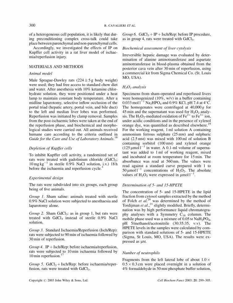
In our experimental model, long-term ischaemiafollowed by reperfusion induced hepatic necrosis inboth non-preconditioned and preconditioned animals.However, a marked reduction of irreversible cellinjury was detectable when IP had previously beenapplied (Figure 1).
Among non-parenchymal liver cells, Kupffer cellsare thought to constitute the major source of toxicmediators, namely ROS, released immediately after
Figure 1. Ischaemic preconditioning and gadolinium chloride treatment attenuate the post-ischaemic irreversible damage of the liver. Dataare presented as means� SE. p< 0.05; a versus sham; b versus Isch/Rep; $ versus Isch/Rep from 0.9% NaCl-treated rats
ischaemic preconditioning modulates kupffer cell activity 301
Copyright # 2003 John Wiley & Sons, Ltd. Cell Biochem Funct 2003; 21: 299–305.
reperfusion.1,3,6 Cytolysis was also significantly at-tenuated in animals previously treated with GdCl3(Figure 1). However, the degree of ischaemic tolerancewas not improved if preconditioning was previouslyapplied to Kupffer cell-depleted rats (Figure 1).
In this study, we examined the changes in concentra-tions of H2O2 and 5- and 15-HPETE during the courseof the ischaemia/reperfusion injury, as by-products ofthe activity of the Kupffer cell’s NADPH oxidase and5-lipoxygenase.4,25,26 To exclude neutrophils as pos-sible sources of 5-lipoxygenase-derived hydroxyperox-ides,27 we evaluated the number of naphthol-AS-D-chloroacetate-positive infiltrating leukocytes.
After ischaemia and reperfusion, a 2.6-fold increasein the concentrations of H2O2 was seen in the group ofrats treated with 0.9% NaCl. This level of oxidativestress was reduced by 31% however, when the animalswere previously submitted to GdCl3 treatment(Table 1). Similarly, IP was able to protect againstpost-reperfusion generation of H2O2 in the 0.9%
NaCl-treated group (47% decrease). However, whenthe IP procedure was applied to the Kupffer cell-depleted livers, no additional attenuation over thelevel of H2O2 production was observed (Table 1).
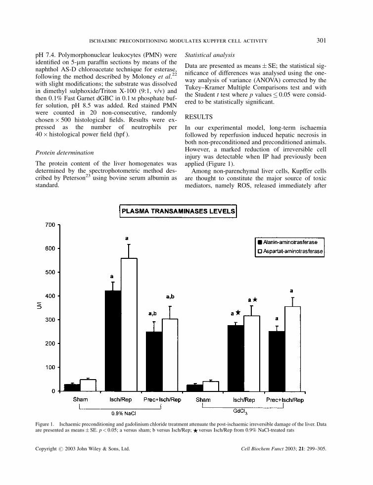
Similarly, GdCl3 treatment and IP were able toattenuate the increased production of 5-lipoxygen-ase-derived lipid hydroperoxides following ischae-mia/reperfusion (Figure 2). However, in the liver
Table 1. Hepatic concentration of hydrogen peroxide afterreperfusion (mmoles)
Groups
0.9% NaCl treated GdCl3 treated
Sham 1.20� 0.08 1.40� 0.06Isch/Rep 3.20� 0.09* 2.20� 0.17*,z
PrecþIsch/Rep 1.70� 0.17*,y 2.00� 0.17y
Data are presented as means�SE.p< 0.05; *versus sham; yversus Isch/Rep; zversus Isch/Rep from0.9% NaCl-treated rats.
Figure 2. Ischaemic preconditioning and gadolinium chloride treatment attenuate the post-ischaemic generation of 5- and 15-hydroperoxyeicosatetraenoic acids. Data are presented as means� SE. p< 0.05; a versus sham; b versus Isch/Rep; $ versus Isch/Rep from0.9% NaCl-treated rats
302 b. cavalieri ET AL.
Copyright # 2003 John Wiley & Sons, Ltd. Cell Biochem Funct 2003; 21: 299–305.
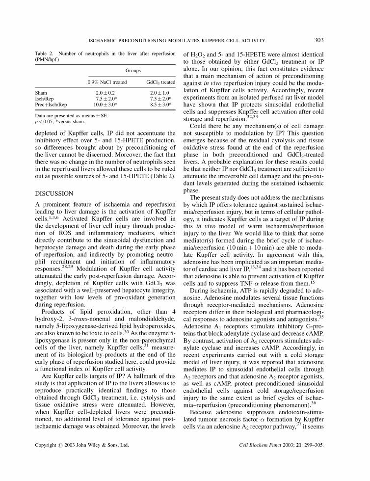
depleted of Kupffer cells, IP did not accentuate theinhibitory effect over 5- and 15-HPETE production,so differences brought about by preconditioning ofthe liver cannot be discerned. Moreover, the fact thatthere was no change in the number of neutrophils seenin the reperfused livers allowed these cells to be ruledout as possible sources of 5- and 15-HPETE (Table 2).
DISCUSSION
A prominent feature of ischaemia and reperfusionleading to liver damage is the activation of Kupffercells.1,3,6 Activated Kupffer cells are involved inthe development of liver cell injury through produc-tion of ROS and inflammatory mediators, whichdirectly contribute to the sinusoidal dysfunction andhepatocyte damage and death during the early phaseof reperfusion, and indirectly by promoting neutro-phil recruitment and initiation of inflammatoryresponses.28,29 Modulation of Kupffer cell activityattenuated the early post-reperfusion damage. Accor-dingly, depletion of Kupffer cells with GdCl3 wasassociated with a well-preserved hepatocyte integrity,together with low levels of pro-oxidant generationduring reperfusion.
Products of lipid peroxidation, other than 4hydroxy-2, 3-trans-nonenal and malondialdehyde,namely 5-lipoxygenase-derived lipid hydroperoxides,are also known to be toxic to cells.30 As the enzyme 5-lipoxygenase is present only in the non-parenchymalcells of the liver, namely Kupffer cells,31 measure-ment of its biological by-products at the end of theearly phase of reperfusion studied here, could providea functional index of Kupffer cell activity.
Are Kupffer cells targets of IP? A hallmark of thisstudy is that application of IP to the livers allows us toreproduce practically identical findings to thoseobtained through GdCl3 treatment, i.e. cytolysis andtissue oxidative stress were attenuated. However,when Kupffer cell-depleted livers were precondi-tioned, no additional level of tolerance against post-ischaemic damage was obtained. Moreover, the levels
of H2O2 and 5- and 15-HPETE were almost identicalto those obtained by either GdCl3 treatment or IPalone. In our opinion, this fact constitutes evidencethat a main mechanism of action of preconditioningagainst in vivo reperfusion injury could be the modu-lation of Kupffer cells activity. Accordingly, recentexperiments from an isolated perfused rat liver modelhave shown that IP protects sinusoidal endothelialcells and suppresses Kupffer cell activation after coldstorage and reperfusion.32,33
Could there be any mechanism(s) of cell damagenot susceptible to modulation by IP? This questionemerges because of the residual cytolysis and tissueoxidative stress found at the end of the reperfusionphase in both preconditioned and GdCl3-treatedlivers. A probable explanation for these results couldbe that neither IP nor GdCl3 treatment are sufficient toattenuate the irreversible cell damage and the pro-oxi-dant levels generated during the sustained ischaemicphase.
The present study does not address the mechanismsby which IP offers tolerance against sustained ischae-mia/reperfusion injury, but in terms of cellular pathol-ogy, it indicates Kupffer cells as a target of IP duringthis in vivo model of warm ischaemia/reperfusioninjury to the liver. We would like to think that somemediator(s) formed during the brief cycle of ischae-mia/reperfusion (10 minþ 10 min) are able to modu-late Kupffer cell activity. In agreement with this,adenosine has been implicated as an important media-tor of cardiac and liver IP,13,34 and it has been reportedthat adenosine is able to prevent activation of Kupffercells and to suppress TNF-� release from them.15
During ischaemia, ATP is rapidly degraded to ade-nosine. Adenosine modulates several tissue functionsthrough receptor-mediated mechanisms. Adenosinereceptors differ in their biological and pharmacologi-cal responses to adenosine agonists and antagonists.35
Adenosine A1 receptors stimulate inhibitory G-pro-teins that block adenylate cyclase and decrease cAMP.By contrast, activation of A2 receptors stimulates ade-nylate cyclase and increases cAMP. Accordingly, inrecent experiments carried out with a cold storagemodel of liver injury, it was reported that adenosinemediates IP to sinusoidal endothelial cells throughA2 receptors and that adenosine A2 receptor agonists,as well as cAMP, protect preconditioned sinusoidalendothelial cells against cold storage/reperfusioninjury to the same extent as brief cycles of ischae-mia–reperfusion (preconditioning phenomenon).36
Because adenosine suppresses endotoxin-stimu-lated tumour necrosis factor-� formation by Kupffercells via an adenosine A2 receptor pathway,37 it seems
Table 2. Number of neutrophils in the liver after reperfusion(PMN/hpf )
Groups
0.9% NaCl treated GdCl3 treated
Sham 2.0� 0.2 2.0� 1.0Isch/Rep 7.5� 2.0* 7.5� 2.0*PrecþIsch/Rep 10.0� 3.0* 8.5� 3.0*
Data are presented as means�SE.p< 0.05; *versus sham.
ischaemic preconditioning modulates kupffer cell activity 303
Copyright # 2003 John Wiley & Sons, Ltd. Cell Biochem Funct 2003; 21: 299–305.
reasonable to believe that adenosine released by pre-conditioning interferes in some step(s) of the mechan-ism involved in the activation of the Kupffer cells’NADPH oxidase and 5-lipoxygenase enzymes, i.e.blocking the post-reperfusion release of toxic media-tors: H2O2 and 5- and 15-HPETE. Accordingly, it hasbeen documented that intracellular cAMP-elevatingagents have been shown to be associated with thedownregulation of the oxidative burst and the inhibi-tion of the 5-lipoxygenase translocation and activationin both neutrophils and cells of the macrophage/monocyte lineage.38–41
ACKNOWLEDGEMENTS
This work was supported by grants from Italian Min-istry of the University (PRIN 1999, 2000, 2001), Cen-tro Nazionale delle Ricerche (Progetto FinalizzatoBiotecnologie), Turin University.
REFERENCES
1. Jaeschke H, Farhood A. Neutrophil and Kupffer cell inducedoxidant stress and ischemia–reperfusion injury in rat liver. Am JPhysiol 1991; 260: G335–G342.
2. Mochida S, Arai M, Ohno A, Masaki N, Ogata I, Fujiwara K.Oxidative stress in hepatocytes and stimulatory state of Kupffercells after reperfusion differ between warm and cold ischemia inrats. Liver 1994; 14: 234–240.
3. Cutrın JC, Llesuy S, Boveris A. Primary role of Kupffer cell-hepatocyte communication in the expression of oxidative stressin the post-ischaemic liver. Cell Biochem Funct 1998; 16:65–72.
4. Arii S, Imamura M. Physiological role of sinusoidal endothelialcells and Kupffer cells and their implication in the pathogenesisof liver injury. J Hepatobiliary Pancreat Surg 2000; 7: 40–48.
5. Rudiger HA, Clavien P-A. Tumor necrosis factor-�, but not Fas,mediates hepatocellullar apoptosis in the murine ischemic liver.Gastroenterology 2002; 122: 202–210.
6. Marzi I, Cowper K, Takei Y, Lindert K, Lemasters JJ, ThurmanRG. Methyl palmitate prevents Kupffer cell activation andimproves survival after orthotopic liver transplant in the rat.Transplant Int 1991; 4: 215–220.
7. Imamura H, Sutto F, Brault A, Huet P-M. Role of Kupffer cellsin cold ischemia/reperfusion injury of rat liver. Gastroenterol-ogy 1995; 109: 189–197.
8. Kozaki K, Egawa H, Bermudez L, Keefe EB, So SK, EsquivelCO. Effects of pentoxifylline pretreatment on Kupffer cells inrat liver transplantation. Hepatology 1995; 21: 1079–1082.
9. Urata K, Brault A, Rocheleau B, Huet PM. Role of Kupffer cellsin the survival after liver transplantation with long portal veinclamping times. Transplant Int 2000; 13: 420–427.
10. Murry CE, Jennings RB, Reimer KA. Preconditioning withischemia: a delay of lethal cell injury in ischemic myocardium.Circulation 1986; 74: 1124–1136.
11. Murry CE, Richard VJ, Jennings RB, Reimer KA. Myocardialprotection is lost before contractile function recovers fromischemic preconditioning. Am J Physiol 1991; 260: H796–H804.
12. Peralta C, Closa D, Hotter G, Gelpı E, Prats N, Rosello-CatafauJ. Liver ischemic preconditioning is mediated by the inhibitoryaction of nitric oxide on endothelin. Biochem Biophys ResCommun 1996; 229: 264–270.
13. Peralta C, Hotter G, Closa D, Bulbena O, Rosello-Catafau J.Protective effect of preconditioning on the injury associatedwith hepatic ischemia-reperfusion: role of nitric oxide andadenosine. Hepatology 1997; 25: 934–937.
14. Peralta C, Bartrons R, Riera R, et al. Hepatic preconditioningpreserves energy metabolism during sustained ischemia. Am JPhysiol 2000; 279: G163–G171.
15. Peralta C, Fernamndez L, Panes J, et al. Preconditioningprotects against systemic disorders associated with hepaticischemia–reperfusion through blockade of tumor necrosis fac-tor-induced P-selectin up-regulation in the rat. Hepatology2001; 33: 100–113.
16. Peralta C, Bulbena O, Xaus C, et al. Ischemic preconditioning:defense mechanism against the reactive oxygen species gener-ated after hepatic ischemia reperfusion. Transplantation 2002;73: 1–9.
17. Cavalieri B, Perrelli M-G, Aragno M, et al. Ischemic precon-ditioning attenuates the oxidant-dependent mechanisms ofreperfusion cell damage and death in rat liver. Liver Transplant2002; 8: 990–999.
18. US National Academy of Sciences. Guide for the Care andUse of Laboratory Animals, (NIH publication 86-23revised). US National Institutes of Health: Bethesda, MA,USA, 1985.
19. Wolff SP. Ferrous ion oxidation in presence of ferric ionindicator xylenol orange for measurement of hydroperoxides.Methods Enzymol 1994; 233: 182–189.
20. Folch J, Lees M, Sloane-Stanley GH. A simple method for theisolation and purification of total lipids from animals tissues.J Biol Chem 1957; 226: 497–509.
21. Tordijman C, Serkiz B, Rambaud F, Bonnet J, Volland JP. HPLCquantification of cyclooxygenase and lipoxygenase metabolitesof arachidonic acid from rat polymorphonuclear leukocytes.J Chromat 1990; 532: 135–143.
22. Moloney WC, McPherson K, Fliegelman L. Esterase activity inleukocytes demonstrated by the use of naphthol AS-D chlor-oacetate substrate. J Histochem Cytochem 1960; 8: 200–207.
23. Peterson GL. A simplification of the protein assay method ofLowry et al. which is more generally applicable. Anal Biochem1977; 83: 346–356.
24. Dieter P, Schulze-Specking A, Decker K. Differential inhibitionof prostaglandin and superoxide production by dexamethasonein primary cultures of rat Kupffer cells. Eur J Biochem 1986;159: 451–457.
25. Sakagami Y, Mizoguchi Y, Seki S, Kobayashi K, Morisawa S,Yamamoto S. Release of peptide leukotrienes from rat Kupffercells. Bichem Biophys Res Commun 1988; 156: 217–221.
26. Ouwendijk RJT, Zijlstra FJ, van den Broek AMWC, Brouwer A,Wilson JHP, Vincent JE. Comparison of the production ofeicosanoids by human and rat peritoneal macrophages andrat Kupffer cells. Prostaglandins 1988; 35: 437–446.
27. Kawada N, Ueda N, Mizoguchi Y, et al. Increased 5-lipoxy-genase activity in massive hepatic cell necrosis in the ratcorrelates with neutrophil infiltration. Hepatology 1992; 16:462–468.
28. Mawet E, Shiratori Y, Hikiba Y, et al. Cytokine-induced neu-trophil chemoattractant release from hepatocytes is modulatedby Kupffer cells. Hepatology 1996; 23: 353–358.
29. Wanner GA, Muller PEM, Ertel W, Bauer M, Menger MD,Messmer K. Differential effect of anti-TNF-� antibody on
304 b. cavalieri ET AL.
Copyright # 2003 John Wiley & Sons, Ltd. Cell Biochem Funct 2003; 21: 299–305.
proinflammatory cytokine release by Kupffer cells followingliver ischemia and reperfusion. Shock 1999; 11: 391–395.
30. Keppler D. Leukotrienes and other eicosanoids in liverpathophysiology. In The Liver: Biology and Pathobiology,Arias IM, Boyer JL, Fausto N, Jakoby WB, Schachter DA,Shafritz DA (eds). Raven Press, Ltd: New York, 1994;1015–1029.
31. Shimada K, Navarro J, Goeger DE, Mustafa SB, Weigel PH,Weinman SA. Expression and regulation of leukotriene-synthesis enzymes in rat liver cells. Hepatology 1998; 28:1275–1281.
32. Arai M, Thurman RG, Lemasters JJ. Involvement of Kupffercells and sinusoidal endothelial cells in ischemic precondition-ing to rat livers stored for transplantation. Transplant Proc 1999;31: 425–427.
33. Arai M, Thurman RG, Lemasters JJ. Ischemic preconditioningof rat livers against cold storage-reperfusion injury: role ofnonparenchymal cells and the phenomenom of heterologouspreconditioning. Liver Transplant 2001; 7: 292–299.
34. Cohen MV, Baines CP, Downey JM. Ischemic preconditioning:from adenosine receptor to KATP channel. Annu Rev Physiol2000; 62: 79–109.
35. Ralevic V, Burnstock G. Receptors for purines and pyrimidines.Pharmacol Rev 1998; 50: 413–492.
36. Arai M, Thurman RG, Lemasters JJ. Contribution of AdenosineA2 receptors and cyclic adenosine monophosphate to protectiveischemic preconditioning of sinusoidal endothelial cells againststorage/reperfusion injury in rats livers. Hepatology 2000; 32:297–302.
37. Reinstein LJ, Lichtman SN, Currin RT, Wang J, Thurman RG,Lemasters JJ. Supression of lipopolysaccharide-stimulatedrelease of tumor necrosis factor by adenosine: evidence forA2 receptors on rat Kupffer cells. Hepatology 1994; 19: 1445–1452.
38. Ottonello L, Morone MP, Dapino P, Dallegri F. cAMP-elevatingagents down regulate the oxidative burst induced by granulo-cyte-macrophage colony stimulating factor (GM-CSF) in adher-ent neutrophils. Clin Exp Immunol 1995; 101: 502–506.
39. Berger CEM, Horrocks BR, Datta HK. CAMP-dependentinhibition is dominant in regulating superoxide productionin the bone-resorbing osteoclats. J Endocrinol 1998; 158:311–318.
40. Bigby TD. The Yin and the Yan of 5-lipoxygenase pathwayactivation. Mol Pharmacol 2002; 62: 200–202.
41. Flamand N, Surette ME, Picard S, Bourgoin S, Borgeat P.Cyclic AMP-mediated inhibition of 5-lipoxygenase transloca-tion and leukotriene biosynthesis in human neutrophils. MolPharmacol 2002; 62: 250–256.
ischaemic preconditioning modulates kupffer cell activity 305
Copyright # 2003 John Wiley & Sons, Ltd. Cell Biochem Funct 2003; 21: 299–305.