iron overload and iron chelation: the inside story course...iron overload and iron chelation: the...
TRANSCRIPT
Iron Overload and Iron Chelation:The Inside Story
Jerry L. Spivak, MDProfessor of Medicine and Oncology
Johns Hopkins University School of MedicineBaltimore, Maryland
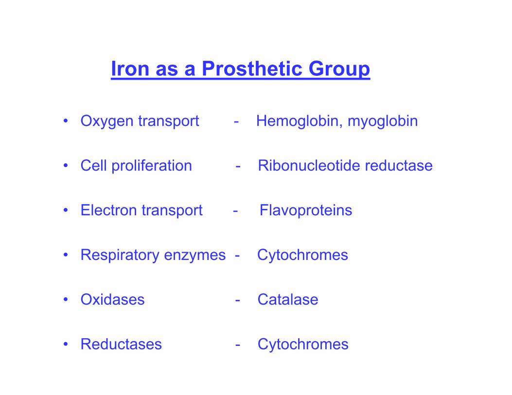
Iron as a Prosthetic Group
• Oxygen transport - Hemoglobin, myoglobin
• Cell proliferation - Ribonucleotide reductase
• Electron transport - Flavoproteins
• Respiratory enzymes - Cytochromes
• Oxidases - Catalase
• Reductases - Cytochromes
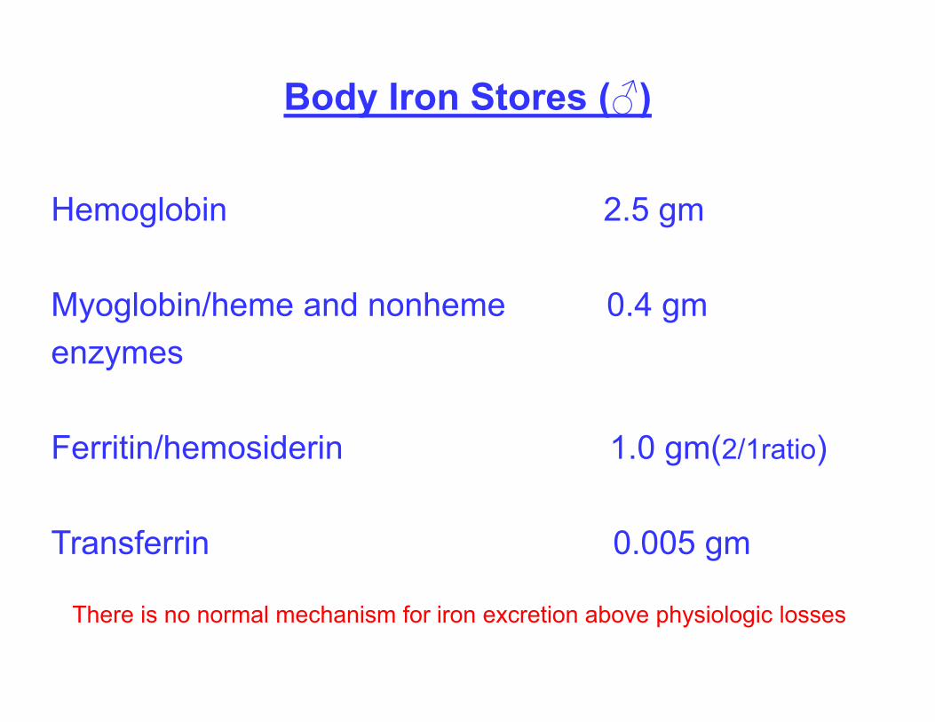
Body Iron Stores (♂)
Hemoglobin 2.5 gm
Myoglobin/heme and nonheme 0.4 gmenzymes
Ferritin/hemosiderin 1.0 gm(2/1ratio)
Transferrin 0.005 gm
There is no normal mechanism for iron excretion above physiologic losses
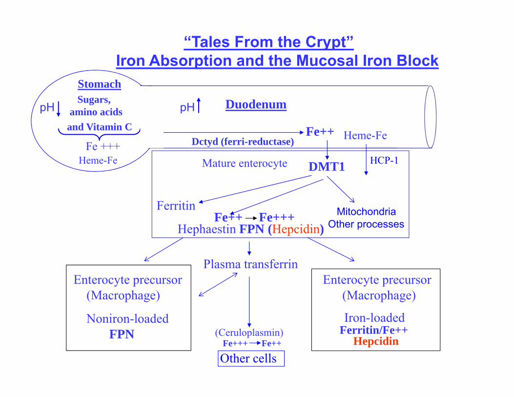
“Tales From the Crypt”Iron Absorption and the Mucosal Iron Block
DuodenumSugars, amino acids
and Vitamin C Fe++
Plasma transferrinEnterocyte precursor
(Macrophage)
Noniron-loadedFPN
Enterocyte precursor(Macrophage)
Iron-loaded
Mature enterocyte DMT1
Hephaestin FPN (Hepcidin)
HepcidinFerritin/Fe++
FerritinFe++ Fe+++
Heme-FeDctyd (ferri-reductase)
Other cells
Stomach
Fe +++Heme-Fe
pH pH
HCP-1
MitochondriaOther processes
(Ceruloplasmin)Fe+++ Fe++
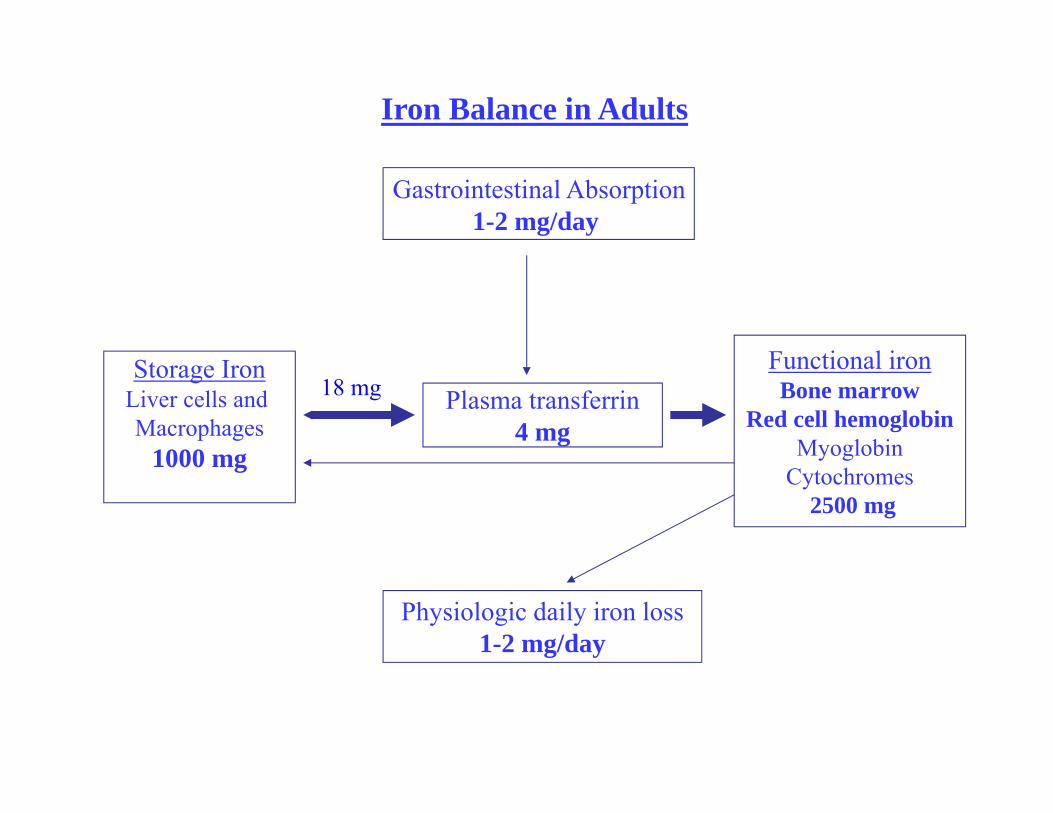
Iron Balance in Adults
Gastrointestinal Absorption1-2 mg/day
Physiologic daily iron loss1-2 mg/day
Plasma transferrin4 mg
Storage IronLiver cells and Macrophages
1000 mg
Functional ironBone marrow
Red cell hemoglobinMyoglobin
Cytochromes2500 mg
18 mg
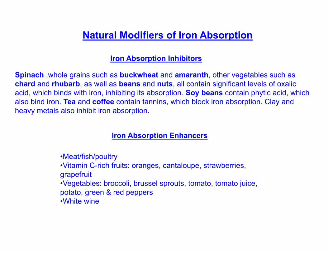
Spinach ,whole grains such as buckwheat and amaranth, other vegetables such as chard and rhubarb, as well as beans and nuts, all contain significant levels of oxalic acid, which binds with iron, inhibiting its absorption. Soy beans contain phytic acid, which also bind iron. Tea and coffee contain tannins, which block iron absorption. Clay and heavy metals also inhibit iron absorption.
Iron Absorption Enhancers
•Meat/fish/poultry •Vitamin C-rich fruits: oranges, cantaloupe, strawberries, grapefruit•Vegetables: broccoli, brussel sprouts, tomato, tomato juice, potato, green & red peppers •White wine
Natural Modifiers of Iron Absorption
Iron Absorption Inhibitors
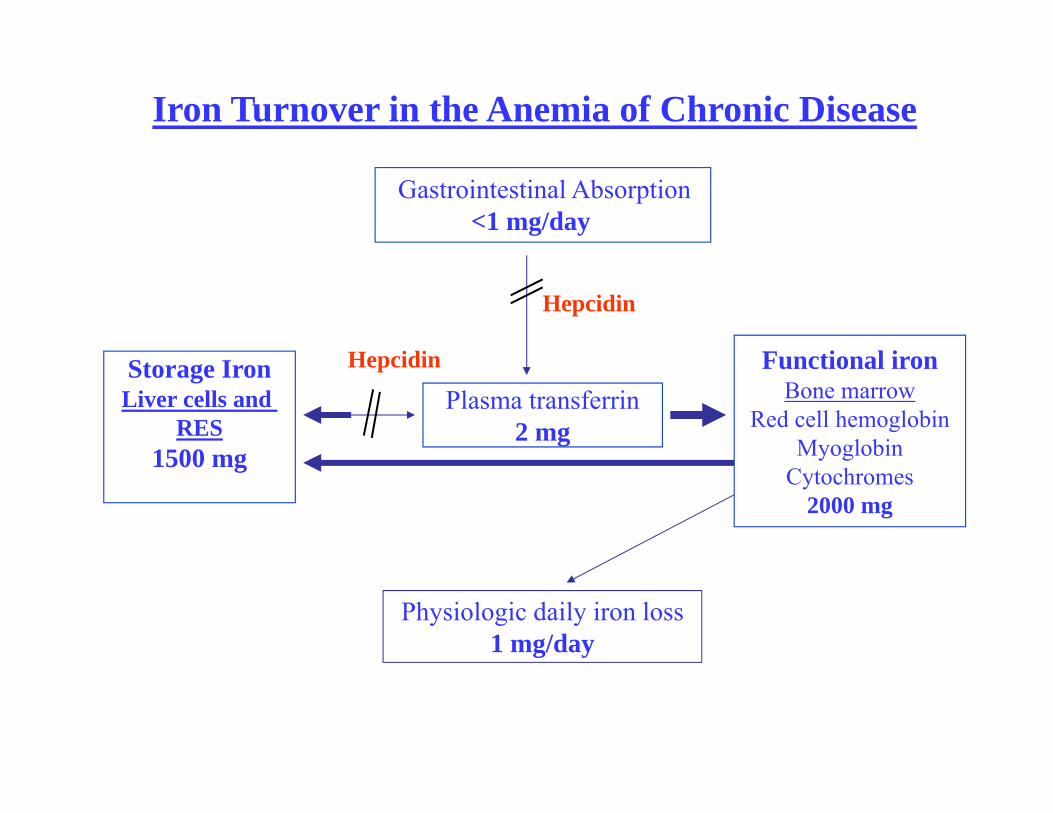
Iron Turnover in the Anemia of Chronic Disease
Gastrointestinal Absorption<1 mg/day
Physiologic daily iron loss1 mg/day
Plasma transferrin2 mg
Storage IronLiver cells and
RES1500 mg
Functional ironBone marrow
Red cell hemoglobinMyoglobin
Cytochromes2000 mg
Hepcidin
Hepcidin
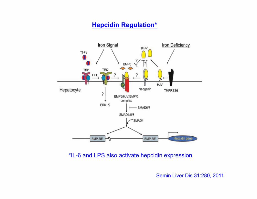
Semin Liver Dis 31:280, 2011
Hepcidin Regulation*
*IL-6 and LPS also activate hepcidin expression
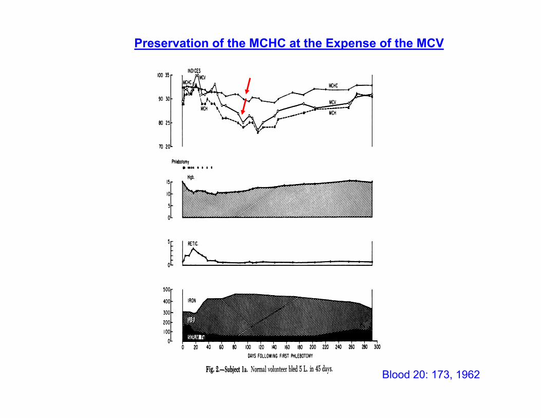
Preservation of the MCHC at the Expense of the MCV
Blood 20: 173, 1962
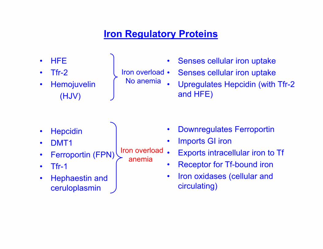
Iron Regulatory Proteins
• HFE• Tfr-2• Hemojuvelin
(HJV)
• Hepcidin • DMT1• Ferroportin (FPN)• Tfr-1• Hephaestin and
ceruloplasmin
• Senses cellular iron uptake• Senses cellular iron uptake• Upregulates Hepcidin (with Tfr-2
and HFE)
• Downregulates Ferroportin• Imports GI iron• Exports intracellular iron to Tf • Receptor for Tf-bound iron• Iron oxidases (cellular and
circulating)
Iron overload No anemia
Iron overloadanemia
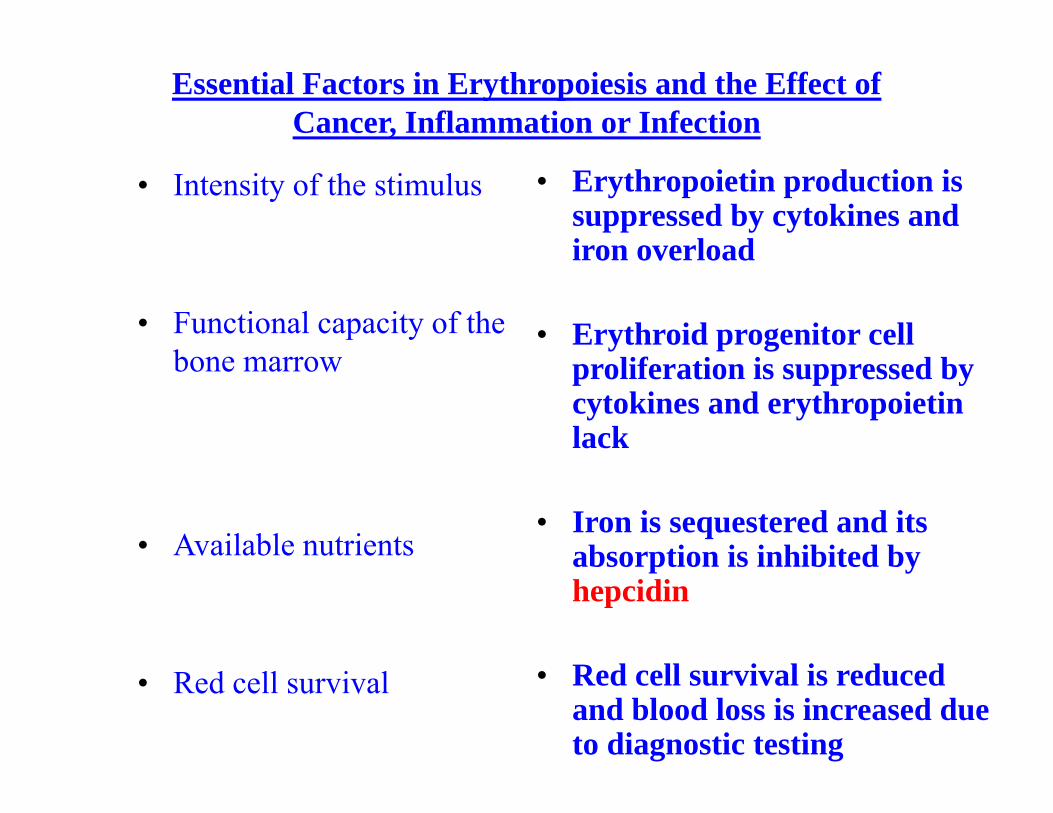
Essential Factors in Erythropoiesis and the Effect of Cancer, Inflammation or Infection
• Intensity of the stimulus
• Functional capacity of the bone marrow
• Available nutrients
• Red cell survival
• Erythropoietin production is suppressed by cytokines and iron overload
• Erythroid progenitor cell proliferation is suppressed by cytokines and erythropoietin lack
• Iron is sequestered and its absorption is inhibited by hepcidin
• Red cell survival is reduced and blood loss is increased due to diagnostic testing
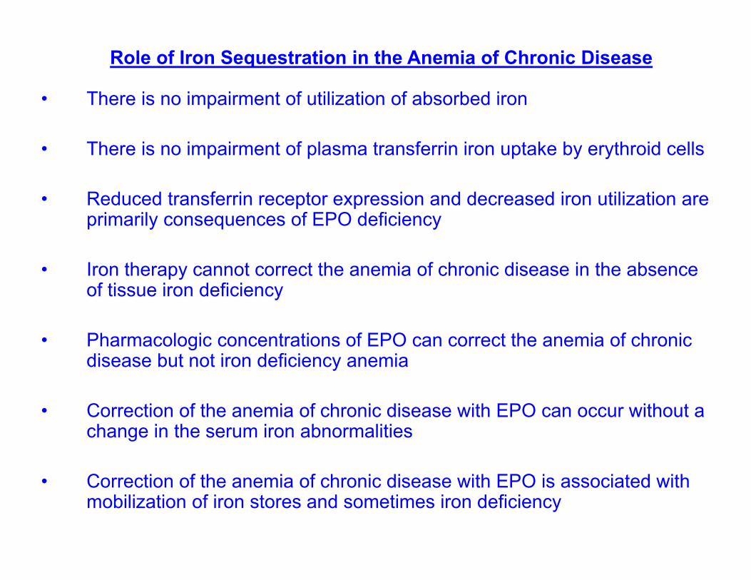
Role of Iron Sequestration in the Anemia of Chronic Disease
• There is no impairment of utilization of absorbed iron
• There is no impairment of plasma transferrin iron uptake by erythroid cells
• Reduced transferrin receptor expression and decreased iron utilization are primarily consequences of EPO deficiency
• Iron therapy cannot correct the anemia of chronic disease in the absence of tissue iron deficiency
• Pharmacologic concentrations of EPO can correct the anemia of chronic disease but not iron deficiency anemia
• Correction of the anemia of chronic disease with EPO can occur without a change in the serum iron abnormalities
• Correction of the anemia of chronic disease with EPO is associated with mobilization of iron stores and sometimes iron deficiency
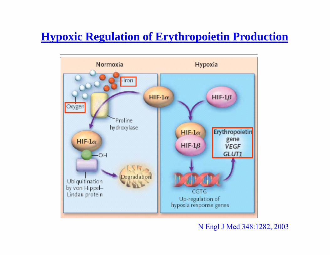
Hypoxic Regulation of Erythropoietin Production
N Engl J Med 348:1282, 2003
Ser
um E
po (m
U/m
l)
Time (days)–2 0 2 4 6 8 10 12
0
1600
1400
1200
800
600
400
200
1000
Hgb
, g/d
L
10
9
7
6
5
8
sEpo, mU/mL
Hb, g/dL
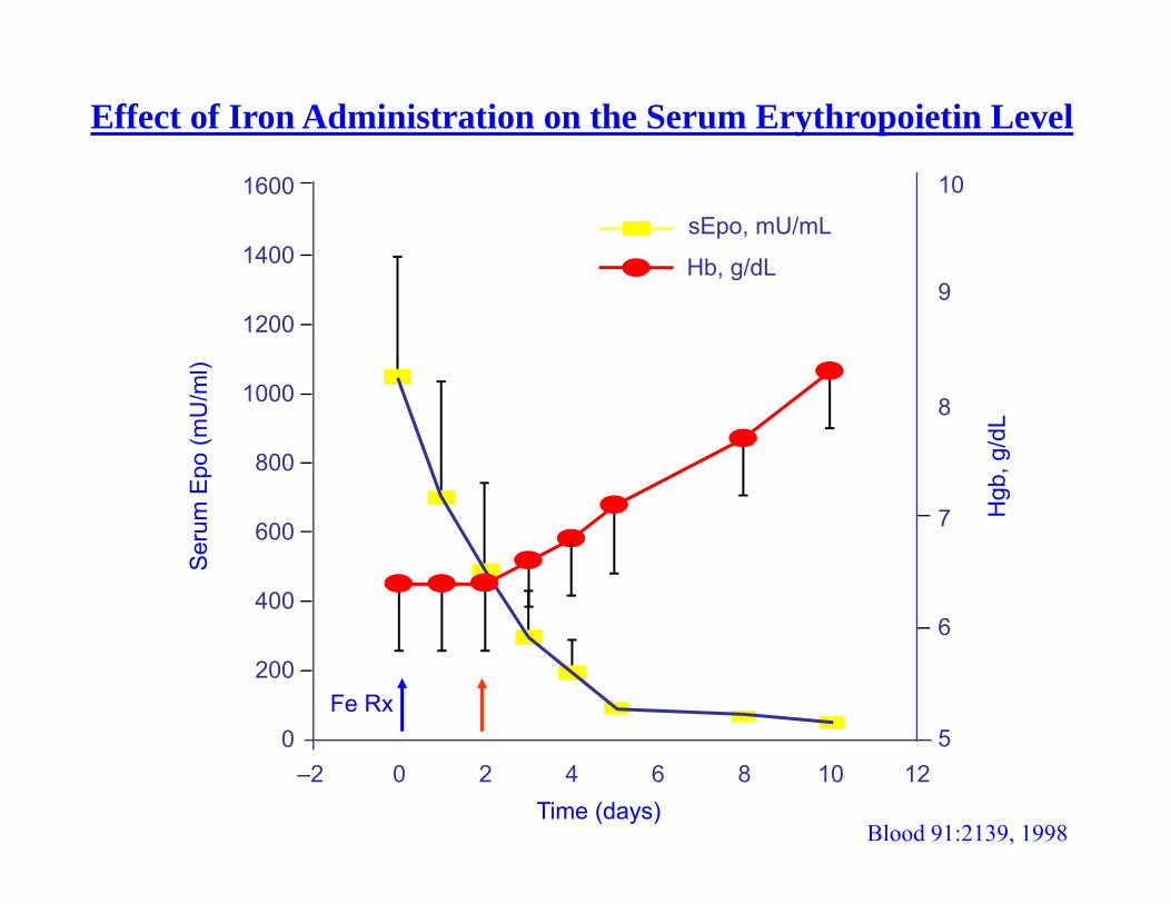
Blood 91:2139, 1998
Effect of Iron Administration on the Serum Erythropoietin Level
Fe Rx
Br J Haematol 94:288, 1996
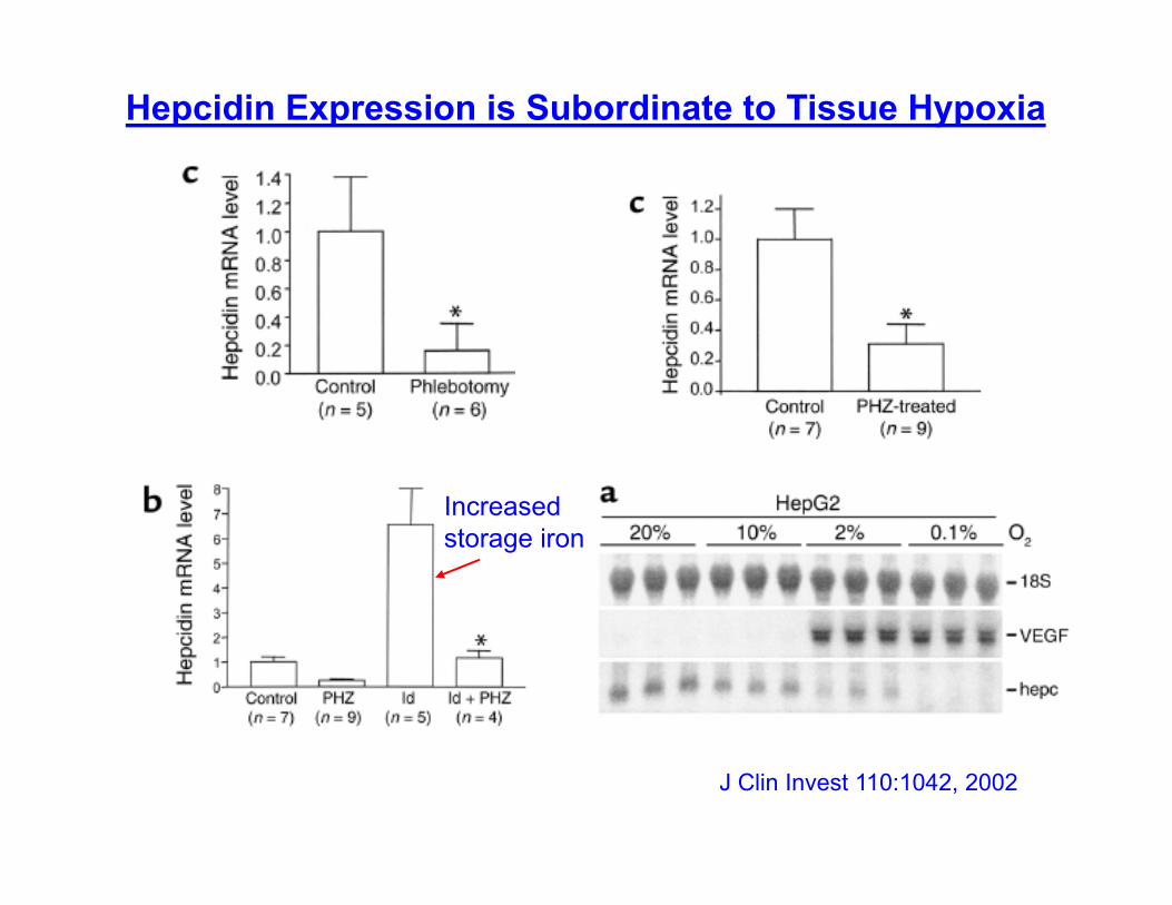
J Clin Invest 110:1042, 2002
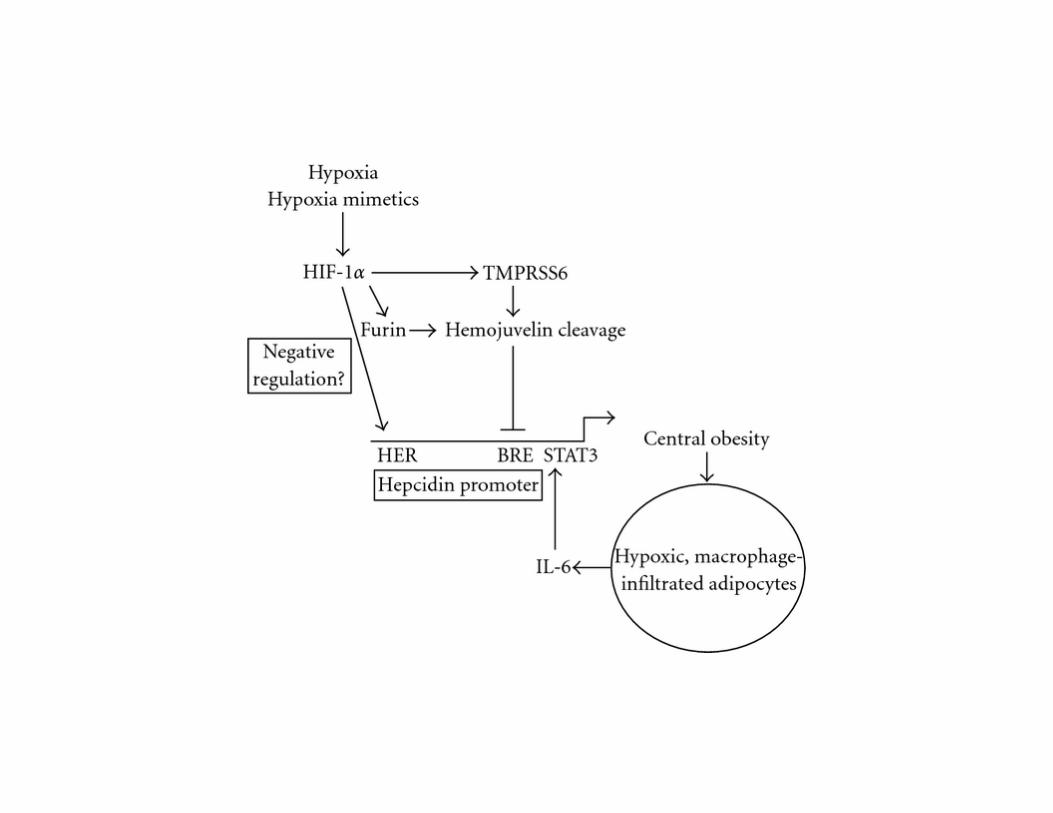
Hepcidin Expression is Subordinate to Tissue Hypoxia
Increasedstorage iron
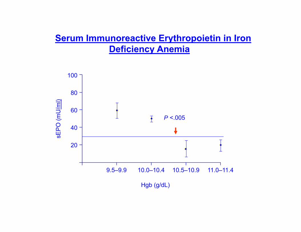
Serum Immunoreactive Erythropoietin in Iron Deficiency Anemia
sEP
O (m
U/m
l)
Hgb (g/dL)
9.5–9.9 10.0–10.4 10.5–10.9 11.0–11.4
P <.005
0
100
80
60
40
20
Time (months)
Hem
oglo
bin
gm
%
B J Haematol 94:288, 1996
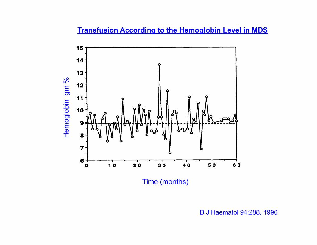
Transfusion According to the Hemoglobin Level in MDS
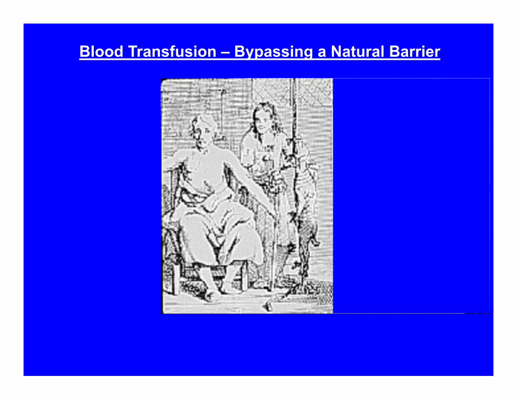
Blood Transfusion – Bypassing a Natural Barrier
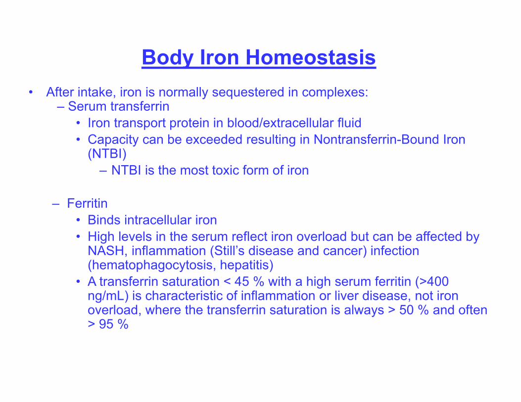
Body Iron Homeostasis• After intake, iron is normally sequestered in complexes:
– Serum transferrin• Iron transport protein in blood/extracellular fluid• Capacity can be exceeded resulting in Nontransferrin-Bound Iron
(NTBI) – NTBI is the most toxic form of iron
– Ferritin• Binds intracellular iron• High levels in the serum reflect iron overload but can be affected by
NASH, inflammation (Still’s disease and cancer) infection (hematophagocytosis, hepatitis)
• A transferrin saturation < 45 % with a high serum ferritin (>400 ng/mL) is characteristic of inflammation or liver disease, not iron overload, where the transferrin saturation is always > 50 % and often > 95 %
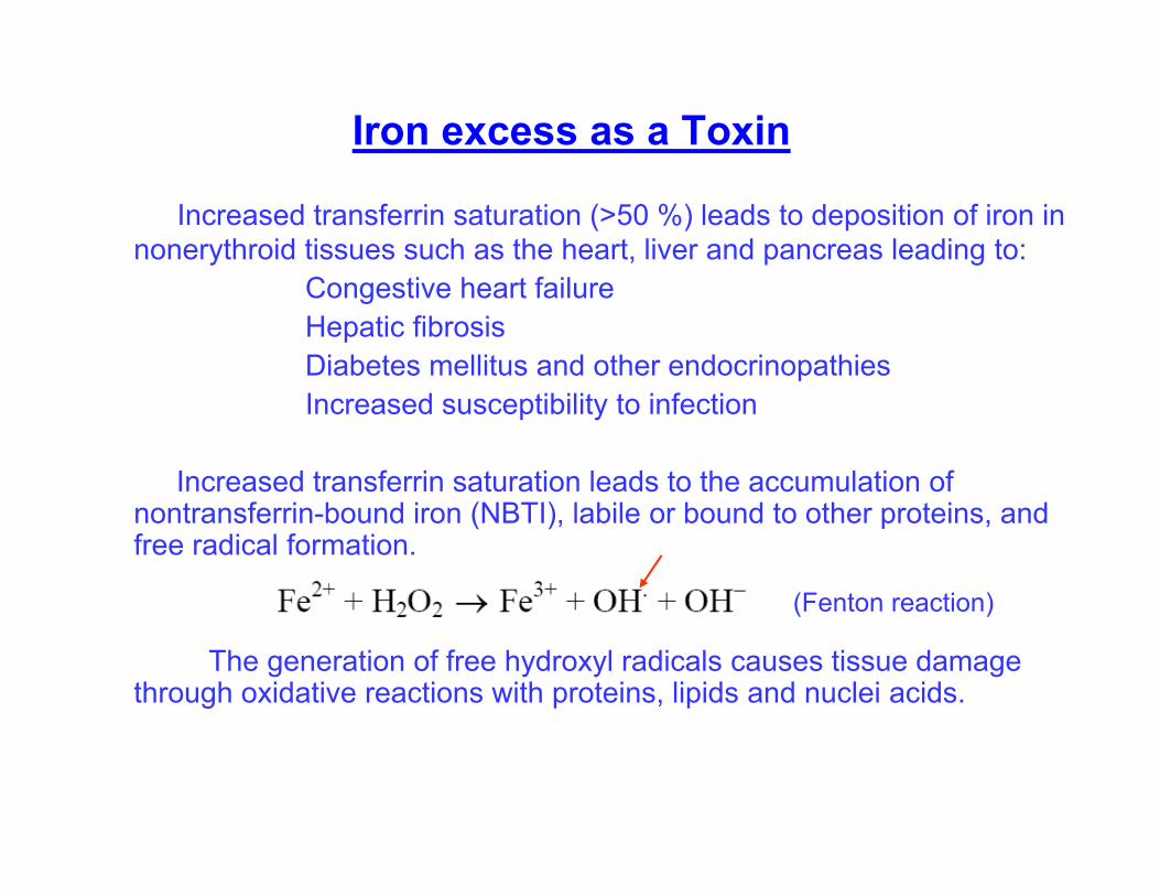
Iron excess as a Toxin
Increased transferrin saturation (>50 %) leads to deposition of iron in nonerythroid tissues such as the heart, liver and pancreas leading to:
Congestive heart failureHepatic fibrosisDiabetes mellitus and other endocrinopathiesIncreased susceptibility to infection
Increased transferrin saturation leads to the accumulation of nontransferrin-bound iron (NBTI), labile or bound to other proteins, and free radical formation.
The generation of free hydroxyl radicals causes tissue damage through oxidative reactions with proteins, lipids and nuclei acids.
(Fenton reaction)
Int J Hematol 76:219, 2002
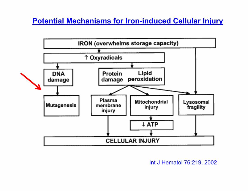
Potential Mechanisms for Iron-induced Cellular Injury
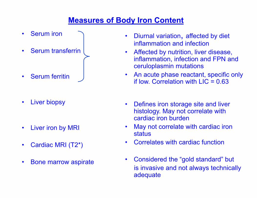
Measures of Body Iron Content• Serum iron
• Serum transferrin
• Serum ferritin
• Liver biopsy
• Liver iron by MRI
• Cardiac MRI (T2*)
• Bone marrow aspirate
• Diurnal variation, affected by diet inflammation and infection
• Affected by nutrition, liver disease, inflammation, infection and FPN and ceruloplasmin mutations
• An acute phase reactant, specific only if low. Correlation with LIC = 0.63
• Defines iron storage site and liver histology. May not correlate with cardiac iron burden
• May not correlate with cardiac iron status
• Correlates with cardiac function
• Considered the “gold standard” butis invasive and not always technically adequate
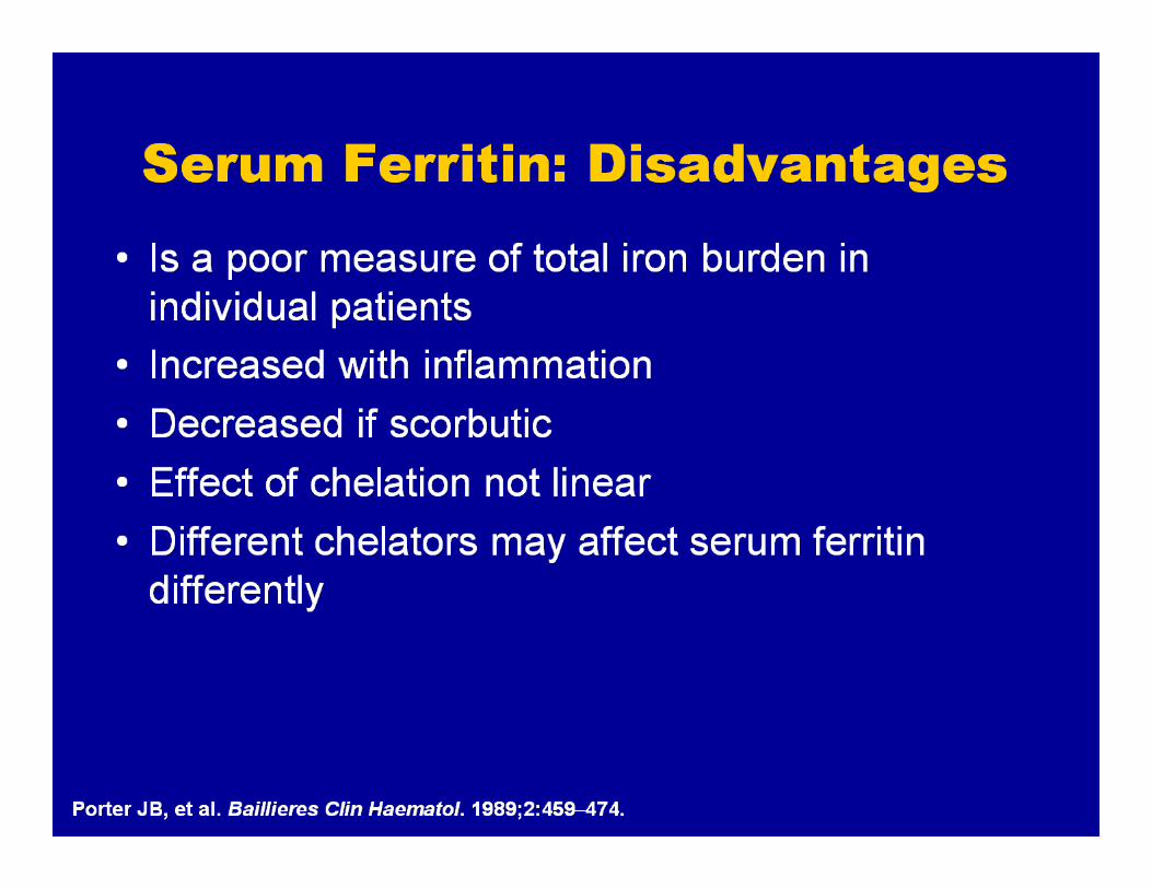
Serum Ferritin: Disadvantages

LIC Predicts Total Body Iron
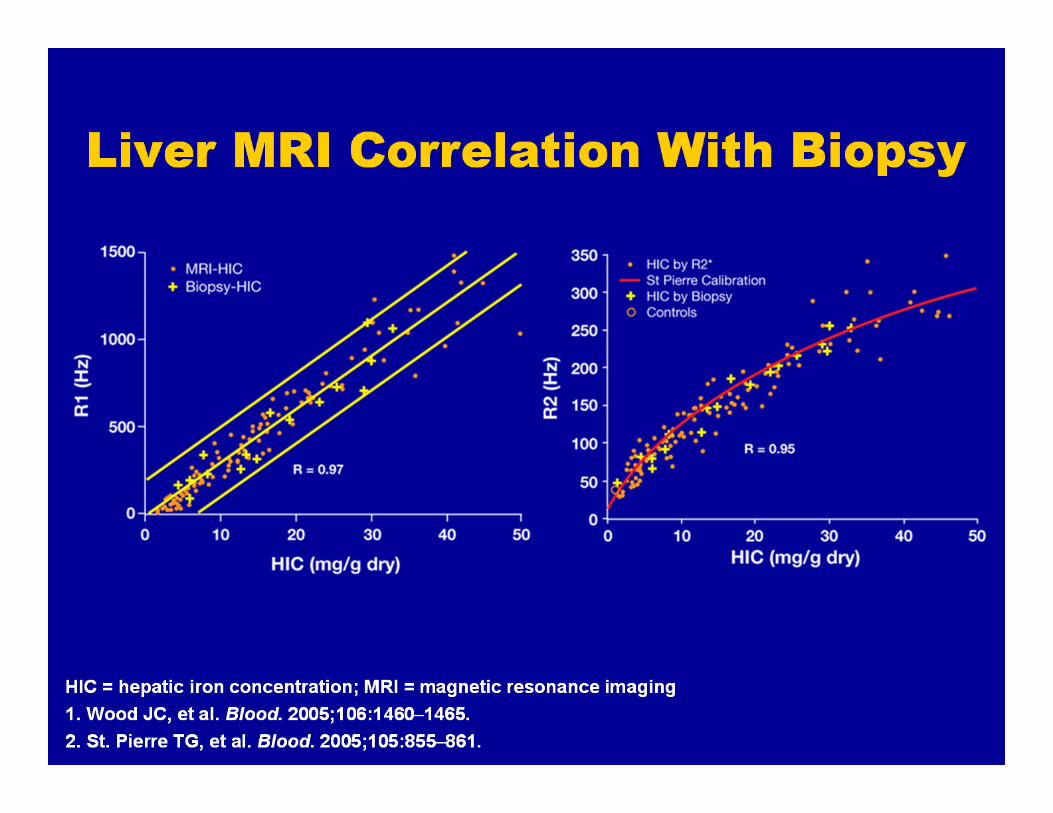
Liver MRI Correlation With Biopsy
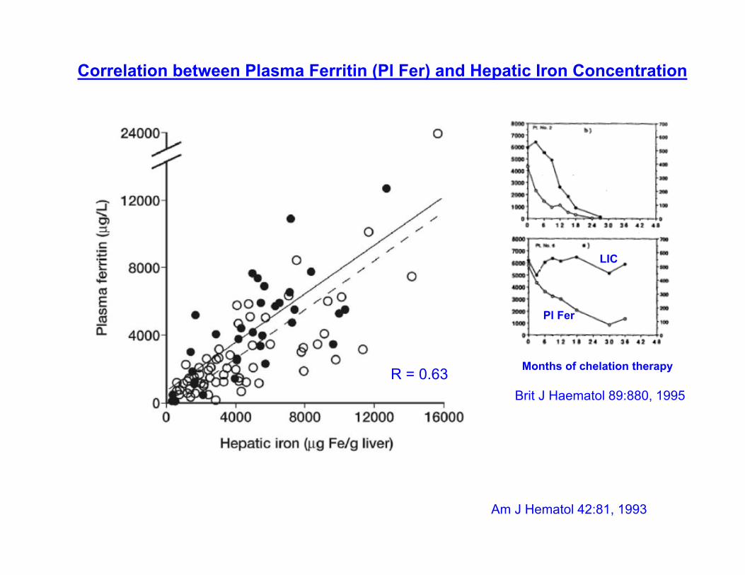
Correlation between Plasma Ferritin (Pl Fer) and Hepatic Iron Concentration
Am J Hematol 42:81, 1993
R = 0.63 Months of chelation therapy
Pl Fer
LIC
Brit J Haematol 89:880, 1995
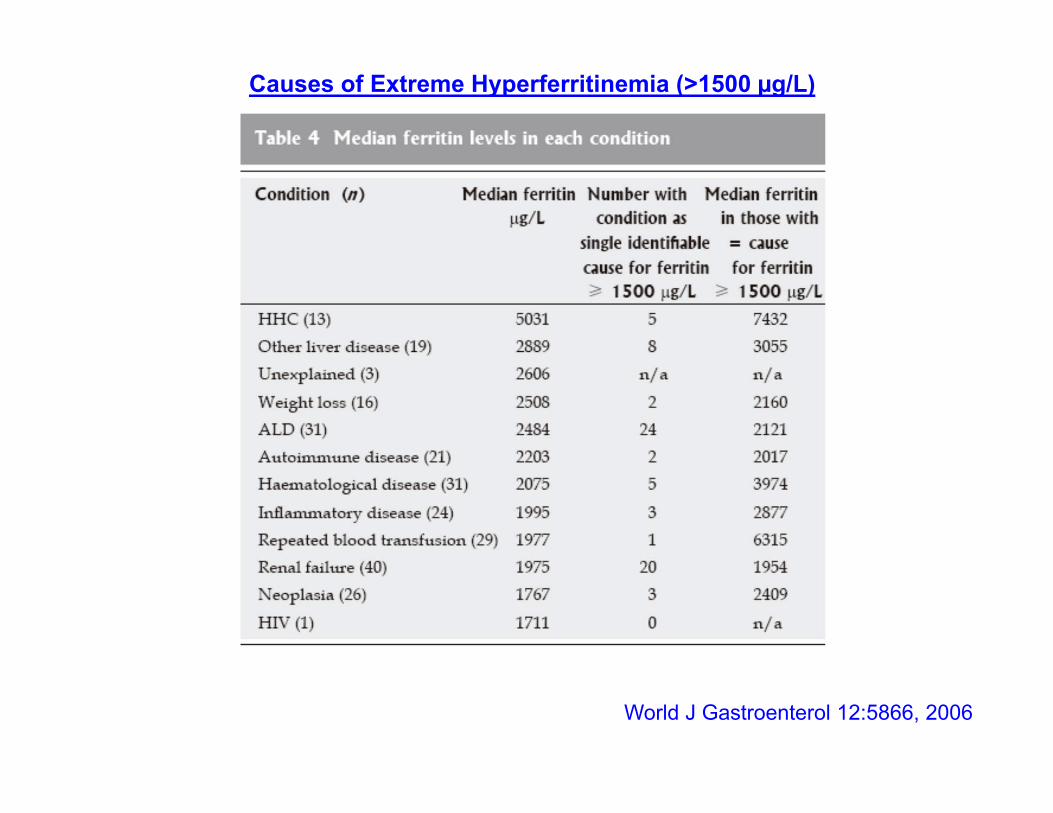
World J Gastroenterol 12:5866, 2006
Causes of Extreme Hyperferritinemia (>1500 µg/L)
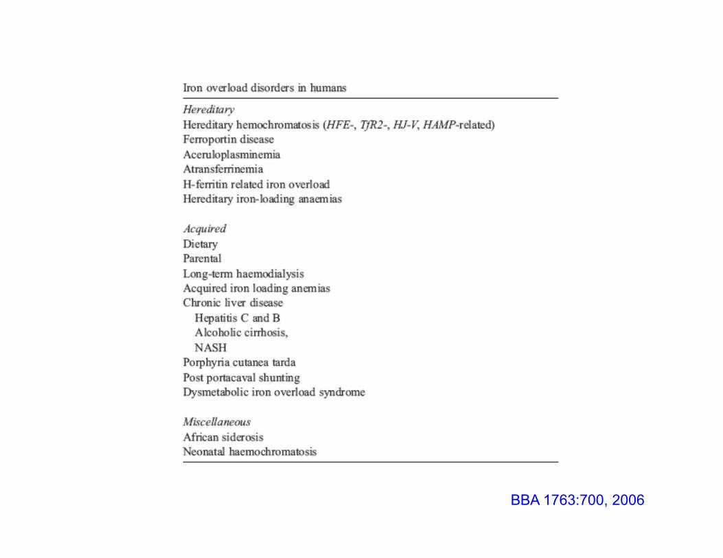
BBA 1763:700, 2006
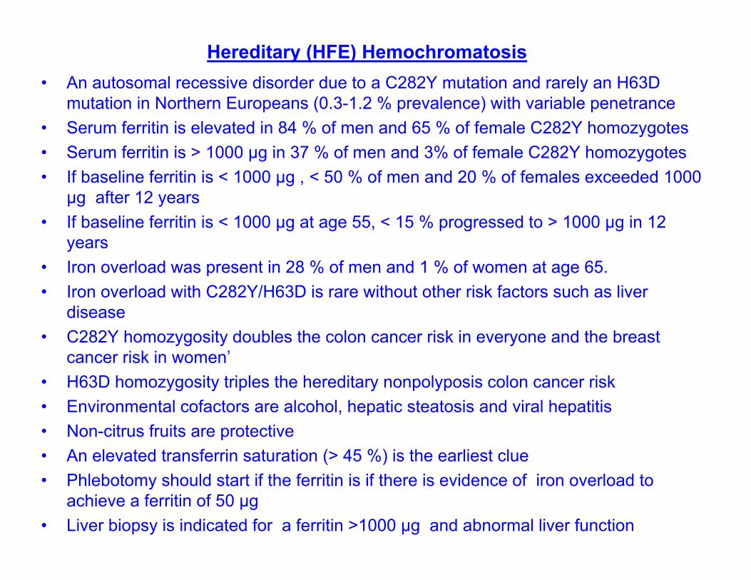
Hereditary (HFE) Hemochromatosis• An autosomal recessive disorder due to a C282Y mutation and rarely an H63D
mutation in Northern Europeans (0.3-1.2 % prevalence) with variable penetrance• Serum ferritin is elevated in 84 % of men and 65 % of female C282Y homozygotes• Serum ferritin is > 1000 µg in 37 % of men and 3% of female C282Y homozygotes• If baseline ferritin is < 1000 µg , < 50 % of men and 20 % of females exceeded 1000
µg after 12 years• If baseline ferritin is < 1000 µg at age 55, < 15 % progressed to > 1000 µg in 12
years • Iron overload was present in 28 % of men and 1 % of women at age 65.• Iron overload with C282Y/H63D is rare without other risk factors such as liver
disease• C282Y homozygosity doubles the colon cancer risk in everyone and the breast
cancer risk in women’• H63D homozygosity triples the hereditary nonpolyposis colon cancer risk• Environmental cofactors are alcohol, hepatic steatosis and viral hepatitis• Non-citrus fruits are protective• An elevated transferrin saturation (> 45 %) is the earliest clue• Phlebotomy should start if the ferritin is if there is evidence of iron overload to
achieve a ferritin of 50 µg• Liver biopsy is indicated for a ferritin >1000 µg and abnormal liver function
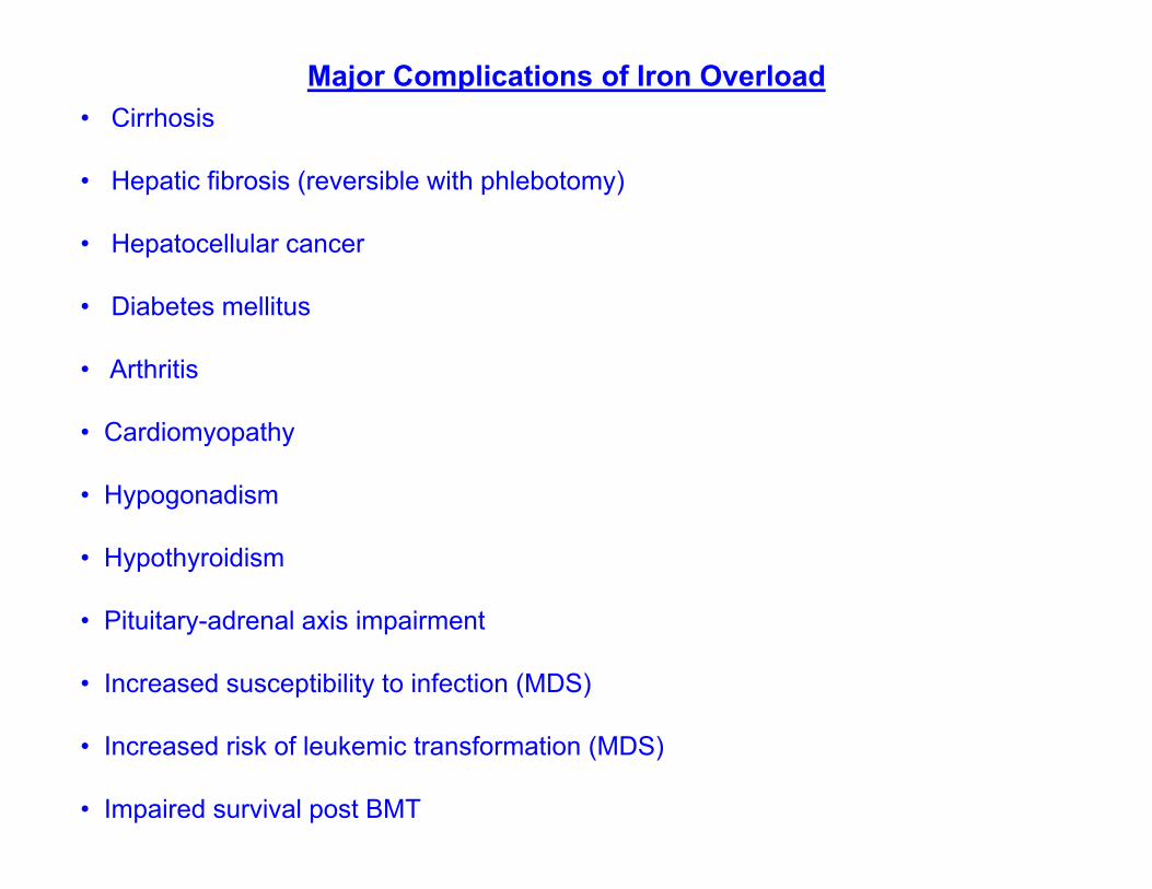
Major Complications of Iron Overload• Cirrhosis
• Hepatic fibrosis (reversible with phlebotomy)
• Hepatocellular cancer
• Diabetes mellitus
• Arthritis
• Cardiomyopathy
• Hypogonadism
• Hypothyroidism
• Pituitary-adrenal axis impairment
• Increased susceptibility to infection (MDS)
• Increased risk of leukemic transformation (MDS)
• Impaired survival post BMT
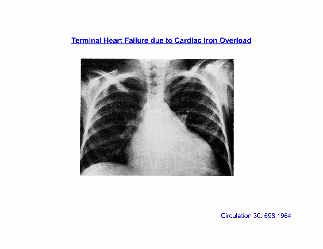
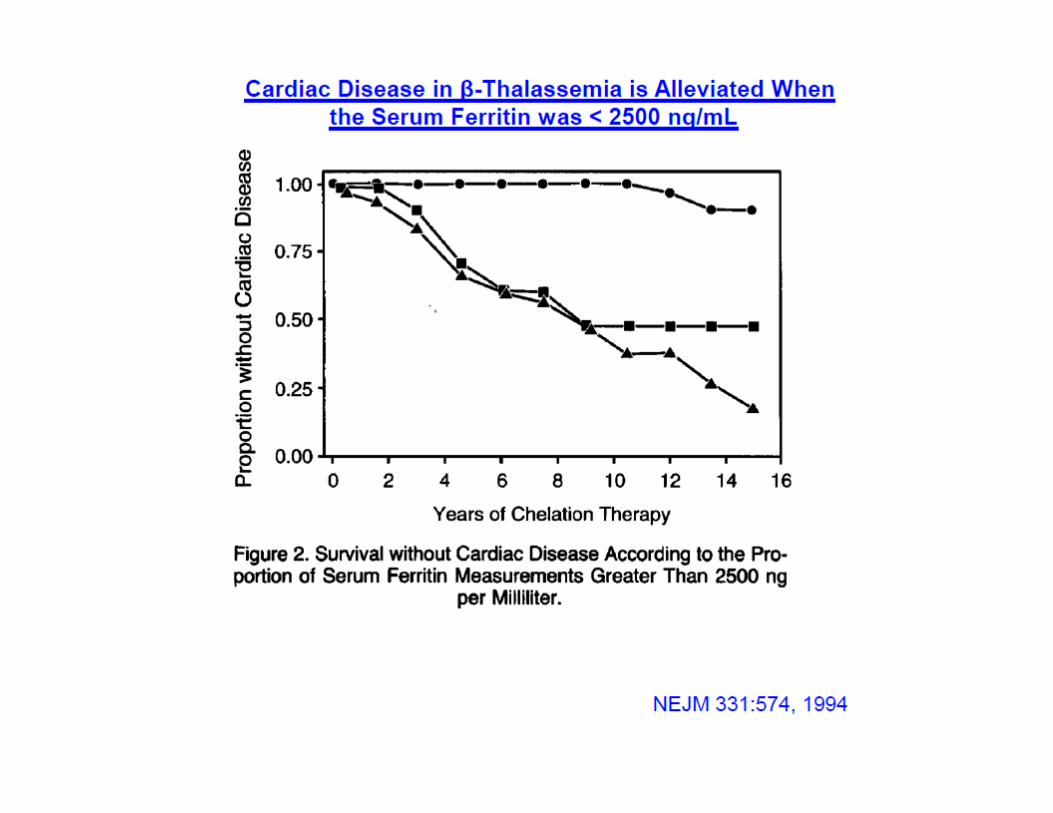
Circulation 30: 698,1964
Terminal Heart Failure due to Cardiac Iron Overload
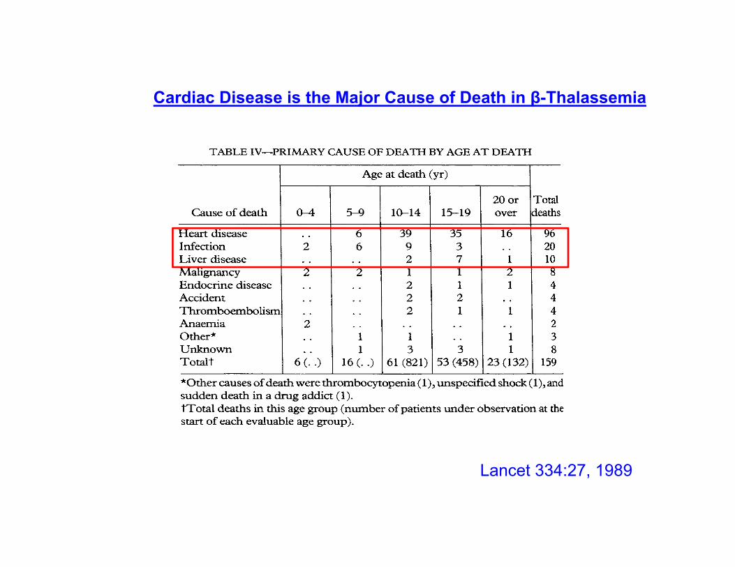
Lancet 334:27, 1989
Cardiac Disease is the Major Cause of Death in β-Thalassemia
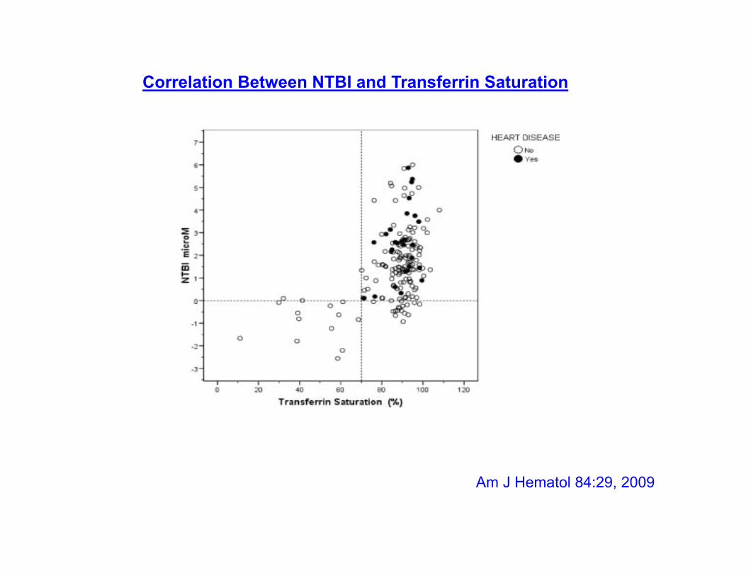
Am J Hematol 84:29, 2009
Correlation Between NTBI and Transferrin Saturation

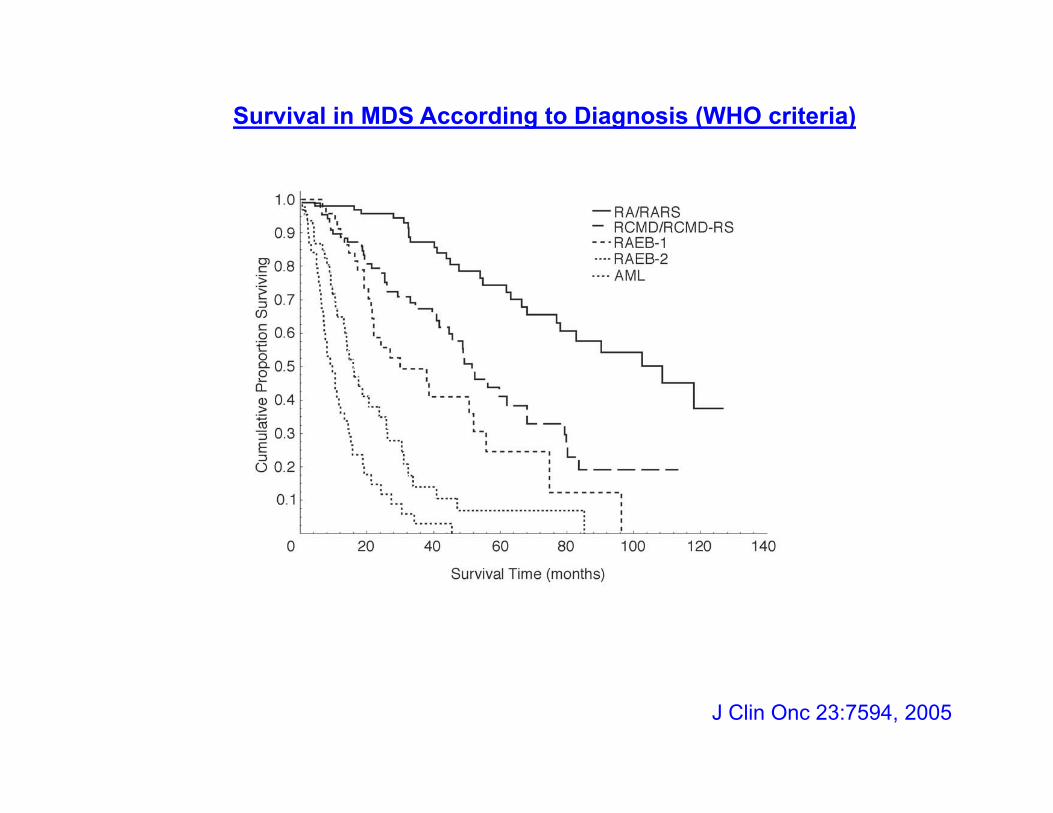
Survival in MDS According to Diagnosis (WHO criteria)
J Clin Onc 23:7594, 2005
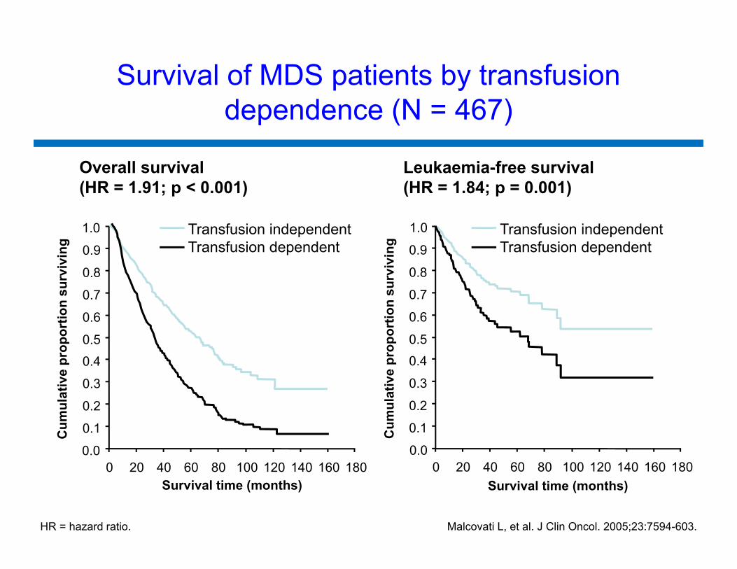
Overall survival(HR = 1.91; p < 0.001)
Leukaemia-free survival(HR = 1.84; p = 0.001)
Survival of MDS patients by transfusion dependence (N = 467)
180
Cum
ulat
ive
prop
ortio
n su
rviv
ing
Transfusion independentTransfusion dependent
0.0
0.1
0.2
0.3
0.40.5
0.6
0.7
0.8
0.9
1.0
0 20 40 60 80 100 120 140 160
Cum
ulat
ive
prop
ortio
n su
rviv
ing
0.0
0.1
0.2
0.3
0.4
0.5
0.6
0.7
0.8
0.9
1.0
0 20 40 60 80 100 120 140 160 180Survival time (months) Survival time (months)
Transfusion independentTransfusion dependent
Malcovati L, et al. J Clin Oncol. 2005;23:7594-603. HR = hazard ratio.
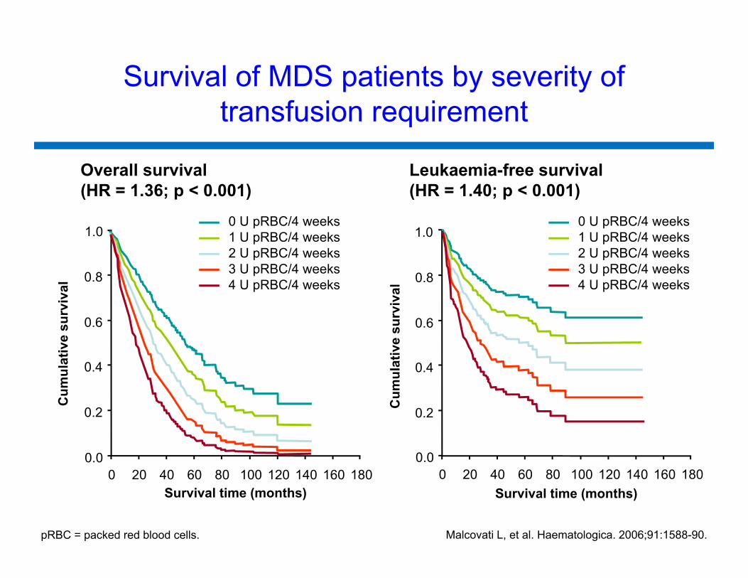
Survival of MDS patients by severity of transfusion requirement
Survival time (months)
0.2
0.4
0.6
0.8
1.0
Survival time (months)
0.0
0.2
0.4
0.6
0.8
1.0
0 20 40 60 80 100 120 140 160 180
Cum
ulat
ive
surv
ival
0 U pRBC/4 weeks1 U pRBC/4 weeks2 U pRBC/4 weeks3 U pRBC/4 weeks4 U pRBC/4 weeks
Malcovati L, et al. Haematologica. 2006;91:1588-90.
0 U pRBC/4 weeks1 U pRBC/4 weeks2 U pRBC/4 weeks3 U pRBC/4 weeks4 U pRBC/4 weeks
Cum
ulat
ive
surv
ival
pRBC = packed red blood cells.
Overall survival(HR = 1.36; p < 0.001)
Leukaemia-free survival(HR = 1.40; p < 0.001)
1800 20 40 60 80 100 120 140 1600.0
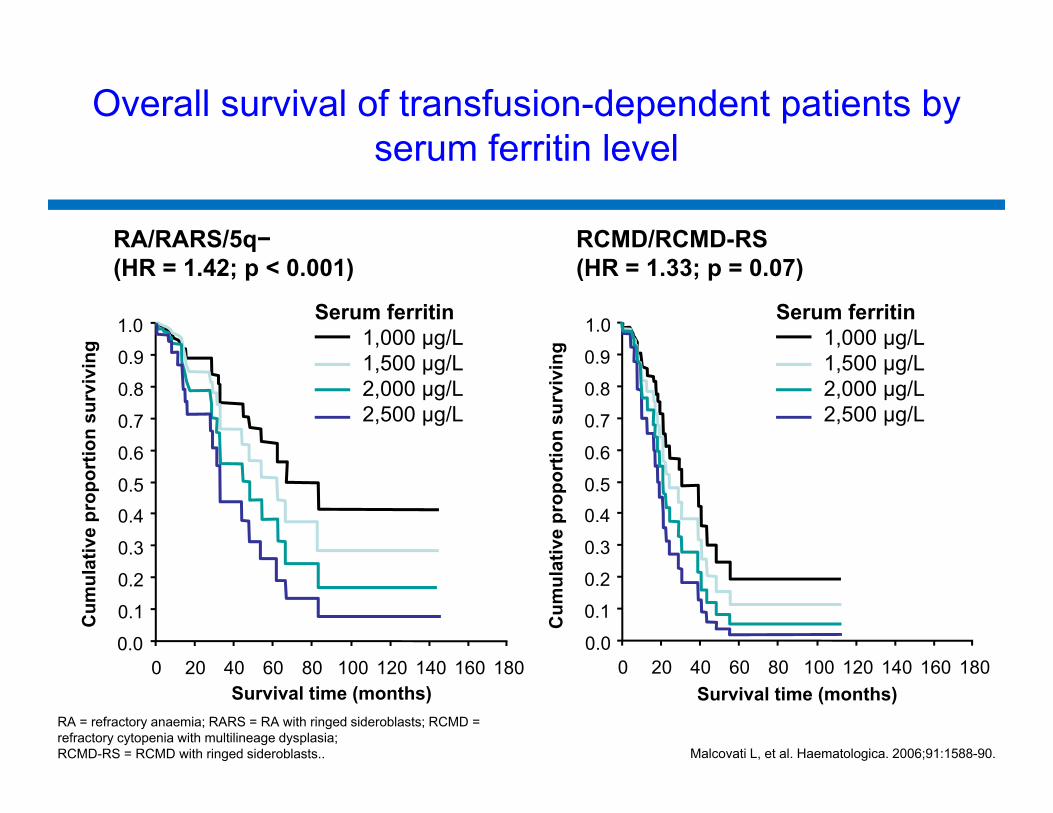
RA/RARS/5q−(HR = 1.42; p < 0.001)
RCMD/RCMD-RS(HR = 1.33; p = 0.07)
Malcovati L, et al. Haematologica. 2006;91:1588-90.
Overall survival of transfusion-dependent patients by serum ferritin level
180
Cum
ulat
ive
prop
ortio
n su
rviv
ing
0.0
0.1
0.2
0.3
0.40.5
0.6
0.7
0.8
0.9
1.0
0 20 40 60 80 100 120 140 160
Cum
ulat
ive
prop
ortio
n su
rviv
ing
0.0
0.1
0.2
0.3
0.4
0.5
0.6
0.7
0.8
0.9
1.0
0 20 40 60 80 100 120 140 160 180Survival time (months) Survival time (months)
Serum ferritin 1,000 µg/L1,500 µg/L2,000 µg/L2,500 µg/L
Serum ferritin 1,000 µg/L1,500 µg/L2,000 µg/L2,500 µg/L
RA = refractory anaemia; RARS = RA with ringed sideroblasts; RCMD = refractory cytopenia with multilineage dysplasia; RCMD-RS = RCMD with ringed sideroblasts..
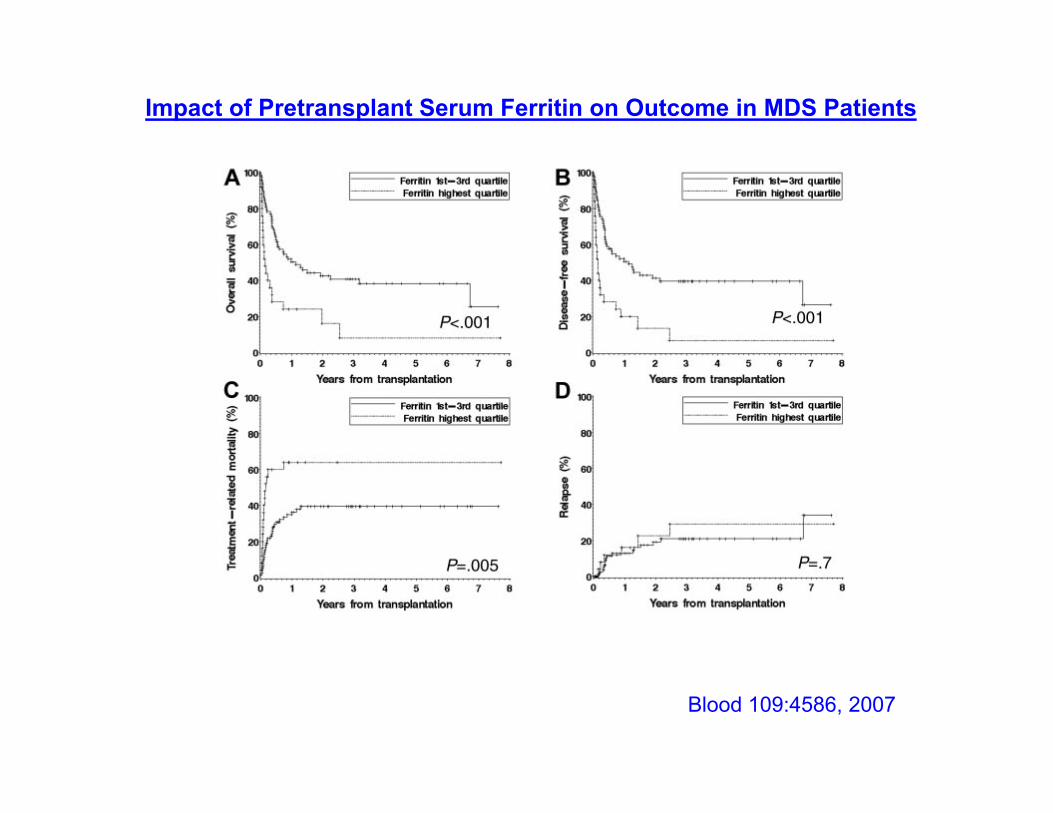
Blood 109:4586, 2007
Impact of Pretransplant Serum Ferritin on Outcome in MDS Patients
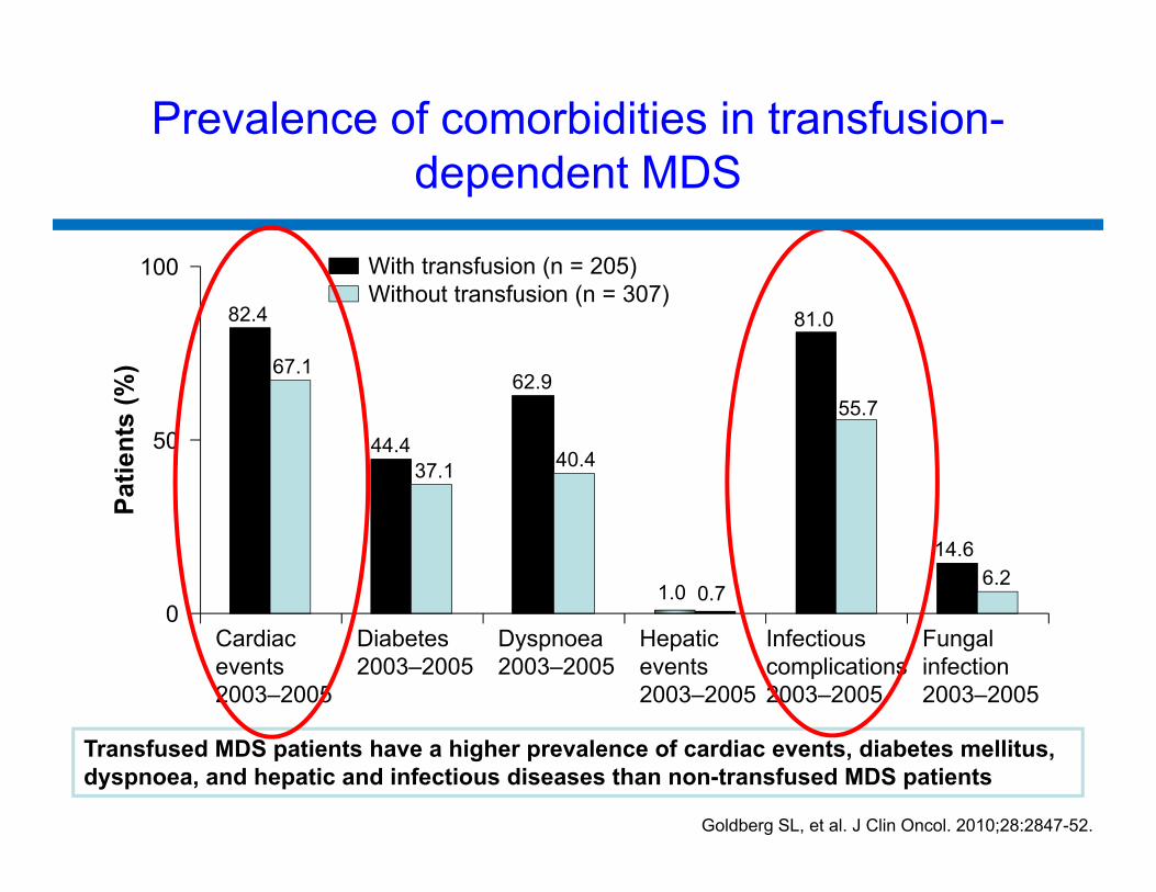
Prevalence of comorbidities in transfusion-dependent MDS
Transfused MDS patients have a higher prevalence of cardiac events, diabetes mellitus, dyspnoea, and hepatic and infectious diseases than non-transfused MDS patients
Goldberg SL, et al. J Clin Oncol. 2010;28:2847-52.
82.4
44.4
62.9
1.0
81.0
14.6
67.1
37.1 40.4
0.7
55.7
6.2
0
50
100
Cardiacevents2003–2005
Diabetes2003–2005
Dyspnoea2003–2005
Hepaticevents2003–2005
Infectiouscomplications2003–2005
Fungalinfection2003–2005
Patie
nts
(%)
With transfusion (n = 205)Without transfusion (n = 307)
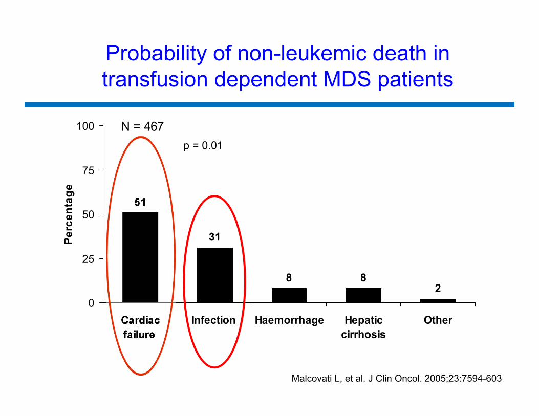
Probability of non-leukemic death in transfusion dependent MDS patients
51
31
8 82
0
25
50
75
100
Cardiacfailure
Infection Haemorrhage Hepaticcirrhosis
Other
Perc
enta
ge
N = 467p = 0.01
Malcovati L, et al. J Clin Oncol. 2005;23:7594-603
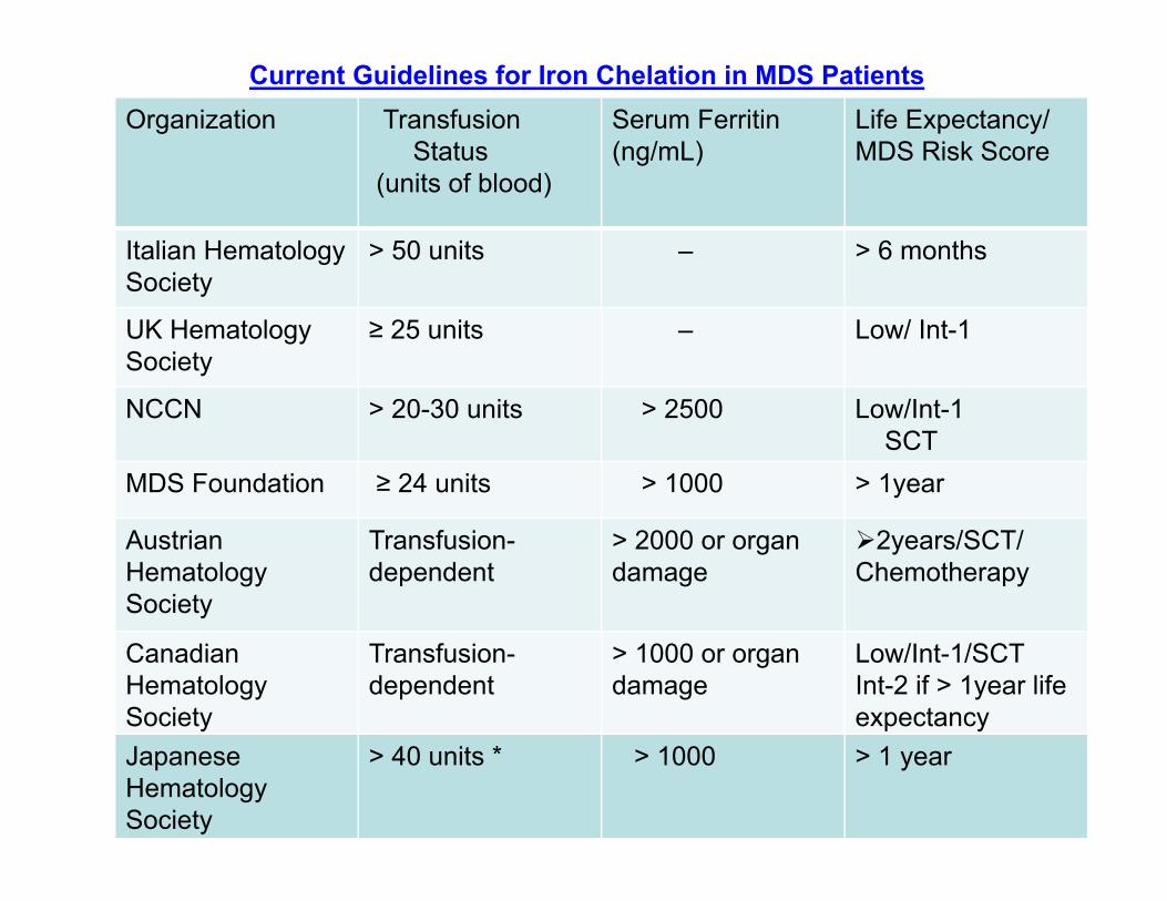
Current Guidelines for Iron Chelation in MDS PatientsOrganization Transfusion
Status (units of blood)
Serum Ferritin (ng/mL)
Life Expectancy/MDS Risk Score
Italian HematologySociety
> 50 units – > 6 months
UK Hematology Society
≥ 25 units – Low/ Int-1
NCCN > 20-30 units > 2500 Low/Int-1SCT
MDS Foundation ≥ 24 units > 1000 > 1year
Austrian Hematology Society
Transfusion-dependent
> 2000 or organdamage
2years/SCT/Chemotherapy
Canadian HematologySociety
Transfusion-dependent
> 1000 or organ damage
Low/Int-1/SCTInt-2 if > 1year life expectancy
JapaneseHematology Society
> 40 units * > 1000 > 1 year
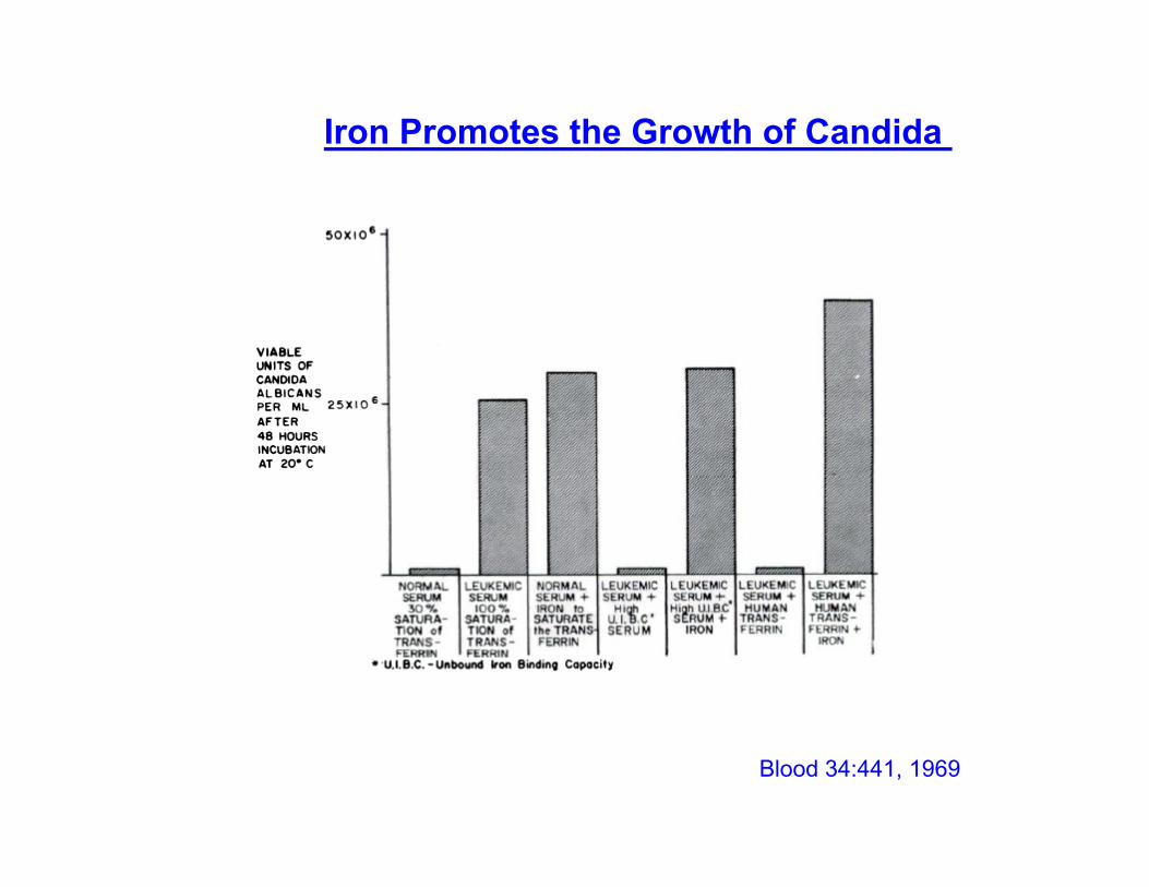
Blood 34:441, 1969
Iron Promotes the Growth of Candida
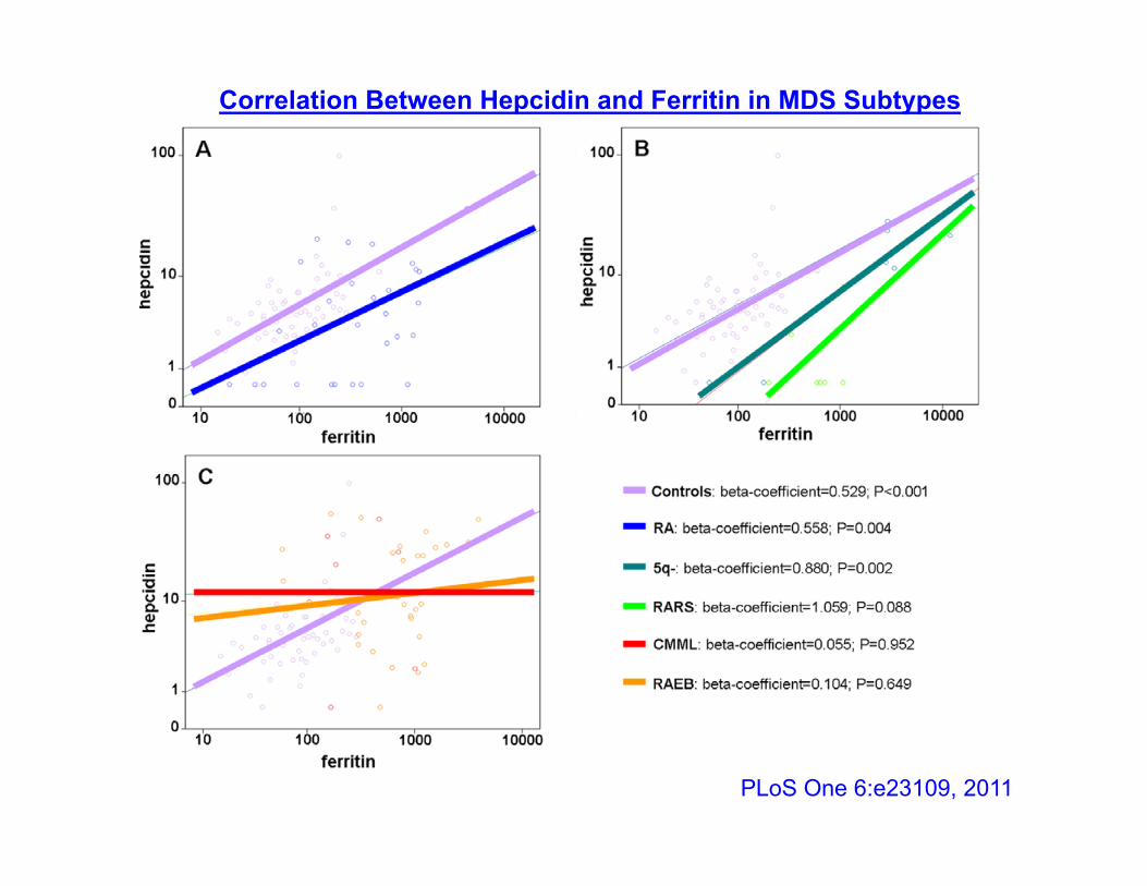
PLoS One 6:e23109, 2011
Correlation Between Hepcidin and Ferritin in MDS Subtypes
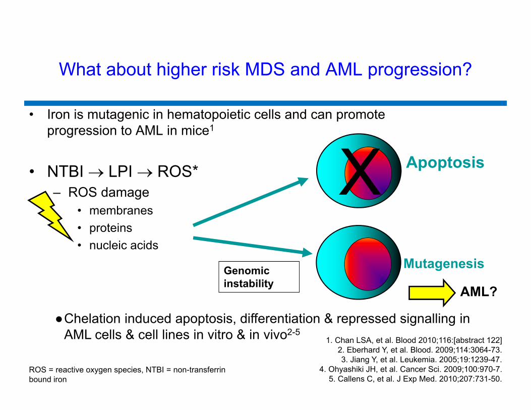
What about higher risk MDS and AML progression?
• Iron is mutagenic in hematopoietic cells and can promote progression to AML in mice1
• NTBI LPI ROS*– ROS damage
• membranes• proteins• nucleic acids
XMutagenesis
Apoptosis
ROS = reactive oxygen species, NTBI = non-transferrin bound iron
Genomic instability AML?
●Chelation induced apoptosis, differentiation & repressed signalling in AML cells & cell lines in vitro & in vivo2-5
1. Chan LSA, et al. Blood 2010;116:[abstract 122]2. Eberhard Y, et al. Blood. 2009;114:3064-73.3. Jiang Y, et al. Leukemia. 2005;19:1239-47.
4. Ohyashiki JH, et al. Cancer Sci. 2009;100:970-7.5. Callens C, et al. J Exp Med. 2010;207:731-50.
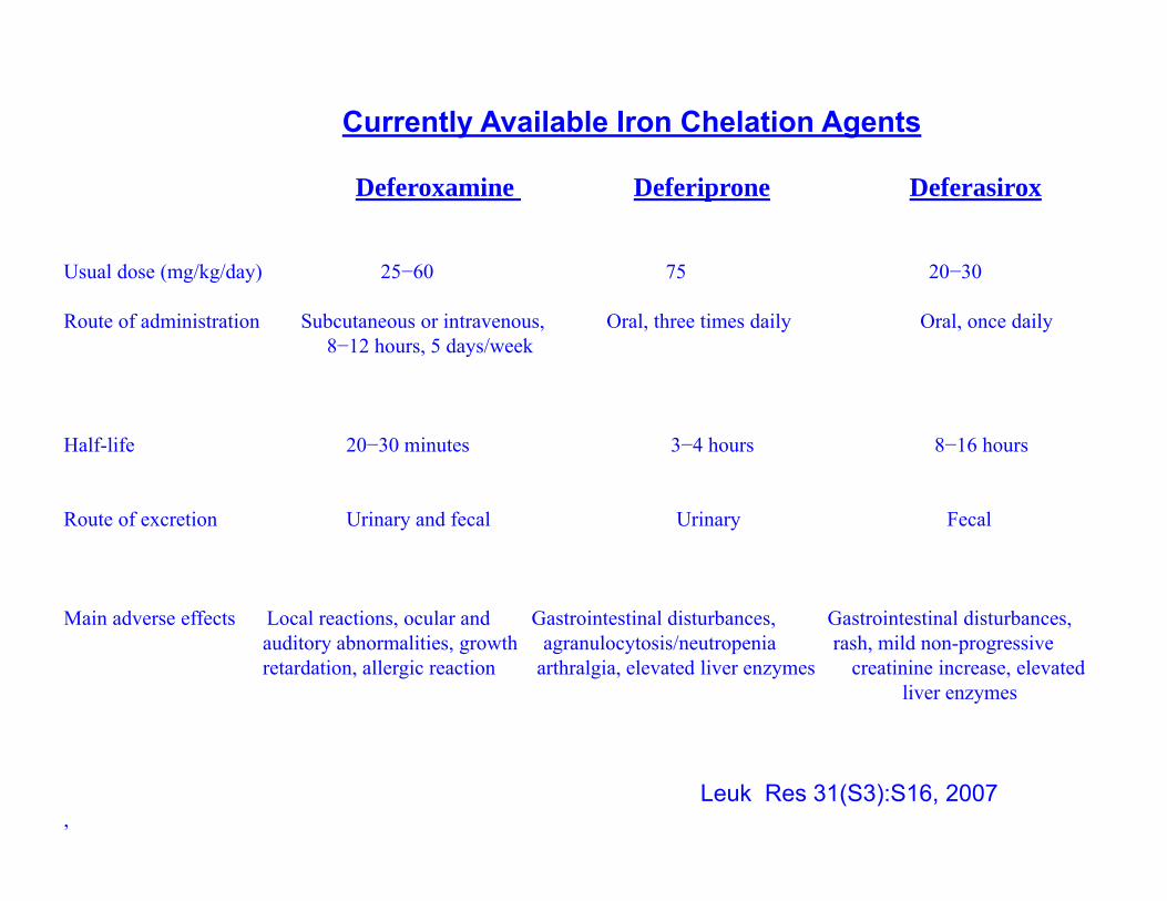
Currently Available Iron Chelation Agents
Deferoxamine Deferiprone Deferasirox
Usual dose (mg/kg/day) 25−60 75 20−30
Route of administration Subcutaneous or intravenous, Oral, three times daily Oral, once daily8−12 hours, 5 days/week
Half-life 20−30 minutes 3−4 hours 8−16 hours
Route of excretion Urinary and fecal Urinary Fecal
Main adverse effects Local reactions, ocular and Gastrointestinal disturbances, Gastrointestinal disturbances, auditory abnormalities, growth agranulocytosis/neutropenia rash, mild non-progressive retardation, allergic reaction arthralgia, elevated liver enzymes creatinine increase, elevated
liver enzymes
Leuk Res 31(S3):S16, 2007,
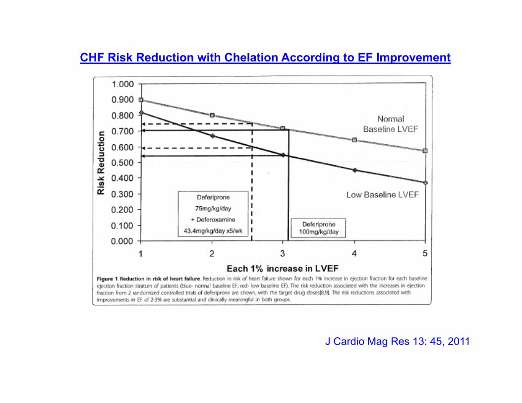
J Cardio Mag Res 13: 45, 2011
CHF Risk Reduction with Chelation According to EF Improvement
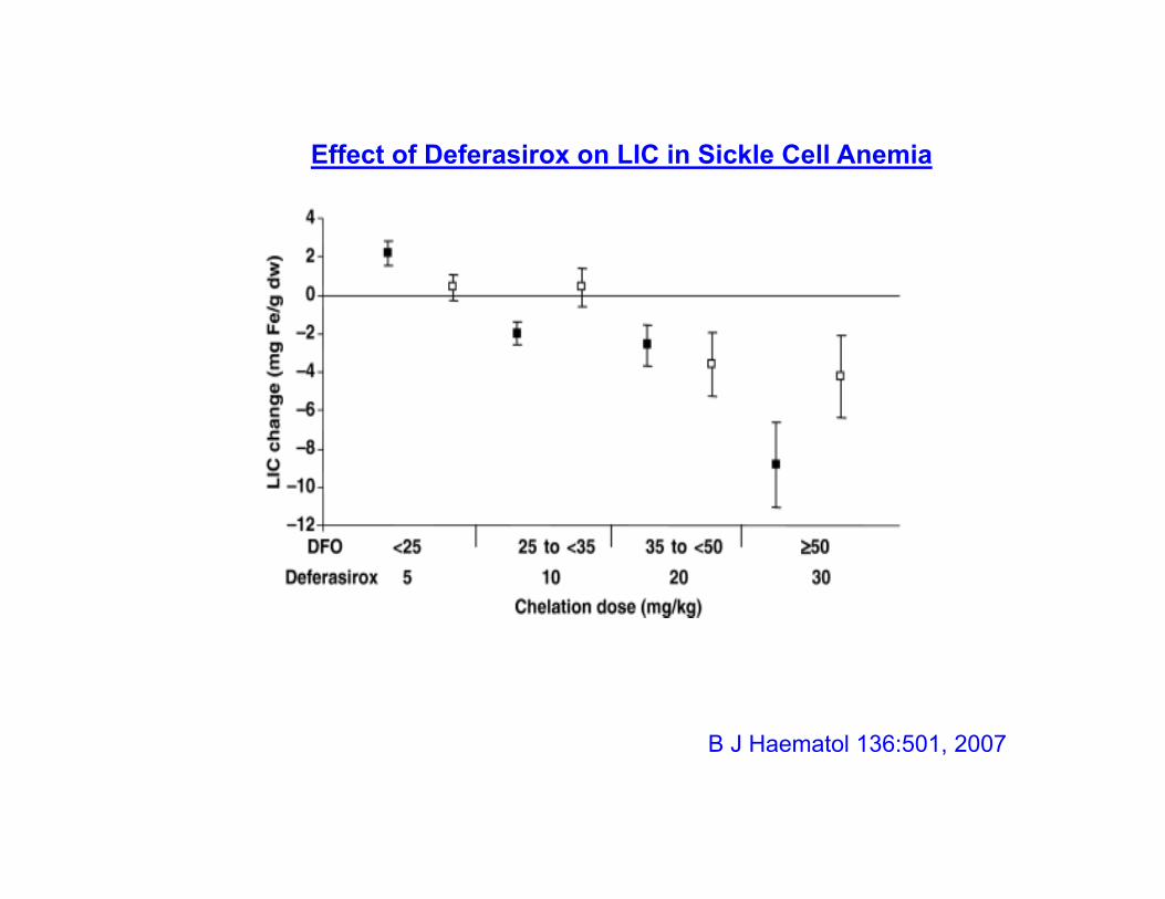
B J Haematol 136:501, 2007
Effect of Deferasirox on LIC in Sickle Cell Anemia
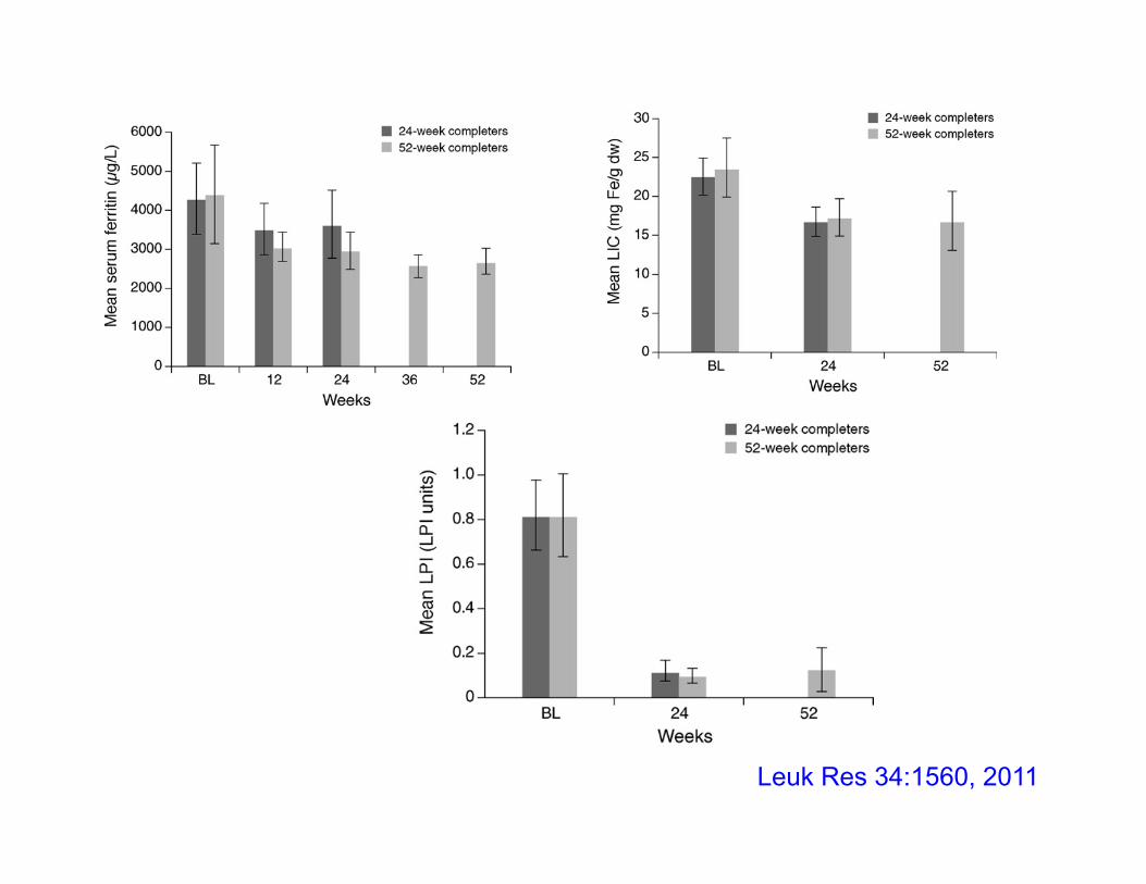
Leuk Res 34:1560, 2011
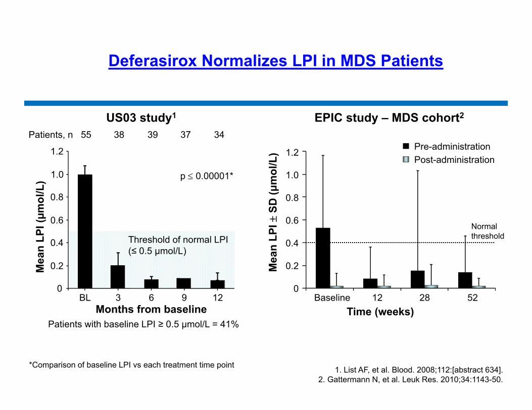
Deferasirox Normalizes LPI in MDS Patients
Pre-administrationPost-administration
0
0.2
0.4
0.6
0.8
1.0
1.2
Mea
n LP
I SD
(μm
ol/L
)
Baseline 12 28 52Time (weeks)
Normal threshold
1. List AF, et al. Blood. 2008;112:[abstract 634].2. Gattermann N, et al. Leuk Res. 2010;34:1143-50.
Patients, n 55 38 39 37 34
Patients with baseline LPI ≥ 0.5 μmol/L = 41%
Threshold of normal LPI (≤ 0.5 µmol/L)
Mea
n LP
I (µm
ol/L
)
0
0.2
0.4
0.6
0.8
1.0
1.2
BL 3 6 9 12Months from baseline
p 0.00001*
*Comparison of baseline LPI vs each treatment time point
US03 study1 EPIC study – MDS cohort2
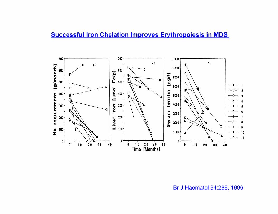
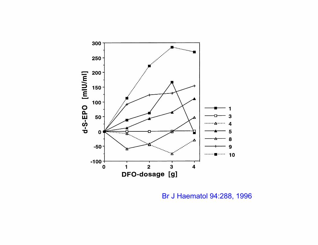
Br J Haematol 94:288, 1996
Successful Iron Chelation Improves Erythropoiesis in MDS
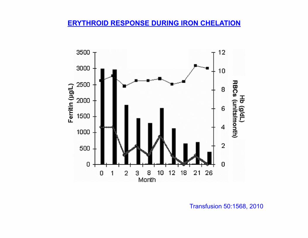
ERYTHROID RESPONSE DURING IRON CHELATION
Transfusion 50:1568, 2010
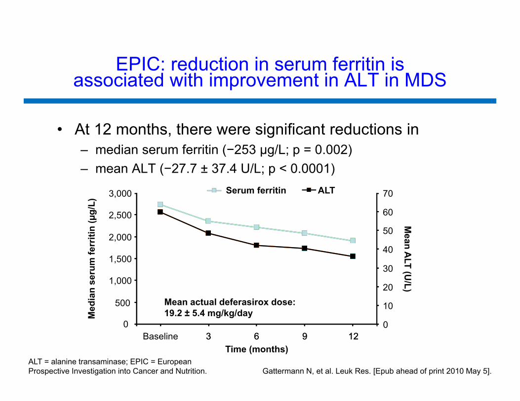
EPIC: reduction in serum ferritin is associated with improvement in ALT in MDS
• At 12 months, there were significant reductions in– median serum ferritin (−253 µg/L; p = 0.002)– mean ALT (−27.7 ± 37.4 U/L; p < 0.0001)
0
500
1,000
1,500
2,000
2,500
3,000
3 6 9 12Baseline 3 6 9 12Time (months)
Med
ian
seru
m fe
rriti
n (µ
g/L)
Mean A
LT (U/L)
ALT Serum ferritin ALT
Mean actual deferasirox dose: 19.2 ± 5.4 mg/kg/day
0
10
20
30
40
50
60
70
ALT = alanine transaminase; EPIC = European Prospective Investigation into Cancer and Nutrition. Gattermann N, et al. Leuk Res. [Epub ahead of print 2010 May 5].
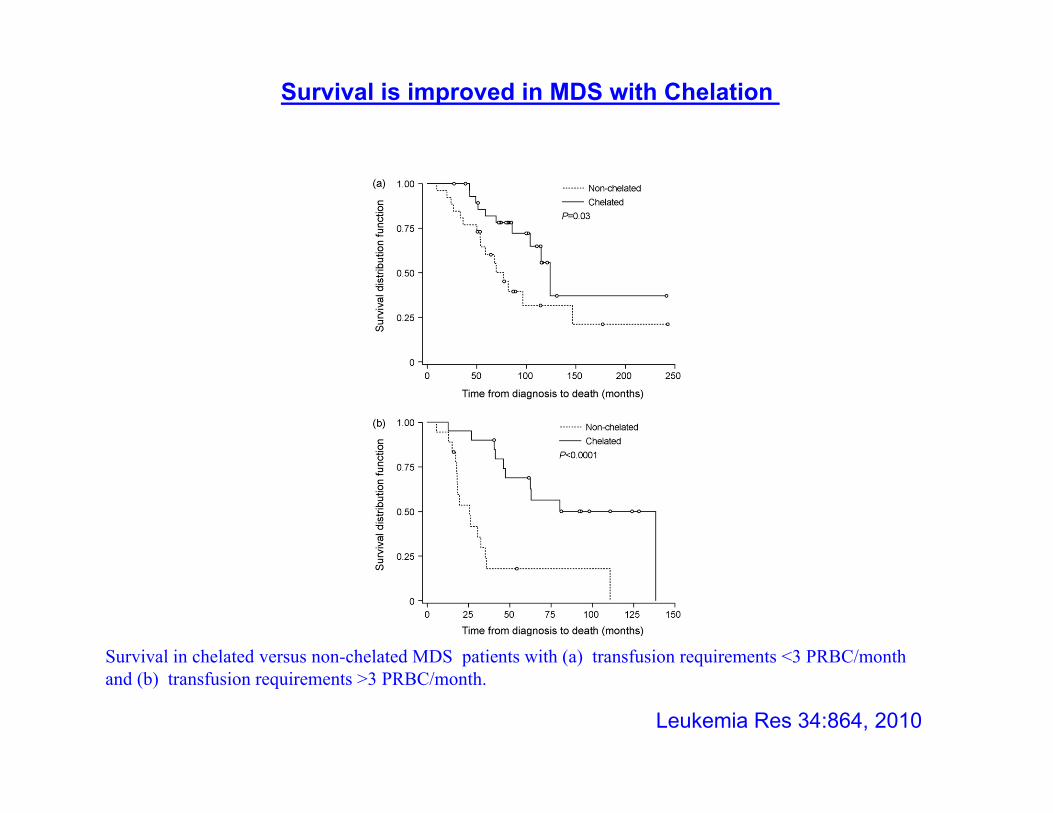
Survival in chelated versus non-chelated MDS patients with (a) transfusion requirements <3 PRBC/month and (b) transfusion requirements >3 PRBC/month.
Leukemia Res 34:864, 2010
Survival is improved in MDS with Chelation
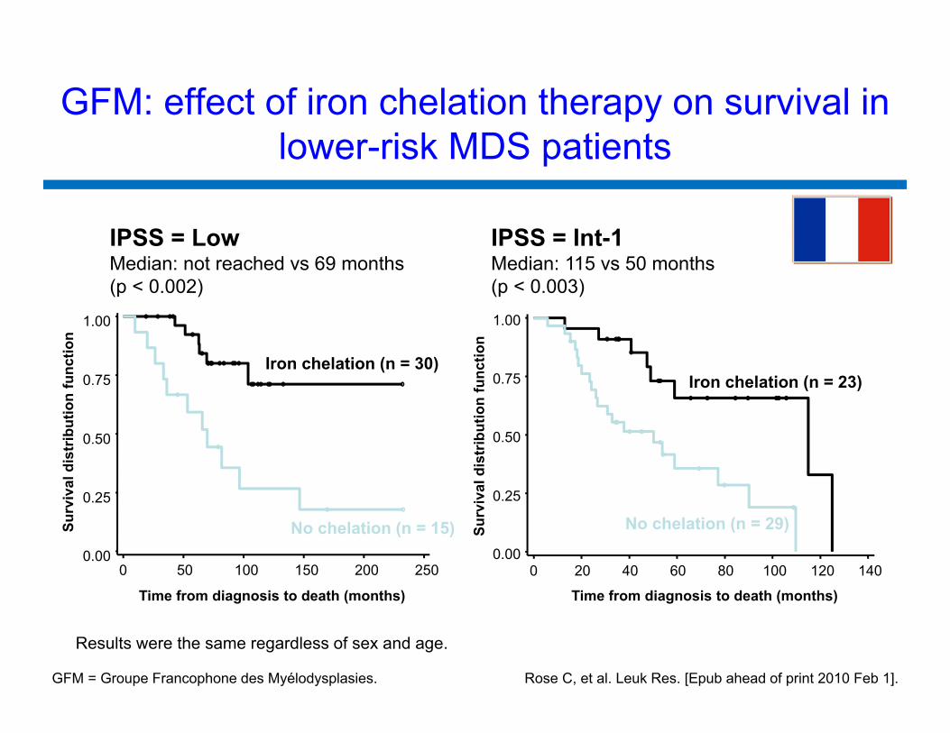
IPSS = LowMedian: not reached vs 69 months (p < 0.002)
Surv
ival
dis
trib
utio
n fu
nctio
n
0.00
0.25
0.50
0.75
1.00
Time from diagnosis to death (months)
0 50 100 150 200 250
Iron chelation (n = 30)
No chelation (n = 15) Surv
ival
dis
trib
utio
n fu
nctio
n
0.00
0.25
0.50
0.75
1.00
Time from diagnosis to death (months)
0 20 40 60 80 100 120 140
No chelation (n = 29)
IPSS = Int-1Median: 115 vs 50 months (p < 0.003)
Results were the same regardless of sex and age.
Rose C, et al. Leuk Res. [Epub ahead of print 2010 Feb 1].
Iron chelation (n = 23)
GFM: effect of iron chelation therapy on survival in lower-risk MDS patients
GFM = Groupe Francophone des Myélodysplasies.
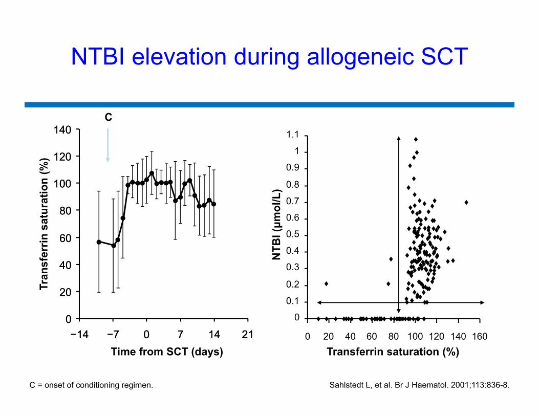
NTBI elevation during allogeneic SCT
C
Sahlstedt L, et al. Br J Haematol. 2001;113:836-8.
0
20
40
60
80
100
120
140
−14 −7 0 7 14 21
Tran
sfer
rin s
atur
atio
n (%
)
Time from SCT (days)
0
0.1
0.2
0.3
0.4
0.5
0.6
0.7
0.8
0.9
1
1.1
0 20 40 60 80 100 120 140 160
NTB
I (μm
ol/L
)
C = onset of conditioning regimen.
0
20
40
60
80
100
120
140
−14 −7 0 7 14 21Transferrin saturation (%)
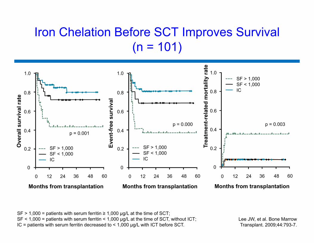
Lee JW, et al. Bone MarrowTransplant. 2009;44:793-7.
SF > 1,000SF < 1,000IC
p = 0.001
0.2
0.4
0.6
0.8
1.0
0
0 12 24 36 48 60
SF > 1,000SF < 1,000IC
p = 0.000
0 12 24 36 48 60
0.2
0.4
0.6
0.8
1.0
0
SF > 1,000SF < 1,000IC
p = 0.003
0 12 24 36 48 60
0.2
0.4
0.6
0.8
1.0
0
Ove
rall
surv
ival
rate
Even
t-fre
e su
rviv
al
Trea
tmen
t-rel
ated
mor
talit
y ra
te
Months from transplantation Months from transplantation Months from transplantation
SF > 1,000 = patients with serum ferritin ≥ 1,000 µg/L at the time of SCT;SF < 1,000 = patients with serum ferritin < 1,000 µg/L at the time of SCT, without ICT;IC = patients with serum ferritin decreased to < 1,000 µg/L with ICT before SCT.
Iron Chelation Before SCT Improves Survival (n = 101)
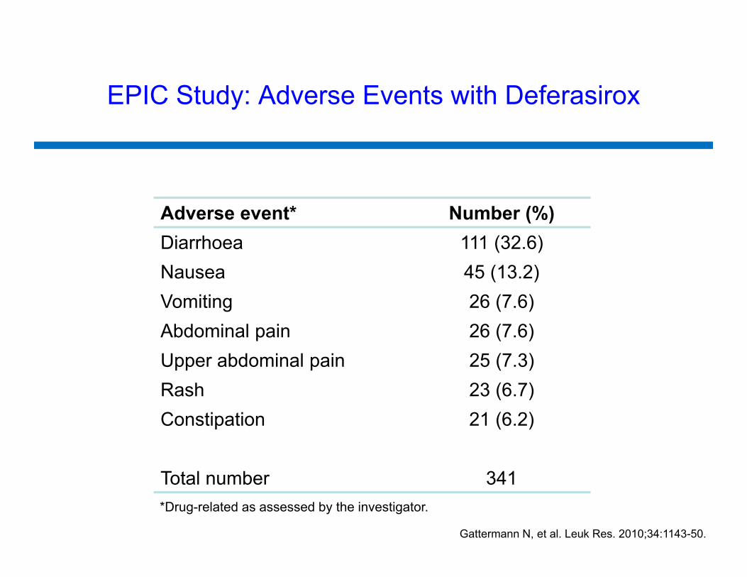
Adverse event* Number (%)Diarrhoea 111 (32.6)Nausea 45 (13.2)Vomiting 26 (7.6)Abdominal pain 26 (7.6)Upper abdominal pain 25 (7.3)Rash 23 (6.7)Constipation 21 (6.2)
Total number 341*Drug-related as assessed by the investigator.
EPIC Study: Adverse Events with Deferasirox
Gattermann N, et al. Leuk Res. 2010;34:1143-50.
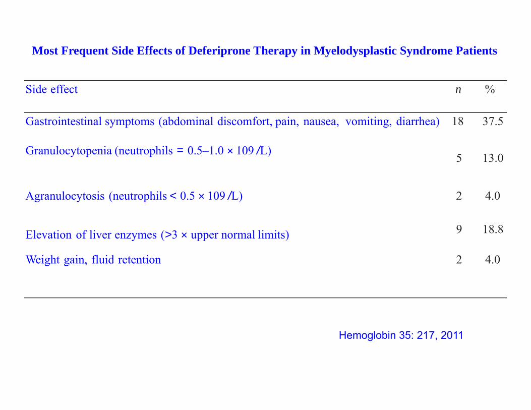
Side effect n %
Gastrointestinal symptoms (abdominal discomfort, pain, nausea, vomiting, diarrhea) 18 37.5
Granulocytopenia (neutrophils = 0.5–1.0 × 10⁹ /L) 5 13.0
Agranulocytosis (neutrophils < 0.5 × 10⁹ /L) 2 4.0
Elevation of liver enzymes (>3 × upper normal limits) 9 18.8
Weight gain, fluid retention 2 4.0
Most Frequent Side Effects of Deferiprone Therapy in Myelodysplastic Syndrome Patients
Hemoglobin 35: 217, 2011
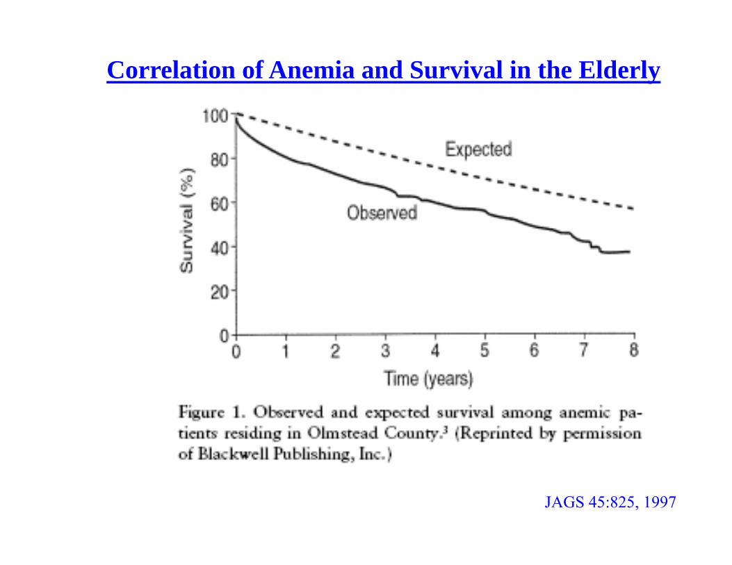
Correlation of Anemia and Survival in the Elderly
JAGS 45:825, 1997
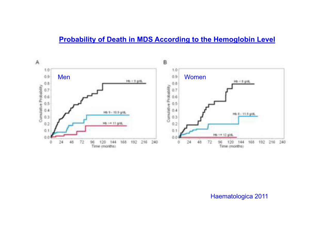
Men Women
Haematologica 2011
Probability of Death in MDS According to the Hemoglobin Level
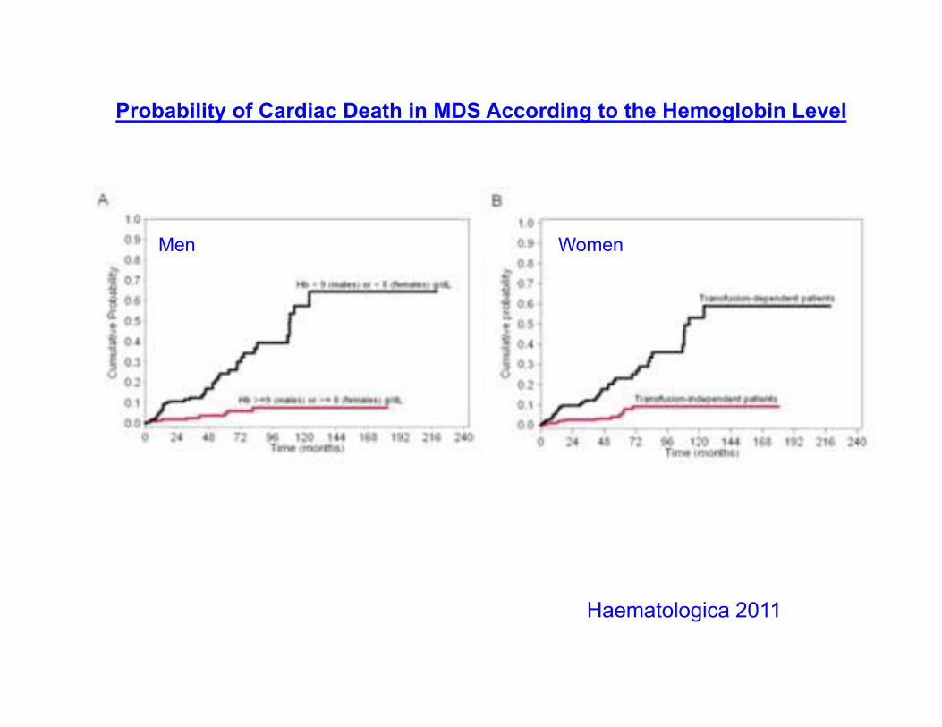
Haematologica 2011
Probability of Cardiac Death in MDS According to the Hemoglobin Level
Men Women
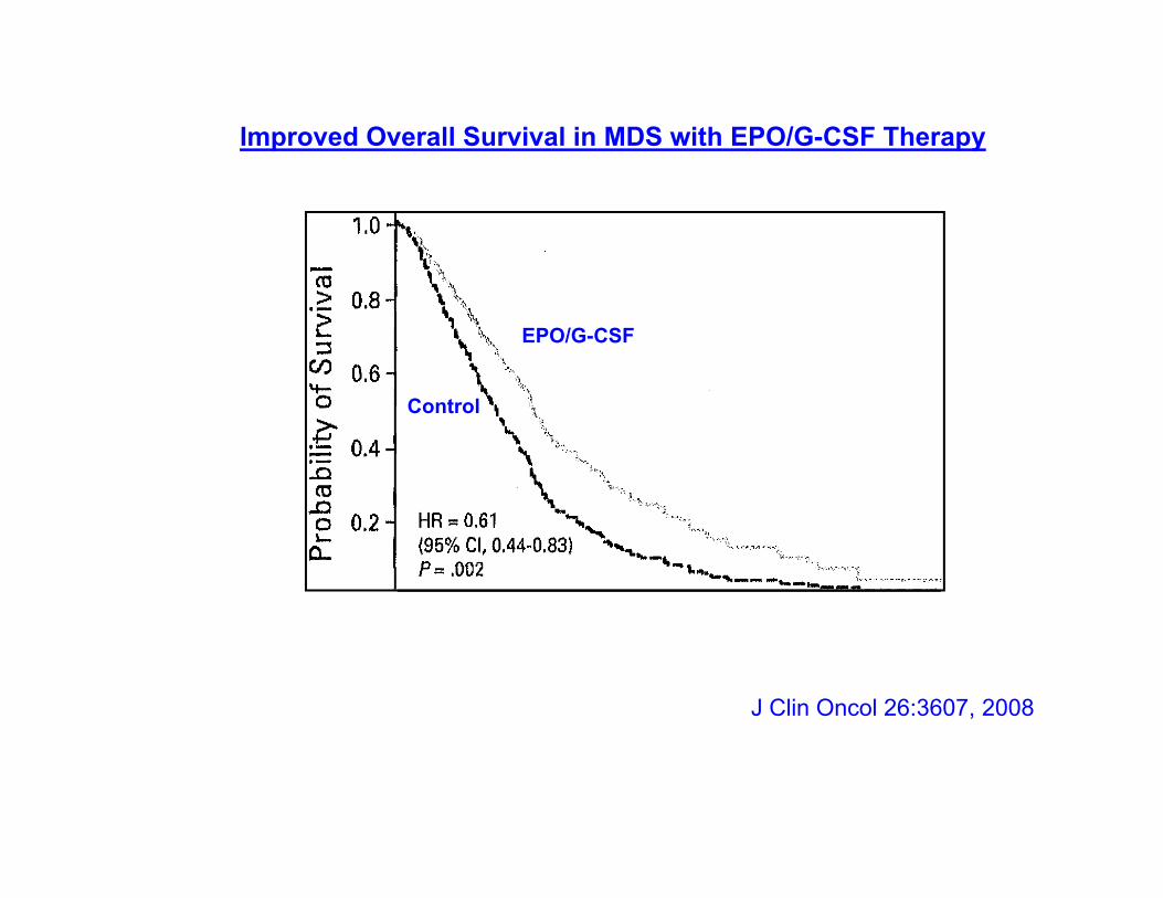
EPO/G-CSF
Control
Improved Overall Survival in MDS with EPO/G-CSF Therapy
J Clin Oncol 26:3607, 2008
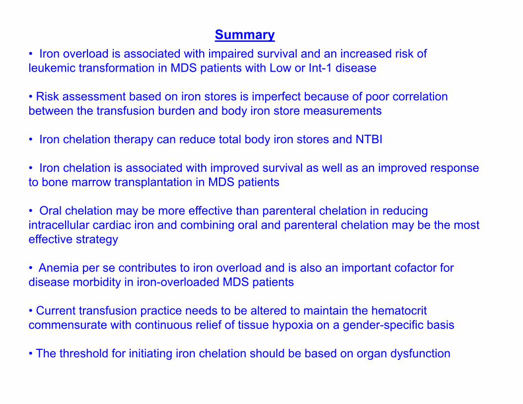
Summary• Iron overload is associated with impaired survival and an increased risk of leukemic transformation in MDS patients with Low or Int-1 disease
• Risk assessment based on iron stores is imperfect because of poor correlation between the transfusion burden and body iron store measurements
• Iron chelation therapy can reduce total body iron stores and NTBI
• Iron chelation is associated with improved survival as well as an improved response to bone marrow transplantation in MDS patients
• Oral chelation may be more effective than parenteral chelation in reducing intracellular cardiac iron and combining oral and parenteral chelation may be the most effective strategy
• Anemia per se contributes to iron overload and is also an important cofactor for disease morbidity in iron-overloaded MDS patients
• Current transfusion practice needs to be altered to maintain the hematocrit commensurate with continuous relief of tissue hypoxia on a gender-specific basis
• The threshold for initiating iron chelation should be based on organ dysfunction

Gold is for the mistress, silver for the maid,
copper for the craftsman cunning at his trade
but iron said the Baron sitting in his hall, iron,
cold iron is the master of them all.
Rudyard Kipling
Which of these Individuals is Tebowing?
A B C
Answer: