investigation of role of aspartame on apoptosis process in ... · investigation of role of...
TRANSCRIPT
Saudi Journal of Biological Sciences (2016) 23, 503–506
King Saud University
Saudi Journal of Biological Sciences
www.ksu.edu.sawww.sciencedirect.com
ORIGINAL ARTICLE
Investigation of role of aspartame on apoptosis
process in HeLa cells
* Corresponding author. Tel.: +82 10 2201 3740.
E-mail address: [email protected] (D.H. Kim).
Peer review under responsibility of King Saud University.
Production and hosting by Elsevier
http://dx.doi.org/10.1016/j.sjbs.2015.06.0011319-562X ª 2015 The Authors. Production and hosting by Elsevier B.V. on behalf of King Saud University.This is an open access article under the CC BY-NC-ND license (http://creativecommons.org/licenses/by-nc-nd/4.0/).
Muthuraman Pandurangan, Gansukh Enkhtaivan, Bhupendra Mistry,
Murugesan Chandrasekaran, Rafi Noorzai, Doo Hwan Kim *
Dept. of Bioresources & Food Science, Konkuk University, Seoul, Republic of Korea
Received 25 December 2014; revised 26 May 2015; accepted 3 June 2015
Available online 15 June 2015
KEYWORDS
Aspartame;
HeLa;
SRB;
mRNA;
p53
Abstract Aspartame is an artificial sweetener used as an alternate for sugar in several foods and
beverages. The study reports that consumption of aspartame containing product could lead to can-
cer. However, the effect of aspartame on apoptosis process in cancer is not yet understood clearly.
HeLa cells were exposed to different concentrations (0.01–0.05 mg/ml) of aspartame for 48 h.
Cytotoxicity of aspartame on cancer cells was determined by SRB assay. The result indicates no
significant changes on cell viability. Aspartame suppresses apoptosis process in cancer cells by
down-regulation of mRNA expression of tumor suppressor gene p53, and pro-apoptotic gene
bax. It up-regulates anti-apoptotic gene bcl-2 mRNA expression. In addition, Ki 67 and PCNA
mRNA, and protein expressions were determined. Taking all these together, we conclude that
aspartame may be a potent substance to slow-down the apoptosis process in HeLa cells. Further
works are ongoing to understand the biochemical and molecular mechanism of aspartame in cancer
cells.ª 2015 The Authors. Production and hosting by Elsevier B.V. on behalf of King Saud University. This is
an open access article under the CCBY-NC-ND license (http://creativecommons.org/licenses/by-nc-nd/4.0/).
1. Introduction
Aspartame is widely used as an artificial sweetener in several
foods (Magnuson et al., 2007). It is commonly found in lowcalorie beverages and desserts (Oyama et al., 2002).Chemically, it is a dipeptide of aspartyl-phenylalanine methyl
ester which facilitates its intestinal hydrolysis and absorptionof amino acids (aspartic acid and phenylalanine) together withfree methanol (Lipton et al., 1991). Aspartic acid and pheny-
lalanine are commonly found in natural proteins, which arebeneficial under normal circumstances (Woodrow, 1984).Consumption of aspartame has been linked with several neuro-
logical and behavioral disturbances (Humphries et al., 2008).However, most of the results yielded inconclusive correlations.
Aspartame is distributed under several trade names and
approved by the FDA. However, the safety of aspartamewas renewed by a report suggesting that an increase in thenumber of people with brain tumors might be associated withthe introduction and use of this sweetener in the USA. A study
reports that lymphomas and leukemias were induced in rats
504 M. Pandurangan et al.
fed with very high doses of aspartame (Soffritti et al., 2005).Saccharin is another artificial sweetener which increases inci-dence of urinary bladder cancer at high doses in male rats.
The aim of the present study was to investigate the short-term effect of aspartame on apoptosis in cancer cells.
2. Materials and methods
2.1. Materials
HeLa cells were obtained from American Type CultureCollection. DMEM and MTT were provided by Sigma–
Aldrich Chemical (St Louis, Mo). Aspartame was providedby Supelco. Fetal bovine serum (FBS), penicillin–streptomycinand trypsin–EDTA were purchased from Gibco. Primers of
p53, bax, bcl-2, Ki 67 and PCNA were purchased fromMacro Gen (South Korea). Primary antibodies were pur-chased from Bioscience Technology (Seoul, South Korea).
2.2. Methods
2.2.1. SRB assay for cell viability
The effect of aspartame on HeLa cells was measured by theSRB assay (Vichai and Kirtikara, 2006). HeLa cells wereseeded at a density of 2.2 · 104 cells/well into 96-well plates
and allowed to adhere for 24 h at 37 �C. Then the cells weretreated with aspartame at different concentrations (0.01–0.05 mg/ml) for 48 h. At the end of the treatments, cells were
fixed with acetone and air dried. After fully dried, each wellwas added with 100% of SRB solution (0.4% w/v) and incu-bated for 3 h at room temperature. Microplate was washedwith 1% of acetic acid and then dried under drying oven.
Stained HeLa cells were photographed. Finally, 10 mm ofTris-base was added and kept overnight. Following complete
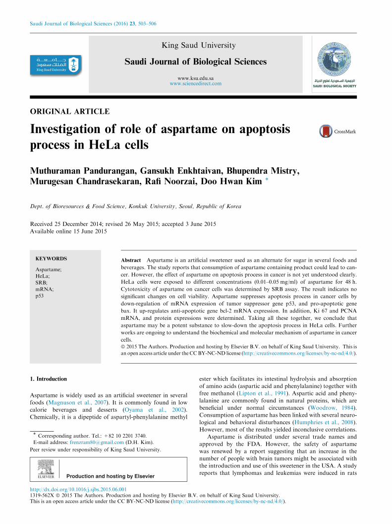
Figure 1 Effects of various concentrations of aspartame on cell viab
into 96 well micro plates and allowed to adhere for 24 h and treated w
dissolution of SRB in Tris-base, the absorbance was measuredat 540 nm.
2.2.2. qPCR
HeLa cells were cultured in T75 flask and treated with aspar-tame (0.01–0.03 mg/ml) for 48 h. Total RNA was isolatedfrom the control and aspartame treated samples
(Chomczynski and Mackey, 1995). The first-strand c-DNAwas synthesized from 1 lg of the total RNA using the M-MLV reverse transcriptase with the anchored oligo d(T)12–18primer. qPCR was performed using a cDNA equivalent of10 ng of total RNA from each sample with primers specificfor p53, bax, bcl-2, Ki 67, PCNA and a housekeeping gene
GAPDH. The reaction was carried out in 10 ll using SYBRGreen Master Mix (Bioneer) according to the manufacturers’instructions. Relative ratios were calculated based on the
2�DDCT method (Pfaffl, 2001). PCR was monitored using theCFX96� Real-Time System (Bio-Rad).
2.2.3. Western blot analysis
HeLa cells were cultured in T75 flask and treated with aspar-tame (0.03 mg/ml) for 48 h. Control and aspartame treatedHeLa cells were washed three times with ice-cold PBS and
lysed with 10 mM Tris–HCl (pH 7.5), 100 mM NaCl, 1%NP-40, 50 mM NaF, 2 mM EDTA (pH 8.0), 1 mM PMSF,10 lg/ml leupeptin, 10 lg/ml aprotinin. Equal amounts oflysate protein samples were run on SDS polyacrylamide gel,
and then transferred onto a PVDF membrane. Non-specificbinding was blocked by soaking the membrane in Tris bufferedsaline-Tween (TBST) buffer that contained 5% nonfat dry
milk for 1 h. The membrane was probed overnight with anantibody against Ki 67 and PCNA. Following washing withTBST buffer, the membrane was incubated for 1 h with horse-
radish peroxidase conjugated goat anti-rabbit IgG. The
ility. HeLa cells were seeded at seeding densities of 2 · 104 cells/ml
ith 0.01–0.05 mg/ml of aspartame for 48 h.
Aspartame and cancer 505
protein levels of Ki 67 and PCNA were determined by usingenhanced chemiluminescence kit (Muthuraman et al., 2014).
3. Results and discussion
Aspartame is one of the artificial sweeteners, used in a widevariety of foods and beverages. It was first approved by the
US Food and Drug Administration (FDA) for use in solid food
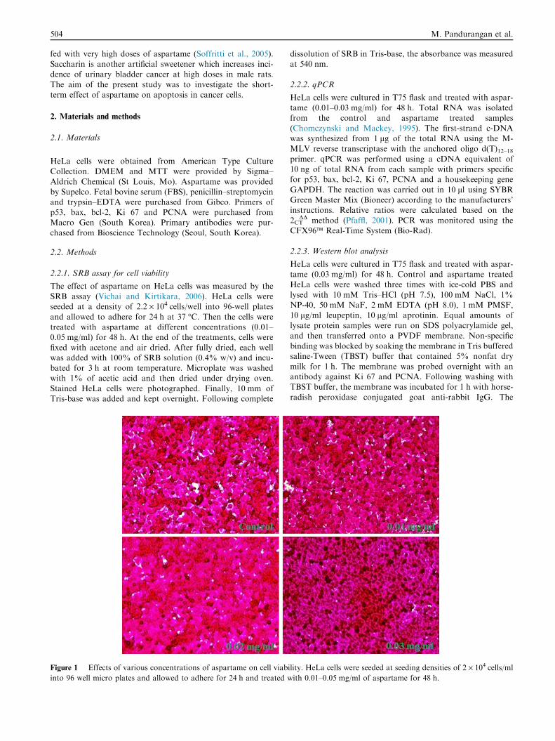
Figure 2 Expression of p53, bax and bcl-2 in the HeLa cells. HeL
Expressions of p53, bax and bcl-2 mRNA are related to GAPDH and
mean ± SEM, n= 6. *p < 0.05.
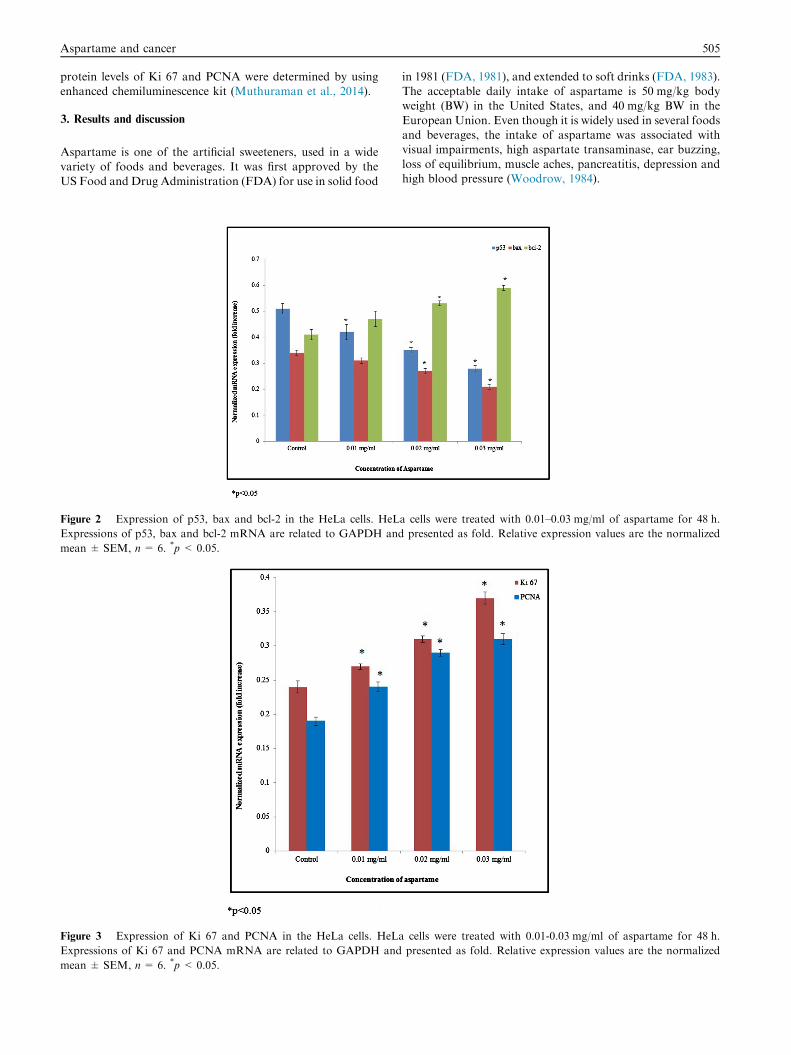
Figure 3 Expression of Ki 67 and PCNA in the HeLa cells. HeLa
Expressions of Ki 67 and PCNA mRNA are related to GAPDH and
mean ± SEM, n= 6. *p < 0.05.
in 1981 (FDA, 1981), and extended to soft drinks (FDA, 1983).The acceptable daily intake of aspartame is 50 mg/kg bodyweight (BW) in the United States, and 40 mg/kg BW in the
European Union. Even though it is widely used in several foodsand beverages, the intake of aspartame was associated withvisual impairments, high aspartate transaminase, ear buzzing,
loss of equilibrium, muscle aches, pancreatitis, depression andhigh blood pressure (Woodrow, 1984).
a cells were treated with 0.01–0.03 mg/ml of aspartame for 48 h.
presented as fold. Relative expression values are the normalized
cells were treated with 0.01-0.03 mg/ml of aspartame for 48 h.
presented as fold. Relative expression values are the normalized
506 M. Pandurangan et al.
HeLa cells were exposed to different concentrations ofaspartame for 48 h. Aspartame in concentrations of 0.01–0.05 mg/ml showed no significant changes in cancer cells vali-
dated by the SRB assay (Fig. 1). HeLa cells exposed to differ-ent concentrations of aspartame for 48 h showed significantchanges in the mRNA expression of apoptotic genes such as
p53, bax, and bcl-2.The mRNA expression level of tumor suppressor gene p53
and pro-apoptotic gene bax was significantly down-regulated.
The mRNA expression of p53 was significantly reduced 0.17,0.31 and 0.45 folds at 0.01, 0.02 and 0.03 mg/ml of aspartametreatment respectively. The mRNA expression of bax was sig-nificantly reduced 0.09, 0.2 and 0.38 folds at 0.01, 0.02 and
0.03 mg/ml of aspartame treatment respectively, whereas bcl-2 mRNA expression significantly upregulated 0.14, 0.29 and0.44 folds at 0.01, 0.02 and 0.03 mg/ml of aspartame treatment
respectively (Fig. 2, *p < 0.05). The mRNA expression of Ki67 was significantly upregulated 0.12, 0.29 and 0.54 folds at0.01, 0.02 and 0.03 mg/ml of aspartame treatment respectively.
The mRNA expression of PCNA was significantly upregulated0.26, 0.52 and 0.63 folds at 0.01, 0.02 and 0.03 mg/ml of aspar-tame treatment respectively (Fig. 3, *p< 0.05). Protein expres-
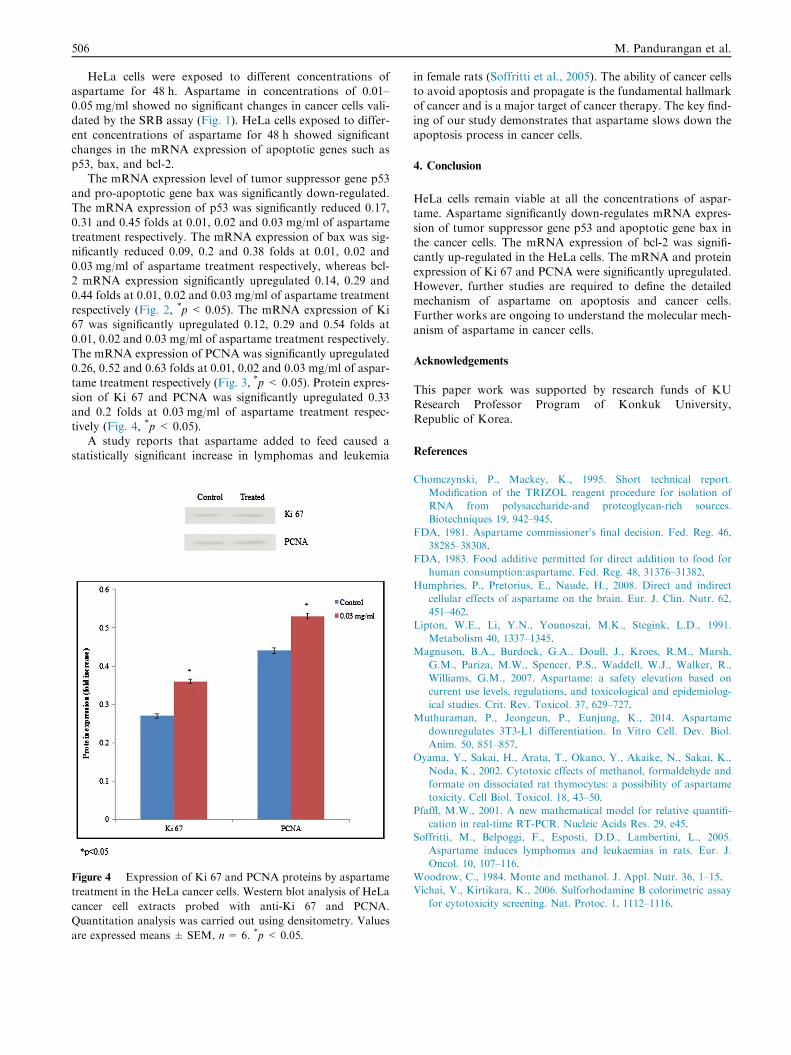
sion of Ki 67 and PCNA was significantly upregulated 0.33and 0.2 folds at 0.03 mg/ml of aspartame treatment respec-tively (Fig. 4, *p< 0.05).
A study reports that aspartame added to feed caused a
statistically significant increase in lymphomas and leukemia
Figure 4 Expression of Ki 67 and PCNA proteins by aspartame
treatment in the HeLa cancer cells. Western blot analysis of HeLa
cancer cell extracts probed with anti-Ki 67 and PCNA.
Quantitation analysis was carried out using densitometry. Values
are expressed means ± SEM, n= 6. *p < 0.05.
in female rats (Soffritti et al., 2005). The ability of cancer cellsto avoid apoptosis and propagate is the fundamental hallmarkof cancer and is a major target of cancer therapy. The key find-
ing of our study demonstrates that aspartame slows down theapoptosis process in cancer cells.
4. Conclusion
HeLa cells remain viable at all the concentrations of aspar-tame. Aspartame significantly down-regulates mRNA expres-
sion of tumor suppressor gene p53 and apoptotic gene bax inthe cancer cells. The mRNA expression of bcl-2 was signifi-cantly up-regulated in the HeLa cells. The mRNA and protein
expression of Ki 67 and PCNA were significantly upregulated.However, further studies are required to define the detailedmechanism of aspartame on apoptosis and cancer cells.
Further works are ongoing to understand the molecular mech-anism of aspartame in cancer cells.
Acknowledgements
This paper work was supported by research funds of KUResearch Professor Program of Konkuk University,
Republic of Korea.
References
Chomczynski, P., Mackey, K., 1995. Short technical report.
Modification of the TRIZOL reagent procedure for isolation of
RNA from polysaccharide-and proteoglycan-rich sources.
Biotechniques 19, 942–945.
FDA, 1981. Aspartame commissioner’s final decision. Fed. Reg. 46,
38285–38308.
FDA, 1983. Food additive permitted for direct addition to food for
human consumption:aspartame. Fed. Reg. 48, 31376–31382.
Humphries, P., Pretorius, E., Naude, H., 2008. Direct and indirect
cellular effects of aspartame on the brain. Eur. J. Clin. Nutr. 62,
451–462.
Lipton, W.E., Li, Y.N., Younoszai, M.K., Stegink, L.D., 1991.
Metabolism 40, 1337–1345.
Magnuson, B.A., Burdock, G.A., Doull, J., Kroes, R.M., Marsh,
G.M., Pariza, M.W., Spencer, P.S., Waddell, W.J., Walker, R.,
Williams, G.M., 2007. Aspartame: a safety elevation based on
current use levels, regulations, and toxicological and epidemiolog-
ical studies. Crit. Rev. Toxicol. 37, 629–727.
Muthuraman, P., Jeongeun, P., Eunjung, K., 2014. Aspartame
downregulates 3T3-L1 differentiation. In Vitro Cell. Dev. Biol.
Anim. 50, 851–857.
Oyama, Y., Sakai, H., Arata, T., Okano, Y., Akaike, N., Sakai, K.,
Noda, K., 2002. Cytotoxic effects of methanol, formaldehyde and
formate on dissociated rat thymocytes: a possibility of aspartame
toxicity. Cell Biol. Toxicol. 18, 43–50.
Pfaffl, M.W., 2001. A new mathematical model for relative quantifi-
cation in real-time RT-PCR. Nucleic Acids Res. 29, e45.
Soffritti, M., Belpoggi, F., Esposti, D.D., Lambertini, L., 2005.
Aspartame induces lymphomas and leukaemias in rats. Eur. J.
Oncol. 10, 107–116.
Woodrow, C., 1984. Monte and methanol. J. Appl. Nutr. 36, 1–15.
Vichai, V., Kirtikara, K., 2006. Sulforhodamine B colorimetric assay
for cytotoxicity screening. Nat. Protoc. 1, 1112–1116.