introduction to pathology/ cell injuryjumed16.weebly.com/uploads/8/8/5/1/88514776/lecture_4.pdf ·...
TRANSCRIPT
INTRODUCTION TO PATHOLOGY/ cell injury
Dr Heyam Awad
MD, FRCPATH, Jordanian Board
LECTURE 4
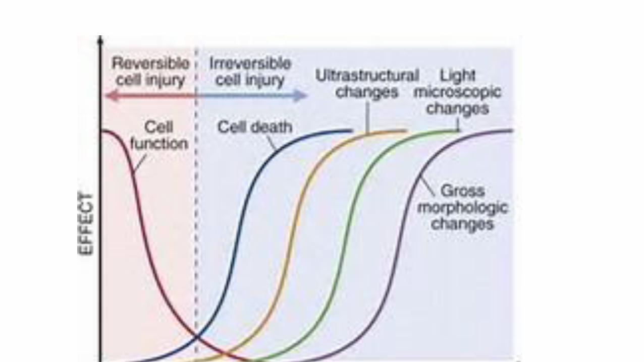
Morphology of cell injury
• The first effect of all injuries is on the biochemical and molecular level
• Functional derangement happens next
• Ultrastructural changes seen by electron microscopy follow
• Then light microscopic changes occur
• The last visible change is at the gross; macroscopic level.
example
• In heart attack= myocardial infarction ,first change is at the molecular level: so cardiac enzymes increase in the blood. Example troponin
• Then cardiac muscle becomes non contractile (loss of function). This happens within 1-2 minutes of ischemia… note that loss of function doesn't mean cell death
• Cells die within 20-20 minutes of ischemia
• It takes 2-3 hours to see any EM changes
• Light microscopic changes need 6-12 hours to be noted
• Macroscopic changes are the last to be seen; within 12-24 hours
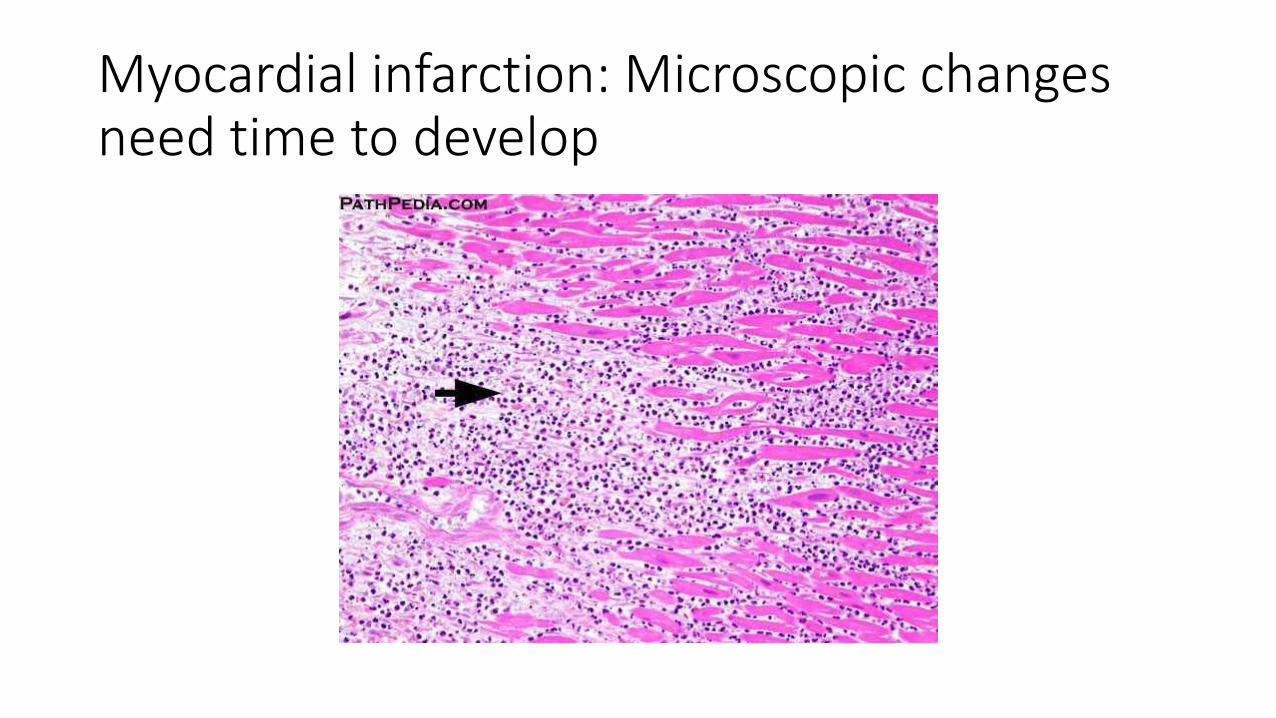
Myocardial infarction: Microscopic changes need time to develop
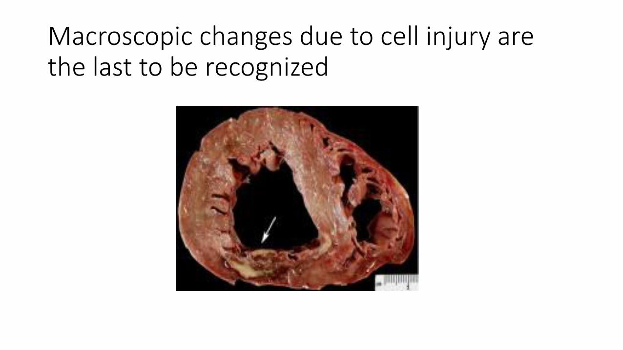
Macroscopic changes due to cell injury are the last to be recognized
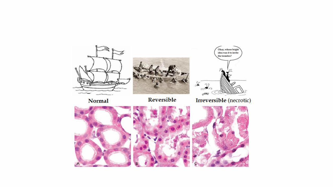
Morphology of reversible cell injury
• The main two morphologic changes in reversible cell injury are:
• 1. cellular swelling
• 2. fatty change
swelling
• Results from failure of the sodium potassium pump due to ATP depletion
• It is the first manifestation of all forms of injury
• It is reversible
• The organ affected will have increased weight
• Microscopy shows small clear vacuoles within the cytoplasm this is called hydropic change or vacular degeneration
• The organelles within the cells are also swollen
Fatty change
• Occurs mainly in hypoxic injury and in toxic and metabolic injury.
• Microscopy: lipid vacuoles in the cytoplasm
• Seen mainly in cells that participate in fat metabolism like hepatocytes and myocardial cells
• It is reversible.
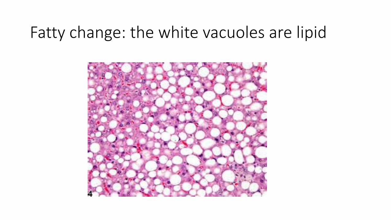
Fatty change: the white vacuoles are lipid
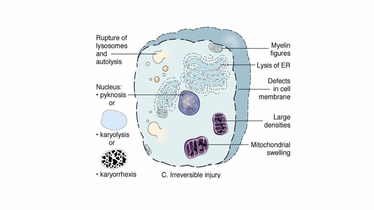
Electron microscopic changes in reversible injury • Plasma membrane blebbing
• Mitochondrial swelling with appearance of amorphous densities
• Dilation of endoplasmic reticulum with detachment of ribosomes and dissociation with polysomes
• Nuclear changes with clumping of chromatin.. This happens due to changes in pH and can be reversible.
• Formation of phospholipid aggregates called myelin figures which are derived from damaged cellular membranes
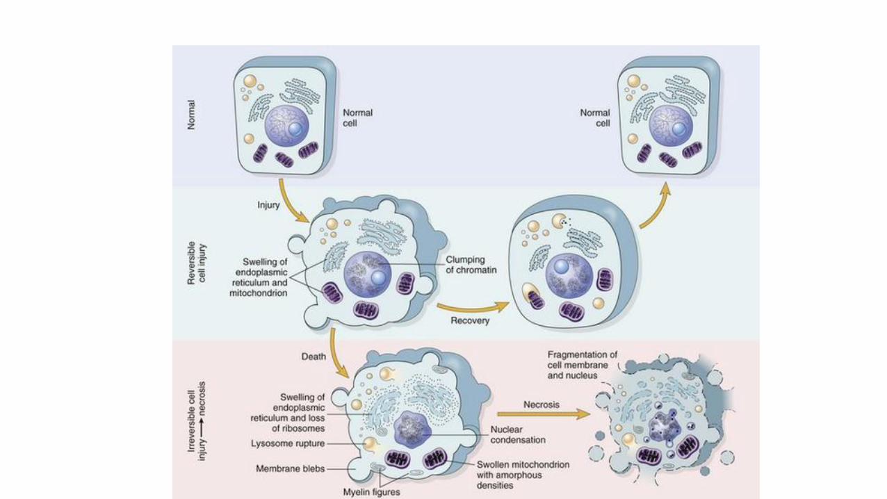
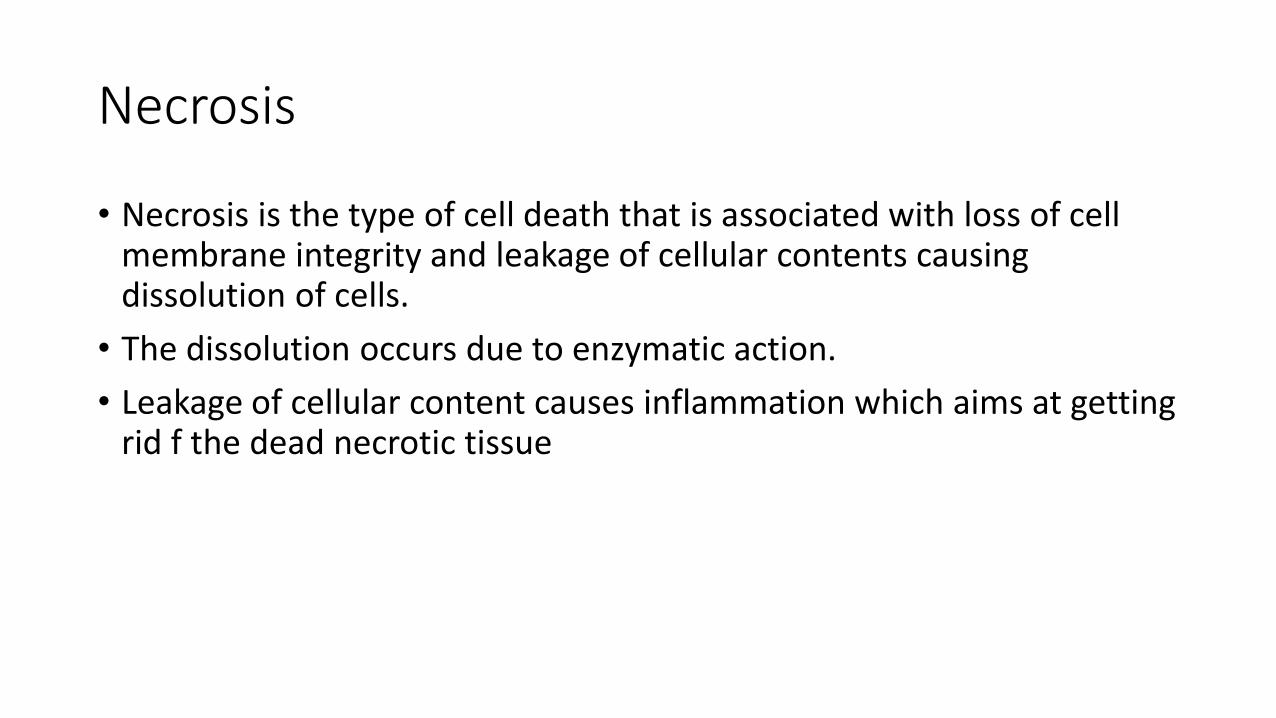
Necrosis
• Necrosis is the type of cell death that is associated with loss of cell membrane integrity and leakage of cellular contents causing dissolution of cells.
• The dissolution occurs due to enzymatic action.
• Leakage of cellular content causes inflammation which aims at getting rid f the dead necrotic tissue
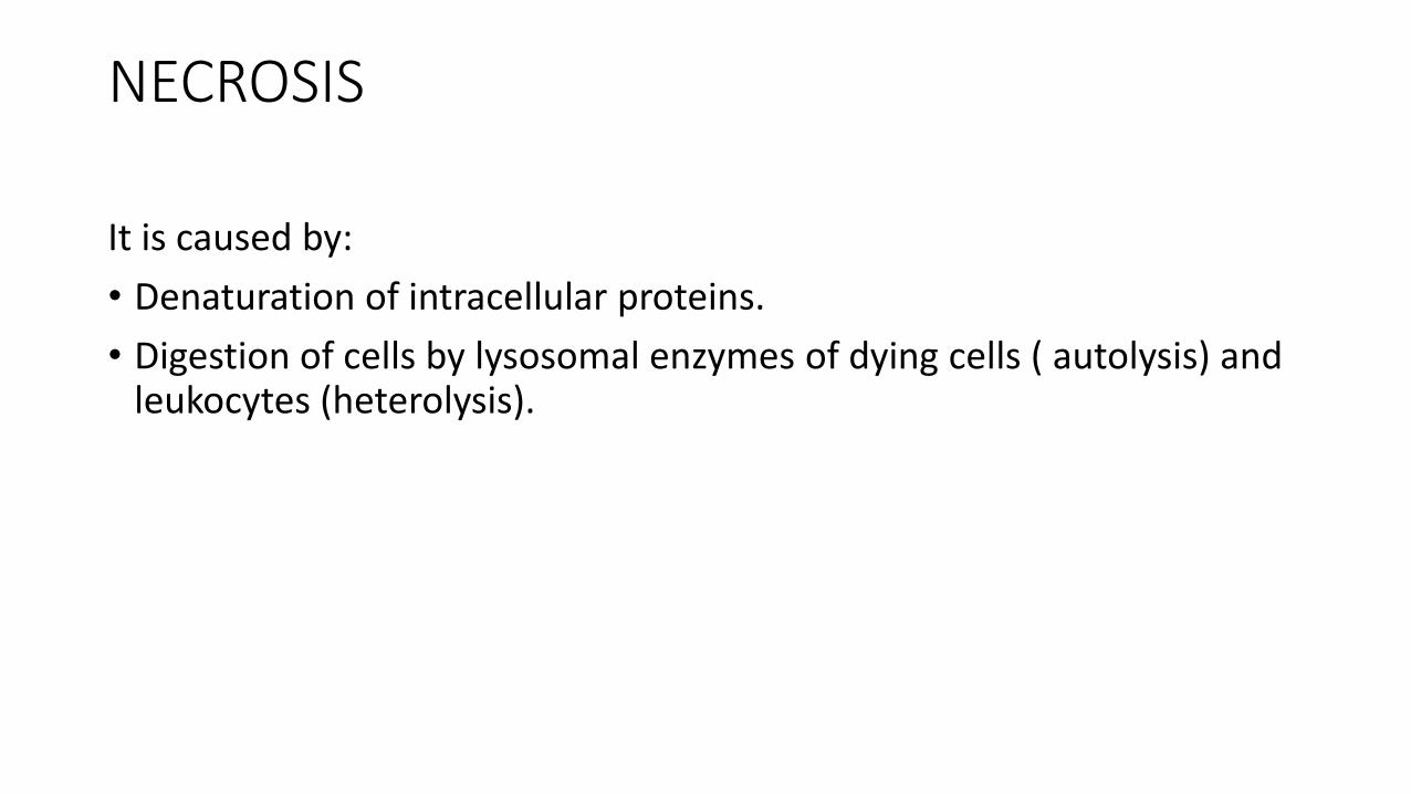
NECROSIS It is caused by:
• Denaturation of intracellular proteins.
• Digestion of cells by lysosomal enzymes of dying cells ( autolysis) and leukocytes (heterolysis).
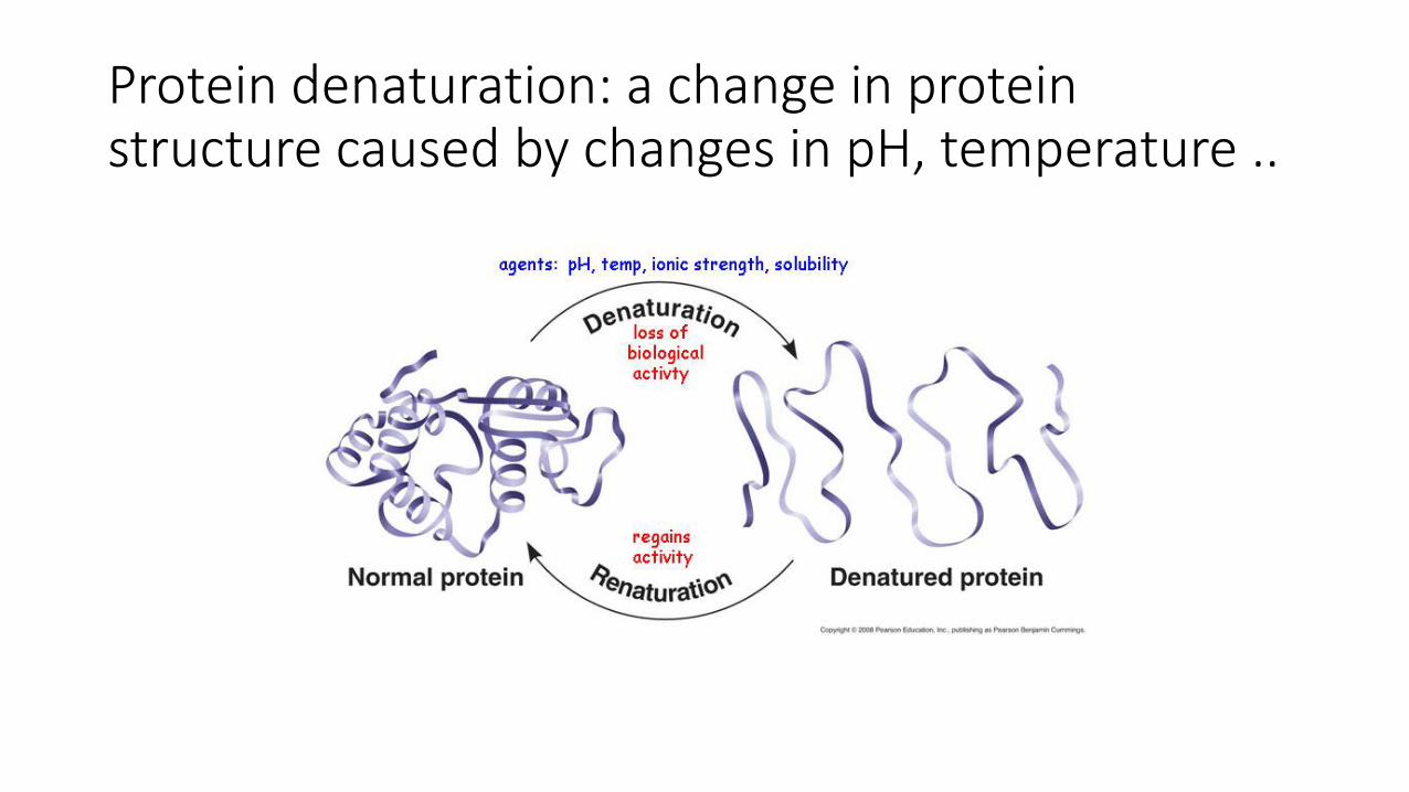
Protein denaturation: a change in protein structure caused by changes in pH, temperature ..
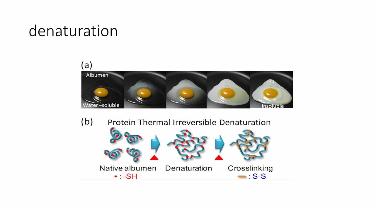
denaturation
Morphology of necrosis
• There are cytoplasmic and nuclear changes.
• Cytoplasmic changes: increased eosinophilia (pink staining from the eosin dye).
• Increased eosinophilia is due to 1. increased binding of eosin to dentures proteins and 2. to loss of basophilia (decreases hematoxylin binding to RNA, because RNA in the cytoplasm decreases.
• Due to these changes cells appear homogenous and glassy
• Myelin figures can be a nidus for calcium deposition and calcifications might occur.
Nuclear changes in necrosis one of three patterns
1. karyolysis: decreased chromatin basophilia secondary to deoxyribonuclease (DNAase) activity.
2. pyknosis: nuclear shrinkage and increased basophilia (DNA condenses into a solid shrunken mass.
3. karyorrhexis, fragmentation then disappearance of nucleus.
EM changes
1)Discontinuities in plasma and organelle
membranes.
2) Marked dilation of mitochondria and large amorphous densities.
3)Disruption of lysosomes.
4) Intracytoplasmic myelin figures
Myelin figures
• Myelin figures: aggregates of damaged cell membranes (phospholipids).
Fate of Myelin figures:
• phagocytosed by other cells
• or further degraded into fatty acids and calcify
Patterns of necrosis
• Denaturation of protein predominates…. Coagulative necrosis.
• Enzymatic digestion predominates… liquefactive necrosis.
• Special circumstances: caseous necrosis and fat necrosis.
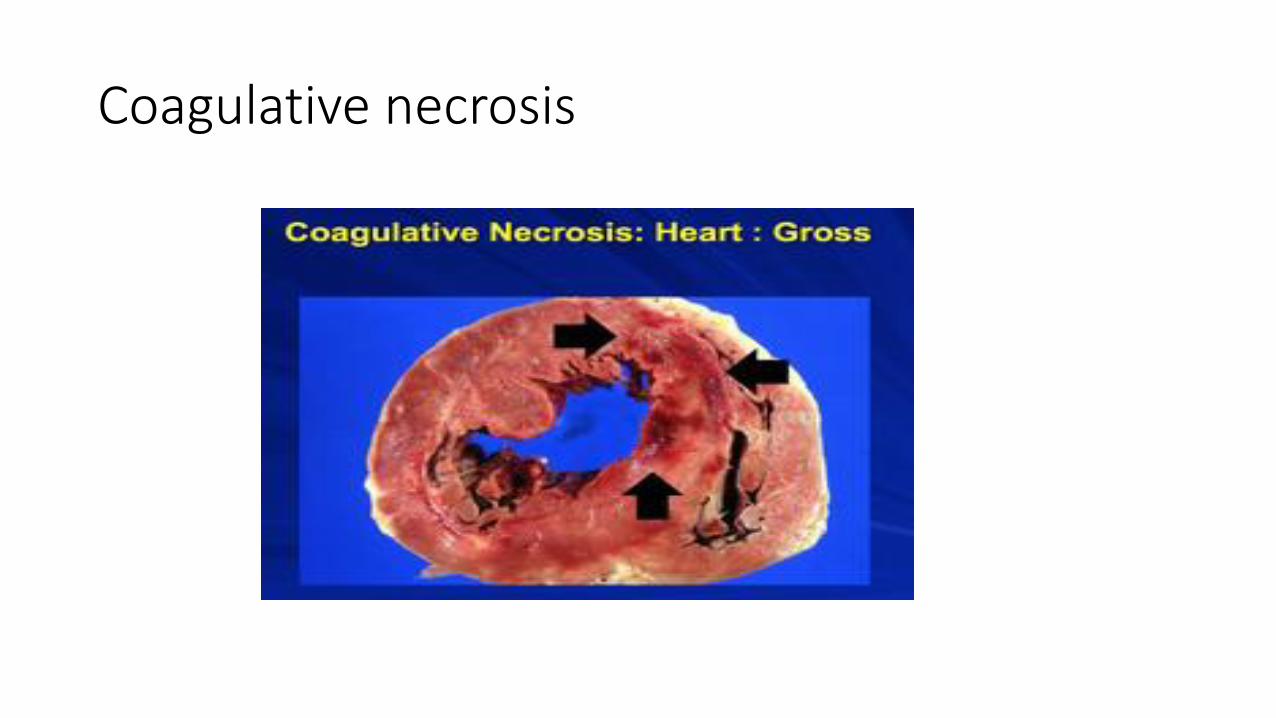
Coagulative necrosis
• preserved architecture of dead tissue .
• Denaturation of structural proteins and enzymes… so no cellular proteolysis.
• Eosiniphilic anucleated cells
• Cells are removed by inflammatory cells.
• Ischemia in all solid organs except the brain may lead to coagulative necrosis.
Coagulative necrosis
Liquifactive necrosis
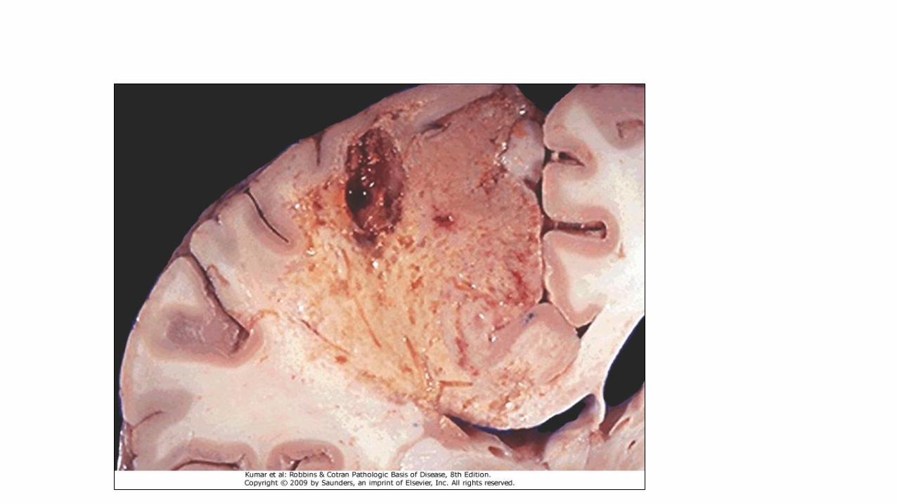
• digestion of the dead cells resulting into a liquid jelly-like mass.
• In focal bacterial or fungal infections and in hypoxic death in central nervous system.
Caseous necrosis
• White cheese like friable necrosis.
• Prototype: Tuberculosis
• Typical finding is granuloma :Collection of fragmented or lysed cells with amorphous granular eosinophilic debris surrounded by macrophages.
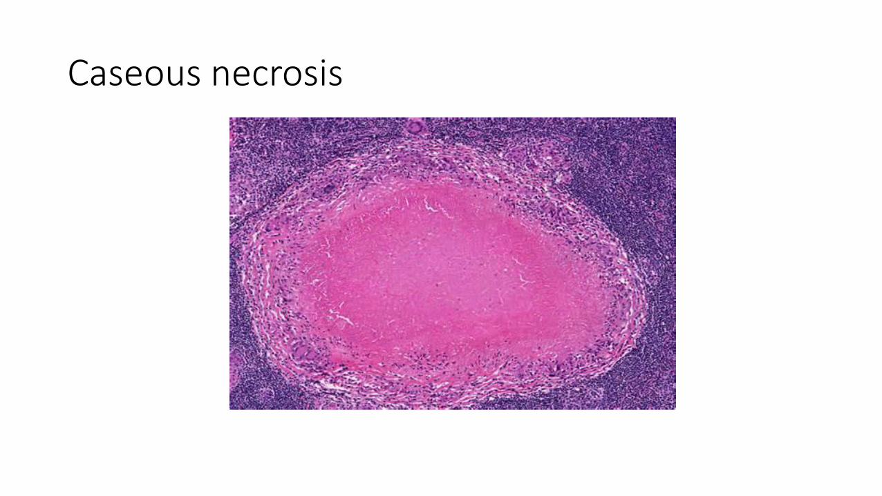
Caseous necrosis
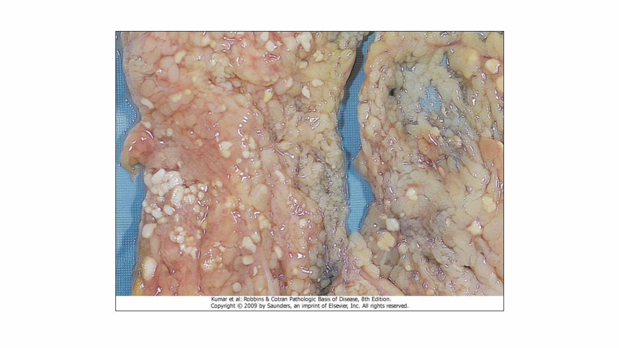
Fat necrosis
• used as a clinical terms and not a specific type.
• Necrosis of fat.
• Typical example: pancreatic enzymes (lipases) release in acute pancreatitis.
Fate of necrotic tissue • Phagocytosis.
• Replacement by scar.
• Regeneration.
• Calcification.