intratracheal exposure to multi-walled carbon nanotubes induces a nonalcoholic steatohepatitis-like...
TRANSCRIPT
http://informahealthcare.com/nanISSN: 1743-5390 (print), 1743-5404 (electronic)
Nanotoxicology, Early Online: 1–11! 2014 Informa UK Ltd. DOI: 10.3109/17435390.2014.963186
ORIGINAL ARTICLE
Intratracheal exposure to multi-walled carbon nanotubes induces anonalcoholic steatohepatitis-like phenotype in C57BL/6J mice
Ji-Eun Kim1,2, Somin Lee1,3, Ah Young Lee1, Hwi Won Seo4, Chanhee Chae4, and Myung-Haing Cho1,2,3,5
1Laboratory of Toxicology, BK21 PLUS Program for Creative Veterinary Science Research, Research Institute for Veterinary Science and College of
Veterinary Medicine, Seoul National University, Seoul, Korea, 2Graduate School of Convergence Science and Technology, Seoul National University,
Suwon, Korea, 3Graduate Group of Tumor Biology, Seoul National University, Seoul, Korea, 4Laboratory of Veterinary Pathology, College of
Veterinary Medicine, Seoul National University, Seoul, Korea, and 5Advanced Institute of Convergence Technology, Seoul National University,
Suwon, Korea
Abstract
The effects of multi-walled carbon nanotubes (MWCNTs) exposure have garnered great interestin the field of public health, due to the high aspect ratio of MWCNTs. Because of worldwideincreases in obesity prevalence, nonalcoholic fatty liver disease (NAFLD) is now the mostcommon prevalent liver disease and is considered to be a component of metabolic syndrome,which is a cluster of disorders that also includes dyslipidemia, diabetes mellitus, arteriosclerosis,and hypertension. Exposure to MWCNTs is known to be a risk factor for lung and cardiovasculardiseases, but its effect on NAFLD is unknown. In this study, we investigated the effects ofintratracheal exposure of two different types of MWCNTs, namely, pristine multi-walled carbonnanotubes (PMWCNTs) and acid-treated multi-walled carbon nanotubes (TMWCNTs), on liverpathogenesis. Direct instillation of a test material into the lungs has been employed as aquantitatively reliable alternative method of inhalation exposure. The 10% weight loss dosewas assessed in three months of subchronic study and is defined here as the maximumtolerated dose (MTD) of PMWCNTs and TMWCNTs; by this metric, MTD for a 1-year exposure ofMWCNTs was determined to be 0.1 mg/mouse. Mice exposed to PMWCNTs and TMWCNTs forone year developed a nonalcoholic steatohepatitis (NASH)-like phenotype, characterized byinflammation, hepatic steatosis, and fibrosis. Furthermore, PMWCNTs induced a more severeNASH-like phenotype than TMWCNTs, which was related to consistent up-regulation ofinterleukin (IL)-6 and plasminogen activator inhibitor (PAI)-1. Impaired cholesterol homeostasis,overexpression of NF-kBp65, and suppression of peroxisome proliferator-activated receptorgamma (PPARg) in the liver were also observed.
Keywords
Cholesterol, multi-walled carbon nanotubes,nonalcoholic fatty liver disease, risk factor
History
Received 3 February 2014Revised 4 September 2014Accepted 4 September 2014Published online 29 September 2014
Introduction
Concerns over unanticipated effects of multi-walled carbonnanotubes (MWCNTs) have been emphasized in recent studies,due to their massive use in industrial fields (Berber et al., 2000;De Volder et al., 2013; Hu et al., 2010). Recent reports havedemonstrated that exposure to fine particulate matter is closelyrelated to the onset of cardiovascular disease, nonalcoholic fattyliver disease (NAFLD), and metabolic disease in tissues that werenot directly exposed (Chen & Schwartz, 2008; Kramer et al.,2010; Sun et al., 2009). NAFLD is rapidly becoming one of themost common chronic liver diseases in industrialized countriesdue to the growing prevalence of obesity (Della Corte et al.,2013), but limited data are available concerning environmentalrisk factors associated with NAFLD progression.
NAFLD is a spectrum of nonalcoholic liver diseases rangingfrom simple triglyceride accumulation in hepatocytes (hepaticsteatosis), known as nonalcoholic fatty liver (NAFL), to nonalco-holic steatohepatitis (NASH, steatosis with inflammation), fibro-sis, and irreversible cirrhosis (Brunt, 2001). Day and James (1998)
characterized the progression of NAFLD using a ‘‘two-hit’’ model.Steatosis is the ‘‘first hit’’, which increases the vulnerability ofthe liver to diverse ‘‘second hits’’ induced by oxidative stress,inflammatory cytokines, endotoxins, saturated fatty acids, or otherliver injuries. These factors in turn promote the ensuing inflam-mation, cell death, and fibrosis that are the key histologic hallmarksof NASH (Day & James, 1998).
Recently, studies with round particulate matter have shownthat inhaled particles can activate Kupffer cells in liver tissue,suggesting that these inhaled particles play a role for as riskfactors for NASH progression (Laing et al., 2010; Tan et al., 2009;Zheng et al., 2013). In the case of MWCNTs, Ji et al. (2009)demonstrated the hepatotoxicity of intravenously (i.v.) adminis-tered MWCNTs 15 and 60 days after exposure, describinglymphatic infiltration and oxidative damage, in addition toreporting that carboxy-modified MWCNTs are less hepatotoxicthan pristine MWCNTs (Ji et al., 2009). Although other studies onhepatic inflammation due to MWCNTs have been reported(Jain et al., 2011; Patlolla et al., 2011), these studies used i.v.MWCNT administration, rather than inhalation exposure, andonly short-term exposure periods of less than two months wereconsidered. These reports on short-term i.v. MWCNT exposuredid not describe pronounced modulatory effects of exposure onhepatic pathways associated with metabolic disease, but reported
Correspondence: Myung-Haing Cho, Laboratory of Toxicology, Collegeof Veterinary Medicine, Seoul National University, Seoul 151–742, Korea.Tel: +822 880 1276. Fax: +822 8731268. E-mail: [email protected]
Nan
otox
icol
ogy
Dow
nloa
ded
from
info
rmah
ealth
care
.com
by
Uni
vers
ity o
f O
tago
on
10/0
6/14
For
pers
onal
use
onl
y.
inflammatory responses and up-regulation of toxicity relatedserum enzymes (Patlolla et al., 2011). Consequently, there is alack of data on long-term direct exposure (more than one year) toMWCNTs through the lungs in relation to NASH.
In this study, we used tracheal instillation to expose the lungsof mice to defined doses of MWCNTs and evaluated histopath-ology, as well as metabolic and inflammatory pathways, in thelivers one year after exposure to PMWCNTs and TMWCNTs.We demonstrated that exposure to PMWCNTs and TMWCNTscaused a NASH-like phenotype and impaired cholesterol homeo-stasis in mice. Through comparison of the effects of PMWCNTsand TMWCNTs, we revealed differences between PMWCNT- andTMWCNT-induced NASH phenotypes and related signaltransduction.
Methods
Multi-walled carbon nanotube preparation
MWCNTs with varied physicochemical characteristics wereprepared for use as previously reported (Kim et al., 2010). Aspurchased PMWCNTs, CM-95� (purity 495%, diameter:12.5 ± 2.5 nm, length: 13.0 ± 1.5mm), synthesized by chemicalvapor deposition (CVD) were purchased from Hanhwa Nanotech(Seoul, Korea). Acid treatments to prepare TMWCNTs (purity498%, diameter: 7.5 ± 2.5 nm, length: 400 ± 99.4 nm) were con-ducted as previously described (Osorio et al., 2008), usingPMWCNTs. In a H2SO4/HNO3 (3:1, v/v) mixture, PMWCNTswere sonicated for 25 min and then refluxed for 90 min at 120 �C,followed by several washes with deionized water until pH 5.5.Washed TMWCNTs were filtered with a 0.8-mm pore size filter(DM Metricel 800; Pall Life Science, Port Washington, NY) anddried at 120 �C for 1 h. After sterilizing the PMWCNTs andTMWCNTs with hot air for 16 h at 180 �C to inactivate anycontaminating endotoxins, they were suspended in sterilizednormal saline and sonicated for 15 min before administration.
Characterization of PMWCNTs and TMWCNTs
Field emission scanning electron microscopy (FE-SEM) imagesof PMWCNTs and TMWCNTs were obtained using a JSM-6700Fmicroscope (JEOL, Tokyo, Japan) at an acceleration voltage of10 kV. The PMWCNTs and TMWCNTs were suspended indimethylformamide (D8654, Sigma-Aldrich, St. Louis, MO)solution and prepared by loading a droplet of sample solutiononto a silicon wafer, which was dried on a hot plate. Energy-filtering transmission electron microscope (EF-TEM) images ofMWCNTs were obtained using a LIBRA 120 TEM (Carl Zeiss,Oberkochen, Germany) at an acceleration voltage of 120 kV.Samples were prepared by loading a droplet of sample solutiononto a copper grid, which was dried at room temperature. Foraccuracy, the size distribution of each sample was taken as theaverage of six measurements. The Raman scattering signal wascollected in a 180� scattering geometry and detected by a Ramansystem consisting of a spectrometer equipped with a thermoelec-trically cooled CCD detector (LabRam 300; Horiba, Ltd., Kyoto,Japan). A 514.5-nm laser line from a continuous wave Ar ion laser(35-MAP-321; Melles Griot, Carlsbad, CA) was used as aphotoexcitation source with a laser power of approximately2 mW directed at the sample. The purity was measured bythermogravimetric analysis (TGA) using a Q-5000IR instrument(TA instruments, Brussels, Belgium). MWCNTs were heated to800 �C at a heating rate of 10 �C/min in an air atmosphere.
Animal experiments
Six-week old C57BL/6J male mice were purchased from ORIENTBIO (Seongnam, Korea) and raised under a 12-h light/dark cycle
in a laboratory animal facility with a temperature of 23 ± 2 �C anda relative humidity level of 50 ± 20%. Normal laboratory mousefeed (#38057, Cargill Agri Purina, Seongnam, Korea) and waterwere available ad libitum. Animal study methods were acceptedby the animal care and use committee at Seoul NationalUniversity (SNU-080215-1). Mice were quarantined for 1 weekand randomly separated into different groups (n¼ 5 per group)based on body weight prior to the experiments. The endotrachealintubation for the instillation procedures was conducted using afiber-optic arm with a strong external halogen light source, asreported previously (Spoelstra et al., 2007). Under anesthesiainduced by intraperitoneal injection of 150 mg/kg ketamine and15 mg/kg xylazine, 0.01 or 0.1 mg of PMWCNTs or TMWCNTswere instilled to the lung using a 22-gauge i.v. catheter in a totalsuspended volume of 50mL of sterilized normal saline. The doseof 0.1 mg for the 1-year exposure to MWCNTs was determinedbased on the maximum tolerated dose (MTD), which is the ten-percent weight loss dose for PMWCNTs and TMWCNTs ascalculated from the results of our previous 3-month subchronicexposure experiment (Kim et al., 2010). As a vehicle control,50 mL of sterilized normal saline was instilled intratracheally tomice. Animals were then sacrificed at one month or one yearfollowing exposure after 4 h of fasting. Blood was collectedfrom the abdominal vein and organs were collected. Serumwas obtained by centrifugation at 3000 rpm for 20 min and storedat �70 �C.
Tissue processing for paraffin-embedded tissue block
The lung and liver tissues were fixed in 10% neutral bufferedformalin, paraffin processed, and sectioned at 3mm. Forhisopathological analysis, the tissue sections were deparaffinizedin xylene, rehydrated through alcohol gradients (100%, 90%, 80%,70%, and 50%, 5 min for each), and then stained as follows.
Lung histopathology
Rehydrated tissue slides were stained with hematoxylin and eosin(H&E) (HHS16, Sigma-Aldrich) according to the manufacturer’sprotocols. Cover slips were mounted using Faramount aqueousmounting medium (Dako Cytomation, Copenhagen, Denmark),and the slides were observed using a light microscope (Carl Zeiss,Thornwood, NY).
Liver histopathology
The rehydrated liver tissue slides stained with H&E (HHS16,Sigma-Aldrich), Masson’s trichrome stain (HT15, Sigma-Aldrich), or periodic acid-Schiff (PAS) (395B, Sigma-Aldrich)according to the manufacturer’s protocols. Mounting of cover slipand observations were as mentioned above in lung histologysection. Scoring of steatosis and ballooning of hepatocytedegeneration and inflammation was performed in a blindedfashion by veterinary pathologists based on a previously describedmethod (Kleiner et al., 2005).
Oil red O staining
Liver tissues were fixed in 4% paraformaldehyde at 4 �C for twodays, transferred to a 20% sucrose solution for two days, and thenembedded with Tissue-Tek OCT (Sakura, Torrance, CA). Frozen-cut liver sections (10 mm) were cut with a microtome (Leica,Nussloch, Germany) and mounted on slides. Rehydrated liversections were stained with 0.5% Oil Red O stock solution inpropylene glycol (O1516, Sigma-Aldrich) for 20 mins at 60 �C,counterstained with Mayer’s hematoxylin, and mounted withglycerin jelly.
2 J.-E. Kim et al. Nanotoxicology, Early Online: 1–11
Nan
otox
icol
ogy
Dow
nloa
ded
from
info
rmah
ealth
care
.com
by
Uni
vers
ity o
f O
tago
on
10/0
6/14
For
pers
onal
use
onl
y.
Western blot analysis
The lungs were homogenized with lysis buffer and proteinconcentrations were measured with a Bradford kit (Bio-Rad,Hercules, CA). An equal amount of protein (25 mg) was loadedand separated by 10–15% sodium dodecyl sulfate (SDS)-poly-acrylamide gel electrophoresis and transferred to a nitrocellulosemembrane. Membranes were then blocked in Tris-buffered saline-Tween (TTBS) containing 5% skim milk for 1 h at roomtemperature and incubated overnight at 4 �C with the primaryantibody. Anti-F4/80 (ab6640; a mouse homolog of humanEGF-like module-containing mucin-like hormone receptor-like 1(EMR1)), anti-ATP-binding cassette transporter A1 (ABCA1)(ab18180), and anti-alpha-smooth muscle actin (a-SMA;ab5694) antibodies were purchased from Abcam (Cambridge,UK). Anti-nuclear factor of kappa light polypeptide geneenhancer in B-cells inhibitor-alpha (IkBa) (sc-371), anti-nuclearfactor kappa-light-chain-enhancer of activated B cells (NF-kB)p65 (sc-372), anti-peroxisome proliferator-activated receptorgamma (PPARg) (sc-7273), and anti-vascular cell adhesionprotein-1 (VCAM-1) (sc-1504) antibodies were purchased fromSanta Cruz Biotechnology, Inc. (Santa Cruz, CA). The anti-glyceraldehyde 3-phosphate dehydrogenase (GAPDH) antibodywas purchased from Abfrontier (Seoul, Korea) and theanti-macrophage galactose-specific lectin-2 (Mac-2) antibodywas purchased from Accurate Chemical & Scientific Co.(ACL8942PE-3, Westbury, NY). Anti-phospho-50-adenosinemonophosphate-activated protein kinase alpha (AMPKa)(#2531) and anti-alpha-tubulin (#2148) antibodies were purchasedfrom Cell Signaling Technology (Danvers, MA). The membraneswere incubated with secondary antibodies conjugated withhorseradish peroxidase (HRP) (Invitrogen, Carlsbad, CA) for 2 hat room temperature in 5% skim milk. Bands of interest werevisualized with an LAS-3000 luminescent image analyzer(Fujifilm, Tokyo, Japan) and quantified using MultiGaugesoftware (version 2.02, Fujifilm, Tokyo, Japan).
Immunohistochemistry (IHC)
Formalin-fixed, paraffin-embedded tissues were sectioned at 4mmand transferred to ProbeOn Plus� slides (#22230900, FisherScientific, Pittsburgh, PA). The lung sections were deparaffinizedin xylene and rehydrated through alcohol gradients, and thenwashed and incubated in 3% hydrogen peroxide (AppliChem,Darmstadt, Germany) for 30 min to quench endogenous peroxid-ase activity. After washing with phosphate-buffered saline (PBS),the tissue sections were ‘‘ringed’’ with a DAKO-pen (DAKO,Carpinteria, CA). Next, sections were boiled for 10 min in citratebuffer for antigen retrieval and rinsed with 1� TTBS (100 mMTris-HCl, 150 mM NaCl, pH 7.5). Endogenous peroxidase wasquenched for immunohistochemistry. Non-specific binding siteswere blocked with 3% bovine serum albumin (BSA), and primaryantibodies were applied on tissue sections overnight at 4 �C. Thenext day, the tissue sections were washed and incubated withsecondary horseradish peroxide conjugated antibodies (1:50) for2 h at room temperature. The sections were rinsed with 1� TTBSand then 3,3-diaminobenzidine (DAB) was applied accordingly(Vector Laboratories, Burlingame, CA). After washing with tapwater, tissue sections were counterstained with Mayer’s hema-toxylin (DAKO, Carpinteria, CA) for 10 min, washed again inrunning tap water for 10 min, dehydrated, and immersed inxylene. Cover slips were mounted using Permount (FisherScientific, Pittsburgh, PA), and the slides were observed using alight microscope (Carl Zeiss, Thornwood, NY). Staining wasassessed by counting the number of positive cells in randomlyselected field images viewed with appropriate magnificationusing In Studio software version 3.01 (Pixera, San Jose, CA).
The images were counted using the ImageJ program (developedby Wayne Rasband, National Institutes of Health, Bethesda, MD).
Serum lipoprotein and cytokine analysis
Collected serum samples were stored at �70 �C before batchassay for levels of total cholesterol (TC), triglyceride (TG), high-density lipoprotein (HDL)-cholesterol (HDL-C), interleukin (IL)-6, monocyte chemotactic protein (MCP)-1, plasminogen activatorinhibitor (PAI)-1, tumor necrosis factor (TNF)-a, and serumamyloid A3 (SAA3). TC and TG were analyzed using the AmplexRed cholesterol assay kit (A12216, Life Technologies; Carlsbad,CA) and Triglyceride G Test kit (Wako Chemicals; Neuss,Germany), respectively, according to the manufacturer’s instruc-tions. HDL-C levels in the supernatants were measured using theAmplex Red cholesterol assay kit after serum precipitation withphosphotungstate-magnesium (13.6 mmol/L of magnesium chlor-ide, 1.6 mmol/L of phosphotungstate; Merck 14993, Munich,Germany). The MILLIPLEXMAP Mouse Adipokine MagneticBead Panel (MADKMAG-71 K; Millipore, Billerica, MA) wasused for cytokine analysis, and the SAA-3 ELISA kit(EZMSAA3-12 K, Millipore) was used to detect serum amyloidA levels according to the manufacturer’s protocol.
Statistical analysis
Results are shown as the mean ± S.E.M. of three experiments.Statistical analyses were performed using Student’s t-test(Graphpad Software, San Diego, CA). A p value 50.05 wasconsidered significant, while a p value 50.01 was consideredhighly significant.
Results
Carbon nanotube characterization
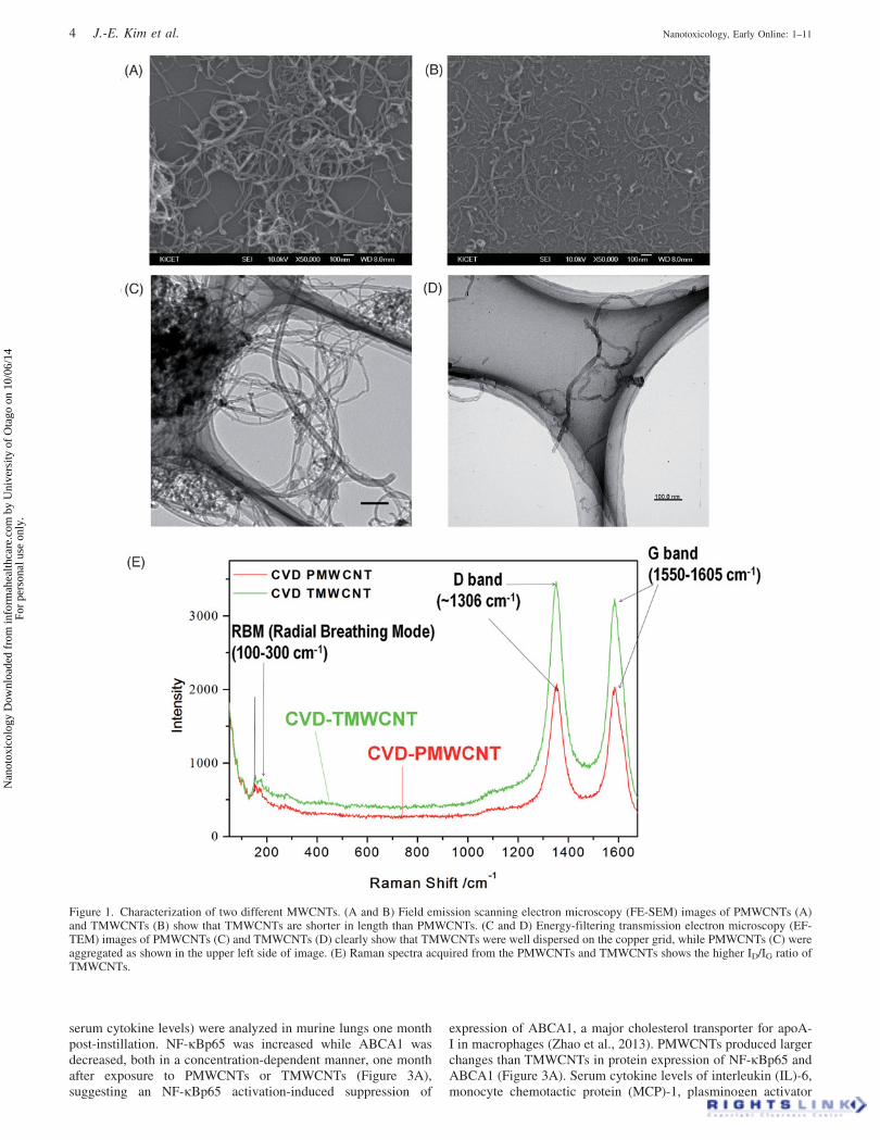
FE-SEM images (Figure 1A and B) and EF-TEM images(Figure 1C and D) of PMWCNTs and TMWCNTs clearlyshowed that TMWCNTs were well dispersed and shortenedcompared to PMWCNTs. The mean length and diameter of thePMWCNTs was 15 ± 5.0 mm and 13.5 ± 1.50 nm, respectively; theTMWCNTs were 400 ± 99.4 nm in length and 7.5 ± 2.5 nm indiameter. Raman spectra acquired from the PMWCNTs andTMWCNTs are shown in Figure 1(E). The higher graphite/defect(ID/IG) ratio of the TMWCNTs as compared to the PMWCNTssuggested functionalization of the TMWCNTs by acid treatment(Table 1).
Lung inflammation by MWCNT instillation andinflammatory cell infiltration in the liver one monthafter administration
To elucidate the effects of tracheal instillation of PMWCNTs andTMWCNTs on the liver, histopathological analysis was performedon lung and liver tissues of exposed mice one month post-instillation. Hematoxylin and eosin (H&E) staining (Figure 2A)was used to detect lung and liver inflammation. Bronchialhyperplasia induced by PMWCNTs and lymphocytic infiltrationinduced by TMWCNTs in the lung are shown in Figure 2(A).Lymphocytic infiltration in the liver one month post-instillationindicated liver inflammation one month after exposure(Figure 2B).
PMWCNTs and TMWCNTs induce systemic cytokinerelease and affect serum cholesterol homeostasis
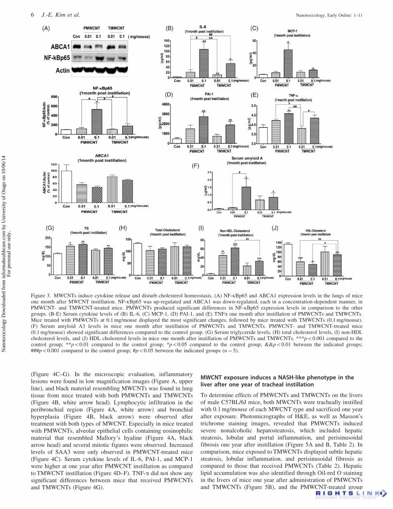
To evaluate the impact of tracheal instillation of PMWCNTs andTMWCNTs on serum cholesterol homeostasis and inflammatorycytokine levels, NF-kBp65 and ABCA1 protein levels (as well as
DOI: 10.3109/17435390.2014.963186 MWCNT induces nonalcoholic steatohepatitis-like phenotype 3
Nan
otox
icol
ogy
Dow
nloa
ded
from
info
rmah
ealth
care
.com
by
Uni
vers
ity o
f O
tago
on
10/0
6/14
For
pers
onal
use
onl
y.
serum cytokine levels) were analyzed in murine lungs one monthpost-instillation. NF-kBp65 was increased while ABCA1 wasdecreased, both in a concentration-dependent manner, one monthafter exposure to PMWCNTs or TMWCNTs (Figure 3A),suggesting an NF-kBp65 activation-induced suppression of
expression of ABCA1, a major cholesterol transporter for apoA-I in macrophages (Zhao et al., 2013). PMWCNTs produced largerchanges than TMWCNTs in protein expression of NF-kBp65 andABCA1 (Figure 3A). Serum cytokine levels of interleukin (IL)-6,monocyte chemotactic protein (MCP)-1, plasminogen activator
Figure 1. Characterization of two different MWCNTs. (A and B) Field emission scanning electron microscopy (FE-SEM) images of PMWCNTs (A)and TMWCNTs (B) show that TMWCNTs are shorter in length than PMWCNTs. (C and D) Energy-filtering transmission electron microscopy (EF-TEM) images of PMWCNTs (C) and TMWCNTs (D) clearly show that TMWCNTs were well dispersed on the copper grid, while PMWCNTs (C) wereaggregated as shown in the upper left side of image. (E) Raman spectra acquired from the PMWCNTs and TMWCNTs shows the higher ID/IG ratio ofTMWCNTs.
4 J.-E. Kim et al. Nanotoxicology, Early Online: 1–11
Nan
otox
icol
ogy
Dow
nloa
ded
from
info
rmah
ealth
care
.com
by
Uni
vers
ity o
f O
tago
on
10/0
6/14
For
pers
onal
use
onl
y.

inhibitor (PAI)-1, and tumor necrosis factor (TNF)-a weremeasured one month post-instillation of PMWCNTs andTMWCNTs. Mice treated with 0.1 mg of PMWCNTs displayedthe most significant changes, followed by TMWCNT-treated mice(0.1 mg/mouse; Figure 3B–E). Increased levels of SAA3 wereobserved in 0.1 mg of PMWCNT and TMWCNT-treated mice(Figure 3F). Serum triglyceride levels (Figure 3G) were
significantly different at one month post-instillation withPMWCNTs and TMWCNTs compared to the control group, butthere was no significant difference between the effects ofPMWCNTs and TMWCNTs. High-density lipoprotein (HDL)-cholesterol (HDL-C) levels (Figure 3J) were decreased signifi-cantly in a dose-dependent manner in mice at one monthpost-instillation with PMWCNTs. In comparison, PMWCNTsproduced a significant decrease in HDL-C compared to thatproduced by TMWCNTs (Figure 3J), corroborating the resultsfor ABCA1 expression shown in Figure 3(A).
Increased serum cytokine and SAA3 levels were sustainedone year after PMWCNT instillation
To assess whether inflammation is alleviated one year afterMWCNT administration, microscopic evaluation of the lungs(Figure 4A and B) and serum cytokine analysis were conducted
Figure 2. Inflammation in the lungs and inflammatory cell infiltration in the livers of mice one month after intratracheally instilled PMWCNTs andTMWNCTs. The tissues were sectioned at 3-mm thickness in paraffin blocks and stained with hematoxylin and eosin (H&E) for histopathologicalanalysis. (A) Lung histopathology at one month after treatment with saline, PMWCNTs, and TMWCNTs. White arrows indicate inflammatory cellinfiltration induced by both types of MWCNTs and blue arrows indicate macrophages engulfing both types of MWCNTs. Arrow head indicates trachealhyperplasia. (B) Liver histopathology one month after treatment with saline, PMWCNTs, and TMWCNTs. Arrows indicate inflammatory cellinfiltration induced by both types of MWCNTs. Representative figures of five individuals from each group are shown. Magnification levels and scalebar sizes are indicated in the figures.
Table 1. Characteristics of PMWCNT and TMWCNT.
PMWCNT TMWCNT
Purity 96.236% 98.460%Diameter 13.5 ± 1.50 nm 7.5 ± 2.5 nmLength 15 ± 5.0 mm 400 ± 99.4 nmID/IG 1.064 1.216
DOI: 10.3109/17435390.2014.963186 MWCNT induces nonalcoholic steatohepatitis-like phenotype 5
Nan
otox
icol
ogy
Dow
nloa
ded
from
info
rmah
ealth
care
.com
by
Uni
vers
ity o
f O
tago
on
10/0
6/14
For
pers
onal
use
onl
y.
(Figure 4C–G). In the microscopic evaluation, inflammatorylesions were found in low magnification images (Figure A, upperline), and black material resembling MWCNTs was found in lungtissue from mice treated with both PMWCNTs and TMWCNTs(Figure 4B, white arrow head). Lymphocytic infiltration in theperibronchial region (Figure 4A, white arrow) and bronchialhyperplasia (Figure 4B, black arrow) were observed aftertreatment with both types of MWCNT. Especially in mice treatedwith PMWCNTs, alveolar epithelial cells containing eosinophilicmaterial that resembled Mallory’s hyaline (Figure 4A, blackarrow head) and several mitotic figures were observed. Increasedlevels of SAA3 were only observed in PMWCNT-treated mice(Figure 4C). Serum cytokine levels of IL-6, PAI-1, and MCP-1were higher at one year after PMWCNT instillation as comparedto TMWCNT instillation (Figure 4D–F). TNF-a did not show anysignificant differences between mice that received PMWCNTsand TMWCNTs (Figure 4G).
MWCNT exposure induces a NASH-like phenotype in theliver after one year of tracheal instillation
To determine effects of PMWCNTs and TMWCNTs on the liversof male C57BL/6J mice, both MWCNTs were tracheally instilledwith 0.1 mg/mouse of each MWCNT type and sacrificed one yearafter exposure. Photomicrographs of H&E, as well as Masson’strichrome staining images, revealed that PMWCNTs inducedsevere nonalcoholic hepatosteatosis, which included hepaticsteatosis, lobular and portal inflammation, and perisinusoidalfibrosis one year after instillation (Figure 5A and B, Table 2). Incomparison, mice exposed to TMWCNTs displayed subtle hepaticsteatosis, lobular inflammation, and perisinusoidal fibrosis ascompared to those that received PMWCNTs (Table 2). Hepaticlipid accumulation was also identified through Oil-red O stainingin the livers of mice one year after administration of PMWCNTsand TMWCNTs (Figure 5B), and the PMWCNT-treated group
Figure 3. MWCNTs induce cytokine release and disturb cholesterol homeostasis. (A) NF-kBp65 and ABCA1 expression levels in the lungs of miceone month after MWCNT instillation. NF-kBp65 was up-regulated and ABCA1 was down-regulated, each in a concentration-dependent manner, inPMWCNT- and TMWCNT-treated mice. PMWCNTs produced significant differences in NF-kBp65 expression levels in comparison to the othergroups. (B-E) Serum cytokine levels of (B) IL-6, (C) MCP-1, (D) PAI-1, and (E) TNFa one month after instillation of PMWCNTs and TMWCNTs.Mice treated with PMWCNTs at 0.1 mg/mouse displayed the most significant changes, followed by mice treated with TMWCNTs (0.1 mg/mouse).(F) Serum amyloid A3 levels in mice one month after instillation of PMWCNTs and TMWCNTs. PMWCNT- and TMWCNT-treated mice(0.1 mg/mouse) showed significant differences compared to the control group. (G) Serum triglyceride levels, (H) total cholesterol levels, (I) non-HDLcholesterol levels, and (J) HDL cholesterol levels in mice one month after instillation of PMWCNTs and TMWCNTs. ***p50.001 compared to thecontrol group; **p50.01 compared to the control group; *p50.05 compared to the control group; &&p50.01 between the indicated groups;###p50.001 compared to the control group; #p50.05 between the indicated groups (n¼ 5).
6 J.-E. Kim et al. Nanotoxicology, Early Online: 1–11
Nan
otox
icol
ogy
Dow
nloa
ded
from
info
rmah
ealth
care
.com
by
Uni
vers
ity o
f O
tago
on
10/0
6/14
For
pers
onal
use
onl
y.

showed more severe effects. Periodic acid-Schiff’s (PAS) stainingshowed reduced glycogen storage in the livers of mice one yearafter exposure to TMWCNTs or PMWCNTs as compared to thesaline-exposed control mice. The PMWCNT-exposed groupinhibited glycogen storage more severely than the TMWCNT-exposed group (Figure 5B). The accumulation of hepatic lipidswas further identified by the increased level of hepatic triglycer-ide (TG; Figure 5C). Decreased HDL-C levels and increased TGlevels in the sera of mice that were intratracheally exposed toPMWCNT and TMWCNT also indicated abnormal lipid homeo-stasis (Figure 5C–F).
Inflammatory signaling, PPAR signaling, and Kupffer cellswere activated in the livers one year after trachealinstillation of PMWCNTs and TMWCNTs
Western blot analysis in the livers of mice one year afteradministration of PMWCNTs and TMWCNTs showed decreasedexpression levels of PPARg and phospho-AMPKa, but increasedexpression of a-SMA (Figure 6A). Inflammation-related proteinsincluding phospho-IkB, NF-kBp65, and VCAM-1 were increasedin the liver one year after administration of PMWCNTs andTMWCNTs (Figure 6B). Immunohistochemistry (Figure 6C) andwestern blotting (Figure 6D) of F4/80, a macrophage surface
marker, indicated that macrophages are activated one year afterexposure to PMWCNTs or TMWCNTs. Furthermore, MAC-2,another macrophage maker, was increased remarkably one yearafter tracheal instillation of PMWCNTs as compared toTMWCNTs (Figure 6E).
Discussion
In this study, we show that exposure of the lungs to MWCNTs caninduce a NASH-like phenotype and alter lipid homeostasis inmice one year after exposure. Furthermore, by comparing twodifferent MWCNTs, non-functionalized hydrophobic PMWCNTsand functionalized hydrophilic TMWCNTs, we found that theNASH-like phenotype produced by PMWCNTs was approxi-mately twice (according to the score in Table 2) as severe as thatproduced by TMWCNTs. These effects were accompanied byelevated levels of serum cytokines, serum SAA3, and proteinexpression levels in the liver one year after exposure toPMWCNTs or TMWCNTs (Figures 4C–F and 6). For thisstudy, we used intratracheal instillation instead of inhalation.Intratracheal instillation can cause a rapid bolus deposition ofMWCNTs in the lung (Driscoll et al., 2000), but we used thismethod because it allowed for precise control of the applied dosesof PMWCNTs and TMWCNTs, which was necessary to compare
Figure 4. PMWCNT instillation induced sustained inflammation in mice after one year. (A and B) Microphotographs of H&E-stained slides of lungtissue one year after PMWCNT and TMWCNT instillation. Lymphatic infiltration was observed in MWCNT-treated lungs (A, white arrow), andalveolar epithelial cells containing eosinophilic material that resembled Mallory’s hyaline were observed in PMWCNT-treated mice (A, black arrowhead). Accumulated MWCNTs (B, upper panel) and tracheal hyperplasia (B, black arrow) in the lungs were also observed. The serum level of SAA3(C), and serum cytokine levels of (D) IL-6, (E) PAI-1, (F) MCP-1, and (G) TNF-a in mice one year after PMWCNT and TMWCNT instillationwere measured. Each bar represents the mean ± S.E.M. **p50.01 compared to the control group; *p50.05 compared to the control group; ##p50.01between the indicated groups (n¼ 5).
DOI: 10.3109/17435390.2014.963186 MWCNT induces nonalcoholic steatohepatitis-like phenotype 7
Nan
otox
icol
ogy
Dow
nloa
ded
from
info
rmah
ealth
care
.com
by
Uni
vers
ity o
f O
tago
on
10/0
6/14
For
pers
onal
use
onl
y.

their effects. The findings of this study using tracheal instillationare also supported by the results obtained using a round particlesinhalation system by Zheng et al. (2013). Currently, limitedstudies are present investigating the pathophysiological effects onthe liver after exposures to MWCNTs via the lungs. Thoughtracheal installation was used in this study, our findings maycontribute to better understanding of the potential toxicity ofMWCNTs on the liver.
For the studies of the effects of MWCNTs one year afteradministration, male C57BL/6J mice were exposed to a singledose (0.1 mg/mouse) of PMWCNTs or TMWCNTs. Assumingmouse alveolar epithelium surface area of 0.05 m2 (Stone et al.,1992), the 0.1 mg MWCNT lung burden would result in 2 mgMWCNT/m2 of alveolar epithelium. For the human alveolarepithelial surface area of 102 m2, the equivalent human lungburden would be 204 mg (Stone et al., 1992). Furthermore,according to a draft established by the National Institute forOccupational Safety and Health, when the dose was converted tohuman work place exposure level, CNT lung burden equivalent tothat of workers exposed for approximately 40 years recommendedexposure limit (REL) for CNTs of 7 mg/m3 per 8 hour work shift(NIOSH, 2010), assuming given minute ventilation of 20 L/minute for a person performing light work (Galer et al., 1992) andan alveolar deposition fraction of 30% (Bates et al., 1966) (for 250work days per year). Therefore, the mentioned dose was selected
Figure 5. MWCNT exposure induces a NASH-like phenotype in the mouse liver one year after tracheal instillation. (A) Photomicrographs of H&Estained slide showing the PMWCNTs and TMWCNTs induced nonalcoholic hepatosteatosis in the livers of one year after instillation. (B) Paraffin-embedded liver tissue sections of one year after administration of PMWCNTs and TMWCNTs were stained with Oil red O (lipid droplet), Masson’strichrome (collagen fiber), and PAS (hepatic glycogen). Representative figures of five individuals from each group. Magnification and scale bar areindicated in the figures. (C) Serum triglyceride, (D) serum total-cholesterol, (E) serum non-HDL-cholesterol, and (F) serum HDL-cholesterol of oneyear after administration of PMWCNTs and TMWCNTs. Each bar represents the mean ± S.E.M. ***Statistically different (p50.001) compared to thecontrol group. **Statistically different (p50.01) compared to the control group. *Statistically different (p50.05) compared to control group.#Statistically different (p50.05) between two indicated groups (n¼ 5).
Table 2. Histopathological scoring for NASH in the livers of C57BL/6Jmice one year after instillation of MWCNTs.
Steatosis Ballooning Fibrosis
Con 0.25 ± 0.29 0.13 ± 0.25 0.0 ± 0.00PMWCNT 1.6 ± 0.55 1.8 ± 0.45 0.8 ± 0.45TMWCNT 0.6 ± 0.55 1.0 ± 0.71 0.4 ± 0.55
8 J.-E. Kim et al. Nanotoxicology, Early Online: 1–11
Nan
otox
icol
ogy
Dow
nloa
ded
from
info
rmah
ealth
care
.com
by
Uni
vers
ity o
f O
tago
on
10/0
6/14
For
pers
onal
use
onl
y.

for mechanistic study in the liver of mice exposed to MWCNTsthrough the lung route.
At one month post-instillation, lymphocytic infiltrations inlung and liver were observed (Figure 2A and B), as previouslyreported by Reddy et al. (2010), who described translocationof MWCNTs to extra-pulmonary organs using histopathologicalanalysis. In addition, increased expression of NF-kBp65 in thelung was associated with decreased expression of the ATP-binding cassette transporter A1 (ABCA1) (Figure 3A), which iscritical in exporting cholesterol from macrophages and plays aprotective role in the development of atherosclerosis (Vaismanet al., 2012). Decreased ABCA1 expression likely resulted indecreased serum HDL-C (Figure 3J), as it has been reported thatincreased endothelial-specific expression of ABCA1 was found toraise serum HDL-C (Vaisman et al., 2012). Decreased ABCA1(Figure 3A) expression also inhibits the efflux of macrophagelipids and promotes inflammatory cytokine release (Figure 3B–E;Zhao et al., 2013). The increased SAA3 levels in mice treatedwith PMWCNTs and TMWCNTs suggest a disturbance in lipidhomeostasis (Figure 3F and J), as pulmonary acute phase responsehas been reported to impact lipid homeostasis, leading to loweredHDL levels (Bourdon et al., 2012) and is also considered as a riskfactor for cardiovascular diseases (Saber et al., 2013). Thesecomplex results suggest that exposure to PMWCNTs andTMWCNTs via the lungs can induce systemic inflammationand interfere with cholesterol homeostasis in the lungs.
At one year post-treatment, although a large part of theadministered MWCNTs had been removed from the lung,infiltrated lymphocytes were still observed to be accumulated inboth lungs of mice treated with PMWCNTs and TMWCNTs(Figure 4A and B). In contrast, SAA3 and serum levels ofcytokines IL-6 and PAI-1 remained increased only in PMWCNT-treated mice one year after administration (Figure 4C–E),indicating the severity of the disturbed lipid homeostasis and
NASH. IL-6 was reported to enhance liver inflammation andtumorigenesis induced by obesity (Park et al., 2010) and was alsoreported to be increased in human NASH (Wieckowska et al.,2008). PAI-1, which inhibits fibrinolysis in clot degradation(Erdely et al., 2009), is involved in neutrophil recruitment to thealveolar compartment (Arndt et al., 2005) and is reported to beincreased in human NASH (Targher et al., 2007). Increased PAI-1expression in PMWCNT-treated mice suggests that PMWCNTexposure is a bigger risk factor for NASH, as compared toTMWCNT exposure (Figure 5A and Table 2). PMWCNTsproduced a prominent NASH-like phenotype manifested by anapproximate two-fold increase in inflammatory response score,lipid accumulation, ballooning degeneration, and collagen depos-ition (Table 2). There have been several reports on thebiodistribution and clearance of administered MWCNTs (Denget al., 2007; Mercer et al., 2013; Reddy et al., 2010; Singh et al.,2006). In a report by Singh et al. (2006), carboxylated-CNTs wereadministered through intravenous (i.v.) injection and showed noorgan-specific accumulation over time. Deng et al. (2007)reported that functionalized CNTs administered via i.v. injectionwere observed in liver Kupffer cells 7 days after exposure.However, they did not show translocation of MWCNTs admin-istered through intratracheal instillation (i.t.) to the liver, andfound that MWCNTs administered in this manner were detectedin the lung 28 days after injection. Reddy et al. (2010) reportedextrapulmonary toxicity of intratracheally administered non-functionalized MWCNTs 3 months after administration, butthey did not provide definite evidence of the translocation ofPMWCNTs via methods such as TEM or dark field imaging.Recently, Czarny et al. (2014) reported gradual accumulation ofMWCNTs in the spleen and the liver one year after intraphar-yngeal administration, using 14C-radiolabeled MWCNTs withtissue radioimaging. Because their method does not use aradioactive tag on the CNT surface, the potential instability of
Figure 6. Inflammatory signaling, PPARg signaling, and Kupffer cells were activated in the mouse liver one year after tracheal instillation ofPMWCNTs and TMWCNTs. (A) Western blot analysis of PPARg, phospho-AMPKa, and a-SMA in the mouse liver one year after administrationof PMWCNTs and TMWCNTs. (B) Western blot analysis of phospho-IkB, NF-kBp65, and VCAM-1 in the mouse liver one year after administrationof PMWCNTs and TMWCNTs (n¼ 5). (C) Immunohistochemistry and (D) western blots of F4/80 (a macrophage surface marker), and(E) immunohistochemistry of MAC-2 were conducted to determine macrophage infiltration in the mouse liver one year after administrationof PMWCNTs and TMWCNTs. Representative figures of five individuals from each group are shown. Magnification levels and scale bar sizesare indicated in the figures. **p50.01 compared to the control group; *p50.05 compared to the control group (n¼ 5).
DOI: 10.3109/17435390.2014.963186 MWCNT induces nonalcoholic steatohepatitis-like phenotype 9
Nan
otox
icol
ogy
Dow
nloa
ded
from
info
rmah
ealth
care
.com
by
Uni
vers
ity o
f O
tago
on
10/0
6/14
For
pers
onal
use
onl
y.

the tag and alteration of the physicochemical properties of CNTs,which could change their in vivo distribution, can be excluded asfactors that can possibly cause false results. Given these reports, ifwe assume that our TMWCNTs correspond to functionalizedMWCNTs and PMWCNTs correspond to non-functionalizedMWCNTs, we speculate that the differences in the accumulationof PMWCNTs and TMWCNTs in the liver could contribute to thedifferences in their pathophysiology and induced NASH pheno-type. In a previous study, we showed faster clearance ofTMWCNTs as compared to PMWCNTs, which also supportsthis explanation (Kim et al., 2010). In addition, previous studieson the hepatotoxicity of using pristine and functionalizedMWCNTs administered via i.v. injection also concluded thatfunctionalized MWCNTs were less hepatotoxic than pristineMWCNTs (Jain et al., 2011; Ji et al., 2009). The continuousrelease of cytokines into the serum over the span of one year andthe accumulation of MWCNTs in the liver contributed to theseverity of NASH induced by MWCNT exposure.
Interactions between metabolic pathways and inflammationmediated by macrophages and adipocytes are important in thedevelopment of NASH (Hui et al., 2013). As shown in Figure 6,those interactions and inflammations were shown in the liver thatwas indicative of MWCNT-induced NASH. The presence ofmacrophages (Figure 6C and D) activated by adipokinedysregulation (Figure 4D–F) perpetuates a vicious cycle ofmacrophage recruitment (Figure 6E) and inflammatory cytokineproduction. This state of chronic inflammation stimulates NF-kB(Figures 3A and 6B) and acute phase response protein SAA3(Figures 3F and 4C). PPARg-mediated increases in adiponectinexpression are necessary for the maintenance of lipid and glucosehomeostasis (Tsuchida et al., 2005) and for the prevention ofinflammation in Kupffer cells and hepatocytes (Feige et al.,2006). Because adiponectin mediates its antisteatotic effect byactivating AMPK (Yamauchi et al., 2002), leading to enhancedfatty acid oxidation, the observed decreases in PPARg andp-AMPKa in the livers of PTMWCNT- and TMWCNT-treatedmice (Figure 6A) indicates an increase in liver inflammation(Figure 6B) and reduced glycogen storage (Figure 5B-PASstaining). Alpha-smooth muscle actin (a-SMA), which is secretedfrom activated hepatic stellate cells and is reported to be unrelatedto alcoholic liver disease, but correlated with the severity ofsteatosis (Reeves et al., 1996), was also increased in the livers ofPMWCNT- and TMWCNT-treated mice after one year, support-ing the severity of NASH produced by PMWCNTs andTMWCNTs (Figure 6A).
Our results shows that exposure to PMWCNTs andTMWCNTs through the lungs can induce NASH-like phenotypesin mice one year after administration, which are related tosystemic inflammation and disturbed lipid homeostasis in thelungs and the liver. PMWCNT exposure resulted in more severeNASH than TMWCNT, as indicated by pathological scores twiceas high as those produced by the latter agent, and the severity washighly correlated with the degree of accumulation of MWCNTs inthe liver, as well as the sustained increased serum levels of IL-6,MCP-1, and SAA3.
Conclusions
This study showed that MWCNTs administered through the lungsinduce NASH-like phenotypes in mice one year after administra-tion, and the consequences of exposure to PMWCNTs were moresevere than those of exposure to TMWCNTs. The predisposingfactor for NASH development appeared to be the sustainedrelease of several cytokines, which can alter lipid homeostasis andthe translocation of MWCNTs to the liver. Given the growingnumber of NASH patients and the close relationship of NASH
with type 2 diabetes, cardiovascular disease, and hepatocellularcarcinoma, it is important to understand MWCNT exposure as arisk factor for NASH. This comparison study suggests the need forfuture in-depth research on metabolic alterations produced byMWCNT exposure via comparative inhalation studies in variousmodels of disease predisposition.
Acknowledgments
Part of this work was supported by a National Research Foundationgrants (NRF-2012M3A9B6055304 and NRF-2014M3A7B6034499)funded by the Ministry of Science, ICT & Future Planning of Korea.J.E.K. and A.Y.L. are supported by the Brain Korea 21 Program forVeterinary Science of Seoul National University. M.H.C. was alsopartially supported by the Research Institute for Veterinary Science, SeoulNational University.
Declaration of interest
The authors declare that they have no competing interests.
References
Arndt PG, Young SK, Worthen GS. 2005. Regulation of lipopolysac-charide-induced lung inflammation by plasminogen activator inhibitor-1 through a JNK-mediated pathway. J Immunol 175:4049–59.
Bates DV, Fish BR, Hatch TF, Mercer TT, Morrow PE. 1966. Depositionand retention models for internal dosimetry of the human respiratorytract. Task group on lung dynamics. Health Phys 12:173–207.
Berber S, Kwon YK, Tomanek D. 2000. Unusually high thermalconductivity of carbon nanotubes. Phys Rev Lett 84:4613–16.
Bourdon JA, Halappanavar S, Saber AT, Jacobsen NR, Williams A,Wallin H, et al. 2012. Hepatic and pulmonary toxicogenomic profilesin mice intratracheally instilled with carbon black nanoparticles revealpulmonary inflammation, acute phase response, and alterations in lipidhomeostasis. Toxicol Sci 127:474–84.
Brunt EM. 2001. Nonalcoholic steatohepatitis: definition and pathology.Semin Liver Dis 21:3–16.
Chen JC, Schwartz J. 2008. Metabolic syndrome and inflammatoryresponses to long-term particulate air pollutants. Environ HealthPerspect 116:612–17.
Czarny B, Georgin D, Berthon F, Plastow G, Pinault M, Patriarche G,et al. 2014. Carbon nanotube translocation to distant organs afterpulmonary exposure: insights from in situ 14c-radiolabelling and tissueradioimaging. ACS Nano 8:5715–24.
Day CP, James OF. 1998. Steatohepatitis: a tale of two ‘‘hits’’?Gastroenterology 114:842–5.
De Volder MF, Tawfick SH, Baughman RH, Hart AJ. 2013. Carbonnanotubes: present and future commercial applications. Science 339:535–9.
Della Corte C, Liccardo D, Mosca A, Vania A, Nobili V. 2013. Non-alcoholic fatty liver disease. Paediatr Child Health, 23:529–34.
Deng X, Jia G, Wang H, Sun H, Wang X, Yang S, et al. 2007.Translocation and fate of multi-walled carbon nanotubes in vivo.Carbon 45:1419–24.
Driscoll KE, Costa DL, Hatch G, Henderson R, Oberdorster G, Salem H,Schlesinger RB. 2000. Intratracheal instillation as an exposuretechnique for the evaluation of respiratory tract toxicity: uses andlimitations. Toxicol Sci 55:24–35.
Erdely A, Hulderman T, Salmen R, Liston A, Zeidler-Erdely PC,Schwegler-Berry D, et al. 2009. Cross-talk between lung and systemiccirculation during carbon nanotube respiratory exposure. Potentialbiomarkers. Nano Lett 9:36–43.
Feige JN, Gelman L, Michalik L, Desvergne B, Wahli W. 2006. Frommolecular action to physiological outputs: peroxisome proliferator-activated receptors are nuclear receptors at the crossroads of keycellular functions. Prog Lipid Res 45:120–59.
Galer DM, Leung HW, Sussman RG, Trzos RJ. 1992. Scientific andpractical considerations for the development of occupational exposurelimits (oels) for chemical substances. Regul Toxicol Pharmacol 15:291–306.
Hu L, Hecht DS, Gruner G. 2010. Carbon nanotube thin films:fabrication, properties, and applications. Chem Rev 110:5790–844.
10 J.-E. Kim et al. Nanotoxicology, Early Online: 1–11
Nan
otox
icol
ogy
Dow
nloa
ded
from
info
rmah
ealth
care
.com
by
Uni
vers
ity o
f O
tago
on
10/0
6/14
For
pers
onal
use
onl
y.

Hui E, Xu A, Yang BH, Lam KS. 2013. Obesity as the common soil ofnon-alcoholic fatty liver disease and diabetes: role of adipokines.J Diabetes Investig 4:413–25.
Jain S, Thakare VS, Das M, Godugu C, Jain AK, Mathur R, et al. 2011.Toxicity of multiwalled carbon nanotubes with end defects criticallydepends on their functionalization density. Chem Res Toxicol 24:2028–39.
Ji Z, Zhang D, Li L, Shen X, Deng X, Dong L, et al. 2009.The hepatotoxicity of multi-walled carbon nanotubes in mice.Nanotechnology 20:445101. doi:10.1088/0957-4484/20/44/445101.
Kim JE, Lim HT, Minai-Tehrani A, Kwon JT, Shin JY, Woo CG, et al.2010. Toxicity and clearance of intratracheally administered multi-walled carbon nanotubes from murine lung. J Toxicol EnvironHealth A 73:1530–43.
Kleiner DE, Brunt EM, Van Natta M, Behling C, Contos MJ, CummingsOW, et al. 2005. Design and validation of a histological scoring systemfor nonalcoholic fatty liver disease. Hepatology 41:1313–21.
Kramer U, Herder C, Sugiri D, Strassburger K, Schikowski T, Ranft U,Rathmann W. 2010. Traffic-related air pollution and incident type 2diabetes: results from the SALIA cohort study. Environ HealthPerspect 118:1273–9.
Laing S, Wang G, Briazova T, Zhang C, Wang A, Zheng Z, et al. 2010.Airborne particulate matter selectively activates endoplasmic reticulumstress response in the lung and liver tissues. Am J Physiol Cell Physiol299:C736–49.
Mercer RR, Scabilloni JF, Hubbs AF, Wang L, Battelli LA, Mckinney W,et al. 2013. Extrapulmonary transport of MWCNT following inhalationexposure. Part Fibre Toxicol 10:38. doi: 10.1186/1743-8977-10-38.
NIOSH. 2010. Current Intelligence Bulletin: occupational exposure tocarbon nanotubes and nanofibers. Cincinnati, Ohio: US Department ofHealth and Human Services Centers for Disease Control andPrevention, National Institute for Occupational Safety and Health;draft document.
Osorio A, Silveira I, Bueno V, Bergmann C. 2008. H2SO4/HNO3/HCl-Functionalization and its effect on dispersion of carbon nanotubes inaqueous media. Appl Surf Sci 255:2485–9.
Park EJ, Lee JH, Yu G-Y, He G, Ali SR, Holzer RG, et al. 2010. Dietaryand genetic obesity promote liver inflammation and tumorigenesis byenhancing IL-6 and TNF expression. Cell 140:197–208.
Patlolla AK, Berry A, Tchounwou PB. 2011. Study of hepatotoxicity andoxidative stress in male Swiss-Webster mice exposed to functionalizedmulti-walled carbon nanotubes. Mol Cell Biochem 358:189–99.
Reddy AR, Krishna DR, Reddy YN, Himabindu V. 2010. Translocationand extra pulmonary toxicities of multi wall carbon nanotubes in rats.Toxicol Mech Methods 20:267–72.
Reeves HL, Burt AD, Wood S, Day CP. 1996. Hepatic stellate cellactivation occurs in the absence of hepatitis in alcoholic liver diseaseand correlates with the severity of steatosis. J Hepatol 25:677–83.
Saber AT, Lamson JS, Jacobsen NR, Ravn-Haren G, Hougaard KS,Nyendi AN, et al. 2013. Particle-induced pulmonary acute phaseresponse correlates with neutrophil influx linking inhaled particles andcardiovascular risk. PLoS One 8:e69020.
Singh R, Pantarotto D, Lacerda L, Pastorin G, Klumpp C, Prato M, et al.2006. Tissue biodistribution and blood clearance rates of intravenouslyadministered carbon nanotube radiotracers. Proc Natl Acad Sci USA103:3357–62.
Spoelstra EN, Ince C, Koeman A, Emons VM, Brouwer LA, van LuynMJ, et al. 2007. A novel and simple method for endotracheal intubationof mice. Lab Anim 41:128–35.
Stone KC, Mercer RR, Gehr P, Stockstill B, Crapo JD. 1992. Allometricrelationships of cell numbers and size in the mammalian lung. Am JRespir Cell Mol Biol 6:235–43.
Sun Q, Yue P, Deiuliis JA, Lumeng CN, Kampfrath T, Mikolaj MB, et al.2009. Ambient air pollution exaggerates adipose inflammation andinsulin resistance in a mouse model of diet-induced obesity.Circulation 119:538–46.
Tan HH, Fiel MI, Sun Q, Guo J, Gordon RE, Chen LC, et al. 2009.Kupffer cell activation by ambient air particulate matter exposure mayexacerbate non-alcoholic fatty liver disease. J Immunotoxicol 6:266–75.
Targher G, Bertolini L, Scala L, Zenari L, Lippi G, Franchini M,Arcaro G. 2007. Plasma PAI-1 levels are increased in patients withnonalcoholic steatohepatitis. Diabetes Care 30:e31–2.
Tsuchida A, Yamauchi T, Takekawa S, Hada Y, Ito Y, Maki T, KadowakiT. 2005. Peroxisome proliferator–activated receptor (PPAR) a activa-tion increases adiponectin receptors and reduces obesity-relatedinflammation in adipose tissue comparison of activation of PPARa,PPARg, and their combination. Diabetes 54:3358–70.
Vaisman BL, Demosky SJ, Stonik JA, Ghias M, Knapper CL, SampsonML, et al. 2012. Endothelial expression of human ABCA1 in miceincreases plasma HDL cholesterol and reduces diet-induced athero-sclerosis. J Lipid Res 53:158–67.
Wieckowska A, Papouchado BG, Li Z, Lopez R, Zein NN, Feldstein AE.2008. Increased hepatic and circulating interleukin-6 levels in humannonalcoholic steatohepatitis. The Am J Gastroenterol 103:1372–9.
Yamauchi T, Kamon J, Minokoshi Y, Ito Y, Waki H, Uchida S, et al.2002. Adiponectin stimulates glucose utilization and fatty-acid oxida-tion by activating AMP-activated protein kinase. Nat Med 8:1288–95.
Zhao GJ, Tang SL, Lv YC, Ouyang XP, He PP, Yao F, et al. 2013.Antagonism of betulinic acid on LPS-mediated inhibition of ABCA1and cholesterol efflux through inhibiting nuclear factor-kappaBsignaling pathway and miR-33 expression. PLoS One 8:e74782.
Zheng Z, Xu X, Zhang X, Wang A, Zhang C, Huttemann M, et al. 2013.Exposure to ambient particulate matter induces a NASH-like pheno-type and impairs hepatic glucose metabolism in an animal model.J Hepatol 58:148–54.
DOI: 10.3109/17435390.2014.963186 MWCNT induces nonalcoholic steatohepatitis-like phenotype 11
Nan
otox
icol
ogy
Dow
nloa
ded
from
info
rmah
ealth
care
.com
by
Uni
vers
ity o
f O
tago
on
10/0
6/14
For
pers
onal
use
onl
y.