intraoperative navigation-assisted surgical orbital …...intraoperative navigation-assisted...
TRANSCRIPT

Intraoperative Navigation-assisted Surgical Orbital Floor Reconstruction in Orbital Fracture Treatment: A Case Report
Shintaro SUKEGAWA1), Takahiro KANNO1,2), Yuta KOYAMA3), Akane SHIBATA1), Kenichi MATSUMOTO1), Yuka SUKEGAWA-TAKAHASHI1), Kyosuke SAKAIDA1), Shigeto TANAKA3),Yoshihiko FURUKI1)
1)Division of Oral and Maxillofacial Surgery, Kagawa Prefectural Central Hospital, Takamatsu, 760-8557, Japan2)Department of Oral and Maxillofacial Surgery, Shimane University Faculty of Medicine, Izumo, 693-8501, Japan3)Division of Ophthalmology, Kagawa Prefectural Central Hospital, Takamatsu, 760-8557, Japan(Received December 1, 2016; Accepted December 13, 2016)
The main goal of three-dimensional reconstruction of the orbital wall fracture is restoration of the orig-inal orbital volume. Although orbital reconstruction using implant materials is effective, it is difficult to assess the accuracy during surgery. Additionally, complete reconstruction of the orbital floor is dif-ficult in patients with massive comminution. There-fore, a conventional technique that preoperatively creates orbital reconstruction materials on the basis of individually precise model reproductions of the orbital forms was developed. However, this method was limited because custom-made preparation and fabrication of the pre-bending plate is time con-suming, making it inapplicable during emergencies. On the other hand, computer-assisted intraoperative navigation systems have recently evolved to improve precision and simplify the surgical procedure, result-ing in improved execution and predictability. Here we describe our feasible application of an intraop-erative navigation-assisted orbital floor reconstruc-tion using the mirroring technique in orbital fracture treatment.
Key words: intraoperative navigation system, orbital reconstruction, mirroring technique
INTRODUCTION
The main goal of three-dimensional (3D) recon-struction of the orbital wall fracture is restoration of the original orbital volume and prevention of long-term complications, such as diplopia, enophthalmos, hypoglobus, facial disproportion, and decreased globe mobility [1-4]. Several studies have reported the use of implant materials, such as bone, cartilage, collagen membrane, titanium, and resorbable mesh, for reconstruction of the orbit [4-8]. Although orbit-al reconstruction in orbital fracture treatment using these materials is effective, it is difficult to assess its accuracy during surgery. Therefore, a new tech-nique that preoperatively creates orbital reconstruc-tion materials using an individually precise model reproduction of the orbital form was developed [9, 10]. Although this method easily reproduces ac-curate orbital positioning and shape, the prolonged time required to create and prepare these custom-made pre-bending plate systems make it inapplicable during emergencies [10].
Complete reconstruction of the orbital floor is difficult in patients presenting with massive com-minution. In such cases, computer-assisted planning has been shown to be an effective technique for re-establishing orbital symmetry [10, 11]. Moreover, computer-assisted intraoperative navigation has re-cently evolved to improve precision and simplify the surgical procedure by minimizing surgical invasive-ness. The development of intraoperative navigation surgery has improved the execution and predictabil-ity, allowing for greater precision during maxillofa-
Corresponding author: Sukegawa ShintaroDivision of Oral and Maxillofacial Surgery, Kagawa Prefec-tural Central Hospital, 1-2-1 Asahi-cho, Takamatsu,Kagawa 760-8557, JapanTel: +81-87-811-3333Fax: +81-87-802-1188E-mail: [email protected]
87
Shimane J. Med. Sci., Vol.33 pp.87-92, 2017

cial surgery.This is a case report on an intraoperative navi-
gation-assisted orbital floor reconstruction using the mirroring technique performed at our hospital.
CASE REPORT
PatientIn August 2016, a 42-year-old Japanese female
suffered injury from a fall and was brought by am-bulance to the Emergency and Critical Center of our hospital. She presented with malocclusion and dip-lopia of the right eye, and was referred to the Divi-sion of oral and maxillofacial surgery. On clinical examination, enophthalmos of the right eye and dip-lopia on upward gaze were observed. These findings were confirmed by the Division of Ophthalmology. Computed tomography (CT) scanning identified a defect in the mid-right orbital floor (25.3 × 18.1 mm) with herniation of the orbital contents into the underlying maxillary sinus accompanied by a right Le Fort I fracture (Fig. 1). The patient provided informed consent for orbital floor reconstruction, and open reduction and internal fixation.
Technique3D navigation-guided surgery was chosen for this
case as it provides high accuracy. A maxillofacial CT scan was preoperatively obtained, and the im-aging data was collected in Digital Imaging and Communication in Medicine format and transferred to a Medtronic StealthStation S7 workstation using Synergy Fusion Cranial 2.2.6 software (Medtronic, Inc., Louisville, Colorado, United States). The CT data of the median sagittal plane was used as the reference plane. The normal anatomic structures of the target area were mirrored from the healthy side, such that the desired contour of the injured orbital shape could be visualized. The mirrored image gen-erated was superimposed onto the fractured side in the original CT scan image in 2 and 3 dimensions, to allow accurate and symmetric positioning of the bones. The mirrored images were then manually adjusted to fit as precisely as possible to fixed ana-tomic landmarks, such as the pterygoid plates and the petrous apex of the temporal bone. A Patient Tracker EM was affixed to the forehead to act as the reference array to track the navigation probe
(Fig. 2A). To perform patient-to-CT data registra-tion, the navigation instrumentation probe was used to trace the reference array, soft tissue landmarks of
Fig. 1.(A) Coronal and (B) Sagittal computed tomography images showing a defect in the mid-left orbital floor, with herniation of the orbital contents into the underlying maxillary sinus. (C) 3D CT showing right Le Fort I fracture.
88 Sukegawa et al.
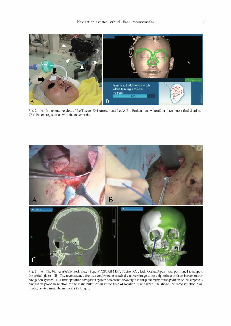
Fig. 2.(A) Intraoperative view of the Tracker EM (arrow) and the AxiEm Emitter (arrow head) in place before final draping.(B)Patient registration with the tracer probe.
Fig. 3.(A) The bio-resorbable mesh plate (SuperFIXSORB MX®, Takiron Co., Ltd., Osaka, Japan) was positioned to support the orbital globe.(B) The reconstructed site was confirmed to match the mirror image using a tip pointer with an intraoperative navigation system.(C) Intraoperative navigation system screenshot showing a multi-plane view of the position of the surgeon’s navigation probe in relation to the mandibular lesion at the time of location. The dashed line shows the reconstruction plan image, created using the mirroring technique.
89Navigation-assisted orbital floor reconstruction

the face, and hard tissue points, such as the tooth cusps and incisal edges. After completion of data registration, continuous 3D tracking of the naviga-tion probe was made available to the surgeon in real time (Fig. 2B).
Surgery During surgery under general anesthesia, the orbit-
al floor fracture was explored via a sub-tarsal inci-sion. Herniated orbital tissue was reduced to restore the intra-orbital structures, as were the fractured bone fragments. The 3D positioning and matching of the planned and actual positions of the orbital floor defect were verified by means of a pointer. A bio-resorbable and osteoconductive bioactive mesh plate system [8](SuperFIXSORB MX®, Takiron Co., Ltd., Osaka, Japan)was positioned to support the orbital globe (Fig. 3A) and evaluated to ensure good orbital reconstruction using the set reconstruc-tion material. The reconstructed site was confirmed to match the mirror image with the help of a tip pointer and an intraoperative navigation system (Fig. 3B, C). Additionally, passive movement of the globe was evaluated at the end of the surgical pro-cedure.
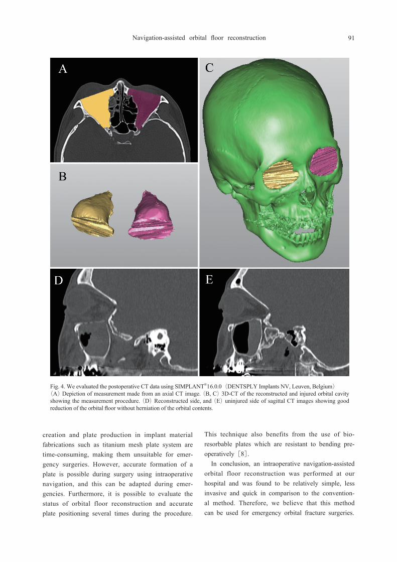
Volume evaluationThe bony orbit was electronically segmented into
axial slices and controlled in the coronal and sagittal view for volume analysis. The postoperative CT data was evaluated using SIMPLANT®16.0.0(DENTSPLY Implants NV, Leuven, Belgium). The details of the technique used for volume segmentation and calculation in our series have been described previ-ously [12]. The anterior border of the orbit was defined by a straight line passing through points on the lateral orbital rim and medial orbital rim. For every axial slice, a sub-volume was determined by calculating the number of pixels within the defined region, with each pixel representing a voxel value. The overall volume of the orbit was calculated in mm3, and compared between the left and right sides
(Fig. 4).The results were clinically and radiographically
evaluated by an ophthalmologist using CT. The pa-tient did not exhibit any intraoperative complications or evidence of enophthalmos, assessed based on
Hertel exophthalmometry. The volume of the recon-structed orbit was 2252 mm3, while that of the un-injured orbit was 2258 mm3. There were no appar-ent differences in orbit volume between the injured and reconstructed sides.
DISCUSSION
Our clinical field of maxillofacial surgeries require high levels of accuracy because of the presence of critical contiguous organs. Real-time 3D computer-navigated surgery enables precise surgical perfor-mance and minimal invasion, resulting in lower risk of complications [13]. Hence, this system has been technologically developed, along with simultaneous advancement of computer-aided modeling software. This has dramatically improved surgical strategies in maxillofacial surgery, especially with respect to pre-diction of suitable bone repositioning for correction of post-traumatic malformations [4, 14].
Previous limited studies focusing on correction of orbital and orbito-zygomatic mid-face fractures by computer-assisted surgery were all based on 3D repositioning of the bones in a virtual environment. This technique uses the virtual osteotomized and segmented bone on the uninjured side as a template that is superimposed on the injured side by a spe-cific mirroring algorithm along the midsagittal plane
[15, 16]. This computational template is then virtu-ally reduced by checking that the alignment is accu-rate within the 3 orthogonal CT scan cross sections as well as in 3D reconstructions. The advantage of this approach is that the surgeon can visualize 3D where the template should be reconstructed to achieve symmetry with the uninjured side. In the present case, preoperative preparation could be easily performed, resulting in accurate orbital reconstruc-tion.
In conventional orbital reconstruction, accurate orbital reconstruction plates are created by construct-ing a model for custom-made preparation, such as a stereolithographic model, using 3D computer simulation data mirroring the healthy side [9, 10]. This can also be performed using CAD/CAM along with the computer simulation data [17]. Although these methods are very useful for individually pre-cise orbital reconstruction, they are limited as model
90 Sukegawa et al.
creation and plate production in implant material fabrications such as titanium mesh plate system are time-consuming, making them unsuitable for emer-gency surgeries. However, accurate formation of a plate is possible during surgery using intraoperative navigation, and this can be adapted during emer-gencies. Furthermore, it is possible to evaluate the status of orbital floor reconstruction and accurate plate positioning several times during the procedure.
This technique also benefits from the use of bio-resorbable plates which are resistant to bending pre-operatively [8].
In conclusion, an intraoperative navigation-assisted orbital floor reconstruction was performed at our hospital and was found to be relatively simple, less invasive and quick in comparison to the convention-al method. Therefore, we believe that this method can be used for emergency orbital fracture surgeries.
Fig. 4. We evaluated the postoperative CT data using SIMPLANT®16.0.0(DENTSPLY Implants NV, Leuven, Belgium)(A) Depiction of measurement made from an axial CT image. (B, C) 3D-CT of the reconstructed and injured orbital cavity showing the measurement procedure. (D) Reconstructed side, and (E) uninjured side of sagittal CT images showing good reduction of the orbital floor without herniation of the orbital contents.
91Navigation-assisted orbital floor reconstruction

DECLARATIONS
Conflicts of interestNone of the authors have any financial or per-
sonal relationships with people or organizations that could inappropriately influence their work.
ETHICS STATEMENT/ confirmation of patient permission
The authors confirm that the patient undergoing the procedure described in our technical note was fully informed about his condition and consented to the clinical and surgical procedures, which included taking photographs of the lesion and the procedure. The authors confirm that all personal details of the patient were removed from the paper and all supple-mentary materials before submission.
REFERENCES
1) Sidebottom AJ, Sissons G. Radiographic screen-ing for midfacial fracture in A&E. Br J Radiol 1999;72:523-4.
2) Gellrich NC, Schramm A, Hammer B, et al. Computer-assisted secondary reconstruction of uni-lateral posttraumatic orbital deformity. Plast Re-constr Surg 2002;110:1417-29.
3) Carinci F, Zollino I, Brunelli G, Cenzi R. Or-bital fractures: a new classification and staging of 190 patients. J Craniofac Surg 2006;17:1040-4.
4) Essig H, Dressel L, Rana M, et al. Precision of posttraumatic primary orbital reconstruction using individually bent titanium mesh with and without navigation: a retrospective study. Head Face Med 2013;9:1.
5) Gunarajah DR, Samman N. Biomaterials for repair of orbital floor blowout fractures: a system-atic review. J Oral Maxillofac Surg 2013;71:550-70.
6) de Souza Kruschewsky L, Novais T, Daltro C, et al. Fractured orbital wall reconstruction with an auricular cartilage graft or absorbable polyacid copolymer. J Craniofac Surg 2011;22:1256-9.
7) Burnstine MA. Clinical recommendations for repair of orbital facial fractures. Curr Opin Oph-thalmol 2003;14:236-40.
8) Kanno T, Tatsumi H, Karino M, Koike T, Ide T, Sekine J. The applicability of an unsintered hydroxyapatite particles/poly-L-lactide composite sheet with tack fixation for orbital fracture recon-struction. J Hard Tissue Biol 2016;25:329-34.
9) Mustafa SF, Evans PL, Bocca A, Patton DW, Sugar AW, Baxter PW. Customized titanium re-construction of post-traumatic orbital wall defects: a review of 22 cases. Int J Oral Maxillofac Surg 2011;40:1357-62.
10) Sukegawa S, Kanno T, Shibata A, Takahashi Y, Furuki Y. Use of a titanium mesh plate with high three-dimensional flexibility to repair an orbital floor fracture: clinical note. Clin Surg 2016;1:1001.
11) Mustafa SF, Key SJ, Evans PL, Sugar AW. Virtual reconstruction of defects of the orbital floor using the morphometry of the opposite maxillary sinus. Br J Oral Maxillofac Surg 2010;48:392-3.
12) Scolozzi P, Jaques B. Comuter-aided volume measurement of post-traumatically reconstructed orbits with AO titanium mesh plates: accuracy and relibability. Ophthal Plast Reconstr Surg 2008;24:384-9.
13) Ohba S, Yoshimura H, Ishimaru K, Awara K, Sano K. Application of a real-time three-dimensional navigation system to various oral and maxillofacial surgical procedures. Odontology 2015;103:360-6.
14) Tarsitano A, Badiali G, Pizzigallo A, Marchetti C. Orbital reconstruction: patient-specific orbital floor reconstruction using a mirroring technique and a customized titanium mesh. J Craniofac Surg 2016 ;doi:10.1097/SCS.0000000000002907.
15) Westendorff C, Gulicher D, Dammann F, Rein-ert S, Hoffmann J. Computer-assisted surgical treatment of orbitozygomatic fractures. J Cranio-fac Surg 2006;17:837-42.
16) Fuller SC, Strong EB. Computer applications in facial plastic and reconstructive surgery. Curr Opin Otolaryngol Head Neck Surg 2007;15:233-7.
17) Stoor P, Suomalainen A, Lindqvist C, et al. Rapid prototyped patient specific implants for re-construction of orbital wall defects. J Craniomax-illofac Surg 2014;42:1644-9.
92 Sukegawa et al.