intracranial skip metastasis from parotid and facial skin tumors
TRANSCRIPT
INTRACRANIAL SKIP METASTASISFROM PAROTID AND FACIAL SKINTUMORS: MECHANISM, DIAGNOSIS,AND TREATMENTP.P. Kumar, MD, A.A. Patil, MD, F.P. Ogren, MD, S.L. Johansson, MD, andM.A. Reeves, BSRT(R,T), CMDOmaha, Nebraska
Perineural and endoneural tumor spread is aform of metastatic disease in which the primarytumors spread along neural pathways and gainaccess to noncontiguous regions. Althoughrare, this type of skip metastasis into thecranial cavity occurs from tumors of the parotidgland and facial skin. Recognition of thisprocess, evaluation of the patient with properdiagnostic procedures, and its treatment arepresented. (J NatI Med Assoc. 1993;85:369-374.)
Key words * head tumors * neck tumors* skip metastasis * brachytherapy
Intracranial metastases are important complicationsof systemic cancer, occurring in 30% of patients withcancer. They are a particular problem in melanoma,breast carcinoma, small-cell lung carcinomas, andnon-Hodgkin's lymphoma because of the increasingability to control the disease elsewhere in the body. In50% of patients with intracranial metastases, the diseaseis found only in the central nervous system. In one largesurgical series, the primary sites of origin were: the lung(40%), melanoma (11%), kidney (11%), colon (8%),soft-tissue sarcoma (8%), breast (6%), and other sites(15%).1 Forty-nine percent of the patients had solitary
From the Departments of Radiation Oncology, Neurosurgery,ENT, and Pathology, University of Nebraska Medical Center,Omaha, Nebraska. Requests for reprints should be addressedto Dr P.P. Kumar, Dept of Radiation Oncology [114-R], VAMedical Ctr, Mountain Home, TN 37684.
intracranial metastases, and 11% had more than fivelesions on computed tomography (CT) scan of the head.
Because intracranial metastases is a blood-bornedisease, most lesions occur in distant arterial fieldsexcept for a preferential posterior fossa distribution forabdominal and pelvic primary tumors. Textbooksordinarily give scant attention to the problem of spreadalong nerves. The first significant mention of neoplasticinvasion of nerves was made by Cruveilheir2 in 1842. In1862, Neumann3 reported a carcinoma of the lower lipthat had extended into both mental nerves.
In 1905, Ernst4 was one of the first to publicize thefact that carcinoma may spread via the perineurallymphatics. It was his opinion, however, that the nervefibers themselves escaped invasion despite the permea-tion of the lymphatic channels of the endoneurium andperineurium. He recognized that the nerve trunk and itslymphatics might be a pathway for spread of carcinomabeyond the limits of its growth. In 1921, Shattock5reported a case of carcinoma of the tongue withinvasion of the lymphatic channels of the lingual nerve.He believed that "There seems to be no reason whyunder such circumstances and the ease with which cellsmay be shifted, the proliferation might not extend to thecentral nervous system."
Perineural lymphatic extension from primary lesionsof the breast was observed by Jentzen6 and Askanazy.7These authors stated that cells of some carcinomas havea strong tendency to grow along nerve trunks and theirsheaths and to produce carcinomatous foci in nerves ata distance from the primary tumor. Their term for thisproperty was neurotropism, a term that possiblydeserves wider acceptance with reference to tumorsexhibiting this peculiar manner of spread.
JOURNAL OF THE NATIONAL MEDICAL ASSOCIATION, VOL. 85, NO. 5 369
NEURAL SPREAD OF HEAD & NECK TUMORS
Quattlebaum8 found that adenoid cystic carcinoma ofthe parotid gland tends to invade perineural lymphatics.The tendency for this tumor to involve nerves hasbecame generally recognized. Dagnelie9 described indetail a cylindroma of the palate that extended along theperipheral nerves to the central nervous system. Studyof amputation specimens by Barber et al° disclosedspread in nerves in a significant number of extremitiesamputated for sarcoma. Mohs,"I in discussing hischemosurgical technique for the treatment of skincancer, also recognized that tumors of the skin mayextend along nerves and may not be detected by theusual examinations.
Other authorsl2l'4 have remarked on the tendency ofepithelial tumors to involve the regional nerves, andthey regard the perineural and endoneural spaces aspotential routes for distant spread of the disease.Ackerman and Del Regato'5 have urged that the usualchannels be suspect in every tumor case.
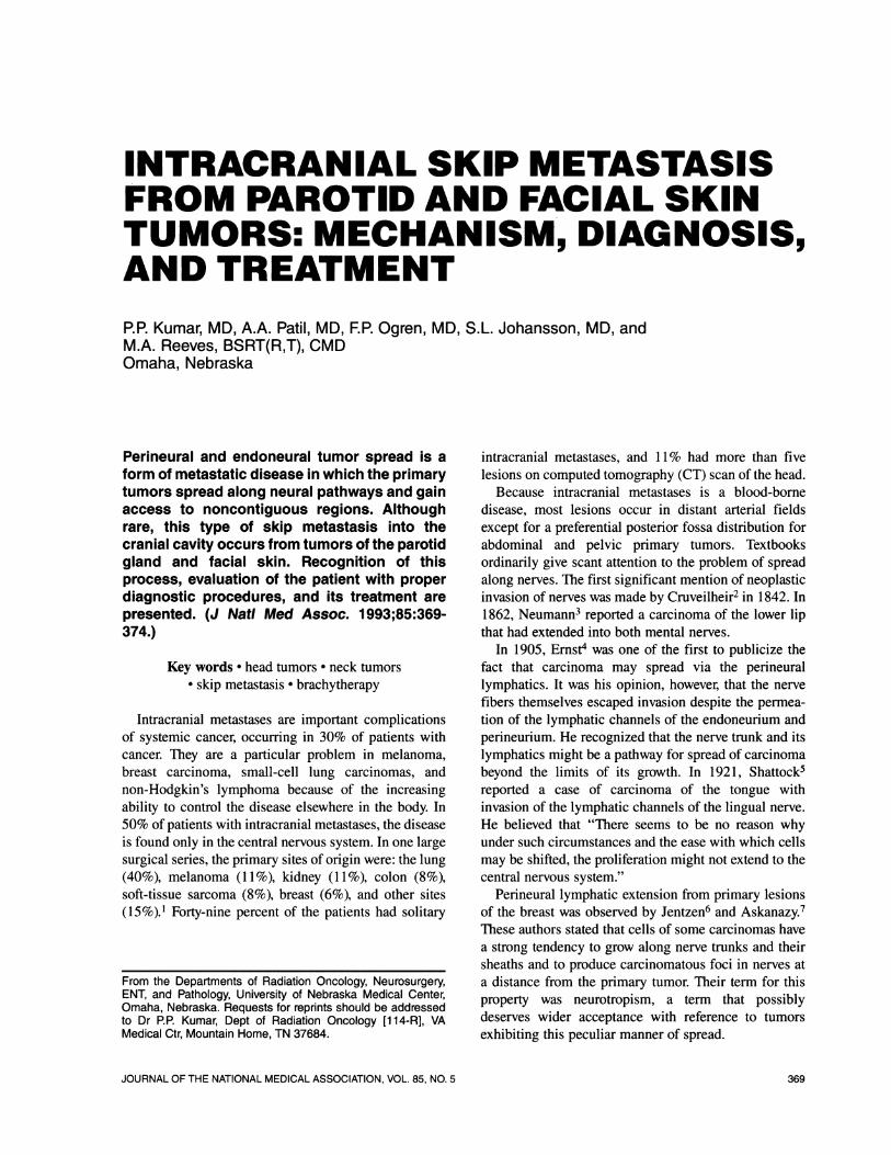
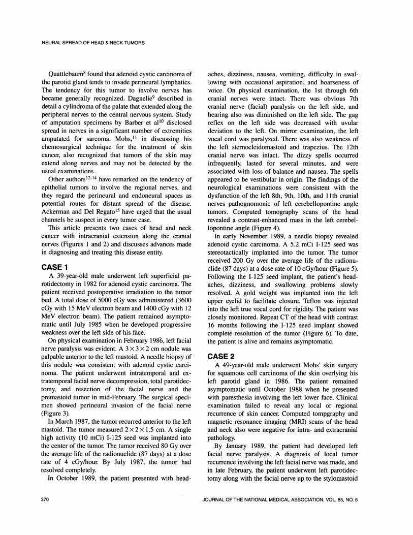
This article presents two cases of head and neckcancer with intracranial extension along the cranialnerves (Figures 1 and 2) and discusses advances madein diagnosing and treating this disease entity.
CASE 1A 39-year-old male underwent left superficial pa-
rotidectomy in 1982 for adenoid cystic carcinoma. Thepatient received postoperative irradiation to the tumorbed. A total dose of 5000 cGy was administered (3600cGy with 15 MeV electron beam and 1400 cGy with 12MeV electron beam). The patient remained asympto-matic until July 1985 when he developed progressiveweakness over the left side of his face.On physical examination in February 1986, left facial
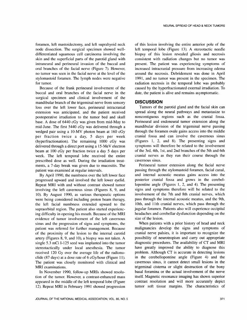
nerve paralysis was evident. A 3 X 3 X 2 cm nodule waspalpable anterior to the left mastoid. A needle biopsy ofthis nodule was consistent with adenoid cystic carci-noma. The patient underwent intratemporal and ex-tratemporal facial nerve decompression, total parotidec-tomy, and resection of the facial nerve and thepremastoid tumor in mid-February. The surgical speci-men showed perineural invasion of the facial nerve(Figure 3).
In March 1987, the tumor recurred anterior to the leftmastoid. The tumor measured 2 X 2 X 1.5 cm. A singlehigh activity (10 mCi) 1-125 seed was implanted intothe center of the tumor. The tumor received 80 Gy overthe average life of the radionuclide (87 days) at a doserate of 4 cGy/hour. By July 1987, the tumor hadresolved completely.
In October 1989, the patient presented with head-
aches, dizziness, nausea, vomiting, difficulty in swal-lowing with occasional aspiration, and hoarseness ofvoice. On physical examination, the 1st through 6thcranial nerves were intact. There was obvious 7thcranial nerve (facial) paralysis on the left side, andhearing also was diminished on the left side. The gagreflex on the left side was decreased with uvulardeviation to the left. On mirror examination, the leftvocal cord was paralyzed. There was also weakness ofthe left sternocleidomastoid and trapezius. The 12thcranial nerve was intact. The dizzy spells occurredinfrequently, lasted for several minutes, and wereassociated with loss of balance and nausea. The spellsappeared to be vestibular in origin. The findings of theneurological examinations were consistent with thedysfunction of the left 8th, 9th, 10th, and 11th cranialnerves pathognomonic of left cerebellopontine angletumors. Computed tomography scans of the headrevealed a contrast-enhanced mass in the left cerebel-lopontine angle (Figure 4).
In early November 1989, a needle biopsy revealedadenoid cystic carcinoma. A 5.2 mCi 1-125 seed wasstereotactically implanted into the tumor. The tumorreceived 200 Gy over the average life of the radionu-clide (87 days) at a dose rate of 10 cGy/hour (Figure 5).Following the 1-125 seed implant, the patient's head-aches, dizziness, and swallowing problems slowlyresolved. A gold weight was implanted into the leftupper eyelid to facilitate closure. Teflon was injectedinto the left true vocal cord for rigidity. The patient wasclosely monitored. Repeat CT of the head with contrast16 months following the 1-125 seed implant showedcomplete resolution of the tumor (Figure 6). To date,the patient is alive and remains asymptomatic.
CASE 2A 49-year-old male underwent Mohs' skin surgery
for squamous cell carcinoma of the skin overlying hisleft parotid gland in 1986. The patient remainedasymptomatic until October 1988 when he presentedwith paresthesia involving the left lower face. Clinicalexamination failed to reveal any local or regionalrecurrence of skin cancer. Computed tompgraphy andmagnetic resonance imaging (MRI) scans of the headand neck also were negative for intra- and extracranialpathology.By January 1989, the patient had developed left
facial nerve paralysis. A diagnosis of local tumorrecurrence involving the left facial nerve was made, andin late February, the patient underwent left parotidec-tomy along with the facial nerve up to the stylomastoid
370 JOURNAL OF THE NATIONAL MEDICAL ASSOCIATION, VOL. 85, NO. 5
NEURAL SPREAD OF HEAD & NECK TUMORS
foramen, left mastoidectomy, and left suprahyoid necknode dissection. The surgical specimen showed well-differentiated squamous cell carcinoma involving theskin and the superficial parts of the parotid gland withintraneural and perineural invasion of the buccal andoral branches of the facial nerve (Figure 7). However,no tumor was seen in the facial nerve at the level of thestylomastoid foramen. The lymph nodes were negativefor tumor.
Because of the frank perineural involvement of thebuccal and oral branches of the facial nerve in thesurgical specimen and clinical involvement of themandibular branch of the trigeminal nerve from sensoryloss over the left lower face, perineural intracranialextension was anticipated, and the patient receivedpostoperative irradiation to the tumor bed and skullbase. A dose of 6440 cGy was given from mid-May tomid-June. The first 5440 cGy was delivered through awedged pair using a 10-MV photon beam at 160 cGyper fraction twice a day, 5 days per week(hyperfractionation). The remaining 1000 cGy wasdelivered through a direct port using a 15-MeV electronbeam at 100 cGy per fraction twice a day 5 days perweek. The left temporal lobe received the entireprescribed dose as well. During the irradiation treat-ments, a 7-day break was given due to mucositis. Thepatient was examined at regular intervals.By April 1990, the numbness over the left lower face
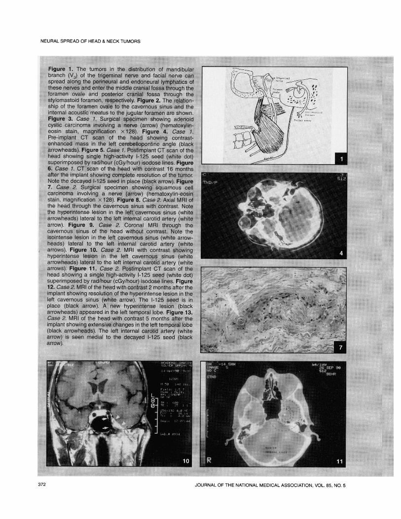
progressed upward and involved the left lower eyelid.Repeat MRI with and without contrast showed tumorinvolving the left cavernous sinus (Figures 8, 9, and10). By August 1990, as various therapeutic optionswere being considered including proton beam therapy,the left facial numbness extended upward to thesupraorbital region. The patient also started experienc-ing difficulty in opening his mouth. Because of the MRIevidence of tumor involvement of the left cavernoussinus and the progression of signs and symptoms, thepatient was referred for further management. Becauseof the proximity of the lesion to the internal carotidartery (Figures 8, 9, and 1O), a biopsy was not taken. Asingle 5.3 mCi 1-125 seed was implanted into the tumorstereotactically under local anesthesia. The tumorreceived 120 Gy over the average life of the radionu-clide (87 days) at a dose rate of 6 cGy/hour (Figure 11).The patient was closely monitored with clinical andMRI examinations.
In November 1990, follow-up MRIs showed resolu-tion of the tumor. However, a contrast-enhanced massappeared in the middle of the left temporal lobe (Figure12). Repeat MRI in February 1991 showed progression
of this lesion involving the entire anterior pole of theleft temporal lobe (Figure 13). A stereotactic needlebiopsy of this lesion revealed gliosis and necrosisconsistent with radiation changes but no tumor waspresent. The patient was experiencing symptoms ofincreased intracranial pressure from increasing edemaaround the necrosis. Debridement was done in April1991, and no tumor was present in the specimen. Theradiation necrosis in the temporal lobe was probablycaused by the hyperfractionated external irradiation. Todate, the patient is alive and remains asymptomatic.
DISCUSSIONTumors of the parotid gland and the facial skin can
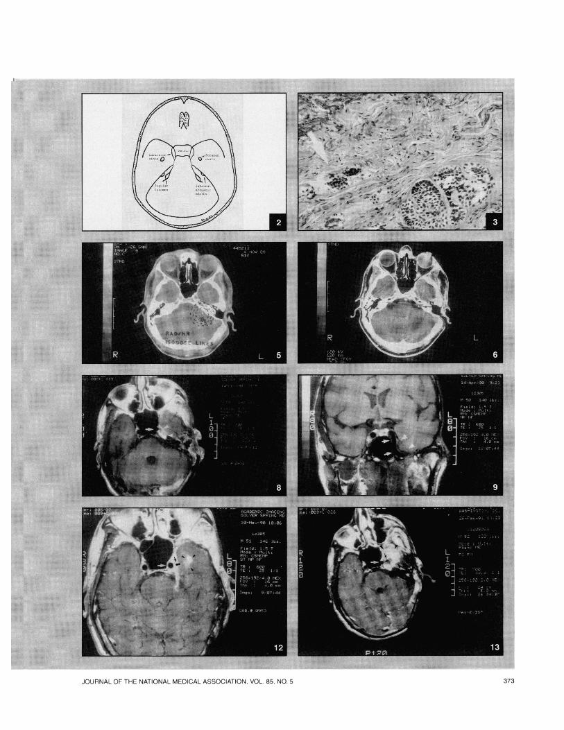
spread along the neural pathways and metastasize tononcontiguous regions such as the cranial fossa.Perineural and endoneural tumor extension along themandibular division of the trigeminal nerve passingthrough the foramen ovale gains access into the middlecranial fossa and can involve the cavernous sinus(Figures 1, 2, and 8). The presenting signs andsymptoms will therefore be related to the involvementof the 3rd, 4th, 1st, and 2nd branches of the 5th and 6thcranial nerves as they run their course through thecavernous sinus.
Perineural tumor extension along the facial nervepassing through the stylomastoid foramen, facial canal,and internal acoustic meatus gains access into theposterior cranial fossa and grows in the cerebel-lopontine angle (Figures 1, 2, and 4). The presentingsigns and symptoms therefore will be related to theinvolvement of the 7th and 8th cranial nerves, whichpass through the internal acoustic meatus, and the 9th,10th, and 11th cranial nerves, which pass through thejugular foramen. Patients also will experience occipitalheadaches and cerebellar dysfunction depending on thesize of the lesion.When patients with a prior history of head and neck
malignancies develop the signs and symptoms ofcranial nerve palsies, it is important to recognize thepossibility of neurotropism and carry out appropriatediagnostic procedures. The availability of CT and MRIhave greatly improved the ability to diagnose thisproblem. Although CT is accurate in detecting lesionsin the cerebellopontine angle (Figure 4) and thecavernous sinus, it cannot detect small lesions in thetrigeminal cisterna or slight destruction of the bonybasal foramina or the actual involvement of the nerveitself. Magnetic resonance imaging has shown superiorcontrast resolution and will more accurately depicttumor soft tissue margins. The characteristics of
JOURNAL OF THE NATIONAL MEDICAL ASSOCIATION, VOL. 85, NO. 5 371
NEURAL SPREAD OF HEAD & NECK TUMORS
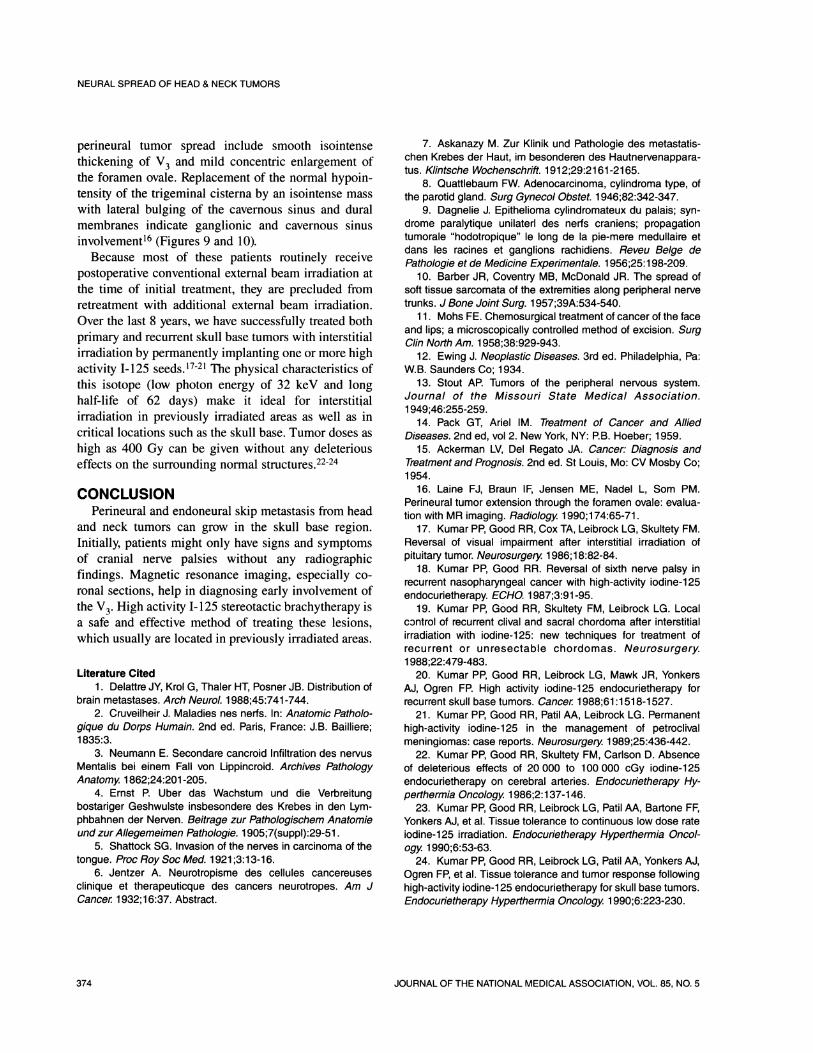
Figure 1. The tumors:..in the distribution. of mandibular*branch ....(V3) o ..f the trig.ern..inal .ne .eand faciale canspread:: alon th eierl n noertypaiso*these nerves and effnter the middlecraninal fosal Ythog theforamen ovale and posterio.r c.ranial fossa thro.ugh the..stylomastoid for .....a.men, r.espectively. Figure 2 .:The reltin-ship of the forarnen ovate.to the cavernous sinus and theinternal acoustic meatuJs to the jugular forame.n are shown.Figure 3.. 3:Case 1.t.Suc.... l specimen ......howing.adenodc ystic carcinomainvoling. a nerv (ro) (hematoxylin-eosin stain, magnfication x 12). FIgre 4. Case 1.Pre-implant CT scan f the head .show ng contrast-*enhance....d mass lIn the left bellopt a.arrowhe ...ads).Fi e5.Case ..... ....stimpla..t.CT scan o.thehead showing single high-activity 1-125 seed (white dot)superimposed by rad/h .o.ur..:(cGy/h.our) isi...odose...lines..F.gure.6. Case ..1. CT. sca.noft.h.e hed with cntt 16 months;after the implant showing co.....mplete re.slution of the tumor.N<ote t:he decayed 1-125:seed in pl.ace (black arrow). Figure..7. Ca.se.2. Su........rgicalspecimen ........sh.owin ...g squamous. ..
carcinoma involving a .n;erve (arow) (hemao xylin-osinstain, magnification x 128). FIgure 8. Case 2. Axial MRI ofthe head through the caveernoussinus with. contrst Notet inte nse leasion i the lfc ernous sinus hitearrowheads) lateral to the left lnternal cafro d arerswhite.arrow). Fsgure 9. Case 2. Coronal MRI through thecavernous sin.us of the..he.a.d .:witho.ut co..ntrast. Not.>..e.. the:i.sointense lesionin the. left cavernous snus (white arrow-heads) lateral to the left internal carotid artery(whitearrows). Figure 10. Case2.aMR with contrast showing.hyperintense lesion i.n:.the. left cavernous sinus...(white.arrowheads) latera to the lef intera carotid artery (whitearrows). Figure 11. Case 2. Postimplant CT scan of thehead showing a singlehignh-actvity 1-125 sed (white dot)..superimposedby red/hour (cGy/hour) isodo.se li:nes...Figure..12. Case 2 CMRI of the head with con.trast months after theimplant showing resolution. of the hyperintense lesion.in theleft cavernous sinus (white arrow). The !-1295 seed is in:place (black arrow). .A..ne.w hyperintense..l.esion (bl ckarrowheads) appeared in the left temporal lobe. F:igure 13.Casen2n. MRI of t hehead with contrast 5 months after the...implant s.howing .....extensve c....han.n the left temporal lobe(black arrowheads). Th.e left........ .intern.al carotid arery (white:arrow) is seen medial to the decayed 1-125 seed (blackarrow). . ...........
JOURNAL OF THE NATIONAL MEDICAL ASSOCIATION, VOL. 85, NO. 5372
JOURNAL OF THE NATIONAL MEDICAL ASSOCIATION, VOL. 85, NO. 5 373
NEURAL SPREAD OF HEAD & NECK TUMORS
perineural tumor spread include smooth isointensethickening of V3 and mild concentric enlargement ofthe foramen ovale. Replacement of the normal hypoin-tensity of the trigeminal cisterna by an isointense masswith lateral bulging of the cavernous sinus and duralmembranes indicate ganglionic and cavernous sinusinvolvement16 (Figures 9 and 10).
Because most of these patients routinely receivepostoperative conventional external beam irradiation atthe time of initial treatment, they are precluded fromretreatment with additional external beam irradiation.Over the last 8 years, we have successfully treated bothprimary and recurrent skull base tumors with interstitialirradiation by permanently implanting one or more highactivity 1-125 seeds.17-2' The physical characteristics ofthis isotope (low photon energy of 32 keV and longhalf-life of 62 days) make it ideal for interstitialirradiation in previously irradiated areas as well as incritical locations such as the skull base. Tumor doses ashigh as 400 Gy can be given without any deleteriouseffects on the surrounding normal structures.22-24
CONCLUSIONPerineural and endoneural skip metastasis from head
and neck tumors can grow in the skull base region.Initially, patients might only have signs and symptomsof cranial nerve palsies without any radiographicfindings. Magnetic resonance imaging, especially co-ronal sections, help in diagnosing early involvement ofthe V3. High activity I-125 stereotactic brachytherapy isa safe and effective method of treating these lesions,which usually are located in previously irradiated areas.
Literature Cited1. Delattre JY, Krol G, Thaler HT, Posner JB. Distribution of
brain metastases. Arch Neurol. 1988;45:741-744.2. Cruveilheir J. Maladies nes nerfs. In: Anatomic Patholo-
gique du Dorps Humain. 2nd ed. Paris, France: J.B. Bailliere;1835:3.
3. Neumann E. Secondare cancroid Infiltration des nervusMentalis bei einem Fall von Lippincroid. Archives PathologyAnatomy. 1862;24:201-205.
4. Ernst P. Uber das Wachstum und die Verbreitungbostariger Geshwulste insbesondere des Krebes in den Lym-phbahnen der Nerven. Beitrage zur Pathologischem Anatomieund zur Allegemeimen Pathologie. 1 905;7(suppl):29-51.
5. Shattock SG. Invasion of the nerves in carcinoma of thetongue. Proc Roy Soc Med. 1921;3:13-16.
6. Jentzer A. Neurotropisme des cellules cancereusesclinique et therapeuticque des cancers neurotropes. Am JCancer 1932;16:37. Abstract.
7. Askanazy M. Zur Klinik und Pathologie des metastatis-chen Krebes der Haut, im besonderen des Hautnervenappara-tus. Klintsche Wochenschrift. 1912;29:2161-2165.
8. Quattlebaum FW. Adenocarcinoma, cylindroma type, ofthe parotid gland. Surg Gynecol Obstet. 1946;82:342-347.
9. Dagnelie J. Epithelioma cylindromateux du palais; syn-drome paralytique unilaterl des nerfs craniens; propagationtumorale "hodotropique" le long de la pie-mere medullaire etdans les racines et ganglions rachidiens. Reveu Belge dePathologie et de Medicine Experimentale. 1956;25:198-209.
10. Barber JR, Coventry MB, McDonald JR. The spread ofsoft tissue sarcomata of the extremities along peripheral nervetrunks. J Bone Joint Surg. 1 957;39A:534-540.
11. Mohs FE. Chemosurgical treatment of cancer of the faceand lips; a microscopically controlled method of excision. SurgClin North Am. 1958;38:929-943.
12. Ewing J. Neoplastic Diseases. 3rd ed. Philadelphia, Pa:W.B. Saunders Co; 1934.
13. Stout AP. Tumors of the peripheral nervous system.Journal of the Missouri State Medical Association.1 949;46:255-259.
14. Pack GT, Ariel IM. Treatment of Cancer and AlliedDiseases. 2nd ed, vol 2. New York, NY: P.B. Hoeber; 1959.
15. Ackerman LV, Del Regato JA. Cancer: Diagnosis andTreatment and Prognosis. 2nd ed. St Louis, Mo: CV Mosby Co;1954.
16. Laine FJ, Braun IF, Jensen ME, Nadel L, Som PM.Perineural tumor extension through the foramen ovale: evalua-tion with MR imaging. Radiology. 1990;174:65-71.
17. Kumar PP, Good RR, Cox TA, Leibrock LG, Skultety FM.Reversal of visual impairment after interstitial irradiation ofpituitary tumor. Neurosurgery. 1986;1 8:82-84.
18. Kumar PP, Good RR. Reversal of sixth nerve palsy inrecurrent nasopharyngeal cancer with high-activity iodine-125endocurietherapy. ECHO. 1987;3:91-95.
19. Kumar PP, Good RR, Skultety FM, Leibrock LG. Localcontrol of recurrent clival and sacral chordoma after interstitialirradiation with iodine-125: new techniques for treatment ofrecurrent or unresectable chordomas. Neurosurgery.1 988;22:479-483.
20. Kumar PP, Good RR, Leibrock LG, Mawk JR, YonkersAJ, Ogren FP. High activity iodine-125 endocurietherapy forrecurrent skull base tumors. Cancer 1988;61 :1518-1527.
21. Kumar PP, Good RR, Patil AA, Leibrock LG. Permanenthigh-activity iodine-125 in the management of petroclivalmeningiomas: case reports. Neurosurgery. 1989;25:436-442.
22. Kumar PP, Good RR, Skultety FM, Carlson D. Absenceof deleterious effects of 20 000 to 100 000 cGy iodine-125endocurietherapy on cerebral arteries. Endocurietherapy Hy-perthermia Oncology. 1986;2:137-146.
23. Kumar PP, Good RR, Leibrock LG, Patil AA, Bartone FF,Yonkers AJ, et al. Tissue tolerance to continuous low dose rateiodine-125 irradiation. Endocurietherapy Hyperthermia Oncol-ogy. 1990;6:53-63.
24. Kumar PP, Good RR, Leibrock LG, Patil AA, Yonkers AJ,Ogren FP, et al. Tissue tolerance and tumor response followinghigh-activity iodine-125 endocurietherapy for skull base tumors.Endocurietherapy Hyperthermia Oncology. 1990;6:223-230.
374 JOURNAL OF THE NATIONAL MEDICAL ASSOCIATION, VOL. 85, NO. 5