intracranial hypertension (pseudotumor cerebri)€¦ · 9/11/11! 3! symptoms consciousness is never...
TRANSCRIPT
9/11/11
1
Richard E. Castillo, O.D., D.O The Oklahoma College of Optometry NORTHEASTERN STATE UNIVERSITY
Director, Ophthalmic Surgery Service
9/11/11
Intracranial Hypertension (Pseudotumor cerebri)
Introduction
� IIH is a condition marked by: � Unkown cause � Elevated cerebrovascular fluid
(CSF) pressure � Papilledema
� Also referred to as � Pseudotumor cerebri
syndrome � Benign intracranial
hypertension
9/11/11
Incidence
9/11/11
� General population: 1:100,000
� Obese women of childbearing age: 19:100,000
� Can develop in children � Male = female � Typically precipitated by
antibiotic or steroid withdrawal

9/11/11
2
Age at Diagnosis
9/11/11
Major Symptoms
9/11/11
� Headache � Worse in AM � Exacerbated by Valsalva maneuver
� Transient visual obscurations � 1-5 sec. “graying-out” � Induced by orthostatic changes
� Horizontal diplopia � Sixth nerve Palsy
Less common symptoms
9/11/11
� Pulsatile tinnitus � Pain associated with
� Neck � Shoulders � Back

9/11/11
3
Symptoms
Consciousness is
Never Altered
9/11/11
Physical signs
� Cardinal sign is Papilledema � Usually bilateral � May be
asymmetrical � Occasionally
unilateral
9/11/11
ONH
9/11/11
9/11/11
4
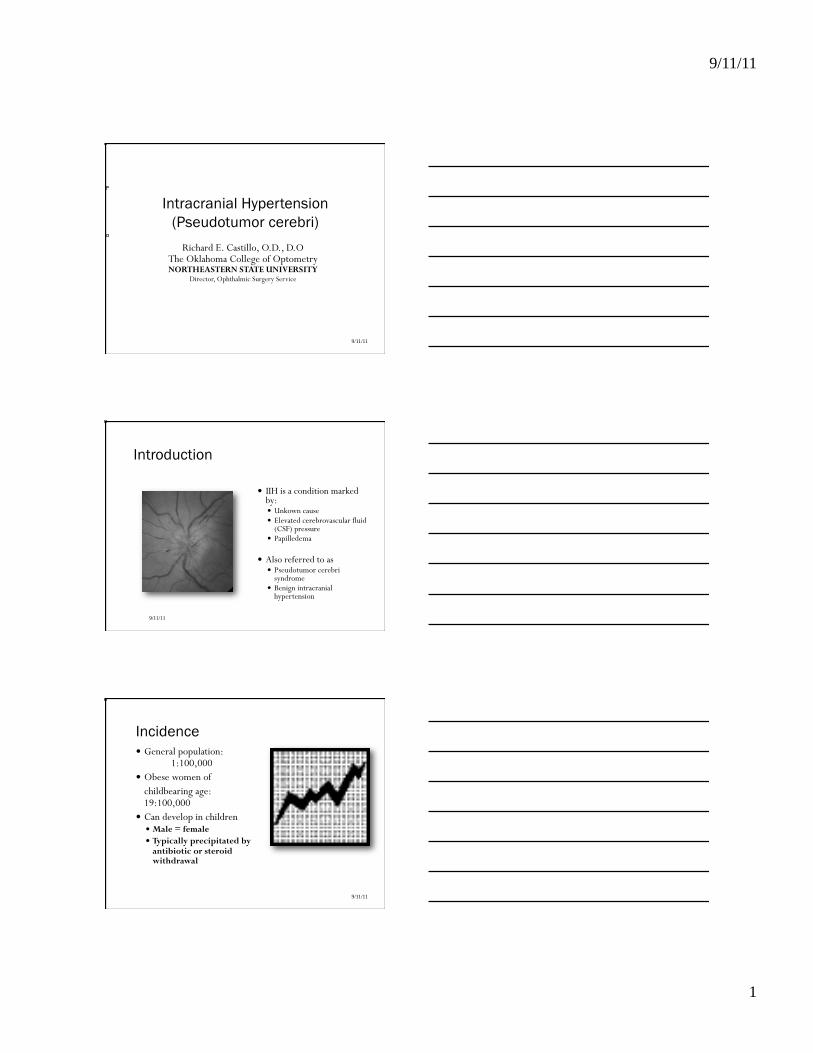
Permanent Sequelae
� Visual loss 2˚ to papilledema � Occurs in10 - 25% of
patients � Including children!
9/11/11
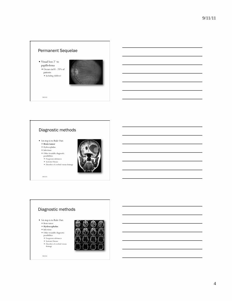
Diagnostic methods
� 1st step is to Rule Out: � Brain tumor � Hydrocephalus � Infections � Other treatable diagnostic
possibilities � Exogenous substances � Systemic Disease � Disorders of cerebral venous drainage
9/11/11
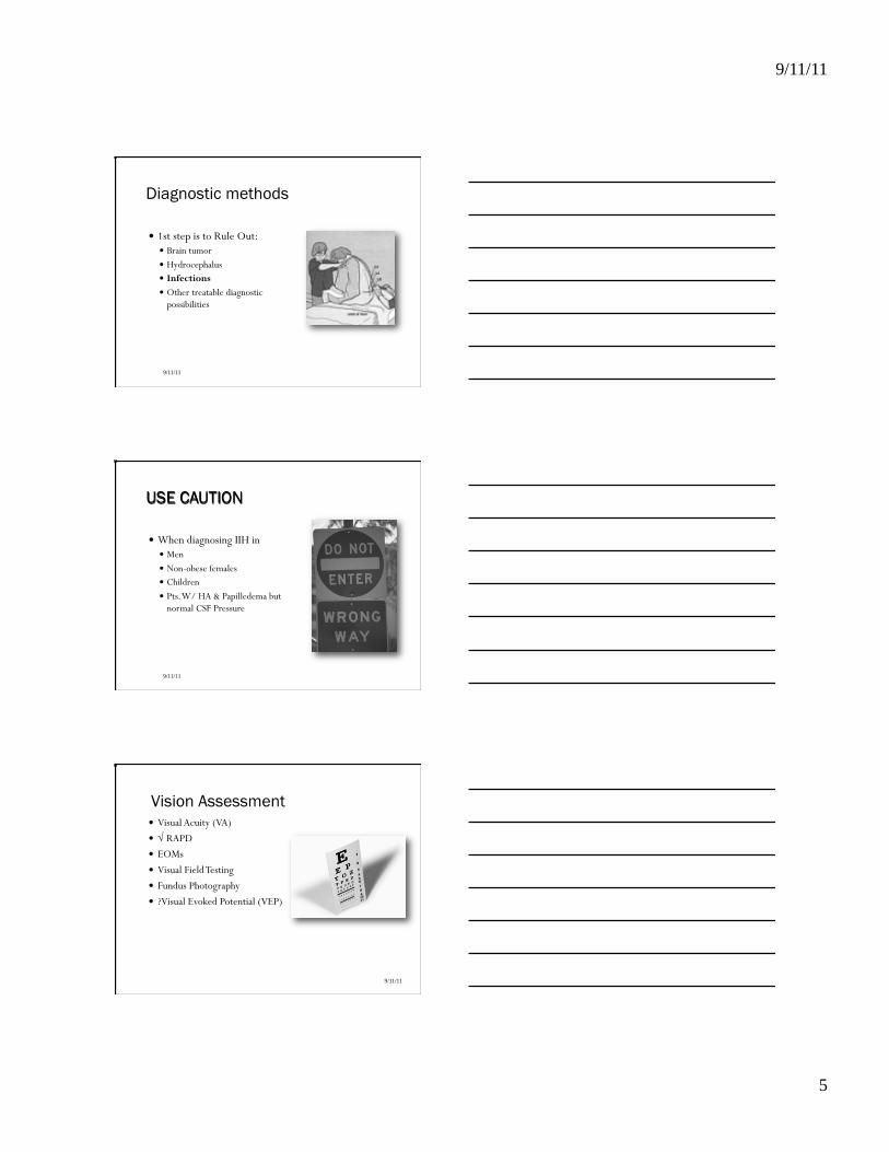
Diagnostic methods
� 1st step is to Rule Out: � Brain tumor � Hydrocephalus � Infections � Other treatable diagnostic
possibilities � Exogenous substances � Systemic Disease � Disorders of cerebral venous
drainage
9/11/11
9/11/11
5
Diagnostic methods
� 1st step is to Rule Out: � Brain tumor � Hydrocephalus � Infections � Other treatable diagnostic
possibilities
9/11/11
USE CAUTION
� When diagnosing IIH in � Men � Non-obese females � Children � Pts. W/ HA & Papilledema but
normal CSF Pressure
9/11/11
Vision Assessment
9/11/11
� Visual Acuity (VA) � √ RAPD � EOMs � Visual Field Testing � Fundus Photography � ?Visual Evoked Potential (VEP)
9/11/11
6
Lab Studies
� Lumbar puncture (LP) is MANDATORY � CSF ≥ 250 mm H2O
� CSF cytology to exclude: � Inflammation � Tumor cells � Infection
9/11/11
Typical CSF in IIH
� Normal or low protein (< 20 mg%)
� Normal glucose � Normal cell count
9/11/11
Additional lab
� ANA � RPR, VDRL � FTA-ABS, MHA-TP � Serum Calcium
� (R/O hypo-parathyroidism)
9/11/11
9/11/11
7
Endocrine studies
� Only if suspicion of � Hyperadrenalism
(Cushing’s) � Hypoadrenalism
(Addison’s) � Hypoparathyroidism
9/11/11
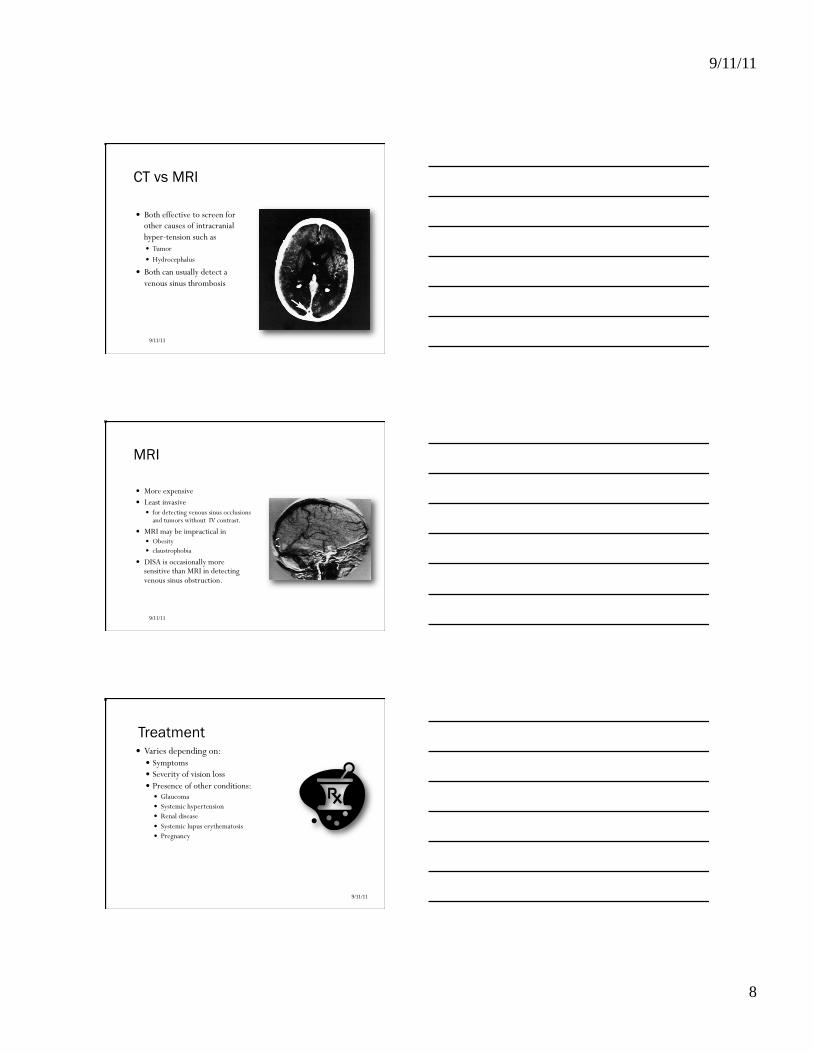
Imaging Techniques
� CT � MRI � DISA � Arteriography
9/11/11
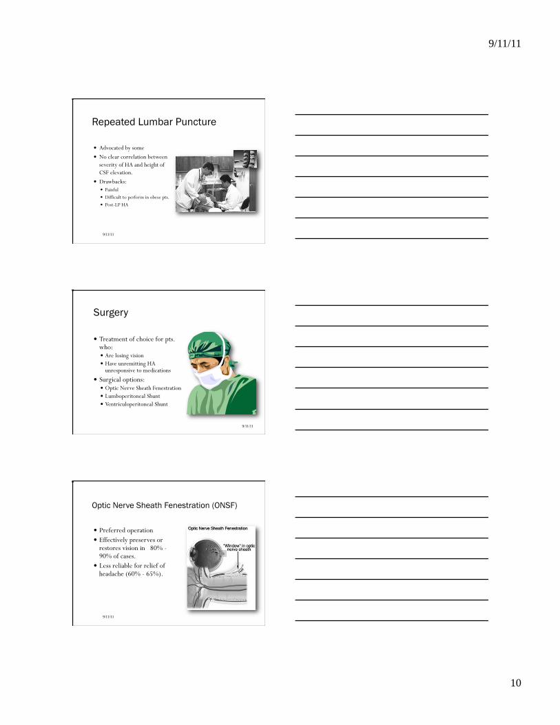
CT Findings
� Normal or small ventricles
� 25% - 50% of patients exhibit an empty sella
9/11/11
9/11/11
8
CT vs MRI
� Both effective to screen for other causes of intracranial hyper-tension such as � Tumor � Hydrocephalus
� Both can usually detect a venous sinus thrombosis
9/11/11
MRI
� More expensive � Least invasive
� for detecting venous sinus occlusions and tumors without IV contrast.
� MRI may be impractical in � Obesity � claustrophobia
� DISA is occasionally more sensitive than MRI in detecting venous sinus obstruction.
9/11/11
Treatment
9/11/11
� Varies depending on: � Symptoms � Severity of vision loss � Presence of other conditions:
� Glaucoma � Systemic hypertension � Renal disease � Systemic lupus erythematosis � Pregnancy

9/11/11
9
Specific Treatment Modalities
� Diet � Medication
� Acetazolimide � Furosamide
� Repeated lumbar puncture
� Surgery
9/11/11
Diet
� Weight-reduction diet should be offered to all IIH pts.
� Pts. should be sent for professional dietary instruction.
9/11/11
Medication
9/11/11
� Headache � Beta-adrenergic blockers � Calcium channel blockers � Tricyclic antidepresants
� Visual Symptoms � Carbonic anhydrase inhibitors � Furosemide � Steroids
9/11/11
10
Repeated Lumbar Puncture
� Advocated by some � No clear correlation between
severity of HA and height of CSF elevation.
� Drawbacks: � Painful � Difficult to perform in obese pts. � Post-LP HA
9/11/11
Surgery
9/11/11
� Treatment of choice for pts. who: � Are losing vision � Have unremitting HA
unresponsive to medications
� Surgical options: � Optic Nerve Sheath Fenestration � Lumboperitoneal Shunt � Ventriculoperitoneal Shunt
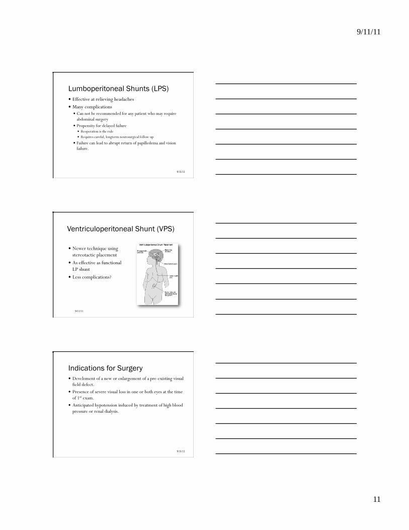
Optic Nerve Sheath Fenestration (ONSF)
� Preferred operation � Effectively preserves or
restores vision in 80% - 90% of cases.
� Less reliable for relief of headache (60% - 65%).
9/11/11
9/11/11
11
Lumboperitoneal Shunts (LPS)
9/11/11
� Effective at relieving headaches � Many complications
� Can not be recommended for any patient who may require abdominal surgery
� Propensity for delayed failure � Reoperation is the rule � Requires careful, longterm neurosurgical follow-up
� Failure can lead to abrupt return of papilledema and vision failure.
Ventriculoperitoneal Shunt (VPS)
� Newer technique using stereotactic placement
� As effective as functional LP shunt
� Less complications?
9/11/11
Indications for Surgery
9/11/11
� Develoment of a new or enlargement of a pre-existing visual field defect.
� Presence of severe visual loss in one or both eyes at the time of 1st exam.
� Anticipated hypotension induced by treatment of high blood pressure or renal dialysis.
9/11/11
12
Indications for Surgery
9/11/11
� Psychosocial reasons such as pts. inability to perform VF testing, noncompliance with medical treatment, or an itinerant life-style.
� Headache unresponsive to standard headache regimens.
Specific Treatment Problems
� The Asymptomatic Patient � May not need Tx � Follow q1-3 months
� Headache � Meds →→→→ LPS/VPS
� Serious Visual Loss � ONSF
� Rapidly Progressive Visual Loss � ONSF+ LPS/VPS+ Meds � If HTN do not drop BP to quickly
� Pregnancy with Headaches � Beta-Blockers, LP, LPS/VPS, bedrest, ONSF
� Renal Disease � ONSF � Beware hypotensive episodes
9/11/11
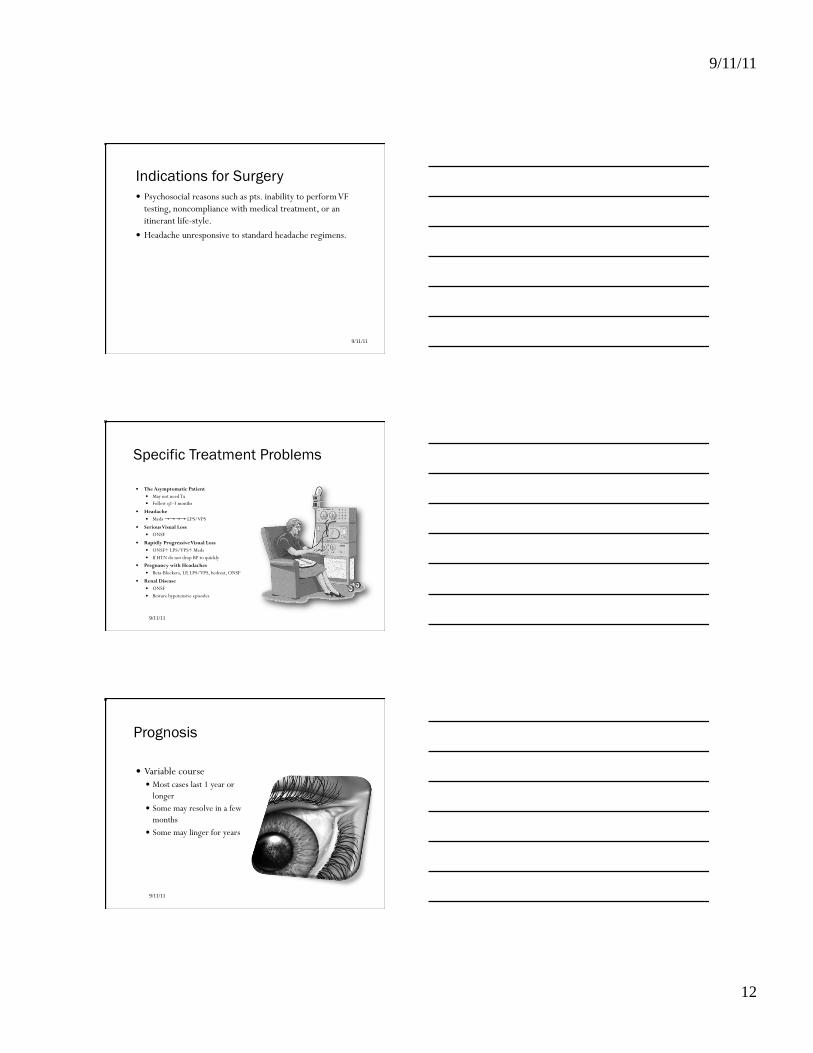
Prognosis
� Variable course � Most cases last 1 year or
longer � Some may resolve in a few
months � Some may linger for years
9/11/11
9/11/11
13
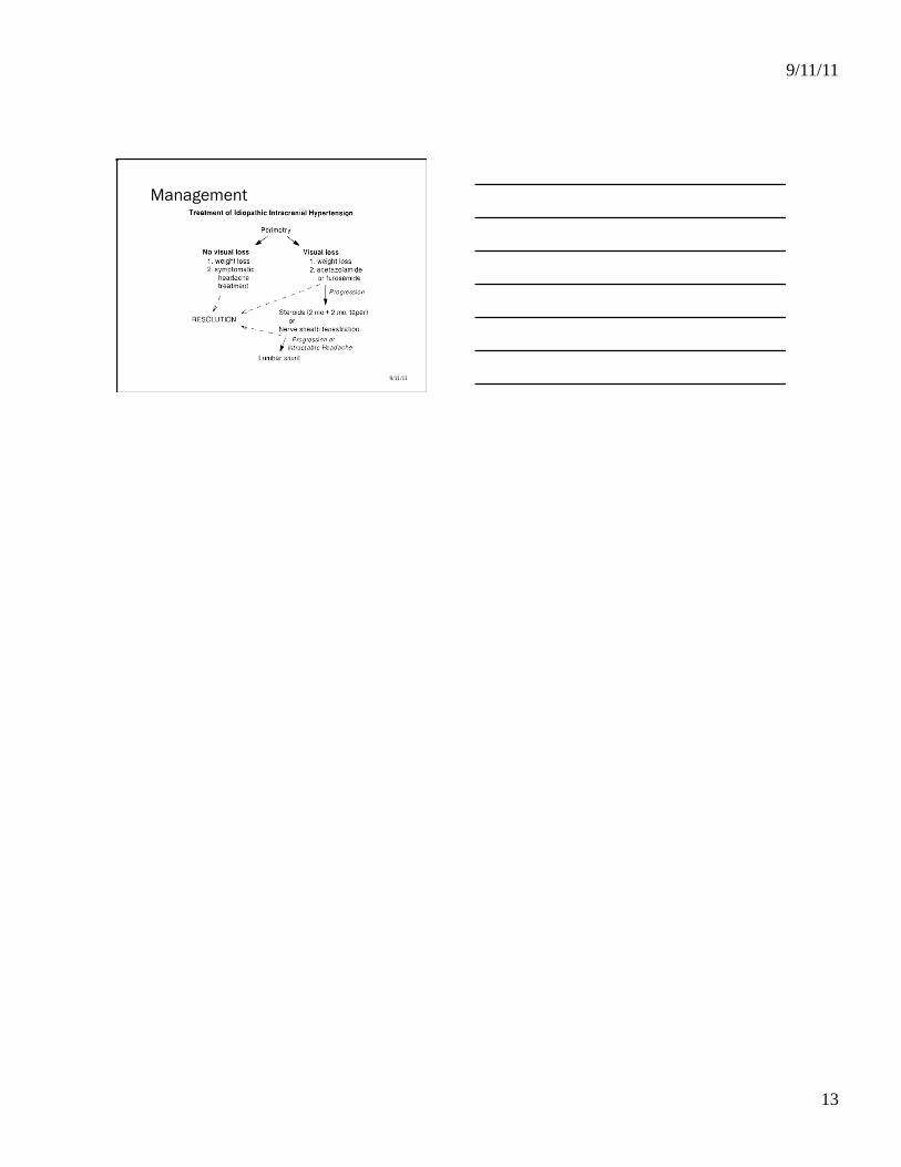
Management
9/11/11