interceptive orthodontic treatment: efficient early...
TRANSCRIPT
Interceptive Orthodontic Treatment: Efficient Early Correction of Malocclusions
Cameron Mashouf, DDS, MS Kayhan L. Mashouf, DMD, MSD
Updated Version, July 2017
www.interceptiveortho.com
1
Introduction When deciding on a treatment protocol for young children (7- 8 years old) with newly erupted crooked teeth, a dentist faces questions such as whether treatment should be recommended or not, and if orthodontic treatment is recommended, what technical protocol should be suggested to the concerned parents of a young child? The social aspects of this subject should be considered when evaluating the timing of orthodontic treatment. By age 8, children’s criteria for attractiveness are the same as those of adults, and the appearance of the smile is considered to be an important criterion when judging facial attractiveness [1]. Thus, interceptive treatment, such as the correction of jaw deformities and dental irregularities, can help raise a young child’s self-esteem. While there are some who question the benefits of interceptive treatment [2-6], there are others who have argued in favor of some form of intervention [7-10]. A survey by the College of Diplomates of the American Board of Orthodontics (CDABO) shows that a majority of the ABO diplomats value interceptive orthodontics and are actively involved in some sort of mixed dentition treatment [11]. One thing that is clear is that there has been minimal progress in the development of appliances and techniques that can efficiently move young children’s teeth [12]. Functional appliances used alone or in combination with fixed appliances have not produced predictable results quickly [13,14]. This paper is intended for dental and orthodontic professionals, and it presents new approaches that include expansion and use of deciduous molars and canines as anchors in the treatment of many mixed dentition cases such as: anterior crowding, open bite, overbite, and crossbite.
2
Correcting Crowding: Creating Space through Expansion The primary way to create space in the mixed dentition protocol proposed in this paper is through expansion of the transverse dimension. The recommended period to begin this protocol is at 7-8 years of age. This coincides with the eruption of the permanent first molars and permanent incisors during the early mixed dentition period. One of the key benefits of this early expansion is a reduction in the need to remove deciduous teeth in grade school children and permanent teeth in middle school and high school children. The protocol follows McNamara’s method [15], with some changes to make it more practical. These changes include avoiding occlusal coverage for the maxillary expander and using fixed expansion in the mandible instead of a removable Schwarz. Early expansion of the maxilla is a stable and effective way to correct arch length deficiencies [16-19]. Conversely, the effectiveness of expansion in the mandibular arch has been disputed [20-24]. Disagreement with regard to the effectiveness of mandibular arch expansion may be related to the differences in the timing of treatment or the methods being used. The expansion appliances used in this protocol for the maxillary and the mandibular arches take advantage of different growth mechanisms in the corresponding jawbones. In the maxilla, the increase in transverse dimension is accomplished through skeletal expansion at the intermaxillary suture. In the mandible, this dimension is increased through dentoaveolar expansion of the buccal segments. Maxillary Expansion Expansion of the maxilla is achieved with a 2-banded maxillary expansion appliance (MEA) attached to the first permanent molars. This produces expansion of the maxilla equivalent to the more traditional 4-banded appliance [25,26].
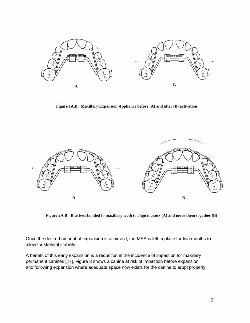
A 12mm “Variety” expansion screw ∗ is used with additional 0.036” arms extending from the first permanent molars mesially to the deciduous canines on the palatal side (Figure 1A). The appliance is activated once per day until the palatal cusps of the maxillary posterior teeth touch the buccal cusps of the mandibular posterior teeth (Figure 1B).
The next step involves bonding brackets to the maxillary teeth. Deciduous molars and canines are bonded at the same time as the permanent incisors. Resilient arch wires are used to align the incisors and move them together, using the space developed through expansion (Figure 2A,B).
∗ (Dentaurum, Ispringen, Germany)
3
Once the desired amount of expansion is achieved, the MEA is left in place for two months to allow for skeletal stability.
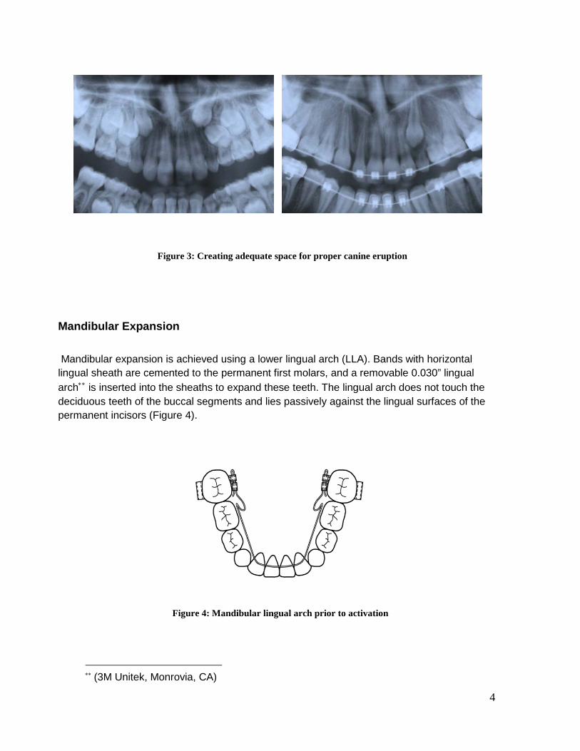
A benefit of this early expansion is a reduction in the incidence of impaction for maxillary permanent canines [27]. Figure 3 shows a canine at risk of impaction before expansion and following expansion where adequate space now exists for the canine to erupt properly.
Figure 1A,B: Maxillary Expansion Appliance before (A) and after (B) activation
Figure 2A,B: Brackets bonded to maxillary teeth to align incisors (A) and move them together (B)
A B
A B
4
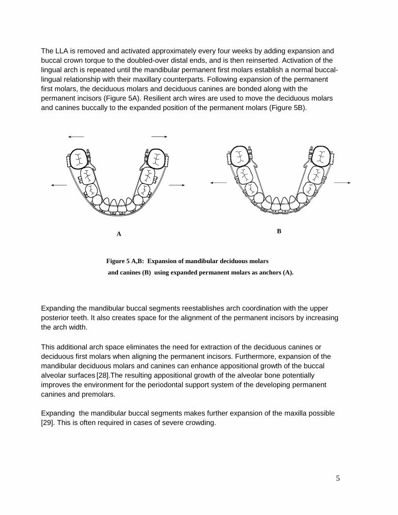
Mandibular Expansion
Mandibular expansion is achieved using a lower lingual arch (LLA). Bands with horizontal lingual sheath are cemented to the permanent first molars, and a removable 0.030” lingual arch∗∗ is inserted into the sheaths to expand these teeth. The lingual arch does not touch the deciduous teeth of the buccal segments and lies passively against the lingual surfaces of the permanent incisors (Figure 4).
∗∗ (3M Unitek, Monrovia, CA)
Figure 4: Mandibular lingual arch prior to activation
Figure 3: Creating adequate space for proper canine eruption
5
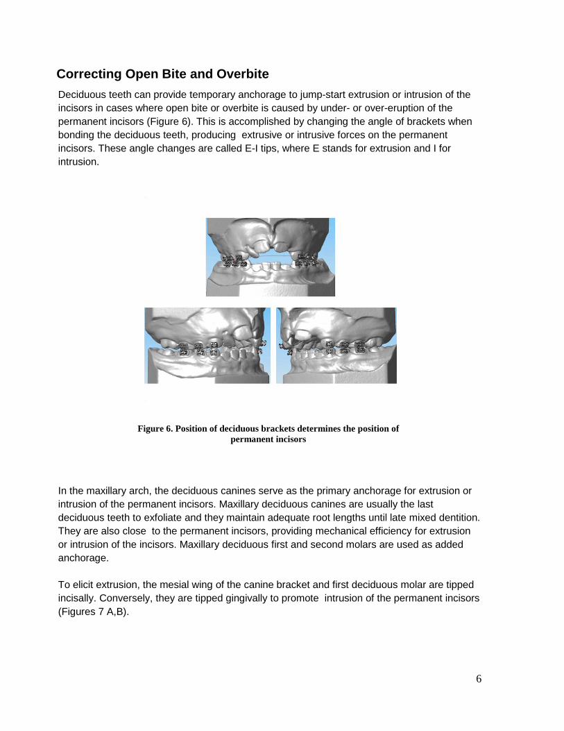
The LLA is removed and activated approximately every four weeks by adding expansion and buccal crown torque to the doubled-over distal ends, and is then reinserted. Activation of the lingual arch is repeated until the mandibular permanent first molars establish a normal buccal-lingual relationship with their maxillary counterparts. Following expansion of the permanent first molars, the deciduous molars and deciduous canines are bonded along with the permanent incisors (Figure 5A). Resilient arch wires are used to move the deciduous molars and canines buccally to the expanded position of the permanent molars (Figure 5B).
Expanding the mandibular buccal segments reestablishes arch coordination with the upper posterior teeth. It also creates space for the alignment of the permanent incisors by increasing the arch width.
This additional arch space eliminates the need for extraction of the deciduous canines or deciduous first molars when aligning the permanent incisors. Furthermore, expansion of the mandibular deciduous molars and canines can enhance appositional growth of the buccal alveolar surfaces [28].The resulting appositional growth of the alveolar bone potentially improves the environment for the periodontal support system of the developing permanent canines and premolars.
Expanding the mandibular buccal segments makes further expansion of the maxilla possible [29]. This is often required in cases of severe crowding.
Figure 5 A,B: Expansion of mandibular deciduous molars
and canines (B) using expanded permanent molars as anchors (A).
A B
6
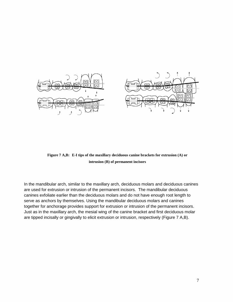
Correcting Open Bite and Overbite Deciduous teeth can provide temporary anchorage to jump-start extrusion or intrusion of the incisors in cases where open bite or overbite is caused by under- or over-eruption of the permanent incisors (Figure 6). This is accomplished by changing the angle of brackets when bonding the deciduous teeth, producing extrusive or intrusive forces on the permanent incisors. These angle changes are called E-I tips, where E stands for extrusion and I for intrusion.
In the maxillary arch, the deciduous canines serve as the primary anchorage for extrusion or intrusion of the permanent incisors. Maxillary deciduous canines are usually the last deciduous teeth to exfoliate and they maintain adequate root lengths until late mixed dentition. They are also close to the permanent incisors, providing mechanical efficiency for extrusion or intrusion of the incisors. Maxillary deciduous first and second molars are used as added anchorage. To elicit extrusion, the mesial wing of the canine bracket and first deciduous molar are tipped incisally. Conversely, they are tipped gingivally to promote intrusion of the permanent incisors (Figures 7 A,B).
Figure 6. Position of deciduous brackets determines the position of permanent incisors
7
In the mandibular arch, similar to the maxillary arch, deciduous molars and deciduous canines are used for extrusion or intrusion of the permanent incisors. The mandibular deciduous canines exfoliate earlier than the deciduous molars and do not have enough root length to serve as anchors by themselves. Using the mandibular deciduous molars and canines together for anchorage provides support for extrusion or intrusion of the permanent incisors. Just as in the maxillary arch, the mesial wing of the canine bracket and first deciduous molar are tipped incisally or gingivally to elicit extrusion or intrusion, respectively (Figure 7 A,B).
Figure 7 A,B: E-I tips of the maxillary deciduous canine brackets for extrusion (A) or
intrusion (B) of permanent incisors
8
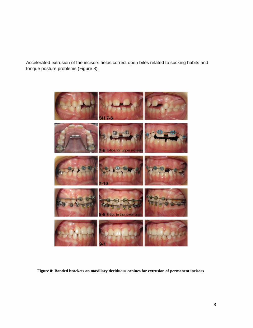
Accelerated extrusion of the incisors helps correct open bites related to sucking habits and tongue posture problems (Figure 8).
Figure 8: Bonded brackets on maxillary deciduous canines for extrusion of permanent incisors
9
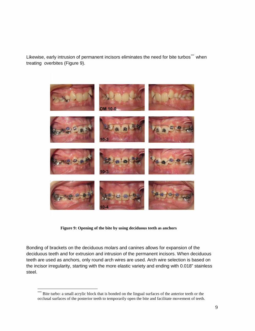
Likewise, early intrusion of permanent incisors eliminates the need for bite turbos*** when treating overbites (Figure 9).
Figure 9: Opening of the bite by using deciduous teeth as anchors
Bonding of brackets on the deciduous molars and canines allows for expansion of the deciduous teeth and for extrusion and intrusion of the permanent incisors. When deciduous teeth are used as anchors, only round arch wires are used. Arch wire selection is based on the incisor irregularity, starting with the more elastic variety and ending with 0.018” stainless steel.
*** Bite turbo: a small acrylic block that is bonded on the lingual surfaces of the anterior teeth or the occlusal surfaces of the posterior teeth to temporarily open the bite and facilitate movement of teeth.
10
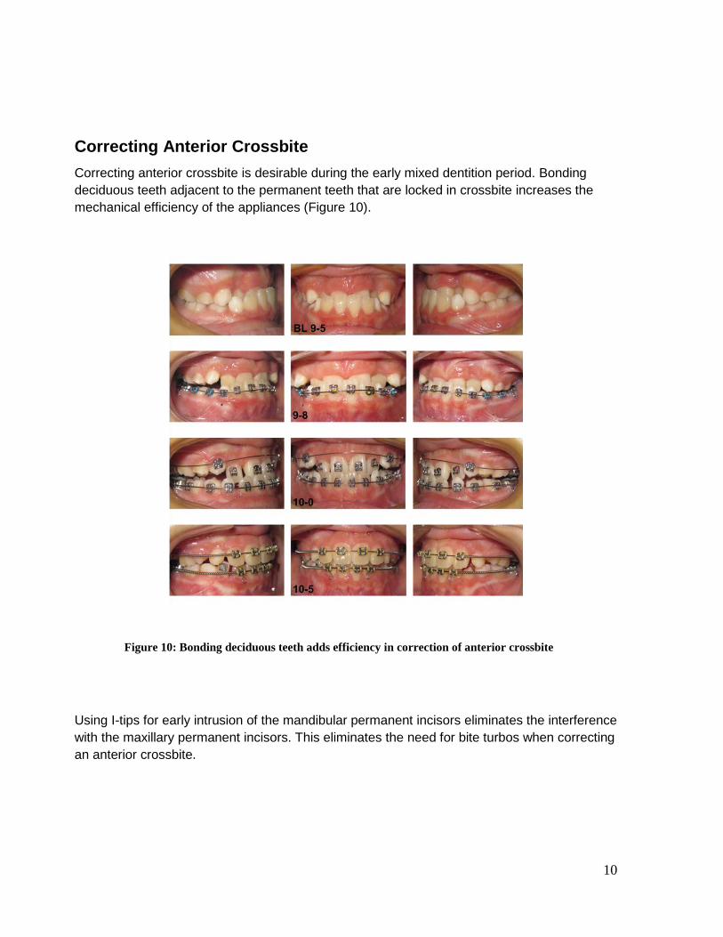
Correcting Anterior Crossbite Correcting anterior crossbite is desirable during the early mixed dentition period. Bonding deciduous teeth adjacent to the permanent teeth that are locked in crossbite increases the mechanical efficiency of the appliances (Figure 10).
Figure 10: Bonding deciduous teeth adds efficiency in correction of anterior crossbite
Using I-tips for early intrusion of the mandibular permanent incisors eliminates the interference with the maxillary permanent incisors. This eliminates the need for bite turbos when correcting an anterior crossbite.
11
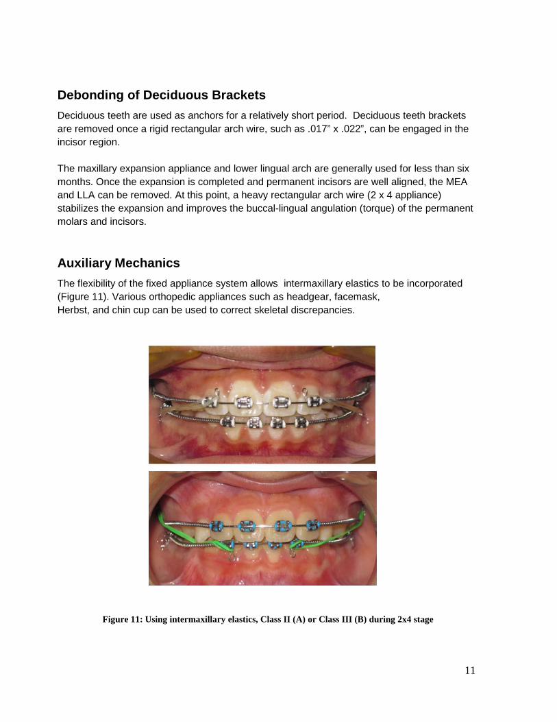
Debonding of Deciduous Brackets Deciduous teeth are used as anchors for a relatively short period. Deciduous teeth brackets are removed once a rigid rectangular arch wire, such as .017” x .022”, can be engaged in the incisor region. The maxillary expansion appliance and lower lingual arch are generally used for less than six months. Once the expansion is completed and permanent incisors are well aligned, the MEA and LLA can be removed. At this point, a heavy rectangular arch wire (2 x 4 appliance) stabilizes the expansion and improves the buccal-lingual angulation (torque) of the permanent molars and incisors. Auxiliary Mechanics The flexibility of the fixed appliance system allows intermaxillary elastics to be incorporated (Figure 11). Various orthopedic appliances such as headgear, facemask, Herbst, and chin cup can be used to correct skeletal discrepancies.
Figure 11: Using intermaxillary elastics, Class II (A) or Class III (B) during 2x4 stage
12
Conclusion Creating a normal occlusal relationship and a balanced neuromuscular environment at an early age can help the normal growth of the facial skeleton in an otherwise healthy child [30]. Although some debate still exists regarding interceptive orthodontics, early treatment is advantageous in correcting certain forms of malocclusion such as crowding, overbite, open bite, and crossbite [31-36]. The mixed dentition protocol presented in this paper uses expansion in the transverse dimension as the main method to create space. An MEA is used to expand the maxilla. Early expansion of the maxillary skeletal complex in non-crossbite individuals can correct maxillary arch length deficiencies [15,18]. Using maxillary deciduous canines as anchorage helps align the maxillary permanent incisors. In the mandible, expansion of the buccal segments, including deciduous molars and canines, can increase the arch length to accommodate crowded permanent incisors. The protocol uses E-I tips to carefully position the deciduous brackets and improve the mechanical efficiency of appliances to accelerate the correction of open bite, overbite, and crossbite conditions. Benefits of the protocol include: 1. Reduces the occurrence of impaction of maxillary permanent canines 2. Eliminates the need to extract the deciduous canines or deciduous first molars 3. Eliminates the need to extract premolars 4. Raises a young child’s self-esteem To get more information about mixed dentition orthodontics and to participate in an open discussion about the subject, please visit www.interceptiveortho.com.
13
References
1. Devi R, Oliva B, Macri L, Clementini M, De Vito E, Nicolotti N, La Torre G. The
impact of social context on the perception of dental appearance in 8-9 years old
children. Italian Journal of Public Health 6:172-176, 2009.
2. King GJ, McGorray SP, Wheeler TT, Dolce C, Taylor M. Comparison of peer
assessment ratings (PAR) from 1-phase and 2-phase treatment protocols for
class II malocclusions. Am J Orthod Dentofacial Orthop 123: 489-96, 2003.
3. Tulloch JFC, Proffit WR, Phillips C. Outcomes in a 2-phase randomized clinical
trial of early Class II treatment. Am J Orthod Dentofacial Orthop 125:657-67,
2004.
4. Dolce C, McGorray S, Brazeau L, King G, Wheeler T. Timing of Class II
treatment: Skeletal changes comparing 1-phase and 2-phase treatment. Am J
Orthod Dentofacial Orthop 132:481-9, 2007.
5. O’Brien K, Wright J, Conboy F, Appelbe P, Davies L, Connolly I, et al. Early
treatment for Class II Division 1 malocclusion with the Twin-block appliance: A
multi-center, randomized, controlled trial. Am J Orthod Dentofacial Orthop
135:573-9, 2009.
6. Wortham JR, Dolce C, McGorray SP, Le H, King GJ, Wheeler TT. Comparison of
arch dimension changes in 1-phase vs 2-phase treatment of Class II
malocclusion. Am J Orthod Dentofacial Orthop 136:65-74, 2009.
7. Oh H, Baumrind S, Korn EL, Dugoni SA, Boero R, Aubert M, Boyd R. A
retrospective study of Class II mixed-dentition treatment. Angle Orthod
2017;87(1):56-67.
8. Dugoni SA. Comprehensive mixed dentition treatment. Am J Orthod Dentofacial
Orthop 1998;131(1):75-84.
14
9. Mirabelli JT, Huang GJ, Siu CH, King GJ. The effectiveness of phase I
orthodontic treatment in a Medicaid population. Am J Orthod Dentofacial Orthop
2005;127(5)592-598.
10. Kerosuo H, Heikinheimo K, Nystrom M, Vakiparta M. Outcome and long-term
stability of an early orthodontic treatment strategy in public health care. Eur J
Orthod 2013;35(2):183-189.
11. Bishara SE, Justus R, Graber TM. Proceedings of the Workshop Discussions on
Early Treatment. Am J Orthod Dentofacial Orthop 113:5-6, 1998.
12. Carlson DS. Biological rationale for early treatment of dentofacial deformities.
Am J Orthod Dentofacial Orthop 121:554-8, 2002.
13. Pancherz H. Treatment timing and outcome. Am J Orthod Dentofacial
Orthop 121:559, 2002.
14. Freeman CS, McNamara JA, Baccetti T, Franchi L, Graff TW. Treatment effects
of the bionator and high-pull facebow combination followed by fixed appliances
in patients with increased vertical dimensions, Am J Orthod Dentofacial Orthop
131:184-95, 2007.
15. McNamara JA Jr. Early intervention in the transverse dimension: Is it worth the
effort?. Am J Orthod Dentofacial Orthop 121:572-4, 2002.
16. McInaney JB, Adams RM, Freeman M. A nonextraction approach to crowded
dentitions in young children: early recognition and treatment. J Am Dent Assoc
101:251-7. 1980.
17. Haas AJ. Long-term posttreatment evaluation of rapid palatal expansion. Angle
Orthod 50:189-217, 1980.
15
18. Geran RG, McNamara JA Jr, Baccetti T, Franchi L, Shapiro LM. A prospective
long-term study on the effects of rapid maxillary expansion in the early mixed
dentition. Am J Orthod Dentofacial Orthop 129:631-40, 2006.
19. Vargo J, Buschang PH, Boley JC, English JD, Behrents RG, Owen AH III.
Treatment effects and short-term relapse of maxillomandibular expansion during
the early to mid mixed dentition. Am J Orthod Dentofacial Orthop 131:456-63,
2007.
20. BeGole EA, Fox DL, Sadowsky C. Analysis of change in arch form with premolar
expansion. Am J Orthod Dentofacial Orthop 113:307-15, 1998.
21. Johnston LE Jr. Answers in search of questioners. Am J Orthod Dentofacial
Orthop 121:552-3, 2002.
22. Gianelly AA. Treatment of crowding in the mixed dentition. Am J Orthod
Dentofacial Orthop 121:569-71, 2002.
23. Little RM. Stability and relapse: Early treatment of arch length deficiency. Am J
Orthod Dentofacial Orthop 121:578-81, 2002.
24. Tai K, Hotokezaka H, Park JH, Tai H, Miyajima K, Choi M, et al. Preliminary
cone-beam computed tomography study evaluating dental and skeletal changes
after treatment with a Mandibular Schwarz appliance. Am J Orthod Dentofacial
Orthop 138:262-3, 2010.
25. Lamparski DG Jr, Rinchuse DJ, Close JM, Sciote JJ. Comparison of skeletal and
dental changes between 2-point and 4-point rapid palatal expanders. Am J
Orthod Dentofacial Orthop 123:321-8, 2003.
26. Davidovitch M, Efstathiou S, Sarne O, Vardimon AD. Skeletal and dental
response to rapid maxillary expansion with 2- versus 4-band appliance. Am J
Orthod Dentofacial Orthop 127:483-92, 2005.
16
27. Baccetti T, Mucedero M, Leonardi M, Cozza P. Interceptive treatment of palatal
impaction of maxillary canines with rapid maxillary expansion: A randomized
clinical trial. Am J Orthod Dentofacial Orthop 136:657-61, 2009.
28. Moss ML. The functional matrix hypothesis revisited. 2. The role of an osseous
connected cellular network. Am J Orthod Dentofacial Orthop 112:221-26, 1997.
29. O’Grady PW, McNamara JA, Baccetti T, Franchi L. A long-term evaluation of the
Mandibular Schwarz appliance and the acrylic splint expander in early mixed
dentition patients. Am J Orthod Dentofacial Orthop 130:202-13, 2006.
30. Hinton RJ, Carlson DS. Effect of function on growth and remodeling of the
temporomandibular joint. In: McNeil C,editor. Science and practice of occlusion.
Chicago:Quintessence p. 95-110, 1997.
31. Hamilton DC. The emancipation of dentofacial orthopedics. Am J Orthod
Dentofacial Orthop 113:7-10, 1998.
32. Arvystas MG. The rationale for early orthodontic treatment. Am J Orthod
Dentofacial Orthop 113:15-8, 1998.
33. White L. Early orthodontic intervention. Am J Orthod Dentofacial Orthop 113:24-8,
1998.
34. Woodside DG. Do functional appliances have an orthopedic effect?. Am J Orthod
Dentofacial Orthop 113:11-4, 1998.
35. Pangrazio-Kulbersh V, Kaczynski R, Shunock M. Early treatment outcome
assessed by the Peer Assessment Rating index. Am J Orthod Dentofacial Orthop
115:544-50, 1999.
36. Dugoni SA, Lee JS, Varela J, Dugoni AA. Early mixed dentition treatment:
postretention evaluation of stability and relapse. The Angle Orthodontist;65:311-
20,1995.