intensity modulated radiation therapy for craniospinal...
TRANSCRIPT
Intensity Modulated Radiation Therapy of Medulloblastoma using Helical TomoTherapy:
Initial Experience from planning to delivery
Reena PhurailatpamTejpal Gupta, Rakesh Jalali, Zubin Master, Bhooshan Zade, RajivSarin, Anusheel Munshi, Deepak Deshpande, Shyam Shrivastava,
Ketayun DinshawDepartments of Radiation Oncology and Medical Physics, Tata Memorial Hospital and
Advanced Centre for Treatment Research and Education in Cancer, Tata Memorial Centre, Mumbai, India.
24/04/2009 2
Aim
• To establish feasibility of Intensity Modulated Radiation Therapy (IMRT) for craniospinal irradiation (CSI) using Helical TomoTherapy (IMRT_Tomo)
• To report initial experience of its implementation in the clinic.
• Dosimetric comparison of IMRT_Tomo with conventional Linear Accelerator based 3DCRT (3DCRT_LA), and IMRT (IMRT_LA) plans
24/04/2009 3
Materials and Methods:
Phase I : Dosimetric comparison of 3DCRT, IMRT_LAand IMRT_Tomo
• Selected CT datasets of 4 previously treated patients (5-14 yrs) of medulloblastoma having spinal lengths up to 48 cm
• Delineated Target and OARs on Coherence dosimetrist VSim Workstation
• For each patients, 3DCRT and IMRT_LA plan were generated on Eclips TPS (V 7.3.1) configured with millennium 120 MLC from Varian
24/04/2009 4
• IMRT_Tomo plans were generated using the same patient datasets on TomoPlanning System (V 2.2.4).
• All plans were generated using 6 MV X-rays
• A dose of 35 Gy in 21 fractions was prescribed to planning target volume (PTV) of Brain and Spine.
• Planning Goal : – At least 95% volume of target (PTV_brain and PTV_spine) received
at least 95% of the prescription dose while restricting the maximum dose limit to 107%
– Reduced dose to OARs
24/04/2009 5
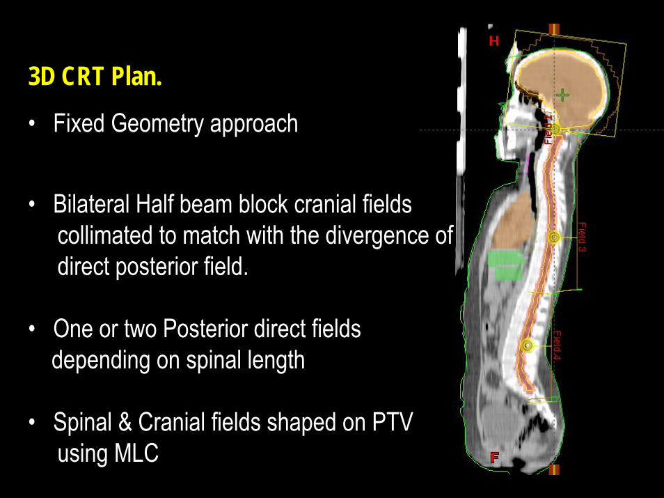
3D CRT Plan.
• Fixed Geometry approach
• Bilateral Half beam block cranial fields collimated to match with the divergence of direct posterior field.
• One or two Posterior direct fieldsdepending on spinal length
• Spinal & Cranial fields shaped on PTVusing MLC
24/04/2009 6
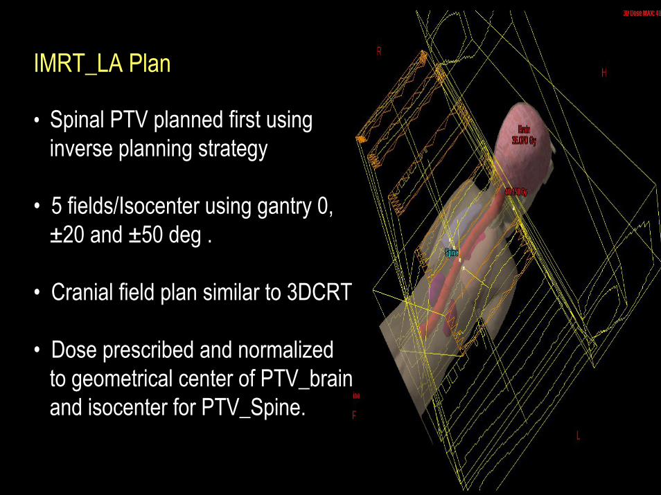
IMRT_LA Plan
• Spinal PTV planned first using inverse planning strategy
• 5 fields/Isocenter using gantry 0, ±20 and ±50 deg .
• Cranial field plan similar to 3DCRT
• Dose prescribed and normalizedto geometrical center of PTV_brainand isocenter for PTV_Spine.
24/04/2009 7
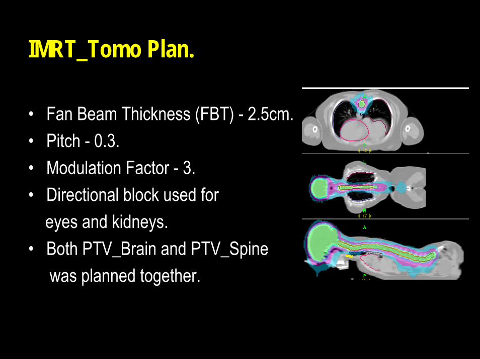
IMRT_Tomo Plan.
• Fan Beam Thickness (FBT) - 2.5cm.• Pitch - 0.3.• Modulation Factor - 3.• Directional block used for
eyes and kidneys.• Both PTV_Brain and PTV_Spine
was planned together.
24/04/2009 8
TC=(VT,Pi / VT) × 100%.
CI = {VT,Pi × VT,Pi } / {VT × VPi }
Plan Evaluation for Target
• Target Volume coverage (TC)
• Dose Uniformity Index (DHI) = D95/D5
• Conformity index (CI)
VT,Pi - volume of target enclosed by the prescription dose VPi - volume of tissues including target covered by the prescription dose VT - volume of target; D95 and D5- dose to 95% and 5% volume of the PTV
24/04/2009 9
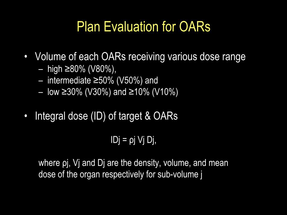
• Volume of each OARs receiving various dose range– high ≥80% (V80%), – intermediate ≥50% (V50%) and – low ≥30% (V30%) and ≥10% (V10%)
• Integral dose (ID) of target & OARs
IDj = ρj Vj Dj,
where ρj, Vj and Dj are the density, volume, and meandose of the organ respectively for sub-volume j
Plan Evaluation for OARs
24/04/2009 10
a b c
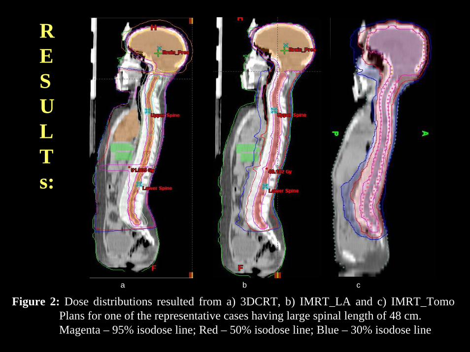
Figure 2: Dose distributions resulted from a) 3DCRT, b) IMRT_LA and c) IMRT_Tomo Plans for one of the representative cases having large spinal length of 48 cm. Magenta – 95% isodose line; Red – 50% isodose line; Blue – 30% isodose line
RESULTs:
24/04/2009 11
PTV_Brain PTV_Spine
3D-RT IMRT TOMO 3D-RT IMRT TOMO
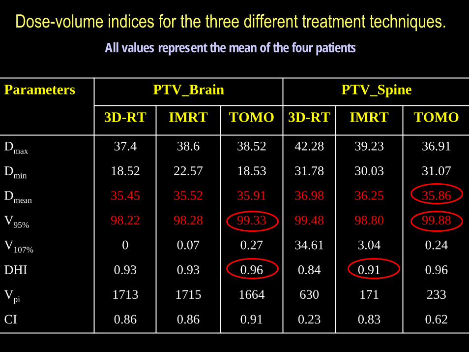
Dmax 37.4 38.6 38.52 42.28 39.23 36.91
Dmin 18.52 22.57 18.53 31.78 30.03 31.07
Dmean 35.45 35.52 35.91 36.98 36.25 35.86
V95% 98.22 98.28 99.33 99.48 98.80 99.88
V107% 0 0.07 0.27 34.61 3.04 0.24
DHI 0.93 0.93 0.96 0.84 0.91 0.96
Vpi 1713 1715 1664 630 171 233
CI 0.86 0.86 0.91 0.23 0.83 0.62
Parameters
Dose-volume indices for the three different treatment techniques.All values represent the mean of the four patients
24/04/2009 12
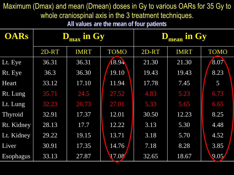
Maximum (Dmax) and mean (Dmean) doses in Gy to various OARs for 35 Gy to whole craniospinal axis in the 3 treatment techniques.
All values are the mean of four patients
Dmax in Gy Dmean in Gy2D-RT IMRT TOMO 2D-RT IMRT TOMO
Lt. Eye 36.31 36.31 18.94 21.30 21.30 8.07Rt. Eye 36.3 36.30 19.10 19.43 19.43 8.23Heart 33.12 17.10 11.94 17.78 7.45 5Rt. Lung 35.71 24.5 27.52 4.83 5.23 6.73Lt. Lung 32.23 26.73 27.01 5.33 5.65 6.65Thyroid 32.91 17.37 12.01 30.50 12.23 8.25Rt. Kidney 28.13 17.7 12.22 3.13 5.30 4.48Lt. Kidney 29.22 19.15 13.71 3.18 5.70 4.52Liver 30.91 17.35 14.76 7.18 8.28 3.85Esophagus 33.13 27.87 17.08 32.65 18.67 9.05
OARs
24/04/2009 13
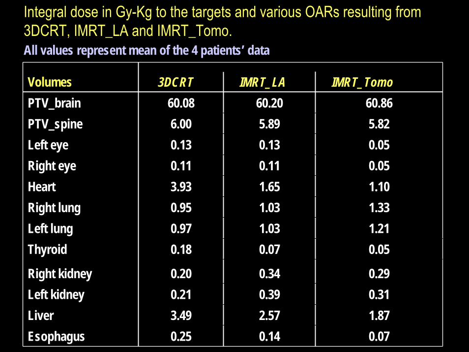
Volumes 3DCRT IMRT_LA IMRT_TomoPTV_brain 60.08 60.20 60.86PTV_spine 6.00 5.89 5.82Left eye 0.13 0.13 0.05Right eye 0.11 0.11 0.05Heart 3.93 1.65 1.10Right lung 0.95 1.03 1.33Left lung 0.97 1.03 1.21Thyroid 0.18 0.07 0.05
Right kidney 0.20 0.34 0.29Left kidney 0.21 0.39 0.31Liver 3.49 2.57 1.87Esophagus 0.25 0.14 0.07
Integral dose in Gy-Kg to the targets and various OARs resulting from 3DCRT, IMRT_LA and IMRT_Tomo. All values represent mean of the 4 patients’ data
24/04/2009 14
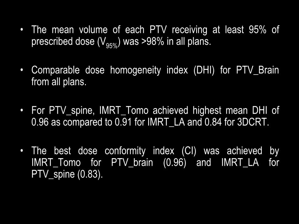
• The mean volume of each PTV receiving at least 95% of prescribed dose (V95%) was >98% in all plans.
• Comparable dose homogeneity index (DHI) for PTV_Brain from all plans.
• For PTV_spine, IMRT_Tomo achieved highest mean DHI of 0.96 as compared to 0.91 for IMRT_LA and 0.84 for 3DCRT.
• The best dose conformity index (CI) was achieved by IMRT_Tomo for PTV_brain (0.96) and IMRT_LA for PTV_spine (0.83).
24/04/2009 15
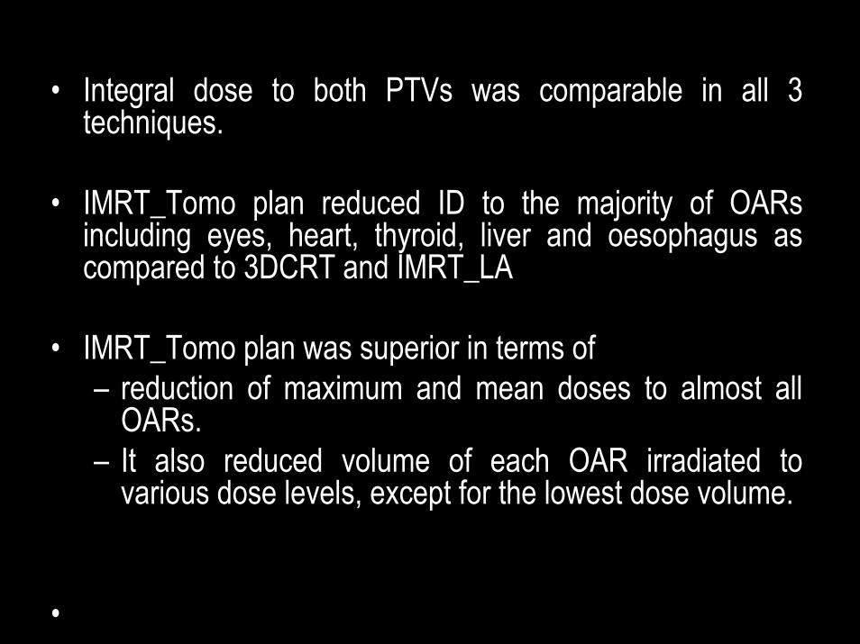
• Integral dose to both PTVs was comparable in all 3 techniques.
• IMRT_Tomo plan reduced ID to the majority of OARs including eyes, heart, thyroid, liver and oesophagus as compared to 3DCRT and IMRT_LA
• IMRT_Tomo plan was superior in terms of – reduction of maximum and mean doses to almost all
OARs.– It also reduced volume of each OAR irradiated to
various dose levels, except for the lowest dose volume.
•
24/04/2009 16
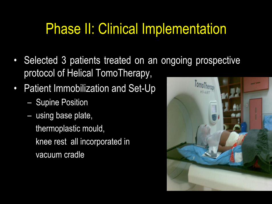
Phase II: Clinical Implementation
• Selected 3 patients treated on an ongoing prospective protocol of Helical TomoTherapy,
• Patient Immobilization and Set-Up – Supine Position– using base plate,
thermoplastic mould,knee rest all incorporated in vacuum cradle
24/04/2009 17
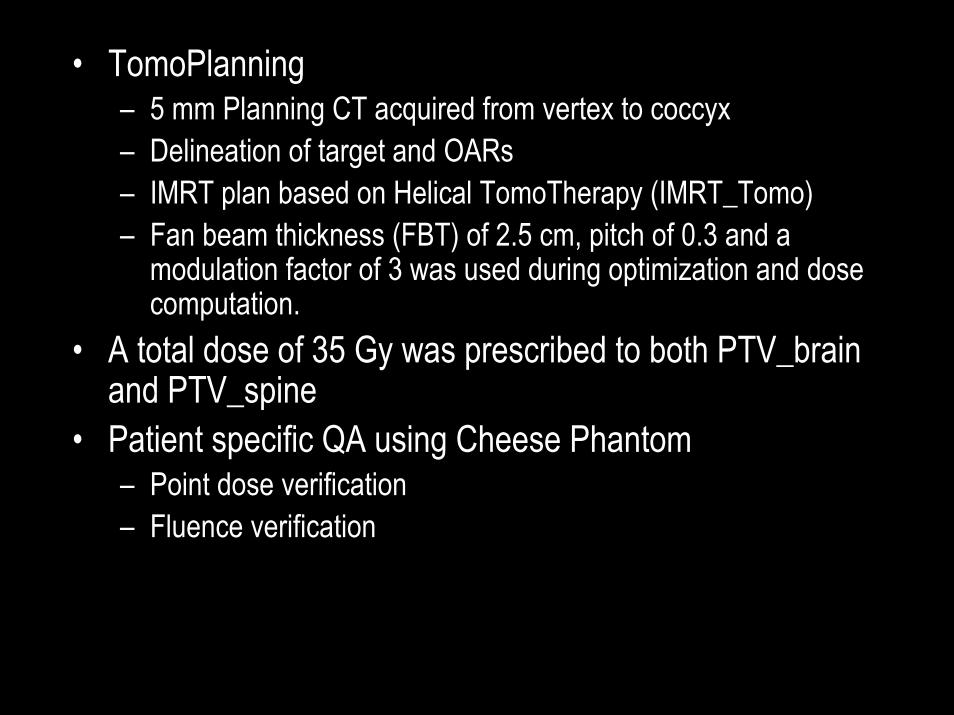
• TomoPlanning– 5 mm Planning CT acquired from vertex to coccyx– Delineation of target and OARs– IMRT plan based on Helical TomoTherapy (IMRT_Tomo)– Fan beam thickness (FBT) of 2.5 cm, pitch of 0.3 and a
modulation factor of 3 was used during optimization and dose computation.
• A total dose of 35 Gy was prescribed to both PTV_brain and PTV_spine
• Patient specific QA using Cheese Phantom– Point dose verification– Fluence verification
24/04/2009 18
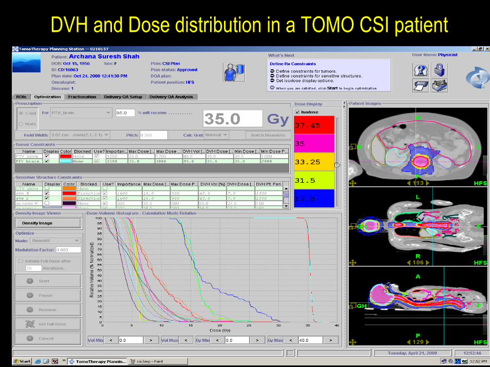
DVH and Dose distribution in a TOMO CSI patient
24/04/2009 19
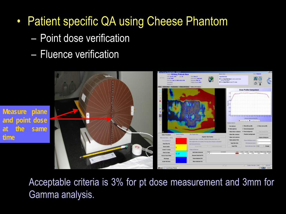
• Patient specific QA using Cheese Phantom– Point dose verification– Fluence verification
Measure plane and point dose at the same time
Acceptable criteria is 3% for pt dose measurement and 3mm for Gamma analysis.
24/04/2009 20
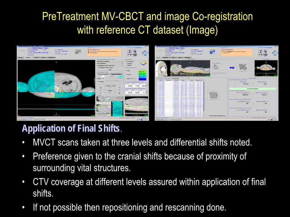
PreTreatment MV-CBCT and image Co-registration with reference CT dataset (Image)
Application of Final Shifts.• MVCT scans taken at three levels and differential shifts noted.• Preference given to the cranial shifts because of proximity of
surrounding vital structures.• CTV coverage at different levels assured within application of final
shifts.• If not possible then repositioning and rescanning done.
24/04/2009 21
Conclusion • Craniospinal irradiation remains one of the most challenging
processes in radiation planning, delivery, and verification
• IMRT_Tomo for CSI is technically easier and dosimetrically favorable as compared to IMRT_LA and 3DCRT in terms of – target volume coverage, – dose homogeneity, conformity, – OAR sparing and – reduction of integral doses to non-target tissues
• The In-build image-guidance allows precise patient positioning and accurate dose delivery
• In case of non-availability of TomoTherapy, IMRT for CSI can be realized on conventional linear accelerator even for spinal lengths exceeding maximum allowable field sizes using appropriate intensity feathering techniques.
24/04/2009 22
• Although time and labor intensive, challenges in successful implementation of IMRT_Tomo for CSI can be circumvented provided they are preempted during the planning phase.
• During clinical implementation, practical issues that arose included – challenges in whole body immobilization, – areas to be imaged daily with MVCT, – co-registration efficiency, – Longer beam-on time.– intrafraction motion, and impact of differential shifts of different
parts of the body, which were handled using appropriate methodology resulting in increased daily time on the machine