insights into the biosynthesis and stability of the lasso peptide capistruin
TRANSCRIPT
Chemistry & Biology
Article
Insights into the Biosynthesis and Stabilityof the Lasso Peptide CapistruinThomas A. Knappe,1 Uwe Linne,1 Lars Robbel,1 and Mohamed A. Marahiel1,*1Department of Chemistry, Philipps-University Marburg, Hans-Meerwein-Strasse, D-35032 Marburg, Germany
*Correspondence: [email protected]
DOI 10.1016/j.chembiol.2009.11.009
SUMMARY
Capistruin is a 19-residue ribosomally synthesizedlasso peptide encoded by the capABCD gene clusterin Burkholderia thailandensis. It is composed of anN-terminal 9-residue macrolactam ring, throughwhich the 10-residue C-terminal tail is threaded.Using a heterologous capistruin production systemin Escherichia coli, we have generated 48 mutantsof the precursor protein CapA to gain insights intocapistruin biosynthesis. Only 4 residues (Gly1, Arg11,Val12, and Ile13) of the lasso sequence were found tobe critical for maturation. Tandem mass spectro-metric fragmentation studies of capistruin F16A/F18A proved Arg15 to be responsible for the trappingof the C-terminal tail. Substituting Arg15 and Phe16by alanine revealed a temperature-sensitive capi-struin derivative, which unfolds into a branchedcyclic peptide upon heating. In conclusion, ourglobal mutagenic approach revealed a low overallspecificity of the biosynthetic machinery and impor-tant structure-stability correlations.
INTRODUCTION
Lasso peptides are a structurally unique class of ribosomally
synthesized, bioactive natural products of bacterial origin con-
sisting of 16–21 amino acids (Rebuffat et al., 2004; Severinov
et al., 2007). They are composed of an 8- or 9-residue N-terminal
macrolactam ring generated upon a condensation reaction
between the a-NH2 group of an N-terminal Gly or Cys and the
side-chain carboxyl group of an Asp9 or Glu8 (Bayro et al.,
2003; Frechet et al., 1994; Iwatsuki et al., 2006; Katahira et al.,
1995, 1996; Knappe et al., 2008; Rosengren et al., 2003; Wilson
et al., 2003). Their primary structure can therefore be described
as a branched cyclic peptide composed of an N-terminal macro-
lactam ring and a linear C-terminal tail. This tail threads through
the macrocycle and is trapped by steric hindrance of bulky side
chains within the ring generating the common lariat-protoknot
structure of lasso peptides. This rigid and compact lasso fold
results in remarkable stability against proteolytic degradation
and thermal or chaotrope induced denaturation (Knappe et al.,
2008; Rosengren et al., 2004). Lasso peptides are classified
depending on the presence (class I) or absence (class II) of
four conserved cysteine residues, which are involved in the
1290 Chemistry & Biology 16, 1290–1298, December 24, 2009 ª2009
formation of two intramolecular disulfide bonds (Rebuffat et al.,
2004).
By nuclear magnetic resonance (NMR) spectroscopic studies,
six bacterial peptides sharing the described primary structure
have been proven to adopt the lariat-protoknot structure, namely
RES-701-1 (Katahira et al., 1995; Morishita et al., 1994), MS-271
(Katahira et al., 1996; Yano et al., 1996), and RP 71955 (Frechet
et al., 1994; Helynck et al., 1993) from different Streptomyces
species; lariatin A (Iwatsuki et al., 2006) from Rhodococcus
sp.; microcin J25 (Bayro et al., 2003; Rosengren et al., 2003;
Wilson et al., 2003) (MccJ25) from Escherichia coli; and capi-
struin (Knappe et al., 2008) from Burkholderia thailandensis
E264. Their biological activities range from inhibition of HIV
replication (Helynck et al., 1993) (RP 71955) to blockage of
Gram-negative bacterial RNA polymerase (Adelman et al.,
2004; Mukhopadhyay et al., 2004) (microcin J25). The biosyn-
thetic gene clusters are only known for the best-studied repre-
sentative MccJ25 (Duquesne et al., 2007a, 2007c; Solbiati
et al., 1999) and the recently described capistruin (Knappe
et al., 2008).
Capistruin is a 19-amino-acid class II lasso peptide
comprising an isopeptide bond between Gly1 and Asp9 resulting
in a 9-residue macrolactam ring. NMR spectroscopic studies re-
vealed Arg15 to be the first amino acid within the threading tail
located below the macrocyclic ring, and due to the steric
hindrance of the arginine side chain it is proposed to be respon-
sible for the trapping of the linear C-terminal tail. Capistruin is the
first lasso peptide discovered by a genome mining based
approach and displays antibacterial activity against closely
related Burkholderia and Pseudomonas strains. As in the
mcjABCD gene cluster responsible for MccJ25 production, the
capistruin biosynthetic gene cluster consists of four genes en-
coding (I) the 47-amino-acid capistruin precursor protein
CapA, (II) the two processing enzymes CapB and CapC, and
(III) the export and immunity protein CapD (Figure 1). It is
assumed that the two processing enzymes CapB/CapC convert
the ribosomally synthesized precursor protein CapA into the
mature lasso peptide capistruin, as it was shown for the matura-
tion of MccJ25 (Duquesne et al., 2007b). CapC is homologous to
asparagine synthetase B catalyzing the transfer of ammonia
from the amide donor glutamine to the side-chain carboxyl group
of aspartic acid, which is activated in an initial reaction as adeny-
late (Larsen et al., 1999). Therefore, CapC is most likely involved
in the side-chain carboxyl group activation of aspartic acid
at position 9 generating the electrophile for the condensation
reaction. CapB is a putative protease showing weak homology
to bacterial transglutaminases (Makarova et al., 1999). It is
Elsevier Ltd All rights reserved
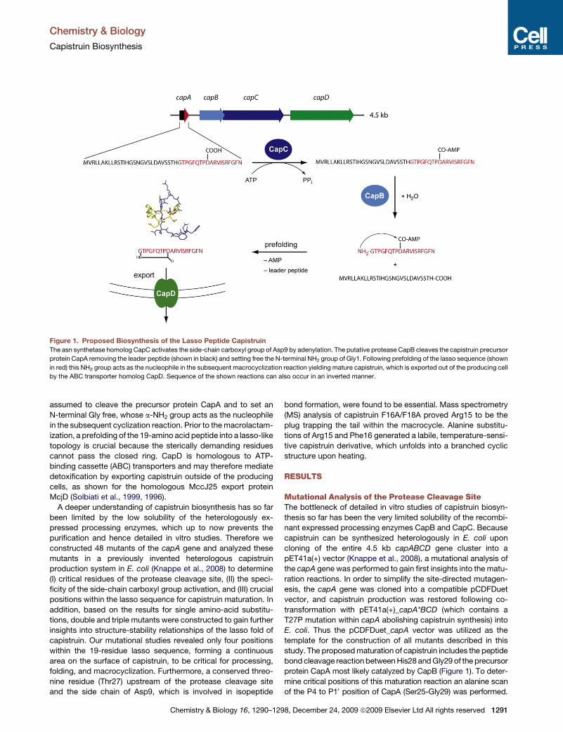
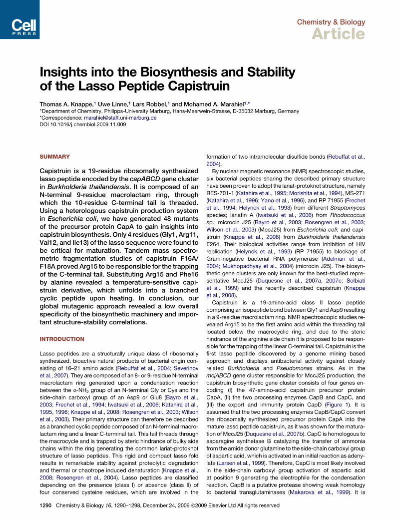
Figure 1. Proposed Biosynthesis of the Lasso Peptide Capistruin
The asn synthetase homolog CapC activates the side-chain carboxyl group of Asp9 by adenylation. The putative protease CapB cleaves the capistruin precursor
protein CapA removing the leader peptide (shown in black) and setting free the N-terminal NH2 group of Gly1. Following prefolding of the lasso sequence (shown
in red) this NH2 group acts as the nucleophile in the subsequent macrocyclization reaction yielding mature capistruin, which is exported out of the producing cell
by the ABC transporter homolog CapD. Sequence of the shown reactions can also occur in an inverted manner.
Chemistry & Biology
Capistruin Biosynthesis
assumed to cleave the precursor protein CapA and to set an
N-terminal Gly free, whose a-NH2 group acts as the nucleophile
in the subsequent cyclization reaction. Prior to the macrolactam-
ization, a prefolding of the 19-amino acid peptide into a lasso-like
topology is crucial because the sterically demanding residues
cannot pass the closed ring. CapD is homologous to ATP-
binding cassette (ABC) transporters and may therefore mediate
detoxification by exporting capistruin outside of the producing
cells, as shown for the homologous MccJ25 export protein
McjD (Solbiati et al., 1999, 1996).
A deeper understanding of capistruin biosynthesis has so far
been limited by the low solubility of the heterologously ex-
pressed processing enzymes, which up to now prevents the
purification and hence detailed in vitro studies. Therefore we
constructed 48 mutants of the capA gene and analyzed these
mutants in a previously invented heterologous capistruin
production system in E. coli (Knappe et al., 2008) to determine
(I) critical residues of the protease cleavage site, (II) the speci-
ficity of the side-chain carboxyl group activation, and (III) crucial
positions within the lasso sequence for capistruin maturation. In
addition, based on the results for single amino-acid substitu-
tions, double and triple mutants were constructed to gain further
insights into structure-stability relationships of the lasso fold of
capistruin. Our mutational studies revealed only four positions
within the 19-residue lasso sequence, forming a continuous
area on the surface of capistruin, to be critical for processing,
folding, and macrocyclization. Furthermore, a conserved threo-
nine residue (Thr27) upstream of the protease cleavage site
and the side chain of Asp9, which is involved in isopeptide
Chemistry & Biology 16, 1290–129
bond formation, were found to be essential. Mass spectrometry
(MS) analysis of capistruin F16A/F18A proved Arg15 to be the
plug trapping the tail within the macrocycle. Alanine substitu-
tions of Arg15 and Phe16 generated a labile, temperature-sensi-
tive capistruin derivative, which unfolds into a branched cyclic
structure upon heating.
RESULTS
Mutational Analysis of the Protease Cleavage SiteThe bottleneck of detailed in vitro studies of capistruin biosyn-
thesis so far has been the very limited solubility of the recombi-
nant expressed processing enzymes CapB and CapC. Because
capistruin can be synthesized heterologously in E. coli upon
cloning of the entire 4.5 kb capABCD gene cluster into a
pET41a(+) vector (Knappe et al., 2008), a mutational analysis of
the capA gene was performed to gain first insights into the matu-
ration reactions. In order to simplify the site-directed mutagen-
esis, the capA gene was cloned into a compatible pCDFDuet
vector, and capistruin production was restored following co-
transformation with pET41a(+)_capA*BCD (which contains a
T27P mutation within capA abolishing capistruin synthesis) into
E. coli. Thus the pCDFDuet_capA vector was utilized as the
template for the construction of all mutants described in this
study. The proposed maturation of capistruin includes the peptide
bond cleavage reaction between His28 and Gly29 of the precursor
protein CapA most likely catalyzed by CapB (Figure 1). To deter-
mine critical positions of this maturation reaction an alanine scan
of the P4 to P10 position of CapA (Ser25-Gly29) was performed.
8, December 24, 2009 ª2009 Elsevier Ltd All rights reserved 1291
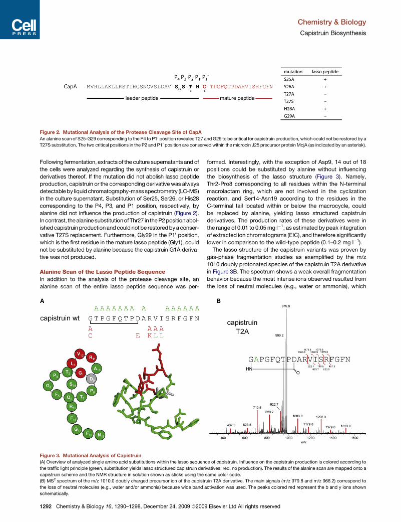
Figure 2. Mutational Analysis of the Protease Cleavage Site of CapA
An alanine scan of S25-G29 corresponding to the P4 to P10 position revealed T27 and G29 to be critical for capistruin production, which could not be restored by a
T27S substitution. The two critical positions in the P2 and P10 position are conserved within the microcin J25 precursor protein McjA (as indicated by an asterisk).
Chemistry & Biology
Capistruin Biosynthesis
Following fermentation, extracts of the culture supernatants and of
the cells were analyzed regarding the synthesis of capistruin or
derivatives thereof. If the mutation did not abolish lasso peptide
production, capistruin or the corresponding derivative was always
detectable by liquid chromatography-mass spectrometry (LC-MS)
in the culture supernatant. Substitution of Ser25, Ser26, or His28
corresponding to the P4, P3, and P1 position, respectively, by
alanine did not influence the production of capistruin (Figure 2).
Incontrast, the alanine substitutionofThr27 in the P2 positionabol-
ished capistruin production and could not be restored by a conser-
vative T27S replacement. Furthermore, Gly29 in the P10 position,
which is the first residue in the mature lasso peptide (Gly1), could
not be substituted by alanine because the capistruin G1A deriva-
tive was not produced.
Alanine Scan of the Lasso Peptide SequenceIn addition to the analysis of the protease cleavage site, an
alanine scan of the entire lasso peptide sequence was per-
Figure 3. Mutational Analysis of Capistruin
(A) Overview of analyzed single amino acid substitutions within the lasso sequenc
the traffic light principle (green, substitution yields lasso structured capistruin deri
capistruin scheme and the NMR structure in solution shown as sticks using the s
(B) MS2 spectrum of the m/z 1010.0 doubly charged precursor ion of the capistr
the loss of neutral molecules (e.g., water and/or ammonia) because wide band a
schematically.
1292 Chemistry & Biology 16, 1290–1298, December 24, 2009 ª2009
formed. Interestingly, with the exception of Asp9, 14 out of 18
positions could be substituted by alanine without influencing
the biosynthesis of the lasso structure (Figure 3). Namely,
Thr2-Pro8 corresponding to all residues within the N-terminal
macrolactam ring, which are not involved in the cyclization
reaction, and Ser14-Asn19 according to the residues in the
C-terminal tail located within or below the macrocycle, could
be replaced by alanine, yielding lasso structured capistruin
derivatives. The production rates of these derivatives were in
the range of 0.01 to 0.05 mg l�1, as estimated by peak integration
of extracted ion chromatograms (EIC), and therefore significantly
lower in comparison to the wild-type peptide (0.1–0.2 mg l�1).
The lasso structure of the capistruin variants was proven by
gas-phase fragmentation studies as exemplified by the m/z
1010 doubly protonated species of the capistruin T2A derivative
in Figure 3B. The spectrum shows a weak overall fragmentation
behavior because the most intense ions observed resulted from
the loss of neutral molecules (e.g., water or ammonia), which
e of capistruin. Influence on the capistruin production is colored according to
vatives; red, no production). The results of the alanine scan are mapped onto a
ame color code.
uin T2A derivative. The main signals (m/z 979.8 and m/z 966.2) correspond to
ctivation was used. The peaks colored red represent the b and y ions shown
Elsevier Ltd All rights reserved
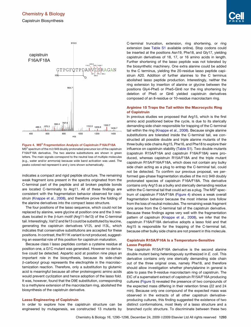
Figure 4. MS2 Fragmentation Analysis of Capistruin F16A/F18A
MS2 spectrum of the m/z 949 doubly protonated precursor ion of the capistruin
F16A/F18A derivative. The two alanine substitutions are shown in green
letters. The main signals correspond to the neutral loss of multiple molecules
(e.g., water and/or ammonia) because wide band activation was used. The
peaks colored red represent b and y ions shown schematically.
Chemistry & Biology
Capistruin Biosynthesis
indicates a compact and rigid peptide structure. The remaining
weak fragment ions present in the spectra originated from the
C-terminal part of the peptide and all broken peptide bonds
are located C-terminally to Arg11. All of these findings are
consistent with the fragmentation behavior observed for capi-
struin (Knappe et al., 2008), and therefore prove the folding of
the alanine derivatives into the compact lasso structure.
The four positions of the lasso sequence, which could not be
replaced by alanine, were glycine at position one and the 3 resi-
dues located in the b-turn motif (Arg11-Ile13) of the C-terminal
tail. Interestingly, Val12 and Ile13 could be substituted by leucine,
generating the capistruin derivatives V12L and I13L, which
indicates that conservative substitutions are accepted for these
positions. In contrast, the R11K variant is not produced, suggest-
ing an essential role of this position for capistruin maturation.
Because class I lasso peptides contain a cysteine residue at
position one, a G1C mutant was generated. However no deriva-
tive could be detected. Aspartic acid at position nine plays an
important role in the biosynthesis, because its side-chain
b-carboxyl group represents the electrophile in the macrolac-
tamization reaction. Therefore, only a substitution by glutamic
acid is meaningful because all other proteinogenic amino acids
would prevent cyclization and hence adoption of the lasso fold.
It was, however, found that the D9E substitution, corresponding
to a methylene extension of the macrolactam ring, abolished the
biosynthesis of the capistruin derivative.
Lasso Engineering of CapistruinIn order to explore how the capistruin structure can be
engineered by mutagenesis, we constructed 13 mutants by
Chemistry & Biology 16, 1290–129
C-terminal truncation, extension, ring shortening, or ring
extension (see Table S1 available online). Stop codons could
be inserted at the positions Asn19, Phe18, and Gly17, yielding
capistruin derivatives of 18, 17, or 16 amino acids in length.
Further shortening of the lasso peptide was not tolerated by
the biosynthetic machinery. One extra alanine could be added
to the C terminus, yielding the 20-residue lasso peptide capi-
struin A20. Addition of further alanines to the C terminus
abolished lasso peptide production. Interestingly, neither the
ring extension by insertion of alanine or glycine between the
positions Gly4-Phe5 or Phe5-Gln6 nor the ring shortening by
deletion of Phe5 or Gln6 yielded capistruin derivatives
composed of an 8-residue or 10-residue macrolactam ring.
Arginine 15 Traps the Tail within the Macrocyclic Ringof CapistruinIn previous studies we proposed that Arg15, which is the first
amino acid positioned below the cycle, is due to its sterically
demanding side chain responsible for trapping of the C-terminal
tail within the ring (Knappe et al., 2008). Because single alanine
substitutions are tolerated inside the C-terminal tail, we con-
structed all possible double and triple alanine mutants of the
three bulky side chains Arg15, Phe16, and Phe18 to explore their
influence on capistruin stability (Table S1). Two double mutants
(capistruin R15A/F16A and capistruin F16A/F18A) were pro-
duced, whereas capistruin R15A/F18A and the triple mutant
capistruin R15A/F16A/F18A, which does not contain any bulky
side chain acting as a plug to entrap the C-terminal tail, could
not be detected. To confirm our previous proposal, we per-
formed gas-phase fragmentation studies of the m/z 949 doubly
protonated species of capistruin F16A/F18A. This derivative
contains only Arg15 as a bulky and sterically demanding residue
within the C-terminal tail that could act as a plug. The MS2 spec-
trum of capistruin F16A/F18A (Figure 4) shows a weak overall
fragmentation behavior because the most intense ions follow
from the loss of neutral molecules. The remaining weak fragment
ions arose from the C-terminal tail of the capistruin derivative.
Because these findings agree very well with the fragmentation
pattern of capistruin (Knappe et al., 2008), we infer that the
capistruin F16A/F18A derivative adopts a lasso fold and that
Arg15 is responsible for the trapping of the C-terminal tail,
because other bulky side chains are not present in this molecule.
Capistruin R15A/F16A Is a Temperature-SensitiveLasso PeptideThe capistruin R15A/F16A derivative is the second alanine
double mutant being heterologously synthesized in E. coli. This
derivative contains only one sterically demanding side chain
out of the three original ones, namely Phe18, and therefore
should allow investigation whether phenylalanine in general is
able to pass the 9-residue macrolactam ring of capistruin. The
EIC of a supernatant extract of capistruin R15A/F16A producing
cultures (Figure 5) revealed the presence of two compounds of
the expected mass differing in their retention times (22 and 24
min). Because only one compound of the expected mass was
observed in the extracts of all other capistruin derivative-
producing cultures, this finding suggested the existence of two
distinct conformations, most likely of a lasso structure and a
branched cyclic structure. To discriminate between these two
8, December 24, 2009 ª2009 Elsevier Ltd All rights reserved 1293
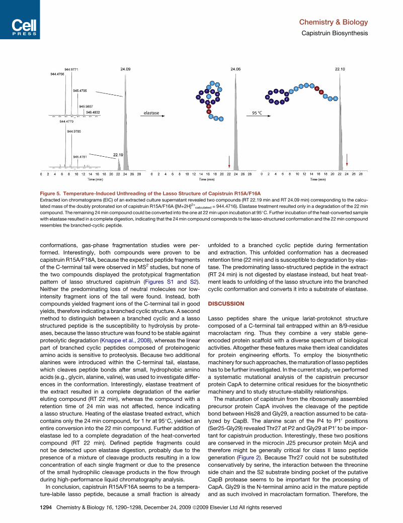
Figure 5. Temperature-Induced Unthreading of the Lasso Structure of Capistruin R15A/F16A
Extracted ion chromatograms (EIC) of an extracted culture supernatant revealed two compounds (RT 22.19 min and RT 24.09 min) corresponding to the calcu-
lated mass of the doubly protonated ion of capistruin R15A/F16A ([M+2H]2+calculated = 944.4716). Elastase treatment resulted only in a degradation of the 22 min
compound. The remaining 24 min compound could be converted into the one at 22 min upon incubation at 95�C. Further incubation of the heat-converted sample
with elastase resulted in a complete digestion, indicating that the 24 min compound corresponds to the lasso-structured conformation and the 22 min compound
resembles the branched-cyclic peptide.
Chemistry & Biology
Capistruin Biosynthesis
conformations, gas-phase fragmentation studies were per-
formed. Interestingly, both compounds were proven to be
capistruin R15A/F18A, because the expected peptide fragments
of the C-terminal tail were observed in MS2 studies, but none of
the two compounds displayed the prototypical fragmentation
pattern of lasso structured capistruin (Figures S1 and S2).
Neither the predominating loss of neutral molecules nor low-
intensity fragment ions of the tail were found. Instead, both
compounds yielded fragment ions of the C-terminal tail in good
yields, therefore indicating a branched cyclic structure. A second
method to distinguish between a branched cyclic and a lasso
structured peptide is the susceptibility to hydrolysis by prote-
ases, because the lasso structure was found to be stable against
proteolytic degradation (Knappe et al., 2008), whereas the linear
part of branched cyclic peptides composed of proteinogenic
amino acids is sensitive to proteolysis. Because two additional
alanines were introduced within the C-terminal tail, elastase,
which cleaves peptide bonds after small, hydrophobic amino
acids (e.g., glycin, alanine, valine), was used to investigate differ-
ences in the conformation. Interestingly, elastase treatment of
the extract resulted in a complete degradation of the earlier
eluting compound (RT 22 min), whereas the compound with a
retention time of 24 min was not affected, hence indicating
a lasso structure. Heating of the elastase treated extract, which
contains only the 24 min compound, for 1 hr at 95�C, yielded an
entire conversion into the 22 min compound. Further addition of
elastase led to a complete degradation of the heat-converted
compound (RT 22 min). Defined peptide fragments could
not be detected upon elastase digestion, probably due to the
presence of a mixture of cleavage products resulting in a low
concentration of each single fragment or due to the presence
of the small hydrophilic cleavage products in the flow through
during high-performance liquid chromatography analysis.
In conclusion, capistruin R15A/F16A seems to be a tempera-
ture-labile lasso peptide, because a small fraction is already
1294 Chemistry & Biology 16, 1290–1298, December 24, 2009 ª2009
unfolded to a branched cyclic peptide during fermentation
and extraction. This unfolded conformation has a decreased
retention time (22 min) and is susceptible to degradation by elas-
tase. The predominating lasso-structured peptide in the extract
(RT 24 min) is not digested by elastase instead, but heat treat-
ment leads to unfolding of the lasso structure into the branched
cyclic conformation and converts it into a substrate of elastase.
DISCUSSION
Lasso peptides share the unique lariat-protoknot structure
composed of a C-terminal tail entrapped within an 8/9-residue
macrolactam ring. Thus they combine a very stable gene-
encoded protein scaffold with a diverse spectrum of biological
activities. Altogether these features make them ideal candidates
for protein engineering efforts. To employ the biosynthetic
machinery for such approaches, the maturation of lasso peptides
has to be further investigated. In the current study, we performed
a systematic mutational analysis of the capistruin precursor
protein CapA to determine critical residues for the biosynthetic
machinery and to study structure-stability relationships.
The maturation of capistruin from the ribosomally assembled
precursor protein CapA involves the cleavage of the peptide
bond between His28 and Gly29, a reaction assumed to be cata-
lyzed by CapB. The alanine scan of the P4 to P10 positions
(Ser25-Gly29) revealed Thr27 at P2 and Gly29 at P10 to be impor-
tant for capistruin production. Interestingly, these two positions
are conserved in the microcin J25 precursor protein McjA and
therefore might be generally critical for class II lasso peptide
generation (Figure 2). Because Thr27 could not be substituted
conservatively by serine, the interaction between the threonine
side chain and the S2 substrate binding pocket of the putative
CapB protease seems to be important for the processing of
CapA. Gly29 is the N-terminal amino acid in the mature peptide
and as such involved in macrolactam formation. Therefore, the
Elsevier Ltd All rights reserved
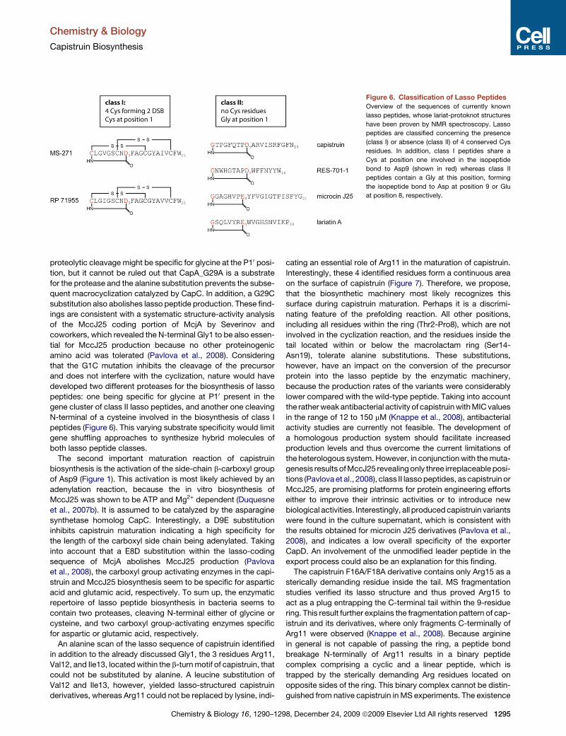
Figure 6. Classification of Lasso Peptides
Overview of the sequences of currently known
lasso peptides, whose lariat-protoknot structures
have been proven by NMR spectroscopy. Lasso
peptides are classified concerning the presence
(class I) or absence (class II) of 4 conserved Cys
residues. In addition, class I peptides share a
Cys at position one involved in the isopeptide
bond to Asp9 (shown in red) whereas class II
peptides contain a Gly at this position, forming
the isopeptide bond to Asp at position 9 or Glu
at position 8, respectively.
Chemistry & Biology
Capistruin Biosynthesis
proteolytic cleavage might be specific for glycine at the P10 posi-
tion, but it cannot be ruled out that CapA_G29A is a substrate
for the protease and the alanine substitution prevents the subse-
quent macrocyclization catalyzed by CapC. In addition, a G29C
substitution also abolishes lasso peptide production. These find-
ings are consistent with a systematic structure-activity analysis
of the MccJ25 coding portion of McjA by Severinov and
coworkers, which revealed the N-terminal Gly1 to be also essen-
tial for MccJ25 production because no other proteinogenic
amino acid was tolerated (Pavlova et al., 2008). Considering
that the G1C mutation inhibits the cleavage of the precursor
and does not interfere with the cyclization, nature would have
developed two different proteases for the biosynthesis of lasso
peptides: one being specific for glycine at P10 present in the
gene cluster of class II lasso peptides, and another one cleaving
N-terminal of a cysteine involved in the biosynthesis of class I
peptides (Figure 6). This varying substrate specificity would limit
gene shuffling approaches to synthesize hybrid molecules of
both lasso peptide classes.
The second important maturation reaction of capistruin
biosynthesis is the activation of the side-chain b-carboxyl group
of Asp9 (Figure 1). This activation is most likely achieved by an
adenylation reaction, because the in vitro biosynthesis of
MccJ25 was shown to be ATP and Mg2+ dependent (Duquesne
et al., 2007b). It is assumed to be catalyzed by the asparagine
synthetase homolog CapC. Interestingly, a D9E substitution
inhibits capistruin maturation indicating a high specificity for
the length of the carboxyl side chain being adenylated. Taking
into account that a E8D substitution within the lasso-coding
sequence of McjA abolishes MccJ25 production (Pavlova
et al., 2008), the carboxyl group activating enzymes in the capi-
struin and MccJ25 biosynthesis seem to be specific for aspartic
acid and glutamic acid, respectively. To sum up, the enzymatic
repertoire of lasso peptide biosynthesis in bacteria seems to
contain two proteases, cleaving N-terminal either of glycine or
cysteine, and two carboxyl group-activating enzymes specific
for aspartic or glutamic acid, respectively.
An alanine scan of the lasso sequence of capistruin identified
in addition to the already discussed Gly1, the 3 residues Arg11,
Val12, and Ile13, located within the b-turn motif of capistruin, that
could not be substituted by alanine. A leucine substitution of
Val12 and Ile13, however, yielded lasso-structured capistruin
derivatives, whereas Arg11 could not be replaced by lysine, indi-
Chemistry & Biology 16, 1290–129
cating an essential role of Arg11 in the maturation of capistruin.
Interestingly, these 4 identified residues form a continuous area
on the surface of capistruin (Figure 7). Therefore, we propose,
that the biosynthetic machinery most likely recognizes this
surface during capistruin maturation. Perhaps it is a discrimi-
nating feature of the prefolding reaction. All other positions,
including all residues within the ring (Thr2-Pro8), which are not
involved in the cyclization reaction, and the residues inside the
tail located within or below the macrolactam ring (Ser14-
Asn19), tolerate alanine substitutions. These substitutions,
however, have an impact on the conversion of the precursor
protein into the lasso peptide by the enzymatic machinery,
because the production rates of the variants were considerably
lower compared with the wild-type peptide. Taking into account
the rather weak antibacterial activity of capistruin with MIC values
in the range of 12 to 150 mM (Knappe et al., 2008), antibacterial
activity studies are currently not feasible. The development of
a homologous production system should facilitate increased
production levels and thus overcome the current limitations of
the heterologous system. However, in conjunction with the muta-
genesis results of MccJ25 revealing only three irreplaceable posi-
tions (Pavlova et al., 2008), class II lasso peptides, as capistruin or
MccJ25, are promising platforms for protein engineering efforts
either to improve their intrinsic activities or to introduce new
biological activities. Interestingly, all produced capistruin variants
were found in the culture supernatant, which is consistent with
the results obtained for microcin J25 derivatives (Pavlova et al.,
2008), and indicates a low overall specificity of the exporter
CapD. An involvement of the unmodified leader peptide in the
export process could also be an explanation for this finding.
The capistruin F16A/F18A derivative contains only Arg15 as a
sterically demanding residue inside the tail. MS fragmentation
studies verified its lasso structure and thus proved Arg15 to
act as a plug entrapping the C-terminal tail within the 9-residue
ring. This result further explains the fragmentation pattern of cap-
istruin and its derivatives, where only fragments C-terminally of
Arg11 were observed (Knappe et al., 2008). Because arginine
in general is not capable of passing the ring, a peptide bond
breakage N-terminally of Arg11 results in a binary peptide
complex comprising a cyclic and a linear peptide, which is
trapped by the sterically demanding Arg residues located on
opposite sides of the ring. This binary complex cannot be distin-
guished from native capistruin in MS experiments. The existence
8, December 24, 2009 ª2009 Elsevier Ltd All rights reserved 1295
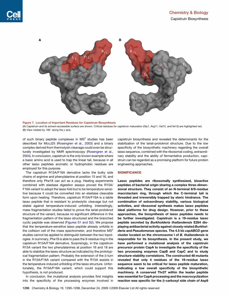
Figure 7. Location of Important Residues for Capistruin Biosynthesis
(A) Capistruin and its solvent-accessible surface are shown. Critical residues for capistruin maturation (Gly1, Arg11, Val12, and Ile13) are highlighted red.
(B) View rotated by 180� along the y axis.
Chemistry & Biology
Capistruin Biosynthesis
of such binary peptide complexes in MS2 studies has been
described for MccJ25 (Rosengren et al., 2003) and a binary
complex derived from thermolysin cleavage could even be struc-
turally investigated by NMR spectroscopy (Rosengren et al.,
2004). In conclusion, capistruin is the only known example where
a basic amino acid is used to trap the linear tail, because in all
other lasso peptides aromatic or hydrophobic residues are
employed for this purpose.
The capistruin R15A/F16A derivative lacks the bulky side
chains of arginine and phenylalanine at position 15 and 16, and
therefore only Phe18 can act as a plug. Heating experiments
combined with elastase digestion assays proved the R15A/
F16A variant to adopt the lasso fold but to be temperature sensi-
tive because it could be converted into an elastase cleavable
form upon heating. Therefore, capistruin R15A/F16A is a labile
lasso peptide that is resistant to proteolytic cleavage but not
stable against temperature-induced unfolding. Interestingly,
mass fragmentation studies failed to prove the lariat-protoknot
structure of the variant, because no significant difference in the
fragmentation pattern of the lasso structured and the branched
cyclic peptide was observed (Figures S1 and S2). We propose
that the temperature-sensitive lasso peptide already unfolds in
the collision cell of the mass spectrometer, and therefore MS2
studies cannot be applied to distinguish between the two topol-
ogies. In summary, Phe18 is able to pass the 9-residue ring in the
capistruin R15A/F16A derivative. Surprisingly, in the capistruin
R15A variant the two phenylalanines at position 16 and 18 are
able to stabilize the lasso structure as indicated by the prototyp-
ical fragmentation pattern. Probably the extension of the b-turn
in the R15A/F16A variant compared with the R15A assists in
the temperature-induced unfolding of the lasso structure. Unfor-
tunately, the R15A/F18A variant, which could support this
hypothesis, is not produced.
In conclusion, the mutational analysis provides first insights
into the specificity of the processing enzymes involved in
1296 Chemistry & Biology 16, 1290–1298, December 24, 2009 ª2009
capistruin biosynthesis and revealed the determinants for the
stabilization of the lariat-protoknot structure. Due to the low
specificity of the biosynthetic machinery regarding the overall
lasso sequence, combined with the ribosomal coding, extraordi-
nary stability and the ability of fermentative production, capi-
struin can be regarded as a promising platform for future protein
engineering approaches.
SIGNIFICANCE
Lasso peptides are ribosomally synthesized, bioactive
peptides of bacterial origin sharing a complex three-dimen-
sional structure. They consist of an N-terminal 8/9-residue
macrolactam ring, through which the C-terminal tail is
threaded and irreversibly trapped by steric hindrance. The
combination of extraordinary stability, various biological
activities, and ribosomal synthesis makes lasso peptides
ideal platforms for drug design. However, prior to these
approaches, the biosynthesis of lasso peptides needs to
be further investigated. Capistruin is a 19-residue lasso
peptide secreted by Burkholderia thailandensis E264 dis-
playing antibacterial activity against closely related Burkhol-
deria and Pseudomonas species. The 4.5 kb capABCD gene
cluster located on the chromosome I of B. thailandensis is
responsible for its biosynthesis. In the present study, we
have performed a mutational analysis of the capistruin
precursor protein CapA to investigate the specificity of the
two processing enzymes CapB and CapC and to study
structure-stability correlations. The constructed 48 mutants
revealed that only 4 residues of the 19-residue lasso
sequence seem to be critical for lasso peptide maturation,
indicating a low overall specificity of the biosynthetic
machinery. A conserved Thr27 within the leader peptide
was essential for CapA processing and the macrocyclization
reaction was specific for the b-carboxyl side chain of Asp9
Elsevier Ltd All rights reserved
Chemistry & Biology
Capistruin Biosynthesis
at the branching point. The double alanine mutant capistruin
F16A/F18A displayed a prototypical capistruin mass frag-
mentation pattern, which proved Arg15 to be solely respon-
sible for the trapping of the C-terminal tail within the macro-
lactam ring. Substitution of Arg15 and Phe16 by alanine
generated a temperature-sensitive capistruin variant, which
adopts the lasso fold, but unfolds into a branched cyclic,
elastase-sensitive peptide upon heating. Our results provide
first significant insights into capistruin biosynthesis
regarding specificity of the biosynthetic machinery and
stability determinants, and should facilitate future protein
engineering and gene shuffling approaches to generate
lasso peptides of designed stability and biological activity.
EXPERIMENTAL PROCEDURES
Bacterial Strains and General Methods
Escherichia coli NEB 10-beta was purchased from New England Biolabs
and used as general host for cloning. E. coli BL21(DE3) was purchased from
Invitrogen and used for heterologous production of capistruin and derivatives
thereof. Burkholderia thailandensis E264 was purchased from German Collec-
tion of Microorganisms and Cell Cultures (DSMZ). Oligonucleotides were
purchased from Eurofins MWG Operon. Pancreatic elastase was purchased
from Sigma. DNA dideoxy sequencing confirmed the identity of constructed
plasmids.
Cloning and Site-Directed Mutagenesis of pCDF-Duet_capA
Phusion High-Fidelity DNA-Polymerase (New England Biolabs) was used for
cloning and site directed mutagenesis following the instructions of the manu-
facturer.
The capA gene of the capistruin biosynthetic gene cluster was amplified
from genomic DNA of B. thailandensis E264 by polymerase chain reaction
using the forward primer (NdeI) ATATCATATGGTT CGACTTTTGGCGAA
GCTGC and the reverse primer (XhoI) ATATCTCGAGTTAATTGAAC CCGAA
GCGCGAAATGACG. The resulting amplicon was digested with NdeI and
XhoI (New England Biolabs) and cloned into pCDFDuet-1 (Novagen).
pCDFDuet_capA was used as a template for site-directed mutagenesis.
Mutagenesis was performed with Phusion High-Fidelity DNA-Polymerase in
HF buffer in the presence of 5% dimethyl sulfoxide. For each targeted codon,
a pair of �35 nt complementary oligonucleotide primers introducing the
designated substitutions was used. Mutagenized plasmid DNA was trans-
formed into E. coli NEB 10-beta. After overnight incubation on LB agar plates
containing 100 mg ml�1 spectinomycin, plasmids were prepared from indi-
vidual transformants and analyzed by sequencing.
Heterologous Production and Extraction of Capistruin
and Capistruin Derivatives
For heterologous production of capistruin or derivatives thereof, E. coli
BL21(DE3) cells were cotransformed with pCDFDuet_capA or a pCDFDuet
vector containing a capA mutant and pET41a(+)_capA*BCD (containing
a T27P mutation within CapA suppressing capistruin synthesis). Cells were
grown in LB medium containing 100 mg ml�1 spectinomycin and 50 mg ml�1
kanamycin at 37�C overnight. M20 medium supplemented with spectinomycin
(100 mg ml�1), kanamycin (50 mg ml�1), thiamine (2 mg ml�1), and biotin (2 mg
ml�1) was inoculated with the starter culture to an OD600 of 0.01 and cultivated
at 37�C. Cells were induced by addition of IPTG to a final concentration of
0.05 mM at an OD600 of 0.6 and harvested after further incubation for 12 to
48 hr at 37�C by centrifugation. Cell pellet was extracted twice with 100 ml
methanol. The culture supernatant was applied to solid phase extraction using
XAD16 resin (Sigma) (�5 g per liter culture supernatant). Upon incubation of
the culture supernatant with XAD16 resin, the supernatant was removed by
filtration and the resin was washed with water and eluted with methanol. Meth-
anol extracts of the pellet and the supernatant were evaporated to dryness,
dissolved in 1.6 ml 20% methanol, and analyzed by LC-MS. Production rates
of the various capistruin derivatives were generally lower than the wild-type
Chemistry & Biology 16, 1290–129
peptide (0.1–0.2 mg l�1) and were estimated to be in the range of 0.01–
0.05 mg l�1 based on peak integration of EICs.
Mass Spectrometric Analysis
The mass spectrometric characterization of capistruin or its derivatives was
performed with an LTQ-FT instrument (Thermo Fisher Scientific, Bremen,
Germany) connected to a microbore 1100 HPLC system (Agilent, Waldbronn,
Germany). Separation of extracted capistruin or capistruin derivatives
from contaminants was achieved using a 125/2 Nucleodur C18ec column
(Macherey-Nagel, Duren, Germany) applying the following gradient of water/
0.05% formic acid (solvent A) and acetonitrile/0.045% formic acid (solvent
B) at a column temperature of 40�C and a flow rate of 0.2 ml min�1: Linear
increase from 10% B to 40% B within 30 min followed by a linear increase
to 95% B in 5 min and holding 95% B for additional 2 min.
CID fragmentation studies within the linear ion trap were either done using
online LC-MS or utilizing purified capistruin samples. The purified samples
were analyzed using a syringe pump at a flow rate of 10 ml min�1. Usually
the doubly charged ions were selected for fragmentation in the ion trap,
because they were the dominant species in the spectrum. The energy for frag-
mentation was set to 35 in all cases.
Stability Studies of Capistruin R15A/F16A and R15A
An extract of the supernatant of a 6 l culture was prepared as described above
and dissolved in 1.6 ml 20 mM Tris/HCl (pH 8.0). In a 30 ml reaction, 25 ml of the
extract was incubated in the presence or absence of 0.025 units elastase
(Sigma) for 1 hr at 25�C (20 mM Tris/HCl [pH 8.0] was used to fill up to
30 ml). Afterwards the samples were heated for 1 hr at 95�C, cooled down to
25�C, and incubated again in the presence or absence of additional 0.025 units
elastase for 1 hr at 25�C. After each single incubation step, 20 ml of the reaction
mixtures was analyzed by LC-MS.
SUPPLEMENTAL DATA
Supplemental Data include two figures and one table and can be found with
this article online at http://www.cell.com/chemistry-biology/supplemental/
S1074-5521(09)00400-1.
ACKNOWLEDGMENTS
We would like to thank the members of the Marahiel group and Severine Zirah
and Sylvie Rebuffat (Museum National d’Histoire Naturelle, Paris) for fruitful
discussions. We gratefully acknowledge financial support from the Deutsche
Forschungsgemeinschaft (M.A.M.), the Fonds der chemischen Industrie
(M.A.M. and T.A.K.), and the Deutscher Akademischer Austauschdienst
(M.A.M. and T.A.K.).
Received: September 29, 2009
Revised: October 26, 2009
Accepted: November 4, 2009
Published: December 23, 2009
REFERENCES
Adelman, K., Yuzenkova, J., La Porta, A., Zenkin, N., Lee, J., Lis, J.T.,
Borukhov, S., Wang, M.D., and Severinov, K. (2004). Molecular mechanism
of transcription inhibition by peptide antibiotic Microcin J25. Mol. Cell 14,
753–762.
Bayro, M.J., Mukhopadhyay, J., Swapna, G.V., Huang, J.Y., Ma, L.C., Sineva,
E., Dawson, P.E., Montelione, G.T., and Ebright, R.H. (2003). Structure of
antibacterial peptide microcin J25: a 21-residue lariat protoknot. J. Am.
Chem. Soc. 125, 12382–12383.
Duquesne, S., Destoumieux-Garzon, D., Peduzzi, J., and Rebuffat, S. (2007a).
Microcins, gene-encoded antibacterial peptides from enterobacteria. Nat.
Prod. Rep. 24, 708–734.
Duquesne, S., Destoumieux-Garzon, D., Zirah, S., Goulard, C., Peduzzi, J.,
and Rebuffat, S. (2007b). Two enzymes catalyze the maturation of a lasso
peptide in Escherichia coli. Chem. Biol. 14, 793–803.
8, December 24, 2009 ª2009 Elsevier Ltd All rights reserved 1297
Chemistry & Biology
Capistruin Biosynthesis
Duquesne, S., Petit, V., Peduzzi, J., and Rebuffat, S. (2007c). Structural and
functional diversity of microcins, gene-encoded antibacterial peptides from
enterobacteria. J. Mol. Microbiol. Biotechnol. 13, 200–209.
Frechet, D., Guitton, J.D., Herman, F., Faucher, D., Helynck, G., Monegier du
Sorbier, B., Ridoux, J.P., James-Surcouf, E., and Vuilhorgne, M. (1994).
Solution structure of RP 71955, a new 21 amino acid tricyclic peptide active
against HIV-1 virus. Biochemistry 33, 42–50.
Helynck, G., Dubertret, C., Mayaux, J.F., and Leboul, J. (1993). Isolation of RP
71955, a new anti-HIV-1 peptide secondary metabolite. J. Antibiot. (Tokyo) 46,
1756–1757.
Iwatsuki, M., Tomoda, H., Uchida, R., Gouda, H., Hirono, S., and Omura, S.
(2006). Lariatins, antimycobacterial peptides produced by Rhodococcus sp.
K01-B0171, have a lasso structure. J. Am. Chem. Soc. 128, 7486–7491.
Katahira, R., Shibata, K., Yamasaki, M., Matsuda, Y., and Yoshida, M. (1995).
Solution structure of endothelin B receptor selective antagonist RES-701-1
determined by 1H NMR spectroscopy. Bioorg. Med. Chem. 3, 1273–1280.
Katahira, R., Yamasaki, M., Matsuda, Y., and Yoshida, M. (1996). MS-271,
a novel inhibitor of calmodulin-activated myosin light chain kinase from
Streptomyces sp.–II. Solution structure of MS-271: characteristic features of
the ‘‘lasso’’ structure. Bioorg. Med. Chem. 4, 121–129.
Knappe, T.A., Linne, U., Zirah, S., Rebuffat, S., Xie, X., and Marahiel, M.A.
(2008). Isolation and structural characterization of capistruin, a lasso peptide
predicted from the genome sequence of Burkholderia thailandensis E264.
J. Am. Chem. Soc. 130, 11446–11454.
Larsen, T.M., Boehlein, S.K., Schuster, S.M., Richards, N.G., Thoden, J.B.,
Holden, H.M., and Rayment, I. (1999). Three-dimensional structure of
Escherichia coli asparagine synthetase B: a short journey from substrate to
product. Biochemistry 38, 16146–16157.
Makarova, K.S., Aravind, L., and Koonin, E.V. (1999). A superfamily of
archaeal, bacterial, and eukaryotic proteins homologous to animal transgluta-
minases. Protein Sci. 8, 1714–1719.
Morishita, Y., Chiba, S., Tsukuda, E., Tanaka, T., Ogawa, T., Yamasaki, M.,
Yoshida, M., Kawamoto, I., and Matsuda, Y. (1994). RES-701-1, a novel and
selective endothelin type B receptor antagonist produced by Streptomyces
sp. RE-701. I. Characterization of producing strain, fermentation, isolation,
physico-chemical and biological properties. J. Antibiot. (Tokyo) 47, 269–275.
1298 Chemistry & Biology 16, 1290–1298, December 24, 2009 ª2009
Mukhopadhyay, J., Sineva, E., Knight, J., Levy, R.M., and Ebright, R.H. (2004).
Antibacterial peptide microcin J25 inhibits transcription by binding within and
obstructing the RNA polymerase secondary channel. Mol. Cell 14, 739–751.
Pavlova, O., Mukhopadhyay, J., Sineva, E., Ebright, R.H., and Severinov, K.
(2008). Systematic structure-activity analysis of microcin J25. J. Biol. Chem.
283, 25589–25595.
Rebuffat, S., Blond, A., Destoumieux-Garzon, D., Goulard, C., and Peduzzi, J.
(2004). Microcin J25, from the macrocyclic to the lasso structure: implications
for biosynthetic, evolutionary and biotechnological perspectives. Curr. Protein
Pept. Sci. 5, 383–391.
Rosengren, K.J., Blond, A., Afonso, C., Tabet, J.C., Rebuffat, S., and Craik,
D.J. (2004). Structure of thermolysin cleaved microcin J25: extreme stability
of a two-chain antimicrobial peptide devoid of covalent links. Biochemistry
43, 4696–4702.
Rosengren, K.J., Clark, R.J., Daly, N.L., Goransson, U., Jones, A., and Craik,
D.J. (2003). Microcin J25 has a threaded sidechain-to-backbone ring structure
and not a head-to-tail cyclized backbone. J. Am. Chem. Soc. 125, 12464–
12474.
Severinov, K., Semenova, E., Kazakov, A., Kazakov, T., and Gelfand, M.S.
(2007). Low-molecular-weight post-translationally modified microcins. Mol.
Microbiol. 65, 1380–1394.
Solbiati, J.O., Ciaccio, M., Farias, R.N., Gonzalez-Pastor, J.E., Moreno, F., and
Salomon, R.A. (1999). Sequence analysis of the four plasmid genes required to
produce the circular peptide antibiotic microcin J25. J. Bacteriol. 181, 2659–
2662.
Solbiati, J.O., Ciaccio, M., Farias, R.N., and Salomon, R.A. (1996). Genetic
analysis of plasmid determinants for microcin J25 production and immunity.
J. Bacteriol. 178, 3661–3663.
Wilson, K.A., Kalkum, M., Ottesen, J., Yuzenkova, J., Chait, B.T., Landick, R.,
Muir, T., Severinov, K., and Darst, S.A. (2003). Structure of microcin J25,
a peptide inhibitor of bacterial RNA polymerase, is a lassoed tail. J. Am.
Chem. Soc. 125, 12475–12483.
Yano, K., Toki, S., Nakanishi, S., Ochiai, K., Ando, K., Yoshida, M., Matsuda,
Y., and Yamasaki, M. (1996). MS-271, a novel inhibitor of calmodulin-activated
myosin light chain kinase from Streptomyces sp.–I. Isolation, structural
determination and biological properties of MS-271. Bioorg. Med. Chem. 4,
115–120.
Elsevier Ltd All rights reserved