insight into renal vascular and nonvascular interventions · "insight into renal vascular and...
TRANSCRIPT
"Insight into Renal Vascular
and Nonvascular Interventions
Dr. Anatoly Shuster, Department of Diagnostic Imaging, TBRHSC
Assistant Professor, Lakehead University, NOSM

Conflict of Interest Declaration:
Nothing to Disclose
Presenter: Anatoly Shuster
Title of Presentation: "Insight into Renal
Vascular and Nonvascular Interventions”
I have no financial or personal
relationships to disclose
Renal Arterial Stenosis
Anatomy:
Renal arteries (RA) arise from the lateral surface of the aorta
at about the L1-L2 level
Right RA runs posterior to the IVC
Left RA passes behind the left renal vein (RV)
At the renal hilum RA bifurcates into ventral and dorsal rami
Accessory RA supply one or both kidneys in 25-35%; may
originate from aorta or iliac artery; most supply the lower pole
Renal Arterial Stenosis
Anatomy:
Kidney is the “end organ”
Communications between extrarenal arteries (aorta, lumbar
arteries, internal iliac artery, inferior adrenal artery) and
intrarenal arteries (segmental, intralobar, arcuate) exist:
capsular, peripelvic, periureteric systems (Abrams/Cornell)
Atherosclerotic Renovascular Disease
Etiology:
Nephrosclerosis: global damage of distal intrarenal vessels
Renovascular Hypertension (RVH): stenosis/occlusion of main,
accessory, or branch RA
Reduction in intrarenal arterial pressure sensed by juxta-
glomerular apparatus >> triggered renin-angiotensin-aldosterone
system >> vasoconstriction + sodium and water retention
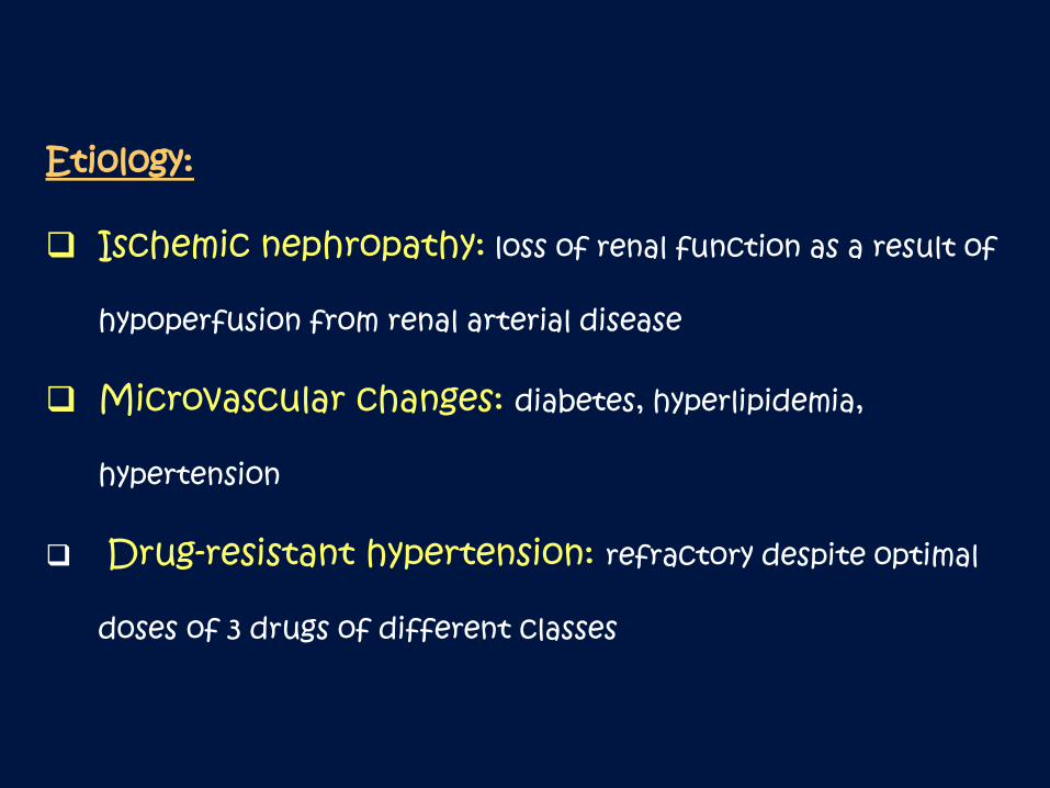
Etiology:
Ischemic nephropathy: loss of renal function as a result of
hypoperfusion from renal arterial disease
Microvascular changes: diabetes, hyperlipidemia,
hypertension
Drug-resistant hypertension: refractory despite optimal
doses of 3 drugs of different classes
Renal Arterial Stenosis
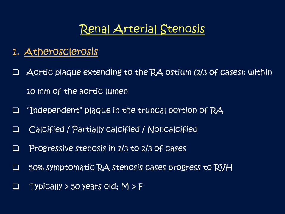
1. Atherosclerosis
Aortic plaque extending to the RA ostium (2/3 of cases): within
10 mm of the aortic lumen
“Independent” plaque in the truncal portion of RA
Calcified / Partially calcified / Noncalcified
Progressive stenosis in 1/3 to 2/3 of cases
50% symptomatic RA stenosis cases progress to RVH
Typically > 50 years old; M > F
Renal Arterial Stenosis
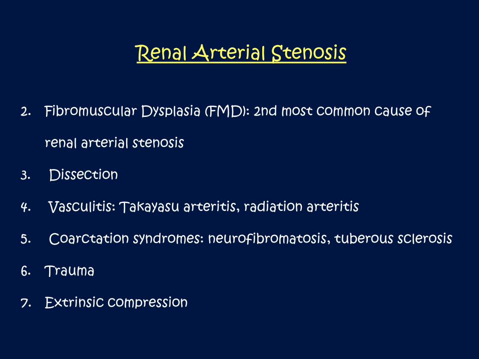
2. Fibromuscular Dysplasia (FMD): 2nd most common cause of
renal arterial stenosis
3. Dissection
4. Vasculitis: Takayasu arteritis, radiation arteritis
5. Coarctation syndromes: neurofibromatosis, tuberous sclerosis
6. Trauma
7. Extrinsic compression
Renal Arterial Stenosis
Diagnosis:
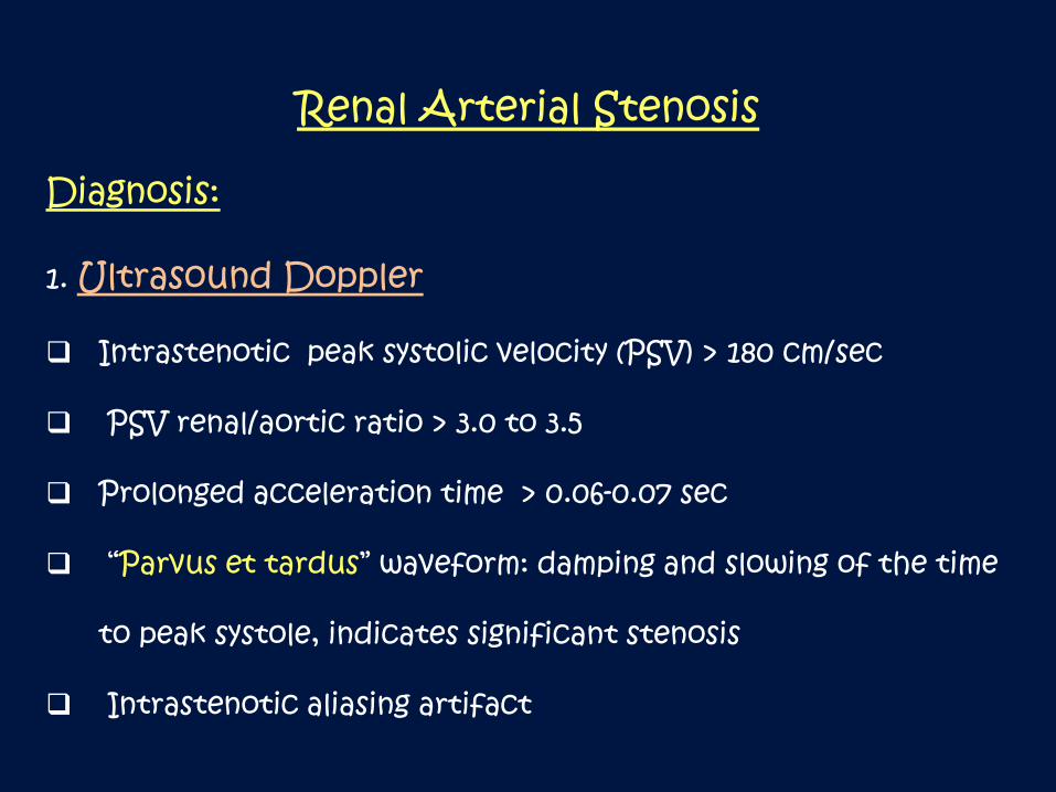
1. Ultrasound Doppler
Intrastenotic peak systolic velocity (PSV) > 180 cm/sec
PSV renal/aortic ratio > 3.0 to 3.5
Prolonged acceleration time > 0.06-0.07 sec
“Parvus et tardus” waveform: damping and slowing of the time
to peak systole, indicates significant stenosis
Intrastenotic aliasing artifact
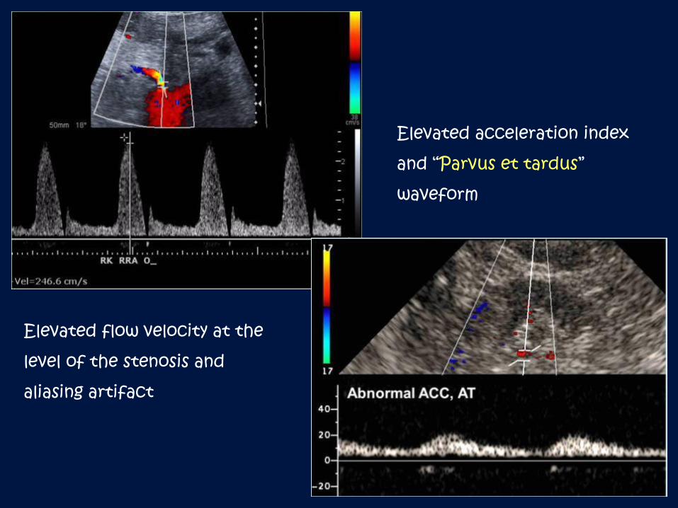
Elevated flow velocity at the
level of the stenosis and
aliasing artifact
Elevated acceleration index
and “Parvus et tardus”
waveform
Renal Arterial Stenosis
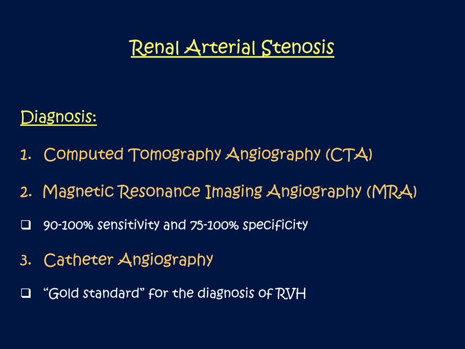
Diagnosis:
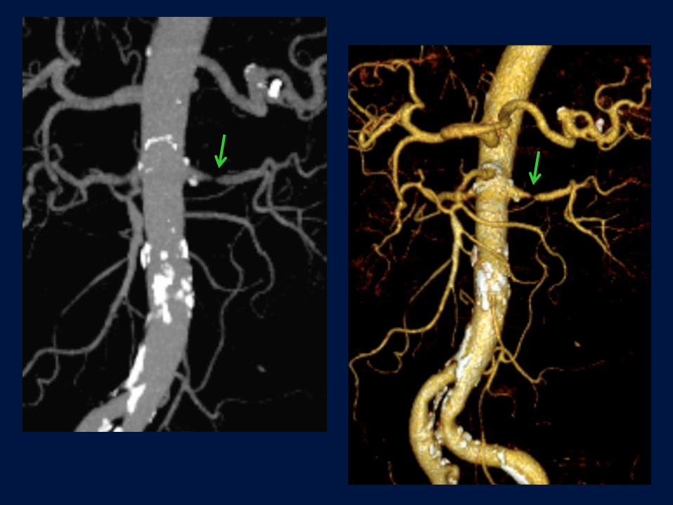
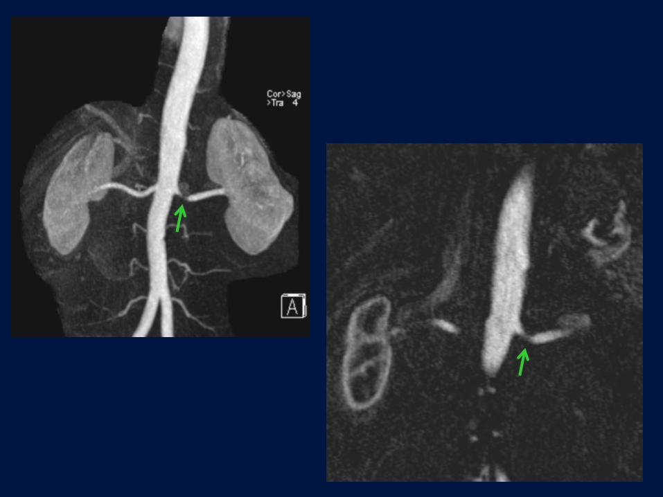
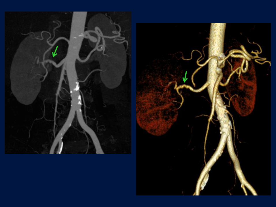
1. Computed Tomography Angiography (CTA)
2. Magnetic Resonance Imaging Angiography (MRA)
90-100% sensitivity and 75-100% specificity
3. Catheter Angiography
“Gold standard” for the diagnosis of RVH
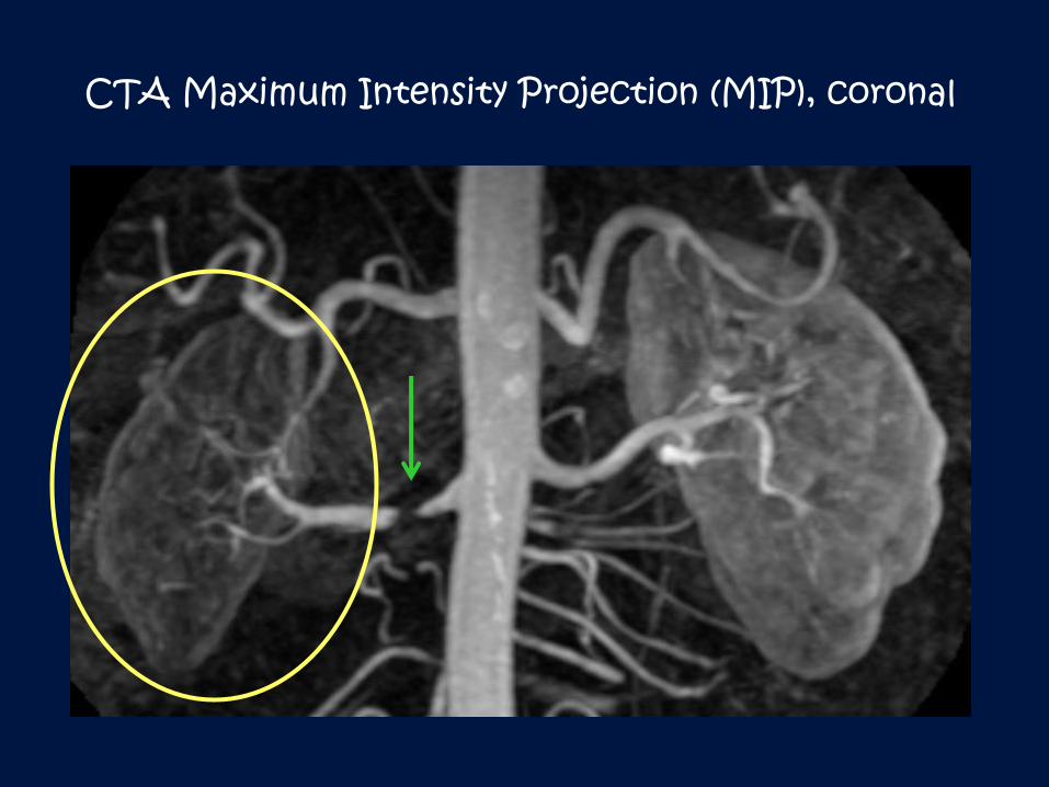
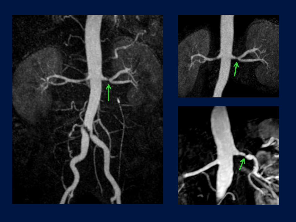
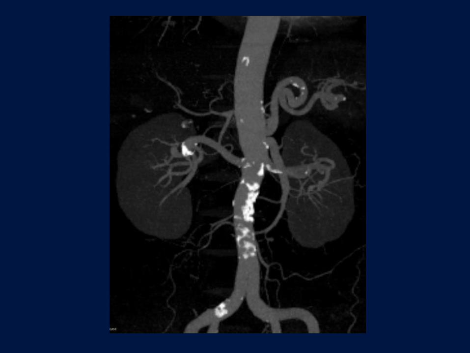
CTA Maximum Intensity Projection (MIP), coronal
Renal Arterial Stenosis
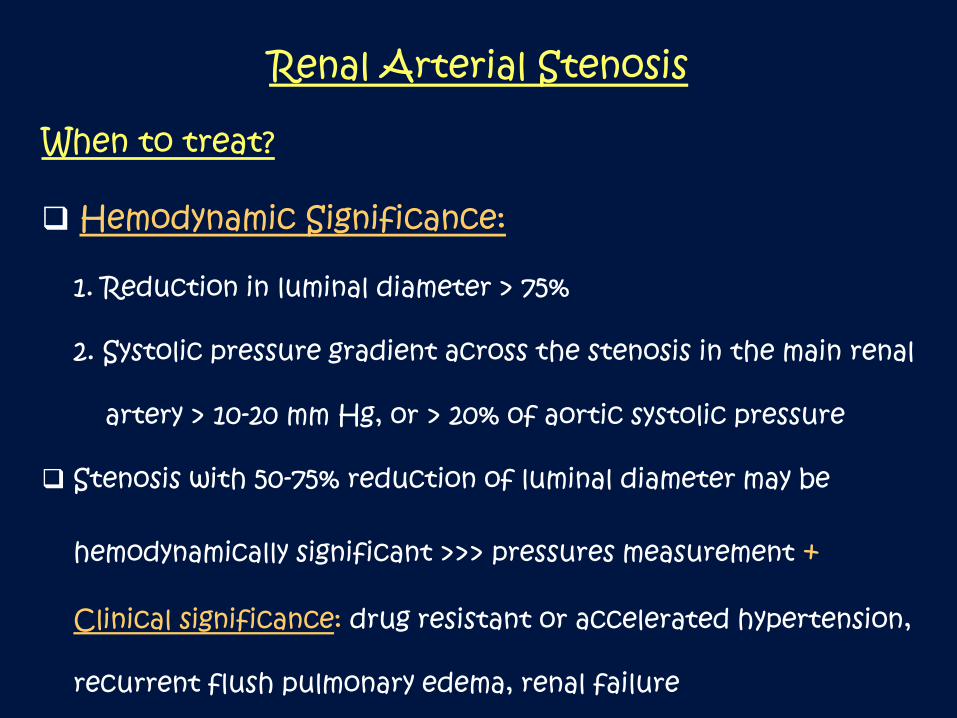
When to treat?
Hemodynamic Significance:
1. Reduction in luminal diameter > 75%
2. Systolic pressure gradient across the stenosis in the main renal
artery > 10-20 mm Hg, or > 20% of aortic systolic pressure
Stenosis with 50-75% reduction of luminal diameter may be
hemodynamically significant >>> pressures measurement +
Clinical significance: drug resistant or accelerated hypertension,
recurrent flush pulmonary edema, renal failure
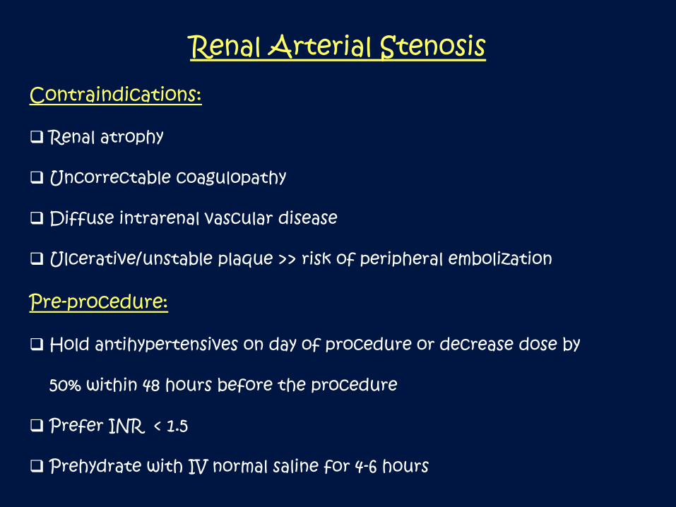
Renal Arterial Stenosis
Contraindications:
Renal atrophy
Uncorrectable coagulopathy
Diffuse intrarenal vascular disease
Ulcerative/unstable plaque >> risk of peripheral embolization
Pre-procedure:
Hold antihypertensives on day of procedure or decrease dose by
50% within 48 hours before the procedure
Prefer INR < 1.5
Prehydrate with IV normal saline for 4-6 hours
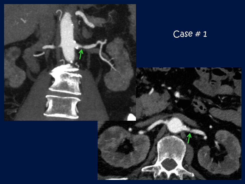
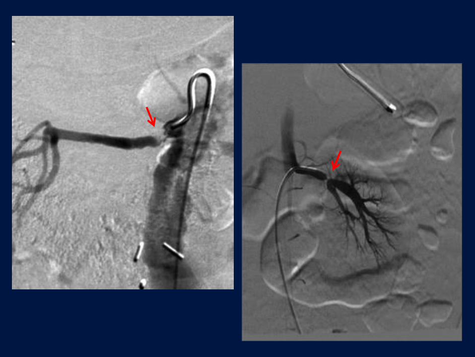
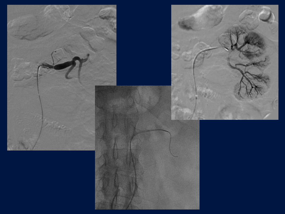
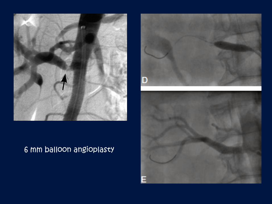
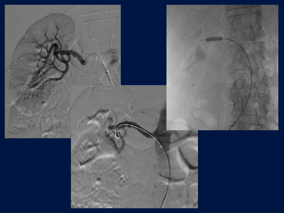
Case # 1
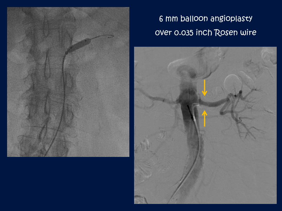
6 mm balloon angioplasty
0ver 0.035 inch Rosen wire
Renal Artery Intervention - Endovascular Techniques, Thomas Zeller, MD, Aljoscha Rastan, MD, Elias Noory, MD; Vascular Disease Management 2011;8:E21–E27
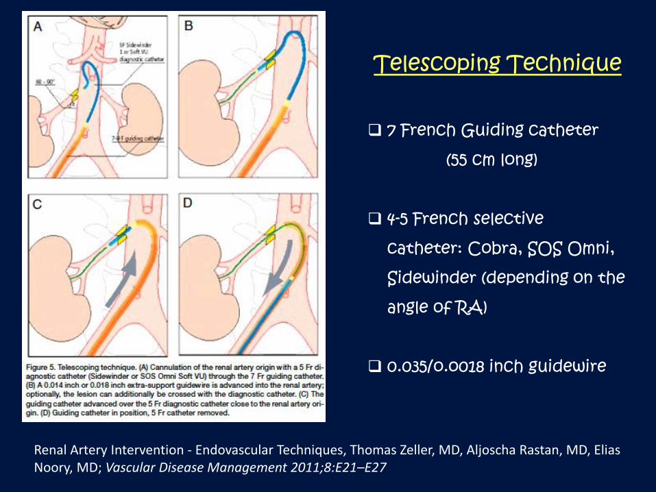
Telescoping Technique
7 French Guiding catheter
(55 cm long)
4-5 French selective
catheter: Cobra, SOS Omni,
Sidewinder (depending on the
angle of RA)
0.035/o.o018 inch guidewire
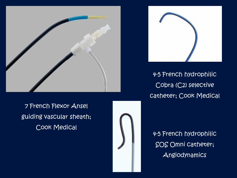
7 French Flexor Ansel
guiding vascular sheath;
Cook Medical
4-5 French hydrophilic
Cobra (C2) selective
catheter; Cook Medical
4-5 French hydrophilic
SOS Omni catheter;
Angiodynamics
6 mm balloon angioplasty
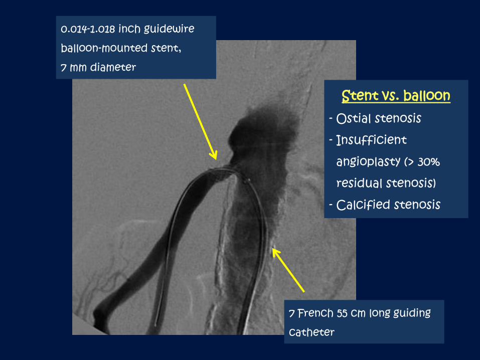
0.014-1.018 inch guidewire
balloon-mounted stent,
7 mm diameter
7 French 55 cm long guiding
catheter
Stent vs. balloon- Ostial stenosis
- Insufficient
angioplasty (> 30%
residual stenosis)
- Calcified stenosis
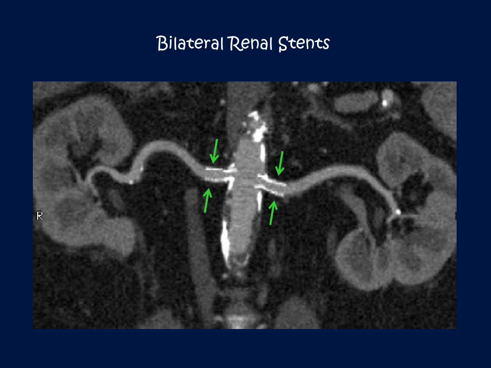
Bilateral Renal Stents
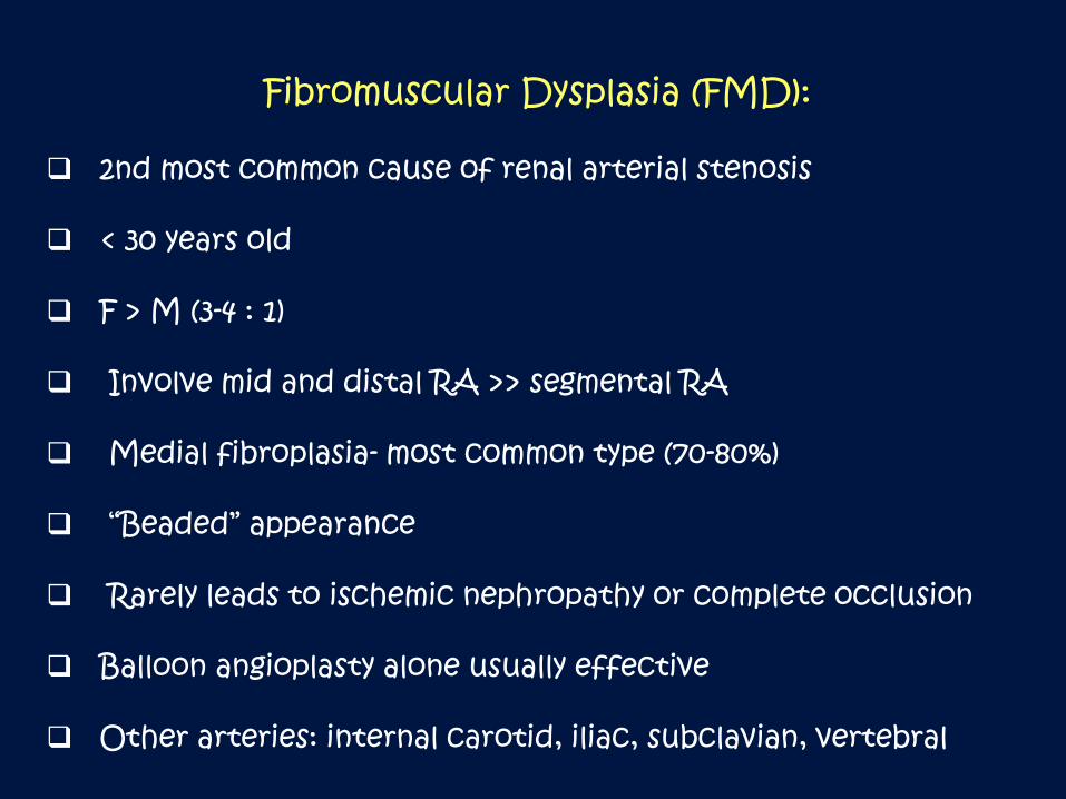
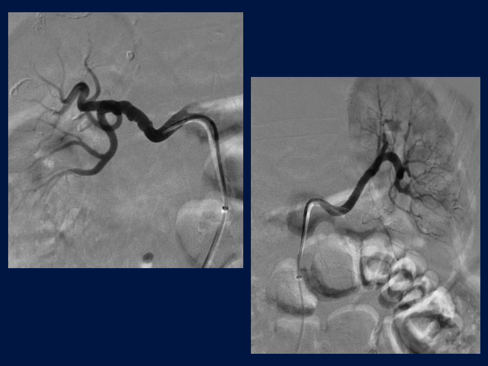
Fibromuscular Dysplasia (FMD):
2nd most common cause of renal arterial stenosis
< 30 years old
F > M (3-4 : 1)
Involve mid and distal RA >> segmental RA
Medial fibroplasia- most common type (70-80%)
“Beaded” appearance
Rarely leads to ischemic nephropathy or complete occlusion
Balloon angioplasty alone usually effective
Other arteries: internal carotid, iliac, subclavian, vertebral
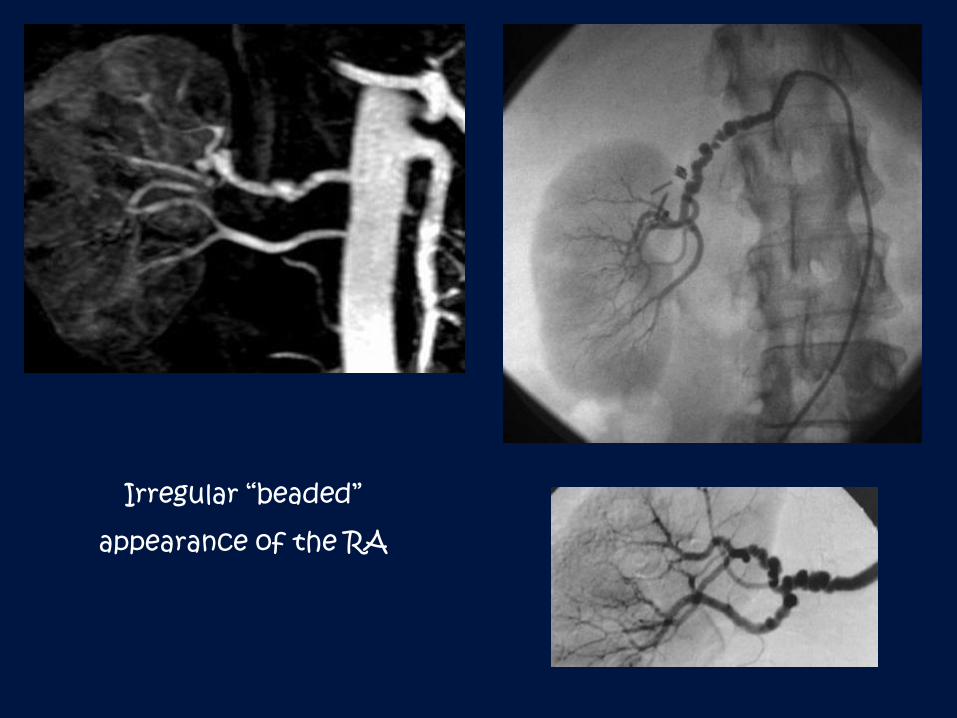
Irregular “beaded”
appearance of the RA
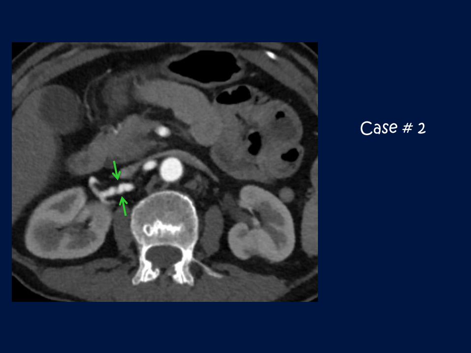
Case # 2
Complications
5-10% of cases
RA dissection or rupture >> stent placement >> surgery
RA thrombosis
Distal thrombus microembolization
Access site complications: hematoma, CFA pseudoaneurysm
Contrast nephropathy
Renal Arterial Embolization
RA Aneurysm
True aneurysms: dysplastic, FMD, connective tissue disorders:
neurofibromatosis, Ehlers-Danlos syndrome, vasculitis: poliarteritis
nodosa (multiple aneurysms) and Takayasu arteritis, congenital
False aneurysms: trauma, inflammation/Infection, post-transplant,
dissection, drug use (cocaine, methamphetamines), tumor related
Arteriovenous Fistulas and Malformations
Traumatic Hemorrhage
Grade IV injures
Renal Arterial Embolization
Dysplastic aneurysms:
Near the first bifurcation of the main RA
75% of patients have elevated blood pressure
Succular or fusiform
Complications: rupture, thrombosis
Risk of rupture is heightened in pregnant women
When to treat: “rule of 2 cm”, regardless of size in women of child-
bearing potential, symptomatic patients, all pseudoaneurysms
Endovascular treatment options: covered stent placement for main
RA aneurysms, embolization with microcoils or glue for intrarenal aneurysms
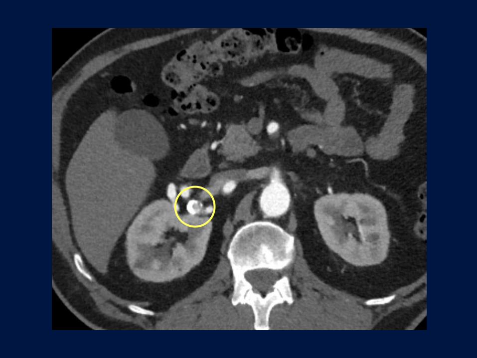
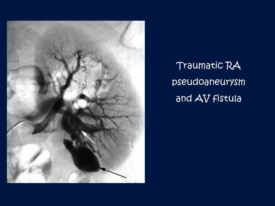
Traumatic RA
pseudoaneurysm
and AV fistula
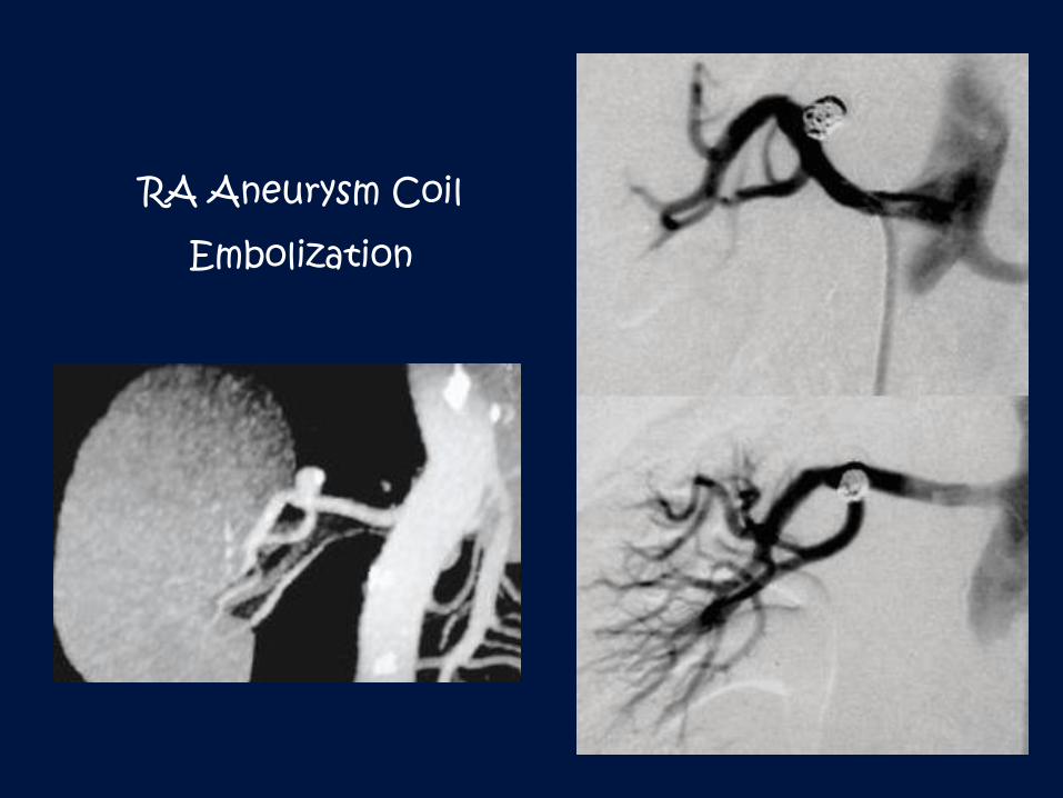
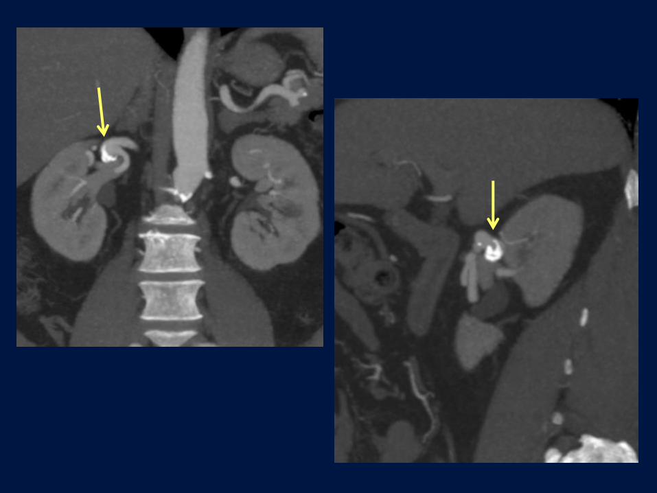
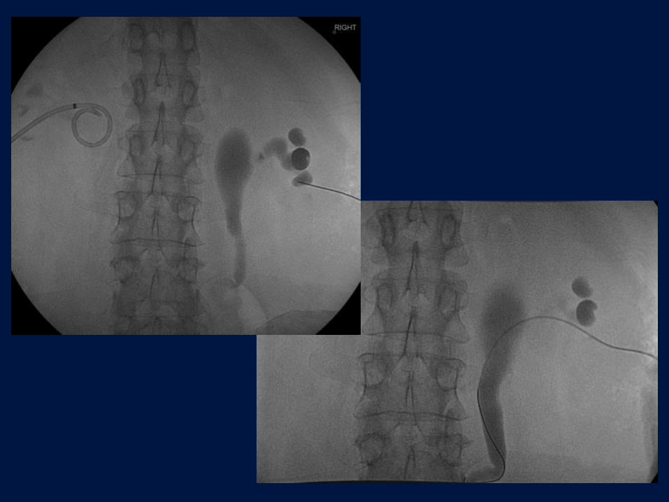
RA Aneurysm Coil
Embolization
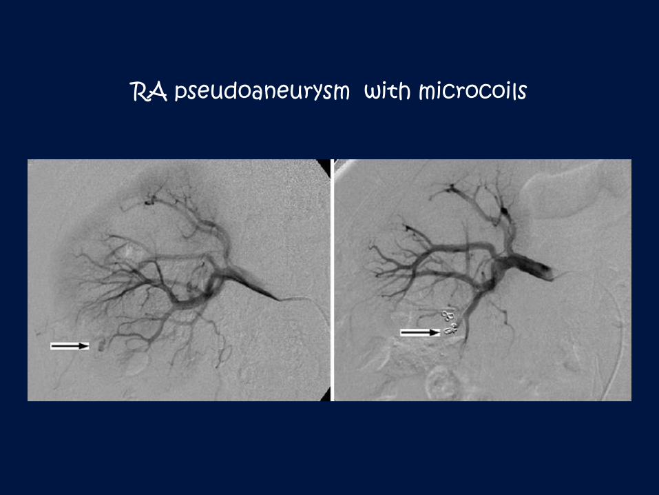
RA pseudoaneurysm with microcoils
Renal Arterial Dissection
Extension of aortic dissection
Trauma: iatrogenic (e.g., catheterization, injury by guidewire ),
blunt or penetrating trauma
FMD
Segmental Arterial Mediolysis
Spontaneous
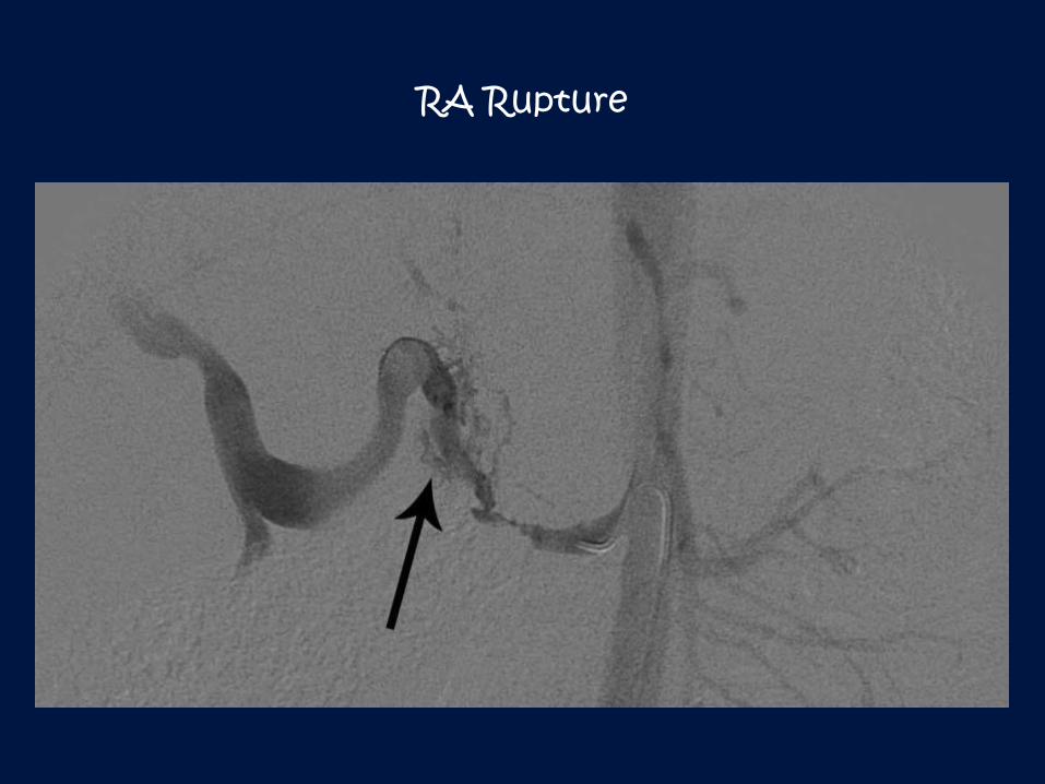
RA Rupture
Renal Transplant Vascular Complications Develop up to 25% of cases
Arterial stenosis - most common problem, 4-10% of cases, occurs
between 3 months to 2 years after placement, usually located at the
anastomosis
Arterial thrombosis - result of operative injury to the donor or
recipient artery, arterial kinking, acute rejection, hypotension,
thrombophilic state, atherosclerosis
Renal vein thrombosis
Vascular injury, pseudoaneurysm or arteriovenous fistula formation
from percutaneous biopsy
Renal Neoplasms
Benign
Adenoma/Oncocytoma
Angiomylolipoma (tuberous sclerosis: multiple bilateral lesions)
Malignant:
Renal Cell Carcinoma (RCC); von Hippel-Lindau disease
Transitional Cell Carcinoma (TCC)
Wilms Tumor
Metastases (including lymphoma)
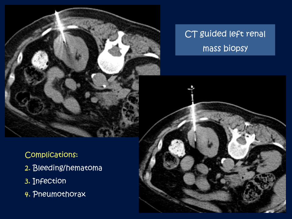
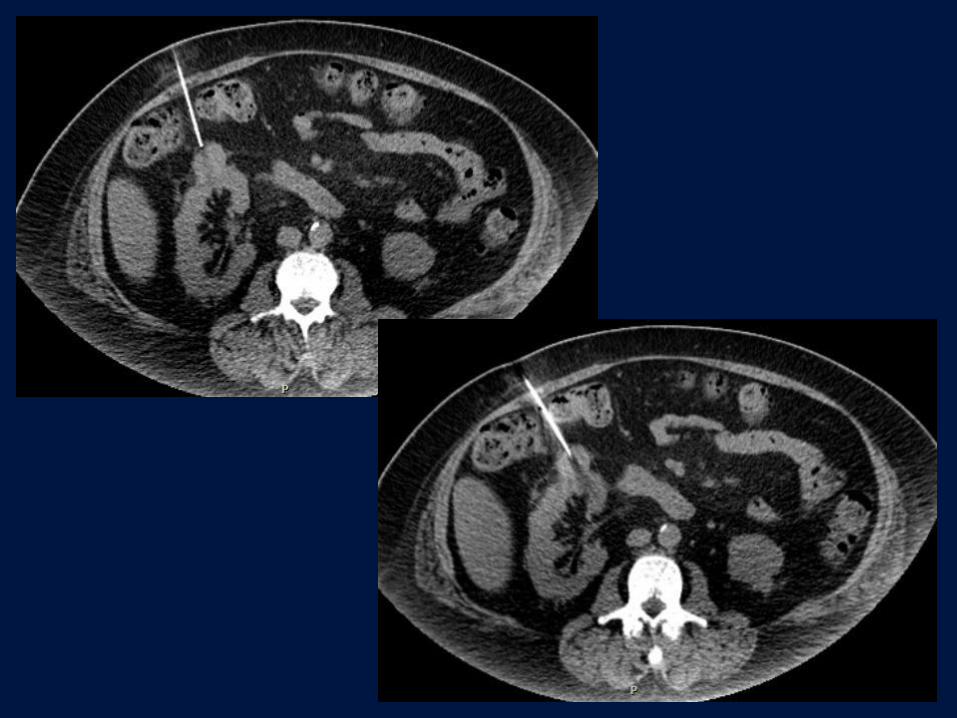
CT guided left renal
mass biopsy
Complications:
2. Bleeding/hematoma
3. Infection
4. Pneumothorax
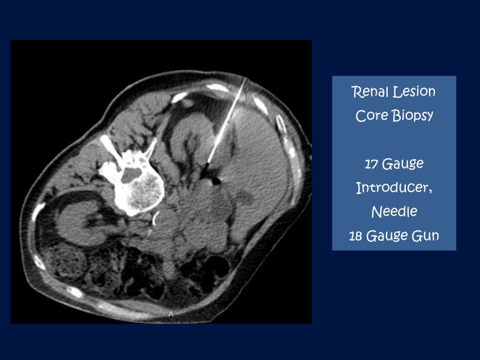
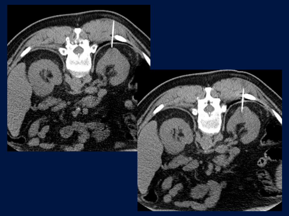
Renal Lesion
Core Biopsy
17 Gauge
Introducer,
Needle
18 Gauge Gun
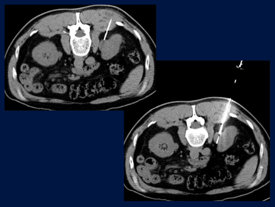
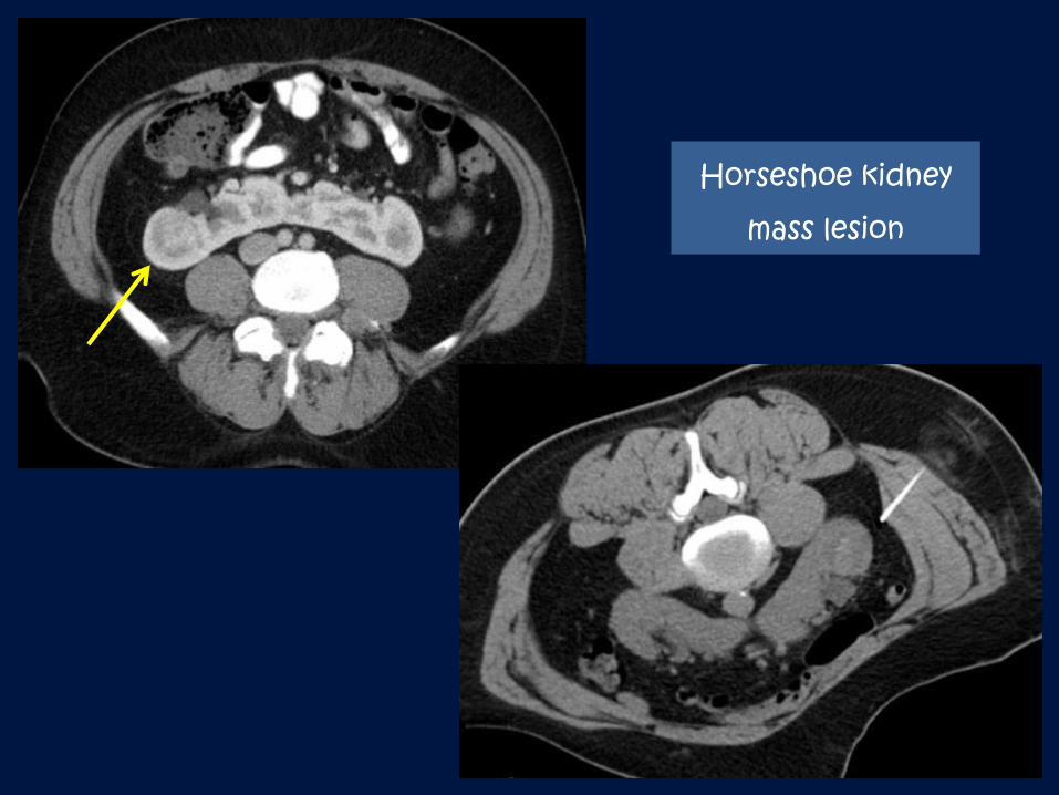
Horseshoe kidney
mass lesion
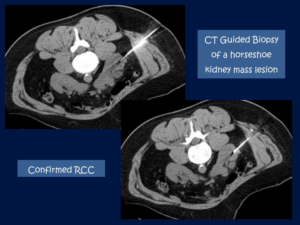
CT Guided Biopsy
of a horseshoe
kidney mass lesion
Confirmed RCC
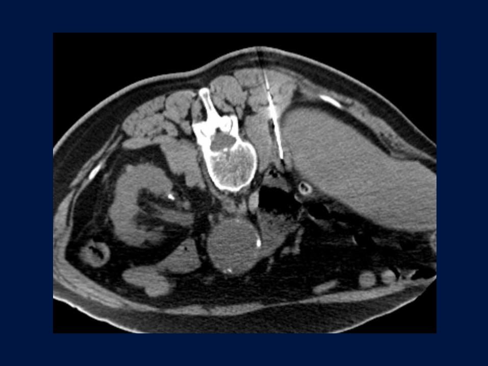
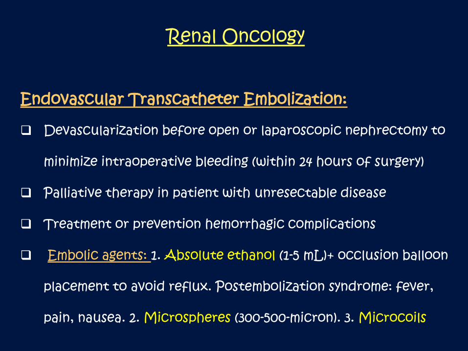
Renal Oncology
Endovascular Transcatheter Embolization:
Devascularization before open or laparoscopic nephrectomy to
minimize intraoperative bleeding (within 24 hours of surgery)
Palliative therapy in patient with unresectable disease
Treatment or prevention hemorrhagic complications
Embolic agents: 1. Absolute ethanol (1-5 mL)+ occlusion balloon
placement to avoid reflux. Postembolization syndrome: fever,
pain, nausea. 2. Microspheres (300-500-micron). 3. Microcoils
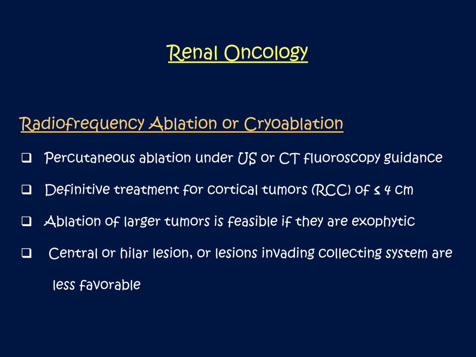
Renal Oncology
Radiofrequency Ablation or Cryoablation
Percutaneous ablation under US or CT fluoroscopy guidance
Definitive treatment for cortical tumors (RCC) of ≤ 4 cm
Ablation of larger tumors is feasible if they are exophytic
Central or hilar lesion, or lesions invading collecting system are
less favorable
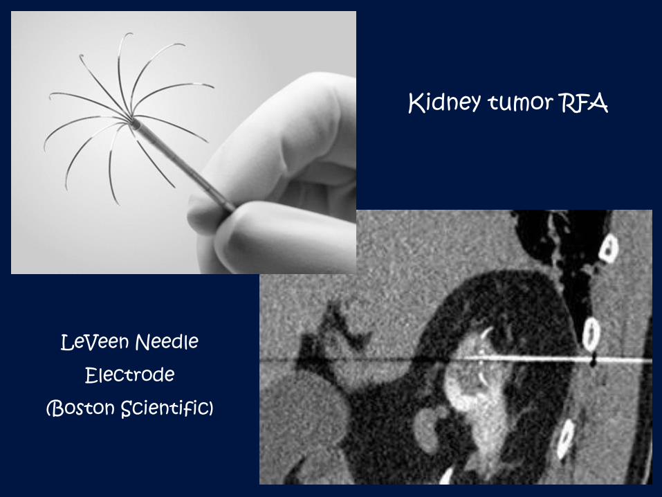
LeVeen Needle
Electrode
(Boston Scientific)
Kidney tumor RFA
Percutaneous Nephrostomy
US/Fluoroscopy guided or CT guided (in obese patients)
Indications:
Hydronephrosis + Infection
Hydronephrosis + Pain
Hydronephrosis + Renal failure
Diversion of Urine: traumatic urinary tract injury, malignant or
inflammatory urinary fistula, hemorrhagic cystitis
Access for diagnostic or therapeutic interventions
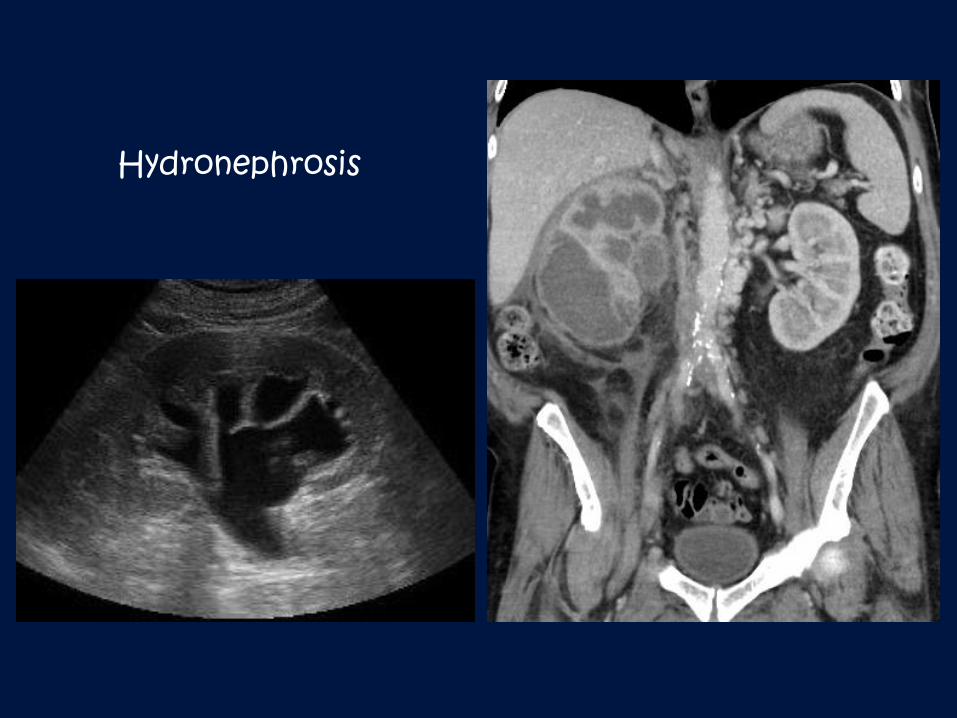
Hydronephrosis
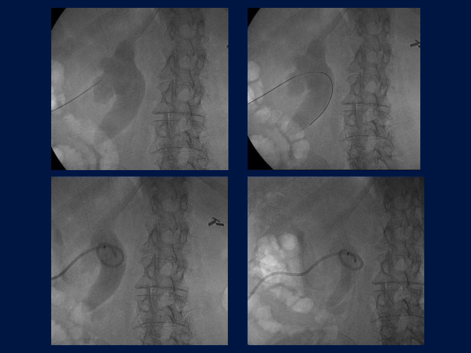

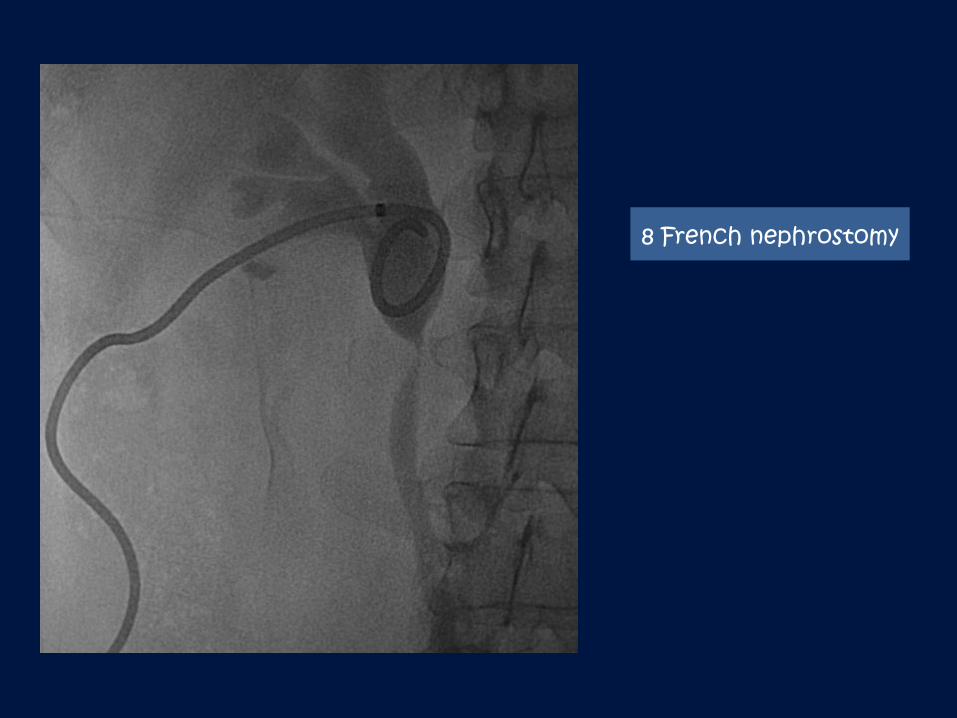
8 French nephrostomy
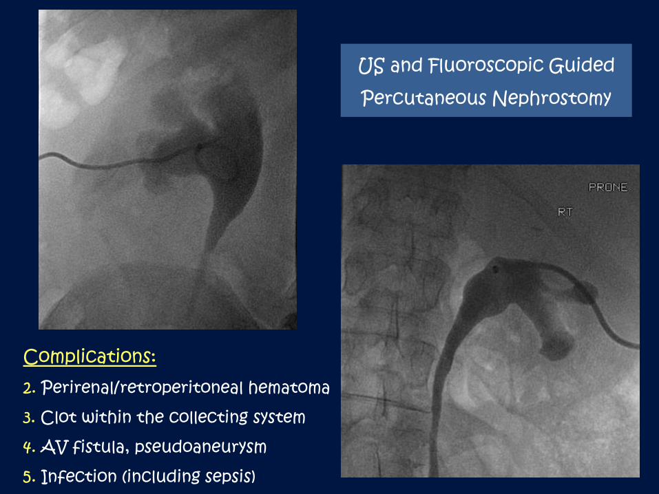
US and Fluoroscopic Guided
Percutaneous Nephrostomy
Complications:2. Perirenal/retroperitoneal hematoma
3. Clot within the collecting system
4. AV fistula, pseudoaneurysm
5. Infection (including sepsis)
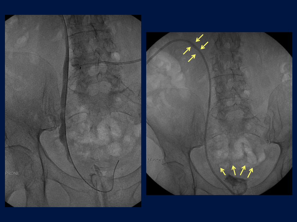
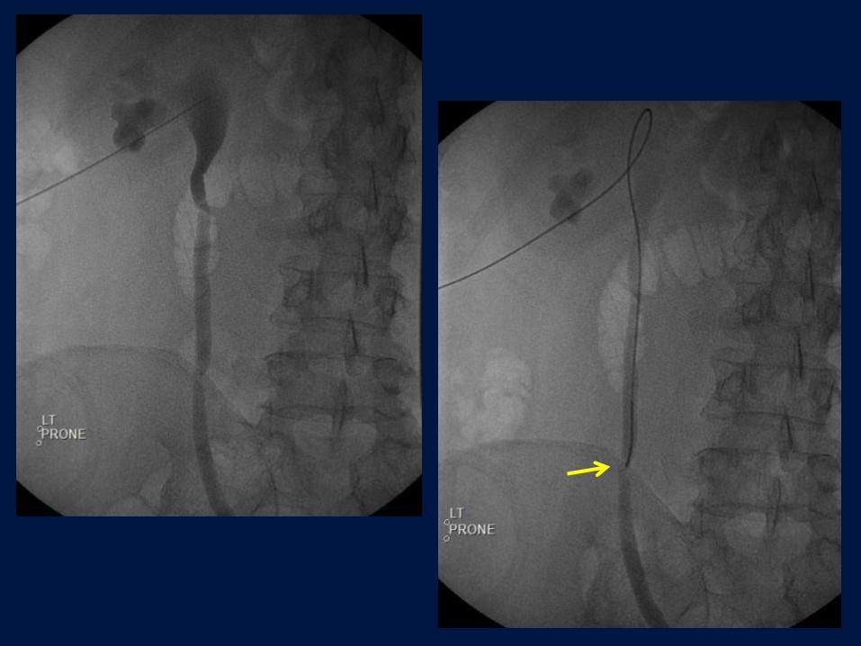
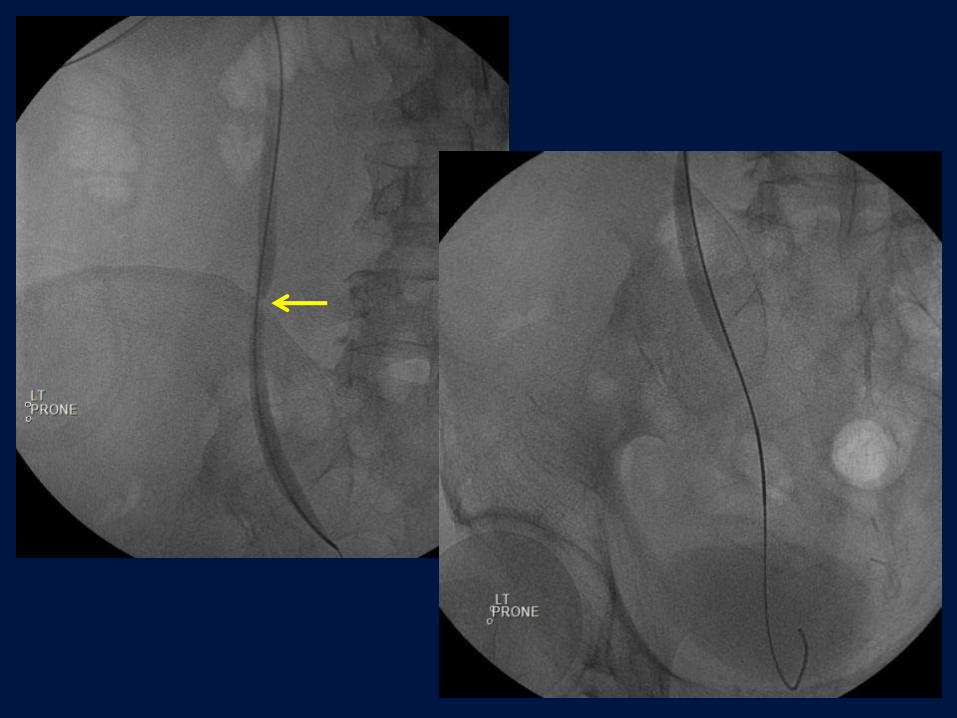
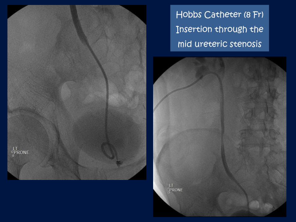
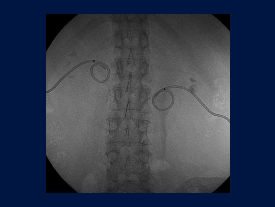
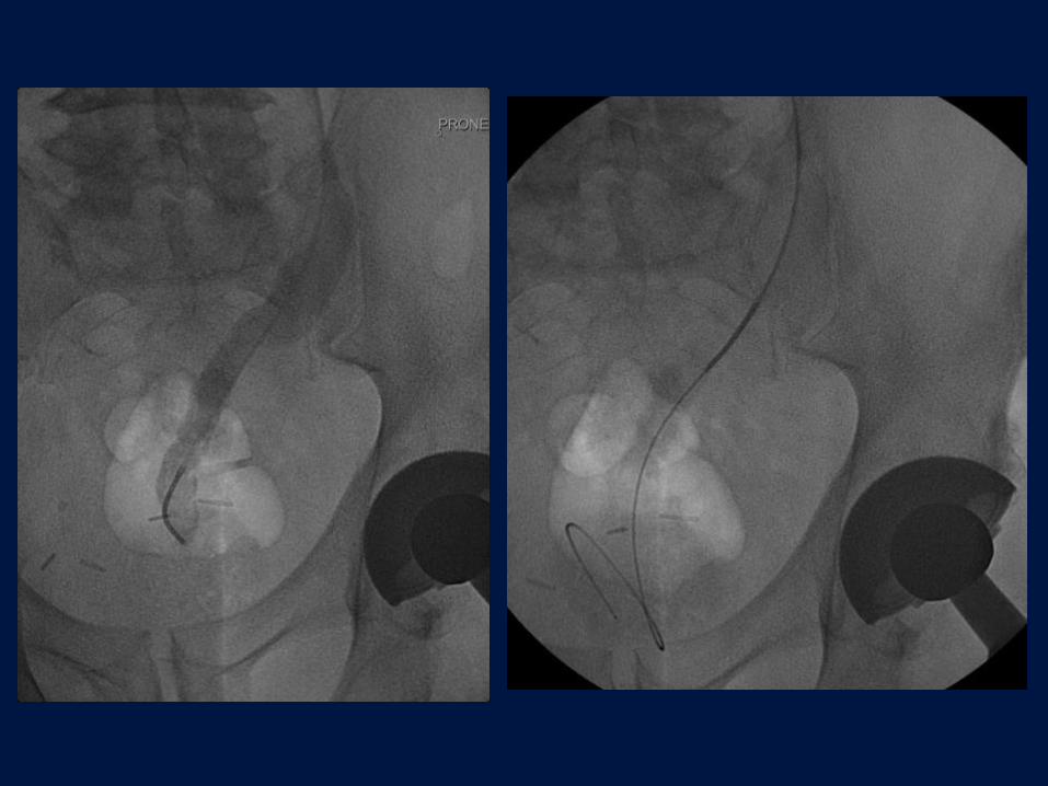
Hobbs Catheter (8 Fr)
Insertion through the
mid ureteric stenosis
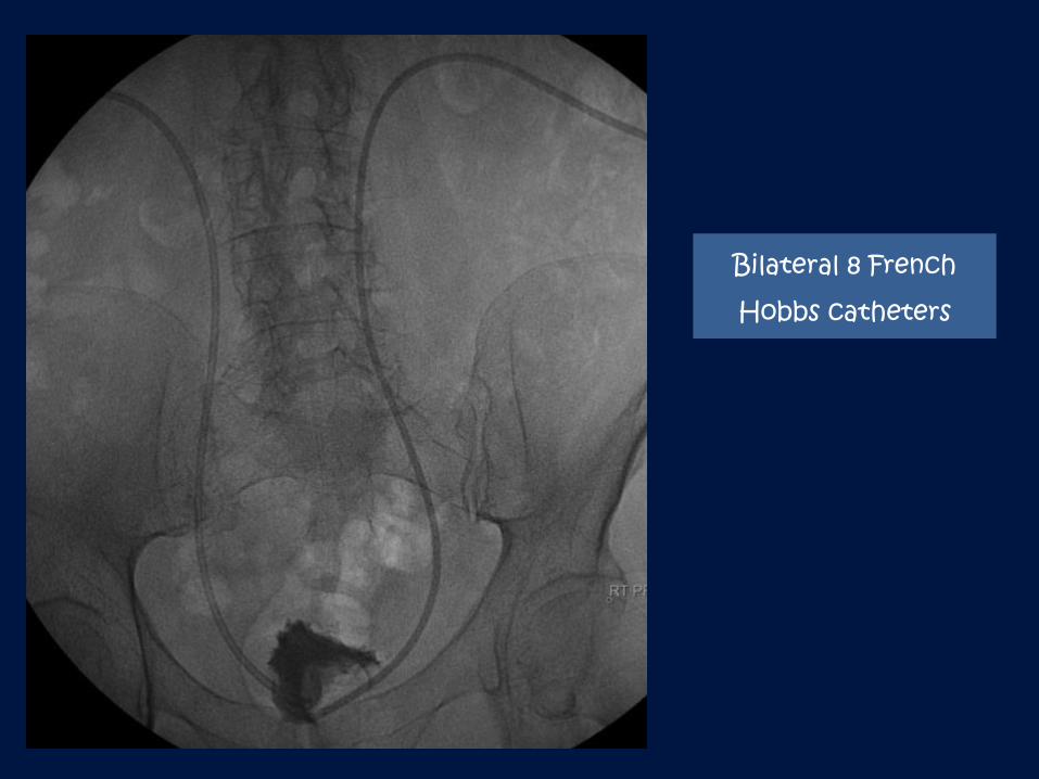
Bilateral 8 French
Hobbs catheters
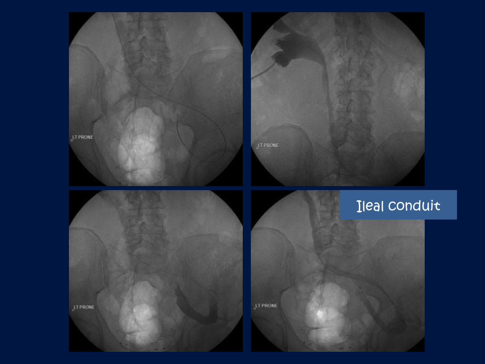
Ileal conduit
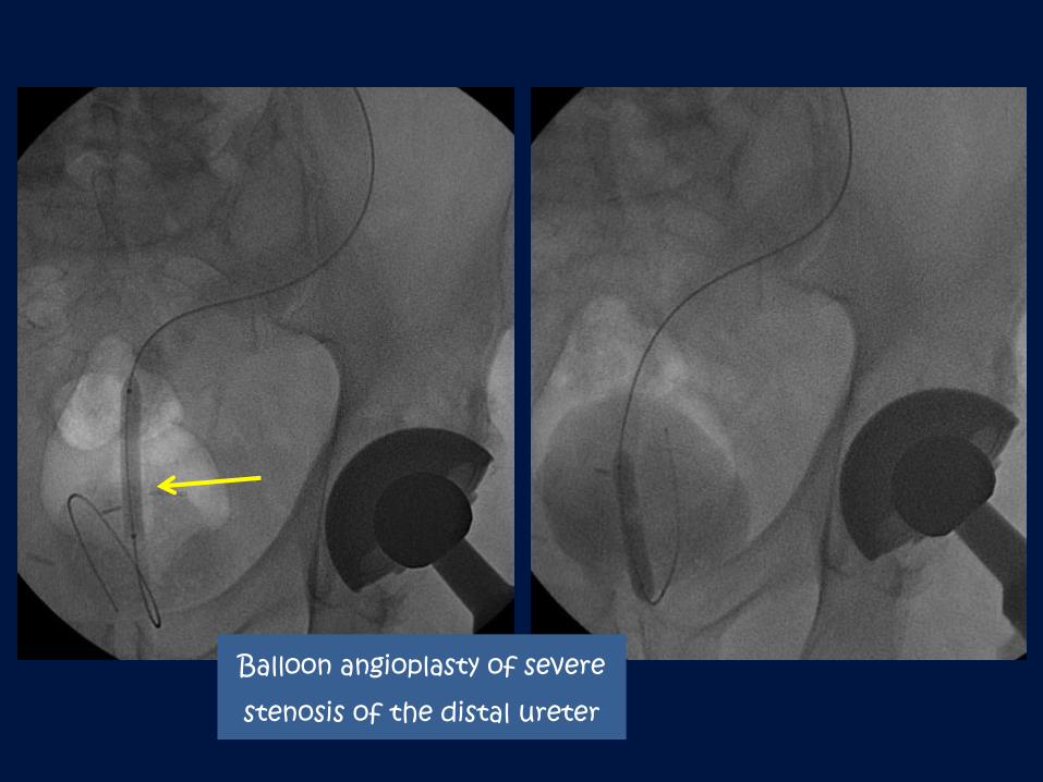
Balloon angioplasty of severe
stenosis of the distal ureter
