innovative endovascular approach to pulmonary · pdf fileinnovative endovascular approach to...
TRANSCRIPT
Innovative Endovascular Approach to PulmonaryEmbolism by Ultrasound Enhanced Thrombolysis
Prof. Ralf R.Kolvenbach MD,PhD,FEBVS

Conflict of Interest
• BTG
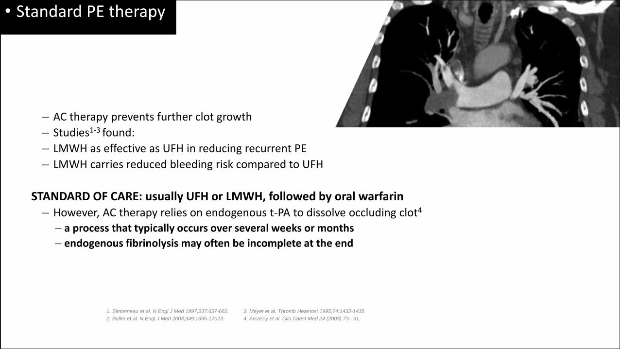
ANTICOAGULATION (AC) – HEPARIN – AC therapy prevents further clot growth
– Studies1-3 found:
– LMWH as effective as UFH in reducing recurrent PE
– LMWH carries reduced bleeding risk compared to UFH
STANDARD OF CARE: usually UFH or LMWH, followed by oral warfarin– However, AC therapy relies on endogenous t-PA to dissolve occluding clot4
– a process that typically occurs over several weeks or months
– endogenous fibrinolysis may often be incomplete at the end
• Standard PE therapy
1. Simonneau et al. N Engl J Med 1997;337:657-662.
2. Buller et al. N Engl J Med 2003;349:1695-17023.
3. Meyer et al. Thromb Heamost 1995;74:1432-1435
4. Arcasoy et al. Clin Chest Med 24 (2003) 73– 91.
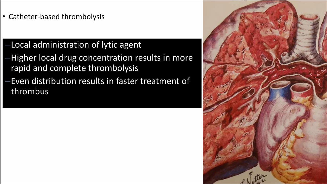
–Local administration of lytic agent
–Higher local drug concentration results in more rapid and complete thrombolysis
–Even distribution results in faster treatment of thrombus
• Catheter-based thrombolysis
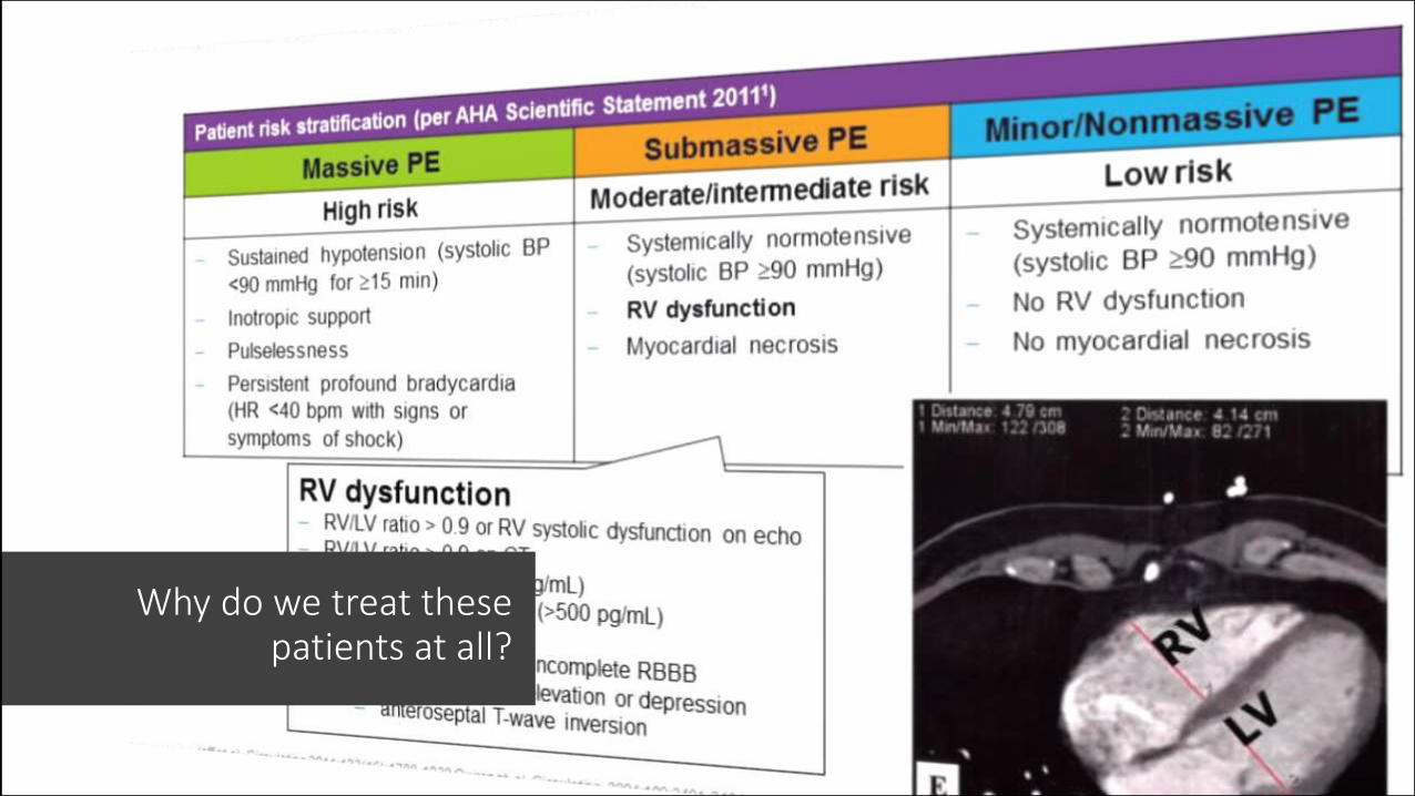
Why do we treat these patients at all?
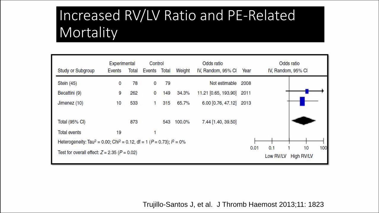
Increased RV/LV Ratio and PE-Related Mortality
Trujillo-Santos J, et al. J Thromb Haemost 2013;11: 1823
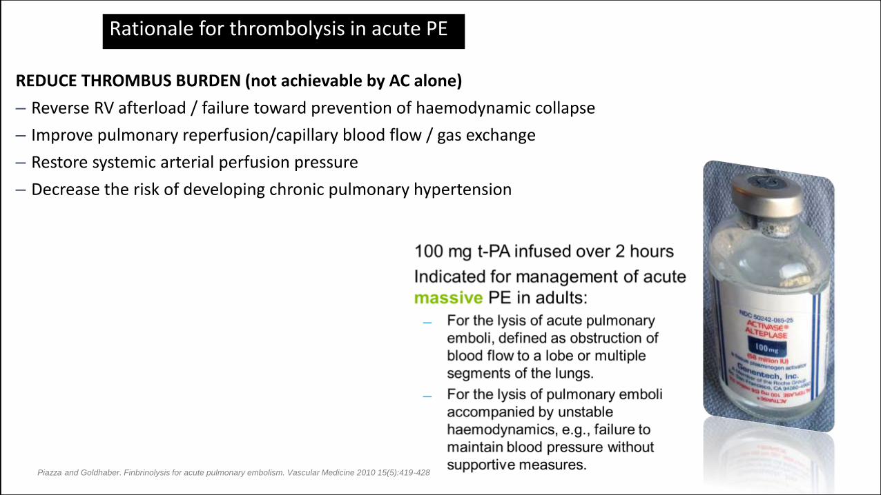
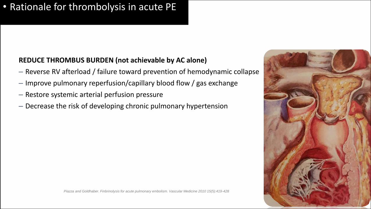
REDUCE THROMBUS BURDEN (not achievable by AC alone)
– Reverse RV afterload / failure toward prevention of haemodynamic collapse
– Improve pulmonary reperfusion/capillary blood flow / gas exchange
– Restore systemic arterial perfusion pressure
– Decrease the risk of developing chronic pulmonary hypertension
Rationale for thrombolysis in acute PE
Piazza and Goldhaber. Finbrinolysis for acute pulmonary embolism. Vascular Medicine 2010 15(5):419-428
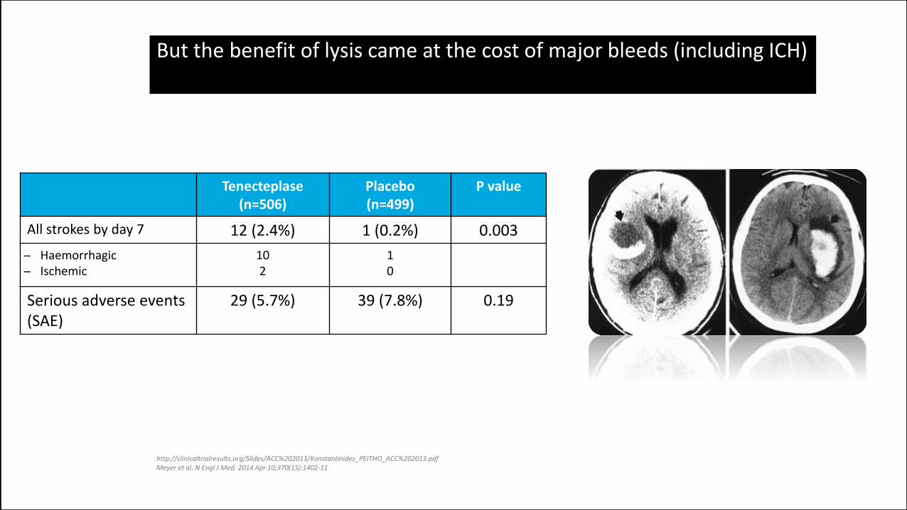
But the benefit of lysis came at the cost of major bleeds (including ICH)
Tenecteplase(n=506)
Placebo(n=499)
P value
All strokes by day 7 12 (2.4%) 1 (0.2%) 0.003
Haemorrhagic Ischemic
102
10
Serious adverse events (SAE)
29 (5.7%) 39 (7.8%) 0.19
http://clinicaltrialresults.org/Slides/ACC%202013/Konstantinides_PEITHO_ACC%202013.pdfMeyer et al. N Engl J Med. 2014 Apr 10;370(15):1402-11
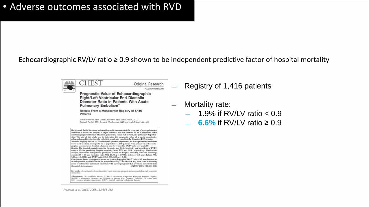
Echocardiographic RV/LV ratio ≥ 0.9 shown to be independent predictive factor of hospital mortality
• Adverse outcomes associated with RVD
Registry of 1,416 patients
Mortality rate:
1.9% if RV/LV ratio < 0.9
6.6% if RV/LV ratio ≥ 0.9
Fremont et al. CHEST 2008;133:358-362
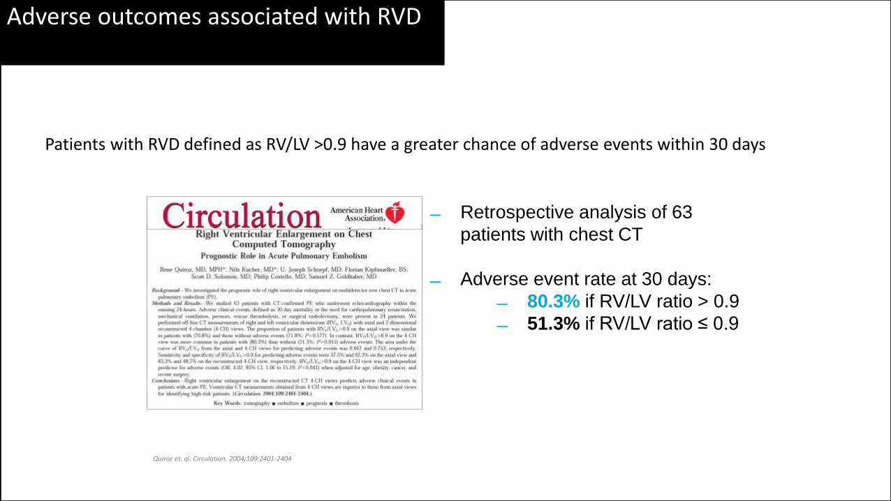
Patients with RVD defined as RV/LV >0.9 have a greater chance of adverse events within 30 days
Adverse outcomes associated with RVD
Retrospective analysis of 63
patients with chest CT
Adverse event rate at 30 days:
80.3% if RV/LV ratio > 0.9
51.3% if RV/LV ratio ≤ 0.9
Quiroz et. al. Circulation. 2004;109:2401-2404
REDUCE THROMBUS BURDEN (not achievable by AC alone)
– Reverse RV afterload / failure toward prevention of hemodynamic collapse
– Improve pulmonary reperfusion/capillary blood flow / gas exchange
– Restore systemic arterial perfusion pressure
– Decrease the risk of developing chronic pulmonary hypertension
• Rationale for thrombolysis in acute PE
Piazza and Goldhaber. Finbrinolysis for acute pulmonary embolism. Vascular Medicine 2010 15(5):419-428
– ULTIMA trial
– SEATTLE II trial
– Meta-analysis of historical published data
– Recent single-center studies
• Review of the clinical evidence for EKOS® for the treatment of PE
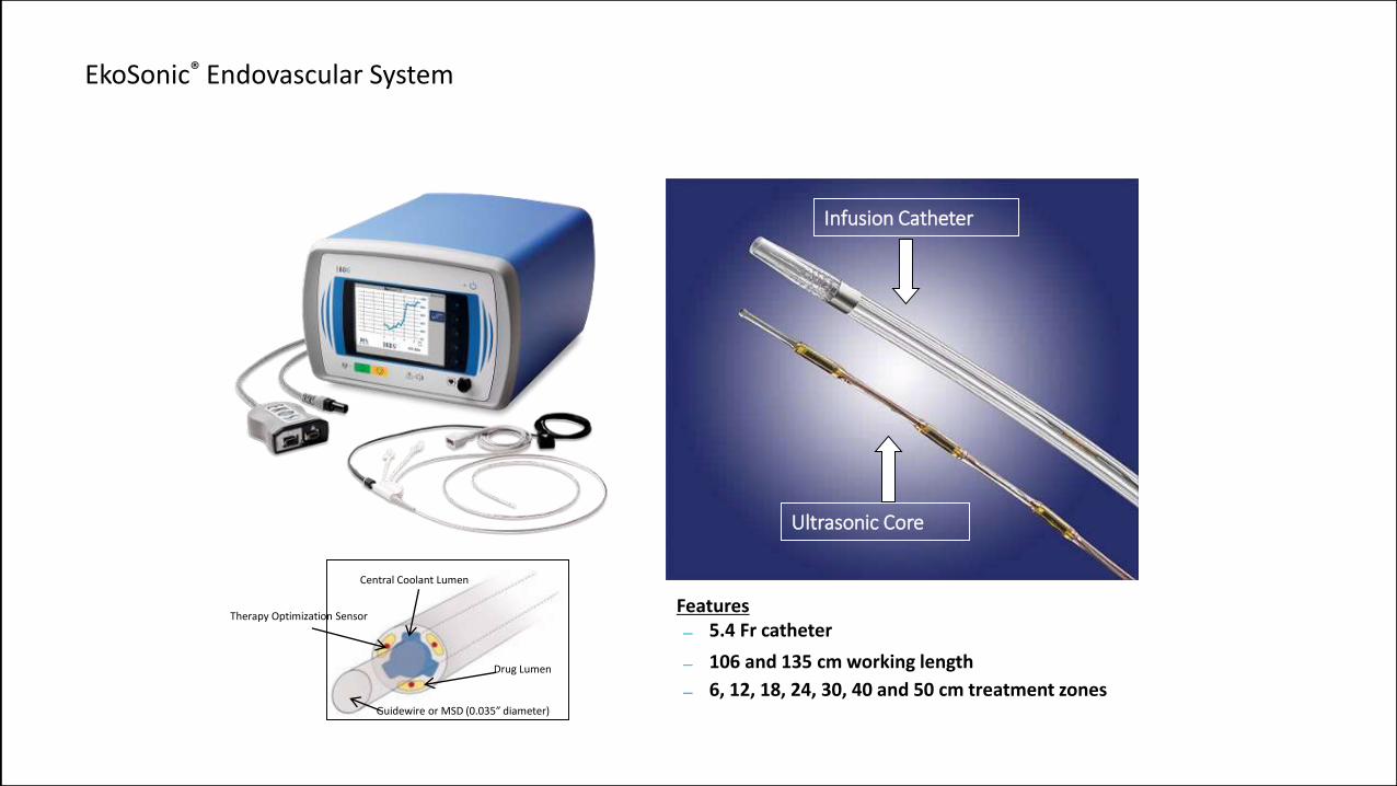
EkoSonic® Endovascular System
Features 5.4 Fr catheter
106 and 135 cm working length
6, 12, 18, 24, 30, 40 and 50 cm treatment zones
Infusion Catheter
Ultrasonic Core
Central Coolant Lumen
Therapy Optimization Sensor
Drug Lumen
Guidewire or MSD (0.035” diameter)
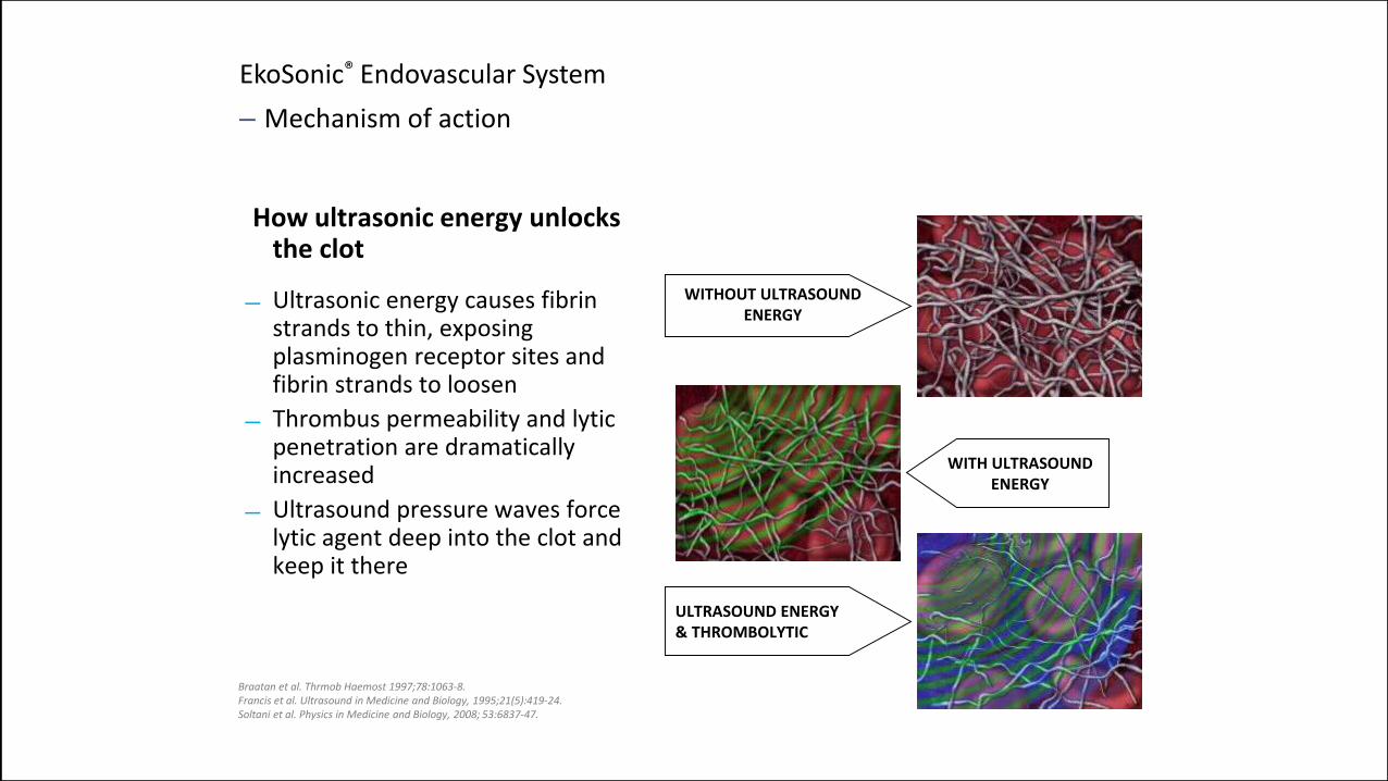
EkoSonic® Endovascular System
– Mechanism of action
WITH ULTRASOUNDENERGY
WITHOUT ULTRASOUNDENERGY
How ultrasonic energy unlocks the clot
Ultrasonic energy causes fibrin strands to thin, exposing plasminogen receptor sites and fibrin strands to loosen
Thrombus permeability and lytic penetration are dramatically increased
Ultrasound pressure waves force lytic agent deep into the clot and keep it there
ULTRASOUND ENERGY& THROMBOLYTIC
Braatan et al. Thrmob Haemost 1997;78:1063-8.Francis et al. Ultrasound in Medicine and Biology, 1995;21(5):419-24.Soltani et al. Physics in Medicine and Biology, 2008; 53:6837-47.
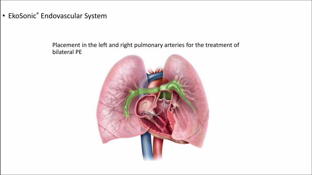
Placement in the left and right pulmonary arteries for the treatment of bilateral PE
• EkoSonic® Endovascular System
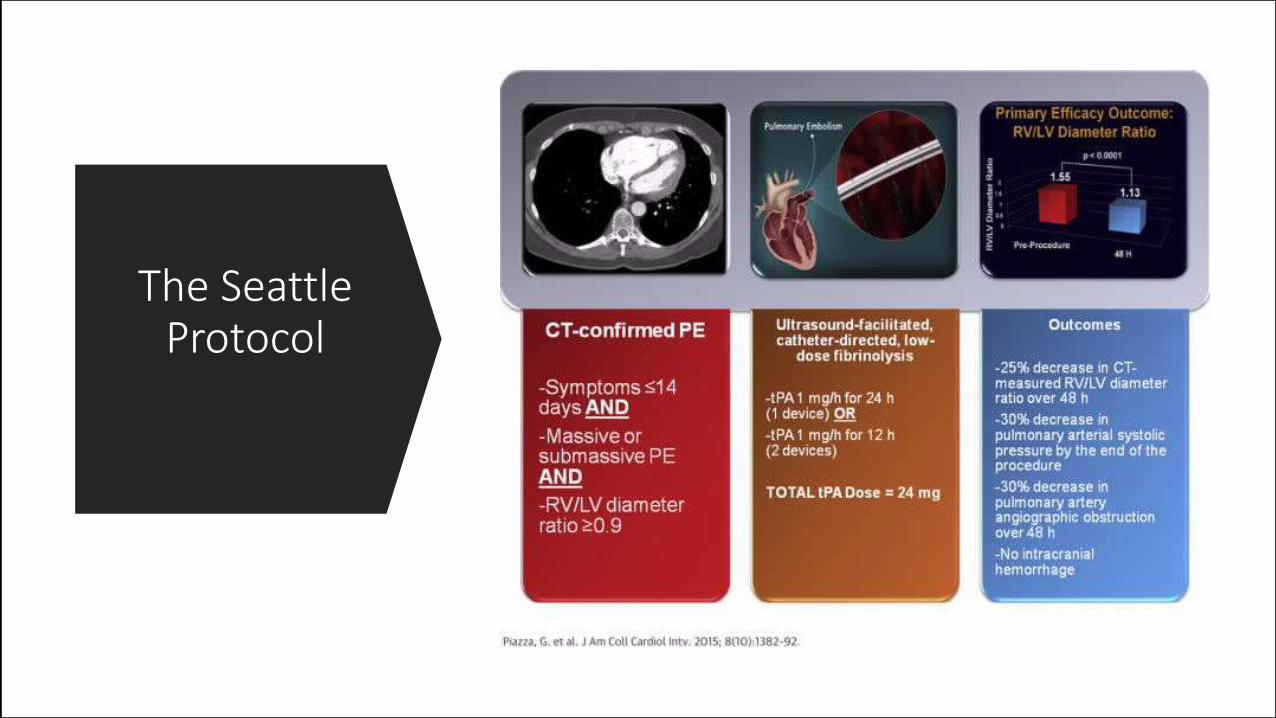
The Seattle Protocol
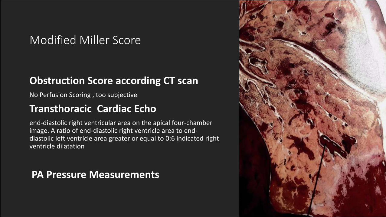
Modified Miller Score
Obstruction Score according CT scanNo Perfusion Scoring , too subjective
Transthoracic Cardiac Echoend-diastolic right ventricular area on the apical four-chamber image. A ratio of end-diastolic right ventricle area to end-diastolic left ventricle area greater or equal to 0:6 indicated right ventricle dilatation
PA Pressure Measurements
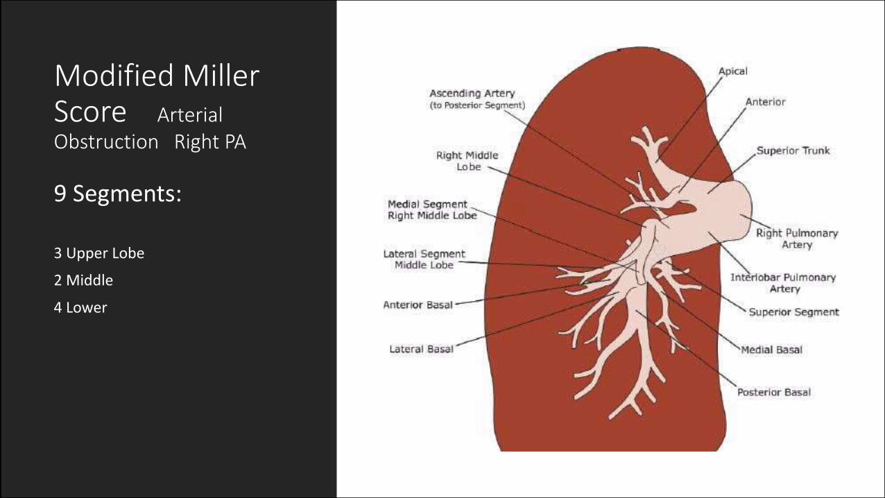
Modified Miller Score ArterialObstruction Right PA
9 Segments:
3 Upper Lobe
2 Middle
4 Lower
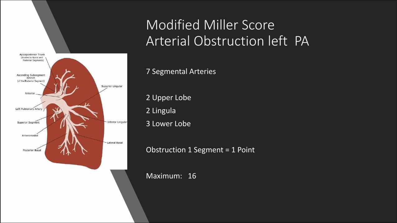
Modified Miller ScoreArterial Obstruction left PA
7 Segmental Arteries
2 Upper Lobe
2 Lingula
3 Lower Lobe
Obstruction 1 Segment = 1 Point
Maximum: 16
– Ultrasound-facilitated fibrinolysis using EKOS®
– If unilateral PE: tPA 1 mg/hr using one device for 24 hours– If bilateral PE: tPA 1 mg/hr per device (using two simultaneously) for 12 hours
– Follow up at 48 +/- 6 hours– CT measurement of RV/LV ratio– Echocardiogram to estimate PA systolic pressure
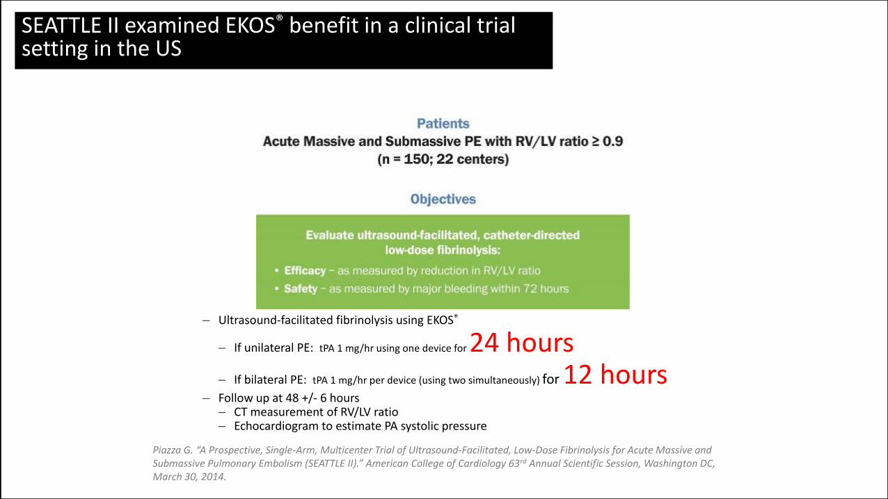
SEATTLE II examined EKOS® benefit in a clinical trial setting in the US
Piazza G. “A Prospective, Single-Arm, Multicenter Trial of Ultrasound-Facilitated, Low-Dose Fibrinolysis for Acute Massive and Submassive Pulmonary Embolism (SEATTLE II).” American College of Cardiology 63rd Annual Scientific Session, Washington DC, March 30, 2014.
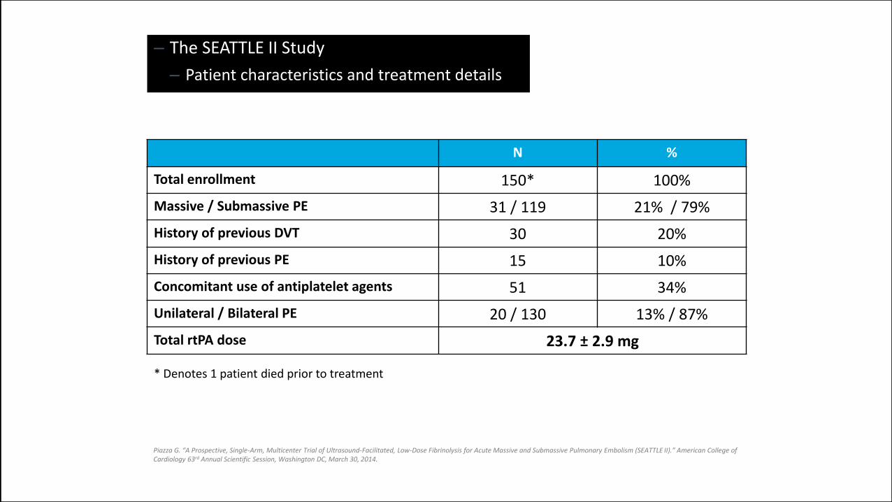
– The SEATTLE II Study
– Patient characteristics and treatment details
N %
Total enrollment 150* 100%
Massive / Submassive PE 31 / 119 21% / 79%
History of previous DVT 30 20%
History of previous PE 15 10%
Concomitant use of antiplatelet agents 51 34%
Unilateral / Bilateral PE 20 / 130 13% / 87%
Total rtPA dose 23.7 ± 2.9 mg
* Denotes 1 patient died prior to treatment
Piazza G. “A Prospective, Single-Arm, Multicenter Trial of Ultrasound-Facilitated, Low-Dose Fibrinolysis for Acute Massive and Submassive Pulmonary Embolism (SEATTLE II).” American College of Cardiology 63rd Annual Scientific Session, Washington DC, March 30, 2014.
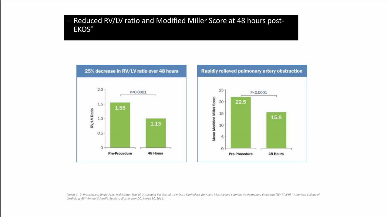
– Reduced RV/LV ratio and Modified Miller Score at 48 hours post-EKOS®
Piazza G. “A Prospective, Single-Arm, Multicenter Trial of Ultrasound-Facilitated, Low-Dose Fibrinolysis for Acute Massive and Submassive Pulmonary Embolism (SEATTLE II).” American College of Cardiology 63rd Annual Scientific Session, Washington DC, March 30, 2014.
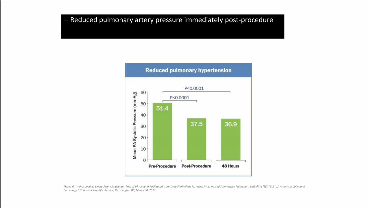
– Reduced pulmonary artery pressure immediately post-procedure
Piazza G. “A Prospective, Single-Arm, Multicenter Trial of Ultrasound-Facilitated, Low-Dose Fibrinolysis for Acute Massive and Submassive Pulmonary Embolism (SEATTLE II).” American College of Cardiology 63rd Annual Scientific Session, Washington DC, March 30, 2014.
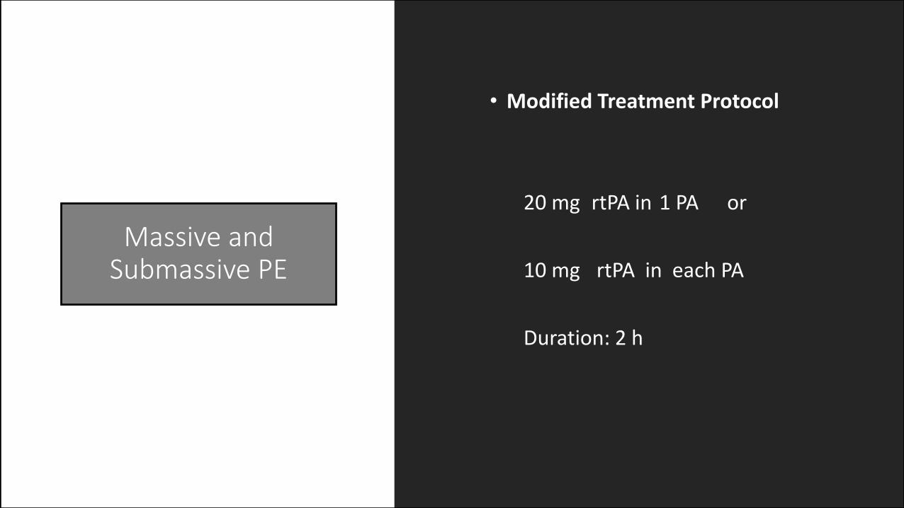
Massive andSubmassive PE
• Modified Treatment Protocol
20 mg rtPA in 1 PA or
10 mg rtPA in each PA
Duration: 2 h
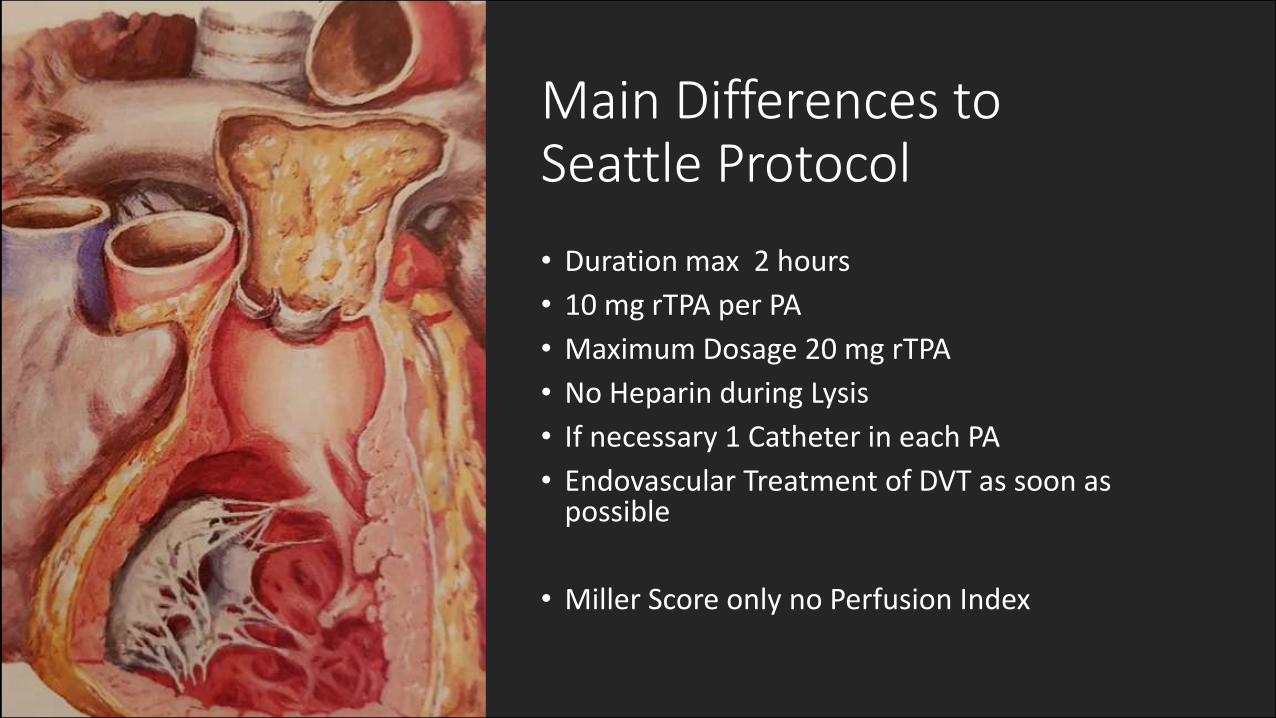
Main Differences toSeattle Protocol
• Duration max 2 hours
• 10 mg rTPA per PA
• Maximum Dosage 20 mg rTPA
• No Heparin during Lysis
• If necessary 1 Catheter in each PA
• Endovascular Treatment of DVT as soon as possible
• Miller Score only no Perfusion Index
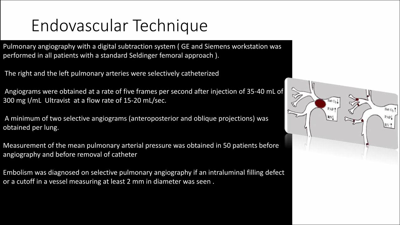
Endovascular TechniquePulmonary angiography with a digital subtraction system ( GE and Siemens workstation was performed in all patients with a standard Seldinger femoral approach ).
The right and the left pulmonary arteries were selectively catheterized
Angiograms were obtained at a rate of five frames per second after injection of 35-40 mL of 300 mg I/mL Ultravist at a flow rate of 15-20 mL/sec.
A minimum of two selective angiograms (anteroposterior and oblique projections) was obtained per lung.
Measurement of the mean pulmonary arterial pressure was obtained in 50 patients before angiography and before removal of catheter
Embolism was diagnosed on selective pulmonary angiography if an intraluminal filling defect or a cutoff in a vessel measuring at least 2 mm in diameter was seen .
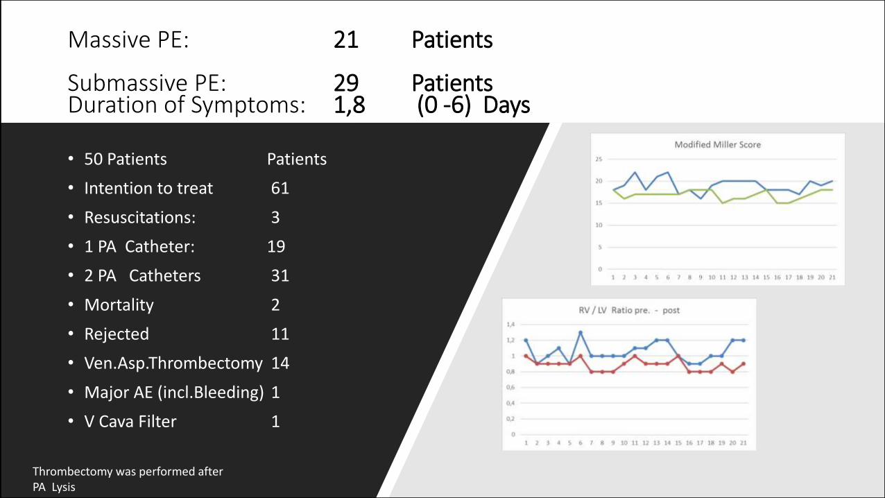
Massive PE: 21 Patients
Submassive PE: 29 PatientsDuration of Symptoms: 1,8 (0 -6) Days
• 50 Patients Patients
• Intention to treat 61
• Resuscitations: 3
• 1 PA Catheter: 19
• 2 PA Catheters 31
• Mortality 2
• Rejected 11
• Ven.Asp.Thrombectomy 14
• Major AE (incl.Bleeding) 1
• V Cava Filter 1
Thrombectomy was performed after PA Lysis
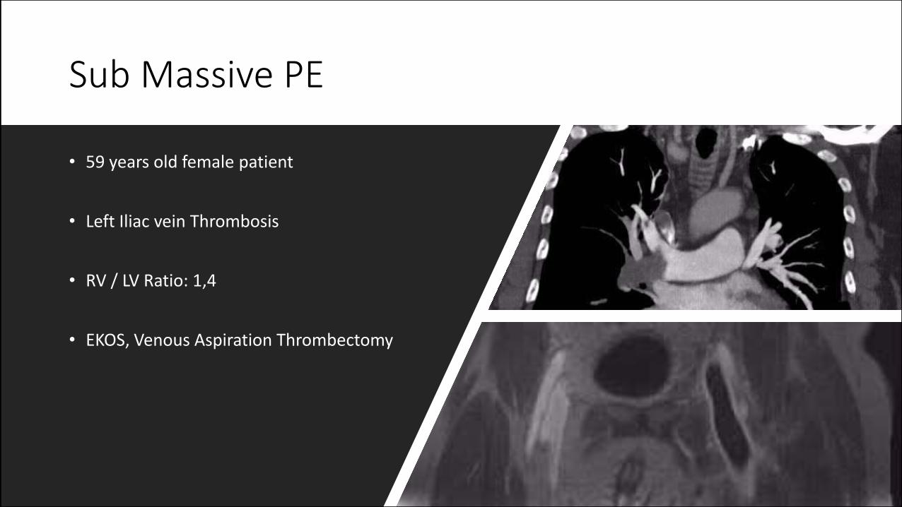
Sub Massive PE
• 59 years old female patient
• Left Iliac vein Thrombosis
• RV / LV Ratio: 1,4
• EKOS, Venous Aspiration Thrombectomy
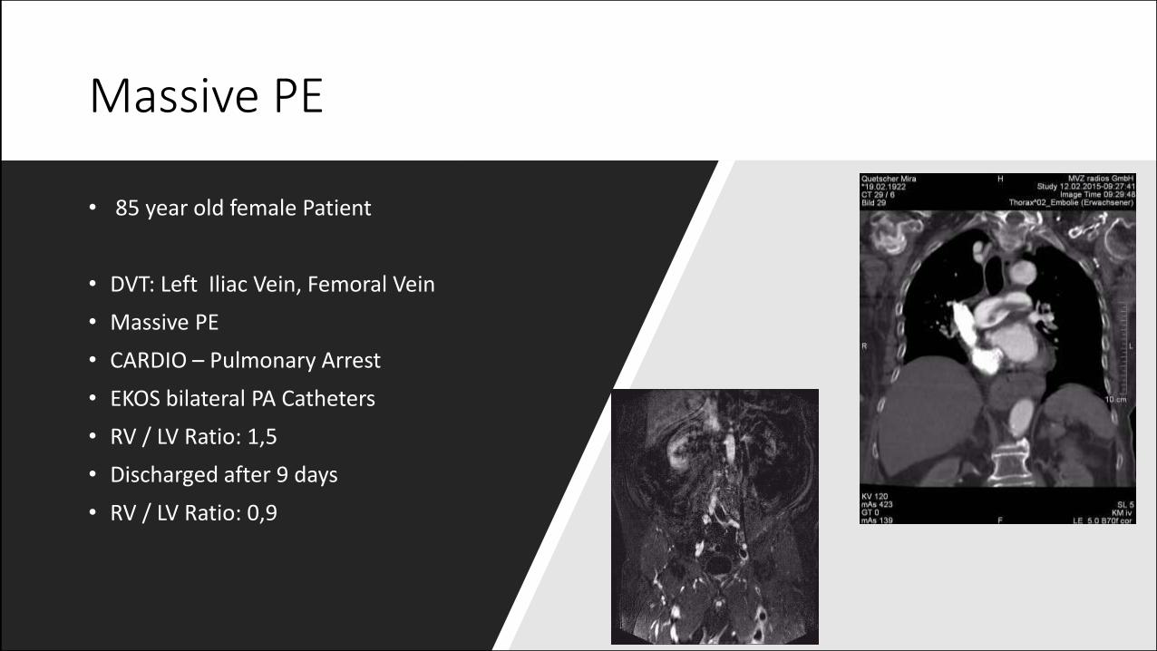
Massive PE
• 85 year old female Patient
• DVT: Left Iliac Vein, Femoral Vein
• Massive PE
• CARDIO – Pulmonary Arrest
• EKOS bilateral PA Catheters
• RV / LV Ratio: 1,5
• Discharged after 9 days
• RV / LV Ratio: 0,9
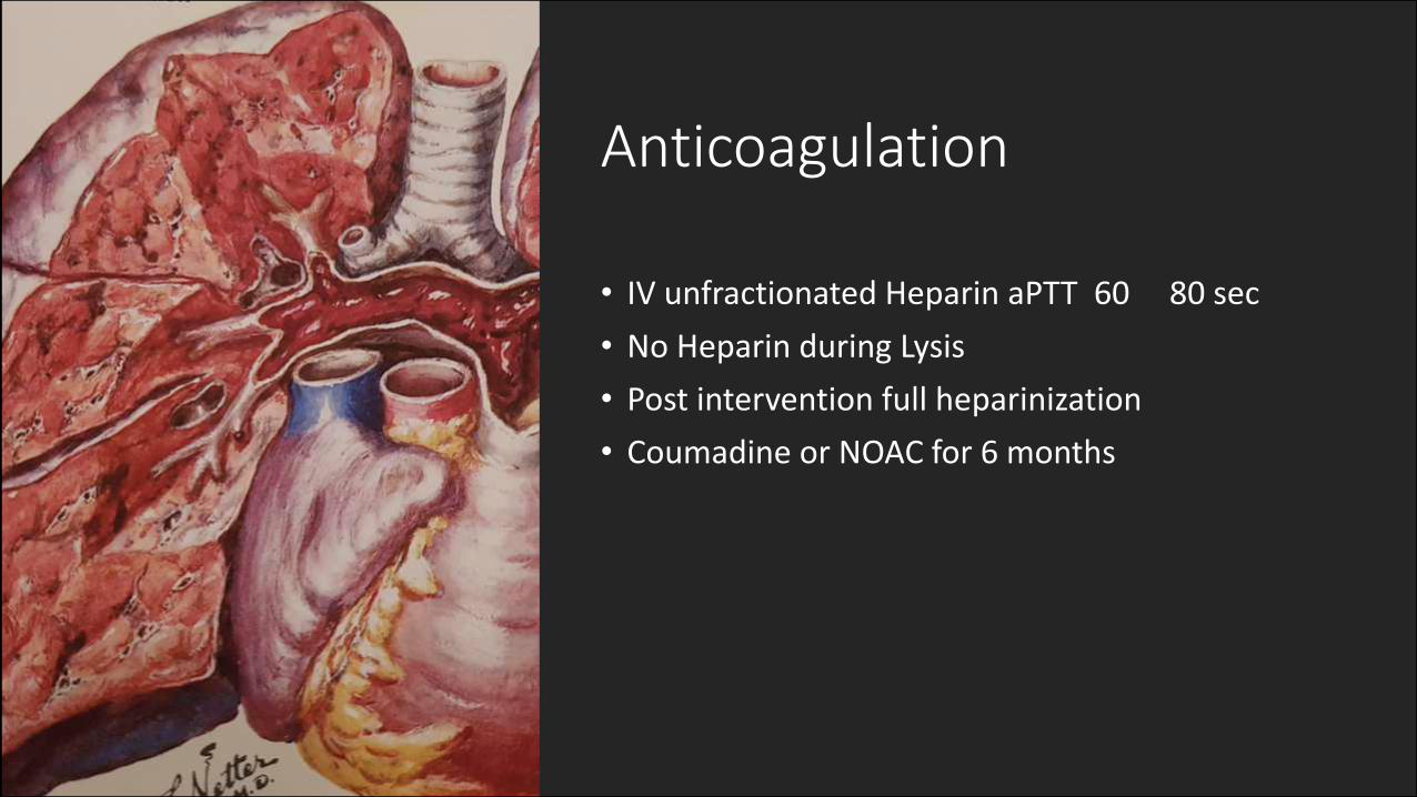
Anticoagulation
• IV unfractionated Heparin aPTT 60 80 sec
• No Heparin during Lysis
• Post intervention full heparinization
• Coumadine or NOAC for 6 months
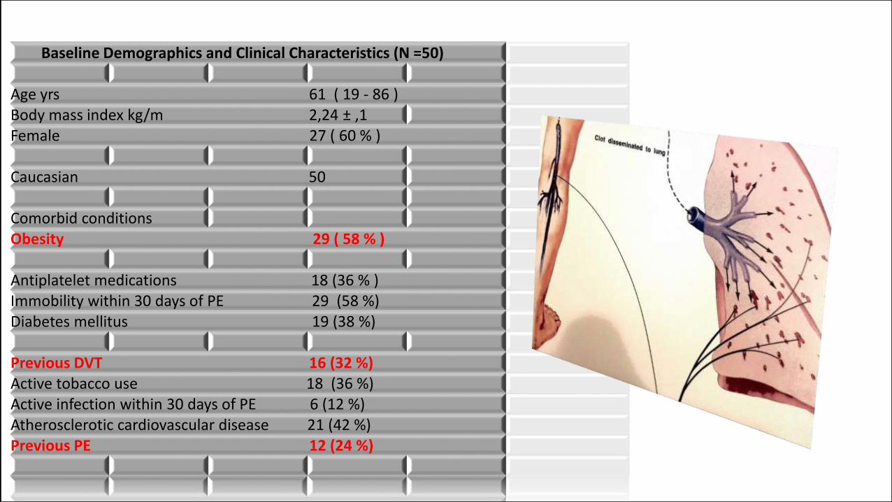
Baseline Demographics and Clinical Characteristics (N =50)
Age yrs 61 ( 19 - 86 ) Body mass index kg/m 2,24 ± ,1 Female 27 ( 60 % )
Caucasian 50
Comorbid conditions Obesity 29 ( 58 % )
Antiplatelet medications 18 (36 % ) Immobility within 30 days of PE 29 (58 %) Diabetes mellitus 19 (38 %)
Previous DVT 16 (32 %) Active tobacco use 18 (36 %) Active infection within 30 days of PE 6 (12 %) Atherosclerotic cardiovascular disease 21 (42 %) Previous PE 12 (24 %)
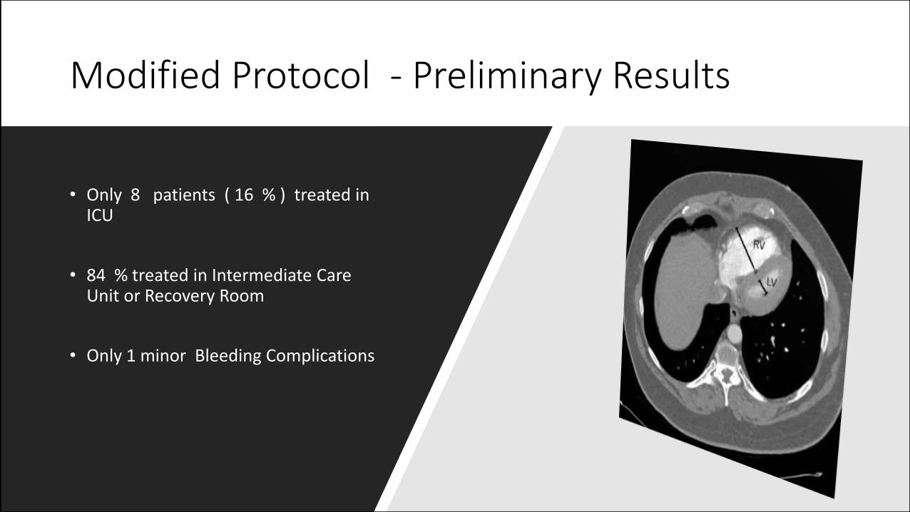
Modified Protocol - Preliminary Results
• Only 8 patients ( 16 % ) treated in ICU
• 84 % treated in Intermediate Care Unit or Recovery Room
• Only 1 minor Bleeding Complications
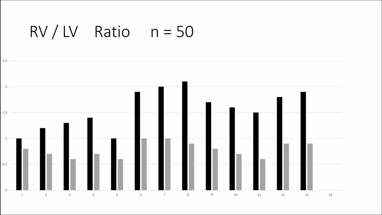
RV / LV Ratio n = 50
0
0.5
1
1.5
2
2.5
1 2 3 4 5 6 7 8 9 10 11 12 13 14
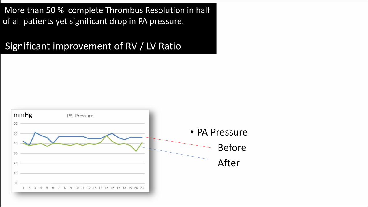
• PA Pressure
Before
After
mmHg
More than 50 % complete Thrombus Resolution in half of all patients yet significant drop in PA pressure.
Significant improvement of RV / LV Ratio
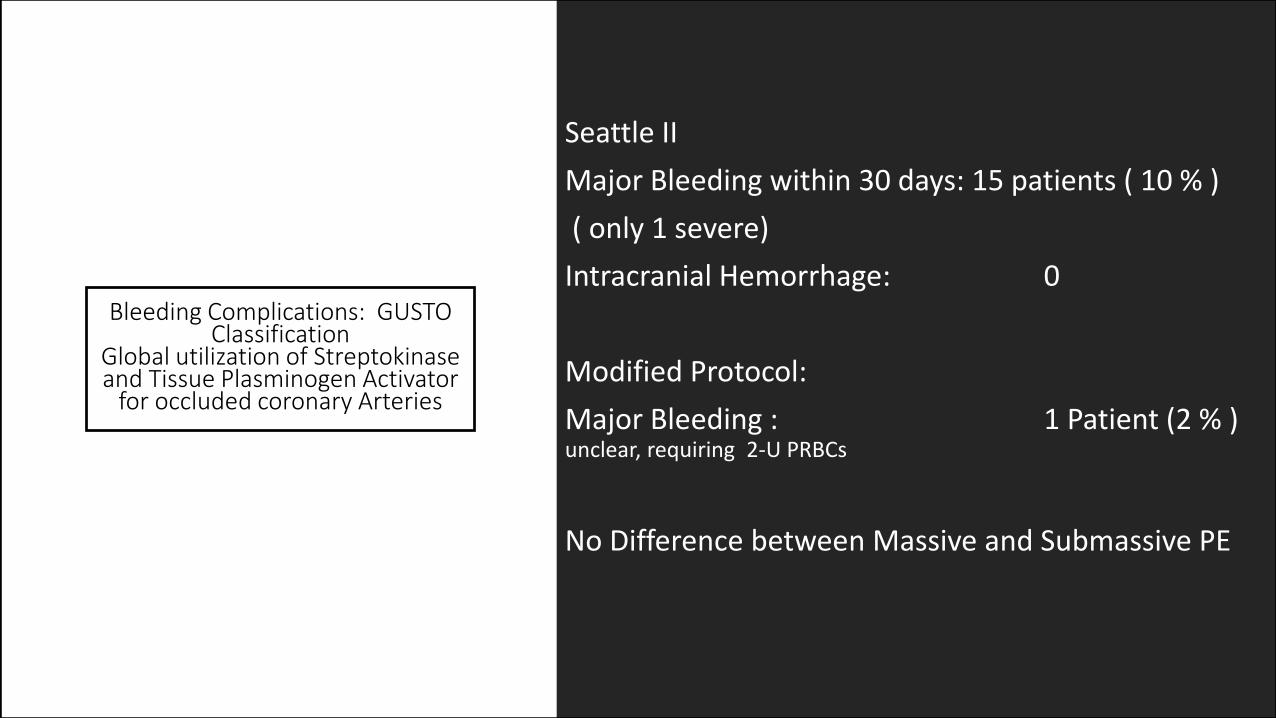
Bleeding Complications: GUSTO Classification
Global utilization of Streptokinase and Tissue Plasminogen Activator
for occluded coronary Arteries
Seattle II
Major Bleeding within 30 days: 15 patients ( 10 % )
( only 1 severe)
Intracranial Hemorrhage: 0
Modified Protocol:
Major Bleeding : 1 Patient (2 % ) unclear, requiring 2-U PRBCs
No Difference between Massive and Submassive PE
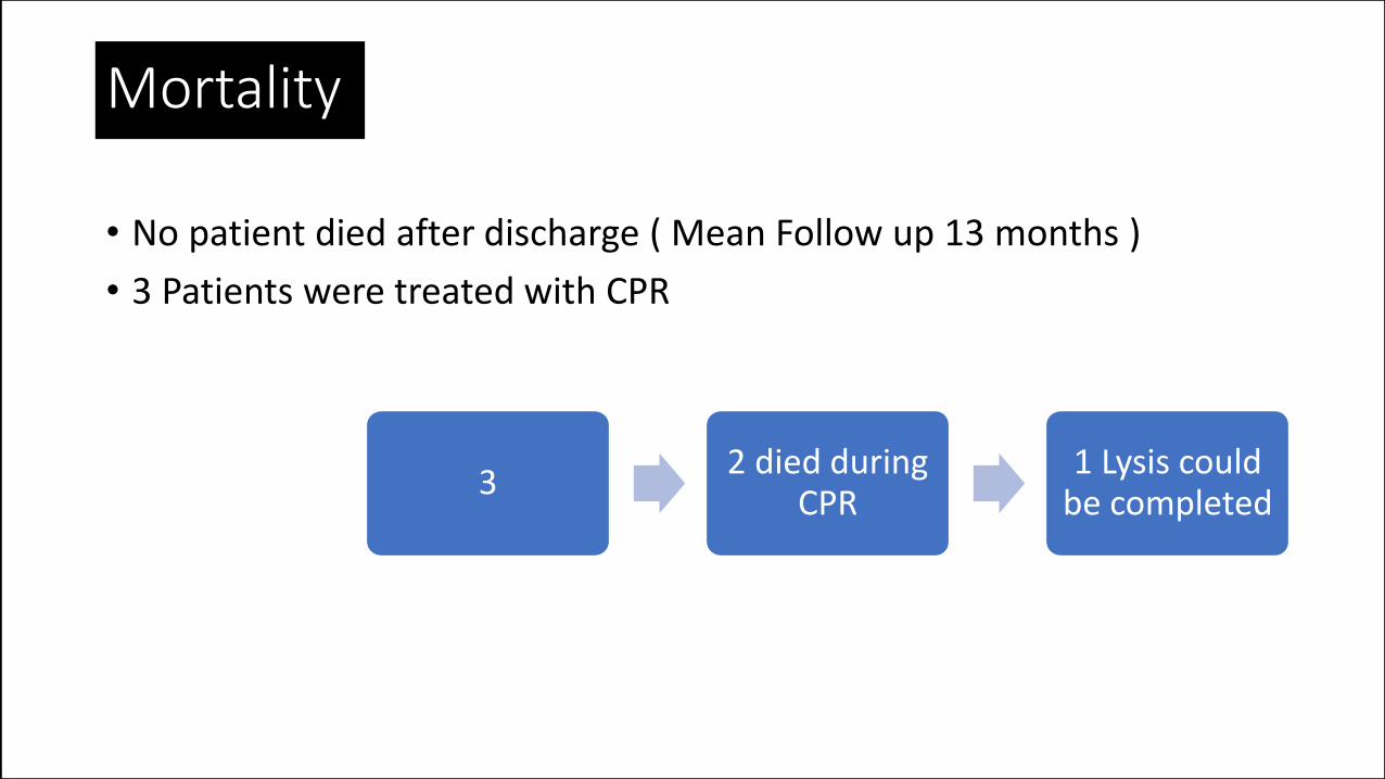
Mortality
• No patient died after discharge ( Mean Follow up 13 months )
• 3 Patients were treated with CPR
32 died during
CPR1 Lysis could
be completed
Advantages of a modified Protocol
• Less logistical efforts required, because of fast track protocol
• No Intensive Care resources necessary. Lower dependency unit issufficient.
• No Heparin during ultra sound assisted lysis further reduces risk of bleeding complications
• Faster hospital discharge possible
Conclusion
• Ultrasound-facilitated low-dose fibrinolysis for acute PE improves RV function and decreases pulmonary hypertension.
• By minimizing the risk of intracranial bleed, it represents a potential “game-changer” in the treatment of high-risk PE patients.
• The modified protocol further reduces the risk of bleeding.
• Less ICU capacity is required.
• Results are comparable to Seattle Protocol

Innovative Endovascular Approach to PulmonaryEmbolism by Ultrasound Enhanced Thrombolysis
Prof. Ralf R.Kolvenbach MD,PhD,FEBVS